Embed Size (px)
Citation preview
THE DIRECT DETERMINATION OF THEMOLECULAR STRUCTURES OF
NATURAL PRODUCTS
A. MeL. MATHIESON
C.S.I.R.O. Chemical Research Laboratories, Melbourne, Australia
INTRODUCTIONWhen I came to look again at the title of my talk, it appeared to me that
it had not quite succeeded in defining the scope of my subject adequately.Let me say, therefore, that my purpose is to show that, given the empiricalformula of a suitable derivative of a natural product in a satisfactory crystalline form, it is possible to arrive at the three-dimensional molecular structurein a direct manner and with only minirnum assumptions, these being of ageneral type.
The task of preparing this lecture was influenced, although not madeeasier, by the increase in the numbers of moderately complex moleculeswhich have been subjected to X-ray analysis in the last few years. Itappears probable that this increased interest (particularly in natural products) is not a passing phase, but a consistent change in attitude which maylead, by its results, to a reconsideration of the role of X-ray analysis in thisfield. Prospects exist for the growth of specialist groups to determinestructures of natural products by the application of this technique.
There is little need to dwell on the reasons for determining molecularstructure-such information is basic to any understanding of how moleculescan be chemically modified with intention and how they may be interrelated biogenetically. Although two-dimensional structural formulaehave served well in the past, it is clear from the extensive discussion of theinfluence of conformation and the increased interest in absolute configuration that a fairly detailed three-dimensional picture of the molecule is nowrequired. This being so, the determination of molecular structure in suchdetail is the essential first stage in the study of a new compound and onewhich must be solved in as direct and rapid a way as is consistent withaccuracy. A solution by classical methods through chemical degradation,and then confirmation by synthesis, offers an attractive and exciting taskfor many organic chemists; but even for moderately complex compounds,the number of man-years involved in the first step, i.e. solution of thestructure, is becoming burdensome, particularly for those compounds withinter-linked ring systems-e.g. the recent description of the chemical constitution of limonin which involved 14 authors1. In this field, X-raystructure analysis may occupy a unique role since, with certain conditionssatisfied, it can be of general application. It is one in which the latentpower of computers may be very effectively placed at the service of organicchemistry.
505
(1)
A. MeL. MATHIESON
I want to illustrate, by selected examples, how X-ray analysis has beenof help in this aspect of organic chemistry, with particular reference tonatural productss", and how the co-ordination of facilities has greatlyincreased the prospect of this assistance in the future. To do so, we needfirst to deal with certain of the features which render the X-ray methodpeculiarly suited to this task.
The physical methods of determining molecular structure fall into twogeneral types, and it is useful to appreciate the clear distinction betweenthem. The first group deals with" fragmentary" information, i.e. it givesinformation primarily about" bits" of the molecule (groups or radicals).Of such techniques, the main representative is that which measures changesin energy between the states of a molecule-ranging from ultra-violetthrough to microwave absorption. In many respects, these techniquesresemble chemical degradative methods which also yield "fragments",recognition of which permit these" fragments" to be fitted together toderive the molecular structure. Perhaps because of this similarity, theorganic chemist has been quick to appreciate and accept these techniquesand make wide application of them. The second type of physical methodis the diffraction technique which, by contrast with the first group, does notoffer partial information-it represents an all-or-none approach to molecularstructure. Wide use has not been made of this technique in the past but,as a result mainly of the availability of large electronic computers, thisattitude may be changing. For this reason now is an appropriate time toindicate the scope of the technique.
X-RAY ANALYSIS
Outline of the X-ray methodWith regard to this aspect of the all-or-none approach, we may consider
the diffraction of X-rays by a single crystal, taken over all orientations.Experimentally, we end up with an array on films of spots of varyingintensity, each spot being related to a set of parallel planes in the crystalreferred to by their Miller indices, hkl, and its intensity being related to anamplitude value, IF I. SO we have an extensive list of measurements,IF(hkl) I. These arise from the periodic smooth distribution of electrondensity within the crystal, p(xyz) , where x, y, and Z are fractions of therepeat distances of the unit cell a.bxc. There are two relationships, (1) and(2), between these quantities.
1 +00
p(Xrz) = V.Ih.Ik .ItF(hkl) exp {- 27Ti(hX + tr + IZ)}-00
F(hkl)' = Vc fff p(xyz) exp {27Ti(hx + ky + lz)} dxdydz
- .Ifr(hkl) exp {27Ti(hxr + kYr + IZr)}r
(2)
(2a)
It is not our intention to explain here in any detail the two distributions,p(xyz) and F(hkl), except to point out that their relationship is a mutually
506
DIRECT DETERMINATION OF MOLECULAR STRUCTURES
dependent or reciprocal one. Thus, in a strict sense, to find out aboutone structure factor, F(hkl), we need to know all about p(xyz) , i.e. thecontents of the unit cell, with reasonable precision. Conversely, to determine the value of the electron density p at any given point x,y,z in the unitcell, we need all the F(hkl) values. It is evident, therefore, in the approachimposed by this technique, that the available experimental informationthe IFI values-cannot be treated piecemeal. We must determine allatom positions to be completely satisfied with, and certain of, our end-result.Fortunately, the X-ray method does not demand a one-step deduction ofthe completely correct structure, but is amenable to a gradual approach tothe true solution by iterative processes.
So far, then, the procedure appears straightforward. It is a matter ofcollecting the IFI data and combining them according to equation (1).However, this conceals the fact that each F can be a complex quantity,IF Icos a+ i IF I sin a, and two parameters are necessary to defineF, namely the modulus, IFI, and the phase angle, a. We can measureIF I, but, in general, cannot determine a experimentally for a singlecrystal using one radiation. Should the crystal contain a centre of symmetry, then a reduces to 0 or 7T, i.e. F rrmy be + F (cos 0°) or - F(cos 7T).If, however, the structure is asymmetric, as is extremely probable for anatural product, then a may range from 0 to 2 7T, and it is obvious that thesituation becomes complex. The possibility of a practicable solution of thecrystal structure is then closely bound up with the size of the molecule andthe number of molecules constituting the asymmetric unit, i.e. the totalnumber of atomic parameters that must be determined. As to the variousproposals for the solution of the general phase problem, i.e. for any arbitrarilyselected crystal, I must refer you to more extensive treatments", but here, ifwe set ourselves the more limited task of determining certain organicstructures with some prospect of success, we must narrow our field to consideration of the "heavy atom" method as a means of yielding a firstapproximation to the phase angle. This does not mean that other methodscannot be used to solve such structures, e.g, hydroxydihydroeremophilone7,
but to use them must involve a certain amount of chemical information,and, further, they are not yet adequately developed to deal with complexasymmetric structures. The heavy-atom method offers the possibility ofdetermining a structure with the minimum of assumptions and the minimumof chemical knowledge, and, because of these conditions, parts of the processcan be made automatic. In principle, the restrictive conditions of theheavy-atom method mean that we cannot tackle any crystal offered, butmust select a suitable derivative which contains one or more (but not toomany) atoms of atomic number higher than those constituting the mainbulk of the molecule, i.e. C, Nand O. For the small sacrifice of the addedcomplexity of the heavy atom(s), we gain a more certain starting point inanalysis.
The relative weight of the heavy atomIn considering the general strategy for the structure determination of a
compound of natural origin, the question arises as to what is the mostsuitable weight of the heavy atom with respect to the remainder of the
507
A. MeL. MATHIESON
molecule. Choice is frequently restricted by chemical considerations orthe crystallizability of derivatives, but some numerical estimate is useful.There are several factors involved. The value of IF I is formed of a vectorsum of the atomic contributions (see equation (2) ) which, for convenience,we may consider in two parts-that due to the N heavy atoms NAH, andthat due to the n light atoms nAL in which we are mainly interested sincethey constitute the molecular skeleton(s). The use of a very heavy atomwill, by itself, give a very close approximation to the" true" phase angle,but due to the error inherent in the measurement of IFol (5-10 per cent),we will not gain very exact information on the contribution of AL. On theother hand, if AH is not heavy enough, then there may arise uncertaintiesduring the analysis which may retard progress-in the extreme, the analysismay not be able to start since it may not be possible to locate AH withcertainty in the first stage. A useful measure for selection in compounds
N n
of moderate complexity is given by "EZiI/1:ZL where ZH and ZL referto the atomic number of the " heavy" and" light" atom types respectively.Values for typical analyses:' covet a wide range with a mean value near 1· O.Values above 2·0 have been used, but, for the vitamin B12-SeCN derivative", a value of 0·46 (based on Co + Se) sufficed, and an even lower valueof 0·22 (for S) in tosyl-prolyl-hydroxyproline" permitted a successful analysis.Given the choice, it is preferable to veer on the low side, 0·6-0·8. Theheaviness index does not provide a complete answer, since the heavy-atomtechnique depends also on the change in distribution of the magnitudes ofIF Iwith increasing number of atoms (see Figure 2 in a review by the author"),This factor becomes of greater importance as the molecular size increases,and its significance is clear in the case of haemoglobin-? and myoglobin-heavy-atom derivatives for which the heaviness index is extremely small.However, for the range of molecular size being considered here, the indexis a useful guide*.
Experimental conditionsA successful analysis is not dependent only on the choice of a suitable
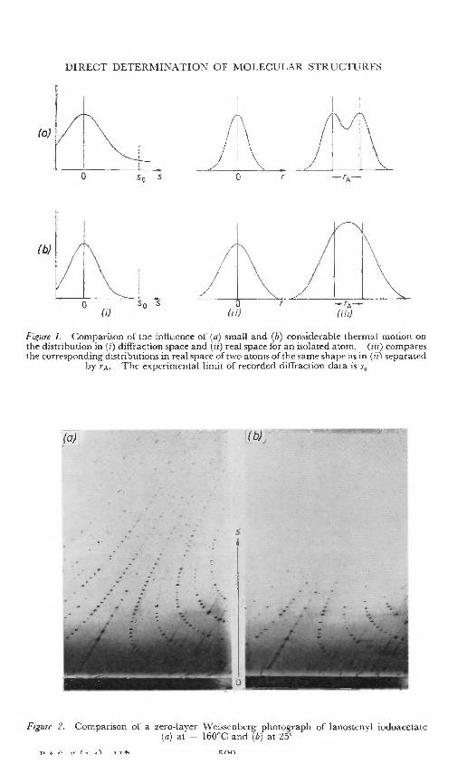
derivative as indicated by the heaviness index, but is intimately tied upwith the range and quality of the diffraction data. This aspect need bementioned only insofar as it influences the analysis both as to speed ofsolution of the structure and the accuracy of the refinement. It is usefulto revert to equations (1) and (2) to note a second aspect of their reciprocalrelationship, i.e. between the shapes of atoms in real space (in terms of theelectron density, p) and the distribution of the diffraction data, F(hkl).For an isolated atom, the distribution in diffraction space is compared with
'the shape of an isolated atom in real space in Figure 1. The broader thepeak in diffraction space which corresponds to more extensive diffractiondata, the sharper is the atom peak in real space, and hence it may be morereadily and accurately located. Conversely, where the range of data islimited, the peak in real space is very diffuse. For adjacent atoms, theability to site them accurately depends on the ability to differentiate theminto separate peaks (see Figure 1 (a) (iii) and (b) (iii». For most organic
* More elaborate discussions of the influence of the heavy atom in determining phase (orsigns) have been given by Luzzati-", Woolfson-" and Sim 14•
508
DIRECT DETERMINAT IO:,\, OF M O LECU LAR STRUCTU RES
(0)
(b)
50 5
Figure I. Comparison of th e influen ce of (a) small a nd (b) considerable thermal motion o nth e distr ib u tion in (i) diffraction spac e and (ii) r eal space for an isola ted atom. (iii) comparesth e correspond ing di str ib u tions in real space of two atoms ofthe same shape as in (iil separated
by rs- The experimen ta l limit of rec orded d iffrac tion data is So
(aF
s
( t» ,
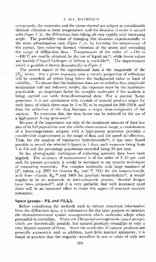
Figure 2. Comparison or a zero-layer Weissenberg pho togra ph of Ianostenyl iodoacetate(a) at - 160°C and (b) at ~5°
A. l\1cL. MATHIESON
compounds, the molecules and the atoms thereof are subject to considerablethermal vibration at room temperature, and the situation is more in accordwith Figure 1 (b), the diffraction data falling off very rapidly with increasingangle. The possibility exists of changing this situation experimentally tothe more advantageous case, Figure 1 (a), by lowering the temperature ofthe crystal, thus reducing thermal vibration of the atoms and extendingthe range of diffraction data. Temperatures of the order of -150 to-isoac are readily obtained by the use of liquid air 15, while lower valuesare feasible if liquid hydrogen or helium is available-", The improvementwhich is possible is shown dramatically in Figure 2.
The second aspect of the experimental data is the magnitude of therr; I terms. For a given exposure, only a certain proportion of reflectionswill be recorded, all others lying below the background value or limit ofvisibility. To ensure that the maximum data are recorded so that amplitudetermination will not influence results, the exposure must be the maximumpracticable-an important factor for complex molecules if the analysis isbeing carried out with three-dimensional data. For a normal X-raygenerator, it is not uncommon with crystals of natural product origin foreach layer, of which there may be 5 to 20, to be exposed for 200-250 h, andthus the collection of the data becomes a major time-component of theanalysis. To overcome this, the time factor can be reduced by the use ofa high-power X-ray generator 1 7•
Because of the importance not only of the maximum amount of data butalso of its full population over the whole observational range, a combinationof a low-temperature adaptor with a high-power generator provides aconsiderable improvement in the range of data and the speed of collection.Thus, for the analysis of himbacine hydrobromide '", it has been foundpossible to record the selected 6 layers in 5 days, each exposure being from;) to 8 h and the percentage population recorded being 90 per cent.
So far, photographic techniques of collecting intensity data have beenimplied. The accuracy of measurement is of the order of 5-10 per centand, for greater accuracy, it would be necessary to use counter techniquesof measuring intensities. For complex molecules with large numbers ofIFI values, e.g. 2927 for vitamin B12 (wet}!", 3351 for the hexacarboxylicacid from vitamin B1 2 20 and 3400 for jacobine bromohydrin21, it wouldrequire to be an automatic or semi-automatic process. Several designshave been proposed 22, and it' is very probable that with increased needthere will be an increased effort to make this aspect of structure analysisautomatic.
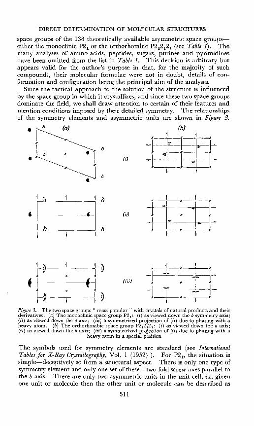
Space groups-v-Pz, and P21212 1
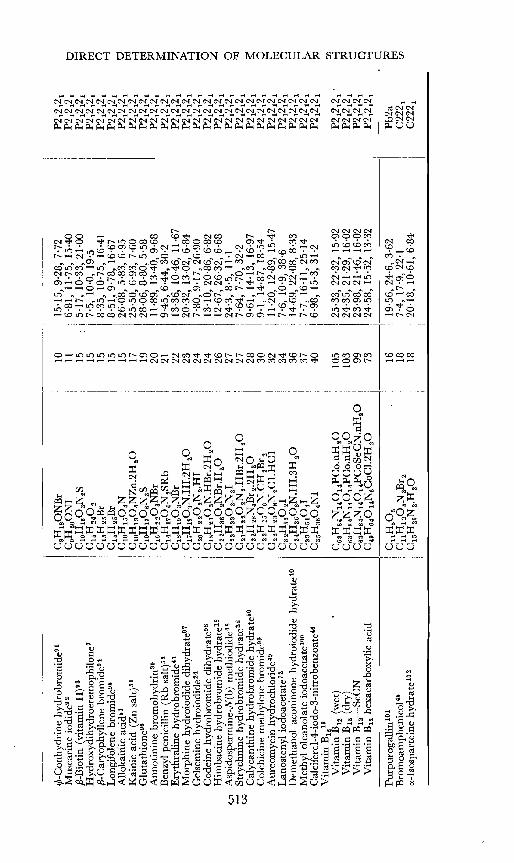
Before considering the methods used to extract structural informationfrom the diffraction data, it is informative for this later purpose to mentionthe three-dimensional spatial arrangements which molecules adopt whenpersuaded to crystallize. There are 230 spatial arrangements (space groups)which are theoretically possible, but natural products crystallize in only avery limited number of these. Since the molecules of natural products aregenerally asymmetric and, in addition, have little internal symmetry, it isfound in practice that the majority crystallize in one or other of only two
510
(b)
C> -I ~j=~I-tn -I -o -o ---1--§ § I
~ I~
~
i:1I
41~(;;)
e --1 ~f f
I
DIRECT DETERMINATION OF MOLECULAR STRUCTURES
space groups of the 138 theoretically available asymmetric space groupseither the monoclinic P2 1 or the orthorhombic P2 12121 (see Table 1). Themany analyses of amino-acids, peptides, sugars, purines and pyrimidineshave been omitted from the list in Table 1. This decision is arbitrary butappears valid for the author's purpose in that, for the majority of suchcompounds, their molecular formulae were not in doubt, details of conformation and configuration being the principal aim of the analyses.
Since the tactical approach to the solution of the structure is influencedby the space group in which it crystallizes, and since these two space groupsdominate the field, we shall draw attention to certain of their features andmention conditions imposed by their detailed symmetry. The relationshipsof the symmetry elements and asymmetric units are shown in Figure 3•
• .., c:!) (a)
j~,I ~
.1 ~ , ·1<: 1
~,
~~1
~ 1 1I
' I . I I I.
0 0 -I--r--:--..,.---1-f I f-i (if;)
I I l I
0 0 ~I---l ~-1-~~ I ~ I I I
I f ~
Figure3. The two space groups" most popular '" with crystals of natural products and theirderivatives: (a) The monoclinic space group P2 1 : (i) as viewed down the b symmetry axis;(ii) as viewed down the a axis; (iii) a symmetrized projection of (ii) due to phasing with aheavy atom. (b) The orthorhombic space group P2 12 12 1 : (i) as viewed down the a axis;(ii) as viewed down the b axis; (iii) a symmetrized projection of (ii) due to phasing with a
heavy atom in a special position
The symbols used for symmetry elements are standard (see InternationalTables for X-Ray Crystallography, Vol. 1 (1952)). For P2 1, the situation issimple-deceptively so from a structural aspect. There is only one type ofsymmetry element and only one set of these-two-fold screw axes parallel tothe b axis. There are only two asymmetric units in the unit cell, i.e. givenone unit or molecule then the other unit or molecule can be described as
511
U1 ......
t-:l
Tab
le1.
Alis
tof
the
maj
ori
tyof
nat
ura
lp
rod
uct
deri
vati
ves
for
whi
cha
deta
iled
stru
ctu
rean
alys
isis
avai
labl
e,g
rou
ped
acco
rdin
gto
the
spac
eg
rou
pin
whi
chth
eycr
ysta
lliz
e
Com
poun
dF
orm
ula
IN
oof
IC
elld
imen
sion
sI
Spac
eG
roup
IA
tom
si
IX-B
rom
ocam
phor
74C
10H
16
OB
rI
117·
38,
7·5
7,9
·12
;{J
=94
°P2
1E
ph
edri
ne
hy
dro
chlo
rid
ets
C10
H1
6O
N.H
Cl
I12
12
·65
,6·0
9,
7·32
;{J
=10
2°15
'P2
1B
rom
odi1
acto
nefr
omja
cob
ines
!C
loH
lS04
Br
I14
9·1
1,6
·42
,10
·84;
{J=
98'2
°P2
1Is
oc1o
vene
hydr
ochl
orid
e76
Cu
Hu
·HC
l
I
156,
35,
13,9
1,7
·89
;fi
=9
5'3
°P2
1E
rgin
eh
yd
rob
rom
ide'
"I
C16
H17
0N
a·H
Br
209,
60,
5·74
,13
·79;
fi=
91
'2°
P21
ce-M
etho
done
hydr
obro
mid
e78
IC
21
H27
0N
.HC
123
10
'69
,8·7
4,
10·7
4;{J
=94
·6°
P21
The
1epo
gine
met
hiod
idew
iC
21H
34O
NI
I
2311
,12,
13,0
4,7·
46;
{J=
109'
5°P2
1IX
-Met
hy1d
ihyd
roth
ebai
nem
eth
iod
ide
79C
21H
280s
NI
I
251
1,0
6,8
,75
,10
·54;
{J=
94°
32'
P21
Cal
ciu
mth
ymid
ylat
es"
ClO
H1
30
8N2
Ca.
6H
20
2614
,40,
6,87
,9
·81
;fi
=9
0·6
°P2
17-
Bro
moc
hole
ster
ylb
rom
ides
-C
27H
uBr2
271
2'0
5,8
·75
,12
·57;
fi=
101°
19'
P21
Cho
1est
eryl
iodi
de-"
C27
H45
II
271
2,5
7,9
,04
,21
,89
;{J
=14
9°P2
1D
es-(
oxym
ethy
lene
)-ly
coct
onin
ehy
droi
odid
ei
hydr
ate?
"C
24
Ha9
06
N.H
I.H
sOI
3412
,79,
9,44
,11
·00;
{J=
97·7
5°P2
1p-
Bro
mob
enzo
ate
dies
ter
ofir
esin
szC
29H
zs06
Br2
I
3528
,75,
7,41
,6
·40
;{J
=92
°P2
1P
hyll
ochl
orin
ees
ters
"C
aaH
38N
40Z
3912
,23,
14·8
4,7·
87;
fi=
94
°59
'P2
1L
umis
tero
l-4-
iodo
-3-n
itro
benz
oate
'<C
35H
4604
NI
4010
,55,
7'6
3,20
·57;
fi=
93
'5°
P21
Jaco
bin
ebr
omoh
ydri
net
hano
late
-"(C
18H
z606
NB
r)2,
C2H
50
H53
10
·11
,15
,53
,14
,90
;11
=11
3°P2
1E
pili
mon
o1io
doac
etat
e'"
I(C
26H
310S
.CO
CH
2I)
274
15,0
3,12
·36,
15·9
3;fi
=9
5'2
°P2
1
Qu
inin
esu
lpha
ted
ihy
dra
te'"
C20
H24
02N
s.H
2S
04.
2H2
030
15
,49
,6,7
4,
20·4
6;fi
=11
4°C
2S
try
chn
ine
sulp
hate
pen
tah
yd
rate
'"C
ZIH
22
02
N2
·HzS
04
·5H
zO34
35,8
5,7,
56,
7·87
;11
=10
7°2
1'
C2
Koj
icac
ids?
C5H
60
49
3,85
,1
8,4
,8,5
5;
11=
96°
P2 1
1n
So
diu
mtr
op
olo
nat
e'"
C7H
50
2Na
913
,91,
3,69
,11
·67;
fi=
93
·1°
P21f
CT
rop
ine
hydr
obro
mid
e'"
CS
H16
0N
Br
106
·32
,20
'2,
7·4
4;
fi=
95°
P2 i
/nN
oo
tkat
inco
pper
com
ple
x"
C30
Has
04C
U17
I8·4
0,11
,96,
15·2
1;11
=11
5·5°
!
P2 1
/aC
ryp
top
leu
rin
em
ethi
odid
e'"
C25
Hao
OaN
I29
:9,
95,
24'2
,9
·95
;fi
=11
2·0°
P2 1
/n
J
~ ~ Q ~ ~ ~ tr:l
en o Z
¢-C
on
hy
dri
ne
hydr
obro
mid
es!
CS
H1S
ON
Br
101
5·1
5,9
·28
,7·
72P2
1212
1M
usca
rine
iodi
de92
C9H
zoO
N1
116,
81,
11·7
5,15
-40
P212
121
tjfi
-Bio
tin
(vit
amin
H)9
3C
lOH
1603
NzS
155·
17,
10
·33
,21
·00
P212
121
~ ~H
ydro
xydi
hydr
oere
mop
hilo
ne7
C1
5H
z4O
Z15
7·5
,10
·0,
19·5
P212
121
~
,8-C
aryo
phyl
lene
bro
mid
e'"
C1
5H
2sB
r15
8,35
,10
·75,
16·4
1P2
12
121
C1L
ongi
fole
nebr
omid
es-
C1s
H2S
Br
158,
51,
9,78
,16
·67
P21
212
1~
All
okai
riic
acid
s!C
lOH
1S0
4N15
26·0
8,5·
83,
6·95
P212
121
tj
Kai
nic
acid
(Zn
salt)
SIC
lOH
130 4
NZ
n.2H
20
1725
,50,
6,93
,7
·60
P212
121
~ ~O
luta
thio
nevs
ClO
H1
70
6NsS
1928
·06,
8,80
,5·
58P2
1212
1~
An
no
tin
ine
brom
ohyd
rirr
'"C
16H
220s
NB
r20
11·8
9,13
-40,
9·68
P212
121
~
Ben
zyl
peni
cill
in(R
bsa
lt)5S
C1
4H
17
04N
zSR
b21
9·45
,6·
44,
30·2
P212
121
~ ~E
ryth
rali
ne
hy
dro
brom
ide4
1C
18H
190s
NB
r22
13,3
6,10
-46,
11·6
7P2
1212
1~
Mo
rph
ine
hydr
oiod
ide
dihy
drat
e'"
C1
7H
I90
sN.H
I,2
H2
023
20·3
2,13
·02,
6·84
P212
12
1I-
jG
else
min
ehy
droi
odid
e-"
CZ
OH
Z20
2N2·
H1
247
'80,
9·17
,26
·90
P212
121
~
Cod
eine
hy
dro
bro
mid
ed
ihy
dra
te'"
Cls
HzI
0sN
.HB
r.2
HzO
2413
·10,
20,8
6,6·
82P2
12
12
10
Him
bac
ine
hy
dro
bro
mid
ehy
drat
e-"
Czz
Hs6
0zN
Br.
HzO
261
2'6
7,2
6·3
2,
6·68
P21
21
21
Z0
1A
spid
ospe
rmin
e-N
(b)
met
hiod
ide"
CZ
SH
330z
N.r
2724
,3,
8,5,
11·1
P21
21
21
0<:;;
Str
ychn
ine
hy
dro
brom
ide
hy
dra
te'"
C2
1H
ZZO
zNz·
HB
r.2H
zO27
7·64
,7·
70,
32·2
P21
21
21
~
Cal
ycan
thin
ehy
dro
brom
ide
hy
dra
te'"
C22
Hzs
N4B
r 2.2
HzO
289·
61,
14·1
3,16
·97
P21
21
21
~C
olch
icin
em
ethy
lene
bro
mid
et"
C2
zHzs
06
N.C
HzB
rz30
9,1,
14·8
7,18
·54
P212
121
0A
ureo
myc
inh
yd
roch
lori
dew
C22
Hzs
OsN
2Cl.
HC
132
11,2
0,12
,89,
15-4
7P2
1212
1~ ~
Lan
oste
nyl
iod
oac
etat
et''
C3z
H5s
0z1
347
'6,
10·9
,38
·6P2
12
12
1C1
Dem
eth
ano
lac
onin
one
hydr
oiod
ide
hy
dra
tes?
CZ
4HSS
OsN
.HI.
3H
zO36
14
'69
,22
·08
,8·
33P2
1212
1cj
Met
hyl
olea
nola
teio
doac
etat
e-v"
CSS
H5
10
4137
7,7
,16
·11
,25
,14
P21
21
21
r-C
alci
fero
l-4-
iodo
-3-n
itro
benz
oate
-"C
ssH
s604
N1
406,
98,
15,3
,31
·2P2
1212
1~
Vit
amin
B12
19o:
Vit
amin
B12
(wet
)C
6sH
s4N
140
14P
Co.
nHzO
105
25
·33
,22
·32
,15
·92
P212
121
.~
Vit
amin
B12
(dry
)C
6sH
s4N
140
14P
Co.
nH.o
103
24
·35
,21
'29
,16
·02
P212
12
1~
Vit
amin
B1
2-S
eC
NC
6sH
s4N
14
01
4P
Co
SeC
N.n
H2
099
23
,98
,21
,46
,16
·02
P21
212
1C1
Vit
amin
B12
hexa
carb
oxyl
icac
idC
49H
6401
4N6C
OC
1.2H
2073
24,5
8,15
·52,
13·3
2P2
1212
1~ c
Pu
rpu
rog
alli
n10
1C
uH
sOs
1619
·56,
24·6
,3·
62P
b2
a~ ~
Bro
mo
amp
hen
ico
l"C
UH
12
0sN
2Br2
187
·4,1
7,9
,22
,1C
222
1rn
cc-I
sosp
arte
ine
hy
dra
te10
2C
1sH
z6N
2·H
20
1820
,18,
10·6
1,6·
84C
222
1
A. MeL. MATHIESON
reproduced by the symmetry element. In P2 12121, there is a set of two-foldscrew axes parallel to each of the three axes-an asymmetric unit is reproduced four times by the operation of the symmetry elements.
For P2 12121 with a single heavy atom in the asymmetric unit, the heavyatoms can contribute to define the phase angle of almost all reflections with,however, the presumably rare possibility that they may lie in special positions,e.g. at a quarter position, Figure 3 (b) (iii), where its effectiveness as a phasefixing agent is compromised, as in the case of gelsemine Irydroiodide-s. Onthe other hand, for P2 1, if there is only one equivalent heavy atom, then itand its symmetry-related atom can be regarded as disposed around a centreof symmetry. We have noted earlier that, where a centre of symmetry isinvolved, the phase angles defined by the heavy atoms degenerate to either0° or 1T. Hence, in this case, so far as the heavy atoms are concerned, thecrystal structure is centrosymmetric and, if the heavy atom is used to initiatethe analysis, it will impose this symmetry upon the diffraction data and thusupon the apparent structure. This situation is depicted in Figure 3 (a) (iii)in contrast with the real situation in Figure 3 (a) (ii). The situation may bedescribed to a first, but necessarily crude, approximation by imputing halfatoms on either side of the introduced reflection plane. For this situation,we therefore face a somewhat more intractable problem-the sites of theAL atoms (if we recognize them) are now of half-weight except on the introduced planes of symmetry. Of the 2n sites, assuming that all are locatedsufficiently accurately and none are spurious, we have to distinguish whichatoms are related to which to form a molecule. At this point, we mustrecognize that for this space group P2 1 with one heavy asymmetric atomwe require to apply tests of chemical sensibility in terms of bond lengths andangles in the recognition of groups of stereochemical significance. Thefirst complete analysis of a natural product-cholesteryl iodide by Carlisleand Crowfoot'v-e-provides an excellent example of this dilemma. Thissituation does not apply to space group P2 1212 1, at least in theory, althoughany chemical generalizations are useful guides at all stages of an analysis.However, for space group P2 1 they are a necessity. For such symmetrizedstructures, it is necessary to tilt the balance towards asymmetry in onedirection or another by a conscious choice, e.g. in gelsemine hydroiodides",acceptance of the existence of the indole ring decided the balance in onedirection and the remainder of the molecular skeleton was defined bygradually working outwards by iterative application of Fourier syntheses.Comparing the two -space groups for an asymmetric unit of one molecule,the amount of diffraction data per molecule is the same, and hence, undercomparable circumstances, it is more desirable to carry out an analysis witha structure crystallizing in P21212 t , since ambiguities are less likely to arise.For this reason also, the comparative numbers of structures in P2 t andP2 12 t2 t which have been analysed (see Table 1), may have no statisticalsignificance in terms of their distribution in Nature, but rather are representative of the relative difficulties of analyses with the respective space groups.
For P2 v the ambiguity arising from the existence of one heavy atom inthe asymmetric unit may theoretically be eliminated by introducing a .second heavy atom. This situation was found to exist in the case of theiodoacetate of epiliminol-", which crystallized with two molecules in the
514
DIRECT DETERMINATION OF MOLECULAR STRUCTURES
asymmetric unit, the two iodine atoms being at differenty levels. However,such a situation may well lead to further complication as was found forjacobine bromohydrins-. Here also there were two molecules in theasymmetric unit, but the situation was degenerate since both Br atoms wereon the same y level.
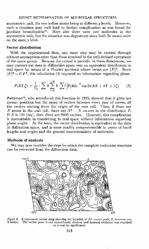
Vector distributionWith the experimental data, one more step may be carried through
without assumptions other than those involved in the well-defined operationsof the space group. Because the crystal is periodic in three dimensions, wemay convert our data in diffraction space into an equivalent distribution inreal space by means of a Fourier synthesis whose terms are IF /2. SinceIF 12 = F.F*, this calculation (3) required no information regarding phase:
I +00
p(Xrz) = Vc
.~ h~ k ~lIF(hkl) 1
2cos 21T(hX + kr + lZ) (3)
-00
Pattcrsorr'", who introduced this function in 1935, showed that it gives notatomic positions but the array of vectors between every pair of atoms, allthe vectors starting from the origin of the unit cell. Thus, if there areN atoms in the unit cell, there are N2 _. N vectors in the distribution P.If N is 100 (say), then there are 9900 vectors. However, this complicationis unavoidable in transferring to real space without information regardingphase angles. At the least, the vector distribution is equivalent to the datain diffraction space, and is more readily comprehensible in terms of bondlengths and angles and the general stereochemistry of molecules.
Methods of analysisWe may now consider the steps by which the complete molecular structure
can be extracted from the diffraction data.
Figure 4. A contoured vector map showing the location of the vector peak, V, between twoS atoms. The vector peak is not immediately obvious and internal evidence was required
to reveal its significance
515
A. MeL. MATHIESON
The first step is to determine the peaks corresponding to the AH-AHvectors in the vector distribution. These may be immediately obvious;but, if the AH atoms are of inadequate weight, then tests involving internalconsistency of the vector distribution may be required to distinguish thetrue AH-AH vector peak from peaks of comparable size, due to coincidenceof several minor vectors, e.g, in tosyl-prolyl-hydroxyproline? (Figure 4) andin cephalosporin C27. Confirmation of the correct selection' is particularlynecessary where there are several atoms of only moderate weight; e.g. inthe carbon tetrachloride adduct of sporidesmin 28 there are seven AH atomsto locate, two S and five Cl. However, we shall assume that the AH-AHvectors can be identified and that, from their location, the sites of the AHatoms in the unit cell can be deduced.
This is the basic information to be used to locate the lighter atoms, sothat they may be grouped to form the molecular skeleton(s) and the atomsdifferentiated into C, Nand O.
If the analysis is started from an empirical formula, we must have no preconceived ideas regarding the molecular shape. In the early stages, it willnot be possible even to tell which regions in the unit cell enclose the molecule(s), particularly if there is more than one molecule in the asymmetricunit. It is important not to superimpose one's ideas upon the data, sinceit is found in practice that it is extremely easy with asymmetric structures topersuade the data in the direction we wish it to go. This arises from theflexibility of definition of the phase angles and the large numbers of parameters involved in this type of analysis. This has been especially noted inthe analysis of the vitamin B12 group of compounds by Hodgkin and herco-workers-"- 20, 29 and we have had similar experience with jacobinebromohydrin21 and thelepogine methiodide'",
For this reason, it is important to handle the diffraction data so that thederived information should have maximum structural significance, a minimum of spurious detail, and lead to the correct solution in a minimum numberof stages. If one deviates from the right track, it will be possible to correctthe error because of the inherent properties of the Fourier method, but thismay require time-consuming cycles of calculation to resolve the dilemma.Since the data initially in the form of a vector distribution will be eventuallyunravelled, and the molecular structure presented more clearly in the formof the electron density distribution, the various approaches to the processof structure analysis by this method may be differentiated by the point atwhich the transfer from the vector map to the direct p map occurs. As wehave noted previously, the vector distribution contains all the structureinformation with the minimum of assumption, and accordingly it appearsadvisable to use this function to extract as much structural information aspossible before transferring to the electron density representation.
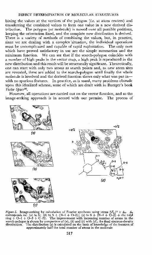
Although the vector distribution was proposed by Patterson in 193523, asystematic method of extracting information was not developed until 1950,when Buerger''! presented his image-seeking approach based on an earliersuggestion by Wrinch32. Other approaches of a similar type were proposedalmost simultaneously by Beevers and Robertsorr'", Clastre and Gay 3 4 andGarrido 35. These processes consist of placing in the vector distribution asearch-polygon which may represent a part or whole of a molecule, com-
516
DIRECT DETERMINATION OF MOLECULAR STRUCTURES
bining the values at the vertices of the polygon (i.e. at atom centres) andtransferring the combined values to form one value in a new derived distribution. The polygon (or molecule) is moved over all possible positions,keeping the orientation fixed, and the complete new distribution is derived.There is a variety of methods of combining the values, but, in practice,since we are dealing with a complex "situation, the individual operationsmust be uncomplicated and capable of rapid exploitation. The only oneswhich have proved satisfactory in use are the simple summation and theminimum function. We can see that if the search-polygon coincides witha number of high peaks in the vector map, a high peak is reproduced in thenew distribution and this result will be structurally significant. Theoretically,one can start with only two atoms as search points and, as new atom sitesare revealed, these are added to the search-polygon until finally the wholemolecule is involved and the derived function shows only what was put inwith no spurious features. In practice, as is usual, many problems obtrudeupon this idealized scheme, some of which are dealt with in Buerger's bookVector Space":
However, all operations are carried out on the vector function, and so theimage-seeking approach is in accord with our premise. The process of
(a)
(b)
-e12
Figure 5. Image-seeking by calculation of Fourier syntheses using terms IFo 12 X <pc. <Pccorresponds in: (a) to S; (b) to S + (N-I + 0-2); (c) to S + (N-I + 0-2) + the tolylring + 0-1 + 0-3 + C-12. The improvement with increasing number of atoms in thesearch-polygon is shown by comparison of (a), (b) and (c) with (d), the final electron-densitydistribution. The distribution (c) is calculated on the basis of knowledge of the location of
approximately half the total number of atoms in the molecule
517
A. MeL. MATHIESON
image-seeking may be carried out by inspection, somewhat more exactly byhand calculation and most expediently by an electronic computer. Alternatively, the combination of the vector distribution by summation may bereplaced exactly and often more conveniently by a Fourier synthesis withterms I F(hkl) 12 X cPc 37, where cPc is the structure factor of the searchpolygon. An illustration of this latter process is given in Figure 5, whichshows the influence of increasing the number of atoms in the search-polygon.The final electron-density map is shown for comparison.
a :0
to
(~OCH3(o~NJJ 0
(j iJ (iii)
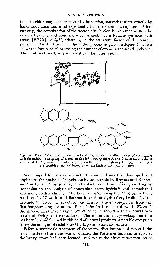
Figure 6. Part of the final three-dimensional electron-density distribution of erythralinehydrobromide. The group of atoms on the left forming rings A and B must be visualizedas rotated 90° to join with the atomic group on the right through ring C. (i), (ii) and (iii)
were possible structural formulae on the basis of chemical evidence
With regard to natural products, this method was first developed andapplied in the analysis of strychnine hydrobromide by Beevers and Robertson 38 in 1950. Subsequently, Przybylska has made use of image-seeking byinspection in the analysis of annotinine bromohydrin39 and demethanolaconinone hydroiodide-". The best example, using the F2 X cPc method,has been by Nowacki and Bonsma in their analysis of erythraline hydrobromidev-. Here the structure was derived almost completely from thefirst image-seeking operation. Part of the final result is shown in Figure 6,the three-dimensional array of atoms being in accord with structural proposals of Prelog and co-workers. The minimum image-seeking functionhas been less widely used in this field of natural products, a notable exceptionbeing the analysis of cellobiosev- by Lipscomb and co-workers.
Before a systematic treatment of the vector distribution had evolved, theusual method of analysis was to discard the Patterson function as soon asthe heavy atoms had been located, and to use the direct representation of
518
DIRECT DETERMINATION OF MOLECULAR STRUCTURES
the electron density. At first, the representation would only approximatecrudely to the" true" distribution, since phase angles were defined by theheavy atoms alone; but as single atoms or groups of atoms were recognized,their contribution to the phase angle was incorporated, and the cycle ofstructure factor and electron-density calculation repeated until the conditions for a satisfactory analysis were achieved, i.e. all peaks structurallysignificant used in (1) and (2)*. For this approach, the classical work
c
c
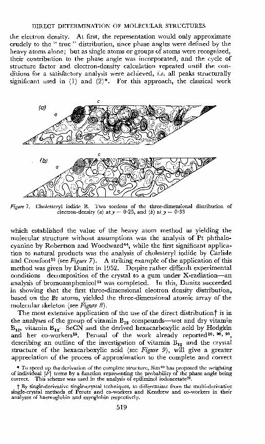
Figure 7. Cholesteryl iodide B. Two sections of the three-dimensional distribution ofelectron-density (a) aty = 0·25, and (b) aty = 0·33
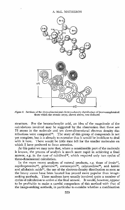
which established the value of the heavy atom method as yielding themolecular structure without assumptions was the analysis of Pt phthalocyanine by Robertson and Woodwardvl, while the first significant application to natural products was the analysis of cholesteryl iodide by Carlisleand Crowfoots" (see Figure 7). A striking example of the application of thismethod was given by Dunitz in 1952. Despite rather difficult experimentalconditions-decomposition of the crystal to a gum under X-radiation-ananalysis of bromoamphenicol-" was completed. In this, Dunitz succeededin showing that the first three-dimensional electron density distribution,based on the Br atoms, yielded the three-dimensional atomic array of themolecular skeleton (see Figure 8).
The most extensive application of the use of the direct distribution'] is inthe analyses of the group of vitamin B12 compounds-wet and dry vitaminB12, vitamin B12-SeCN and the derived hexacarboxylic acid by Hodgkinand her co-workers-''. Perusal of the work already reported-"- 20, 29,
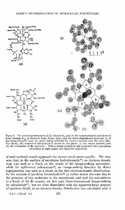
describing an outline of the investigation of vitamin B12 and the crystalstructure of the hexacarboxylic acid (see Figure 9), will give a greaterappreciation of the process of approximation to the complete and correct
* To speed up the derivation of the complete structure, Sim 43 has proposed the weightingof individual IFI terms by a function representing the probability of the phase angle beingcorrect. This scheme was used in the analysis of epiliminol iodoacetate-".
t By single-derivative single-crystal techniques, to differentiate from the multi-derivativesingle-crystal methods of Perutz and co-workers and Kendrew and co-workers in theiranalyses of haemoglobin and myoglobin respectively.
519
A. MeL. MATHIESON
Figure 8. Sections of the three-dimensional electron-density distribution of bromoamphenicolfrom which the atomic ar~ay, shown above, was deduced
structure. For the hexacarboxylic acid, an idea of the magnitude of thecalculations involved may be suggested by the observation that there are73 atoms in the molecule and ten three-dimensional electron density distributions were computed 20. The story of this group of compounds is notyet complete, but it is already so extensive that it would be invidious to dealwith it here. There would be little time left for the smaller molecules onwhich I have preferred to focus attention.
At this point we may note that, where a considerable part of the moleculeis known, the process of analysis is much more rapid in achieving a finalanswer, e.g. in the case of calciferol", which required only two cycles ofthree-dimensional calculation.
In the more recent analyses of natural products, e.g. those of iresinv",aspidospermine-", gelsemine-", aureomycin-P, calycanthine'P, and kainicand allokainic acidss-, the use of the electron-density distribution as soon asthe heavy atoms have been located has proved more popular than imageseeking methods. These analyses have usually involved quite a number ofcycles ofcalculation to arrive at the final answer. It would, however, appearto be profitable to make a careful comparison of this method with that ofthe image-seeking methods, in particular to consider whether a combination
520
DIRECT DETERMINATION OF 1vfC)LECULAR ST-RUCTURES
(a)
(b)
(iJ
OJ) @ t+
~ ~.' \» (c) ,Wf1i~
Figure 9. The molecular structures of (a) vitamin B12 and (b) the hexacarboxylic acid derivedfrom vitamin B l2l as deduced from X-ray data; and the three-dimensional structure (c) ofthe hexacarboxylic acid, the atoms being indicated by contour sections near the atom sites.For clarity, the contoured distribution is shown in two parts: (i) the corrin nucleus; and(ii) the remainder of the molecule. Where atoms overlap in this projected view, groupings
of sections at right angles are displayed adjacently
of such methods would approach the correct result more rapidly. We maynote that, in the analysis of strychnine hydrobromide'", an electron densitymap was used as a check on the results of the image-seeking procedure,while for epilimonol iodoacetatew, an image-seeking function by directsuperposition was used as a check on the first electron-density distribution.In the analysis of jacobine bromohydrin 21 (a rather severe test-case due tothe presence of two molecules in the asyrnmetric unit and the coincidencein y levels of the Br atoms), we first used three-dimensional image-seekingby calculations"; but we were dissatisfied with the apparent large amountof spurious detail, so an electron-density distribution was calculated with a
521
A. MeL. MATHIESON
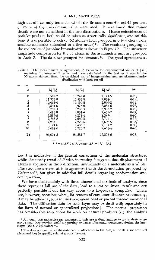
high cut-off, i.e. only terms for which the Br atoms contributed 45 per centor more of their maximum value were used.· It was found that minordetails were not coincident in the two distributions. Hence coincidences ofpositive peaks in both could be taken as structurally significant, and on thisbasis it was possible to extract 52 atoms which grouped into two chemicallysensible molecules (identical to a first order)*. The resultant grouping ofthe molecules of jacobine bromohydrin is shown in Figure 10. The structureamplitude comparison for the 55 atoms in the asymmetric unit are groupedin Table 2. The data are grouped for constant k, The good agreement at
Table 2. The measurement of agreement, R, between the experimental values of IF I,including " unobserved'1 terms, and those calculated for the first set of sites for the55 atoms derived from the combined use of image-seeking and an electron-density
distribution with high cut-off
k ~IFolI
~IFcl ~IMI R*
0 10,588-7 10,241'0 2,177'5 0.20 61 11,375'8 10,588'0 2,089-1 0,18",2 10,661'6 10,159·8 2,066-2 0-19",3 9,394'0 9,925'0 2,065'6 0.22 0
4 8,793-4 9,278·3 2,337'2 0.26 65 8,615'8 8,8744 2,405'7 0.27 96 7,815'5 8,274-4 2,387'1 0-30 57 7,756'2 7,660-8 2,721-1 0.35 18 7,235'1 7,220'6 2,662-5 0.36 89 6,124'6 6,435'7 2,437-2 0.39 8
10 5,662'6 5,723'7 2,456'4 0-434
~k 94,024-3 94,381'7 25,805·6 0.27 4
* R = LI~FII LIFol, where I~FI = IFol-1 Fel
low k is indicative of the general correctness of the molecular structure,while the steady trend of R with increasing k suggests that displacement ofatoms is required in the y direction, individually or a molecule as a whole.The structure arrived at is in agreement with the formulation proposed byGcissman->, but gives in addition full details regarding conformation andconfiguration. .
We have dealt mainly with three-dimensional methods of analysis, sincethese represent full use of the data, lead to a less equivocal result and areperfectly possible if one has easy access to a large-scale computer. Thereare, however, occasions when, for reasons of computer distance or economy,it may be advantageous to use two-dimensional or partial three-dimensionaldata. The diffraction data for each layer may be dealt with separately inthe form of normal or generalized projections'[. The normal projectionhas considerable restrictions for work on natural products (e.g. the analysis
* Although two molecules per asymmetric unit are a disadvantage to an analysis at anearly stage, they provide useful supporting evidence of internal consistency during the laterstages (see also epiliminol").
t This does not contradict the statement made earlier in the text, as the data are not usedpiecemeal but in specific related groups (layers).
522
DIRECT DETER"lI~ATIO"OF MOLECULAR STRUCTL:RES
Figure l O. Grouping of molecules in the crystal structure of jacobine bromohydrin ethanoladduct. Molecules A and B arc not related by symmecry; Band B' are crystallographically
related by a diad screw axis
G
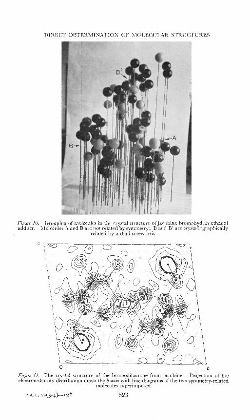
cFigure 1J. The crystal structure of the bromoclilactone from jacobine. Projection of theelectron-density distribution down the b axis with line diagrams of the two symmetry-related
molecules superimposed
523
A. MeL. MATHIESON
of benzyl penicillin'", which was mainly two-dimensional In the earlystages), but it may still be of assistance for smaller molecules. A case inpoint is that of the bromodilactone from jacobine-" (see Figure 11), thestructure analysis of which provides further corroborative evidence of thecorrectness of Geissman's deductions regarding this group of alkaloids fromSenecio jacobaea L.52. For normal projections, the x,y parameters are determined by the position of the peak while, for the corresponding generalized
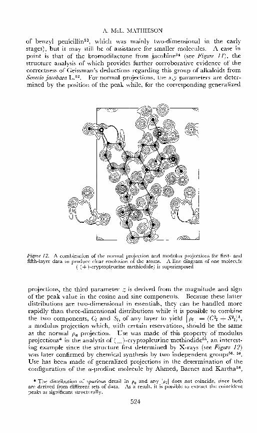
Figure 12. A combination of the normal projection and modulus projections for first- andfifth-layer data to produce clear resolution of the atoms. A line diagram of one molecule
( (±)-cryptopleurine methiodide) is superimposed
projections, the third parameter Z is derived from the magnitude and signof the peak value in the cosine and sine components. Because these latterdistributions are two-dimensional in essentials, they can be handled morerapidly than three-dimensional distributions while it is possible to combinethe two components, Ci and Si, of any layer to yield Ipli = (C2l + S2l)1,a modulus projection which, with certain reservations, should be the sameas the normal Po projection. Use was made of this property of modulusprojections* in the analysis of (±)-cryptopleurine methiodide'", an interesting example since the structure first determined by X-rays (see Figure 12)was later confirmed by chemical synthesis by two independent groups56. 59.
Use has been made of generalized projections in the determination of theconfiguration of the ex-prodine molecule by Ahmed, Barnes and Kartha-",
* The distribution of spurious detail in Po and any !Pli does not coincide, since bothare derived from different sets of data. As a result, it is possible to extract the coincidentpeaks as significant structurally.
524
DIRECT DETERMINATION OF MOLECULAR STRUCTURES
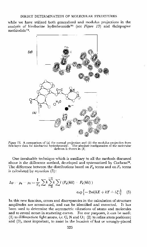
while we have utilized both generalized and modulus projections in theanalysis of himbacine hydrobromide-" (see Figure 13) and the1epoginemethiodide 73.
(0)1
Figure 13. A comparison of (a) the normal projection and (b) the modulus projection fromfirst-layer data for himbacine hydrobromide. The absolute configuration of the molecular
skeleton is shown in (b)
One invaluable technique which is auxiliary to all the methods discussedabove is the difference method, developed and systematized by Cochran'".The difference between the distributions based on Fo terms and on Fe termsis calculated by eq ua tion (5):
I +00
~p = Po - pc = VcLhLkLl (Fo(hkl) - Fc(hkl»
-00
exp {- 21Ti(hX + »r+ lZ)} (5)
In this new function, errors and discrepancies in the calculation of structureamplitudes are accentuated, and can be identified and corrected. It hasbeen used to determine the asymmetric vibrations of atoms and moleculesand to reveal errors in scattering curves. For our purposes, it can be used:(1) to differentiate light atoms, i.e. C, Nand 0; (2) to refine atom positions;and (3), most important, to assist in the location of lost or wrongly-placed
525
A. MeL. MATHIESON
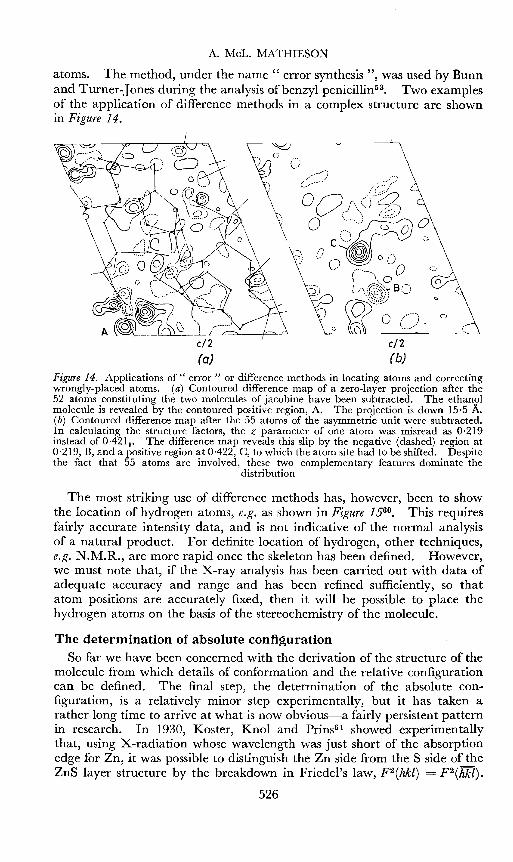
atoms. The method, under the name" error synthesis ", was used by Bunnand Turner-Jones during the analysis of benzyl penicillin'<. Two examplesof the application of difference methods in a complex structure are shownin Figure 14.
o
c'
o
o
en(b)
Figure 14. Applications of" error" or difference methods in locating atoms and correctingwrongly-placed atoms. (a) Contoured difference map of a zero-layer projection after the52 atoms constituting the two molecules of jacobine have been subtracted. The ethanolmolecule is revealed by the contoured positive region, A. The projection is down 15·5 A.(b) Contoured difference map after the 55 atoms of the asymmetric unit were subtracted.In calculating the structure factors, the z parameter of one atom was misread as 0·219instead of 0.421 9 , The difference map reveals this slip by the negative (dashed) region at0,219, B, and a positive region at 0,422, C, to which the atom site had to be shifted. Despitethe fact that 55 atoms are involved, these two complementary features dominate the
distribution
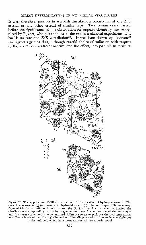
The most striking use of difference methods has, however, been to showthe location of hydrogen atoms, e.g. as shown in Figure 1560• This requiresfairly accurate intensity data, and is not indicative of the normal analysisof a natural product. For definite location of hydrogen, other techniques,e.g. N.M.R., are more rapid once the skeleton has been defined. However,we must note that, if the X-ray analysis has been carried out with data ofadequate accuracy and range and has been refined sufficiently, so thatatom positions are accurately fixed, then it will be possible to place thehydrogen atoms on the basis of the stereochemistry of the molecule.
The determination of absolute configurationSo far we have been concerned with the derivation of the structure of the
molecule from which details of conformation and the relative configurationcan be defined. The final step, the determination of the absolute configuration, is a relatively minor step experimentally, but it has taken arather long time to arrive at what is now obvious-a fairly persistent patternin research. In 1930, Koster, Knol and Prins"! showed experimentallythat, using X-radiation whose wavelength was just short of the absorptionedge for Zn, it was possible to distinguish the Zn side from the S side of theZnS layer structure by the breakdown in Friedel's law, F2(hkl) = F2(hkl).
526
DIRECT DETERMINATION OF MOLECULAR STRUCTURES
It was, therefore, possible to establish the absolute orientation of any ZnScrystal or any other crystal of similar type. Twenty-one years passedbefore the significance of this observation for organic chemistry was recognized by Bijvoet, who put the idea to the test in a classical experiment withNaRb tartrate and ZrK cx-radiation6 2• It was later shown by Petersonv"(in Bijvoet's group) that, although careful choice of radiation with respectto the anomalous scatterer accentuated the effect, it is possible to measure
(b)
o 1 2$.~
e CIo 0
• No C. H
(a)
c/2
Figure 15. The application of difference methods in the location of hydrogen atoms. Thecrystal structure is (±)-aspartic acid hydrochloride. (a) The zero-layer difference mapfrom which the aspartic acid skeleton and the CI- ion have been subtracted, leaving thedistribution corresponding to the hydrogen atoms. (b) A combination of the zero-layerand first-layer cosine and sine generalized difference maps to pick out the hydrogen atomsat different levels of the third (y) dimension. Line diagrams of the four molecular skeletons
in the unit cell, which have been subtracted, are superimposed
527
A. MeL. MATHIESON
the differences in intensity even when the heavy atom absorption edge liesa considerable way from the wavelength of the radiation used. Thisobservation enabled the technique to be applied without special experimentalconditions. Thus, it was shown that it was possible to define the absoluteconfiguration of strychnine hydrobromide'r' using Cu radiation. We haveused this technique to define the absolute configuration of himbacinehydrobromide-" and jacobine bromohydrint '.
For a heavy atom such as I with Cu radiation, the effect of anomalousdispersion is more marked than for Br, sincef" = + 7·2 for CuK radiation(1= fo +1' + if", where io = -53·0). This is reflected in the largerdifferences in intensity, often readily visible to the eye; and, under suchconditions, Przybylska and Marion have defined the absolute configuration'"of (+)-des-(oxymethylene)-lycoctonine hydroiodides" and (+)-demethanolaconionone hydrioiodidew (see Figure 16).
Figure 16. The absolute configuration of the molecular skeleton common to des-(oxymethylene) -lycoctonine and demethanolaconinone
Although of lesser interest, an alternative method more akin to thechemical approach is the introduction of a known absolute centre with theheavy atoms". With this technique, the analysis proceeds normally andthe final structure contains its own reference standard. An example is theanalysis of (+)-S-methyl-L-cysteine-S-oxide6 8• The analysis of vitamin B12
illustrates both methods, since the absolute configuration of the molecule inthe earlier stages of the analysis was defined by reference to the D-ribosecomponent-", while later this decision was confirmed by reference to theanomalous dispersion of the Co atom in the hexacarboxylic acid 20.
The definitive experiment of Bijvoet and his co-workers and its implications have already had considerable influence on the definition of configuration in organic chemistry, and have evoked a new set of rules by Cahn,Ingold and Prelog'" to overcome inconsistencies in nomenclature.
That the anomalous dispersion effect -may be of greater significance thanthe definition of configuration, that it may be of direct use in structureanalysis, was realized early. However, since the intensity differences aresmall, the necessity for very accurate intensity measurement is one whichhas not yet been made completely practicable for complex molecules.Peterson'" illustrated its feasibility and Srinivasan70 has applied it to ananalysis. of L-tyrosine. Okaya, Saito and Pepinsky i! have suggested a neatmethod of unravelling the vector distribution by this technique (see alsoPepinsky") .
528
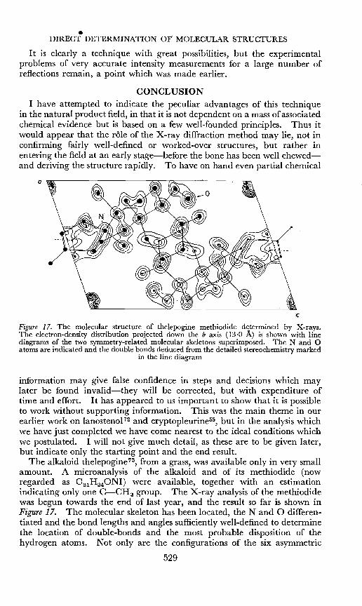
•DIRECT DETERMINATION OF MOLECULAR STRUCTURES
It is clearly a technique with great possibilities, but the experimentalproblems of very accurate intensity measurements for a large number ofreflections remain, a point which was made earlier.
CONCLUSIONI have attempted to indicate the peculiar advantages of this technique
in the natural product field, in that it is not dependent on a mass of associatedchemical evidence but is based on a few well-founded principles. Thus itwould appear that the role of the X-ray diffraction method may lie, not inconfirming fairly well-defined or worked-over structures, but rather inentering the field at an early stage-before the bone has been well chewedand deriving the structure rapidly. To have on hand even partial chemical
a ~-----------z:::. r.:::s::----~----nJIT7\
:c
Figure 17. The molecular structure of thelepogine methiodide determined by X-rays.The electron-density distribution projected down the b axis (13,0 A) is shown with linediagrams of the two symmetry-related molecular skeletons superimposed. The Nand 0atoms are indicated and the double bonds deduced from the detailed stereochemistry marked
in the line diagram
information may give false confidence in steps and decisions which maylater be found invalid-they will be corrected, but with expenditure oftime and effort. It has appeared to us important to show that it is possibleto work without supporting information. This was the main theme in ourearlier work on lanostenol P and cryptopleurine'", but in the analysis whichwe have just completed we have come nearest to the ideal conditions whichwe postulated. I will not give much detail, as these are to be given later,but indicate only the starting point and the end result.
The alkaloid thelepogine 73, from a grass, was available only in very smallamount. A microanalysis of the alkaloid and of its methiodide (nowregarded as C 21H340NI) were, available, together with an estimationindicating only one C-CHa group. The X-ray analysis of the methiodidewas begun towards the end of last year, and the result so far is shown inFigure 17. The molecular skeleton has been located, the Nand 0 differentiated and the bond lengths and angles sufficiently well-defined to determinethe location of double-bonds and the most probable disposition of thehydrogen atoms. Not only are the configurations of the six asymmetric
529
A. MeL. MATHIESON
centres in the molecule placed on a relative basis, but the absolute configuration shown in Figure 17 has been defined by the use of the anomalousdispersion of the I atom to the Cu K; radiation used in the analysis. Onepoint which was unexpected and may be of chemical interest is the existencewithin the molecular skeleton of a pyrrolizidine ring system which has thesame absolute configuration as found in jacobine bromohydrin.
The deduction from the evidence which we have presented is that, in therange of molecules up to 100 atoms (excluding H), and probably beyond,the structure can be solved in a time which compares favourably with otherphysical methods, provided that certain preliminary conditions are satisfiedand a computer of reasonable storage and speed is available. Nor needthis work necessarily involve a large team-the three analyses, of himbacinehydrobromide-", jacobine bromohydrins- and thelepogine onethiodides'',some details of which I have used to illustrate various points, were initiatedand solved in a period of less than two years. Because of the mass of detailrequired for an analysis and the extensive data derived from the work,however, it is important to use this technique for compounds either of greatsignificance in themselves or those which provide a structural key to a groupof compounds.
I wish to express grateful thanks to my colleague, Mr J. Fridrichsons, for hiscollaboration and unflagging support in the work which hasformed the basisofopinionsexpressed here; to Drs]. R. Price, W. D. Crow andC. C.J. Culvenor ofMelbourne,and Drs E . .Ritchie and W. C. Taylor of Sydney, for, without their willing andgenerous co-operation (a) in allowing us to tackle problems which they might well havedesire to retain for their personal study, and (b) in providing excellent crystals of thederivatives, we would not have had such interesting compounds to work on; and}inally, to the invaluable co-operation of the respective staffs of the electronic computersSILLIAC (University ofSydney) and UTECOM (University ofNew South Wales).
References1 D. Arigoni, D. H. R. Barton, E. J. Corey, O. Jeger, L. Cag1ioti, S. Dev, P. G. Ferrini,
E. R. Glazier, A. Melera, S. K. Pradhan, K. Schaffner, S. Sternhell, J. F. Templetonand S. Tobinaga. Experientia, 16, 41 (1960)
2 D. Crowfoot. Ann. Rev. Biochem., 1948, 1153 J. M. Robertson. Third Henderson Memorial Lecture, Royal Institute of Chemistry
Lectures, Monographs and Reports, No.6 (1954)4 A. McL. Mathieson. Revs. Pure and Appl. Chem. (Australia), 5, 113 (1955)6 R. Pepinsky. Record Chem. Progr. (Kresge-Hooker Sci. Lib.), 17, 145 (1956)6 H. Lipson and W. Cochran. The Crystalline State, Vol. 3, Bell & Sons, London (1953)7 D. F. Grant and D. Rogers. Chern. & Ind. (London), 1956, 275
D. F. Grant. Acta Cryst., 10, 498 (1957)8 C. Brink, D. C. Hodgkin, J. Lindsey, J. Pickworth, J. H. Robertson and J. G. White.
Nature, 174,4 (1954)9 A. F. Beecham, J. Fridrichsons and A. MeL. Mathieson. J. Am. Chem. Soc., 80, 4739
(1958)10 M. F. Perutz, M. G. Rossman, A. F. Cullis, H. Muirhead, G. Will and A. C. T. North.
Nature, 185,416 (1960)11 J. C. Kendrew, R. E. Dickerson, B. Strandberg, R. G. Hart, D. R. Davies, D. C. Phillips
and V. C. Shore. Nature, 185, 422 (1960)12 V. Luzzati. Acta Cryst., 6, 142 (1953)13 M. M. Woo1fson. Acta Cryst., 9, 804 (1956)
530
DIRECT DETERMINATION OF MOLECULAR STRUCTURES
14 G. A. Sim. Acta Cryst., 10, 177, 536 (1957)15 J. Fridrichsons and A. MeL. Mathieson. Rev. Sci. Instr., 29, 784 (1958)16 J. H. Robertson. J. Sci. Insir., 37, 41 (1960)17 A. Taylor. J. Sci. Instr, and Phys. in Ind., 26, 225 (1949)
D. A. Davies, A. MeL. Mathieson and G. M. Stiff. Rev. Sci. Instr., 30, 488 (1959)18 J. Fridrichsons and A. McL. Mathieson. Acta Cryst., in the press19 D. C. Hodgkin, J. Kamper, J. Lindsey, M. MacKay, J. Pickworth, J. H. Robertson,
C. B. Shoemaker, J. G. White, R. J. Prosen and K. N. Trueblood. Proc. Roy. Soc.(London), 242A, 228 (1957)
20 D. C. Hodgkin, J. Pickworth, J. H. Robertson, R. J. Prosen, R. A. Sparks and K. N.Trueblood. Proc. Roy. Soc. (London), 251A, 306 (1959)
21 J. Fridrichsons, A. MeL. Mathieson and D. ]. Sutor. Tetrahedron Letters, No. 23, 35(1960)
22 W. L. Bond. Acta Cryst., 8, 741 (1955)U. W. Arndt and D. C. Phillips. Brit. Pat. Application No. 31197/57 (October)A. MeL. Mathieson. Acta Cryst., 11,433 (1958)E. Prince and S. C. Abrahams. Rev. Sci. Instr., 30, 581 (1959)F. Langdon and B. D. Frazer. Rev. Sci. Instr., 30, 997 (1959)J. Ladell and K. Lowitzsch. Acta Cryst., 13, 205 (1960)
28 A. L. Patterson. Z. Krist., 90, 517 (1935)24 F. M. Lovell, R. Pepinsky and A. J. C. Wilson. Tetrahedron Letters, No.4, 1 (1959)25 C. H. Carlisle and D. Crowfoot. Proc. Roy. Soc. (London), 18U, 64 (1945)26 S. Arnott, A. W. Davie, J. M. Robertson, G. A. Sim and D. G. Watson. Experientia,
16,49 (1960)27 S. Abrahamsson, D. C. Hodgkin and E. N. Maslen. To be published28 R. L. M. Synge and E. P. White. Chern. & Ind. (London), 1959, 154629 D. C. Hodgkin, M. J. Kamper, K. N. Trueblood and]. G. White. Z. Krist., 113, 30
(1960)30 J. Fridrichsons and A. McL. Mathieson. Tetrahedron Letters, No. 26, 18 (1960)31 M. J. Buerger. Acta Cryst., 3, 87 (1950)
M. J. Buerger. Acta Cryst., 4, 531 (1951)32 D. M. Wrinch. Phil. Mag., 27, 98 (1939)33 C. A. Beevers and J. H. Robertson. Acta Cryst., 3, 164 (1950)34 J. Clastre and R. Gay. Compt. rend., 230, 1976 (1950)
J. Clastre and R. Gay. J. phys. radium, 11, 75 (1950)3li J. Garrido. Compt. rend., 231, 297 (1950)36 M. J. Buerger. Vector Space, Wiley, New York (1959)37 D. McLachlan and I. D. Thomas. Acta Cryst., 5, 301 (1952)38 C. A. Beevers andJ. H. Robertson. Acta Cryst., 4, 270 (1951)39 M. Przybylska and L. Marion. Can. J. Chem., 35, 1075 (1957)
M. Przybylska and F. R. Ahmed. Acta Cryst., 11, 718 (1958)40 M. Przyby1ska and L. Marion. Can. J. Chem., 37, 1116 (1959)41 W. Nowacki and G. F. Bonsma. Z. Krist., 110, 89 (1958)112 R. A. Jacobson, J. A. Wunderlich and W. N. Lipscomb. Nature, 184, 1719 (1959)43 G. A. Sim. Acta Cryst., 12, 813 (1959)44 J. M. Robertson and 1. Woodward. J. Chem. Soc., 1940, 3645 J. D. Dunitz. J. Am. Chem. Soc., 74, 995 (1952)46 D. C. Hodgkin, M. S. Webster and J. D. Dunitz. Chern. & Ind. (London), 1957, 114847 M. G. Rossman and W. N. Lipscomb. Tetrahedron, 4, 275 (1958)48 J. F. D. Mills and S. C. Nyburg. J. Chern. Soc., 1960, 1458
J. F. D. Mills and S. C. Nyburg. Tetrahedron Letters, No. 11, 1 (1959)49 S. Hirakawa, Y. Okaya, F. M. Lovell and R. Pepinsky. Z. Krist., 112, 439 (1959)50 T. A. Hamor, J. M. Robertson, H. N. Shrivastava and T. V. Silverton. Proc. Chern.
Soc., 1960, 7851 1. Nitta, H. Watase and Y. Tomiie. Nature, 181, 761 (1958)
H. Watase and 1. Nitta. Bull. Chern. Soc. Japan, 30, 889 (1957)H. Watase. Bull. Chern. Soc. Japan, 31, 932 (1958)
52 T. A. Geissman. Australian J. Chem., 12, 247 (1959)sa D. Crowfoot, C. W. Bunn, B. W. Rogers-Low and A. Turner-Jones. A Chemistry of
Penicillin, p. 310, Princeton University Press (1949)
531
A. MeL. MATHIESON
54 J. C. Taylor. To be published55 ]. Fridrichsons and A. McL. Mathieson. Nature, 173, 732 (1954)
]. Fridrichsons and A. MeL. Mathieson. Acta Cryst., 8, 761 (1955)56 C. K. Bradsher and H. Berger. ]. Am. Chern. Soc., 80, 930 (1958)57 P. Marchini and B. Belleau. Can.]. Chem., 36, 581 (1958)58 F. R. Ahmed, W. H. Barnes and G. Kartha. Chern. & Ind. (London), 1959, 48559 W. Cochran. Acta Cryst., 4, 408 (1951)60 B. Dawson. To be published61 D. Koster, K. S. Knol and]. A. Prins. Z. Physik, 63, 345 (1930)62]. M. Bijvoet, A. F. Peerdeman and A. J. van Bommel. Nature, 168,271 (1951)63 S. W. Peterson. Nature, 176, 395 (1955)64 A. F. Peerdeman, Acta Cryst., 9, 824 (1956)65 M. Przybylska and L. Marion. Can.]. Chern., 37, 1843 (1959)66 M. Przybylska and L. Marion. Can.]. Chern., 34, 185 (1956)67 A. MeL. Mathieson. Acta Cryst., 9, 317 (1956)68 R. Hine and D. Rogers. Chern. & Ind. (London), 1956, 142869 R. S. Cahn, C. K. Ingold and V. Prelog, Experientia, 12, 81 (1956)70 R. Srinivasan. Proc. Indian Acad. Sci., 50, 19 (1959)71 Y. Okaya, Y. Saito and R. Pepinsky. Phys. Reu., 98, 1857 (1955)72 R. G. Curtis, J. Fridrichsons and A. McL. Mathieson. Nature, 170, 321 (1952)
j. Fridrichsons and A. MeL. Mathieson. ]. Chern. Soc., 1959, 215973 W. D. Crow. To be published74 E. W. Wiebanga and C. J. Krom. Rec. trau. chim., 65, 663 (1946)75 D. C. Phillips. Acta Cryst., 7, 159 (1954)76 J. S. C1unie and J. M. Robertson. Proc. Chern. Soc., 1960, 8277 J. L. de Vries and R. Pepinsky. Nature, 168,431 (1951)78 A. W. Hanson and F. R. Ahmed. Acta Cryst., 11,669 (1958)79 D. W. Smits and R. Pepinsky. Acta Cryst., 7, 653 (1954)80 P. Horn and V. Luzzati. Nature, 183, 880 (1959)81 H. Burki and W. Nowacki. Z. Krist., 108, 206 (1956)82 M. G. Rossman and W. N. Lipscomb. Tetrahedron, 4, 275 (1958)83 W. Hoppe and G. Will. Z. Krist., 113, 104 (1960)84 D. C. Hodgkin and D. Sayre. ]. Chern. Soc., 1952, 456185 H. Mendel. Proc. Koninkl. Ned. Akad. Wetenschap.,58D, 132 "(1955)86 C. Bokhoven, J. C. Schoone and]. M. Bijvoet. Acta Cryst., 4, 275 (1951)87 H. A. McKinstry, P. E. Eiland and R. Pepinsky. Acta Cryst., 5, 285 (1952)88 Y. Sasada and 1. Nitta. Acta Cryst., 9, 205 (1956)89 ]. W. Wisser,]. Manassen and J. L. de Vries. Acta Cryst., 7, 288 (1954)90 R. B. Campbell and]. M. Robertson. Chern. & Ind. (London), 1952, 126691 H. S. Yanai and W. N. Lipscomb. Tetrahedron, 6, 103 (1959)92 F. Jellinek. Acta Cryst., 10, 277 (1957)93 W. Traub. Nature, 178, 649 (1956)94 J. M. Robertson and G. Todd. Chern. & Ind. (London), 1953, 43796 R. H. Moffett and D. Rogers. Chern. & Ind. (London), 1953,91696 W. B. Wright. Acta Cryst., 11, 632 (1958)97 M. MacKay and D. C. Hodgkin. ]. Chern. Soc., 1955,326198 J. M. Lindsey and W. H. Barnes. Acta Cryst., 8, 227 (1955)99 J. L. de Vries, M. V. King and R. Pepinsky. Acta Cryst., 5, 437 (1952)
100 A. M. Abd El Rahim and C. H. Carlisle. Chern. & Ind. (London), 1954,279101 j. D. Dunitz, Nature, 169, 1087 (1952)102 M. Przybylska and W. H. Barnes. Acta Cryst., 6, 377 (1953)
532


![Algorithm 464 Eigenvalues of a Real, Symmetric, Tridiagonal …nbrenner/Algorithm 467 Matrix... · 2014-02-04 · G.W. Hill [Recd. 24 Aug. 1971, 23 Feb. 1972, 10 July 1972] C.S.I.R.O.,](https://img.pdfslide.us/doc/110x75/5f0211227e708231d40268c1/algorithm-464-eigenvalues-of-a-real-symmetric-tridiagonal-nbrenneralgorithm-467.jpg)


![Renewable resources-based PTT [poly(trimethylene ...iupac.org/publications/pac/pdf/2013/pdf/8503x0521.pdfRenewable resources-based PTT [poly(trimethylene terephthalate)] ... biobased](https://img.pdfslide.us/doc/110x75/5ad6edfa7f8b9af9068b9cb7/renewable-resources-based-ptt-polytrimethylene-iupacorgpublicationspacpdf2013pdf.jpg)