Embed Size (px)
Citation preview
RESEARCH ARTICLE4656
Development 139, 4656-4665 (2012) doi:10.1242/dev.078923© 2012. Published by The Company of Biologists Ltd
INTRODUCTIONThe axial skeleton and skeletal muscles arise from the somites. Assomites form from the anterior end of the presomitic mesoderm(PSM), mesoderm progenitors in the tailbud continually generatenew mesoderm cells and feed them into the posterior PSM (Holley,2007). The number of progenitors and the rate at which theirprogeny differentiate and move from the tailbud into the PSM mustbe controlled to ensure that the correct somite number is reached.Premature exhaustion of progenitors results in premature extinctionof the PSM, a deficit of posterior somites, and therefore a truncatedbody.
Several mouse mutants with a truncated axis have beendescribed and all but one result from lack of mesoderm (Wilson etal., 2009). The one exception carries a null mutation in the bHLHtranscription factor mesogenin 1 (Msgn1). It lacks thoracic, lumbarand sacral vertebrae and skeletal muscles (Yoon and Wold, 2000),but there is no lack of mesoderm progenitors – quite the opposite:the lack of PSM tissue is accompanied by an enlarged tailbudcontaining an excess of cells expressing brachyury – a mesodermal
progenitor marker. This suggests that in the absence of Msgn1,mesoderm progenitors that should normally emerge to becomePSM remain instead in the tailbud. What, then, is the precisefunction of Msgn1? Do the brachyury-expressing progenitorsremain in the tailbud because of a block in differentiation, cellmigration, or both? To investigate this question, we characterisedMsgn1 function through loss- and gain-of-function experimentscombined with live imaging in zebrafish.
In zebrafish, msgn1 is expressed in a domain similar to that inmouse (Yoo et al., 2003), but its function has not been described.However, the zebrafish spadetail (spt, now termed tbx16) mutant,like mouse Msgn1 mutants, shows a large accumulation of cellsexpressing the brachyury-like gene no tail (ntl) in the tailbud(Griffin et al., 1998; Griffin and Kimelman, 2002). It has beenproposed that Spt promotes the differentiation of tailbudprogenitors by inhibiting the progenitor maintenance genes ntl andWnt (Griffin and Kimelman, 2002; Martin and Kimelman, 2008).Spt also controls cell movement during gastrulation (Ho and Kane,1990; Kimmel et al., 1989; Row et al., 2011), suggesting that Sptmight also control motility in the tailbud. However, Spt cannot bethe only factor regulating the transition of tailbud progenitors intoPSM because spt null mutants still form tail somites (Griffen et al.,1998). Msgn1 is thus a candidate additional factor in zebrafishresponsible for the switch from a tailbud progenitor state to a PSMstate.
We show that combined loss of msgn1 and spt leads to completefailure of trunk and tail somite formation accompanied by a largeexcess of ntl-expressing cells in the tailbud. Using a heat shock-inducible transgenic line, we find that a pulse of msgn1 expressioncauses a rapid downregulation of ntl and wnt8, indicating that thesetwo genes, which work in an intricate positive-feedback loop witheach other, are themselves negatively regulated by Msgn1. ThisMsgn1-induced downregulation of ntl and wnt8 expression is
1Instituto de Medicina Molecular e Instituto de Histologia e Biologia doDesenvolvimento, Faculdade de Medicina da Universidade de Lisboa, 1649-028Lisboa, Portugal. 2Instituto Gulbenkian de Ciência, P-2780-156 Oeiras, Portugal.3Department of Molecular and Cell Biology, University of California, Berkeley, CA94720-3200, USA. 4Division of Basic Science, Fred Hutchinson Cancer ResearchCenter Seattle, WA 98109, USA. 5Developmental Genetics Laboratory, CancerResearch UK London Research Institute, 44 Lincoln’s Inn Fields, London WC2A 3PX,UK. 6Vertebrate Development Laboratory, Cancer Research UK London ResearchInstitute, 44 Lincoln’s Inn Fields, London WC2A 3PX, UK.
*Authors for correspondence ([email protected]; [email protected])‡Present address: Department of Molecular Genetics and Department of Molecularand Cellular Biochemistry, The Ohio State University, Columbus, OH 43210, USA
Accepted 13 September 2012
SUMMARYSomites are formed from the presomitic mesoderm (PSM) and give rise to the axial skeleton and skeletal muscles. The PSM isdynamic; somites are generated at the anterior end, while the posterior end is continually renewed with new cells entering fromthe tailbud progenitor region. Which genes control the conversion of tailbud progenitors into PSM and how is this processcoordinated with cell movement? Using loss- and gain-of-function experiments and heat-shock transgenics we show in zebrafishthat the transcription factor Mesogenin 1 (Msgn1), acting with Spadetail (Spt), has a central role. Msgn1 allows progression of thePSM differentiation program by switching off the progenitor maintenance genes ntl, wnt3a, wnt8 and fgf8 in the future PSM cellsas they exit from the tailbud, and subsequently induces expression of PSM markers such as tbx24. msgn1 is itself positively regulatedby Ntl/Wnt/Fgf, creating a negative-feedback loop that might be crucial to regulate homeostasis of the progenitor population untilsomitogenesis ends. Msgn1 drives not only the changes in gene expression in the nascent PSM cells but also the movements by whichthey stream out of the tailbud into the PSM. Loss of Msgn1 reduces the flux of cells out of the tailbud, producing smaller somitesand an enlarged tailbud, and, by delaying exhaustion of the progenitor population, results in supernumerary tail somites. Throughits combined effects on gene expression and cell movement, Msgn1 (with Spt) plays a key role both in genesis of the paraxialmesoderm and in maintenance of the progenitor population from which it derives.
KEY WORDS: Mesogenin 1, Spadetail (Tbx16), Paraxial mesoderm
The differentiation and movement of presomitic mesodermprogenitor cells are controlled by Mesogenin 1Rita Fior1,2,*, Adrienne A. Maxwell3, Taylur P. Ma4, Annalisa Vezzaro5, Cecilia B. Moens4, Sharon L. Amacher3,‡, Julian Lewis6 and Leonor Saúde1,2,*
DEVELO
PMENT
4657RESEARCH ARTICLEMsgn1 function in zebrafish
followed by ectopic activation of an intermediate/anterior PSMmarker, tbx24 (also known as fss and now termed tbx6), in thetailbud, consistent with the idea that Msgn1 throws a switch thatconverts cells from a tailbud progenitor state into a PSM state.msgn1 expression is itself positively regulated by the ntl, Wnt andFgf mesoderm progenitor maintenance genes (Griffin andKimelman, 2002; Goering et al., 2003; Wittler et al., 2007; Wanget al., 2007; Morley et al., 2009; Garnett et al., 2009) (our data),which, by activating msgn1 in a subset of the tailbud cellpopulation, evidently trigger these cells to embark on the PSMdifferentiation pathway. We show that Msgn1 drives not only thedifferentiation but also the migration of such cells out of the tailbudinto the PSM region. By governing the flux of cells from theprogenitor region into the PSM, Msgn1 helps control both the sizeof somites and the size and persistence of the progenitor cellpopulation; loss of Msgn1 activity thus gives rise to additional tailsomites.
MATERIALS AND METHODSZebrafish lines and heat-shock experimentsZebrafish lines: msgn1fh273 [a mutant found by screening ENU-mutagenised F1 fish (Draper et al., 2004)]; sptb104 (Kimmel et al., 1989);ntlb195 (Halpern et al., 1993); hsp70:dkk1-GFPw32 (Stoick-Cooper et al.,2007); and hsp70:dnfgfr1-EGFPpd1 (Lee et al., 2005).
For all heat-shock experiments, embryos were raised at 25°C and heatshocked at 39°C for the indicated time. hsp70:HA-msgn1, hsp70:dkk1-GFP and hsp70:dnfgfr1-EGFP embryos were generated from a crossbetween transgenic heterozygous and wild-type fish, giving batches withan expected mean ratio of 50% transgenics to 50% wild-type siblings.hsp70:HA-msgn1 embryos were sorted into distinct phenotypic classesafter in situ hybridisation (confirmed by genotyping) and hsp70:dkk1-GFPand hsp70:dnfgfr1-EGFP embryos were sorted by GFP expression.
DNA constructsmsgn1 cDNA was amplified from a zebrafish EST (IMAGE:7286125) withprimers (5�-3�) pFWEcoRI (CCGGAATTCATGGCGCAAATCG -ACGTGGATG) and pRXbaI (CTAGTCTAGATCACTGCTGC -TCGAGGATGCC) and cloned into the EcoRI and XbaI sites of pCS2+,and NotI/SP6 was used to produce msgn1 poly(A)-capped RNA andClaI/T7 to produce an antisense RNA probe.
The hsp70:HA-msgn1 transgenic was created by placing msgn1 cDNAcontaining an N-terminal HA tag downstream of the hsp70 heat-shockpromoter in the pT2 vector (UAS-hsp70p-polyA--crystallin promoter-CFP) using primers pFW-HA-ClaI-Kozak (CCATCGATGGCCACC -ATGGCTTCATATCCTTACGATG) and pRStuI (AAAAGGCCTTTTTC -ACTGCTGCTCGAGGATGCC).
The nuclear localisation sequence (Nls)-tagged Kaede was generated byPCR. The first PCR was performed to add a BamHI site and Kozak sequenceto the 5� end and part of the Nls and a linker sequence at the 3� end of Kaede,using primers pFW1 (ATACGCGGATCCGCC GCCGC CATGAG -TCTGATTAAACCAGAAAATG) and pR1 (CTTTTCTT TTCTTTTTT -GGAGAACCCTTGACGTTGTCCGGCAATCC). The second PCR wasperformed to add an EcoRI site and the rest of the Nls sequence at 3� end ofKaede, using primers: pFW1 and pR2 (TATCCGGAATTCTTAGTCAA -CTTTTCTTTTCTTTTTTGGAGA). The resulting PCR product was clonedinto the BamHI and EcoRI sites of pCS2+ and NotI/SP6 was used to produceNls-Kaede poly(A)-capped RNA.
Microinjectionsmsgn1 morpholino (CATGGCGCAAATCGACGTGGATGTG) and astandard control morpholino (CCTCTTACCTCAGTTACAATTTATA)from Gene Tools were injected at 5.7 ng/embryo at the one-cell stage; 100pg msgn1, Nls-Kaede and Kaede mRNAs were injected at the one-cellstage; the DNA plasmids hsp70:ntl and hsp70:cacatenin (Martin andKimelman, 2012) were injected at 30 pg/embryo at the one-cell stage.
In situ hybridisation and immunohistochemistrySingle whole-mount in situ hybridisations were performed as described(Thisse and Thisse, 2008). Double whole-mount fluorescent in situhybridisations were performed as described (Jülich et al., 2005) withmodifications: the red signal was developed with Fast Red (alkalinephosphatase substrate, Roche) and the green signal with tyramide-FITC(horseradish peroxidase substrate, PerkinElmer TSA Plus FluoresceinSystem). For HA immunohistochemistry, embryos were fixed for 2 hoursin 4% paraformaldehyde, incubated with a monoclonal rat anti-HAantibody (3F10, Roche) followed by anti-rat DyLight 488 secondaryantibody (Rockland). F-actin and nuclei were detected with Alexa Fluor488-Phalloidin (Molecular Probes) and DAPI, respectively.
Kaede time-lapse microscopyKaede- and Nls-Kaede mRNA-injected embryos were kept in the dark.Eight-somite stage embryos were mounted in 1.2% low-melting agarose inglass-bottom Petri dishes. Small groups of cells were labelled byphotoconversion in the maturation zone (25 m posterior to the end of thenotochord and in the focal plane of notochord and adaxial cells) and in thePSM (see Fig. 6H), with a 405 nm laser in a Zeiss 510 META confocalmicroscope using a 20� objective. Embryos were imaged in an AndorRevolutionXD spinning disc confocal microscope at 25°C. Stacks of 50optical sections, spaced by 1 m, were collected every 180 or 213 secondsfor up to 2 hours.
Cell track analysis and measurementsTime-lapse movies were analysed using ImageJ software (NIH). Individualcells were tracked using the MtrackJ plug-in. The diving velocity and theanteroposterior (A/P) velocity were measured by following cells over anextended period of time (generally 60 minutes or more), and werecalculated as (zfinal–zinitial)/(tfinal–tinitial) and (yfinal–yinitial)/(tfinal–tinitial), wherez and y denote positions along the superficial/deep (dorsoventral) and A/Paxes, respectively. The centre-to-centre distance between dot-2 and dot-3was 109±5 µm (mean ± s.d.) – the same within narrow limits for all setsof embryos analysed. The mean A/P velocity of dot-3 cells was subtractedfrom the A/P velocity of dot-2 cells to obtain the A/P velocity (Vap) of dot-2 relative to dot-3 cells. Statistical significance of differences betweengenotypes was calculated using unpaired two-tailed Student’s t-test.
RESULTSDepletion of msgn1 and spt leads to completeloss of trunk and tail somitesTo uncover the role of msgn1 in zebrafish, we examined theembryonic phenotype in loss-of-function experiments using atranslation-blocking morpholino (msgn1MO) (supplementarymaterial Fig. S1A). msgn1MO-injected embryos showed increasedntl expression in the tailbud when compared with control siblings(Fig. 1A�,B�), as does the mouse Msgn1 mutant (Yoon and Wold,2000). However, somites formed in zebrafish msgn1 morphants(Fig. 1B�,B�), in contrast to the mouse mutant, in which somiteformation is abolished. To validate the msgn1 knockdown, weanalysed a zebrafish msgn1 nonsense mutant allele, msgn1fh273
(supplementary material Fig. S1B). Homozygous msgn1fh273
mutants are viable and have a phenotype indistinguishable fromthat of the msgn1 morphants (compare Fig. 1B-B� with 1F-F�). Themildness of the msgn1fh273 phenotype is not due to a maternalcontribution, as no maternal msgn1 mRNA was detected either byin situ hybridisation or by RT-PCR and maternal-zygotic andzygotic msgn1fh273 mutants had indistinguishable embryonicphenotypes (data not shown). Another possible explanation for themildness of the phenotype is a second Msgn gene, which is notunlikely given the genome duplication event in teleosts. However,we could not find any evidence for such a gene duplication.
The enlarged population of ntl-expressing tailbud cells in msgn1morphants and mutants is similar to that seen in spt mutants, D
EVELO
PMENT
4658
although less severe (Fig. 1C�) (Griffin and Kimelman, 2002). Totest the hypothesis that Msgn1 and Spt function collaboratively, weinjected msgn1MO into embryos derived from a cross of sptheterozygotes and we also generated msgn1–/–;spt–/– doublemutants. In both cases, the combined loss of Msgn1 and Spt led toa complete failure of trunk and tail somite formation along with agreatly enlarged tailbud that was full of ntl-expressing cells (Fig.
RESEARCH ARTICLE Development 139 (24)
1D�,H�). This severe phenotype is very similar to that described forthe Msgn1 mouse mutant and strongly suggests that cells thatshould have emerged to form PSM remained instead in the tailbudprogenitor region in an immature state. As further evidence of ashared function of msgn1 and spt, loss of one copy of spt in anmsgn1 mutant or morphant background led to an enhanced msgn1phenotype (Fig. 1G-G��; data not shown).
In wild-type embryos, the intermediate/anterior PSM is markedby expression of tbx24 throughout somitogenesis; in spt–/– singlemutants, tbx24 expression is initially defective but is restoredaround the 14-somite stage, correlating with the recovery ofsomitogenesis at this stage in these mutants (Griffin and Kimelman,2002) (Fig. 1C�). By contrast, the combined absence of Msgn1 andSpt leads to a sustained loss of expression of tbx24 (Fig. 1D�,H�)and mespaa (Fig. 1H��), suggesting that embryos lacking Msgn1and Spt function fail to generate somites because their cells areunable to progress along the PSM differentiation pathway.
Our data reveal a PSM formation program that differs betweenzebrafish and mouse. In the mouse, Msgn1 is required for bothtrunk and tail somite formation, but in zebrafish Spt is required fortrunk somite formation and Msgn1 is not, whereas tail somiteformation depends on both Msgn1 and Spt. To establish whetherMsgn1 has a similar genetic relationship with Ntl duringsomitogenesis, we generated msgn1–/–;ntl–/– double mutants; thesedid not display any enhancement of the ntl phenotype, suggestingthat Msgn1 works downstream of Ntl and not in parallel to it(supplementary material Fig. S2).
Msgn1 regulates the transition from the tailbudmaturation zone to the PSMTo determine the step of the PSM differentiation pathway at whichMsgn1 acts, we analysed the msgn1 morphant phenotype andcompared this with the phenotype obtained when msgn1 wasoverexpressed.
During normal development, mesoderm progenitors located inthe dorsal tailbud region, which is known as the progenitor zone(PZ), express ntl and wnt8 (Griffin and Kimelman, 2002) (Fig. 2A,dorsal view). The progeny of these cells that are destined tobecome PSM move ventrally to enter a so-called maturation zone(MZ), where they express msgn1, spt and tbx6l, in addition to ntl(Kanki and Ho, 1997; Griffin and Kimelman, 2002) (Fig. 2Aventral view, 2B-B�). When cells reach the posterior PSM, theydownregulate ntl expression but maintain expression of msgn1, sptand tbx6l (Griffin and Kimelman, 2002; Amacher et al., 2002). Alittle later still, as cells become displaced from the posterior to theintermediate PSM, they start to express tbx24 and will continue todo so until the somite border is completed (Nikaido et al., 2002)(Fig. 2A).
In msgn1 morphants, the tailbud domain marked by ntl and wnt8was clearly expanded in comparison with control siblings (Fig. 2C-F�). Conversely, when msgn1 was overexpressed by mRNAinjection at the one-cell stage, expression of ntl and wnt8 wasseverely reduced and lost prematurely (Fig. 2G-J�), followed laterby a severely truncated tailbud (supplementary material Fig. S3I,J).Strikingly, msgn1 overexpression led also to loss of the notochordas seen both by morphology and loss of midline ntl expression(Fig. 2H,H�; supplementary material Fig. S3L).
Double fluorescent in situ hybridisation for ntl and tbx6l showedthat the tailbud PZ (identified by the expression of ntl but not tbx6l)and the MZ (located more deeply and identified by ntl and tbx6lco-expression) were both expanded in the absence of Msgn1 (Fig.2K-L�). These data suggest that in the absence of Msgn1 there is a
Fig. 1. Msgn1 and Spt are essential for tail somite formation.(A-D,E-H) Live zebrafish embryos typical of their genotypic classes. (A�-D�,E�-H��) In situ hybridisation for ntl and cb1045 (xirp2a), myoD(myod1), tbx24 and mespaa expression in uninjected wild-type (wt)siblings and in the genotypes indicated. spt–/– mutants were derivedfrom a heterozygous spt+/– cross. msgn1–/– mutants, spt+/–;msgn1–/–
mutants and spt–/–;msgn1–/– double mutants were derived from adouble heterozygous msgn1+/–;spt+/– cross. Indicated is the number ofembryos (n) observed with the phenotype shown in each panel, andwhen embryos were derived from mutant crosses the obtained ncorresponded to the expected frequencies for each genotype. Asteriskindicates that wt and msgn1 mutants have an undistinguishablephenotype at the 14-somite stage.
DEVELO
PMENT
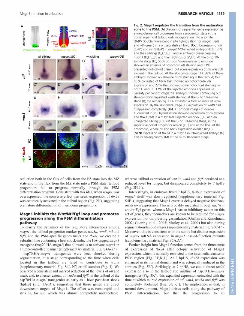
reduction both in the flux of cells from the PZ state into the MZstate and in the flux from the MZ state into a PSM state: tailbudprogenitors fail to progress normally through the PSMdifferentiation program. Consistent with this idea, when msgn1 wasoverexpressed, the converse effect was seen: expression of tbx24was ectopically activated in the tailbud region (Fig. 2N), suggestingpremature differentiation of mesoderm progenitors.
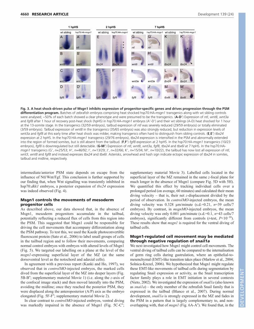
Msgn1 inhibits the Wnt/Ntl/Fgf loop and promotesprogression along the PSM differentiationpathwayTo clarify the dynamics of the regulatory interactions amongmsgn1, the tailbud progenitor marker genes wnt3a, wnt8, ntl andfgf8, and the PSM-specific genes tbx24 and tbx6l, we created azebrafish line containing a heat shock-inducible HA-tagged msgn1transgene (hsp70:HA-msgn1) that allowed us to activate msgn1 ina time-controlled manner (supplementary material Fig. S4A-K�).
hsp70:HA-msgn1 transgenics were heat shocked duringsegmentation, at a stage corresponding to the time when cellslocated in the tailbud are fated to contribute to trunk(supplementary material Fig. S4L-N�) or tail somites (Fig. 3). Weobserved a consistent and marked reduction of the levels of ntl andwnt8, and, to a lesser extent, of wnt3a and fgf8, in the tailbud of thehsp70:HA-msgn1 transgenics as early as 1 hour post-heat shock(hpHS) (Fig. 3A-D�), suggesting that these genes are directdownstream targets of Msgn1. The effect was most rapid andstriking for ntl, which was almost completely undetectable,
4659RESEARCH ARTICLEMsgn1 function in zebrafish
whereas tailbud expression of wnt3a, wnt8 and fgf8 persisted at areduced level for longer, but disappeared completely by 7 hpHS(Fig. 3H-J�).
Interestingly, in embryos fixed 7 hpHS, tailbud expression ofmsgn1 itself was downregulated (supplementary material Fig.S4E�), suggesting that Msgn1 exerts a delayed negative feedbackon its own expression. This is probably mediated through ntl, Wntand/or Fgf genes: whereas Msgn1 has an inhibitory action on thisset of genes, they themselves are known to be required for msgn1expression, not only during gastrulation (Griffin and Kimelman,2002; Goering et al., 2003; Morley et al., 2009) but also duringsegmentation/tailbud stages (supplementary material Fig. S5C-F�).Moreover, this is consistent with the subtle but distinct expansionof msgn1 mRNA expression in msgn1 loss-of-function mutants(supplementary material Fig. S5A,A�).
Further insight into Msgn1 function comes from the timecourseof expression of tbx24 after ectopic activation of Msgn1expression, which is normally restricted to the intermediate/anteriorPSM region (Fig. 3E,K,L). At 2 hpHS, tbx24 expression wasenhanced in its normal domain and was ectopically induced in thesomites (Fig. 3E�). Strikingly, at 7 hpHS, we could detect tbx24expression also in the tailbud and midline of hsp70:HA-msgn1transgenics (Fig. 3K�); this expanded expression coincided with thetime at which tailbud expression of ntl, wnt8, wnt3a and fgf8 wascompletely abolished (Fig. 3G�-J�). The implication is that, innormal development, Msgn1 drives cells along the pathway ofPSM differentiation, but that the progression to an
Fig. 2. Msgn1 regulates the transition from the maturationzone to the PSM. (A) Diagram of sequential gene expression asa mesodermal cell progresses from a progenitor state in thedorsal superficial tailbud until incorporation into a somite. (B-B�) Double fluorescent in situ hybridisation for msgn1 (red)and ntl (green) in a wt zebrafish embryo. (C-J�) Expression of ntl(C-H�) and wnt8 (E-J�) in msgn1MO-injected embryos (D,D�,F,F�)and their siblings (C,C�,E,E�) and in embryos overexpressingmsgn1 (H,H�,J,J�) and their siblings (G,G�,I,I�). At the 8- to 10-somite stage (H), 55% of msgn1-overexpressing embryosshowed an absence of notochord ntl staining and 33%presented notochord breaks, but some expression of ntl was stillevident in the tailbud. At the 20-somite stage (H�), 88% of theseembryos showed an absence of ntl staining in the tailbud; this88% consisted of 66% that showed no notochordal ntlexpression and 22% that showed some notochord staining. Inboth H and H�, 12% of the injected embryos appeared wt.Seventy per cent of msgn1OE embryos showed continuing butstrongly downregulated wnt8 staining at the 8- to 10-somitestage (J); the remaining 30% exhibited a total absence of wnt8expression. By the 20-somite stage (J�), expression of wnt8 haddisappeared completely. (K-L�) Confocal images of doublefluorescent in situ hybridisation showing expression of ntl (green)and tbx6l (red) in a msgn1MO-injected embryo (L,L�) and anuninjected sibling (K,K�) at the 8- to 10-somite stage, in thesuperficial dorsal progenitor region (K,L) and at the level of thenotochord, where ntl and tbx6l expression overlap (K�,L�). (M,N) Expression of tbx24 in a msgn1 mRNA-injected embryo (N)and its sibling control (M) at the 8- to 10-somite stage.
DEVELO
PMENT
4660
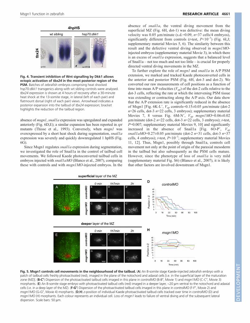
intermediate/anterior PSM state depends on escape from theinfluence of Ntl/Wnt/Fgf. This conclusion is further supported byour finding that, when Wnt signalling was transiently inhibited inhsp70:dkk1 embryos, a posterior expansion of tbx24 expressionwas indeed observed (Fig. 4).
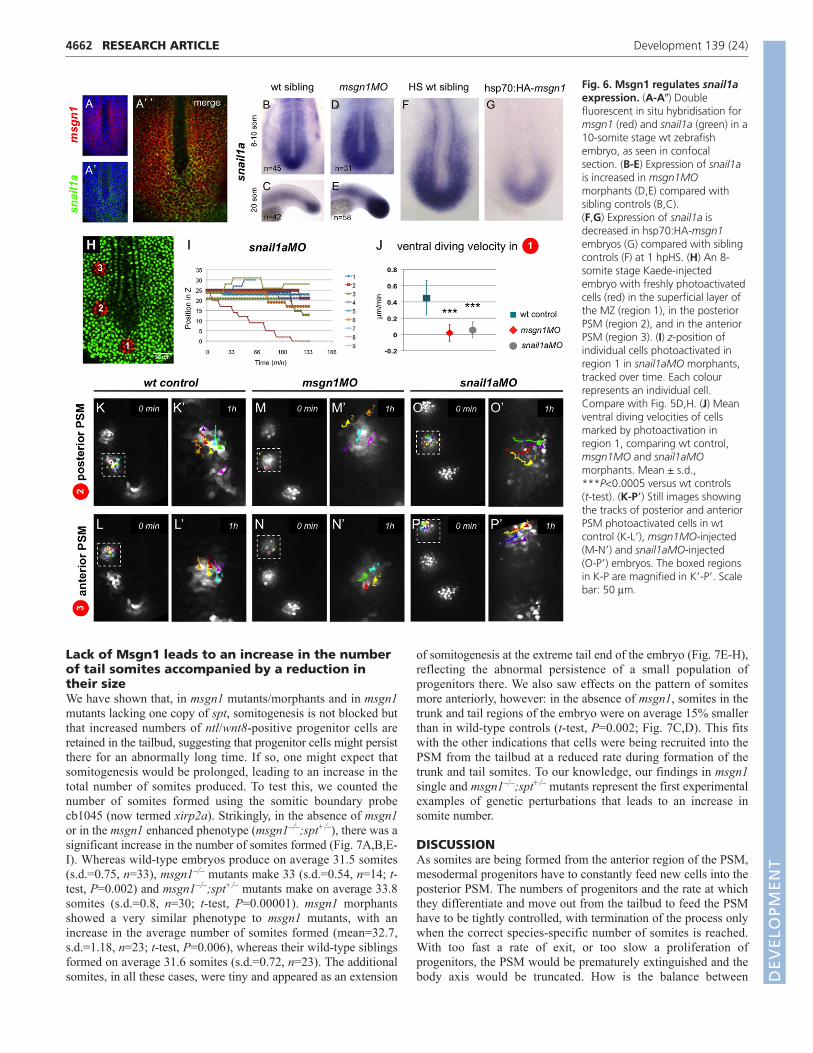
Msgn1 controls the movements of mesodermprogenitor cellsAs described above, our data showed that, in the absence ofMsgn1, mesoderm progenitors accumulate in the tailbud,potentially reflecting a reduced flux of cells from this region intothe PSM. This suggested that Msgn1 could be responsible fordriving the cell movements that accompany differentiation alongthe PSM pathway. To test this, we used the Kaede photoconvertiblefluorescent protein (Sato et al., 2006) to label small groups of cellsin the tailbud region and to follow their movements, comparingnormal control embryos with embryos with altered levels of Msgn1(Fig. 5). We targeted our labelling on a plane at the level of themsgn1-expressing superficial layer of the MZ (at the samedorsoventral level as the notochord and adaxial cells).
In agreement with a previous report (Kanki and Ho, 1997), weobserved that in controlMO-injected embryos, the marked cellsdived from the superficial layer of the MZ into deeper layers (Fig.5B-B�; supplementary material Movie 1) (i.e. along the z-axis ofthe confocal image stack) and then moved laterally into the PSM,avoiding the midline; once they reached the posterior PSM, theywere displaced along the anteroposterior (A/P) axis as the embryoelongated (Fig. 5F-F�; supplementary material Movie 2).
In clear contrast to controlMO-injected embryos, ventral divingwas markedly impaired in the absence of Msgn1 (Fig. 5C-C�;
RESEARCH ARTICLE Development 139 (24)
supplementary material Movie 3). Labelled cells located in thesuperficial layer of the MZ remained in the same z-focal plane formuch longer in the absence of Msgn1 (compare Fig. 5D with 5H).We quantified this effect by tracking individual cells over aprolonged period (on average, 60 minutes) and calculated their meandiving velocity – that is, their net z-displacement divided by theperiod of observation. In controlMO-injected embryos, the meandiving velocity was 0.328 µm/minute (s.d.=0.21, n=39 cells/7embryos). By contrast, in msgnMO-injected embryos, the meandiving velocity was only 0.001 µm/minute (s.d.=0.1, n=43 cells/7embryos), significantly different from controls (t-test, P<10–10).These results show that msgn1 is required for the ventral diving oftailbud cells.
Msgn1-regulated cell movement may be mediatedthrough negative regulation of snail1aWe next investigated how Msgn1 might control cell movements. Theventral diving of tailbud cells can be compared to the internalisationof germ ring cells during gastrulation, where an epithelial-to-mesenchymal (EMT)-like transition takes place (Marlow et al., 2004;Solnica-Krezel, 2006). We hypothesized that Msgn1 might regulatethese EMT-like movements of tailbud cells during segmentation byregulating Snail expression or activity, as the Snail transcriptionfactor family plays a role in EMT initiation in several contexts(Nieto, 2002). We investigated the expression of snail1a (also knownas snai1a) – the only member of the zebrafish Snail family that isexpressed in the tailbud (Blanco et al., 2007). During normaldevelopment, snail1a is strongly expressed in the MZ and fades inthe PSM in a pattern that is largely complementary to, and non-overlapping with, that of msgn1 (Fig. 6A-A�). We found that, in the
Fig. 3. A heat shock-driven pulse of Msgn1 inhibits expression of progenitor-specific genes and drives progression through the PSMdifferentiation program. Batches of zebrafish embryos comprising heat shocked hsp70:HA-msgn1 transgenics along with wt sibling controlswere analysed; ~50% of each batch showed a clear phenotype and were presumed to be the transgenics. (A-D�) Expression of ntl, wnt8, wnt3aand fgf8 after 1 hour of recovery post-heat shock (hpHS) in hsp70:HA-msgn1 embryos (A�-D�) and their wt siblings (A-D) heat shocked for 1 hourat the 13-somite stage. In the transgenics (32/59 embryos), tailbud expression of ntl was severely reduced (29/59 embryos) or totally eliminated(3/59 embryos). Tailbud expression of wnt8 in the transgenics (35/65 embryos) was also strongly reduced, but reduction in expression levels ofwnt3a and fgf8 at this early time after heat shock was milder, making transgenics often hard to distinguish from sibling controls. (E,E�) tbx24expression at 2 hpHS. In the hsp70:HA-msgn1 transgenics (29/76 embryos), tbx24 expression is intensified in the PSM and abnormally extendedinto the region of formed somites, but is still absent from the tailbud. (F,F�) fgf8 expression at 2 hpHS. In the hsp70:HA-msgn1 transgenics (10/23embryos), fgf8 is downregulated but still detectable. (G-M�) Expression of ntl, wnt8, wnt3a, fgf8, tbx24 and tbx6l at 7 hpHS. In the hsp70:HA-msgn1 transgenics (G�, n=25/53; H�, n=46/92; I�, n=13/29; J�, n=32/66; K�, n=15/34; M�, n=10/22), the tailbud has now lost all expression of ntl,wnt3, wnt8 and fgf8 and instead expresses tbx24 and tbx6I. Asterisks, arrowhead and hash sign indicate ectopic expression of tbx24 in somites,tailbud and midline, respectively.
DEVELO
PMENT
absence of msgn1, snail1a expression was upregulated and expandedanteriorly (Fig. 6D,E); a similar expansion has been reported in sptmutants (Thisse et al., 1993). Conversely, when msgn1 wasoverexpressed by a short heat shock during segmentation, snail1aexpression was severely and quickly downregulated 1 hpHS (Fig.6G).
Since Msgn1 regulates snail1a expression during segmentation,we investigated the role of Snail1a in the control of tailbud cellmovements. We followed Kaede photoconverted tailbud cells inembryos injected with snail1aMO (Blanco et al., 2007), comparingthese with controls and with msgn1MO-injected embryos. In the
4661RESEARCH ARTICLEMsgn1 function in zebrafish
absence of snail1a, the ventral diving movement from thesuperficial MZ (Fig. 6H, dot-1) was defective: the mean divingvelocity was 0.05 µm/minute (s.d.=0.09, n=37 cells/4 embryos),significantly different from controls (t-test, P<10–7) (Fig. 6I,J;supplementary material Movies 5, 6). The similarity between thisresult and the defective ventral diving observed in msgn1MO-injected embryos (supplementary material Movie 3), in which thereis an excess of snail1a expression, suggests that a balanced levelof Snail1a – not too much and not too little – is crucial for properlydirected ventral diving movements in the MZ.
To further explore the role of msgn1 and snail1a in A/P PSMextension, we marked and tracked Kaede photoconverted cells inthe anterior and posterior PSM (Fig. 6H, dot-3 and dot-2). Weconverted our raw measurements of cell positions as a function oftime into mean A/P velocities (Vap) of the dot-2 cells relative to thedot-3 cells, reflecting the rate at which the intervening PSM tissuewas extending or contracting along the A/P axis. Our data showthat the A/P extension rate is significantly reduced in the absenceof Msgn1 [Fig. 6K-L�, Vap controls=0.15±0.05 µm/minute (dot-2n=26 cells, dot-3 n=22 cells, 3 embryos); supplementary materialMovies 7, 8 versus Fig. 6M-N�, Vap msgn1MO=0.06±0.02µm/minute (dot-2 n=22 cells, dot-3 n=22 cells, 3 embryos); t-test,P=0.007; supplementary material Movies 9, 10] and significantlyincreased in the absence of Snail1a [Fig. 6O-P�, Vapsnail1aMO=0.27±0.05 µm/minute (dot-2 n=31 cells, dot-3 n=37cells, 4 embryos); t-test, P<10–7; supplementary material Movies11, 12]. Thus, Msgn1, possibly through Snail1a, controls cellmovement not only at the point of origin of the paraxial mesodermin the tailbud but also subsequently as the PSM cells mature.However, since the phenotype of loss of snail1a is very mild(supplementary material Fig. S6) (Blanco et al., 2007), it is likelythat other factors are involved downstream of Msgn1.
Fig. 4. Transient inhibition of Wnt signalling by Dkk1 allowsectopic activation of tbx24 in the most posterior region of thePSM. Batches of zebrafish embryos comprising heat shockedhsp70:dkk1 transgenics along with wt sibling controls were analysed.tbx24 expression is shown at 4 hours of recovery after a 30-minuteheat shock at the 13-somite stage, in lateral (left of each pair) andflatmount dorsal (right of each pair) views. Arrowhead indicates aposterior expansion into the tailbud of tbx24 expression; brackethighlights the reduction of the tailbud region.
Fig. 5. Msgn1 controls cell movements in the neighbourhood of the tailbud. (A) An 8-somite stage Kaede-injected zebrafish embryo with apatch of tailbud cells freshly photoactivated (red), imaged in the plane of the notochord and adaxial cells [i.e. in the superficial layer of the maturationzone (MZ)]. (B-C�) Dispersion of the photoactivated tailbud cells imaged in this plane in controlMO (B-B�, Movie 1) and msgn1MO (C-C�, Movie 3)morphants. (E) An 8-somite stage embryo with photoactivated tailbud cells (red) imaged in a deeper layer, ~20 m ventral to the notochord and adaxialcells (i.e. in a deep layer of the MZ). (F-G�) Dispersion of the photoactivated tailbud cells imaged in this plane in controlMO (F-F�, Movie 2) andmsgn1MO (G-G�, Movie 4) morphants. (D,H) z-position of individual Kaede photoactivated tailbud cells tracked over time in controlMO (D) andmsgn1MO (H) morphants. Each colour represents an individual cell. Loss of msgn1 leads to failure of ventral diving and of the subsequent lateraldispersion. Scale bars: 50 m. D
EVELO
PMENT
4662
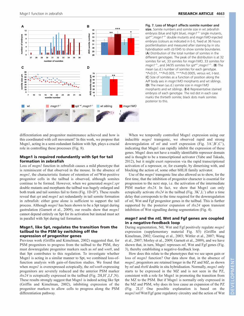
Lack of Msgn1 leads to an increase in the numberof tail somites accompanied by a reduction intheir sizeWe have shown that, in msgn1 mutants/morphants and in msgn1mutants lacking one copy of spt, somitogenesis is not blocked butthat increased numbers of ntl/wnt8-positive progenitor cells areretained in the tailbud, suggesting that progenitor cells might persistthere for an abnormally long time. If so, one might expect thatsomitogenesis would be prolonged, leading to an increase in thetotal number of somites produced. To test this, we counted thenumber of somites formed using the somitic boundary probecb1045 (now termed xirp2a). Strikingly, in the absence of msgn1or in the msgn1 enhanced phenotype (msgn1–/–;spt+/–), there was asignificant increase in the number of somites formed (Fig. 7A,B,E-I). Whereas wild-type embryos produce on average 31.5 somites(s.d.=0.75, n=33), msgn1–/– mutants make 33 (s.d.=0.54, n=14; t-test, P=0.002) and msgn1–/–;spt+/– mutants make on average 33.8somites (s.d.=0.8, n=30; t-test, P=0.00001). msgn1 morphantsshowed a very similar phenotype to msgn1 mutants, with anincrease in the average number of somites formed (mean=32.7,s.d.=1.18, n=23; t-test, P=0.006), whereas their wild-type siblingsformed on average 31.6 somites (s.d.=0.72, n=23). The additionalsomites, in all these cases, were tiny and appeared as an extension
RESEARCH ARTICLE Development 139 (24)
of somitogenesis at the extreme tail end of the embryo (Fig. 7E-H),reflecting the abnormal persistence of a small population ofprogenitors there. We also saw effects on the pattern of somitesmore anteriorly, however: in the absence of msgn1, somites in thetrunk and tail regions of the embryo were on average 15% smallerthan in wild-type controls (t-test, P=0.002; Fig. 7C,D). This fitswith the other indications that cells were being recruited into thePSM from the tailbud at a reduced rate during formation of thetrunk and tail somites. To our knowledge, our findings in msgn1single and msgn1–/–;spt+/– mutants represent the first experimentalexamples of genetic perturbations that leads to an increase insomite number.
DISCUSSIONAs somites are being formed from the anterior region of the PSM,mesodermal progenitors have to constantly feed new cells into theposterior PSM. The numbers of progenitors and the rate at whichthey differentiate and move out from the tailbud to feed the PSMhave to be tightly controlled, with termination of the process onlywhen the correct species-specific number of somites is reached.With too fast a rate of exit, or too slow a proliferation ofprogenitors, the PSM would be prematurely extinguished and thebody axis would be truncated. How is the balance between
Fig. 6. Msgn1 regulates snail1aexpression. (A-A�) Doublefluorescent in situ hybridisation formsgn1 (red) and snail1a (green) in a10-somite stage wt zebrafishembryo, as seen in confocalsection. (B-E) Expression of snail1ais increased in msgn1MOmorphants (D,E) compared withsibling controls (B,C). (F,G) Expression of snail1a isdecreased in hsp70:HA-msgn1embryos (G) compared with siblingcontrols (F) at 1 hpHS. (H) An 8-somite stage Kaede-injectedembryo with freshly photoactivatedcells (red) in the superficial layer ofthe MZ (region 1), in the posteriorPSM (region 2), and in the anteriorPSM (region 3). (I) z-position ofindividual cells photoactivated inregion 1 in snail1aMO morphants,tracked over time. Each colourrepresents an individual cell.Compare with Fig. 5D,H. (J) Meanventral diving velocities of cellsmarked by photoactivation inregion 1, comparing wt control,msgn1MO and snail1aMOmorphants. Mean ± s.d.,***P<0.0005 versus wt controls (t-test). (K-P�) Still images showingthe tracks of posterior and anteriorPSM photoactivated cells in wtcontrol (K-L�), msgn1MO-injected(M-N�) and snail1aMO-injected (O-P�) embryos. The boxed regionsin K-P are magnified in K�-P�. Scalebar: 50 m.
DEVELO
PMENT
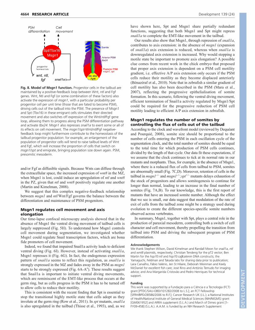
differentiation and progenitor maintenance achieved and how isthis coordinated with cell movement? In this work, we propose thatMsgn1, acting in a semi-redundant fashion with Spt, plays a crucialrole in controlling these processes (Fig. 8).
Msgn1 is required redundantly with Spt for tailformation in zebrafishLoss of msgn1 function in zebrafish causes a mild phenotype thatis reminiscent of that observed in the mouse. In the absence ofmsgn1, the characteristic feature of retention of ntl/Wnt-positiveprogenitor cells in the tailbud is observed, although somitescontinue to be formed. However, when we generated msgn1;sptdouble mutants and morphants the tailbud was hugely enlarged andboth trunk and tail somites fail to form (Fig. 1D-D�). These resultsreveal that spt and msgn1 act redundantly in tail somite formationin zebrafish: either gene alone is sufficient to support the tailprocess. Although msgn1 has been shown to be a Spt target duringgastrulation (Garnett et al., 2009), our results show that msgn1cannot depend entirely on Spt for its activation but instead must actin parallel with Spt during tail formation.
Msgn1, like Spt, regulates the transition from thetailbud to the PSM by switching off theexpression of progenitor genesPrevious work (Griffin and Kimelman, 2002) suggested that, forPSM progenitors to progress from the tailbud to the PSM, theymust downregulate progenitor markers such as ntl and wnt8, andthat Spt contributes to this regulation. To investigate whetherMsgn1 is acting in a similar manner to Spt, we combined loss-of-function analysis with gain-of-function studies. We found thatwhen msgn1 is overexpressed ectopically, the ntl/wnt8-expressingprogenitors are severely reduced and the anterior PSM markertbx24 is ectopically expressed in the tailbud (Fig. 2H,H�,J,J�,N).These results strongly suggest that Msgn1 is indeed acting like Spt(Griffin and Kimelman, 2002), inhibiting expression of theprogenitor markers to allow cells to progress along the PSMdifferentiation pathway.
4663RESEARCH ARTICLEMsgn1 function in zebrafish
When we temporally controlled Msgn1 expression using ourinducible msgn1 transgenic, we observed rapid and strongdownregulation of ntl and wnt8 expression (Fig. 3A�,B�,C�),indicating that Msgn1 can rapidly inhibit the expression of thesegenes. Msgn1 does not have a readily identifiable repressor domainand is thought to be a transcriptional activator (Yabe and Takada,2012), but it might exert repression via the rapid transcriptionalactivation of a repressor, or, for example, by dimerising with, andblocking the action of, some other bHLH family activator.
Use of the msgn1 transgenic line also allowed us to show, for thefirst time, that the inhibition of progenitor character is essential forprogression to the next step, i.e. the activation of the intermediatePSM marker tbx24. In fact, we show that Msgn1 can onlyectopically activate tbx24 in the tailbud (Fig. 3K�,L�) after a timedelay that corresponds to the time required for the downregulationof ntl, Wnt and Fgf progenitor genes in the tailbud. This is furthersupported by the posterior expansion of tbx24 upon transientinhibition of Wnt signalling during segmentation (Fig. 4).
msgn1 and the ntl, Wnt and Fgf genes are coupledin a negative-feedback loopDuring segmentation, Ntl, Wnt and Fgf positively regulate msgn1expression (supplementary material Fig. S5) (Griffin andKimelman, 2002; Goering et al., 2003; Wittler et al., 2007; Wanget al., 2007; Morley et al., 2009; Garnett et al., 2009), and we haveshown that, in turn, Msgn1 represses ntl, Wnt and Fgf genes (Fig.3), thereby establishing a negative-feedback loop.
How does this relate to the phenotypes that we see upon gain orloss of msgn1 function? Our data show that, in the absence ofmsgn1, progenitors are retained longer in the PZ and MZ, as shownby ntl and tbx6l double in situ hybridisation. Normally, msgn1 onlystarts to be expressed in the MZ and is not seen in the PZ,consistent with a role for Msgn1 in promoting the transition fromthe MZ to the PSM. But if Msgn1 is normally only expressed inthe MZ and PSM, why does its loss cause an expansion of the PZ(Fig. 2L)? One possible explanation is based on themsgn1/ntl/Wnt/Fgf gene regulatory circuitry and the action of Wnt
Fig. 7. Loss of Msgn1 affects somite number andsize. Somite numbers and somite size in wt zebrafishembryos (blue and light blue), msgn1–/– single mutants,spt+/–;msgn1–/– double mutants and msgn1MO-injectedembryos (colours as indicated in E-I), fixed at 36 hourspostfertilisation and measured after staining by in situhybridisation with cb1045 to show somite boundaries.(A) Distribution of the total number of somites in thedifferent genotypes. The peak of the distribution is at 32somites for wt, 33 somites for msgn1MO, 33 somites formsgn1–/–, and 34/35 somites for spt+/–;msgn1–/–. (B) Themean (±s.d.) number of somites for each genotype.*P<0.01, **P<0.005, ***P<0.0005, versus wt; t-test. (C) Size of somites as a function of position along theA/P body axis in msgn1MO morphants and wt siblings.(D) The mean (±s.d.) somite size in msgn1MOmorphants and wt siblings. (E-I) Representative stainedembryos of each genotype. The red dot in each casemarks the thirtieth somite; black dots mark somitesposterior to this.
DEVELO
PMENT
4664 RESEARCH ARTICLE Development 139 (24)
and/or Fgf as diffusible signals. Because Wnts can diffuse throughthe extracellular space, the increased expression of wnt8 in the MZ,when Msgn1 is lost, could induce an upregulation of ntl and wnt8in the PZ, given that ntl and wnt8 positively regulate one another(Martin and Kimelman, 2008).
We suggest that this complex negative-feedback relationshipbetween msgn1 and ntl/Wnt/Fgf governs the balance between thedifferentiation and maintenance of PSM progenitors.
Msgn1 regulates cell movement and axiselongationOur time-lapse confocal microscopy analysis showed that in theabsence of Msgn1 the ventral diving movement of tailbud cells islargely suppressed (Fig. 5H). To understand how Msgn1 controlscell movement during segmentation, we investigated whetherMsgn1 could regulate Snail transcription factors, which are bonafide promoters of cell movement.
Indeed, we found that impaired Snail1a activity leads to deficientventral diving (Fig. 6I). However, instead of activating snail1a,Msgn1 represses it (Fig. 6G). In fact, the endogenous expressionpattern of snail1a seems to reflect this regulation, as snail1a isstrongly expressed in the MZ and fades away in the PSM as msgn1starts to be strongly expressed (Fig. 6A-A�). These results suggestthat Snail1a is important to initiate ventral diving movements,which are reminiscent of the EMT-like process that occurs at thegerm ring, but as cells progress in the PSM it has to be turned offto allow cells to reduce their motility.
This is consistent with the recent finding that Spt is essential tostop the transitional highly motile state that cells adopt as theyinvolute at the germ ring (Row et al., 2011). In spt mutants, snail1ais also upregulated in the tailbud (Thisse et al., 1993), and, as we
have shown here, Spt and Msgn1 share partially redundantfunctions, suggesting that both Msgn1 and Spt might represssnail1a to complete the EMT-like movement in the tailbud.
Our results also show that Msgn1, through repression of snail1a,contributes to axis extension: in the absence of msgn1 (expansionof snail1a) axis extension is reduced, whereas when snail1a isdownregulated axis extension is increased. Why would stopping amotile state be important to promote axis elongation? A possibleclue comes from recent work in the chick embryo that proposedthat proper axis extension is dependent on a PSM cell motilitygradient, i.e. effective A/P axis extension only occurs if the PSMcells reduce their motility as they become displaced anteriorly(Bénazéraf et al., 2010). Note that in zebrafish a similar gradient ofcell motility has also been described in the PSM (Mara et al.,2007), reflecting the progressive epithelialisation of somiteformation. In this scenario, following the ventral diving movement,efficient termination of Snail1a activity regulated by Msgn1/Sptcould be required for the progressive reduction of PSM cellmotility leading to efficient A/P axis extension in zebrafish.
Msgn1 regulates the number of somites bycontrolling the flux of cells out of the tailbudAccording to the clock and wavefront model (reviewed by Dequéantand Pourquié, 2008), somite size should be proportional to thenumber of cells entering the PSM in each oscillation cycle of thesegmentation clock, and the total number of somites should be equalto the total time for which production of PSM cells continues,divided by the length of that cycle. Our data fit these expectations, ifwe assume that the clock continues to tick at its normal rate in ourmutants and morphants. Thus, for example, in the absence of Msgn1,where there is a reduced flux of cells from tailbud to PSM, somitesare abnormally small (Fig. 7C,D). Moreover, retention of cells in thetailbud in msgn1–/– and msgn1–/–;spt+/– mutants delays exhaustion ofthe stock of progenitors and allows somitogenesis to continue forlonger than normal, leading to an increase in the final number ofsomites (Fig. 7A,B). To our knowledge, this is the first report ofmutants that have an increased somite number. Although the effectthat we see is small, our data suggest that modulation of the rate ofexit of cells from the tailbud zone might be a strategy used duringevolution to create the different species-specific somite numbersobserved across vertebrates.
In summary, Msgn1, together with Spt, plays a central role in theproduction of paraxial mesoderm, controlling both a switch of cellcharacter and cell movement, thereby propelling the transition fromtailbud into PSM and driving the subsequent program of PSMdifferentiation.
AcknowledgementsWe thank Stephen Wilson, David Kimelman and Randall Moon for snail1a, ntland wnt8 plasmids, respectively; Christian Tendeng for the pT2 vector; BenMartin for the hsp70:ntl and hsp70:cacatenin DNA constructs; theYamaguchi, Feldman and Takada labs for sharing data prior to publication;Lara Carvalho, Fábio Valério, Jen St Hilaire, Deborah Weinman and KeelyMcDaniel for excellent fish care; José Rino and António Temudo for imagingadvice; and Ana Margarida Cristovão and Pedro Henriques for technicalsupport.
FundingThis work was supported by a Fundação para a Ciência e a Tecnologia (FCT)grant [PTDC/SAU-OBD/101282/2008 to L.S.]; an FCT Fellowship[SFRH/BPD/28586/2006 to R.F.]; Cancer Research UK (J.L.); a National Institutesof Health/National Institute of General Medical Sciences (NIH/NIGMS) grant[GM061952] and ARRA supplement (S.L.A.) and March of Dimes grant [1-FY09-458] (S.L.A.). A.A.M. is funded by an NIH Research Supplement
Fig. 8. Model of Msgn1 function. Progenitor cells in the tailbud aremaintained by a positive-feedback loop between Wnt, ntl and Fgfgenes. Wnt, Ntl and Fgf (or some combination of these factors) alsoactivate the expression of msgn1, with a particular probability perprogenitor cell per unit time (those that are fated to become PSM),driving cells out of the tailbud into the PSM. The presence of Msgn1and Spt (Tbx16) in these emigrant cells stimulates their directedmovement and also switches off expression of the Wnt/ntl/Fgf geneloop, allowing them to progress along the PSM differentiation pathwayand activate tbx24. Msgn1 also represses snail1a to exert some or all ofits effects on cell movement. The msgn1/spt-Wnt/ntl/Fgf negative-feedback loop might furthermore contribute to the homeostasis of thetailbud progenitor population: for example, an enlargement of thepopulation of progenitor cells will tend to raise tailbud levels of Wntand Fgf, which will increase the proportion of cells that switch onmsgn1/spt and emigrate, bringing population size down again. PSM,presomitic mesoderm.
DEVELO
PMENT
4665RESEARCH ARTICLEMsgn1 function in zebrafish
[NIH/NIGMS grant GM061952] to promote diversity in health-related research.The msgnfh273 TILLING allele was found with support from NIH grant R01HG002995. Deposited in PMC for release after 12 months.
Competing interests statementThe authors declare no competing financial interests.
Supplementary materialSupplementary material available online athttp://dev.biologists.org/lookup/suppl/doi:10.1242/dev.078923/-/DC1
ReferencesAmacher, S. L., Draper, B. W., Summers, B. R. and Kimmel, C. B. (2002). The
zebrafish T-box genes no tail and spadetail are required for development oftrunk and tail mesoderm and medial floor plate. Development 129, 3311-3323.
Bénazéraf, B., Francois, P., Baker, R. E., Denans, N., Little, C. D. andPourquié, O. (2010). A random cell motility gradient downstream of FGFcontrols elongation of an amniote embryo. Nature 466, 248-252.
Blanco, M. J., Barrallo-Gimeno, A., Acloque, H., Reyes, A. E., Tada, M.,Allende, M. L., Mayor, R. and Nieto, M. A. (2007). Snail1a and Snail1bcooperate in the anterior migration of the axial mesendoderm in the zebrafishembryo. Development 134, 4073-4081.
Dequéant, M. L. and Pourquié, O. (2008). Segmental patterning of thevertebrate embryonic axis. Nat. Rev. Genet. 9, 370-382.
Draper, B. W., McCallum, C. M., Stout, J. L., Slade, A. J. and Moens, C. B.(2004). A high-throughput method for identifying N-ethyl-N-nitrosourea (ENU)-induced point mutations in zebrafish. Methods Cell Biol. 77, 91-112.
Garnett, A. T., Han, T. M., Gilchrist, M. J., Smith, J. C., Eisen, M. B., Wardle, F.C. and Amacher, S. L. (2009). Identification of direct T-box target genes in thedeveloping zebrafish mesoderm. Development 136, 749-760.
Goering, L. M., Hoshijima, K., Hug, B., Bisgrove, B., Kispert, A. andGrunwald, D. J. (2003). An interacting network of T-box genes directs geneexpression and fate in the zebrafish mesoderm. Proc. Natl. Acad. Sci. USA 100,9410-9415.
Griffin, K. J. and Kimelman, D. (2002). One-Eyed Pinhead and Spadetail areessential for heart and somite formation. Nat. Cell Biol. 4, 821-825.
Griffin, K. J., Amacher, S. L., Kimmel, C. B. and Kimelman, D. (1998).Molecular identification of spadetail: regulation of zebrafish trunk and tailmesoderm formation by T-box genes. Development 125, 3379-3388.
Halpern, M. E., Ho, R. K., Walker, C. and Kimmel, C. B. (1993). Induction ofmuscle pioneers and floor plate is distinguished by the zebrafish no tailmutation. Cell 75, 99-111.
Ho, R. K. and Kane, D. A. (1990). Cell-autonomous action of zebrafish spt-1mutation in specific mesodermal precursors. Nature 348, 728-730.
Holley, S. A. (2007). The genetics and embryology of zebrafish metamerism. Dev.Dyn. 236, 1422-1449.
Jülich, D., Geisler, R., Tübingen 2000 Screen Consortium and Holley, S. A.(2005). Integrinalpha5 and delta/notch signaling have complementaryspatiotemporal requirements during zebrafish somitogenesis. Dev. Cell 8, 575-586.
Kanki, J. P. and Ho, R. K. (1997). The development of the posterior body inzebrafish. Development 124, 881-893.
Kimmel, C. B., Kane, D. A., Walker, C., Warga, R. M. and Rothman, M. B.(1989). A mutation that changes cell movement and cell fate in the zebrafishembryo. Nature 337, 358-362.
Lee, Y., Grill, S., Sanchez, A., Murphy-Ryan, M. and Poss, K. D. (2005). Fgfsignaling instructs position-dependent growth rate during zebrafish finregeneration. Development 132, 5173-5183.
Mara, A., Schroeder, J., Chalouni, C. and Holley, S. A. (2007). Priming,initiation and synchronization of the segmentation clock by deltaD and deltaC.Nat. Cell Biol. 9, 523-530.
Marlow, F., Gonzalez, E. M., Yin, C., Rojo, C. and Solnica-Krezel, L. (2004). Notail co-operates with non-canonical Wnt signaling to regulate posterior bodymorphogenesis in zebrafish. Development 131, 203-216.
Martin, B. L. and Kimelman, D. (2008). Regulation of canonical Wnt signaling byBrachyury is essential for posterior mesoderm formation. Dev. Cell 15, 121-133.
Martin, B. L. and Kimelman, D. (2012). Canonical Wnt signaling dynamicallycontrols multiple stem cell fate decisions during vertebrate body formation. Dev.Cell 22, 223-232.
Morley, R. H., Lachani, K., Keefe, D., Gilchrist, M. J., Flicek, P., Smith, J. C.and Wardle, F. C. (2009). A gene regulatory network directed by zebrafish Notail accounts for its roles in mesoderm formation. Proc. Natl. Acad. Sci. USA 106,3829-3834.
Nieto, M. A. (2002). The snail superfamily of zinc-finger transcription factors. Nat.Rev. Mol. Cell Biol. 3, 155-166.
Nikaido, M., Kawakami, A., Sawada, A., Furutani-Seiki, M., Takeda, H. andAraki, K. (2002). Tbx24, encoding a T-box protein, is mutated in the zebrafishsomite-segmentation mutant fused somites. Nat. Genet. 31, 195-199.
Row, R. H., Maître, J. L., Martin, B. L., Stockinger, P., Heisenberg, C. P. andKimelman, D. (2011). Completion of the epithelial to mesenchymal transitionin zebrafish mesoderm requires Spadetail. Dev. Biol. 354, 102-110.
Sato, T., Takahoko, M. and Okamoto, H. (2006). HuC:Kaede, a useful tool tolabel neural morphologies in networks in vivo. Genesis 44, 136-142.
Solnica-Krezel, L. (2006). Gastrulation in zebrafish – all just about adhesion? Curr.Opin. Genet. Dev. 16, 433-441.
Stoick-Cooper, C. L., Weidinger, G., Riehle, K. J., Hubbert, C., Major, M. B.,Fausto, N. and Moon, R. T. (2007). Distinct Wnt signaling pathways haveopposing roles in appendage regeneration. Development 134, 479-489.
Thisse, C. and Thisse, B. (2008). High-resolution in situ hybridization to whole-mount zebrafish embryos. Nat. Protoc. 3, 59-69.
Thisse, C., Thisse, B., Schilling, T. F. and Postlethwait, J. H. (1993). Structure ofthe zebrafish snail1 gene and its expression in wild-type, spadetail and no tailmutant embryos. Development 119, 1203-1215.
Wang, J., Li, S., Chen, Y. and Ding, X. (2007). Wnt/beta-catenin signalingcontrols Mespo expression to regulate segmentation during Xenopussomitogenesis. Dev. Biol. 304, 836-847.
Wilson, V., Olivera-Martinez, I. and Storey, K. G. (2009). Stem cells, signals andvertebrate body axis extension. Development 136, 1591-1604.
Wittler, L., Shin, E. H., Grote, P., Kispert, A., Beckers, A., Gossler, A., Werber,M. and Herrmann, B. G. (2007). Expression of Msgn1 in the presomiticmesoderm is controlled by synergism of WNT signalling and Tbx6. EMBO Rep. 8,784-789.
Yabe, T. and Takada, S. (2012). Mesogenin causes embryonic mesodermprogenitors to differentiate during development of zebrafish tail somites. Dev.Biol. 370, 213-222.
Yoo, K. W., Kim, C. H., Park, H. C., Kim, S. H., Kim, H. S., Hong, S. K., Han, S.,Rhee, M. and Huh, T. L. (2003). Characterization and expression of apresomitic mesoderm-specific mespo gene in zebrafish. Dev. Genes Evol. 213,203-206.
Yoon, J. K. and Wold, B. (2000). The bHLH regulator pMesogenin1 is required formaturation and segmentation of paraxial mesoderm. Genes Dev. 14, 3204-3214.
DEVELO
PMENT