Embed Size (px)
Citation preview
Developmental Cell 10, 355–366, March, 2006 ª2006 Elsevier Inc. DOI 10.1016/j.devcel.2006.02.011
Oscillations of the Snail Genes in the PresomiticMesoderm Coordinate Segmental Patterningand Morphogenesis in Vertebrate Somitogenesis
Jacqueline Kim Dale,1,2 Pascale Malapert,1
Jerome Chal,1 Goncalo Vilhais-Neto,1
Miguel Maroto,1,2 Teri Johnson,1
Sachintha Jayasinghe,1 Paul Trainor,1
Bernhard Herrmann,3 and Olivier Pourquie1,*1Howard Hughes Medical InstituteStowers Institute for Medical Research1000 East 50th StreetKansas City, Missouri 64110
Summary
The segmented body plan of vertebrate embryos arises
through segmentation of the paraxial mesoderm toform somites. The tight temporal and spatial control
underlying this process of somitogenesis is regulated
by the segmentation clock and the FGF signaling wave-front. Here, we report the cyclic mRNA expression of
Snail1 and Snail2 in the mouse and chick presomiticmesoderm (PSM), respectively. Whereas Snail genes’
oscillations are independent of NOTCH signaling, weshow that they require WNT and FGF signaling. Over-
expressing Snail2 in the chick embryo prevents cyclicLfng and Meso1 expression in the PSM and disrupts
somite formation. Moreover, cells misexpressingSnail2 fail to express Paraxis, remain mesenchymal,
and are thereby inhibited from undergoing the epitheli-alization event that culminates in the formation of the
epithelial somite. Thus, Snail genes define a class ofcyclic genes that coordinate segmentation and PSM
morphogenesis.
Introduction
The segmented body plan is a characteristic feature ofthe vertebrate embryo that becomes apparent very earlyin development. The first overt sign of segmentation isseen in the paraxial mesoderm as it progressively gener-ates somites in an anteroposterior (AP) direction. Somi-togenesis is under tight temporal control such that anew pair of somites forms according to a strict periodicschedule that is species specific. The periodicity of thisprocess is under the control of the segmentation clockthat drives oscillating mRNA expression of a numberof ‘‘cyclic genes’’ in the unsegmented presomitic meso-derm (PSM) of vertebrates (Pourquie, 2003).
The majority of cyclic genes are downstream targetsof the NOTCH signaling pathway and code for Hairy/Enhancer of split family members, the glycosyltransfer-ase enzyme LUNATIC FRINGE (LFNG), and the NOTCHligand DELTA. It is now a well accepted fact that this
*Correspondence: [email protected] Present address: Division of Cell and Developmental Biology,
School of Life Sciences, MSI/WTB Complex, Dundee University,
Dow Street, Dundee, DD1 5EH, Scotland, United Kingdom.3 Present address: Charite, Institute of Med. Genetics and MPI of
Mol. Genetics, Berlin, Germany.
pathway plays a crucial role in somitogenesis since mu-tation of several of its components causes severe so-mitic defects in mice or zebrafish embryos (Rida et al.,2004). If the cyclic expression of genes of the NOTCHpathway such as Lfng or Hes7 is perturbed by eithergain or loss of function, somitogenesis is severely dis-rupted (Bessho et al., 2003; Dale et al., 2003; Serthet al., 2003). Nevertheless, even the most severe mouseand zebrafish NOTCH pathway mutants retain somedegree of segmentation at the level of the most anteriorsomites, raising the possibility that there may be otherpathways involved in the mechanism of the segmenta-tion clock that can compensate in part for the loss ofNOTCH (Rida et al., 2004).
Oscillations of Axin2 and Nkd1, two members of theWNT signaling pathway in the mouse PSM, recently im-plicated this pathway in the segmentation clock (Aulehlaet al., 2003; Ishikawa et al., 2004). Disruption of WNT sig-naling in the Wnt3a hypomorphic mutation vestigial tail(vt) leads to a loss of Axin2 and of Nkd1 expression.Interestingly, the dynamic expression of Nkd1 is also de-pendent on NOTCH signaling, as it no longer oscillates inthe absence of Hes7 (Ishikawa et al., 2004). Taken to-gether, these data suggest that the NOTCH and WNTpathways interact within the mechanism of the segmen-tation clock.
The FGF pathway is also known to be crucial to somi-togenesis since it regulates the competence of PSMcells to undergo segmentation and thus controls the re-sponse of PSM cells to the segmentation clock (Dubrulleand Pourquie, 2004). FGF signaling establishes a travel-ing wavefront involved in the conversion of the pulsatilesignal of the clock into the spatial periodic pattern ofsomites (Pourquie, 2003). In mouse, Fgf8 expression isdownregulated in the absence of Wnt3a (Aulehla et al.,2003), and in zebrafish, expression of the NOTCH targetgene her13.2, required for cyclic gene oscillations, isregulated by FGF signaling (Kawamura et al., 2005).Thus, it appears that several levels of crosstalk exist be-tween the NOTCH, the WNT, and the FGF pathways insomitogenesis.
The SNAIL superfamily of transcriptional repressors,most notably SNAIL1 (formerly SNAIL) and SNAIL2 (for-merly SLUG), have been shown to play a critical role invertebrate and invertebrate development and in cancer(Barrallo-Gimeno and Nieto, 2005). These proteins con-trol major morphogenetic processes by controllingepithelium-to-mesenchyme transitions (EMTs) (Barrallo-Gimeno and Nieto, 2005). SNAIL proteins are able torepress genes coding for proteins associated with anepithelial phenotype such as E-CADHERIN or desmo-somal proteins and can activate the expression of mes-enchymal markers (Batlle et al., 2000; Cano et al., 2000;Savagner et al., 1997). In the developing vertebrate em-bryo, the first SNAIL-dependent EMT that occurs takesplace in the primitive streak during gastrulation (Carveret al., 2001; Ciruna and Rossant, 2001). Driven by Snail1activity acting downstream of FGF signaling, epithelialprimitive streak cells fated to become mesodermalprogenitors downregulate E-CADHERIN and undergo

Developmental Cell356
EMTs. Consequently, they delaminate from the streak asindividual mesenchymal cells and subsequently migrateto the various mesodermal locations of the developingembryo (Ciruna and Rossant, 2001). On exiting the prim-itive streak after an EMT, paraxial mesoderm progenitorcells populate the mesenchymal posterior PSM, wherethey are exposed to high levels of FGF signaling (Du-brulle et al., 2001) and maintain strong Snail1 and Snail2expression in mouse and chick, respectively (Seftonet al., 1998). When paraxial mesoderm cells pass athreshold concentration of FGF signaling (termed thedetermination front) in the anterior PSM, they begin toacquire epithelial characteristics (Duband et al., 1987;Dubrulle et al., 2001; Nakaya et al., 2004). At this level,cells also become allocated to genetically defined seg-ments that provide the templates upon which the epithe-lial somites will form at the anterior end of the PSM. Howthe morphogenetic program leading to epithelializationis coordinated to the segmentation process in thePSM is currently unknown.
The beginning of the epithelialization process in thePSM correlates with the downregulation of Snail1 andSnail2 (Sefton et al., 1998), thus raising the possibilitythat these genes are involved in the control of this mor-phological transition. We have closely investigated theexpression profile of Snail1 and Snail2 within the PSMin mouse and chick embryos, respectively, and wefound that their mRNA is rhythmically transcribed witha periodicity that matches the budding off of epithelialsomites. Snail1 oscillates largely in synchrony with theNOTCH cyclic genes, but its expression is independentof NOTCH signaling and relies upon WNT3A signaling.Misexpression of Snail2 in the chick PSM blocks Lfngand Meso1 expression. This disruption of the segmenta-tion clock-driven oscillations suggests a role for Snailgenes in the clock mechanism. Subsequently, cellsoverexpressing Snail2 fail to epithelialize and form so-mites, remaining in a mesenchymal state. While thisphenotype is reminiscent of that seen in embryos misex-pressing FGF in the PSM, Snail2 does not upregulateFGF targets like Brachyury, suggesting that it only medi-ates the morphogenetic aspect of the FGF response inthe PSM. Together, these results indicate that Snailgenes may identify a new class of cyclic genes and pro-vide a link between the segmentation clock and the FGFsignaling wavefront in vertebrate segmentation.
Results
Snail1 and Snail2 mRNA Oscillate in the Mouseand Chick Embryo PSM, Respectively
We have examined the expression of Snail1 mRNA in thePSM of E8.5–E10 mouse embryos by in situ hybridiza-tion. Large batches of embryos were collected and ana-lyzed at each stage. We found that the expression pat-tern varied considerably among the batches of similarstage embryos. Thus, we found some embryos display-ing a broad posterior pattern of expression plus a thinstripe in the anterior region of the PSM just caudal tothe posterior limit of the next somite to be formed (Fig-ures 1A and 1B); other embryos displayed only a wideposterior expression domain in the PSM (Figure 1C),while others displayed only a narrow band of expressionin the anterior-most part of the PSM (Figure 1D). These
different patterns were seen at each of the stages ana-lyzed, strongly suggesting that Snail1 may be oscillatingin the PSM. To directly test if Snail1 expression is oscil-lating, we bisected the posterior region of mice embryos
Figure 1. Oscillations of Snail Genes in Mouse and Chick PSM
(A–D) Posterior lateral view of E9.5 mouse embryos hybridized with
the Snail1 probe.
(E) Schematic representation of the dynamic expression of Snail1
during the formation of one somite in the mouse embryo (120 min).
(F–I) Posterior dorsal view of 2-day-old chick embryos hybridized
with the Snail2 probe.
(J) Schematic representation of the dynamic expression of Snail2
during the formation of one somite in the chick embryo (90 min).

Snail Genes Coordinate Segmentation/Morphogenesis357
Figure 2. Comparison between Snail1, Lfng, and Axin2 Expression in the Mouse Embryo PSM
(A–D) Comparison of Snail1 and Lfng expression detected by whole-mount in situ hybridization in the two embryonic halves of the same E9.5
mouse embryos.
(E) Detection of Snail1 by using an exonic (Ex) and an intronic (In) probe in the two embryonic halves of the same E9.5 embryos.
(F) Comparison of Snail1 and Axin2 expression detected by whole-mount in situ hybridization in the two embryonic halves of the same E9.5
mouse embryos.
Arrowheads mark the level of the anterior front of the posterior dynamic domain of the genes. Anterior is oriented toward the top.
down the midline and fixed one half while culturing theother half in vitro for a defined time period. The twohalves were then analyzed for Snail1 expression (Fig-ure S1; see the Supplemental Data available with this ar-ticle online). When the one half was cultured for 75, 90, or105 min, we found that the expression of Snail1 was dif-ferent from that in the fixed half (Figure S1A, n = 6). How-ever, when we cultured one half for 120 min, this explantformed an extra somite, and the expression domain wasthe same in the cultured and the fixed explants (Fig-ure S1B, n = 5). These data demonstrate that Snail1mRNA is expressed according to a periodic wave inthe mouse PSM (Figure 1E), and that the period of theoscillation is 120 min—the time it takes to form a newpair of somites from the mouse PSM.
There is a striking interchange in both the expressionpatterns and the functions of two members of the Snailsuperfamily, namely, Snail1 and Snail2, in neural crest,nascent mesoderm, and paraxial mesoderm betweenchick and mice embryos (Locascio et al., 2002; Seftonet al., 1998). In the chick embryo, Snail2, not Snail1, isexpressed in the PSM (Figures 1F–1I). We analyzed indetail the PSM expression of Snail2 in a large series ofstage-matched chick embryos and observed a clear dy-namic expression pattern (Figures 1F–1J) similar to thatseen in the mouse PSM for Snail1. Therefore, our dataidentify Snail1 in mouse and Snail2 in chick as cyclicgenes regulated by the segmentation clock.
Snail1 May Define a New Class of Cyclic Genes
The genes of the NOTCH pathway Lfng, Hes1, and Hes7cycle in synchrony in the mouse PSM; whereas the WNTpathway inhibitor Axin2 oscillates out of phase with
them (Aulehla et al., 2003). In order to determine whetherSnail1 cycles in synchrony with any of these genes, webisected the posterior region of mice embryos downthe midline and hybridized one half for Lfng expressionand the other half for Snail1 expression. We foundthat, for the main part of the cycle, the anterior progres-sion domain of the expression front progresses in syn-chrony for the two genes (arrowheads, Figures 2A–2C).However, there are two differences in the expressionprofiles of these genes in the PSM. First, at the onsetof a new wave, Snail1 expression is initiated prior tothat of Lfng in the posterior PSM (Figure 2D), and, sub-sequently, the Lfng expression front catches up withthat of Snail1 (Figure 2A). Second, unlike Snail1, the pos-terior domain of the expression of Lfng is rapidly de-graded as the expression front moves anteriorly up thePSM (Figures 2B–2D). This may be due to different ratesof mRNA stability/degradation for the two genes.
We tested this possibility indirectly by comparing theexpression of Snail1 detected with either an intronicprobe or an exonic probe in bisected explants as de-scribed above (Figure 2E). In all cases, the pattern of ex-pression in each half was the same for the two probes(Figure 2E, n = 11), demonstrating that the domains ofexpression of the exonic probe are sites of active tran-scription. Thus, Snail1 continues to be transcribed in do-mains of the PSM where Lfng transcription has alreadybeen shut off. Due to these two differences, the ratioof embryos that do or do not display the broad posteriordomain of expression is very different for the two genes.Thus, only 20% of the embryos analyzed display only thestripe of Snail1 expression in the anterior-most PSM,which may account for why the authors who originally
Developmental Cell358
described the expression of Snail1 in the PSM did not re-port the dynamic expression in this tissue (Locascioet al., 2002; Nieto et al., 1992; Sefton et al., 1998; Smithet al., 1992). The differences in the expression profilesof the NOTCH pathway-related cyclic genes as com-pared to Snail1 in the mouse PSM suggest that Snail1expression is not regulated in the same way as Lfng,Hes1, and Hes7.
Axin2 was shown to oscillate in opposite phase to theNOTCH pathway-related cyclic genes in the mouse PSM(Aulehla et al., 2003), and, as expected based on the ex-pression described above, which was largely in syn-chrony with Lfng when we directly compared Snail1and Axin2 in half-embryos, they were also observed tobe out of synchrony (Figure 2F, n = 18).
Periodic Expression of Snail1 Is Independentof NOTCH, but Is Downstream of the WNT Pathway
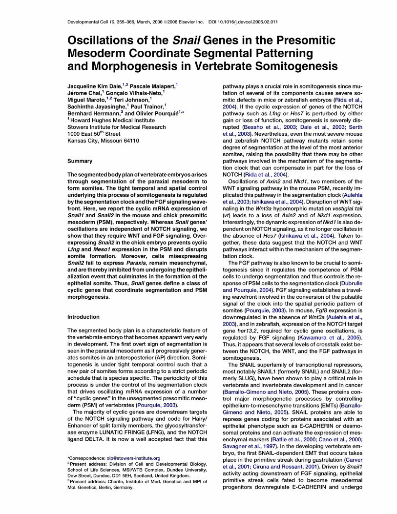
In order to test whether Snail1 is regulated by NOTCH orby WNT signaling, we analyzed the PSM expression ofSnail1 in mutant mice embryos in which these pathwayswere altered. We found that at E9–E9.5, when mutantembryos already exhibit strong segmentation defectsand expression of Lfng and Hes7 is severely disturbed,Snail1 continues to be expressed strongly in the PSMof Notch12/2 (n = 12), Hes72/2 (n = 17), and Lfng2/2
(n = 3) homozygous null embryos at levels equivalentto these seen in heterozygous and wild-type littermates(Figures 3A and 3C–3F). We also analyzed the expres-sion of Snail1 at E8.5 in Rbpjk2/2 homozygous null em-bryos that display the most severe segmentation pheno-type of the NOTCH mutants (Oka et al., 1995). WhereasLfng expression is absent from the PSM of these mu-tants (data not shown) (Morales et al., 2002), we foundthat Snail1 is still expressed (Figure 3B, n = 13/14).Therefore, NOTCH signaling is not required for Snail1expression in the PSM.
We next investigated if NOTCH signaling was requiredfor the oscillations of Snail1 in the mouse PSM. To thisend, we examined Snail1 expression in Hes7 null mousemutants in which oscillations of the NOTCH pathway-related cyclic genes are lost (Bessho et al., 2001). Whilethe anterior stripe of Snail1 expression was absent in thePSM of Hes7 mutants, we did observe different patternsof expression when comparing a large number of em-bryos, implying that a dynamic expression of Snail1 ismaintained in these mutants (Figures 3E and 3F). Thesedata strongly suggest that Snail1 dynamic expression inthe PSM is not dependent on NOTCH activity.
To complement this approach, we conducted a 2 hrin vitro culture of bisected E9.5 posterior mouse embryoexplants in the presence or absence of 10 mM DAPT(Dovey et al., 2001), an inhibitor of g-secretase-mediated NOTCH cleavage. Under these conditions,while cyclic Lfng transcription is abolished in the PSM(Figure 3G, n = 3), Snail1 continues to be expressed (Fig-ure 3H, n = 6). Moreover, the expression domains ofSnail1 are the same in the treated and untreated explanthalves from one embryo, but they differ from thoseof other embryos, suggesting, again, that oscillatingSnail1 expression in the PSM is independent of NOTCHsignaling.
To test if the regulation of Snail2 in the chick embryoPSM is similarly independent of NOTCH signaling, we
overexpressed by in ovo electroporation the activatedform of the NOTCH receptor, or a LUNATIC FRINGE ex-pression construct that was shown to inhibit NOTCHsignaling in the PSM (Dale et al., 2003). In both cases,no effect on Snail2 expression could be observed inthe PSM (data not shown). Together, these data suggestthat, in mouse and chick, the dynamic expression ofSnail genes in the PSM is not regulated by NOTCH sig-naling.
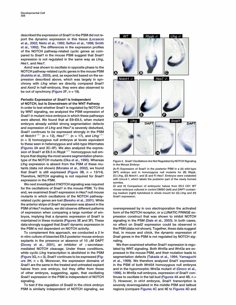
We then examined whether Snail1 expression is regu-lated by WNT signaling. Both Wnt5a and Wnt3a are ex-pressed in the mouse PSM, and their mutation leads tosegmentation defects (Takada et al., 1994; Yamaguchiet al., 1999). We therefore analyzed Snail1 expressionin the PSM of both Wnt5A homozygous null embryosand in the hypomorphic Wnt3a mutant vt (Greco et al.,1996). In Wnt5a null embryos, expression of Snail1 con-tinues to oscillate in the tail bud (Figures 4A and 4B, n =7). However, in vt/vt embryos, Snail1 transcription isseverely downregulated in the middle PSM and tailbudregions (compare Figures 4C and 4E to Figures 4D and
Figure 3. Snail1 Oscillations Are Not Regulated by NOTCH Signaling
in the Mouse Embryo
(A–F) Expression of Snail1 in the posterior PSM in a (A) wild-type
(WT) embryo and in homozygous null mutants for (B) Rbpjk,
(C) Lfng, (D) Notch1, and (E and F) Hes7. Embryos were colabeled
with Uncx4.1, which labels the posterior part of the newly formed
somites.
(G and H) Comparison of embryonic halves from E9.5 CD1 WT
mouse embryos cultured in control DMSO (left) and DAPT-contain-
ing medium (right) hybridized in whole mount for (G) Lfng and (H)
Snail1 expression.
Snail Genes Coordinate Segmentation/Morphogenesis359
Figure 4. Snail1 Expression Is Downstream of Wnt3a Signaling
(A and B) Comparison of Snail1 expression between (A) wild-type (WT) and (B) Wnt5a homozygous null mutant E9.5 mouse embryos.
(C–F) Comparison of Snail1 expression between (C and E) WT and (D and F) vt mutant E10.5 mouse embryos.
Embryos were colabeled with Uncx4.1, which labels the posterior part of the newly formed somites. (A, B, E, and F) lateral views and (C and D)
dorsal views of the PSM region.
4F, n = 29). Thus, like the other cyclic genes character-ized thus far (Aulehla et al., 2003), Snail1 oscillations ap-pear to be dependent on WNT3A signaling.
Snail2 Overexpression in the Chick Embryo
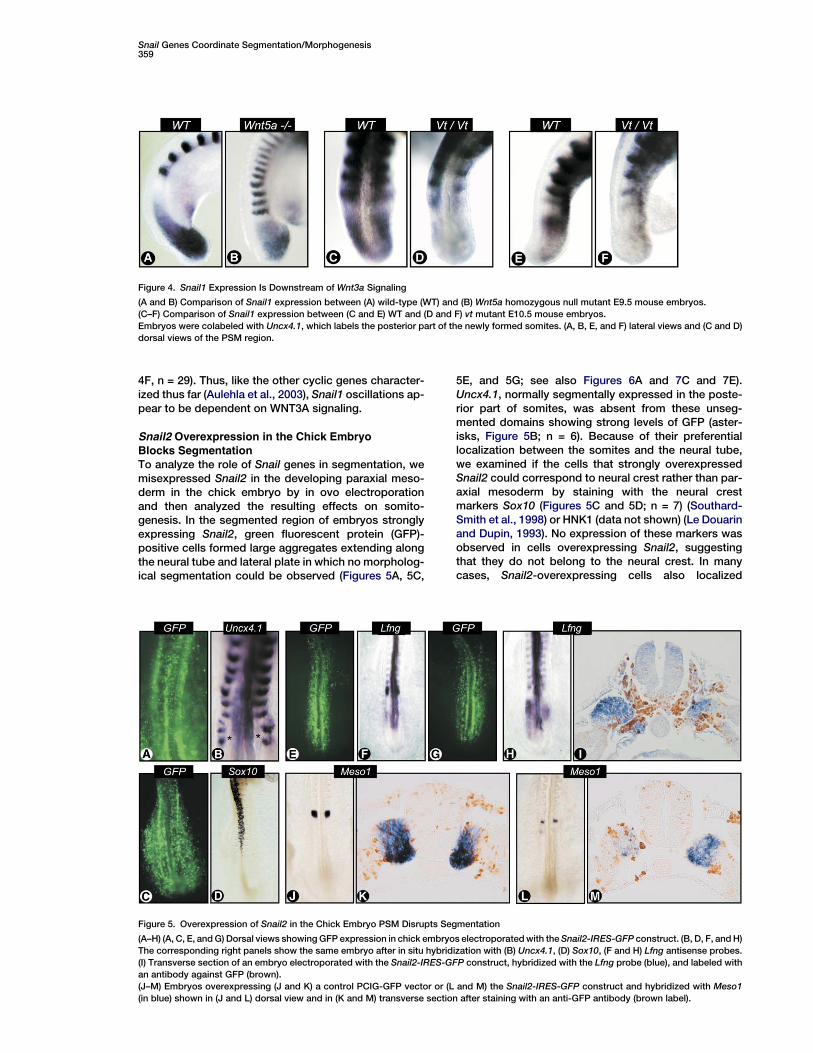
Blocks SegmentationTo analyze the role of Snail genes in segmentation, wemisexpressed Snail2 in the developing paraxial meso-derm in the chick embryo by in ovo electroporationand then analyzed the resulting effects on somito-genesis. In the segmented region of embryos stronglyexpressing Snail2, green fluorescent protein (GFP)-positive cells formed large aggregates extending alongthe neural tube and lateral plate in which no morpholog-ical segmentation could be observed (Figures 5A, 5C,
5E, and 5G; see also Figures 6A and 7C and 7E).Uncx4.1, normally segmentally expressed in the poste-rior part of somites, was absent from these unseg-mented domains showing strong levels of GFP (aster-isks, Figure 5B; n = 6). Because of their preferentiallocalization between the somites and the neural tube,we examined if the cells that strongly overexpressedSnail2 could correspond to neural crest rather than par-axial mesoderm by staining with the neural crestmarkers Sox10 (Figures 5C and 5D; n = 7) (Southard-Smith et al., 1998) or HNK1 (data not shown) (Le Douarinand Dupin, 1993). No expression of these markers wasobserved in cells overexpressing Snail2, suggestingthat they do not belong to the neural crest. In manycases, Snail2-overexpressing cells also localized
Figure 5. Overexpression of Snail2 in the Chick Embryo PSM Disrupts Segmentation
(A–H) (A, C, E, and G) Dorsal views showing GFP expression in chick embryos electroporated with the Snail2-IRES-GFP construct. (B, D, F, and H)
The corresponding right panels show the same embryo after in situ hybridization with (B) Uncx4.1, (D) Sox10, (F and H) Lfng antisense probes.
(I) Transverse section of an embryo electroporated with the Snail2-IRES-GFP construct, hybridized with the Lfng probe (blue), and labeled with
an antibody against GFP (brown).
(J–M) Embryos overexpressing (J and K) a control PCIG-GFP vector or (L and M) the Snail2-IRES-GFP construct and hybridized with Meso1
(in blue) shown in (J and L) dorsal view and in (K and M) transverse section after staining with an anti-GFP antibody (brown label).
Developmental Cell360
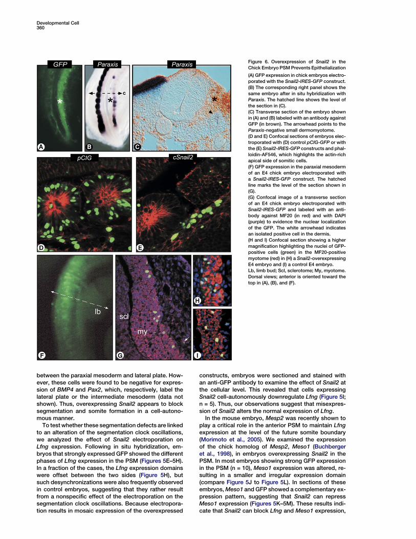
Figure 6. Overexpression of Snail2 in the
Chick Embryo PSM Prevents Epithelialization
(A) GFP expression in chick embryos electro-
porated with the Snail2-IRES-GFP construct.
(B) The corresponding right panel shows the
same embryo after in situ hybridization with
Paraxis. The hatched line shows the level of
the section in (C).
(C) Transverse section of the embryo shown
in (A) and (B) labeled with an antibody against
GFP (in brown). The arrowhead points to the
Paraxis-negative small dermomyotome.
(D and E) Confocal sections of embryos elec-
troporated with (D) control pCIG-GFP or with
the (E) Snail2-IRES-GFP constructs and phal-
loidin-AF546, which highlights the actin-rich
apical side of somitic cells.
(F) GFP expression in the paraxial mesoderm
of an E4 chick embryo electroporated with
a Snail2-IRES-GFP construct. The hatched
line marks the level of the section shown in
(G).
(G) Confocal image of a transverse section
of an E4 chick embryo electroporated with
Snail2-IRES-GFP and labeled with an anti-
body against MF20 (in red) and with DAPI
(purple) to evidence the nuclear localization
of the GFP. The white arrowhead indicates
an isolated positive cell in the dermis.
(H and I) Confocal section showing a higher
magnification highlighting the nuclei of GFP-
positive cells (green) in the MF20-positive
myotome (red) in (H) a Snail2-overexpressing
E4 embryo and (I) a control E4 embryo.
Lb, limb bud; Scl, sclerotome; My, myotome.
Dorsal views; anterior is oriented toward the
top in (A), (B), and (F).
between the paraxial mesoderm and lateral plate. How-ever, these cells were found to be negative for expres-sion of BMP4 and Pax2, which, respectively, label thelateral plate or the intermediate mesoderm (data notshown). Thus, overexpressing Snail2 appears to blocksegmentation and somite formation in a cell-autono-mous manner.
To test whether these segmentation defects are linkedto an alteration of the segmentation clock oscillations,we analyzed the effect of Snail2 electroporation onLfng expression. Following in situ hybridization, em-bryos that strongly expressed GFP showed the differentphases of Lfng expression in the PSM (Figures 5E–5H).In a fraction of the cases, the Lfng expression domainswere offset between the two sides (Figure 5H), butsuch desynchronizations were also frequently observedin control embryos, suggesting that they rather resultfrom a nonspecific effect of the electroporation on thesegmentation clock oscillations. Because electropora-tion results in mosaic expression of the overexpressed
constructs, embryos were sectioned and stained withan anti-GFP antibody to examine the effect of Snail2 atthe cellular level. This revealed that cells expressingSnail2 cell-autonomously downregulate Lfng (Figure 5I;n = 5). Thus, our observations suggest that misexpres-sion of Snail2 alters the normal expression of Lfng.
In the mouse embryo, Mesp2 was recently shown toplay a critical role in the anterior PSM to maintain Lfngexpression at the level of the future somite boundary(Morimoto et al., 2005). We examined the expressionof the chick homolog of Mesp2, Meso1 (Buchbergeret al., 1998), in embryos overexpressing Snail2 in thePSM. In most embryos showing strong GFP expressionin the PSM (n = 10), Meso1 expression was altered, re-sulting in a smaller and irregular expression domain(compare Figure 5J to Figure 5L). In sections of theseembryos, Meso1 and GFP showed a complementary ex-pression pattern, suggesting that Snail2 can repressMeso1 expression (Figures 5K–5M). These results indi-cate that Snail2 can block Lfng and Meso1 expression,

Snail Genes Coordinate Segmentation/Morphogenesis361
thus preventing overexpressing cells from formingsomites.
Snail2 Regulates the Mesenchyme-to-Epithelium
Transformation Leading to Somite FormationThe posterior expression domain of mouse Snail1 andchick Snail2 correlates with the mesenchymal domainin the PSM, suggesting that these genes might be in-volved in the control of the mesenchymal state of thesecells. To test whether Snail2 is involved in the control ofthis transition, we examined the effect of overexpress-ing Snail2 on PSM epithelialization. To that end, we an-alyzed the effect on the expression of Paraxis, a tran-scription factor whose expression is associated withthe onset of epithelialization in the anterior PSM and issubsequently expressed in the epithelial somites anddermomyotomes (Burgess et al., 1995). In chick em-
Figure 7. Snail2 Expression Is Regulated by FGF Signaling
(A and B) Chick embryos treated with (A) DMSO and (B) SU5402 and
hybridized with a Snail2 probe. Asterisks mark the lateral plate.
(C and E) GFP expression in chick embryos electroporated with the
Snail2-IRES-GFP construct.
(D and F) The corresponding right panel shows the same embryo af-
ter in situ hybridization with (D) Brachyury and (F) Tbx6 antisense
probes. Dorsal views; anterior is oriented toward the top.
bryos strongly overexpressing Snail2, Paraxis wasfound to be absent from the GFP-positive regions ofthe anterior PSM and somites (Figures 6A and 6B; n =9). The strongly expressing unsegmented domains lyingbetween the neural tube and somites were often flankedby a row of smaller somites composed of GFP-negativecells (Figures 6A and 6B). In sections, expression ofSnail2 was clearly associated with a loss of the epithelialstructures in the somite or the dermomyotome (asterisk,Figure 6C), and Paraxis was found to be downregulatedin a cell-autonomous fashion in these domains (Fig-ure 6C and data not shown). In some cases, Paraxiswas also found to be downregulated in the remainingsmall dermomyotome, which, nonetheless, does not ex-press Snail2 (arrowhead, Figure 6C). This non-cell-au-tonomous downregulation is probably caused by thephysical separation of the dermomyotome from theoverlying ectoderm by the Snail2-overexpressing cells,since it has been reported that Paraxis expression inthe dermomyotome depends upon close contact withthe ectoderm (Sosic et al., 1997). Furthermore, confocalanalysis of electroporated embryos clearly showed thatGFP-positive cells overexpressing Snail2 were largelyexcluded from epithelial somites (Figures 6D and 6E).We counted the GFP-positive cells in the somitic regionof the paraxial mesoderm in control embryos; 135 GFP-positive cells were observed in 5 somites in 2 controlembryos, whereas no cell was seen outside these so-mites in the paraxial mesoderm region. In 2 embryosoverexpressing Snail2, 27 GFP-positive cells werecounted in 5 somites, while 128 cells were found to lieadjacent to these somites in the paraxial mesoderm re-gion. Therefore, ectopic activation of Snail2 can blockthe incorporation of paraxial mesoderm cells in epithe-lial somites.
We then examined the fate of the Snail2-overexpress-ing cells compared to control GFP-overexpressing cellsin E4 embryos (Figure 6F; n = 4 experimentals and n = 4controls). A difficulty with this experiment is that theplasmids electroporated become diluted in tissuesthat are actively dividing, like the sclerotome or the der-mis. In contrast, because the muscle fibers are postmi-totic, a bright GFP signal is retained in the nuclei evenat late stages like E4 (Figure 6G). Therefore, we havecompared the number of GFP-positive cells per myo-tome in sections of E4 embryos overexpressing Snail2(Figure 6H) and overexpressing the control GFP con-struct (Figure 6I). We found an average of 11.9 GFP-pos-itive cells/myotome (143 cells on 12 sections from 3 con-trol embryos) in control embryos and 12.2 GFP-positivecells/myotome (183 cells on 15 sections from 4 experi-mental embryos) in the Snail2-overexpressing embryos.Therefore, a similar contribution to the myotome is ob-served for control and Snail2-overexpressing embryos,indicating that Snail2-overexpressing cells can differen-tiate into paraxial mesoderm derivatives.
FGF Signaling Is Necessary, but Not Sufficient,for Snail2 Expression in the PSM
In the PSM, the EMT is controlled by the level of FGF sig-naling, and the epithelialization process begins once thecells enter the anterior region characterized by lowerFGF signaling (Delfini et al., 2005; Dubrulle et al., 2001).Our data suggest that this transition could be controlled

Developmental Cell362
by Snail genes acting downstream of FGF. Consistently,Snail1 expression is lost in the PSM of mouse mutantsfor FGFR1 (Ciruna and Rossant, 2001). To test whetherSnail2 is regulated in a similar way in the chick embryo,we treated chick embryo explants for 4 hr in culture with100 mm SU5402, which blocks the activity of FGFR1(Mohammadi et al., 1997). In treated embryos, Snail2was found to be downregulated in the PSM, whereas itwas upregulated in the neural tube and the lateral plate(Figures 7A and 7B; n = 10). Therefore, FGF signaling isrequired for Snail2 expression in the PSM, whereasthis pathway represses its expression in the neuraltube and lateral plate.
The cell-autonomous blockade of segmentation ob-served when overexpressing Snail2 is reminiscent ofthat seen when overactivating the ERK pathway thatacts downstream of FGF signaling in the PSM (Delfiniet al., 2005). In these experiments, electroporation ofa constitutively active form of the MEKK1 kinase, ca-MEKK1, results in an ectopic activation of Brachyury inthe overexpressing cells of the anterior PSM and so-mites. Since Snail genes act downstream of FGF signal-ing in the PSM, we investigated whether Snail2 in thechick may be acting as the transcriptional effector ofthe FGF response. We therefore examined the effect ofmisexpressing Snail2 on the expression of Brachyury,whose expression is associated with posterior PSMidentity (Ciruna and Rossant, 2001; Delfini et al., 2005;Dubrulle et al., 2001). Unlike in FGF8 or activatedMEKK1-overexpressing embryos, no ectopic anteriorexpression of Brachyury was observed in Snail2-over-expressing embryos (Figures 7C and 7D; n = 8). There-fore, Snail2 and Brachyury appear to be regulated inde-pendently by FGF signaling. To further investigate if thisreflects an impairment of maturation of the paraxial me-soderm cells, we also examined the expression of Tbx6,a T box factor expressed specifically in the PSM (Kne-zevic et al., 1997). No expression of Tbx6 could bedetected in cells overexpressing Snail2 in the somiticregion (Figures 7E and 7F; n = 6). These results indicatethat whereas PSM cells overexpressing Snail2 fail to ep-ithelialize and segment, they do not maintain a PSMidentity.
To test whether ectopic FGF signaling in the PSMresults in Snail2 expression, we overexpressed FGF8by electroporation. Whereas somite formation wasblocked, as previously reported (Dubrulle et al., 2001),no ectopic expression of Snail2 was observed in the par-axial mesoderm of FGF8-overexpressing embryos (datanot shown). Together, these experiments suggest thatFGF signaling is necessary, but not sufficient, for Snail2expression in the chick PSM.
Discussion
Here, we show that Snail1 and Snail2 may identify a novelclass of cyclic genes showing periodic expression in themouse and chick PSM, respectively. We observe thatwhile Snail1 expression dynamics show some similari-ties with those of the NOTCH pathway-related cyclicgenes like Lfng, they also exhibit clear differences, par-ticularly at the tail bud level. Thus, Snail1 transcription isinitiated prior to that of the NOTCH cyclic genes at theonset of a new wave of expression in the posterior
PSM. Interfering with the NOTCH pathway does not alterthe dynamic expression profile of the Snail genes, sug-gesting that their regulation is independent of NOTCHsignaling. In contrast, Snail1 expression is lost in theWnt3a hypomorph mutant vt, thus confirming the impor-tant role of this pathway upstream of the segmentationclock mechanism. In the chick embryo PSM, overex-pressing the Snail2 gene downregulates the expressionof Lfng and Meso1 and results in a block of segmenta-tion. Such gain of function ultimately prevents targetedcells from activating Paraxis, thereby blocking them ina mesenchymal state. We show that FGF signaling is re-quired for maintenance of Snail2 expression in the PSM.While the Snail2 overexpression phenotype in the chickembryo strongly resembles that observed after theectopic activation of the FGF pathway in the PSM, noectopic expression of genes associated with the pos-terior PSM identity is seen upon Snail2 misexpression.Together, these data suggest that Snail genes link thesegmentation clock to PSM morphogenesis down-stream of the FGF pathway.
Snail Genes May Define a New Class of Cyclic GenesHere, we show that the mouse Snail1 gene (formerlySnail) and the chick Snail2 gene (formerly Slug) oscillatein the PSM with very similar expression kinetics inthe two species (Figure 1). Such an inversion of expres-sion and function between Snail1 and Snail2 in mouseand chick is not unexpected since it has been observedin most embryonic tissues, including neural crest andmesodermal derivatives (Locascio et al., 2002; Seftonet al., 1998). In heart formation, Snail1 was shown toact downstream of NOTCH to control the EMT leadingto the development of cardiac valvular primordia (Tim-merman et al., 2004). In contrast, in mouse and chickPSM, our data suggest that expression of Snail genesis not regulated by the NOTCH pathway. This is in sharpcontrast to the NOTCH pathway-related cyclic genes,such as Lfng, which are regulated by NOTCH signalingin the PSM (Dale et al., 2003). Also, expression of theSnail genes was found to be downstream of FGF signal-ing in the PSM (Figures 7A and 7B) (Ciruna and Rossant,2001); however, expression of the NOTCH pathway-related genes, like Lfng or Hairy/Hes, was not affectedby SU5402 treatment (Delfini et al., 2005; Dubrulleet al., 2001). Snail1 has been shown to control EMTs inthe mouse paraxial mesoderm (Ciruna and Rossant,2001), and our overexpression data in the chick supportthe notion that Snail2 plays a similar role in the chickPSM. Together, this data argue that Snail1 and Snail2genes are functional homologs in the PSM of mouseand chick embryos, respectively.
The mouse Snail1 homozygous null mutant does notform paraxial mesoderm, thus precluding analysis ofthe role of this gene in the segmentation clock function(Carver et al., 2001). We show that, in the chick PSM,overexpressing Snail2 can repress Lfng expression,consistent with the well-known repressor function ofSnail family genes (Nieto, 2002). SNAIL1 was shown tobind E boxes in the E-CADHERIN promoter (Batlleet al., 2000), and several such sites are found in theLfng promoter, in agreement with the idea that Snailgenes might directly regulate Lfng (Cole et al., 2002; Mo-rales et al., 2002). Strikingly, however, Snail1 and Lfng

Snail Genes Coordinate Segmentation/Morphogenesis363
are coexpressed for a portion of the oscillation cycle, al-though Snail1 transcription continues in the posteriorPSM in embryos that have downregulated Lfng (Figures2C and 2D). This could suggest that accumulation ofSNAIL1 protein during one oscillation cycle is requiredfor repressing Lfng expression in the posterior PSM, orthat Lfng repression by Snail1 is indirect. Therefore, de-spite their coexpression for a significant portion of theoscillation cycle, our data indicate that Snail genes areregulated differently from the NOTCH pathway-relatedcyclic genes.
The link between WNT and NOTCH signaling remainsone of the central unsolved mechanisms underlyingboth the generation and regulation of the segmentationclock. Here, we show that Snail1 transcription is down-stream of WNT3A, but not of NOTCH, signaling. Interest-ingly, however, Snail1 oscillations were out of phasewhen compared to Axin2, suggesting that whereasboth genes are regulated by WNT signaling, the modalityof this regulation might be different. Snail1 transcriptionwas recently shown to be inhibited by GSK3b activation(Bachelder et al., 2005). The WNT pathway oscillationsreported in the PSM (Aulehla et al., 2003) are expectedto result in a periodic inactivation of GSK3b that would,in turn, lift the inhibition of Snail1 transcription and, to-gether with FGF signaling, would lead to periodic Snail1activation in the PSM. The periodic production of theSNAIL1 protein downstream of the WNT oscillationscould trigger the periodic repression of NOTCH targets.The SNAIL1 protein is highly unstable and is targeted fornuclear export and proteasome-mediated degradationby means of GSK3b-mediated phosphorylation (Zhouet al., 2004). Thus, its effect on NOTCH signaling shouldbe transient and periodic. Snail1 oscillations mighttherefore provide a link between the WNT and NOTCHsignaling pathways in the segmentation clock.
Snail Genes Coordinate PSM Morphogenesisand Segmentation
In the mouse embryo, cells of the primitive streak that re-ceive high FGF signaling activate Snail1 expression,which, in turn, represses E-CADHERIN transcription,thus allowing the EMT leading to the formation of meso-dermal tissues such as the posterior PSM (Batlle et al.,2000; Ciruna and Rossant, 2001). The downregulationof E-CADHERIN expression was proposed to releasea pool of b-CATENIN from the cell membrane, thus al-lowing these cells to respond to WNT signaling (Cirunaand Rossant, 2001). The most prominent Snail1 andSnail2 expression domains in the mouse and chickPSM extend from the primitive streak/tail bud to thelevel of the determination front and therefore nicely cor-relate with the mesenchymal area of the PSM (Figure 1)(Sefton et al., 1998). In chick and mouse embryos, E-CADHERIN is specifically excluded from the PSM,where the Snail genes are predominantly expressed(Cano et al., 2000; Thiery et al., 1984). The Snail genes’expression domain within the posterior PSM also corre-lates with the region where the oscillations of the WNTcyclic gene Axin2 occur (Aulehla et al., 2003). Inhibitionof cadherin levels, mediated by Snail genes and associ-ated with the maintenance of the mesenchymal state ofthe PSM, might be required to ensure that a sufficientb-CATENIN pool is available for the WNT-driven oscilla-
tions underlying the segmentation clock. At the determi-nation front, the downregulation of FGF and Wnt signal-ing (Aulehla et al., 2003) would result in the arrest ofoscillating Snail expression. Thus, the repression ofthe epithelial phenotype is relieved at this level, and cad-herin expression levels begin to increase with the pro-gressive epithelialization of the anterior PSM (Dubandet al., 1987; Linask et al., 1998). The arrest of clockgene oscillations at the determination front might there-fore be linked to the reexpression of cadherins and theconsequential reduced availability of b-CATENIN dueto its sequestration at the membrane by cadherins.Such crosstalk between cadherins, WNT, and FGF sig-naling, resulting in the control of b-CATENIN levels,has been reported in several systems (Nelson andNusse, 2004). The Snail genes would thus act as compe-tence factors for the segmentation clock. Because theyact downstream of FGF and Wnt signaling, these geneswould provide a means of temporally integrating theclock mechanism with progression of the wavefront.
The anterior limit of the posterior expression domainof Snail genes in the PSM roughly corresponds to theposition of the determination front. This is the level atwhich periodic NOTCH signaling controlled by the seg-mentation clock triggers the segmental activation ofdownstream targets such as the Mesp family genes(Jen et al., 1999; Morimoto et al., 2005; Takahashiet al., 2000). Here, we show that overexpression ofSnail2 in the chick PSM, prevents expression of theMesp2 homolog, Meso1 (Figures 5J–5M) (Buchbergeret al., 1998). This suggests that Snail genes might re-press expression of Mesp2/Meso1 in most of the PSM,thus restricting expression of these genes to the narrowSnail-negative stripe in the anterior PSM. Such a role inpositioning the NOTCH response has been previouslyreported in the fly, where Snail was proposed to restrictNOTCH signaling at the future midline by a combinationof cell-autonomous and non-cell-autonomous repres-sing and stimulating activities (Cowden and Levine,2002; Morel et al., 2003).
Snail2 overexpression in the chick results in a cell-autonomous blockade of the somitogenesis process ac-companied by a downregulation of Paraxis and Uncx4.1expression in the anterior PSM and somites. This pheno-type is very similar to that observed after either constitu-tive activation of the ERK pathway via misexpression ofa constitutively active form of the MEKK1 kinase in thePSM (Delfini et al., 2005) or overexpression of FGF8 inthe PSM (Dubrulle et al., 2001). However, whereas FGF8and caMEKK1 also ectopically activate expression ofposterior PSM markers like Brachyury in the anteriorPSM, this phenotype was not observed when overex-pressing Snail2. This result is consistent with the homo-zygous null mutation of Snail1 in the mouse in whichBrachyury-positive mesoderm forms but retains a polar-ized epithelial character expressing E-CADHERIN(Carver et al., 2001). Overexpressing FGF8 in the PSMalso blocks the subsequent differentiation of paraxialmesoderm cells (Dubrulle et al., 2001). In contrast,Snail2-overexpressing cells differentiate into muscle,dermis, and sclerotome, suggesting that while pattern-ing is disrupted, paraxial mesoderm differentiation isnot affected. These results are consistent with the phe-notype of the Paraxis null mutant in which the paraxial

Developmental Cell364
mesoderm does not form epithelial somites, but differ-entiates into normal myotomes (Burgess et al., 1996).Therefore, this suggests that Snail does not control thematuration of PSM cells, but rather controls the morpho-genesis along this tissue. Together, our results suggestthat Snail genes play a dual role in the segmentation pro-cess: first, they play a role at the level of the segmenta-tion clock by integrating the NOTCH, WNT, and FGFpathways, and, second, they play a role by controllingthe morphogenetic process associated with epithelialsomite formation.
Experimental Procedures
Embryos
Fertilized chick eggs were obtained from Ozark Hatcheries (Neosho,
MO) and incubated at 38ºC in a humidified incubator. Embryos were
staged according to the developmental table of Hamburger and
Hamilton (HH) (Hamburger and Hamilton, 1992) and by counting so-
mite pairs.
Wild-type CD1 mice embryos were harvested from timed mated
pregnant females between 8.5 and 10 days postcoitum (dpc).
Notch12/2, Lfng2/2, Rbpjk2/2, Hes72/2, Wnt5a2/2, and vt/vt em-
bryos were obtained and genotyped by PCR analysis of the yolk
sacs as described (Aulehla et al., 2003; Bessho et al., 2003; Conlon
et al., 1995; Oka et al., 1995; Yamaguchi et al., 1999; Zhang and Grid-
ley, 1998).
Mouse and Chick Embryo Explant Culture
E9.5 CD1 mice embryos were harvested, and their posterior part was
divided into two halves by cutting along the neural tube. The ex-
plants were cultured in hanging drops of culture medium composed
of DMEM/F12 supplemented with 10% fetal calf serum, 10 ng/ml
bFGF, and 50 U/ml penicillin/streptomycin. Experiments were per-
formed as described by Palmeirim et al. (1997). Half of the explant
was immediately fixed, and the other half was cultured for 75–120
min prior to fixation. The two explants were then analyzed for ex-
pression of Snail1 mRNA by in situ hybridization. Alternatively, the
two halves of the explant were fixed immediately and were used to
compare the expression domains of Snail1 and Lfng or Snail1 and
Axin2 or exonic Snail1 and intronic Snail1 by in situ hybridization.
N-(N-(3,5-difluorophenacetyl)-L-alanyl)-S-phenylglycine t-butyl es-
ter, DAPT (Calbiochem), was diluted into culture medium from stock
solutions in dimethylsulphoxide (DMSO) to maintain a final DMSO
concentration of 1%. Half-embryo assays were performed as de-
scribed above. One half was cultured in medium containing DAPT
(10 mM), whereas the control side was cultured in normal medium.
Both sides were cultured for 2 hr and then analyzed for expression
of Lfng mRNA or Snail1 mRNA by in situ hybridization. Chick embryo
explants were cultured in vitro as described in Delfini et al. (2005).
Posterior half-embryo explants were cultured for 4 hr in a chick cul-
ture medium (5% chick serum, 2.5% FCS, and 1% bicarbonate in
DMEM or L-15, GIBCO-BRL) containing 100 mM of the FGFR inhibitor
SU5402 (Pfizer) dissolved in DMSO. Explants were then fixed and
processed for in situ hybridization.
Plasmids and In Ovo Electroporation
In ovo electroporations were performed as described (Dubrulle
et al., 2001). Eggs were windowed, and the DNA solution was in-
jected between the vitelline membrane and the epiblast in stage-4
to stage-5 HH embryos. The primitive streak was coated with the
DNA solution from the node until mid-streak. Two platinum elec-
trodes tied together were used: one was placed directly on the
streak posterior to the node, and the second was inserted into the
yolk. A series of electric pulses (4 pulses, 30 volts, 50 ms) was
directed with a square wave electroporator (BTX). For electro-
porations, constructs were cloned into the pCIG (Megason and
McMahon, 2002) or pCAGGS (Niwa et al., 1991) expression vectors,
which were purified with an endotoxin-free maxi kit (Qiagen) and
used at a final concentration of 2 mg/ml or 1 mg/ml, respectively,
in a PBS solution containing 1 mM MgCl2 and 0.4 mg/ml fast green
FCF (Sigma). pCIG is a bicistronic vector that drives the expression
of a nuclear GFP reporter in addition to the gene of interest (Mega-
son and McMahon, 2002). Full-length Snail2 was cloned into the
pCIG vector. The FGF8, activated NOTCH, and LFNG expression
constructs have been previously described (Dale et al., 2003;
Dubrulle et al., 2001). Control embryos were either electroporated
with empty pCIG or coelectroporated with pCAAGS-GFP and empty
pCAAGS. After electroporation, eggs were reincubated for 24, 40,
48, or 96 (for E4 embryos) hr and were assayed for GFP expression.
Embryos were then fixed and processed for in situ hybridization or
incubated overnight with phalloidin AF546 (Molecular Probe,
1:100) and analyzed by confocal microscopy.
Whole-Mount In Situ Hybridization and Immunohistochemistry
on Section
Whole-mount in situ hybridizations were performed as described
(Henrique et al., 1995). The chick Snail2, Brachyury, Tbx6, Lfng,
Uncx4.1, Paraxis, and Sox10 (Dale et al., 2003; Knezevic et al.,
1997; Sefton et al., 1998; Sosic et al., 1997; Southard-Smith et al.,
1998) and the mouse Lfng and Axin2 (Aulehla et al., 2003) probes
have been described. The intronic Snail1 probe was produced by
polymerase chain reaction (PCR) amplification of a fragment corre-
sponding to nucleotides 1004–1553 of the mouse Snail1 genomic
sequence (GenBank accession code NT 039210). Some of the la-
beled embryos were embedded for cryosection in gelatin-sucrose
and cut at 14 mm. For some embryos, 10 mm paraffin sections
were also prepared. GFP was detected on section with an anti-
GFP mAb (Roche # 1814460, 1:200) by diaminobenzidine (DAB) or
fluorescence staining. Differentiated muscles were evidenced in
section by using the anti-Myosin Heavy Chain MF20 mAb detected
by fluorescence. Sections were counterstained with DAPI staining
to visualize nuclei.
Supplemental Data
Supplemental Data including Figure S1 are available at http://www.
developmentalcell.com/cgi/content/full/10/3/355/DC1/.
Acknowledgments
We would like to thank Jean-Philippe Rey and the Histology Core Fa-
cility of the Stowers Institute for Medical Research for their help in
processing embryos. We thank Silvia Esteban for the artwork. We
are grateful to members of the Pourquie lab for helpful discussions
and comments on the manuscript. We thank T. Gridley, T. Honjo,
A. McMahon, R. Conlon, and R. Kageyama for providing the Lfng,
RBPjK, WNT5A, Notch1, and Hes7 knockout mice. We thank Angela
Nieto, K. Katsube, S. Mackem, and E. Olson for reagents. This work
has been supported by National Institutes of Health grant R01
HD043158 to O.P. and by the Stowers Institute for Medical
Research. O.P. is an investigator of the Howard Hughes Medical
Institute.
Received: July 9, 2005
Revised: December 7, 2005
Accepted: February 16, 2006
Published online: March 6, 2006
References
Aulehla, A., Wehrle, C., Brand-Saberi, B., Kemler, R., Gossler, A.,
Kanzler, B., and Herrmann, B.G. (2003). Wnt3a plays a major role
in the segmentation clock controlling somitogenesis. Dev. Cell 4,
395–406.
Bachelder, R.E., Yoon, S.O., Franci, C., de Herreros, A.G., and
Mercurio, A.M. (2005). Glycogen synthase kinase-3 is an endoge-
nous inhibitor of Snail transcription: implications for the epithelial-
mesenchymal transition. J. Cell Biol. 168, 29–33.
Barrallo-Gimeno, A., and Nieto, M.A. (2005). The Snail genes as
inducers of cell movement and survival: implications in development
and cancer. Development 132, 3151–3161.
Batlle, E., Sancho, E., Franci, C., Dominguez, D., Monfar, M.,
Baulida, J., and Garcia De Herreros, A. (2000). The transcription fac-
tor snail is a repressor of E-cadherin gene expression in epithelial
tumour cells. Nat. Cell Biol. 2, 84–89.
Snail Genes Coordinate Segmentation/Morphogenesis365
Bessho, Y., Sakata, R., Komatsu, S., Shiota, K., Yamada, S., and
Kageyama, R. (2001). Dynamic expression and essential functions
of Hes7 in somite segmentation. Genes Dev. 15, 2642–2647.
Bessho, Y., Hirata, H., Masamizu, Y., and Kageyama, R. (2003).
Periodic repression by the bHLH factor Hes7 is an essential mecha-
nism for the somite segmentation clock. Genes Dev. 17, 1451–1456.
Buchberger, A., Seidl, K., Klein, C., Eberhardt, H., and Arnold, H.H.
(1998). cMeso-1, a novel bHLH transcription factor, is involved in
somite formation in chicken embryos. Dev. Biol. 199, 201–215.
Burgess, R., Cserjesi, P., Ligon, K.L., and Olson, E.N. (1995). Paraxis:
a basic helix-loop-helix protein expressed in paraxial mesoderm and
developing somites. Dev. Biol. 168, 296–306.
Burgess, R., Rawls, A., Brown, D., Bradley, A., and Olson, E.N.
(1996). Requirement of the paraxis gene for somite formation and
musculoskeletal patterning. Nature 384, 570–573.
Cano, A., Perez-Moreno, M.A., Rodrigo, I., Locascio, A., Blanco,
M.J., del Barrio, M.G., Portillo, F., and Nieto, M.A. (2000). The tran-
scription factor snail controls epithelial-mesenchymal transitions
by repressing E-cadherin expression. Nat. Cell Biol. 2, 76–83.
Carver, E.A., Jiang, R., Lan, Y., Oram, K.F., and Gridley, T. (2001). The
mouse snail gene encodes a key regulator of the epithelial-mesen-
chymal transition. Mol. Cell. Biol. 21, 8184–8188.
Ciruna, B., and Rossant, J. (2001). FGF signaling regulates meso-
derm cell fate specification and morphogenetic movement at the
primitive streak. Dev. Cell 1, 37–49.
Cole, S.E., Levorse, J.M., Tilghman, S.M., and Vogt, T.F. (2002).
Clock regulatory elements control cyclic expression of Lunatic
Fringe during somitogenesis. Dev. Cell 3, 75–84.
Conlon, R.A., Reaume, A.G., and Rossant, J. (1995). Notch1 is re-
quired for the coordinate segmentation of somites. Development
121, 1533–1545.
Cowden, J., and Levine, M. (2002). The Snail repressor positions
Notch signaling in the Drosophila embryo. Development 129,
1785–1793.
Dale, J.K., Maroto, M., Dequeant, M.L., Malapert, P., McGrew, M.,
and Pourquie, O. (2003). Periodic Notch inhibition by Lunatic Fringe
underlies the chick segmentation clock. Nature 421, 275–278.
Delfini, M.C., Dubrulle, J., Malapert, P., Chal, J., and Pourquie, O.
(2005). Control of the segmentation process by graded MAPK/ERK
activation in the chick embryo. Proc. Natl. Acad. Sci. USA 102,
11343–11348.
Dovey, H.F., John, V., Anderson, J.P., Chen, L.Z., de Saint Andrieu,
P., Fang, L.Y., Freedman, S.B., Folmer, B., Goldbach, E., Holsztyn-
ska, E.J., et al. (2001). Functional g-secretase inhibitors reduce
beta-amyloid peptide levels in brain. J. Neurochem. 76, 173–181.
Duband, J.L., Dufour, S., Hatta, K., Takeichi, M., Edelman, G.M., and
Thiery, J.P. (1987). Adhesion molecules during somitogenesis in the
avian embryo. J. Cell Biol. 104, 1361–1374.
Dubrulle, J., and Pourquie, O. (2004). Coupling segmentation to axis
formation. Development 131, 5783–5793.
Dubrulle, J., McGrew, M.J., and Pourquie, O. (2001). FGF signaling
controls somite boundary position and regulates segmentation
clock control of spatiotemporal Hox gene activation. Cell 106,
219–232.
Greco, T.L., Takada, S., Newhouse, M.M., McMahon, J.A., Mc-
Mahon, A.P., and Camper, S.A. (1996). Analysis of the vestigial tail
mutation demonstrates that Wnt-3a gene dosage regulates mouse
axial development. Genes Dev. 10, 313–324.
Hamburger, V., and Hamilton, H.L. (1992). A series of normal stages
in the development of the chick embryo (1951). Dev. Dyn. 195, 231–
272.
Henrique, D., Adam, J., Myat, A., Chitnis, A., Lewis, J., and Ish-
Horowicz, D. (1995). Expression of a Delta homologue in prospective
neurons in the chick. Nature 375, 787–790.
Ishikawa, A., Kitajima, S., Takahashi, Y., Kokubo, H., Kanno, J.,
Inoue, T., and Saga, Y. (2004). Mouse Nkd1, a Wnt antagonist, ex-
hibits oscillatory gene expression in the PSM under the control of
Notch signaling. Mech. Dev. 121, 1443–1453.
Jen, W.C., Gawantka, V., Pollet, N., Niehrs, C., and Kintner, C. (1999).
Periodic repression of Notch pathway genes governs the segmenta-
tion of Xenopus embryos. Genes Dev. 13, 1486–1499.
Kawamura, A., Koshida, S., Hijikata, H., Sakaguchi, T., Kondoh, H.,
and Takada, S. (2005). Zebrafish hairy/enhancer of split protein links
FGF signaling to cyclic gene expression in the periodic segmenta-
tion of somites. Genes Dev. 19, 1156–1161.
Knezevic, V., De Santo, R., and Mackem, S. (1997). Two novel chick
T-box genes related to mouse Brachyury are expressed in different,
non-overlapping mesodermal domains during gastrulation. Devel-
opment 124, 411–419.
Le Douarin, N.M., and Dupin, E. (1993). Cell lineage analysis in neural
crest ontogeny. J. Neurobiol. 24, 146–161.
Linask, K.K., Ludwig, C., Han, M.D., Liu, X., Radice, G.L., and Knud-
sen, K.A. (1998). N-cadherin/catenin-mediated morphoregulation of
somite formation. Dev. Biol. 202, 85–102.
Locascio, A., Manzanares, M., Blanco, M.J., and Nieto, M.A. (2002).
Modularity and reshuffling of Snail and Slug expression during ver-
tebrate evolution. Proc. Natl. Acad. Sci. USA 99, 16841–16846.
Megason, S.G., and McMahon, A.P. (2002). A mitogen gradient of
dorsal midline Wnts organizes growth in the CNS. Development
129, 2087–2098.
Mohammadi, M., McMahon, G., Sun, L., Tang, C., Hirth, P., Yeh, B.K.,
Hubbard, S.R., and Schlessinger, J. (1997). Structures of the tyrosine
kinase domain of fibroblast growth factor receptor in complex with
inhibitors. Science 276, 955–960.
Morales, A.V., Yasuda, Y., and Ish-Horowicz, D. (2002). Periodic Lu-
natic Fringe expression is controlled during segmentation by a cyclic
transcriptional enhancer responsive to Notch signaling. Dev. Cell 3,
63–74.
Morel, V., Le Borgne, R., and Schweisguth, F. (2003). Snail is re-
quired for Delta endocytosis and Notch-dependent activation of sin-
gle-minded expression. Dev. Genes Evol. 213, 65–72.
Morimoto, M., Takahashi, Y., Endo, M., and Saga, Y. (2005). The
Mesp2 transcription factor establishes segmental borders by sup-
pressing Notch activity. Nature 435, 354–359.
Nakaya, Y., Kuroda, S., Katagiri, Y.T., Kaibuchi, K., and Takahashi, Y.
(2004). Mesenchymal-epithelial transition during somitic segmenta-
tion is regulated by differential roles of Cdc42 and Rac1. Dev. Cell 7,
425–438.
Nelson, W.J., and Nusse, R. (2004). Convergence of Wnt, b-catenin,
and cadherin pathways. Science 303, 1483–1487.
Nieto, M.A. (2002). The snail superfamily of zinc-finger transcription
factors. Nat. Rev. Mol. Cell Biol. 3, 155–166.
Nieto, M.A., Bennett, M.F., Sargent, M.G., and Wilkinson, D.G.
(1992). Cloning and developmental expression of Sna, a murine
homologue of the Drosophila snail gene. Development 116, 227–
237.
Niwa, H., Yamamura, K., and Miyazaki, J. (1991). Efficient selection
for high-expression transfectants with a novel eukaryotic vector.
Gene 108, 193–199.
Oka, C., Nakano, T., Wakeham, A., de la Pompa, J.L., Mori, C., Sakai,
T., Okazaki, S., Kawaichi, M., Shiota, K., Mak, T.W., and Honjo, T.
(1995). Disruption of the mouse RBP-J kappa gene results in early
embryonic death. Development 121, 3291–3301.
Palmeirim, I., Henrique, D., Ish-Horowicz, D., and Pourquie, O.
(1997). Avian hairy gene expression identifies a molecular clock
linked to vertebrate segmentation and somitogenesis. Cell 91,
639–648.
Pourquie, O. (2003). The segmentation clock: converting embryonic
time into spatial pattern. Science 301, 328–330.
Rida, P.C., Le Minh, N., and Jiang, Y.J. (2004). A Notch feeling of so-
mite segmentation and beyond. Dev. Biol. 265, 2–22.
Savagner, P., Yamada, K.M., and Thiery, J.P. (1997). The zinc-finger
protein slug causes desmosome dissociation, an initial and neces-
sary step for growth factor-induced epithelial-mesenchymal transi-
tion. J. Cell Biol. 137, 1403–1419.

Developmental Cell366
Sefton, M., Sanchez, S., and Nieto, M.A. (1998). Conserved and di-
vergent roles for members of the Snail family of transcription factors
in the chick and mouse embryo. Development 125, 3111–3121.
Serth, K., Schuster-Gossler, K., Cordes, R., and Gossler, A. (2003).
Transcriptional oscillation of Lunatic fringe is essential for somito-
genesis. Genes Dev. 17, 912–925.
Smith, D.E., Franco del Amo, F., and Gridley, T. (1992). Isolation of
Sna, a mouse gene homologous to the Drosophila genes snail and
escargot: its expression pattern suggests multiple roles during post-
implantation development. Development 116, 1033–1039.
Sosic, D., Brand-Saberi, B., Schmidt, C., Christ, B., and Olson, E.N.
(1997). Regulation of paraxis expression and somite formation by
ectoderm- and neural tube-derived signals. Dev. Biol. 185, 229–243.
Southard-Smith, E.M., Kos, L., and Pavan, W.J. (1998). Sox10 muta-
tion disrupts neural crest development in Dom Hirschsprung mouse
model. Nat. Genet. 18, 60–64.
Takada, S., Stark, K.L., Shea, M.J., Vassileva, G., McMahon, J.A.,
and McMahon, A.P. (1994). Wnt-3a regulates somite and tailbud for-
mation in the mouse embryo. Genes Dev. 8, 174–189.
Takahashi, Y., Koizumi, K., Takagi, A., Kitajima, S., Inoue, T., Koseki,
H., and Saga, Y. (2000). Mesp2 initiates somite segmentation
through the Notch signalling pathway. Nat. Genet. 25, 390–396.
Thiery, J.P., Delouvee, A., Gallin, W.J., Cunningham, B.A., and Edel-
man, G.M. (1984). Ontogenetic expression of cell adhesion mole-
cules: L–CAM is found in epithelia derived from the three primary
germ layers. Dev. Biol. 102, 61–78.
Timmerman, L.A., Grego-Bessa, J., Raya, A., Bertran, E., Perez-
Pomares, J.M., Diez, J., Aranda, S., Palomo, S., McCormick, F.,
Izpisua-Belmonte, J.C., and de la Pompa, J.L. (2004). Notch pro-
motes epithelial-mesenchymal transition during cardiac develop-
ment and oncogenic transformation. Genes Dev. 18, 99–115.
Yamaguchi, T.P., Bradley, A., McMahon, A.P., and Jones, S. (1999).
A Wnt5a pathway underlies outgrowth of multiple structures in the
vertebrate embryo. Development 126, 1211–1223.
Zhang, N., and Gridley, T. (1998). Defects in somite formation in luna-
tic fringe-deficient mice. Nature 394, 374–377.
Zhou, B.P., Deng, J., Xia, W., Xu, J., Li, Y.M., Gunduz, M., and Hung,
M.C. (2004). Dual regulation of Snail by GSK-3b-mediated phosphor-
ylation in control of epithelial-mesenchymal transition. Nat. Cell Biol.
6, 931–940.