Embed Size (px)
Citation preview
The Development of the Facial Muscles in Man ' f 2
RAYMOND F. GASSER3 Department of Anatomy, University of Alabama Medical Center, Birmingham, Alabama
ABSTRACT Morphogenesis and histogenesis of the facial muscles is described in human embryos and fetuses 4.2-360.0 mm (crown-rump length). The microscopic study was performed on 50 specimens that had been variously serially-sectioned and stained. Graphic or wax reconstructions were made from transverse serial sections at 11 representative ages. Three late fetuses and a term infant were studied grossly.
Second branchial arch mesenchyme in early embryos (4.2-6.5 mm) becomes in- creasingly dense, but is not subdivided into distinct premuscle masses. By 8-20 mm, sheet-like collections (laminae) of premyoblasts and early myoblasts extend from the superficial part of the arch into the temporal, occipital, cervical and mandibular regions. Premuscle condensations deep in the arch become the stapedius, posterior digastric and stylohyoid muscles. The infraorbital lamina and the occipital platysma appear by 20-23 mm. The superficial muscles differentiate rapidly between 26 and 37 mm. Most are composed of myoblasts at 41 mm. By 80 mm, all the muscles con- tain myotubes and are in their definitive positions. At 80-140 mm, the myotubes be- come young muscle fibers. From 140 mm to 360 mm (term) the muscles increase in size and gain definitive attachments. Superficial muscles differentiate later than the deep and those in the cervicomandibular and occipital regions differentiate earlier than those in the frontal and midfacial regions. the muscular branches of the facial nerve
This paper presents a detailed descrip- tion of the morphogenesis and histogenesis of the muscles innervated by the seventh cranial or facial nerve in man. Such a study may be helpful in understanding and explaining variations and anomalous ar- rangements of the facial musculature and the general patterns of development in the head and neck regions. Besides func- tioning to express the emotions of man, these muscles are also important in phon- ation, mastication, deglutition, audition and vision. This may help explain their extensive development and their unusual degree of individualization in the human subject. In comparison to the important role this musculature plays in the post- natal life of man, its embryology has re- ceived little attention. Since the time of Rabl (1887) and Gegenbaur (1890) the embryonic development of the facial mus- cles has been associated with the second branchial or hyoid arch. Futamura's ('06) paper is the most extensive report on the morphogenesis of these muscles and is widely quoted in the literature. He de- scribed the ontogeny of the muscles in the light of their phylogeny which makes an already difficult subject more complex. In
AM. J. ANAT., 120: 357-376.
The muscle -masses form puri pussv with which develop deep to them.
addition, the existence of primitive sphinc- ter arrangements which he described at an early age around the eye, nose, mouth and ear is questionable. Because Popowsky (1895) and Huber ('31) used mainly fetal dissections as the source of their informa- tion, they could not adequately cover the early morphogenesis of the facial muscles and even many of the muscles in the fetal specimens were overlooked. Zuckermann- Zicha ('25) restricted her study to the de- velopment of only one facial muscle, the palpebral part of the orbicularis oculi. Both Futamura ('06) and Zuckermann- Zicha ('25) included statements on facial muscle histogenesis in their papers but more complete information during the early periods of prenatal life becomes sig- nificant in the light of Hooker's ('52, '58) and Humphrey's ('64) studies on the ap- pearance of human fetal reflexes in the head and neck areas.
1 Submitted in partial fulfillment of the require- ments for the degree of Doctor of Philosophy in the Department of Anatomy in the Graduate School of the University of Alabama University, Alabama, 1965.
2 Aided in part by gra& HD-00230, National Insti- tutes of Child Health and Human Development, Na- tional Institutes of Health to Dr. Tryphena Hum hrey
3 Present address: Department of Anatomy, %uisi: ana. State University Medical Center, New Orleans, Loulsiana.
357
358 RAYMOND F. GASSER
MATERIALS AND METHODS
The head and neck of 50 serially-sec- tioned human embryos and fetuses 4.2 to 146.0 mm crown-rump length (C-RL) were examined microscopically and seg- regated according to their state of devel- opment into four groups. Three aborted late fetuses (142, 210 and 270 mm C-RL) and a stillborn term infant (360 mm C-RL) were dissected. The three oldest dissected specimens comprised a fifth group. All future references to millimeter measurements in this paper will pertain to C-RL. All references to ages of the specimens studied will be menstrual age calculated from the C-RL according to the tables of Mall (’18) and Streeter (’20) for specimens under and over 100 mm, respectively .
The distribution of the sectioned speci- mens is as follows: Group I, four speci- mens 4.2 to 6.5 mm; group 11, 20 speci- mens 8 to 20 mm; group 111, 18 specimens 20 to 45 mm; group IV, 8 specimens 50 to 146 mm. The material had been sec- tioned in various planes and at various thicknesses, but most were cut transversely at 10 CI. Most of the youngest specimens (4.2-13.5 mm) had been stained with hematoxylin and eosin; specimens 13.5 to 37.0 mm had been stained with a variety of techniques, usually either protargol or a quadruple stain of hematoxylin, orange g, aniline blue and eosin. The 14 sectioned specimens over 37 mm had been stained with the quadruple stain.4
The right sides of 11 transversely-sec- tioned specimens at representative age levels were reconstructed, five graphically and six with wax. As many as 30 meas- urements were made on the right half of the projected image (17X to 70X) in the graphically-reconstructed specimens. A graph was constructed which consisted of horizontal parallel lines. The distance be- tween the lines depended upon the thick- ness of the sections, their magnification and the number of sections measured. The measurements of the sections with the largest rostrodorsal diameter were the first plotted on the graph followed by sections cranial and caudal to them. The picture constructed in this manner is a lateral view of the right half of the head and neck. The sections were subsequently ex-
amined microscopically in order to deter- mine the location, arrangement and ex- tent of the muscle precursors. Premuscle masses were plotted on graphs section by section in the form of dots (mesenchyme) and lines (myoblasts and myotubes). The lines had the same orientation as the pre- muscle cells in the sections.
The wax reconstructions resulted in es- sentially the same type of map with the added feature of having definite external contours. Their construction was similar to the method of Born (1883, 1888). Se- lected sections were projected (7X to 20X) onto sheets of dental tray spacer wax which had a uniform thickness (1.7 mm). The outlines of the projected images were cut into the wax sheets. Each sheet repre- sented the same number of sections. The approximately 100 impressions were stacked in order and secured in position. The right side of the resulting model and a scale were photographed from a central lateral position and prints made at one-to- one magnification. The wax sheets in the model were represented in the print trac- ings as horizontal, parallel lines 1.7 mm apart. The sections were examined micro- scopically and the facial premuscle masses were plotted on the tracings in the same manner as above.
The dissected specimens were obtained relatively fresh and in good condition, em- balmed by way of the umbilical vein with 10% buffered formalin, submerged in this fixative for two days and then washed in tap water for 24 hours. Then they were wrapped in cotton previously soaked in Kaiserling I11 solution, placed in plastic bags and stored as long as six months at 40°F. One week before their dissection, the heads and necks were lightly and care- fully skinned and submerged in Bouin’s fluid. At the time of their dissection, the fetuses were washed for one minute in water. The dissections were performed with a dissecting microscope at magnifi- cations up to 40X using teasing needles, needle forceps and a small pair of scissors.
The types of primordial muscle cells present in various regions of the facial
4 A table giring a complete listing of pertinent data on each mecimen is in the author’s dissertation en- titled “Th; development of the facial muscles and the facial nerve in human embryos,” which is avail- able at the Medical Center Library, University of Alabama Medical Center, Birmingham, Alabama.
FACIAL MUSCLE DEVELOPMENT 359
muscle field were observed in ten variously stained, representative specimens 4.8 to 146.0 After 26 mm, four regions were primarily observed, namely, occipital, orbicularis oris, orbicularis oculi and buc- cinator. The definitions of the various terms used for stages of histogenesis dif- fer considerably from one author to an- other. In order to avoid this confusion, the terms suggested by Boyd (’60) for the histogenesis of nonsomitic muscles have been used, namely, premyoblust, myoblast, myotube and muscle fiber. In order to facilitate a better understanding of facial muscle histogenesis some of Boyd’s stages have been subdivided. The definition of each stage and substage is as follows:
1. Premyoblast; a primordial muscle cell which cannot be distinguished from associated fibroblasts and is without orien- tation.
a. Early; an irregular stellate-shaped cell with little cytoplasm and a large, round, darkly-staining nucleus.
b. Late; an irregular stellate- or spindle- shaped cell with scant cytoplasm and an oval nucleus that stains lightly with finely clumped chromatin.
2. Myoblast; the cell may be uni- or multinucleated, has become elongated but shows no visible transverse striations and little or no cytoplasmic structure. Many groups of nuclei are oriented in the same direction.
a. Early; a uninucleated, short, spindle- shaped cell with the cytoplasm beginning to increase in amount and containing one oval or elongated, ellipsoidal nucleus with one or two nucleoli and finely clumped chromatin. No cytoplasmic structures are visible.
b. Middle; a uni- or multinucleated, long, spindle-shaped cell with elongated, ellipsoidal nuclei containing finely clumped chromatin. No cytoplasmic structures are evident. Cell shape and nucleus are simi- lar to a short, smooth muscle cell.
c. Late; a multinucleated, very long, spindle-shaped cell with elongated nuclei containing finely clumped chromatin. Fine fibrils are sometimes evident in the cyto- plasm. Groups of cells, in longitudinal section, have the appearance of long, paral- lel ribbons.
3. Myotube; a very elongated cell con- taining fibrils that show some transverse striations in the periphery and has an axial core of pale, homogeneous cytoplasm with ellipsoidal, central nuclei.
4. Muscle Fiber; a multinucleated cell with fully established, transverse striations and nuclei situated at the periphery.
a. Early; a muscle fiber with peripheral nuclei that are large in comparison to the total cross-section of the cell.
b. Late; the definitive muscle fiber.
DEVELOPMENTAL OBSERVATIONS
The sequence of development of the facial muscles is presented in five stages. Within each stage there are important, de- velopmental changes, yet there is a natural overlap between stages.
As the individual muscles develop, they separate into a superficial and a deep group. When the separation becomes clear (Stage II), the development of the super- ficial muscles is presented before that of the deep muscles. The muscles innervated by the facial nerve are listed in table 1 where they are grouped according to their location (superficial or deep) and their common, premuscle condensation (lam- ina, mesenchymal collection or complex). The definitive location of the muscles is illustrated in figures 10 and 11 in the 80 mm fetus.
Stage I : second branchial arch mesenchyme (4.2 to 6.5 mm)
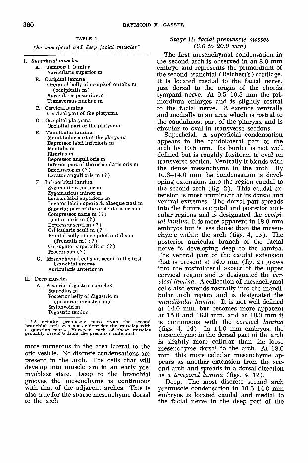
The mesenchyme of the second bran- chial arch in 4.2-6.5 mm embryos is most densely cellular in the middle and ventral regions of the arch. Midway in the arch it extends medially to join with the mesenchyme of the arch on the op- posite side. When traced dorsally from the middle area it becomes less and less cellular (fig. 1) . At the termination of the facial nerve, the mesenchyme sur- rounds the nerve and, at the level of the second arch epibranchial placode, it is located deep and caudal to the nerve. Dor- sal to the arch, the mesenchyme is very much scattered with the cells becoming less numerous. The cells become slightly ~
5 A table giving in outline fprm the state of histo- genesis for each of the ten specimens is in the author’s dissertation. See footnote 4, page 358.
360 RAYMOND F. GASSER
TABLE 1
The superficial and deep facial muscles' Stage II: facial prentuscle masses
(8.0 to 20.0 mm) The first mesenchymal condensation in
the second arch is observed in an 8.0 mm 1. Superficial muscles A.
B.
C.
D.
E.
F.
G.
Temporal lamina Auricularis superior m
Occipital lamina Occipital belly of occipitofrontalis m
Auricularis posterior m Transversus nuchae m
Cervical lamina Cervical part of the platysma
Occipital platysma Occipital part of the platysma
Mandibular lamina Mandibular part of the platysma Depressor labii inferioris m Mentalis m Risonus m Depressor anguli oris m Inferior part of the orbicularis oris m Buccinator m ( ? ) Levator anguli oris m ( ? )
Infraorbital lamina Zygomaticus major m Zygomaticus minor m Levator labii superioris m Levator labii superioris alaeque nasi m Superior part of the orbicularis oris m Compressor naris m ( ? ) Dilator naris m ( ? ) Depressor septi m ( ? ) Orbicularis oculi m ( ? ) Frontal belly of occipitofrontalis m
(frontalism) (??. Corrugator supercilii m ( ? ) Procerus m ( ? )
Mesenchymal cells adjacent to the first branchial groove
Auricularis anterior m
(occipitalis m )
11. Deep muscles A. Posterior digastric complex
Stapedius m Posterior belly of digastric m
(posterior digastric m ) Stylohyoid m Digastric tendon
1 A definite premuscle mass from the second branchial arch was not evident for the muscles with a question mark. However, each o f these muscles probably develops from the precursor mdicated.
more numerous in the area lateral to the otic vesicle. No discrete condensations are present in the arch. The cells that will develop into muscle are in an early pre- myoblast state. Deep to the branchial grooves the mesenchyme is continuous with that of the adjacent arches. This is also true for the sparse mesenchyme dorsal to the arch.
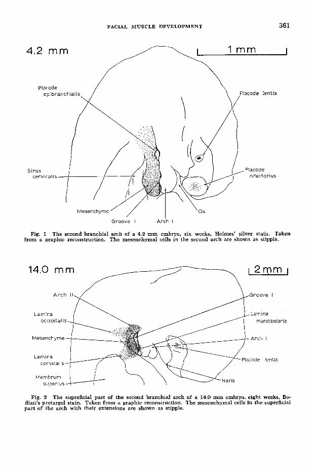
embryo and represents the primordium of the second branchial (Reichert's) cartilage. It is located medial to the facial nerve, just dorsal to the origin of the chorda tympani nerve. At 9.5-10.5 mm the pri- mordium enlarges and is slightly rostral to the facial nerve. It extends ventrally and medially to an area which is rostral to the caudalmost part of the pharynx and is circular to oval in transverse sections.
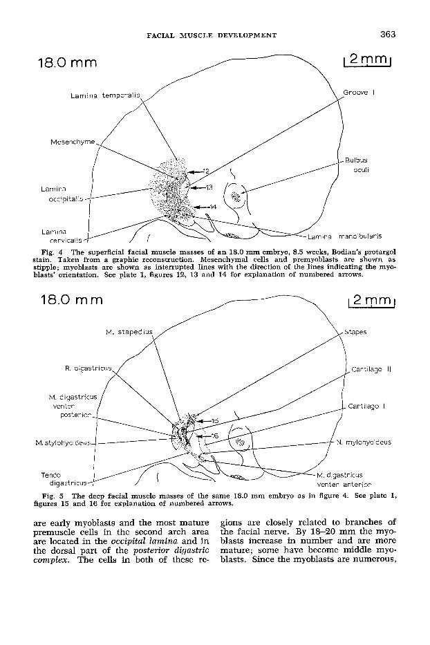
Superficial. A superficial condensation appears in the caudolateral part of the arch by 10.5 mm. Its border is not well defined but is roughly fusiform to oval on transverse section. Ventrally it blends with the dense mesenchyme in the arch. By 10.6-14.0 mm the condensation is devel- oping extensions into the region caudal to the second arch (fig. 2). This caudal ex- tension is most prominent at its dorsal and ventral extremes. The dorsal part spreads into the future occipital and posterior auri- cular regions and is designated the occipi- tal lamina. It is more apparent in 18.0 mm embryos but is less dense than the mesen- chyme within the arch (figs. 4, 13). The posterior auricular branch of the facial nerve is developing deep to the lamina. The ventral part of the caudal extension that is present at 14.0 mm (fig. 2) grows into the rostrolateral aspect of the upper cervical region and is designated the cer- vical lamina. A collection of mesenchymal cells also extends rostrally into the mandi- bular arch region and is designated the mandibular lamina. It is not well defined at 14.0 mm, but becomes more apparent at 15.0 and 16.0 mm, and at 18.0 mm it is continuous with the cervical lamina (figs. 4, 14). In 14.0 mm embryos, the mesenchyme in the dorsal part of the arch is slightly more cellular than the loose mesenchyme dorsal to the arch. At 18.0 mm, this more cellular mesenchyme ap- pears as another extension from the sec- ond arch and spreads in a dorsal direction as a temporal lamina (figs. 4, 12).
Deep. The most discrete second arch premuscle condensation in 10.5-14.0 mm embryos is located caudal and medial to the facial nerve in the deep part of the
4.2 mm
FACIAL MUSCLE DEVELOPMENT 361
1 mm I
Groove I A r i h I
Fig. 1 The second branchial arch of a 4.2 mm embryo, six weeks, Holmes’ silver stain. Taken from a graphic reconstruction. The mesenchymal cells in the second arch are shown as stipple.
12mm I 7 14.0 mm
love I
Fig. 2 The superficial part of the second branchial arch of a 14.0 mm embryo, eight weeks, Bo- dian’s protargol stain. Taken from a graphic reconstruction. The mesenchymal cells in the superficial part of the arch with their extensions are shown as stipple.

362 RAYMOND F. GASSER
arch and is designated the posterior di- gastric complex (fig. 3). At 14 mm it is continuous laterally with the deep aspect of the cervical lamina. The complex in- creases in size as it courses ventrally and then becomes slightly constricted as it pro- ceeds into the mandibular arch where it is continuous with another densely cellular concentration, the mytohyoid complex. The posterior digastric complex receives a series of small, diffuse branches from the caudal aspect of the facial nerve. These branches can be traced ventrally within the complex to the region of the first branchial groove. They could not be traced beyond the constricted part of the condensation in any of the embryos ex- amined. The most dorsal part of the mylohyoid complex receives a small, single branch from the mandibular division of the trigeminal nerve. This continuous group of condensations represents the first appearance of the digastric, stylohyoid and mylohyoid muscles. By 18 mm the dorsal part is dividing into the posterior belly of the digastric (posterior digastric) and stylohyoid muscles (figs. 5, 16). The ventral part is dividing into the anterior belly of the digastric (anterior digastric) and mylohyoid muscles. The intermediate region becom.es more constricted and is developing into the diagastric tendon. The
stylohyoid muscle develops from the rostra1 part of the posterior digastric com- plex immediately caudal to the second arch cartilage.
The dorsal extent of the posterior di- gastric complex is continuous on the me- dial side of the facial nerve with the dor- sal part of the second branchial cartilage. In this region will develop the stapedius muscle and the stapes of the middle ear. At 18.0 mm the stapes is forming in an area adjacent to the dorsal end of the second arch cartilage (fig. 5). The stapes at this time is a ring-like condensation with the stapedial artery coursing through it. A small collection of late premyoblasts is located caudal to the dorsal tip of the second arch cartilage in 14.0 mm em- bryos (fig. 3) and is the first appearance of the stapedius muscle. These premyo- blasts are adjacent to the medial side of the facial nerve and course rostrally to the vicinity of the second arch cartilage. At 18-20 mm the stapedius muscle is better defined (figs. 5, 15) and is composed mostly of early myoblasts which join the cells connecting the stapes and the sec- ond arch cartilage.
Histogenesis. Most of the premuscle cells in 14-16 mm embryos are late pre- myoblasts. None of the cells are mature myoblasts. By 18 mm many of the cells
Fig. 3 The deep part o f the second brancial arch of the same 14.0 mm embryo as in figure 2. All mesenchymal condensations in the deep part of the second arch and one of the condensations in the first arch are shown as stipple.
FACIAL MUSCLE DEVELOPMENT 363
c- 18.0 m m
Fig. 4 The superficial facial muscle masses of a n 18.0 mm embryo, 8.5 weeks, Bodian's protargol stain. Taken from a graphic reconstruction. Mesenchymal cells and premyoblasts are shown as stipple; myoblasts are shown as interrupted lines with the direction of the lines indicating the myo- blasts' orientation. See plate 1, figures 12, 13 and 14 for explanation of numbered arrows.
18.0 m m
M. stylohyoideus \.'..,. h:"' ;,
' N mylohyoideus I
O M . %-, digastricus -, venter anterior
Fig. 5 The deep facial muscle masses of the same 18.0 mm embryo as in figure 4. See plate 1, figures 15 and 16 for explanation of numbered arrows.
are early myoblasts and the most mature gions are closely related to branches of premuscle cells in the second arch area the facial nerve. By 18-20 mm the myo- are located in the occipital lamina and in blasts increase in number and are more the dorsal part of the posterior cligastric mature; some have become middle myo- complex. The cells in both of these re- blasts. Since the myoblasts are numerous,
364 RAYMOND F. GASSER
the laminae and deeper muscle masses are easier to outline, trace and identify.
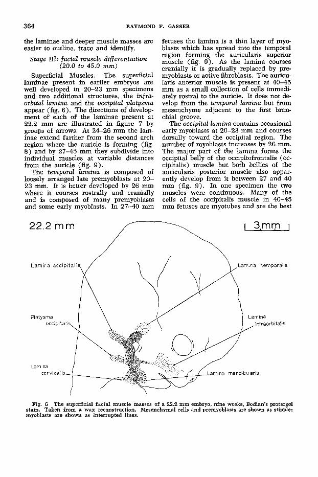
Stage 111: facial muscle differentiation (20.0 to 45.0 mm)
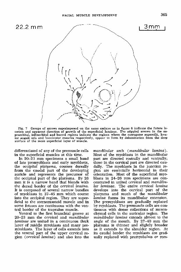
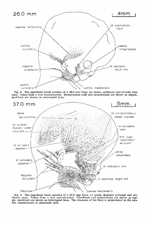
Superficial Muscles. The superficial laminae present in earlier embryos are well developed in 20-23 mm specimens and two additional structures, the infra- orbital lamina and the occipital platysma appear (fig. 6). The directions of develop- ment of each of the laminae present at 22.2 mm are illustrated in figure 7 by groups of arrows. At 24-26 mm the lam- inae extend farther from the second arch region where the auricle is forming (fig. 8) and by 27-45 mm they subdivide into individual muscles at variable distances from the auricle (fig. 9).
The temporal lamina is composed of loosely arranged late premyoblasts at 20- 23 mm. It is better developed by 26 mm where it courses rostrally and cranially and is composed of many premyoblasts and some early myoblasts. In 27-40 mm
22.2 mm
fetuses the lamina is a thin layer of myo- blasts which has spread into the temporal region forming the auricularis superior muscle (fig. 9). As the lamina courses cranially it is gradually replaced by pre- myoblasts or active fibroblasts. The auricu- laris anterior muscle is present at 40-45 mm as a small collection of cells immedi- ately rostra1 to the auricle. It does not de- velop from the temporal lamina but from mesenchyme adjacent to the fmt bran- chial groove.
The occipital lamina contains occasional early myoblasts at 20-23 mm and courses dorsally toward the occipital region. The number of myoblasts increases by 26 mm. The major part of the lamina forms the occipital belly of the occipitofrontalis (oc- cipitalis) muscle but both bellies of the auricularis posterior muscle also appar- ently develop from it between 27 and 40 mm (fig. 9). In one specimen the two muscles were continuous. Many of the cells of the occipitalis muscle in 40-45 mm fetuses are myotubes and are the best
Fig. 6 The superficial facial muscle masses of a 22.2 mm embryo, nine weeks, Bodian’s protargol stain. Taken from a wax reconstruction. Mesenchymal cells and premyoblasts are shown as stipple; myoblasts are shown as interrupted lines.
FACIAL MUSCLE DEVELOPMENT 365
22.2 mm
Fig. 7 Groups of arrows superimposed on the same embryo as in figure 6 indicate the future 10- cation and apparent direction of growth of the superficial laminae. The stippled arrows in the su- praorbital, infraorbital and buccal regions indicate the regions where the conugator supercilii, leva- tor anguli oris and buccinator muscles respectively, appear to form by delamination from the deep surface of the more superficial layer of muscle.
differentiated of any of the premuscle cells in the superficial muscles at this time.
In 20-23 mm specimens a small band of late premyoblasts and early myoblasts, the occipital platysma, courses dorsally from the caudal part of the developing auricle and represents the precursor of the occipital part of the platysma. By 26 mm it is a narrow band that blends with the dorsal border of the cervical lamina. It is composed of several narrow bundles of myoblasts in 37-45 mm which course into the occipital region. They are super- ficial to the sternomastoid muscle and in some fetuses are continuous with the ven- tral border of the trapezius muscle.
Ventral to the first branchial groove at 20-23 mm the cervical and mandibular laminae are united in a continuous thick layer of middle myoblasts and many pre- myoblasts. The layer of cells extends into the ventral part of the upper cervical re- gion (celvical lamina) and also into the
mandibular arch (mandibular lamina) . Most of the myoblasts in the mandibular part are directed rostrally and ventrally; those in the cervical part are directed cau- dally. The myoblasts in the junction re- gion are essentially horizontal in their orientation. Most of the superficial myo- blasts in 24-26 mm specimens are con- centrated in united cervical and mandibu- lar laminae. The entire cervical lamina develops into the cervical part of the platysma and much of the mandibular lamina forms its mandibular extension. The premyoblasts are gradually replaced by myoblasts. The premuscle cells are con- tinuous with dense collections of mesen- chymal cells in the auricular region. The mandibular lamina extends almost to the angle of the mouth. By 27-40 mm the platysma is thinner and slightly broader as it extends to the shoulder region. At its caudal border the myoblasts are grad- ually replaced with premyobalsts or mes-
26.0
Lamina M. zygornaticus
temporal is \ major
I nfraorbitalis
Lamina mandibuiaris
Fig. 8 The superficial facial muscles of a 26.0 mm fetus, ten weeks, erythrosin and toluidin blue stain. Taken from a wax reconstruction. Mesenchymal cells and prernyoblasts are shown as stipple; myoblasts are shown as interrupted lines.
37.0 mm I ,5,mm, I
, occipitofrontalis venter frontalis
M. orbicularis
infraorbitalis
, orbicularis oris
, depressor anguli oris
Lamina mandibularis
Fig. 9 The superficial facial muscles of a 37.0 mm fetus, 11 weeks, Bodian’s protargol and ery- throsin stain. Taken from a wax reconstruction. Fibroblasts and premyoblasts are shown as stip- ple; myoblasts are shown as interrupted lines. The closeness of the lines is proportional to the rela- tive concentration of premuscle cells.
FACIAL MUSCLE DEVELOPMENT 367
enchymal cells which can be traced to the level of the developing clavicle. At 40-45 mm the platysma extends to the midline in the mental and submental regions, but not in the cervical region.
A triangular group of myoblasts is lo- cated at the rostral termination of the mandibular lamina at 26 mm which ex- tends to the angle of the mouth. It is on a plane superficial to the mandibular lam- ina and represents the first appearance of the depressor anguli oris muscle. The di- rection of the myoblasts differs from those in the mandibular lamina by 90 O . The myo- blasts are more closely packed as they approach the angle of the mouth. The muscle is well defmed between 27 and 45 mm and is superficial to the platysma.
The orbicularis oris muscle has begun to develop by 37 mm where it is a loose collection of middle myoblasts which is thicker in the upper than in the lower lip. The myoblasts are sparse in the mid- line. By 40-45 mm the muscle is made up of late myoblasts and completely en- circles the mouth.
In 20-23 mm specimens the mandibular lamina is continuous with a loose collec- tion of mesenchymal cells in the infraor- bital region, the infraorbital lamina. The region of continuity is narrow and is limited to the area immediately rostral to the ventral part of the first branchial groove. Since the infraorbital lamina is highly vascular, it is difficult to determine if its component cells are forming facial muscles or blood vessels. Considerable changes take place by 24-26 mm. In the most lateral aspect of the infraorbital re- gion, a discrete band of myoblasts has formed. It is round to oval in transverse section and represents the first appearance of the zygomaticus major muscle. The muscle courses from the region of the de- veloping zygoma to the angle of the mouth where it is continuous with the depressor anguli oris muscle. Medial to the zygo- maticus major muscle, the infraorbital lamina is composed of premyoblasts and myoblasts which are superficial to the in- fraorbital plexus of nerves. The lamina ex- tends laterally and dorsally into the area immediately above the zygomaticus major muscle. In 27-45 mm fetuses, the zygo- maticus major muscle is a very prominent
band that is continuous along its medial border with the zygomaticus and orbicu- laris oculi muscles. The zygomaticus minor muscle is continuous medially with a sheet of myobalsts that courses into the upper lip and lateral aspect of the nose. No definite levator labial or nasal muscles are identifiable.
At 37 mm, the orbicularis oculi muscle is developing around the eye, but does not yet completely encircle it. The muscle is thickest laterally and thins as it spreads dorsally. The infraorbital portion is partly superficial to the zygomaticus muscles and infraorbital lamina. It courses medially and disappears at the side of the nose. Mesenchymal cells, probably premyoblasts, are arranged in a layer in the superior palpebrum and in the medial part of the orbital region. Only the most medial por- tion of the orbital part of the orbicularis oculi muscle is deficient in 40-45 mm fetuses. Myoblasts are present in most of the orbital part, but the palpebral part re- mains in a premyoblastic state.
The frontal belly of the occipitofrontalis (frontalis) muscle begins to develop in 27-45 mm fetuses, but is not well defmed. Its premuscle cells are mainly premyo- blasts which show little orientation. The majority of the cells cranial to the frontal, temporal and occipital regions are premyo- blasts or active fibroblasts which form a thin layer superficial to the developing calvarium. In 40-45 mm specimens, this layer extends to the vertex of the head, but thins in the midline in the fronto- parietal and occipitoparietal regions. As development proceeds, the cells become oriented mainly in a rostrodorsal direction and form the galea aponeurotica. Myo- blasts connecting the frontalis and oc- cipitalis muscles were not observed in any of the specimens.
The buccinator muscle first appears at 26 mm, but is difficult to outline since it is primarily composed of premyoblasts and early myoblasts. Its dorsal boundary is especially difficult to define since it grad- ually blends with the mesenchymal cells lateral to the pharynx. The buccinator mass becomes superficial as it approaches the angle of the mouth where it joins the deep aspect of the more superficial muscle masses in the region. A nerve supply to

368 RAYMOND F. GASSER
the muscle was not observed until 37 mm. At this time, the muscle is more apparent and is divided in its dorsal half into a large caudal and a small cranial portion with the parotid duct coursing between them. The caudal portion extends farther dorsally, and between 37 and 45 mm, it blends with the superior constrictor of the pharynx. At this age, the muscle is com- pletely pierced by the buccal branch of the mandibular nerve.
By 37 mm, the levator anguli oris muscle has formed deep to the caudal and lateral part of the infraorbital plexus. It begins cranially adjacent to the developing maxil- lary bone and courses caudally and lat- erally to join with the zygomaticus and buccinator muscles at the angle of the mouth. Caudal to the angle of the mouth, the muscle is sometimes continuous with the mandibular part of the platysma and the depressor anguli oris muscle.
All of the deep facial muscles are recognizable in the previous stage but in the present stage they become more distinct and increase in size. Be- tween 20 and 26 mm, the cranial end of the posterior digastric muscle is immedi- ately caudal to the dorsal end of the stapedius muscle. As development pro- ceeds, this distance becomes greater. The stapedius muscle remains close to the me- dial aspect of the facial nerve, sometimes receiving a short, but obvious, branch from the nerve. It is made up of early and middle myoblasts which terminate medial to the hyostapedial connection be- tween the stapes and cranial end of the second arch cartilage.
The stylohyoid muscle is rostral to the caudal half of the posterior digastric mus- cle and dorsal to the second arch cartilage. As it proceeds caudally, it is sometimes lateral to the posterior digastric muscle and sometimes surrounds it before ter- minating in mesenchyme of the hyoid re- gion. The digastric tendon is a dense col- lection of darkly staining cells that courses into the depths of both the posterior and anterior parts of the digastric muscle.
Stage IV: definitive location of the facial muscles (50 to 146 mm)
Superficial Muscles. Additional super- ficial muscles can be identified between
Deep muscles.
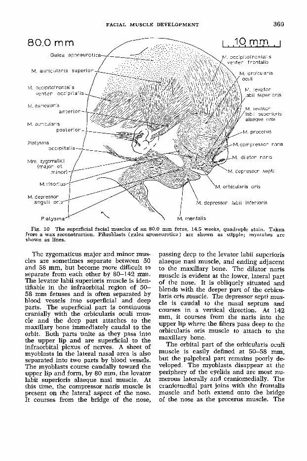
50 and 58 mm and all of these muscles can be recognized and outlined at 80 mm in their definitive position (fig. 10).
At 58 mm, the auricularis anterior mus- cle is a short band of myoblasts coursing cranially from the rostral aspect of the auricle, and at 80 mm it is adjacent to the rostral border of the auricularis superior muscle. NO separation could be seen be- tween these two muscles at 142 mm. Dor- sal to the auricularis posterior muscle a thin and small sheet of muscle is occa- sionally present in the lower occipital or upper cervical region. Its muscle cells are mostly directed transversely and compose the transversus nuchae muscle.
Between 58 and 80 mm, the occipital platysma is a single, discrete band which takes a variable path from the platysma into the occipital region. This muscle could not be identified in the grossly dissected, 142 mm fetus. At 58 mm, the mandibu- lar part of the platysma extends into the mental region where it is continuous with vertically directed muscle cells that ex- tend into the lower lip and represent the depressor labii inferioris muscle. This ar- rangement is observed better at 80 mm where the mentalis muscle is defined for the first time. The risorious muscle de- velops as a thickening along the cranial border of the platysma. Its state of de- velopment is variable.
At 58 mm, the buccinator muscle ex- tends farther dorsally, and by 80 mm it extends to the region of the lateral ptery- goid plate (fig. 11). Between 101 and 146 mm, it unites dorsally with the superior pharyngeal constrictor muscle. A decus- sation of its muscle cells at the angle of the mouth is apparent at 80 mm. By 142 mm, a large fat pad has developed super- ficial to the buccinator but deep to the risorius and platysma muscles. The leva- tor anguli oris muscle grows considerably in thickness and length deep to the in- fraorbital plexus of nerves.
In 58-80 mm fetuses, the orbicularis oris muscle is well developed, completely encircles the mouth, but is difficult to divide into superficial and deep parts. It has become thicker and is continuous with the perioral muscles. Its medial part in the upper lip lags in development.
FACIAL MUSCLE DEVELOPMENT 369
80.0 mm ccipitof rontal is nter frontalis
M. aur icular is super ior
M. occipitof rontalis venter occipi ta l i
M. auricularis
M. auricularis
M. compressor naris
. dilator naris
M. orbicularis oris
. depressor labii inferioris
Fig. 10 The superficial facial muscles of an 80.0 mm fetus, 14.5 weeks, quadruple stain. Taken from a wax reconstruction. Fibroblasts (galea aponeurotica) are shown as stipple; myotubes are shown as lines.
The zygomaticus major and minor mus- cles are sometimes separate between 50 and 58 mm, but become more difficult to separate from each other by 80-142 mm. The levator labii superioris muscle is iden- tifiable in the infraorbital region of 50- 58 mm fetuses and is often separated by blood vessels into superficial and deep parts. The superficial part is continuous cranially with the orbicularis oculi mus- cle and the deep part attaches to the maxillary bone immediately caudal to the orbit. Both parts unite as they pass into the upper lip and are superficial to the infraorbital plexus of nerves. A sheet of myoblasts in the lateral nasal area is also separated into two parts by blood vessels. The myoblasts course caudally toward the upper lip and form, by 80 mm, the levator labii superioris alaeque nasi muscle. At this time, the compressor naris muscle is present on the lateral aspect of the nose. It courses from the bridge of the nose,
passing deep to the levator labii superioris alaeque nasi muscle, and ending adjacent to the maxillary bone. The dilator naris muscle is evident at the lower, lateral part of the nose. It is obliquely situated and blends with the deeper part of the orbicu- laris oris muscle. The depressor septi mus- cle is caudal to the nasal septum and courses in a vertical direction. At 142 mm, it courses from the naris into the upper lip where the fibers pass deep to the orbicularis oris muscle to attach to the maxillary bone.
The orbital part of the orbicularis oculi muscle is easily defined at 50-58 mm, but the palpebral part remains poorly de- veloped. The myoblasts disappear at the periphery of the eyelids and are most nu- merous laterally and craniomedially. The craniomedial part joins with the frontalis muscle and both extend onto the bridge of the nose as the procerus muscle. The
370 RAYMOND F. GASSER
80.0 mm
M. buccinator
M. digastricus venter poster ior
M. orbicularis
R. stylohyoideus
Processus styloideus M. stylohyoideus
digastricus venter anterior
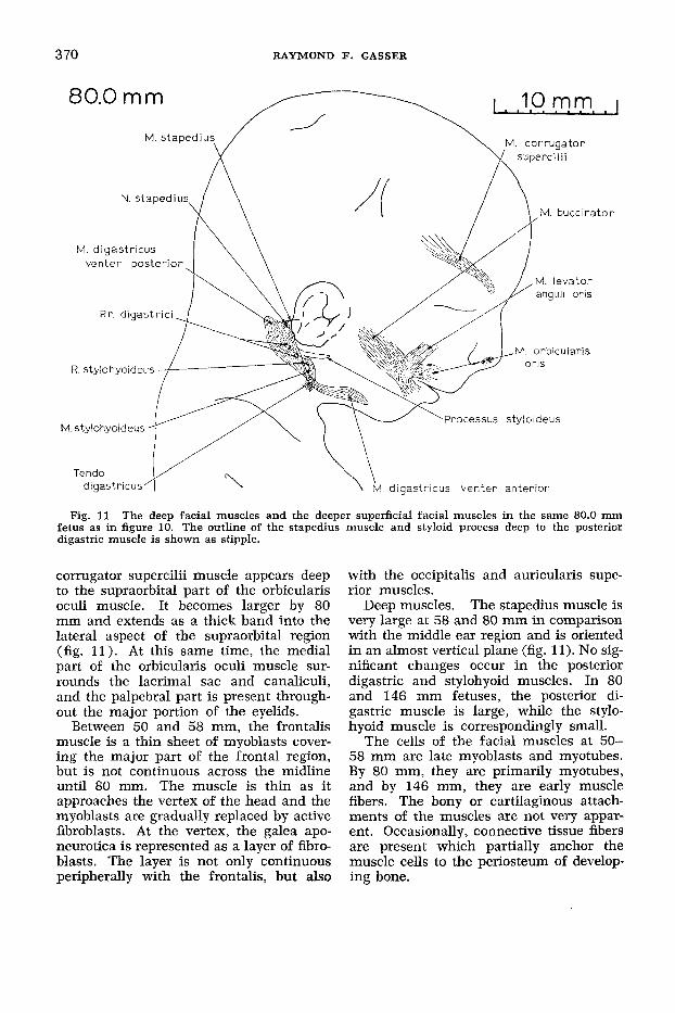
Fig. 11 The deep facial muscles and the deeper superficial facial muscles in the same 80.0 mni fetus as in figure 10. The outline of the stapedius muscle and styloid process deep to the posterior digastric muscle is shown as stipple.
corrugator supercilii muscle appears deep to the supraorbital part of the orbicularis oculi muscle. It becomes larger by 80 mm and extends as a thick band into the lateral aspect of the supraorbital region (fig. 11) . At this same time, the medial part of the orbicularis oculi muscle sur- rounds the lacrimal sac and canaliculi, and the palpebral part is present through- out the major portion of the eyelids.
Between 50 and 58 mm, the frontalis muscle is a thin sheet of myoblasts cover- ing the major part of the frontal region, but is not continuous across the midline until 80 mm. The muscle is thin as it approaches the vertex of the head and the myoblasts are gradually replaced by active fibroblasts. At the vertex, the galea apo- neurotica is represented as a layer of fibro- blasts. The layer is not only continuous peripherally with the frontalis, but also
with the occipitalis and auricularis supe- rior muscles.
The stapedius muscle is very large at 58 and 80 mm in comparison with the middle ear region and is oriented in an almost vertical plane (fig. 11). No sig- nificant changes occur in the posterior digastric and stylohyoid muscles. In 80 and 146 mm fetuses, the posterior di- gastric muscle is large, while the stylo- hyoid muscle is correspondingly small.
The cells of the facial muscles at 50- 58 mm are late myoblasts and myotubes. By 80 mm, they are primarily myotubes, and by 146 mm, they are early muscle fibers. The bony or cartilaginous attach- ments of the muscles are not very appar- ent. Occasionally, connective tissue fibers are present which partially anchor the muscle cells to the periosteum of develop- ing bone.
Deep muscles.
FACIAL MUSCLE
Stage V: facial muscles in dissected late fetuses (21 0, 270 and 360 mm)
The muscles increase in size and ex- tent as the head and neck regions become larger and, in most cases, the boundaries between individual muscles become more prominent. Since the muscles reach their definitive location as early as 58 mm, only gradual changes occur between 146 mm and term.
Superficial muscles. The transversus nuchae muscle is present in the 210 and 270 mm fetuses and is almost continuous with the auricularis posterior muscle. The latter muscle is composed of two bellies in all three specimens. The occipital platysma muscle could not be identified. In the term infant, the platysma extends cau- dally to the level of the nipple. A distinct, thickened risorius muscle is not always present. The boundary between the zygo- maticus muscles is difficult to define, but there is usually a small gap between the zygomaticus minor and levator labii supe- rioris muscles. All of the superficial mus- cles are delicate and most are deep to a layer of subcutaneous fat, but superficial to their nerve supply. Most of the bony at- tachments are established, but are not firmly anchored.
Deep muscles. The stylohyoid muscle is smaller and its fibers are less tightly bound together than those of the digastric muscle. All of the deep muscles are at- tached to bone or cartilage and their at- tachments are generally more firmly an- chored than are the superficial muscles.
DISCUSSION
1. Muscles of expression Futamura ('06) stated that by 31 to
34 days (9-13 mm) the superficial mus- cle blastema becomes voluminous and spreads out dorsally, ventrally and orally. These early, superficial extensions have been designated the occipital (dorsally), cervicaZ (ventrally) and mandibular (or- ally) laminae. As the neck develops and increases in length, the cervical lamina expands to produce the platysma. This manner of development is similar to that described by Rabl (1887) and Futamura ('06). According to Bryce ('23) bundles of fibers sometimes are found in the defini-
DEVELOPMENT 371
tive state extending from the dorsal border of the platysma to the cervical fascia over the sternomastoid and trapezius muscles, and even to the mastoid process. This in- consistent strip of muscle, the occipital platysma, appears very early in develop- ment (22.2 mm), but diminishes in size as development progresses and could not be found in late fetuses. Huber ('31) stated that it is a vestige of the nuchal portion of the platysma which reflects our primate ancestry.
At 6 weeks (18-24 mm), Futamura ('06) described two layers of premuscle tissue; a deep layer that he identified with the sphincter colli of lower vertebrates and a superficial layer which he looked upon as the platysma (colli) of lower forms. In the present study, no attempt is made to describe the ontogeny of the facial mus- cles on the basis of their phylogeny. The muscles are divided into groups according to their manner of development and defmi- tive location. Futamura ('06) described the deep layer as giving origin to primitive sphincter arrangements around the eye, nose, mouth and ear, and except in the case of the mouth, these early sphincters disappear and are replaced by new forma- tions from the superficial layer. The pres- ent study did not reveal sphincters at 18- 24 mm in any of these regions (figs. 4, 6). Sphincters completely encircling the nose and auricle were never observed and the orbicularis oris and oculi muscles do not become complete sphincters until ap- proximately 40-45 mm.
Popowsky (1895) found no superficial muscles at 28 mm except the cervical por- tion of the platysma (cervical lamina). Huber ('31) demonstrated the presence of an additional muscle (auricularis poste- rior) which is identified in the present work as the occipital lamina. The present technique revealed additional regions of muscle development, the temporal and in fraorbital laminae and the occipital platysma, as early as 18-22 mm (figs. 4,6).
Futamura ('06) observed the anlage of the quadratus labii superioris muscle at 6 weeks (18-24 mm) which is similar to
GFutamura ('06) gave an estimated age in days or weeks for his specimens. The millimeter measure- ments in parentheses are the approximate crown- rump lengths of his specimens calculated from Mall's ('10) graph.
372 RAYMOND F. GASSER
what is identified here as the infraorbital lamina. A small connection exists between the infraorbital lamina and the second arch region at 22 mm (fig. 6) , which is less evident at 26 mm (fig. 8) when the lamina is more prominent. The origin of the premuscle cells in the lamina and ex- actly which muscles on the face develop from the lamina is questionable (table 1). The cells of the infraorbital Zamina could be descendants of cells in the second bran- chial arch, their migration taking place at an earlier time when they could not be recognized as premuscle cells and did not form a discrete condensation. The infra- orbital lamina gives origin to all of the muscles in the infraorbital region and prob- ably contributes to the formation of the muscles of the nose, most of the orbicu- laris oculi and some of the orbicularis oris muscles.
By 8 to 9 weeks (32-36 mm), Futa- mura ('06) reported the facial muscles to be well differentiated for the most part. Huber ('31) pointed out that the critical period of development is between the sec- ond and third months of prenatal life. There is a sudden surge in facial muscle differentiation between 26 and 37 mm, but deficiencies are present in the palpe- bra1 part of the orbicularis oculi and in all of the muscles close to the middle of the face. The present findings agree with Zuckermann-Zicha's ('25) report that the palpebral part of the orbicularis oculi mus- cle differentiates between 55 and 80 mm.
The origin of the premuscle cells of the buccinator muscle is questionable. They probably migrated from the deep aspect of the mandibular lamina at an earlier age. The cells of the levator anguli oris mus- cle most likely have the same origin as those in the buccinator muscle.
2. Occipitofrontalis and auricularis superior muscles
Although both bellies of the occipito- frontalis muscles are described definitively as components of a single muscle, they develop separately. At 37 mm (fig. 9), the occipital belly and the auricularis posterior muscle have replaced the oc- cipital lamina. Bryce ('23) mentioned that these two muscles are sometimes united in the definitive condition. The frontal belly
differentiates later in the frontal region and its origin is difficult to establish. The corrugator supercilii muscle probably orig- inates from its deep surface. The auricu- laris superior muscle develops from the temporal lamina. The muscles of the scalp gradually thin as they approach the vertex of the head and become continuous with a layer of fibroblasts from which the galea aponeurotica develops. According to Bryce ('23), some authors regard the aponeurosis as representing the degen- erated portion of a primitive fronto-oc- cipital sheet. A continuous layer of myo- blasts connecting the frontal and occipital areas was not observed in any of the specimens. It is difficult to consider the fibroblasts as dedserentiated myoblasts; they probably differentiated directly from mesenchymal cells.
3. Deep muscles A concept of the posterior digastric com-
plex from which the posterior digastric, stylohyoid and stapedius muscles develop was presented by Rabl in 1887. Futamura ('06) reported seeing twigs from the facial nerve coursing to the complex at 35-36 days (13-15 mm). At this early age, some of the fibers course into the most ventral and slightly constricted part of the com- plex to the caudal aspect of the mandibu- lar arch. No fibers continue into the mylo- hyoid complex since it already receives an innervation from a poorly developed in- ferior dental nerve. These findings indi- cate a dual origin for the digastric muscle, the posterior belly from the posterior di- gastric complex of the second arch and the anterior belly from the mylohyoid complex of the first arch. However, each complex is always continuous with one another deep to the first branchial groove. Gegen- baur (1890) and Futamura ('06) believed that the digastric muscle develops from two independent parts. Some investigators consider the muscle to develop as a sepa- ration from the sternomastoid muscle. A union with this latter muscle was not ob- served in any of the specimens. Consider- ation of past and present data suggests that the posterior digastric muscle as well as the digastric tendon develop from the second arch, and the anterior digastric

FACIAL MUSCLE DEVELOPMENT 373
muscle develops along with the mylohyoid muscle in the ventral part of the first arch.
Ruge ('10) and Bryce ('23) pointed out the rare occurrence of a definitive muscu- lar connection between the occipital fascia and the posterior digastric muscle. This connection probably represents a retention of an earlier connection (14 mm) between the posterior digastric complex and the cervical lamina.
The time and manner of development of the stapedius muscle agrees with Futa- mura's ('06) and Schimert's ('33) descrip- tions, but disagrees with Broman (1898) who believed the muscle develops from the connection between the stapes and the second arch (Reichert's) cartilage. No ex- planation can be given for the variability in the size and orientation of this muscle during development.
4. Histogenesis and pattern of differentiation
In his studies on the labial muscles, Futamura ('06) saw no transverse stria- tions at 22 weeks (170 mm), faint stria- tions at 26 weeks (220 mm) and distinct striations at 30 weeks (250 mm). The present work reveals transverse striations in myotubes (e.g., orbicularis oris muscle) at 58 mm which become very apparent at 80 mm. Early muscle fibers are present at 146 mm.
Contrary to the statements of Popowsky (1895) the superficial musculature at the end of the third month (58 mm) is al- ready differentiated. This is substantiated by Huber ('31 ) who produced contractions of the muscles in a living 55 mrn fetus through electrical stimulation. In their studies on the appearance of human fetal reflexes, Hooker ('52, 58) and Humphrey ('64, '65) observed, at 37-47 mm, a squint type reflex produced by contraction of the orbicularis oculi muscle. The orbital part of the muscle almost completely surrounds the eye at 41 mm and is composed of late myoblasts. At 47-49 mm, these investiga- tors produced a scowl type reflex by con- traction of the corrugator supercilii muscle. This muscle is well differentiated by 58 mm when it is composed of Iate myoblasts and occasional myotubes. By 60-64 mm, Hooker ('52, '58) and Humphrey ('64, '65) produced momentary lip closure and
at 74-79 mm, lip closure was maintained. At 58 mm, the orbicularis oris muscle is well developed, completely encircles the mouth and is composed mostly of late myoblasts with some myotubes. They ob- served a sneer type reflex produced by an elevation of the angle of the mouth and ala of the nose at '74 to 88 mm. All of the facial muscles, including the infraorbital ones, have differentiated by 80 mm and are made up of myotubes. Comparison of the degree of histogenetic development of the premuscle cells with the age at which reflexes can be elicited lends support to the view that late myoblasts, with un- striated fibrils in their cytoplasm, are not only able to contact, but that their con- tractions are influenced to some degree by nerve impulses.
A definite pattern of differentiation is evident in the formation of the facial mus- cles. The present investigation supports Futamura's ('06) findings that differentia- tion of the muscles begins earlier in the deep than in the superficial regions and earlier in the lower lateral than in the upper medial regions. In addition, stud- ies show that the muscles in the occipital region (e.g., occipitalis) differentiate and become striated earlier than the lateral facial muscles (e.g., zygomaticus and de- pressor anguli oris) which in turn differ- entiate before the muscles close to the midline of the face.
Superficial muscle masses spread from the region of the second arch in all direc- tions except one, that is directly rostrally (fig. 7). The most likely reason for this is the presence of the external auditory mea- tus which is replacing the first branchial groove. The deep meatus probably causes cells from the second arch to migrate around it so that cell groups must extend first cranially or caudally before they can course rostrally.
In most of the specimens studied, the development of the peripheral branches of the facial nerve follow pari passu the de- velopment of the facial muscle masses (Gasser, '67). A close relationship exists between the muscles and the nerve through- out development and each appears to influ- ence the histogenesis and morphogenesis of the other. As the muscle masses form, the nerve supply to them differentiates.

374 RAYMOND F. GASSER
The premuscle cells in the vicinity of the nerve fibers are further differentiated than those more distant from the nerve fibers.
ACKNOWLEDGMENTS
Appreciation is expressed to Dr. Try- phena Humphrey, Dr. E. C. Sensenig and to the late Dr. Davenport Hooker for mak- ing their collections of human embryonic material available during this investigation.
LITERATURE CITED
Born, G. 1883 Die Plattenmodellirmethode. Arch. f. Micr. Anat., 22: 584-589.
1888 Noch einmal die Plattenmodellir- method. Z. Wiss. Mikr., 5 : 433-455.
Boyd, J. D. 1960 Development of striated mus- cle. Structure and Function of Muscle. Vol. 1. Ed. by G. H. Bourne. Academic Press, New York. Chap. 111, 63-85.
Broman, I. 1898 Ueber Entwicklungsgeschichte der Gehorknochelchen beim Menschen. Anat. Hefte, 21: 509-661.
Bryce, T. H. 1923 Myology. Quain’s Elements of Anatomy. 11th ed. Vol. 4, part 2. Ed. by E. S. Schafer, J. Symington and T. H. Bryce. Longmans, Green Co., London 31-74.
Futamura, R. 1906 Uber die Entwicklung der Facialismuskulatur des Menschen. Anat. Hefte, 30: 433-516.
Gasser, R. F. 1967 The development of the facial nerve in man. Ann. Otol., in press.
Gegenbaur, C. 1890 Lehrbuch der Anatomie des Menschen. 4 Aufl., Band 2, Wilhelm Engelmann, Leipzig, 448-449.
12
13
14
15
16
Hooker, D. 1952 The Prenatal Origin of Be- havior. 18th Porter Lecture. University of Kansas Press, Lawrence, Chap. 11, 54-86.
Evidence of prenatal function of the central nervous system in man. dames Arthur Lecture on The evolution of the human brain, 1957. The American Museum of Natural
1958
History, New York. Huber. E. 1931 Evolution of Facial Muscula-
ture. and Facial Expression. The Johns Hopkins Press, Baltimore 1-184.
Humphrey, T. 1964 Some correlations between the appearance of human fetal reflexes and the development of the nervous system. Progr. Brain Res., 4: 93-135.
1965 Personal communication. Mall, F. P. 1910 Manual of Human Embryol-
ogy. Vol. 1. Ed. by F. Keibel and F. P. Mall. J. B. Lippincott Co., Philadelphia. Chap. VIII, 180-201.
1918 On the age of human embryos. Am. J. Anat., 23: 397-422.
Popowsky, I. 1895 Zur Entwicklungsgeschichte des N. facialis beim Menschen. Morph. Jahrb., 23: 329-374.
Rabl, K. 1887 iiber das Gebiet des Nervus facialis. Anat. Anz., 2: 219-227.
Ruge, G. 1910 Verbindungen des Platysnia mit der tiefen Musculature des Halses beim Men- schen. Morph. Jahrb., 41: 708-724.
Schimert, J. 1933 Zur Entwicklungsgeschichte des Musculus stapedius beim Menschen. Anat. Anz., 76: 317-332.
Streeter, G. L. 1920 Weight, sitting height, head size, foot length and menstrual age of the human embryo. Contr. Embryol. Carneg. Instn., 12: 143-170.
Zuckermann-Zicha, M. 1925 Sur le d6veloppe- ment de la musculature des paupi&res chez l’homme. Arch. de Biol., 35: 313-323.
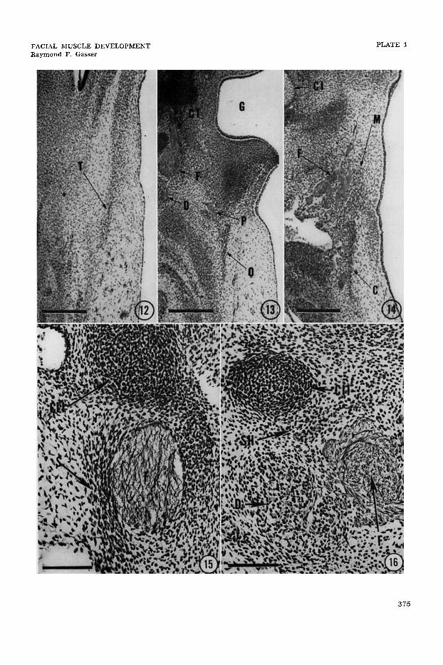
PLATE 1
EXPLANATION O F FIGURES
A section thrcugh the temporal lamina ( T ) of the same 18.0 mm as in figure 4, 8.5 weeks, Bodian’s protargol stain, 10 p. Scale z 300 p. Arrow 12 in figure 4 indicates level and plane of section.
A section through the occipital lamina (0) of the same embryo as in figure 4. Scale = 300 p. Arrow 13 in figure 4 indicates level and plane of section. CT, chorda tympani nerve; D, posterior digastric muscle; F, facial nerve; G. first branchial groove; P, posterior auricu- lar nerve.
A section through the cervical (C) and mandibular ( M ) laminae of the same embryo as in figure 4. Scale = 300 p. Arrow 14 in figure 4 indicates level and plane of section. CI, first branchial (Meckel’s) cartilage; F, terminal branches of facial nerve.
A section through the stapedius muscle ( S ) of the same embryo as in figure 5. Scale= 100 p. Arrow 15 in figure 5 indicates level and plane of section. CII, second branchial (Reichert’s) cartilage; F, facial nerve.
A section through the posterior digastric muscle (D) of the same em- bryo as in figure 5. Scale = 100 p. Arrow 16 in figure 5 indicates level and plane of section. CIII, second branchial (Reichert’s 1 car- tilage; F; facial nerve; SH, stylohyoid muscle.
FACIAL MUSCLE DEVELOPMENT Raymond F. Gasser
PLATE 1
3 75