Embed Size (px)
Citation preview


MUSCLES
• Approximately 40% of your body weight
• Approximately 650 muscles
• Muscles only pull (they can’t push)
• You have over 30 facial muscles
• Eye muscles move more than 100,000 times a day

OverviewOverview of Muscle Tissuesof Muscle Tissues
• Consists of muscle cells that are highly specialized for contraction
• Muscle is the dominant tissue in the heart and in the walls of other hollow organs of the body.
• Essential function of muscle is contraction, or shortening – a unique characteristic that sets it apart from any other body tissue
• Muscles are responsible for essentially all body movement and can be viewed as the “machines” of the body.

There are 3 types of muscle tissue
1. Skeletal
2. Cardiac
3. Smooth
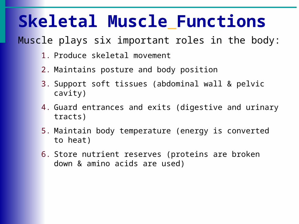
Skeletal Muscle FunctionsMuscle plays six important roles in the body:
1. Produce skeletal movement
2. Maintains posture and body position
3. Support soft tissues (abdominal wall & pelvic cavity)
4. Guard entrances and exits (digestive and urinary tracts)
5. Maintain body temperature (energy is converted to heat)
6. Store nutrient reserves (proteins are broken down & amino acids are used)

Organization of Muscle TissueThree layers of connective tissue are part of each muscle:
1. Epimysium – dense layer of collagen fibers that surround the entire muscle
2. Perimysium – divides the muscle into a series of compartments each containing a bundle of muscle fibers (fascicle); contains collagen & elastic fibers, blood vessels and nerves that maintain blood flow
3. Endomysium – flexible, elastic connective tissue layer; surrounds the individual skeletal muscle cells and interconnects adjacent muscle fibers

Figure 10.1
Arrangement of FasciclesArrangement of Fascicles
Parallel - fascicles run parallel to the long axis of the muscle*Most skeletal muscles are parallel muscles
Fusiform – spindle-shaped muscles

Arrangement of FasciclesArrangement of Fascicles
• Pennate – the fascicles form a common angle with the tendon (rectus femoris and deltoid)
• Convergent – fascicles converge on a common attachment site (pectoralis major)
Figure 10.1

Arrangement of FasciclesArrangement of Fascicles
Figure 10.1
Circular – fascicles are arranged in concentric rings around an opening (orbicularis oris)

Axial and Appendicular Muscles
• Axial muscles arise on the axial skeleton (60% of skeletal muscles) ; position the head and spinal column and move the rib cage
• Appendicular muscles stabilize and move the appendicular skeleton (40% of skeletal muscles)

Naming of Skeletal MusclesNaming of Skeletal Muscles
Slide 6.36aCopyright © 2003 Pearson Education, Inc. publishing as Benjamin Cummings
Direction of muscle fibers
Example: rectus (straight)
Relative size of the muscle
Example: maximus (largest)
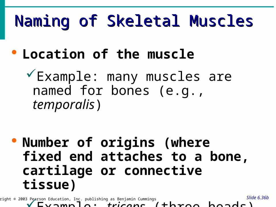
Naming of Skeletal MusclesNaming of Skeletal Muscles
Slide 6.36bCopyright © 2003 Pearson Education, Inc. publishing as Benjamin Cummings
Location of the muscle
Example: many muscles are named for bones (e.g., temporalis)
Number of origins (where fixed end attaches to a bone, cartilage or connective tissue)
Example: triceps (three heads)

Naming of Skeletal MusclesNaming of Skeletal Muscles
Slide 6.37Copyright © 2003 Pearson Education, Inc. publishing as Benjamin Cummings
Location of the muscles origin and insertion (movable end attaches to another structure)
Example: sterno (on the sternum)
Shape of the muscle
Example: deltoid (triangular)
Action of the muscle
Example: flexor and extensor
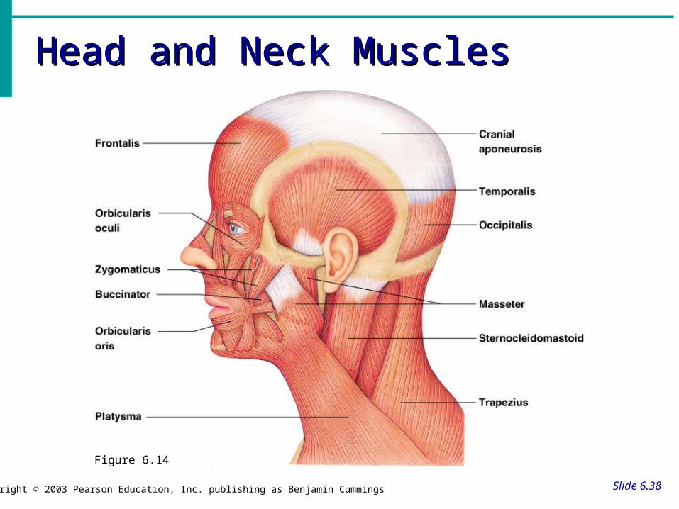
Head and Neck MusclesHead and Neck Muscles
Slide 6.38Copyright © 2003 Pearson Education, Inc. publishing as Benjamin Cummings
Figure 6.14

Trunk MusclesTrunk Muscles
Slide 6.39Copyright © 2003 Pearson Education, Inc. publishing as Benjamin Cummings
Figure 6.15

Deep Trunk and Arm MusclesDeep Trunk and Arm Muscles
Slide 6.40Copyright © 2003 Pearson Education, Inc. publishing as Benjamin Cummings
Figure 6.16

Muscles of the Pelvis, Hip, and ThighMuscles of the Pelvis, Hip, and Thigh
Slide 6.41Copyright © 2003 Pearson Education, Inc. publishing as Benjamin Cummings
Figure 6.18c

Muscles of the Lower LegMuscles of the Lower Leg
Slide 6.42Copyright © 2003 Pearson Education, Inc. publishing as Benjamin Cummings
Figure 6.19

Superficial Muscles: AnteriorSuperficial Muscles: Anterior
Slide 6.43Copyright © 2003 Pearson Education, Inc. publishing as Benjamin Cummings
Figure 6.20

Superficial Muscles: PosteriorSuperficial Muscles: Posterior
Slide 6.44Copyright © 2003 Pearson Education, Inc. publishing as Benjamin Cummings
Figure 6.21

Types of Ordinary Body MovementsTypes of Ordinary Body Movements
Slide 6.32Copyright © 2003 Pearson Education, Inc. publishing as Benjamin Cummings
Flexion – bending at the joint
Extension - straightening at the joint
Hyperextension
Rotation – rotating on axis
Abduction – moving away from the midline
Adduction – moving toward the body
Circumduction – circular movement

Body MovementsBody Movements
Slide 6.33Copyright © 2003 Pearson Education, Inc. publishing as Benjamin Cummings
Figure 6.13

Dorsifelxion
Plantar flexion
Special MovementsSpecial Movements

Special MovementsSpecial Movements
Slide 6.34Copyright © 2003 Pearson Education, Inc. publishing as Benjamin Cummings
Inversion
Eversion

Opposition
Supination
Pronation
Special MovementsSpecial Movements

Three Types of Contractions/Exercises
• These contractions provide resistance to make the muscle work harder for the purpose of developing muscular fitness:
1. Isometric
2. Isotonic
3. Isokinetic
Antagonistic muscles – work in oppositionExample: bicep & tricep (flexor/extensor)

Isometric• Static contractions
• Contract or tighten muscles but fibers do not change length
• There is no movement of the joint to which the muscle is attached
• Examples:
Block tackle in football
Weight – lifter holds barbell above head

Isotonic• Most common type of contraction
• The muscle either shortens (concentric) or lengthens (eccentric) and takes a joint through a full range of motion (ROM) by raising and lowering a fixed resistance
• Examples: Raising and lowering a weight

Isokinetic
• A muscle shortens at constant speed over the full range of motion
• Perform by using special equipment that contains a speed governor so that the speed of the movement is constant no matter how much tension is produced by the muscle
• Example:
Rowing machine

What is a strain?Strains are injuries that involve the stretching or tearing of a musculo-tendinous (muscle and tendon) structure
What is a sprain?A sprain is an injury involving the stretching or tearing of a ligament (tissue that connects bone to bone) or a joint capsule
Strain vs. Sprain

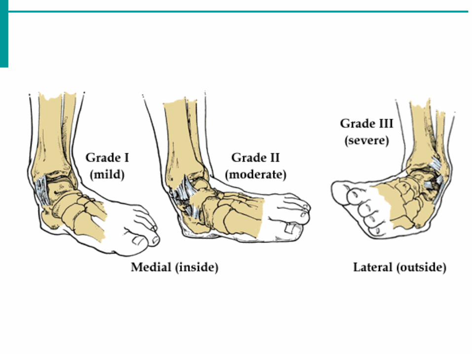
Sprains and Strains are categorized according to severity.
Grade I (mild) sprain or strain involves some stretching or minor tearing of a ligament or muscle.
Grade II (moderate) sprain or strain is a ligament or muscle that is partially torn but still intact.
Grade III (severe) sprain or strain means that the ligament or muscle is completely torn, resulting in joint instability.

Ankle Sprains
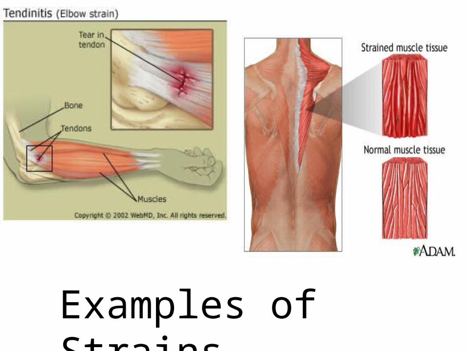
Examples of Strains

First Aid
•R – rest
•I - ice
•C - compression
•E - elevation
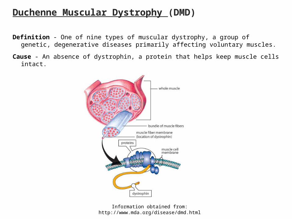
Duchenne Muscular Dystrophy (DMD)
Definition - One of nine types of muscular dystrophy, a group of genetic, degenerative diseases primarily affecting voluntary muscles.
Cause - An absence of dystrophin, a protein that helps keep muscle cells intact.
Information obtained from: http://www.mda.org/disease/dmd.html
DMD continued.....
Onset - Early childhood - about 2 to 6 years.
Symptoms - Generalized weakness first affecting the muscles of the hips, pelvic area, thighs and shoulders. Calves are often enlarged.
Progression - DMD eventually affects all voluntary muscles, and the heart and breathing muscles.
Inheritance - X-linked recessive. DMD primarily affects boys, who inherit the disease through their mothers. Women can be carriers of DMD but usually exhibit no symptoms.