Embed Size (px)
Citation preview
Infected corneal ulceration
What is the normal healing of the cornea?
The cornea is the clear, front part of the eye that acts like a windshield, protecting the inside of the eye while allowing light to pass through. It is formed by 3 layers, from outside to inside: the epithelium, the stroma and the endothelium. A corneal ulcer is a break, a wound in one or more of the layers of the cornea. Normally, when there is a defect or ulcer on the surface of the eye (of the epithelium), the cells at the outer edges grows into the center and healing is often complete within 2-4 days. When the ulcer is deeper, it takes more time to heal. Why is it important to act fast in case of an infected corneal ulcer?
An infected corneal ulceration (infected wound of the clear, front part of the eye) poses a special problem because bacteria that are present are actually delaying the healing process and even digesting the corneal tissues (this is called a melting ulcer). This can lead to corneal perforation (full thickness whole) and cause blindness if not treated promptly.
Infected ulcer in a cat Infected ulcer in a dog Melting ulcer in a dog
What are the treatments for infected corneal ulcers?
• Intensive medical treatment :
Intensive antibiotic treatment (every hour or every 2 hours) is prescribed for the first few days following diagnosis of an infected corneal ulcer. The frequency of treatment (instillation of antibiotic eye drops) is only reduced after the infection is stabilized. Hospitalisation might be necessary in order to get the treatment done. If infected corneal ulcer is not too deep and responds well to medical therapy, surgery may be avoided.
• Combined surgical treatment:
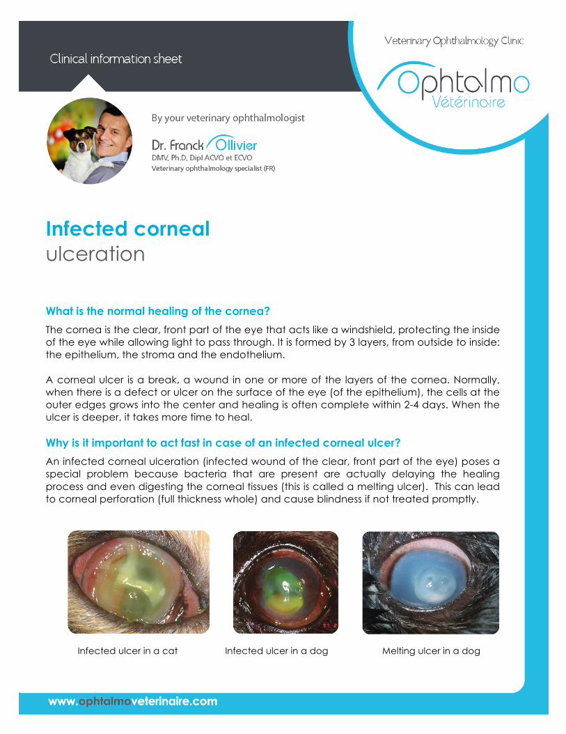
Depending on a variety of factors, your pet may need a surgically placed biodegradable graft, conjunctival graft, corneal graft or corneal glue to provide protection and support for the cornea as it heals. A conjunctival graft represents a technique commonly performed to treat deep corneal ulcers or corneal perforations. It allows an immediate coverage/filling of the corneal defect with the pink tissue around the eye (conjunctiva) used as a bandage over a wound. This tissue is attached to the cornea with very thin stitches. The graft covering the ulcer will remain in place and will form a scar but the base of the graft (the pedicle) can be trimmed 2 months after surgery in order to stop the vascularisation of the graft in order to reduce the corneal scar and increase the visual field.
Corneal ulcer amlost perforating in a dog that needs surgical repair
Same eye : a conjunctival graft (pink tissue around the eye ) is prepared
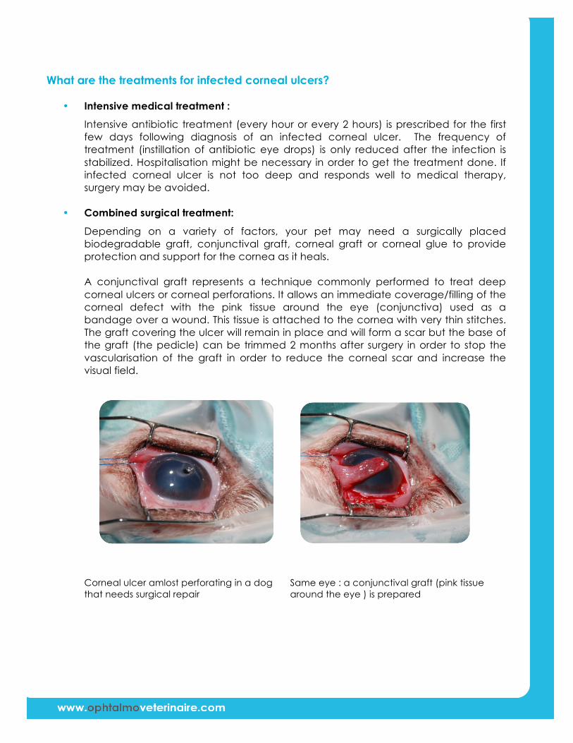
Same eye : the conjunctival graft is sutured over the corneal ulcer
Same eye 3 weeks after the surgery after trimming of the pedicule
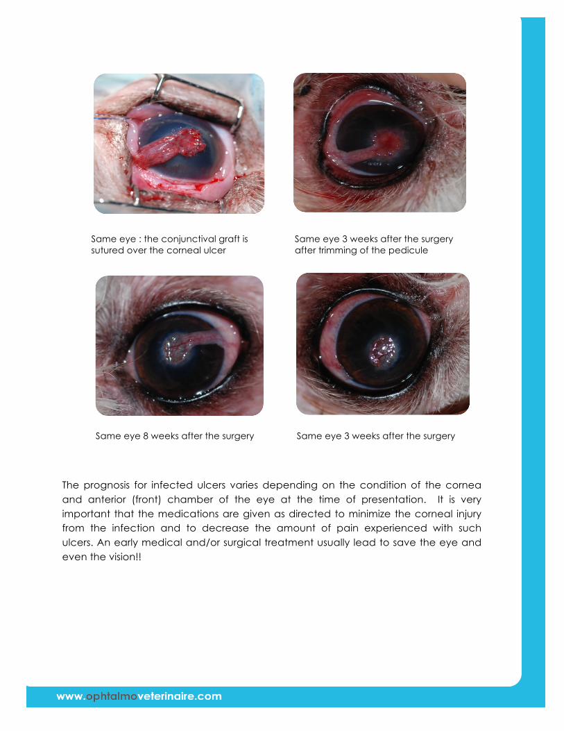
Same eye 8 weeks after the surgery Same eye 3 weeks after the surgery
The prognosis for infected ulcers varies depending on the condition of the cornea and anterior (front) chamber of the eye at the time of presentation. It is very important that the medications are given as directed to minimize the corneal injury from the infection and to decrease the amount of pain experienced with such ulcers. An early medical and/or surgical treatment usually lead to save the eye and even the vision!!


![[Product Monograph Template - Standard]...images in association with red eyes from conjunctival congestion and corneal edema). Instruct patients to consult a physician immediately](https://img.pdfslide.us/doc/110x75/5ea24d1bd166c440bb4fab69/product-monograph-template-standard-images-in-association-with-red-eyes.jpg)