Embed Size (px)
Citation preview
OpenStax-CNX module: m47435 1
The Circulatory System∗
Robert Bear
David Rintoul
Based on Overview of the Circulatory System† by
OpenStax College
This work is produced by OpenStax-CNX and licensed under the
Creative Commons Attribution License 4.0‡
Introduction
Observation by means of the microscope will reveal more wonderful things than those viewedin regard to mere structure and connection: for while the heart is still beating the contrary(i.e., in opposite directions in the di�erent vessels) movement of the blood is observed in thevessels�though with di�culty�so that the circulation of the blood is clearly exposed.
Marcello Malpighi, De Pulmonibus, 1661Malpighi's work (mostly on frogs) outlined the �ner microscopic details of circulation, following the work
of Harvey, who described the circulatory system at a macroscopic level. In all animals, except a few simpletypes, the circulatory system is used to transport nutrients and gases through the body. Simple di�usionallows some water, nutrient, waste, and gas exchange into primitive animals that are only a few cell layersthick; however, bulk �ow is the only method by which the entire body of larger more complex organisms isaccessed.
1 Circulatory System Architecture
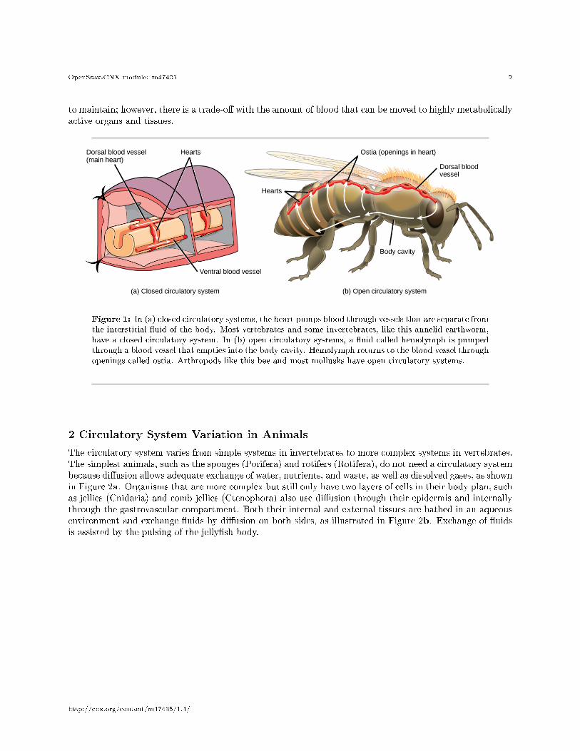
The circulatory system is e�ectively a network of cylindrical vessels: the arteries, veins, and capillaries thatemanate from a pump, the heart. In all vertebrate organisms, as well as some invertebrates, this is a closedsystem, in which the blood is not free in a cavity. In a closed circulatory system, blood is containedinside blood vessels and circulates unidirectionally from the heart around the systemic circulatory route, thenreturns to the heart again, as illustrated in Figure 1a. As opposed to a closed system, arthropods�includinginsects, crustaceans, and most mollusks�have an open circulatory system, as illustrated in Figure 1b. In anopen circulatory system, the �uid is not enclosed in the blood vessels but is pumped into a cavity calleda hemocoel; rather than blood, this �uid is called hemolymph because the blood mixes with the interstitial�uid. As the heart beats and the animal moves, the hemolymph circulates around the organs within thebody cavity and then reenters the hearts through openings called ostia. This movement allows for gas andnutrient exchange. An open circulatory system does not use as much energy as a closed system to operate or
∗Version 1.4: Jul 27, 2014 2:35 pm -0500†http://cnx.org/content/m44801/1.4/‡http://creativecommons.org/licenses/by/4.0/
http://cnx.org/content/m47435/1.4/
OpenStax-CNX module: m47435 2
to maintain; however, there is a trade-o� with the amount of blood that can be moved to highly metabolicallyactive organs and tissues.
Figure 1: In (a) closed circulatory systems, the heart pumps blood through vessels that are separate fromthe interstitial �uid of the body. Most vertebrates and some invertebrates, like this annelid earthworm,have a closed circulatory system. In (b) open circulatory systems, a �uid called hemolymph is pumpedthrough a blood vessel that empties into the body cavity. Hemolymph returns to the blood vessel throughopenings called ostia. Arthropods like this bee and most mollusks have open circulatory systems.
2 Circulatory System Variation in Animals
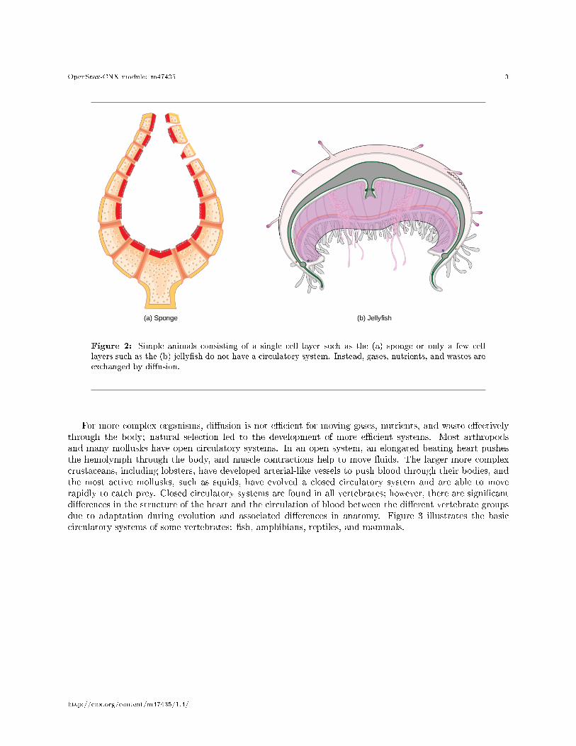
The circulatory system varies from simple systems in invertebrates to more complex systems in vertebrates.The simplest animals, such as the sponges (Porifera) and rotifers (Rotifera), do not need a circulatory systembecause di�usion allows adequate exchange of water, nutrients, and waste, as well as dissolved gases, as shownin Figure 2a. Organisms that are more complex but still only have two layers of cells in their body plan, suchas jellies (Cnidaria) and comb jellies (Ctenophora) also use di�usion through their epidermis and internallythrough the gastrovascular compartment. Both their internal and external tissues are bathed in an aqueousenvironment and exchange �uids by di�usion on both sides, as illustrated in Figure 2b. Exchange of �uidsis assisted by the pulsing of the jelly�sh body.
http://cnx.org/content/m47435/1.4/
OpenStax-CNX module: m47435 3
Figure 2: Simple animals consisting of a single cell layer such as the (a) sponge or only a few celllayers such as the (b) jelly�sh do not have a circulatory system. Instead, gases, nutrients, and wastes areexchanged by di�usion.
For more complex organisms, di�usion is not e�cient for moving gases, nutrients, and waste e�ectivelythrough the body; natural selection led to the development of more e�cient systems. Most arthropodsand many mollusks have open circulatory systems. In an open system, an elongated beating heart pushesthe hemolymph through the body, and muscle contractions help to move �uids. The larger more complexcrustaceans, including lobsters, have developed arterial-like vessels to push blood through their bodies, andthe most active mollusks, such as squids, have evolved a closed circulatory system and are able to moverapidly to catch prey. Closed circulatory systems are found in all vertebrates; however, there are signi�cantdi�erences in the structure of the heart and the circulation of blood between the di�erent vertebrate groupsdue to adaptation during evolution and associated di�erences in anatomy. Figure 3 illustrates the basiccirculatory systems of some vertebrates: �sh, amphibians, reptiles, and mammals.
http://cnx.org/content/m47435/1.4/
OpenStax-CNX module: m47435 4
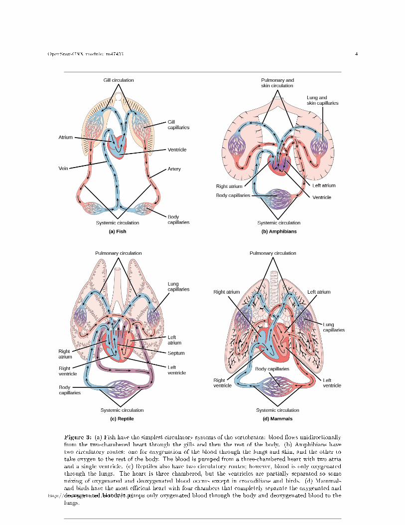
Figure 3: (a) Fish have the simplest circulatory systems of the vertebrates: blood �ows unidirectionallyfrom the two-chambered heart through the gills and then the rest of the body. (b) Amphibians havetwo circulatory routes: one for oxygenation of the blood through the lungs and skin, and the other totake oxygen to the rest of the body. The blood is pumped from a three-chambered heart with two atriaand a single ventricle. (c) Reptiles also have two circulatory routes; however, blood is only oxygenatedthrough the lungs. The heart is three chambered, but the ventricles are partially separated so somemixing of oxygenated and deoxygenated blood occurs except in crocodilians and birds. (d) Mammalsand birds have the most e�cient heart with four chambers that completely separate the oxygenated anddeoxygenated blood; it pumps only oxygenated blood through the body and deoxygenated blood to thelungs.
http://cnx.org/content/m47435/1.4/
OpenStax-CNX module: m47435 5
As illustrated in Figure 3a Fish have a single circuit for blood �ow and a two-chambered heart that hasonly a single atrium and a single ventricle. The atrium collects blood that has returned from the body, andthe ventricle pumps the blood to the gills where gas exchange occurs and the blood is re-oxygenated; thisis called the gill circulation. The blood then continues through the rest of the body before arriving back atthe atrium; this is called the systemic circulation. This unidirectional �ow of blood produces a gradientof oxygenated to deoxygenated blood around the �sh's systemic circuit. The result is a limit in the amountof oxygen that can reach some of the organs and tissues of the body, reducing the overall metabolic capacityof �sh.
In amphibians, reptiles, birds, and mammals, blood �ow is directed in two circuits: one through the lungsand back to the heart, which is called the pulmonary circulation, and the other throughout the rest ofthe body and its organs including the brain (systemic circulation). In amphibians, gas exchange also occursthrough the skin during pulmonary circulation and is referred to as pulmocutaneous circulation.
As shown in Figure 3b, amphibians have a three-chambered heart that has two atria and one ventriclerather than the two-chambered heart of �sh. The two atria (superior heart chambers) receive blood fromthe two di�erent circuits (the lungs and the systems), and then there is some mixing of the blood in theheart's ventricle (inferior heart chamber), which reduces the oxygen concentration in the blood pumpedfrom the ventricle. The advantage to this arrangement is that high pressure in the vessels pushes blood tothe lungs and body. The mixing is mitigated by a ridge within the ventricle that diverts oxygen-rich bloodthrough the systemic circulatory system and deoxygenated blood to the pulmocutaneous circuit. For thisreason, amphibians are often described as having double circulation.
Most reptiles also have a three-chambered heart similar to the amphibian heart that directs blood tothe pulmonary and systemic circuits, as shown in Figure 3c. The ventricle is divided more e�ectively bya partial septum, which results in even less mixing of oxygenated and deoxygenated blood. Some reptiles(alligators and crocodiles) are the most primitive animals to exhibit a four-chambered heart. Crocodilianshave a unique circulatory mechanism where the heart shunts blood from the lungs toward the stomach andother organs during long periods of submergence, for instance, while the animal waits for prey or staysunderwater waiting for prey to rot. One adaptation includes two main arteries that leave the same part ofthe heart: one takes blood to the lungs and the other provides an alternate route to the stomach and otherparts of the body. Two other adaptations include a hole in the heart between the two ventricles, called theforamen of Panizza, which allows blood to move from one side of the heart to the other, and specializedconnective tissue that slows the blood �ow to the lungs. Together these adaptations have made crocodilesand alligators one of the most successful and ancient animal groups on earth.
In mammals and birds, the heart is also divided into four chambers: two atria and two ventricles, asillustrated in Figure 3d. The oxygenated blood is completely separated from the deoxygenated blood, whichimproves the e�ciency of double circulation and is probably required for the warm-blooded lifestyle ofmammals and birds. The four-chambered heart of birds and mammals evolved independently from ancestorswith a three-chambered heart. The independent evolution of the same or a similar biological trait is referredto as convergent evolution.
3 Components of Blood
Oxygen-binding proteins (hemoglobin, hemocyanin, etc.) are one of the main components of blood in allanimals. The blood is more than those proteins, though. Blood is actually a term used to describe the liquidthat moves through the vessels and includes plasma (the liquid portion, which contains water, proteins,salts, lipids, and glucose) and the cells (red and white cells) and cell fragments called platelets. Plasma isactually the major component of blood and contains the water, proteins, electrolytes, lipids, and glucose.The cells are responsible for carrying the gases (red cells) and immune the response (white). The plateletsare responsible for blood clotting. In humans, cellular components make up approximately 45 percent ofthe blood and the liquid plasma 55 percent. Blood is 20 percent of a human's extracellular �uid and eightpercent of the weight of an average human.
http://cnx.org/content/m47435/1.4/
OpenStax-CNX module: m47435 6
3.1 The Role of Blood in the Body
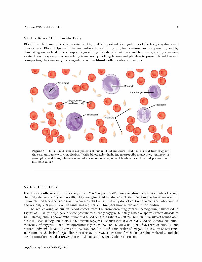
Blood, like the human blood illustrated in Figure 4 is important for regulation of the body's systems andhomeostasis. Blood helps maintain homeostasis by stabilizing pH, temperature, osmotic pressure, and byeliminating excess heat. Blood supports growth by distributing nutrients and hormones, and by removingwaste. Blood plays a protective role by transporting clotting factors and platelets to prevent blood loss andtransporting the disease-�ghting agents or white blood cells to sites of infection.
Figure 4: The cells and cellular components of human blood are shown. Red blood cells deliver oxygen tothe cells and remove carbon dioxide. White blood cells�including neutrophils, monocytes, lymphocytes,eosinophils, and basophils�are involved in the immune response. Platelets form clots that prevent bloodloss after injury.
3.2 Red Blood Cells
Red blood cells, or erythrocytes (erythro- = �red�; -cyte = �cell�), are specialized cells that circulate throughthe body delivering oxygen to cells; they are generated by division of stem cells in the bone marrow. Inmammals, red blood cells are small biconcave cells that at maturity do not contain a nucleus or mitochondriaand are only 7�8 µm in size. In birds and reptiles, erythrocytes have nuclie and mitochondria.
The red coloring of human blood comes from the iron-containing protein hemoglobin, illustrated inFigure 5a. The principal job of these proteins is to carry oxygen, but they also transports carbon dioxide aswell. Hemoglobin is packed into human red blood cells at a rate of about 250 million molecules of hemoglobinper cell. Each hemoglobin molecule binds four oxygen molecules so that each red blood cell carries one billionmolecules of oxygen. There are approximately 25 trillion red blood cells in the �ve liters of blood in thehuman body, which could carry up to 25 sextillion (25 × 1021) molecules of oxygen in the body at any time.In mammals, the lack of organelles in erythrocytes leaves more room for the hemoglobin molecules, and thelack of mitochondria also prevents use of the oxygen for metabolic respiration.
http://cnx.org/content/m47435/1.4/
OpenStax-CNX module: m47435 7
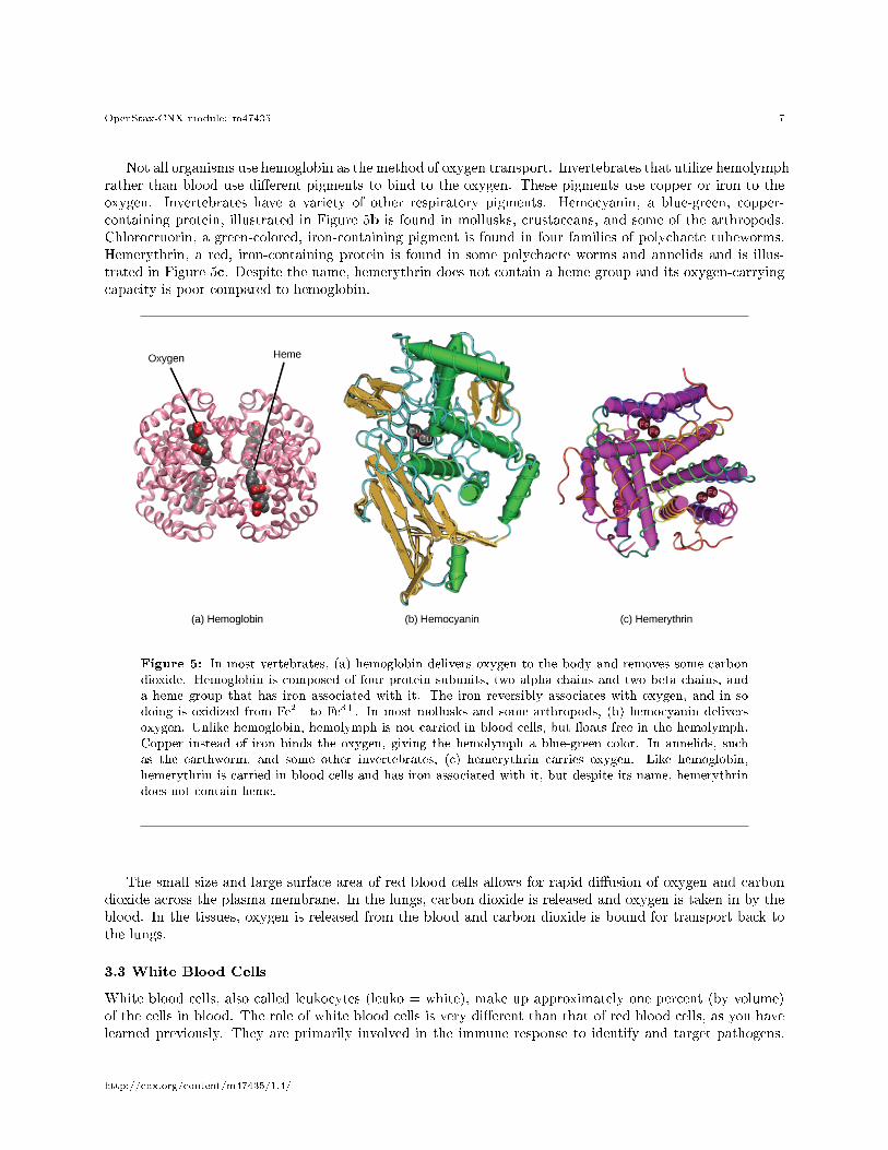
Not all organisms use hemoglobin as the method of oxygen transport. Invertebrates that utilize hemolymphrather than blood use di�erent pigments to bind to the oxygen. These pigments use copper or iron to theoxygen. Invertebrates have a variety of other respiratory pigments. Hemocyanin, a blue-green, copper-containing protein, illustrated in Figure 5b is found in mollusks, crustaceans, and some of the arthropods.Chlorocruorin, a green-colored, iron-containing pigment is found in four families of polychaete tubeworms.Hemerythrin, a red, iron-containing protein is found in some polychaete worms and annelids and is illus-trated in Figure 5c. Despite the name, hemerythrin does not contain a heme group and its oxygen-carryingcapacity is poor compared to hemoglobin.
Figure 5: In most vertebrates, (a) hemoglobin delivers oxygen to the body and removes some carbondioxide. Hemoglobin is composed of four protein subunits, two alpha chains and two beta chains, anda heme group that has iron associated with it. The iron reversibly associates with oxygen, and in sodoing is oxidized from Fe2+ to Fe3+. In most mollusks and some arthropods, (b) hemocyanin deliversoxygen. Unlike hemoglobin, hemolymph is not carried in blood cells, but �oats free in the hemolymph.Copper instead of iron binds the oxygen, giving the hemolymph a blue-green color. In annelids, suchas the earthworm, and some other invertebrates, (c) hemerythrin carries oxygen. Like hemoglobin,hemerythrin is carried in blood cells and has iron associated with it, but despite its name, hemerythrindoes not contain heme.
The small size and large surface area of red blood cells allows for rapid di�usion of oxygen and carbondioxide across the plasma membrane. In the lungs, carbon dioxide is released and oxygen is taken in by theblood. In the tissues, oxygen is released from the blood and carbon dioxide is bound for transport back tothe lungs.
3.3 White Blood Cells
White blood cells, also called leukocytes (leuko = white), make up approximately one percent (by volume)of the cells in blood. The role of white blood cells is very di�erent than that of red blood cells, as you havelearned previously. They are primarily involved in the immune response to identify and target pathogens,
http://cnx.org/content/m47435/1.4/
OpenStax-CNX module: m47435 8
such as invading bacteria, viruses, and other foreign organisms. White blood cells are formed continually;some only live for hours or days, but some live for years.
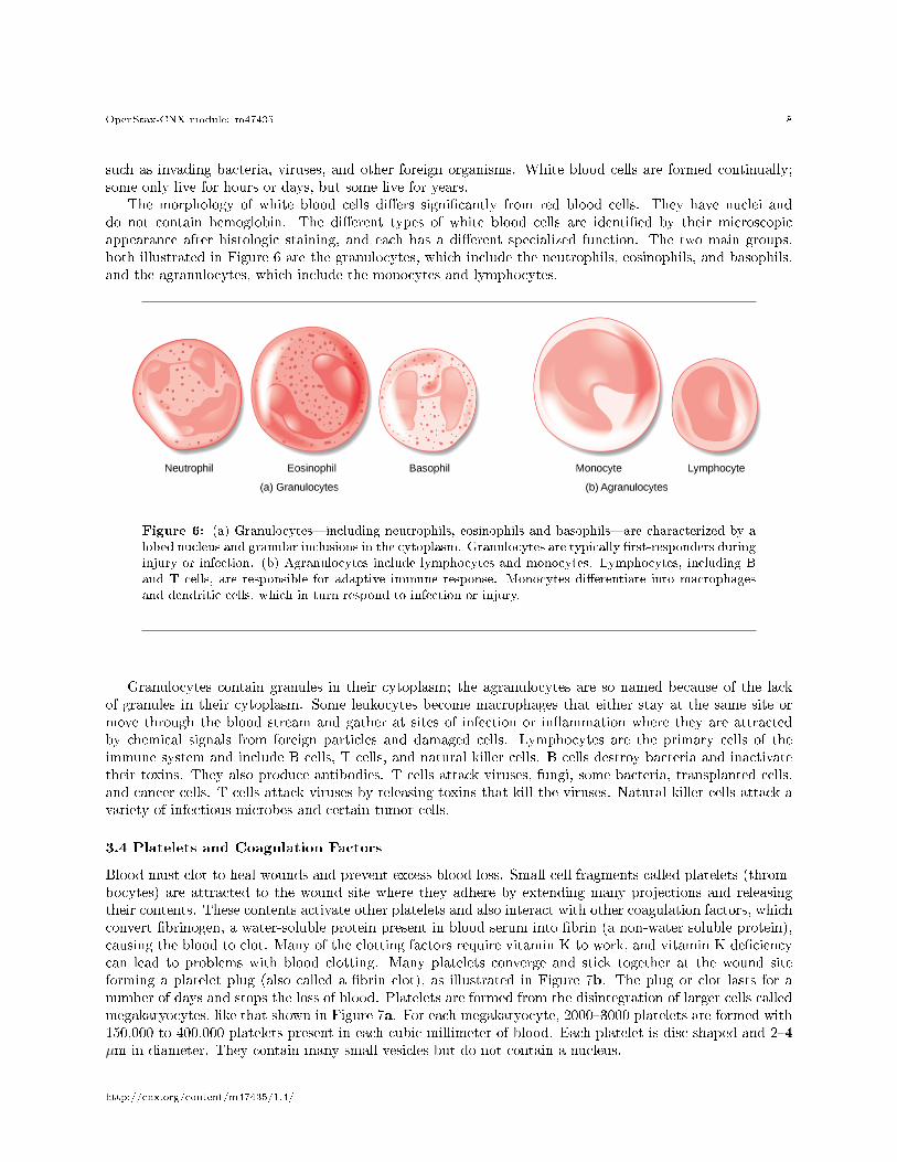
The morphology of white blood cells di�ers signi�cantly from red blood cells. They have nuclei anddo not contain hemoglobin. The di�erent types of white blood cells are identi�ed by their microscopicappearance after histologic staining, and each has a di�erent specialized function. The two main groups,both illustrated in Figure 6 are the granulocytes, which include the neutrophils, eosinophils, and basophils,and the agranulocytes, which include the monocytes and lymphocytes.
Figure 6: (a) Granulocytes�including neutrophils, eosinophils and basophils�are characterized by alobed nucleus and granular inclusions in the cytoplasm. Granulocytes are typically �rst-responders duringinjury or infection. (b) Agranulocytes include lymphocytes and monocytes. Lymphocytes, including Band T cells, are responsible for adaptive immune response. Monocytes di�erentiate into macrophagesand dendritic cells, which in turn respond to infection or injury.
Granulocytes contain granules in their cytoplasm; the agranulocytes are so named because of the lackof granules in their cytoplasm. Some leukocytes become macrophages that either stay at the same site ormove through the blood stream and gather at sites of infection or in�ammation where they are attractedby chemical signals from foreign particles and damaged cells. Lymphocytes are the primary cells of theimmune system and include B cells, T cells, and natural killer cells. B cells destroy bacteria and inactivatetheir toxins. They also produce antibodies. T cells attack viruses, fungi, some bacteria, transplanted cells,and cancer cells. T cells attack viruses by releasing toxins that kill the viruses. Natural killer cells attack avariety of infectious microbes and certain tumor cells.
3.4 Platelets and Coagulation Factors
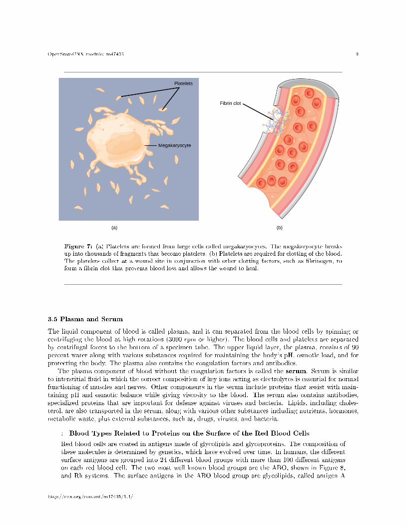
Blood must clot to heal wounds and prevent excess blood loss. Small cell fragments called platelets (throm-bocytes) are attracted to the wound site where they adhere by extending many projections and releasingtheir contents. These contents activate other platelets and also interact with other coagulation factors, whichconvert �brinogen, a water-soluble protein present in blood serum into �brin (a non-water soluble protein),causing the blood to clot. Many of the clotting factors require vitamin K to work, and vitamin K de�ciencycan lead to problems with blood clotting. Many platelets converge and stick together at the wound siteforming a platelet plug (also called a �brin clot), as illustrated in Figure 7b. The plug or clot lasts for anumber of days and stops the loss of blood. Platelets are formed from the disintegration of larger cells calledmegakaryocytes, like that shown in Figure 7a. For each megakaryocyte, 2000�3000 platelets are formed with150,000 to 400,000 platelets present in each cubic millimeter of blood. Each platelet is disc shaped and 2�4µm in diameter. They contain many small vesicles but do not contain a nucleus.
http://cnx.org/content/m47435/1.4/
OpenStax-CNX module: m47435 9
Figure 7: (a) Platelets are formed from large cells called megakaryocytes. The megakaryocyte breaksup into thousands of fragments that become platelets. (b) Platelets are required for clotting of the blood.The platelets collect at a wound site in conjunction with other clotting factors, such as �brinogen, toform a �brin clot that prevents blood loss and allows the wound to heal.
3.5 Plasma and Serum
The liquid component of blood is called plasma, and it can separated from the blood cells by spinning orcentrifuging the blood at high rotations (3000 rpm or higher). The blood cells and platelets are separatedby centrifugal forces to the bottom of a specimen tube. The upper liquid layer, the plasma, consists of 90percent water along with various substances required for maintaining the body's pH, osmotic load, and forprotecting the body. The plasma also contains the coagulation factors and antibodies.
The plasma component of blood without the coagulation factors is called the serum. Serum is similarto interstitial �uid in which the correct composition of key ions acting as electrolytes is essential for normalfunctioning of muscles and nerves. Other components in the serum include proteins that assist with main-taining pH and osmotic balance while giving viscosity to the blood. The serum also contains antibodies,specialized proteins that are important for defense against viruses and bacteria. Lipids, including choles-terol, are also transported in the serum, along with various other substances including nutrients, hormones,metabolic waste, plus external substances, such as, drugs, viruses, and bacteria.
: Blood Types Related to Proteins on the Surface of the Red Blood Cells
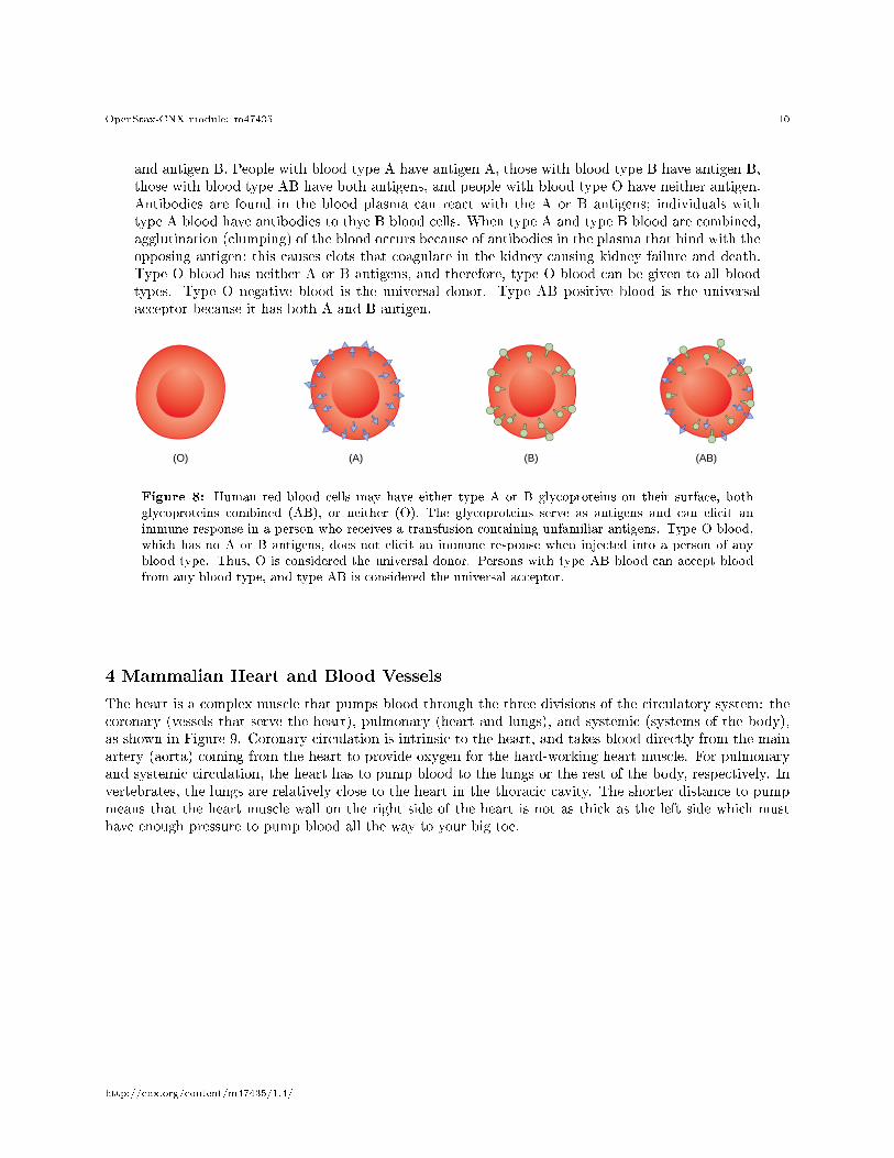
Red blood cells are coated in antigens made of glycolipids and glycoproteins. The composition ofthese molecules is determined by genetics, which have evolved over time. In humans, the di�erentsurface antigens are grouped into 24 di�erent blood groups with more than 100 di�erent antigenson each red blood cell. The two most well known blood groups are the ABO, shown in Figure 8,and Rh systems. The surface antigens in the ABO blood group are glycolipids, called antigen A
http://cnx.org/content/m47435/1.4/
OpenStax-CNX module: m47435 10
and antigen B. People with blood type A have antigen A, those with blood type B have antigen B,those with blood type AB have both antigens, and people with blood type O have neither antigen.Antibodies are found in the blood plasma can react with the A or B antigens; individuals withtype A blood have antibodies to thye B blood cells. When type A and type B blood are combined,agglutination (clumping) of the blood occurs because of antibodies in the plasma that bind with theopposing antigen; this causes clots that coagulate in the kidney causing kidney failure and death.Type O blood has neither A or B antigens, and therefore, type O blood can be given to all bloodtypes. Type O negative blood is the universal donor. Type AB positive blood is the universalacceptor because it has both A and B antigen.
Figure 8: Human red blood cells may have either type A or B glycoproteins on their surface, bothglycoproteins combined (AB), or neither (O). The glycoproteins serve as antigens and can elicit animmune response in a person who receives a transfusion containing unfamiliar antigens. Type O blood,which has no A or B antigens, does not elicit an immune response when injected into a person of anyblood type. Thus, O is considered the universal donor. Persons with type AB blood can accept bloodfrom any blood type, and type AB is considered the universal acceptor.
4 Mammalian Heart and Blood Vessels
The heart is a complex muscle that pumps blood through the three divisions of the circulatory system: thecoronary (vessels that serve the heart), pulmonary (heart and lungs), and systemic (systems of the body),as shown in Figure 9. Coronary circulation is intrinsic to the heart, and takes blood directly from the mainartery (aorta) coming from the heart to provide oxygen for the hard-working heart muscle. For pulmonaryand systemic circulation, the heart has to pump blood to the lungs or the rest of the body, respectively. Invertebrates, the lungs are relatively close to the heart in the thoracic cavity. The shorter distance to pumpmeans that the heart muscle wall on the right side of the heart is not as thick as the left side which musthave enough pressure to pump blood all the way to your big toe.
http://cnx.org/content/m47435/1.4/
OpenStax-CNX module: m47435 11
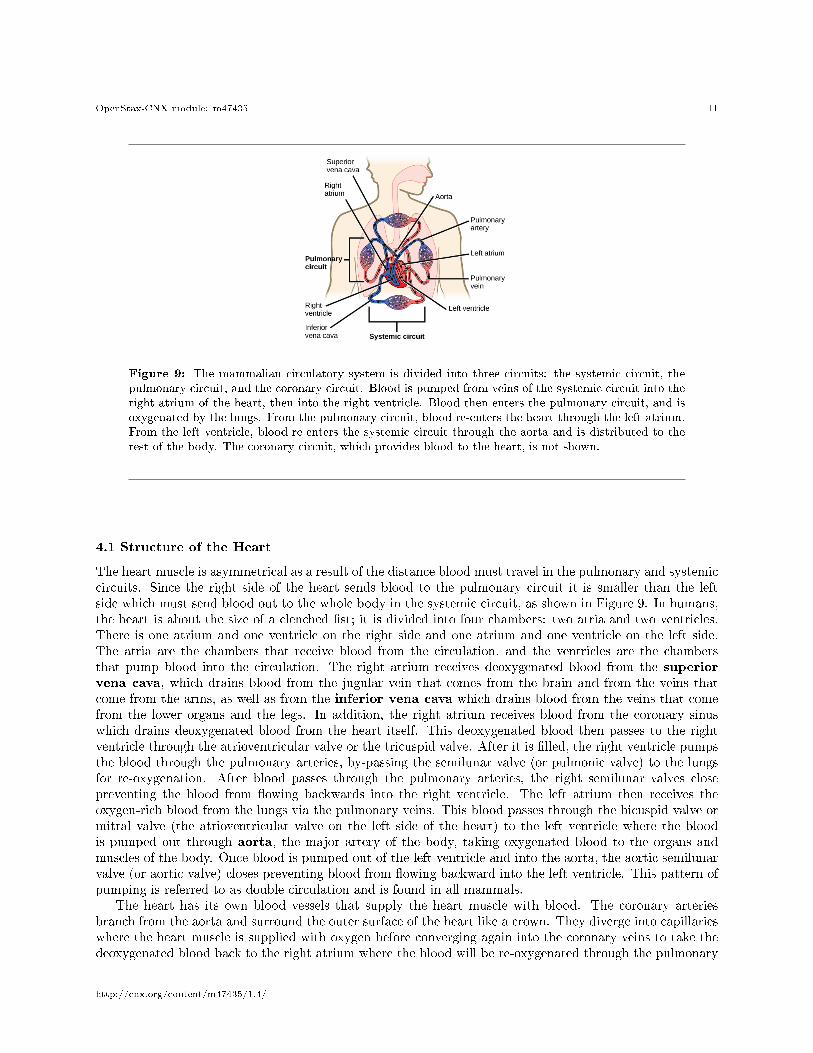
Figure 9: The mammalian circulatory system is divided into three circuits: the systemic circuit, thepulmonary circuit, and the coronary circuit. Blood is pumped from veins of the systemic circuit into theright atrium of the heart, then into the right ventricle. Blood then enters the pulmonary circuit, and isoxygenated by the lungs. From the pulmonary circuit, blood re-enters the heart through the left atrium.From the left ventricle, blood re-enters the systemic circuit through the aorta and is distributed to therest of the body. The coronary circuit, which provides blood to the heart, is not shown.
4.1 Structure of the Heart
The heart muscle is asymmetrical as a result of the distance blood must travel in the pulmonary and systemiccircuits. Since the right side of the heart sends blood to the pulmonary circuit it is smaller than the leftside which must send blood out to the whole body in the systemic circuit, as shown in Figure 9. In humans,the heart is about the size of a clenched �st; it is divided into four chambers: two atria and two ventricles.There is one atrium and one ventricle on the right side and one atrium and one ventricle on the left side.The atria are the chambers that receive blood from the circulation, and the ventricles are the chambersthat pump blood into the circulation. The right atrium receives deoxygenated blood from the superiorvena cava, which drains blood from the jugular vein that comes from the brain and from the veins thatcome from the arms, as well as from the inferior vena cava which drains blood from the veins that comefrom the lower organs and the legs. In addition, the right atrium receives blood from the coronary sinuswhich drains deoxygenated blood from the heart itself. This deoxygenated blood then passes to the rightventricle through the atrioventricular valve or the tricuspid valve. After it is �lled, the right ventricle pumpsthe blood through the pulmonary arteries, by-passing the semilunar valve (or pulmonic valve) to the lungsfor re-oxygenation. After blood passes through the pulmonary arteries, the right semilunar valves closepreventing the blood from �owing backwards into the right ventricle. The left atrium then receives theoxygen-rich blood from the lungs via the pulmonary veins. This blood passes through the bicuspid valve ormitral valve (the atrioventricular valve on the left side of the heart) to the left ventricle where the bloodis pumped out through aorta, the major artery of the body, taking oxygenated blood to the organs andmuscles of the body. Once blood is pumped out of the left ventricle and into the aorta, the aortic semilunarvalve (or aortic valve) closes preventing blood from �owing backward into the left ventricle. This pattern ofpumping is referred to as double circulation and is found in all mammals.
The heart has its own blood vessels that supply the heart muscle with blood. The coronary arteriesbranch from the aorta and surround the outer surface of the heart like a crown. They diverge into capillarieswhere the heart muscle is supplied with oxygen before converging again into the coronary veins to take thedeoxygenated blood back to the right atrium where the blood will be re-oxygenated through the pulmonary
http://cnx.org/content/m47435/1.4/
OpenStax-CNX module: m47435 12
circuit. The heart muscle will die without a steady supply of blood. Atherosclerosis is the blockage ofan artery by the buildup of fatty plaques. Because of the size (narrow) of the coronary arteries and theirfunction in serving the heart itself, atherosclerosis can be deadly in these arteries. The slowdown of blood�ow and subsequent oxygen deprivation that results from atherosclerosis causes severe pain, known as angina,and complete blockage of the arteries will cause myocardial infarction: the death of cardiac muscle tissue,commonly known as a heart attack.
4.2 Arteries, Veins, and Capillaries
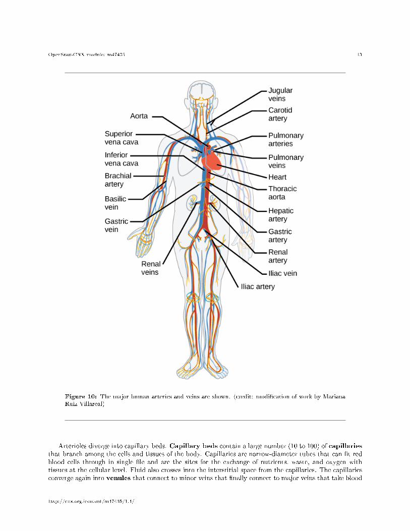
The blood from the heart is carried through the body by a complex network of blood vessels (Figure 10).Arteries take blood away from the heart. The main artery is the aorta that branches into major arteriesthat take blood to di�erent limbs and organs. These major arteries include the carotid artery that takesblood to the brain, the brachial arteries that take blood to the arms, and the thoracic artery that takesblood to the thorax and then into the hepatic, renal, and gastric arteries for the liver, kidney, and stomach,respectively. The iliac artery takes blood to the lower limbs. The major arteries diverge into minor arteries,and then smaller vessels called arterioles, to reach more deeply into the muscles and organs of the body.
http://cnx.org/content/m47435/1.4/
OpenStax-CNX module: m47435 13
Figure 10: The major human arteries and veins are shown. (credit: modi�cation of work by MarianaRuiz Villareal)
Arterioles diverge into capillary beds. Capillary beds contain a large number (10 to 100) of capillariesthat branch among the cells and tissues of the body. Capillaries are narrow-diameter tubes that can �t redblood cells through in single �le and are the sites for the exchange of nutrients, waste, and oxygen withtissues at the cellular level. Fluid also crosses into the interstitial space from the capillaries. The capillariesconverge again into venules that connect to minor veins that �nally connect to major veins that take blood
http://cnx.org/content/m47435/1.4/

OpenStax-CNX module: m47435 14
high in carbon dioxide back to the heart. Veins are blood vessels that bring blood back to the heart. Themajor veins drain blood from the same organs and limbs that the major arteries supply. Fluid is also broughtback to the heart via the lymphatic system.
http://cnx.org/content/m47435/1.4/