Embed Size (px)
Citation preview

The Circulatory System

Functions of the Circulatory System
• Brings blood containing
oxygen, nutrients, and hormones to cells
• Transports CO2 and
other wastes away
from cells

Functions Continued • Fights infection
• Regulates body
temperature
• Helps stabilize pH and ionic concentration of body
fluids.
Functions Continued • Nutrients from the gut to the body
• Urea from the liver to the kidneys
The Need for an Exchange Surface
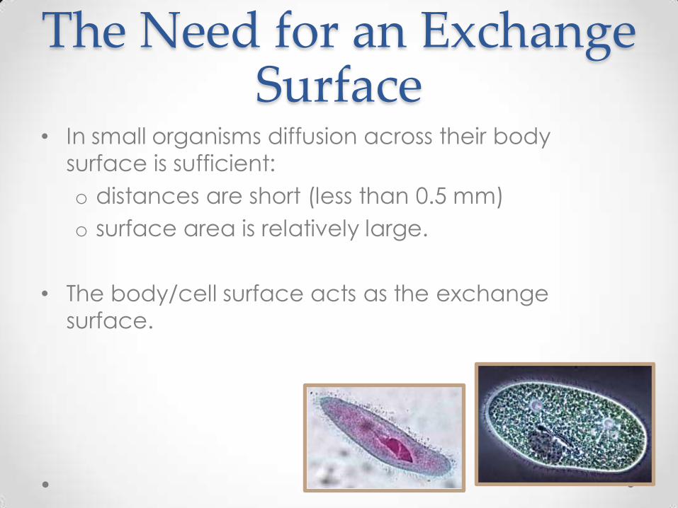
• In small organisms diffusion across their body
surface is sufficient:
o distances are short (less than 0.5 mm)
o surface area is relatively large.
• The body/cell surface acts as the exchange
surface.
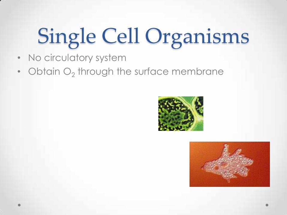
Single Cell Organisms • No circulatory system
• Obtain O2 through the surface membrane

The Need for an Exchange Surface
• Large active organisms cannot rely upon their body
surface:
o surface area relative to volume is insufficient for
exchange
o distances are too great.
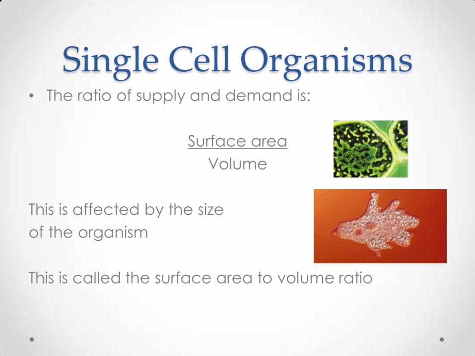
Single Cell Organisms • The ratio of supply and demand is:
Surface area
Volume
This is affected by the size
of the organism
This is called the surface area to volume ratio
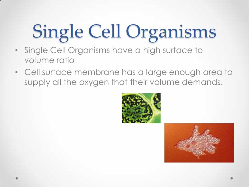
Single Cell Organisms • Single Cell Organisms have a high surface to
volume ratio
• Cell surface membrane has a large enough area to
supply all the oxygen that their volume demands.
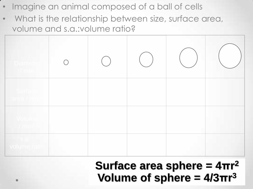
• Imagine an animal composed of a ball of cells
• What is the relationship between size, surface area,
volume and s.a.:volume ratio?
1 2 3
Diameter
/ mm
4
Surface
area / mm2
Volume
/ mm3
s.a. :
volume ratio
5
Surface area sphere = 4πr2
Volume of sphere = 4/3πr3

Surface area sphere = 4πr2
Volume of sphere = 4/3πr3
1 2 3
Diameter
/ mm
4
Surface
area / mm2
Volume
/ mm3
s.a. :
volume
ratio
5
12.6 50.3 113.1 201.1 314.2
4.2 33.5 113.1 268.1 523.6
3 1.5 1 0.75 0.60

Meeting the Demand • Thus there is a need for the following:
• Specialised exchange surface to meet the
demands of high activity levels in multi-cellular
organisms
• Efficient transport system to deliver materials to
and from the exchange surface
What Effect does an Increase in Size have on
the need for a Specialist Exchange Surface in
Larger Animals?
• As size increases, volume increases
disproportionately compared to volume.
• Demands of cells for nutrients and waste removal
also increased disproportionately:
o the surface does not increase sufficiently to
accommodate these extra demands
o must be met by a specialist exchange surface
with enhanced surface area.

General Features of Exchange Surfaces
Permeable
Thin
Moist
Mechanism to maintain diffusion gradients
The large surface can be provided by the body surface in small organisms or by folding of the exchange surface.
This speaks for itself.
Diffusion is only efficient over short distances (< 1mm). Rate is inversely proportional to square of the distance.
O2, CO2 and nutrients diffuse in solution.
Transport system, ventilation mechanism or creation of currents across surface.
Large surface area
relative to volume
Circulatory System
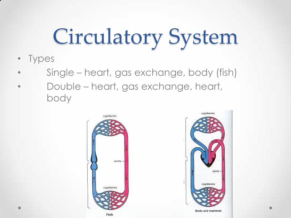
Circulatory System • Types
• Single – heart, gas exchange, body (fish)
• Double – heart, gas exchange, heart,
body

Circulatory System • Two distinct part of the Double System
o Pulmonary (lungs)
o Systemic (body)
o Animation
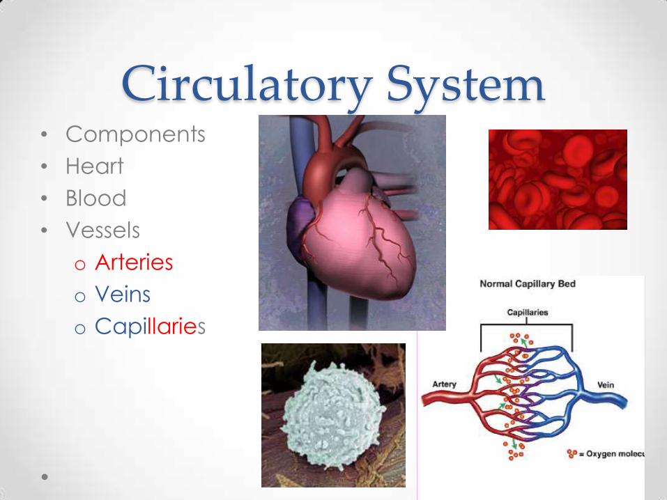
Circulatory System • Components
• Heart
• Blood
• Vessels
o Arteries
o Veins
o Capillaries
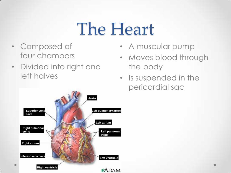
The Heart • A muscular pump
• Moves blood through
the body
• Is suspended in the
pericardial sac
• Composed of
four chambers
• Divided into right and
left halves
Aorta
Left pulmonary artery
Left atrium
Left pulmonary
veins
Left ventricle
Superior vena
cava
Right pulmonary
veins
Right atrium
Right ventricle
Inferior vena cava
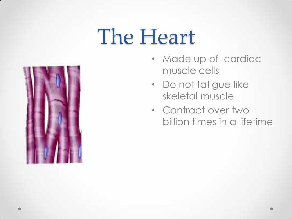
The Heart • Made up of cardiac
muscle cells
• Do not fatigue like
skeletal muscle
• Contract over two
billion times in a lifetime

Pericardium • Protective sac of
connective tissue
• Surrounds the
heart
• Filled with
• fluid
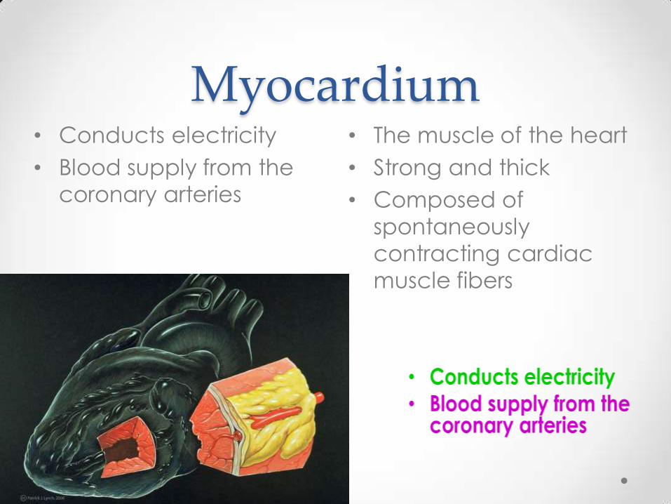
Myocardium • The muscle of the heart
• Strong and thick
• Composed of
spontaneously
contracting cardiac
muscle fibers
• Conducts electricity
• Blood supply from the
coronary arteries

Structures of the Heart • Chambers
o Atria- (2) upper chambers
• Thin walled
• Receive blood from veins
• Send blood to ventricles
o Ventricles- (2) lower chambers
• Thick walled
• Receive blood from atria
• Pump blood out through arteries
• Chambers
o Atria (2) upper chambers
• Thin walled
• Receives blood from veins
• Sends blood to ventricles
o Ventricles (2) lower chambers
• Thick walled
• Received blood from atria
• Pump blood out through arteries
• Septum
o Wall that divides heart into right and left halves
Septum
Pulmonary valve
Right atrium
Tricuspid valve
Right ventricle
Left atrium
Aortic valve
Mitral valve
Left ventricle
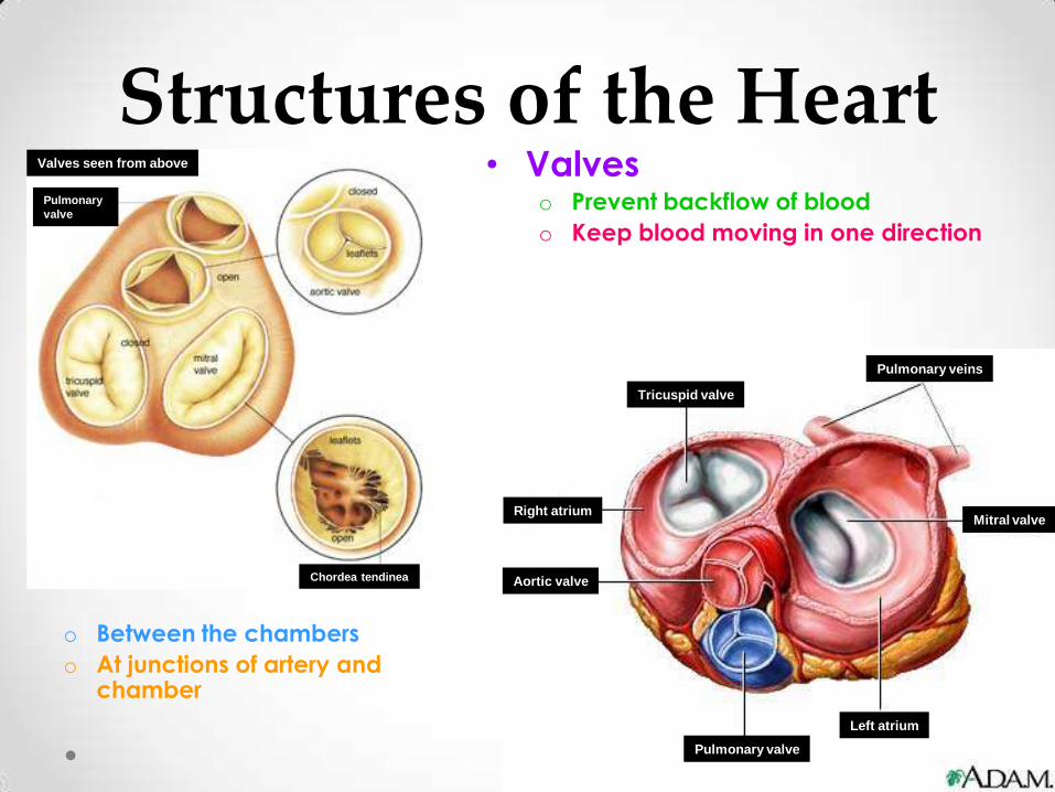
Structures of the Heart • Valves
o Prevent backflow of blood
o Keep blood moving in one direction
o Between the chambers
o At junctions of artery and chamber
Tricuspid valve
Pulmonary veins
Mitral valve
Left atrium
Pulmonary valve
Aortic valve
Right atrium
Valves seen from above
Chordea tendinea
Pulmonary
valve

Structures of the Heart • Bicuspid and Tricuspid Valve (Atrioventricular)
o Control passage of blood from atrium to ventricle
• Semi-lunar valves
o Controls passage of blood from ventricle to
arteries
Papilla
ry
muscl
e
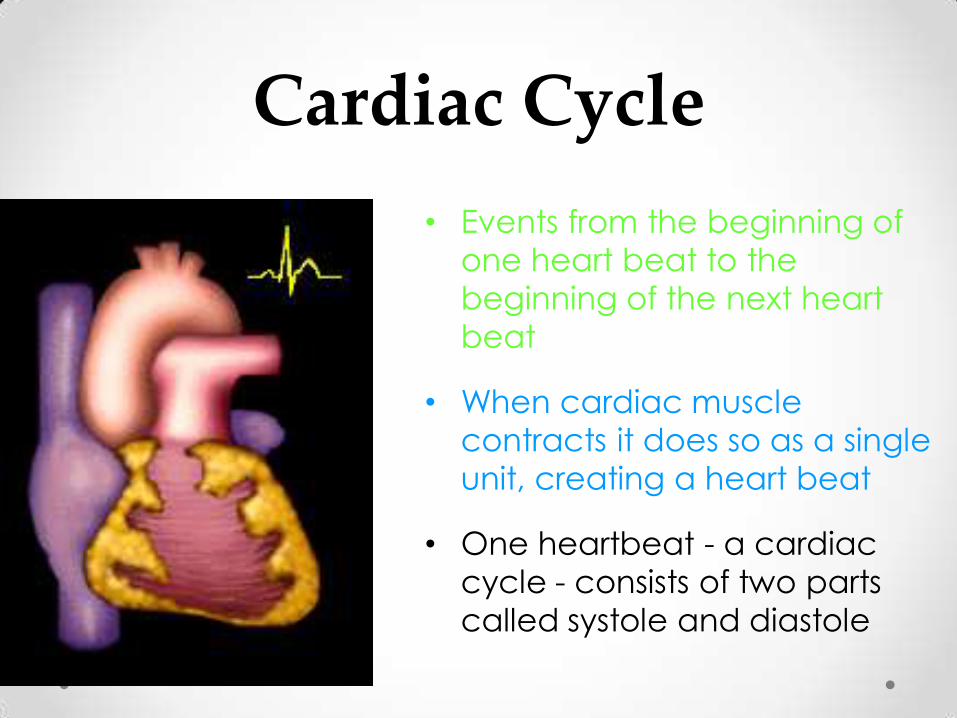
Cardiac Cycle
• Events from the beginning of
one heart beat to the
beginning of the next heart
beat
• When cardiac muscle
contracts it does so as a single
unit, creating a heart beat
• One heartbeat - a cardiac
cycle - consists of two parts
called systole and diastole

Cardiac Cycle • Diastole is the period of
time when the heart
relaxes after
contraction
• Oxygenated blood
from the lungs fills the
left atrium
• Deoxygenated blood
from other parts of the
body fills the right
atrium.
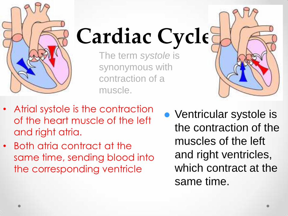
Cardiac Cycle
• Atrial systole is the contraction
of the heart muscle of the left
and right atria.
• Both atria contract at the
same time, sending blood into
the corresponding ventricle
Ventricular systole is
the contraction of the
muscles of the left
and right ventricles,
which contract at the
same time.
The term systole is
synonymous with
contraction of a
muscle.
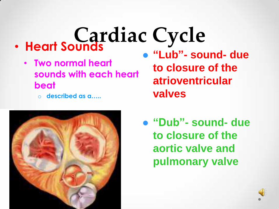
Cardiac Cycle • Two normal heart
sounds with each heart
beat o described as a…..
• Heart Sounds “Lub”- sound- due
to closure of the
atrioventricular
valves
“Dub”- sound- due
to closure of the
aortic valve and
pulmonary valve

Cardiac Cycle • Heart Rate - count of each heart beat
o On average, a heart beats 70 times a minute when at rest
o Calculated as "beats per minute" (bpm).
o The pulse is used measuring the heart rate
o Heart rate is controlled by the medulla in the brain

Cardiac Cycle
• Resting heart rate can be
significantly lower in athletes
o Heart rate increases
when more food and
oxygen are needed by
the cells, or when under
stress

Cardiac Cycle
An electrocardiogram abbreviated as EKG
or ECG is a test that measures the electrical
activity of the heartbeat or one cardiac cycle.

Cardiac Conduction System
• Includes:
o SA node
(pacemaker)
o AV node
Purkinje
fibers
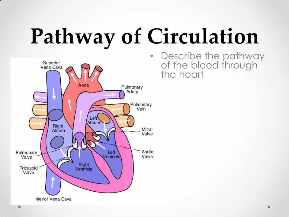
Pathway of Circulation • Describe the pathway
of the blood through the heart
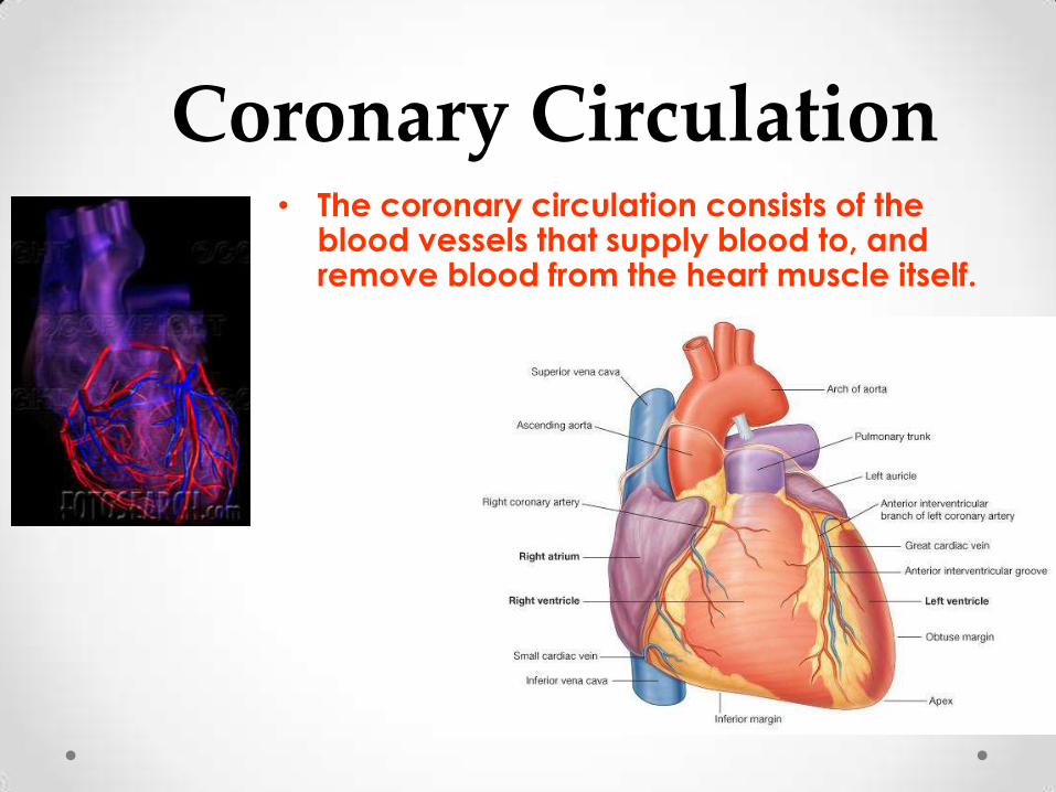
Coronary Circulation • The coronary circulation consists of the
blood vessels that supply blood to, and remove blood from the heart muscle itself.

Coronary Circulation
• The vessels that supply blood high in oxygen to the myocardium are known as coronary arteries.

Arteries, Veins and Capillaries
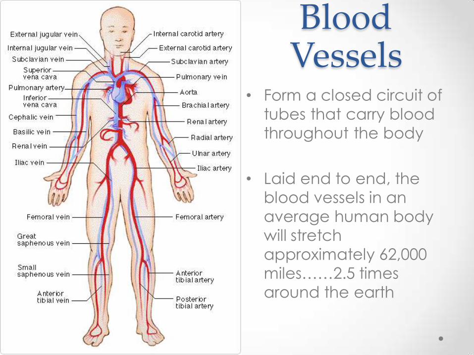
Blood Vessels
• Form a closed circuit of
tubes that carry blood
throughout the body
• Laid end to end, the
blood vessels in an
average human body
will stretch
approximately 62,000
miles……2.5 times
around the earth
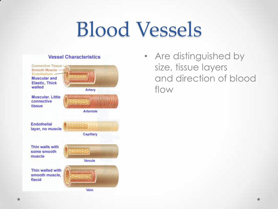
Blood Vessels • Are distinguished by
size, tissue layers
and direction of blood
flow

Blood Vessels • Arteries
o Receive blood from
ventricles
o Take blood away
from the heart
o Usually carry
oxygenated blood
o Thickest vessel walls
o Withstand greater blood pressure
o Are very elastic
o Connect to capillaries
o Aorta is the largest artery
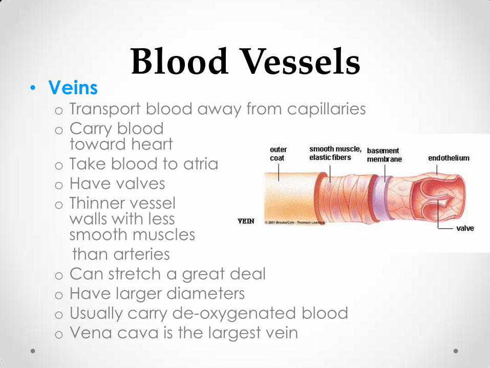
Blood Vessels • Veins
o Transport blood away from capillaries
o Carry blood toward heart
o Take blood to atria
o Have valves
o Thinner vessel walls with less smooth muscles
than arteries
o Can stretch a great deal
o Have larger diameters
o Usually carry de-oxygenated blood
o Vena cava is the largest vein
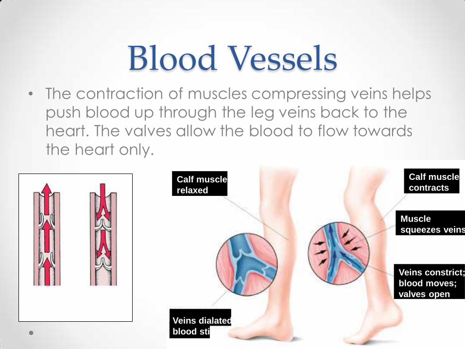
Blood Vessels • The contraction of muscles compressing veins helps
push blood up through the leg veins back to the
heart. The valves allow the blood to flow towards
the heart only.
Calf muscle
relaxed
Calf muscle
contracts
Muscle
squeezes veins
Veins constrict;
blood moves;
valves open
Veins dialated;
blood still;
valves closed
Valves
OPEN
Valves
CLOSED

Blood Vessels • Capillaries
o Smallest of blood vessels
o Only one cell thick (epithelial cell)
o Connect arteries to veins
o Bring oxygen and nutrients to cells
o Removes CO2, urea, and other wastes from cells
o Where blood is under low pressure and moving slowly

Blood Vessels • A network of capillaries
runs close to the cells.
• The capillaries have very thin walls.
Arteriole Venule
Tissue cells Vein Artery capillaries
Capillaries

C B
O L
M O
P O
A D
R
I V
S E
O S
N S
E
O L
F S
Blood

Blood • The life stream of the body, affecting
every cell and
system we have.
• The blood is an
accumulation of
many different
elements, each
working in a specific
way to keep us
alive.

Blood
• Several types of cells suspended in a fluid medium known as plasma.
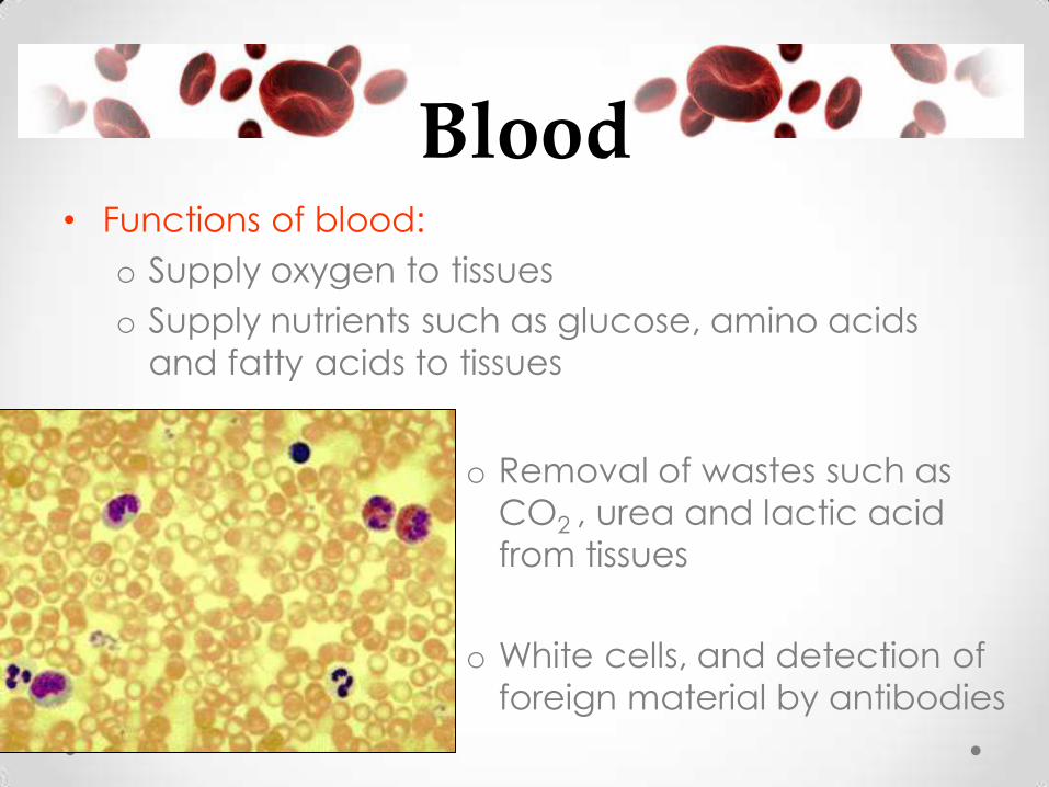
Blood • Functions of blood:
o Supply oxygen to tissues
o Supply nutrients such as glucose, amino acids
and fatty acids to tissues
o Removal of wastes such as
CO2 , urea and lactic acid
from tissues
o White cells, and detection of
foreign material by antibodies
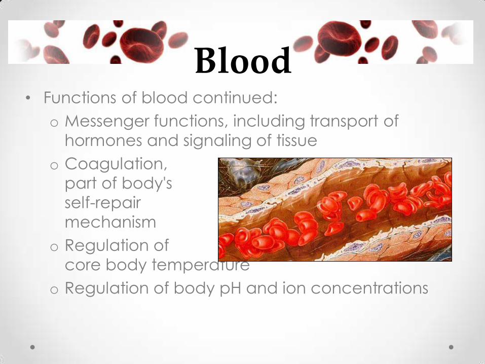
Blood • Functions of blood continued:
o Messenger functions, including transport of
hormones and signaling of tissue
o Coagulation,
part of body's
self-repair
mechanism
o Regulation of
core body temperature
o Regulation of body pH and ion concentrations

Blood • What percent of your body is blood?
• How much blood do we contain?
o On average 4-6 liters
o We contain about a pint of
blood for every 7 kg
of body weight
• Composition of Blood:
o What percent of your blood is
cellular?
o What percent of your blood is
plasma?
8%
45%
55%
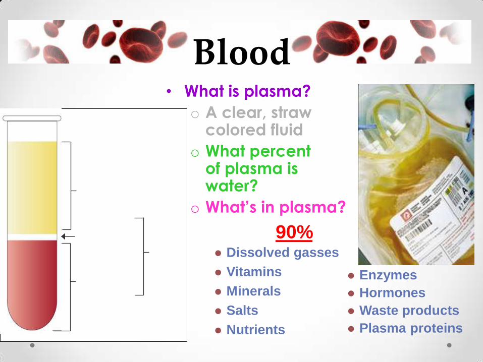
Blood • What is plasma?
o A clear, straw colored fluid
o What percent of plasma is water?
o What’s in plasma?
Dissolved gasses
Vitamins
Minerals
Salts
Nutrients
Enzymes
Hormones
Waste products
Plasma proteins
90% Buffy coat leukocytes
and platelets
(<1% of whole blood)
Erythrocytes
(45% of whole blood)
Plasma
(55% of whole blood)
Formed
elements

Blood • The cellular components are:
o red blood cells
o white blood cells
o platelets
• Blood cells are
formed in bone
marrow

Blood • Red Blood Cell (RBC) Characteristics
o Biconcave disks
o No nucleus
o Contain the iron based pigment hemoglobin
which binds with oxygen to transport it
o Life span about 120 days
o 5 billion/1mL of blood = most numerous
o Are very small
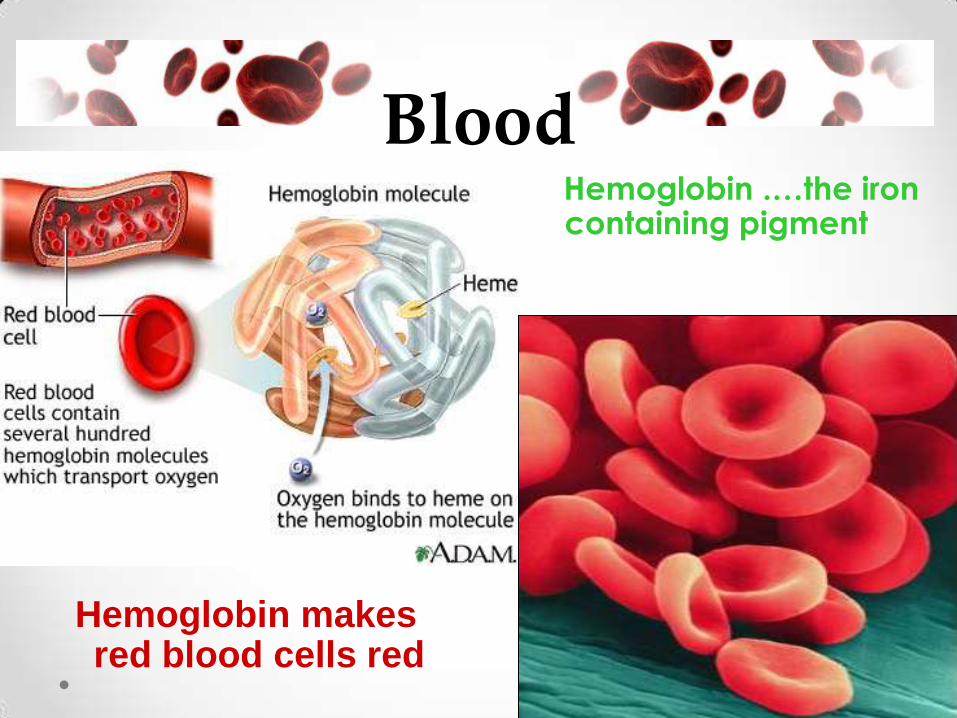
Blood Hemoglobin .…the iron
containing pigment
Hemoglobin makes red blood cells red

Blood • White Blood Cell (WBC)
Characteristics oNo definite shape
oHave nucleus
o Protect body against infection
o Life span varies (3 days-a few months)
o 7,000/1mL of blood
oNumbers increase if
infection is present
o Larger than RBC’s

Blood • Types of white blood cells:
o Monocytes are the
largest
o Neutrophils are the
most numerous
o Lymphocytes are
produced by the
lymph tissue
o Basophils release
histamines

Blood • Types of white blood cells:
The role of monocytes and neutrophyllis are to
phagocytize (engulf and then digest) cellular debris
and pathogens.
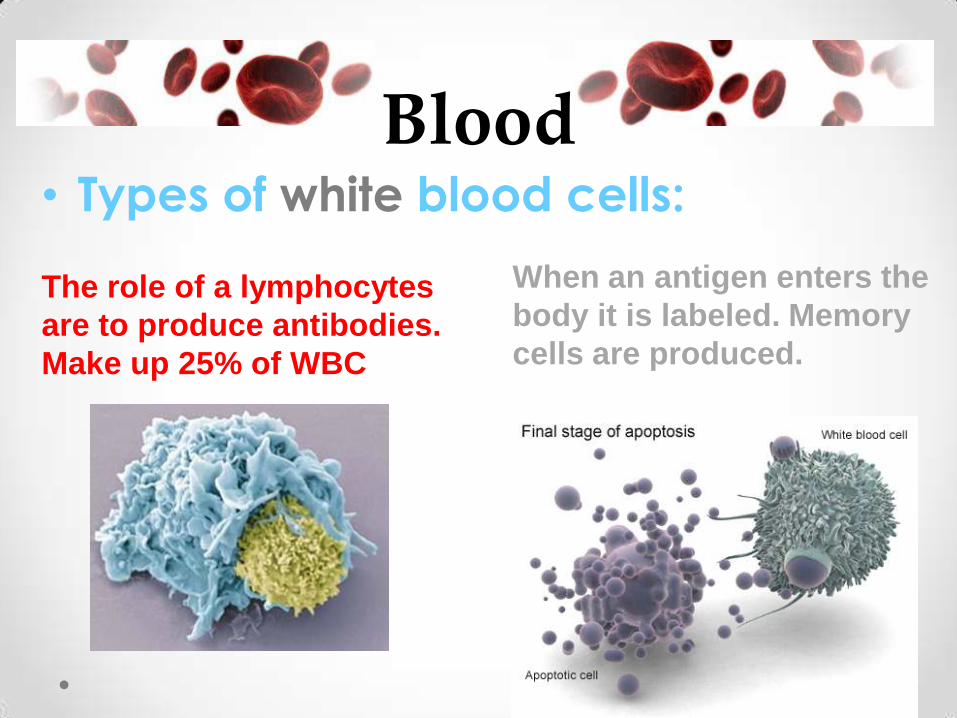
Blood • Types of white blood cells:
When an antigen enters the
body it is labeled. Memory
cells are produced.
The role of a lymphocytes
are to produce antibodies.
Make up 25% of WBC
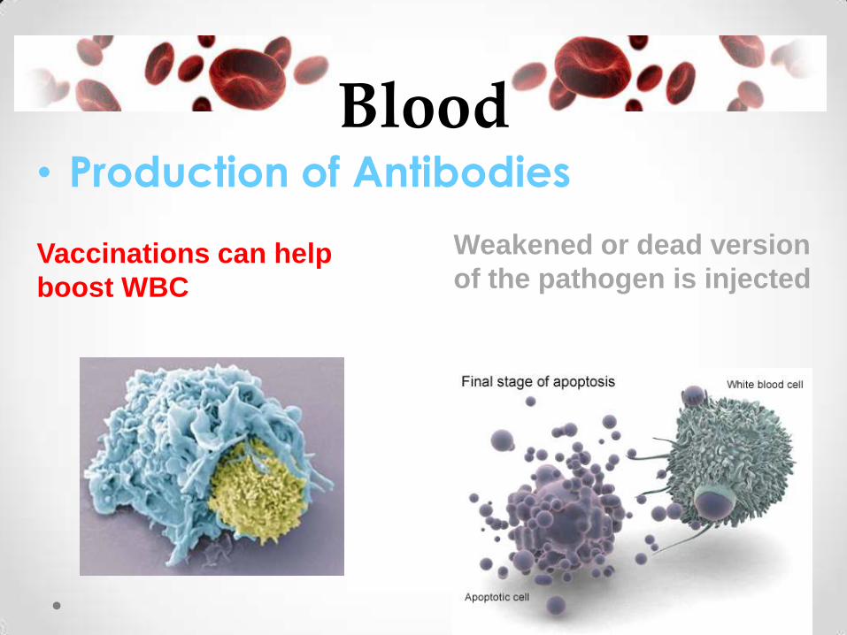
Blood • Production of Antibodies
Weakened or dead version
of the pathogen is injected Vaccinations can help
boost WBC

Blood • Vaccinations
Vaccination tricks the
lymphocytes to produce
memory cells and
antibodies
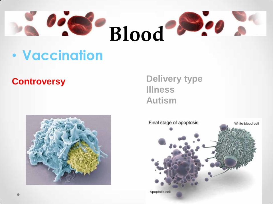
Blood • Vaccination
Delivery type
Illness
Autism
Controversy

Blood • Platelet Characteristics:
o RBC fragments
o Irregularly shaped
o No nucleus
o 150,000-400,000/1mL
o Life span about 7-11 days
o Have a sticky surface
o Responsible for blood clotting (injury healing)

Blood • This is an actual picture of White Blood Cells,
in with some red blood cells. The platelets are stained purple, a T-Lymphocyte white cell is stained green, and a Monocyte white cell is stained gold as seen through a scanning electron microscope.

Blood Clotting • Steps in Blood Clotting:
• platelets clump
• platelets release thromboblastin
• thromboblastin
produces thrombin
• thrombin converts
fibrinogen into fibrin
• fibrin causes a clot
Fibrin
Let’s simplify
this shall we?

Blood Clotting • Blood vessel is injured.
• Platelets clump at the site and produce a substance that produces strands of fibrin.
• Fibrin strands help
to clog the opening or hole in the vessel.

Circulatory System Disorders
• Risk factors
o Older age
o Male gender
o Cigarette smoking
o High cholesterol
o Diabetes
o Stress
o Obesity
o Heredity
o Physical inactivity
o High blood pressure
Heart Disease Quitting smoking, a healthy diet and exercise may
reduce your risk of heart disease
Plaque in
coronary
artery

Circulatory System Disorders
• Starts with damage or injury to the inner layer of an artery
• Fatty deposits called plaque build up in the arteries
• This causes:
o Blockage in artery
o Less flexible vessels
o High Blood Pressure
Atherosclerosis
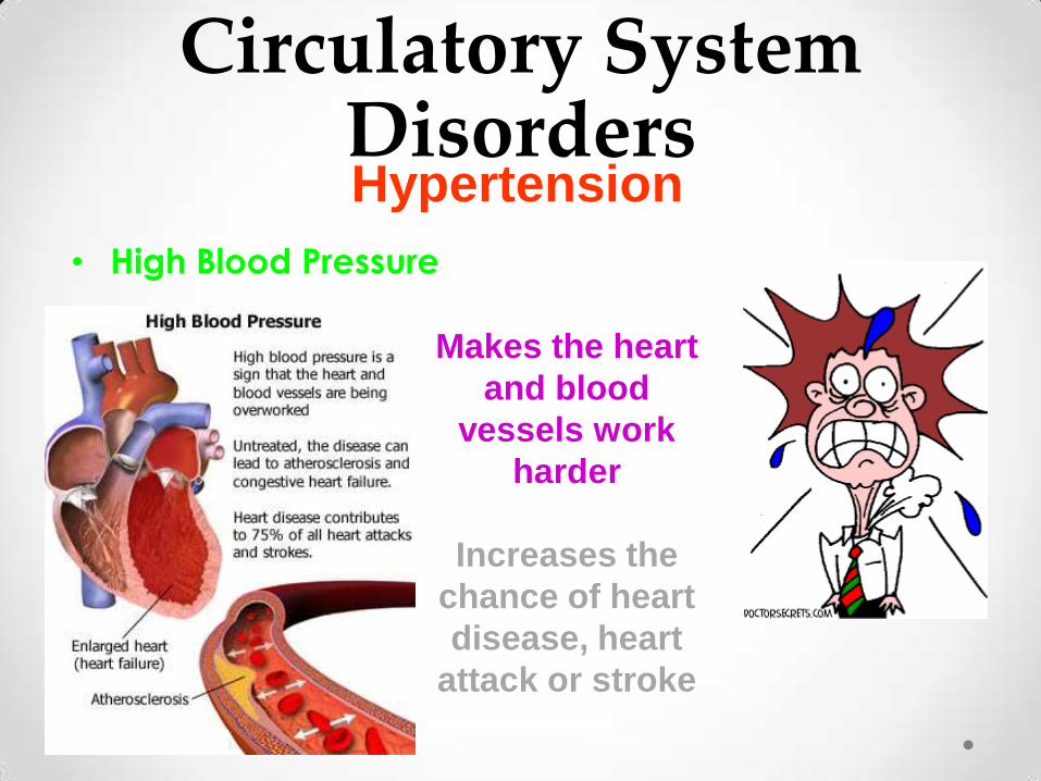
Circulatory System Disorders
• High Blood Pressure
Hypertension
Makes the heart
and blood
vessels work
harder
Increases the
chance of heart
disease, heart
attack or stroke
Circulatory System Disorders
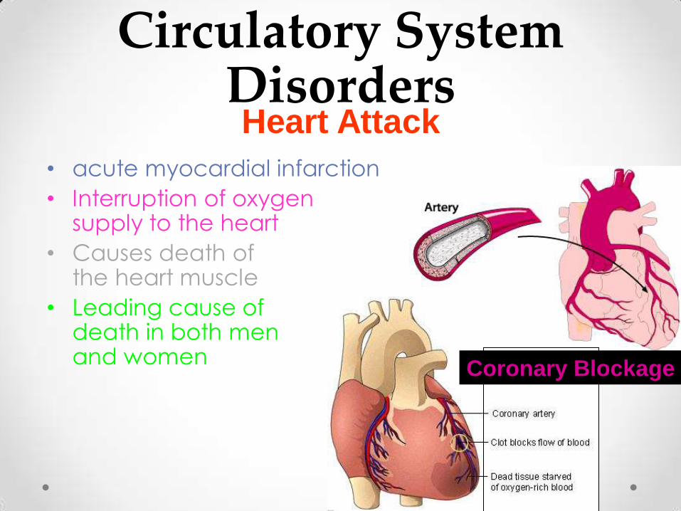
• acute myocardial infarction
• Interruption of oxygen supply to the heart
• Causes death of the heart muscle
• Leading cause of death in both men and women
Heart Attack
Coronary Blockage
Circulatory System Disorders
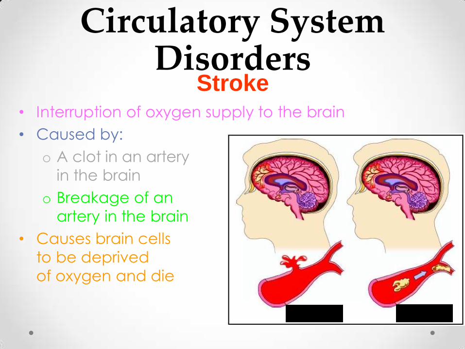
• Interruption of oxygen supply to the brain
• Caused by:
o A clot in an artery
in the brain
o Breakage of an
artery in the brain
• Causes brain cells
to be deprived
of oxygen and die
Stroke
Circulatory System Disorders
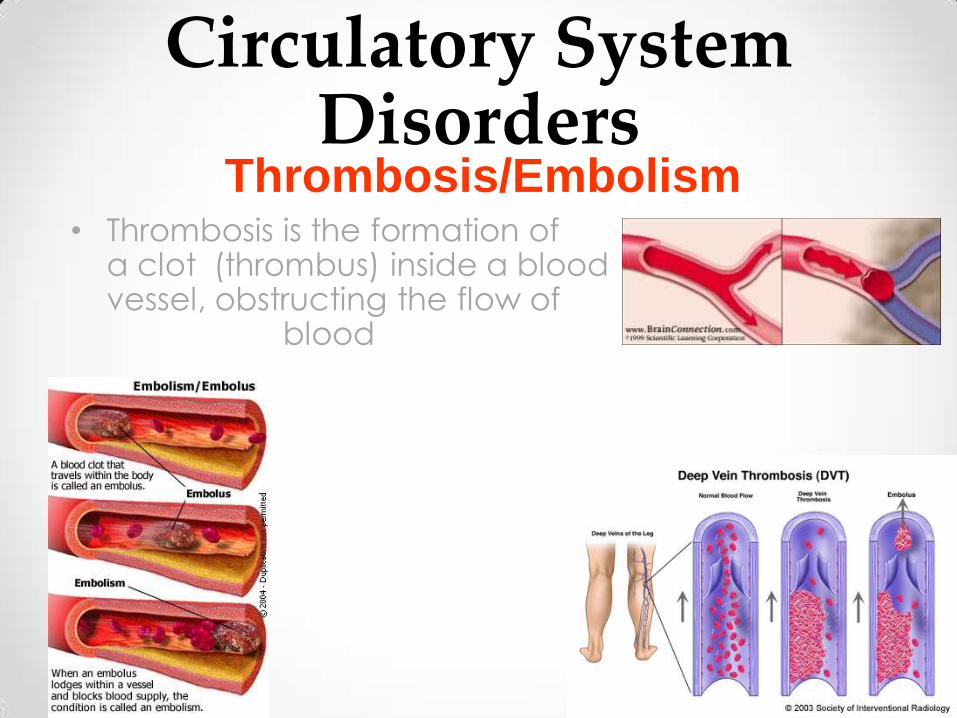
• Thrombosis is the formation of a clot (thrombus) inside a blood vessel, obstructing the flow of blood
Thrombosis/Embolism

Circulatory System Disorders
• The loss of blood from the body
• Hematoma- a collection of blood due to internal bleeding (burse)
Hemorrhage
Gingival Hemorrhage

Circulatory System Disorders
• A rare inherited bleeding disorder in which the blood does not clot normally
• The person is missing or has low levels of certain
proteins in the blood called clotting factors
• Usually occurs only in males
Hem philia
Swelling in left knee joint
due to spontaneous bleeding

Circulatory System Disorders
• Abnormally low number of red blood cells circulating in the body or when the blood does not have enough hemoglobin
• There are different kinds of anemia
o Iron Deficiency
o Vitamin Deficiency
o Hemolytic Anemias
o Sickle Cell Anemia
Anemia

Circulatory System Disorders
• Sickle cell trait- The person is carrying the defective gene, but also has some normal hemoglobin
• Sickle cell anemia- The person has most or all of the normal hemoglobin replaced with the sickle hemoglobin
Sickle Cell Disease
Circulatory System Disorders
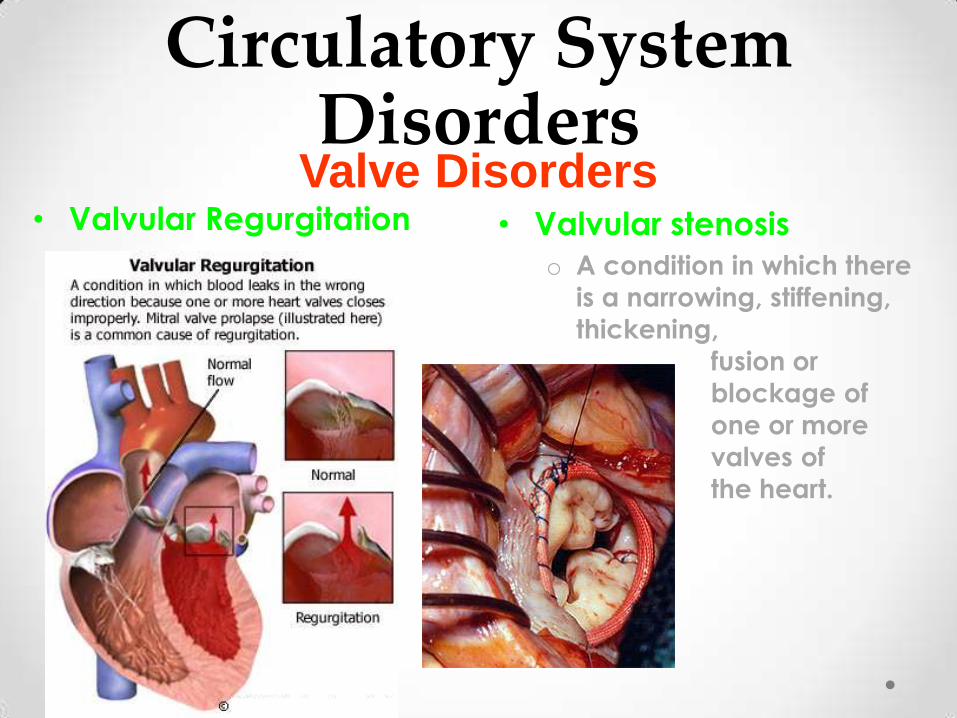
• Valvular Regurgitation • Valvular stenosis
o A condition in which there
is a narrowing, stiffening,
thickening,
fusion or
blockage of
one or more
valves of
the heart.
Valve Disorders
Circulatory System Disorders
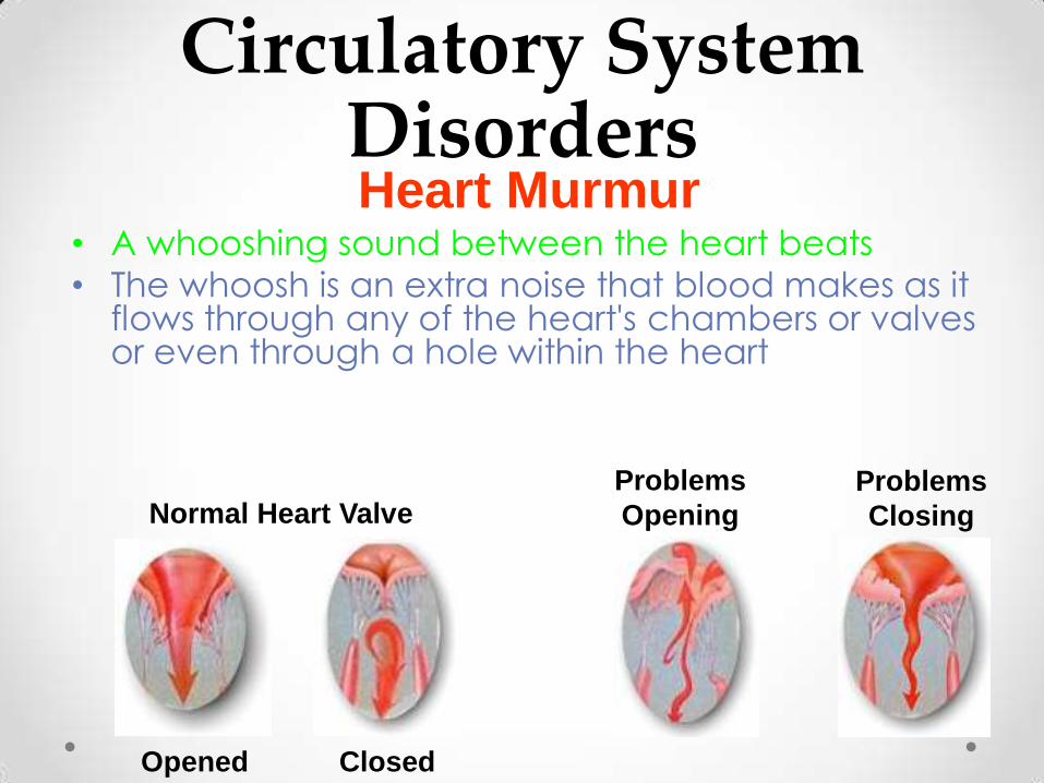
• A whooshing sound between the heart beats
• The whoosh is an extra noise that blood makes as it flows through any of the heart's chambers or valves or even through a hole within the heart
Heart Murmur
Normal Heart Valve
Closed Opened
Problems
Closing
Problems
Opening


![Circulatory System. Figure 24.01 Transports materials throughout body: Nutrients Metabolic wastes Gases (O 2 & CO 2 ) Hormones [regulate body processes]](https://img.pdfslide.us/doc/110x75/56649f285503460f94c4148f/circulatory-system-figure-2401-transports-materials-throughout-body-nutrients.jpg)