Embed Size (px)
Citation preview
Circulatory Systems
Transport & Maintenance

Circulatory Systems• transport to & from tissues
– nutrients, O2; waste, CO2
– hormones• maintain electrolyte balance of intercellular
fluid• transport to/from homeostatic organs
– small intestine delivers nutrients– liver removes wastes, controls nutrients– kidney controls electrolytes, dumps wastes
Circulatory Systems• some animals lack circulatory systems
– aquatic environment fulfills same functions• some animals have open circulatory systems
– the heart pumps interstitial fluid– vessels deliver interstitial fluid to tissues– interstitial fluids leave the vessels & bathe
the cells of the tissues– interstitial fluids return to the heart
• other animals have closed circulatory systems
open circulatory
systemsFigure 49.1
closed circulatory
system of
earthwormFigure 49.2
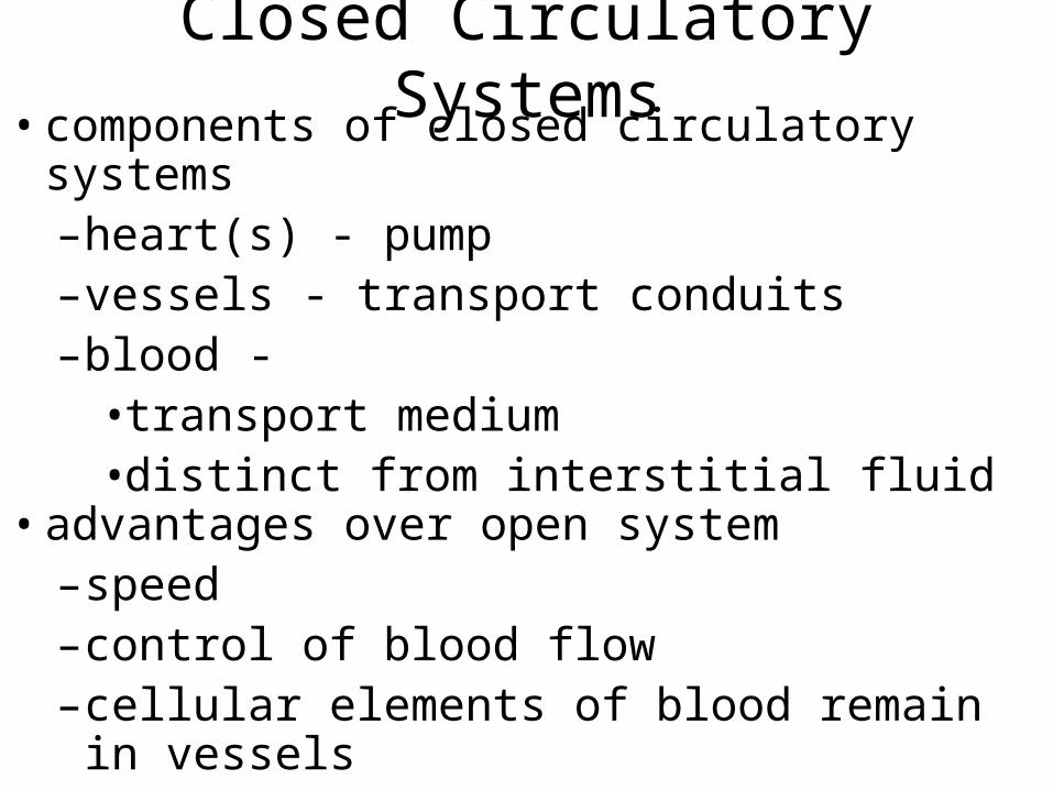
Closed Circulatory Systems• components of closed circulatory systems
– heart(s) - pump– vessels - transport conduits– blood -
• transport medium• distinct from interstitial fluid
• advantages over open system– speed– control of blood flow– cellular elements of blood remain in vessels
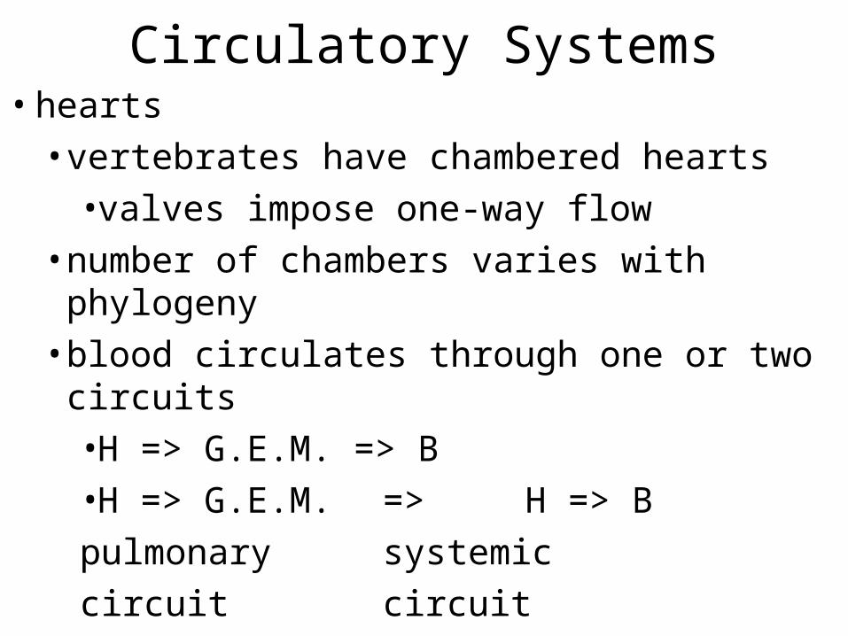
Circulatory Systems• hearts
• vertebrates have chambered hearts
• valves impose one-way flow
• number of chambers varies with phylogeny
• blood circulates through one or two circuits
• H => G.E.M. => B
• H => G.E.M. => H => B
pulmonary systemic
circuit circuit
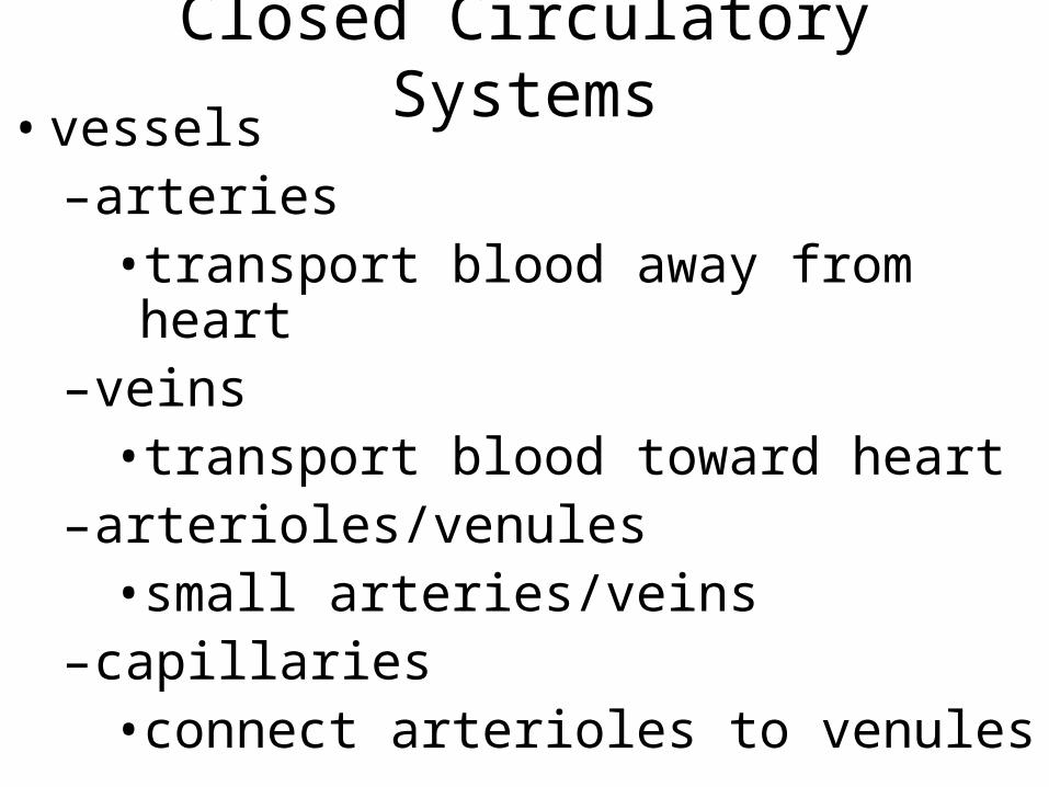
Closed Circulatory Systems• vessels
– arteries • transport blood away from heart
– veins • transport blood toward heart
– arterioles/venules• small arteries/veins
– capillaries• connect arterioles to venules
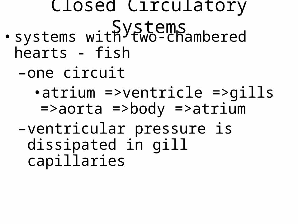
Closed Circulatory Systems• systems with two-chambered hearts - fish
– one circuit• atrium =>ventricle =>gills =>aorta =>body =>atrium
– ventricular pressure is dissipated in gill capillaries
fish circulation schematic
p. 943
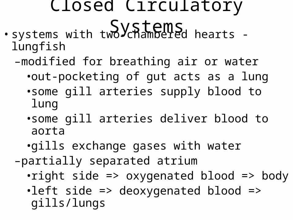
Closed Circulatory Systems• systems with two-chambered hearts - lungfish
– modified for breathing air or water• out-pocketing of gut acts as a lung• some gill arteries supply blood to lung• some gill arteries deliver blood to aorta• gills exchange gases with water
– partially separated atrium• right side => oxygenated blood => body• left side => deoxygenated blood => gills/lungs
lungfish circulation schematicp. 943
* one pair of gill arteries delivers blood to lung* two gill arches deliver blood directly to aorta* “gilled” gill arches exchange gases with blood
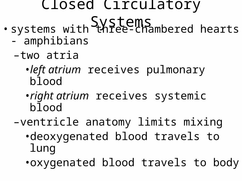
Closed Circulatory Systems• systems with three-chambered hearts -
amphibians– two atria
• left atrium receives pulmonary blood• right atrium receives systemic blood
– ventricle anatomy limits mixing• deoxygenated blood travels to lung• oxygenated blood travels to body
amphibian circulation schematicp. 943
Closed Circulatory Systems• reptilian hearts provide further control
– two atria receive blood from pulmonary & systemic circuits
– partially separated ventricle supplies three vessels• pulmonary artery & two aortas
–when breathing, the right aorta carries deoxygenated blood to the pulmonary circuit
–when not breathing, both aortas carry blood to the systemic circuit
reptilian circulation schematicp. 944
Closed Circulatory Systems• crocodilian hearts have four chambers
– two atria, two ventricles, two aortas• two aortas are bridged near their origins• when breathing, the left ventricle (& aorta) pressure is higher–deoxygenated blood goes to lungs
• when not breathing, right aorta pressure is higher–pulmonary circuit is bypassed
crocodilian schematicp. 944
Closed Circulatory Systems• endotherm hearts have four chambers and one
aorta– systemic/pulmonary circuits are separated– tissues receive highest possible [O2] (P1)
under high pressure– lungs receive lowest possible [O2] (P2)
under lower pressure
endotherm schematic
p. 945
human circulatory
systemFigure 49.3
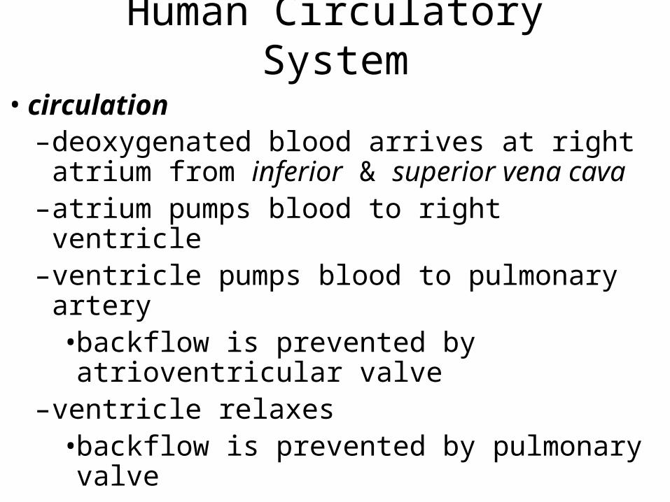
Human Circulatory System
• circulation– deoxygenated blood arrives at right atrium
from inferior & superior vena cava– atrium pumps blood to right ventricle– ventricle pumps blood to pulmonary artery
• backflow is prevented by atrioventricular valve
– ventricle relaxes• backflow is prevented by pulmonary valve
human heart anatomyFigure 49.3
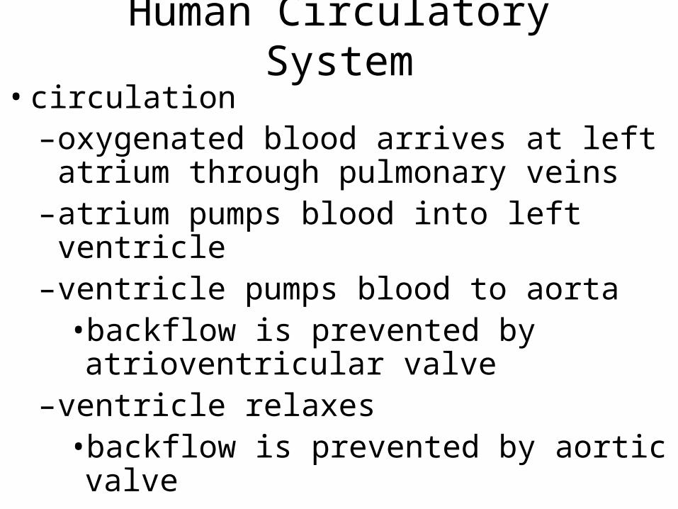
Human Circulatory System
• circulation– oxygenated blood arrives at left atrium
through pulmonary veins– atrium pumps blood into left ventricle– ventricle pumps blood to aorta
• backflow is prevented by atrioventricular valve
– ventricle relaxes• backflow is prevented by aortic valve
human heart anatomyFigure 49.3
Human Circulatory System• cardiac cycle
– systole - contraction of ventricles• maximum pressure generated• major electrical event
– diastole - relaxation of ventricles• minimum pressure• characteristic electrical signatures
ventricular pressures & volumes
Figure 49.4
measuring blood pressureFigure 49.5
Human Circulatory System• heartbeat is myogenic
– pacemaker cells occur at sinoatrial node• resting membrane potential depolarizes• at threshold, voltage gated Ca2+ channels open
• K+ channels open to repolarize cells• K+ channels close slowly, allow gradual depolarization
– autonomic nervous system regulates the rate of depolarization
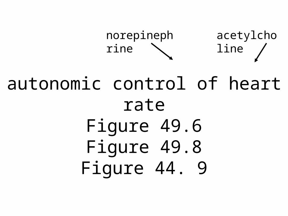
autonomic control of heart rateFigure 49.6Figure 49.8Figure 44. 9
norepinephrine acetylcholine
Human Circulatory System• contraction
– the pacemaker action potential spreads across the atrial walls
– atria contract– action potential is transmitted to ventricles
through the atrioventricular node and the bundle of His
– the action potential spreads to Purkinje fibers in ventricular muscle
– ventricles contract
origin and
spread of
cardiac contractionFigure 49.7
Human Circulatory System• vascular system
– arteries carry blood from heart• elastic tissues absorb pressure of heart contractions
• smooth muscle allows control of blood flow by neural and hormonal signals
artery structureFigure 49.10
Human Circulatory System• vascular system
– capillaries • fed by arterioles; drained by venules• exchange materials between blood & intercellular fluids–high total capacity; slow flow–thin walls
capillary bedFigure 49.10
Human Circulatory System• vascular system
– capillaries • exchange materials by filtration, osmosis & diffusion–water & solutes move through capillary
walls under pressure on the arteriole side
–remaining solutes & diffusing CO2 produce a low osmotic potential
–water returns to capillaries on the venule side

water movement balanced between blood pressure
& osmotic potential
Figure 49.12
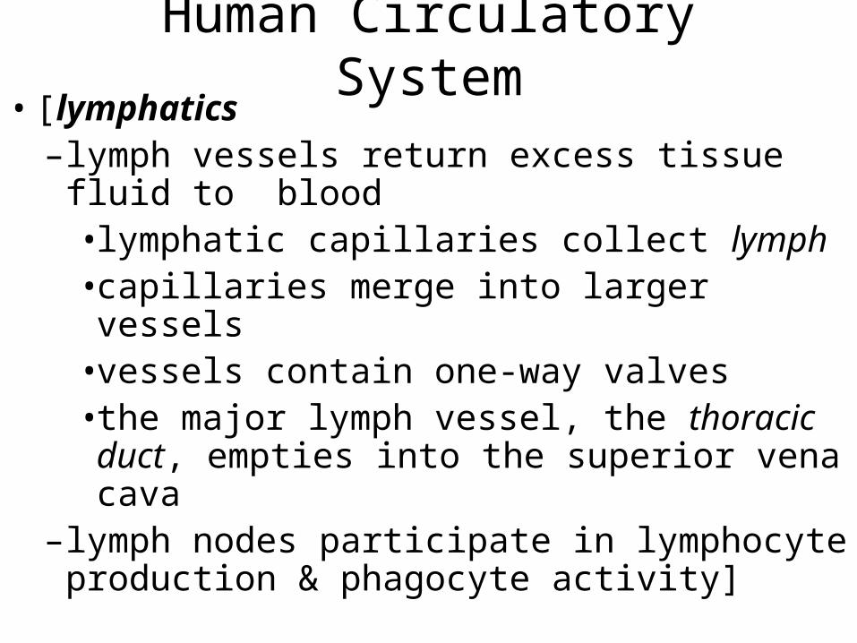
Human Circulatory System• [lymphatics
– lymph vessels return excess tissue fluid to blood• lymphatic capillaries collect lymph• capillaries merge into larger vessels• vessels contain one-way valves• the major lymph vessel, the thoracic duct, empties into the superior vena cava
– lymph nodes participate in lymphocyte production & phagocyte activity]
vein structureFigure 49.10
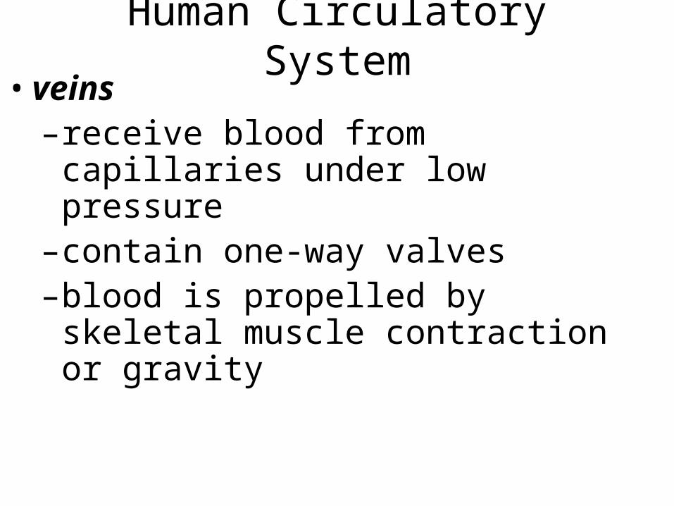
Human Circulatory System• veins
– receive blood from capillaries under low pressure
– contain one-way valves– blood is propelled by skeletal muscle
contraction or gravity
venous return by skeletal muscle contraction and one-way valves
Human Circulatory System• blood - a fluid connective tissue
– fluid matrix - plasma• dissolved gases, ions, proteins, nutrients, hormones, etc.
• many components found in tissue fluid– cellular elements
• red blood cells (erythrocytes)• white blood cells (leukocytes)• platelets
blood componentsFigure 49.15
human blood samplesbeforeand after
centrifugation to separate red blood cells from serum
Human Circulatory System• control & regulation of circulation
– capillaries are subject to auto-regulation• pre-capillary sphincters and arterial smooth muscle are sensitive to –O2 & CO2 concentrations–accumulated waste materials
local control of blood flowFigure 49.17
Human Circulatory System• control & regulation of circulation
– simultaneous auto-regulation of capillary beds produces systemic responses• changes in breathing, heart rate• changes in blood distribution
– systemic control is neural or hormonal• sympathetic stimulation contracts most arteries; dilates skeletal muscle arteries
• hormones constrict arteries in targeted tissues
circulatory regulation at two levelsFigure 49.18
Human Circulatory System• control & regulation of circulation
– autonomic control of circulation originates in medulla of brain stem• inputs arrive from
–stretch receptors–chemosensors–higher brain centers
• responses may be –direct - artery relaxation or contraction–indirect - release of epinephrine
neural control of circulation is
centered in the medullaFigure 49.19