Embed Size (px)
Citation preview
www.sciencedirect.com
c o r t e x x x x ( 2 0 1 2 ) 1e1 2
Available online at
Journal homepage: www.elsevier.com/locate/cortex
Clinical neuroanatomy
The anatomy of fronto-occipital connections from early bluntdissections to contemporary tractography
Stephanie J. Forkel a,b,*, Michel Thiebaut de Schotten a,c, Jamie M. Kawadler a,Flavio Dell’Acqua b,d, Adrian Danek e and Marco Catani a
aNATBRAINLAB, Department of Forensics and Neurodevelopmental Sciences, King’s College London, Institute of Psychiatry, UKbDepartment of Neuroimaging Sciences, King’s College London, Institute of Psychiatry, UKc Inserm e UPMC UMRS 975, Brain and Spine Institute, Groupe Hospitalier Pitie-Salpetriere, Paris, FrancedNIHR Biomedical Research Centre for Mental Health at South London and Maudsley NHS Foundation Trust and Institute of Psychiatry,
King’s College London, UKeNeurology Department, Ludwig-Maximilians-Universitat Munich, Germany
a r t i c l e i n f o
Article history:
Received 4 April 2012
Reviewed 23 May 2012
Revised 15 August 2012
Accepted 7 September 2012
Action editor Gereon Fink
Published online xxx
Keywords:
Fronto-occipital fasciculus
Diffusion MRI
Tractography
White matter
Neuroanatomy
Agenesis of the corpus callosum
* Corresponding author. NATBRAINLAB, DepPsychiatry, London, UK.
E-mail addresses: stephanie.forkel@gmx.
Please cite this article in press as: Forkelcontemporary tractography, Cortex (2012
0010-9452/$ e see front matter ª 2012 Elsevhttp://dx.doi.org/10.1016/j.cortex.2012.09.005
a b s t r a c t
The occipital and frontal lobes are anatomically distant yet functionally highly integrated
to generate some of the most complex behaviour. A series of long associative fibres, such as
the fronto-occipital networks, mediate this integration via rapid feed-forward propagation
of visual input to anterior frontal regions and direct topedown modulation of early visual
processing.
Despite the vast number of anatomical investigations a general consensus on the
anatomy of fronto-occipital connections is not forthcoming. For example, in the monkey
the existence of a human equivalent of the ‘inferior fronto-occipital fasciculus’ (iFOF) has
not been demonstrated. Conversely, a ‘superior fronto-occipital fasciculus’ (sFOF), also
referred to as ‘subcallosal bundle’ by some authors, is reported in monkey axonal tracing
studies but not in human dissections.
In this study our aim is twofold. First, we use diffusion tractography to delineate the
in vivo anatomy of the sFOF and the iFOF in 30 healthy subjects and three acallosal brains.
Second, we provide a comprehensive review of the post-mortem and neuroimaging studies
of the fronto-occipital connections published over the last two centuries, together with the
first integral translation of Onufrowicz’s original description of a human fronto-occipital
fasciculus (1887) and Muratoff’s report of the ‘subcallosal bundle’ in animals (1893).
Our tractography dissections suggest that in the human brain (i) the iFOF is a bilateral
association pathway connecting ventro-medial occipital cortex to orbital and polar frontal
cortex, (ii) the sFOF overlaps with branches of the superior longitudinal fasciculus (SLF) and
probably represents an ‘occipital extension’ of the SLF, (iii) the subcallosal bundle of
Muratoff is probably a complex tract encompassing ascending thalamo-frontal and
descending fronto-caudate connections and is therefore a projection rather than an
associative tract.
artment of Forensics and Neurodevelopmental Sciences, King’s College London, Institute of
net, [email protected] (S.J. Forkel).
SJ, et al., The anatomy of fronto-occipital connections from early blunt dissections to), http://dx.doi.org/10.1016/j.cortex.2012.09.005
ier Srl. All rights reserved.
c o r t e x x x x ( 2 0 1 2 ) 1e1 22
Please cite this article in press as: Forkelcontemporary tractography, Cortex (2012
In conclusion, our experimental findings and review of the literature suggest that
a ventral pathway in humans, namely the iFOF, mediates a direct communication between
occipital and frontal lobes. Whether the iFOF represents a unique human pathway awaits
further ad hoc investigations in animals.
ª 2012 Elsevier Srl. All rights reserved.
1. Introduction tractography dissections of the dorsal and ventral fronto-
The occipital lobes have been intensively investigated in the
last two centuries and many aspects have been clarified with
regard to their anatomy and function. Comparative anatomy
and neurophysiology studies suggest that the occipital lobes
have undergone a complex rearrangement along the
phylogeny scale (Rapoport, 1990; Orban et al., 2004). Primary
visual areas are relatively smaller in humans with the
expansion or addition of associative areas specialised, for
example, in face perception (i.e., ‘fusiform face area’) or word
recognition (i.e., ‘visual word form area’) (Cohen et al., 2000;
Epelbaum et al., 2008). It has been proposed that these rela-
tively ‘new areas of the human brain’ cooperate with more
distant regions through long association tracts, whereas
evolutionary old areas communicate through short associa-
tion fibres (Deacon, 1990).
In the human brain a large system of long range associative
connections, encompassing the inferior longitudinal fascic-
ulus (ILF), cingulum and inferior fronto-occipital fasciculus
(iFOF), mediates fast feed-forward relay of visual input to
anterior multimodal temporal, parietal and frontal regions
and direct topedown modulation by these regions on early
visual areas. The anatomy of the ILF and cingulum has been
described in detail both in human and monkey brains,
whereas the iFOF has been identified only in humans
(Schmahmann et al., 2007; Catani, 2007; Umarova et al., 2010;
Yeterian et al., 2012; Thiebaut de Schotten et al., 2012). The
controversy further involves a dorsal fronto-occipital fascic-
ulus, namely the superior fronto-occipital fasciculus (sFOF),
which has been described in monkey but not in humans
(Schmahmann et al., 2007; Thiebaut de Schotten et al., 2012).
To further complicate the matter, a dorsal fronto-occipital
fasciculus is found in human brains lacking the interhemi-
spheric callosal connection (congenital agenesis), but not in
healthy brains. This bundle is often referred to with the
eponym ‘Probst bundle’. These humanesimian discrepancies
may be attributed to: (i) methodological limitations of post-
mortem and in vivo dissections; (ii) inaccuracy of anatomical
terms to indicate the same tracts in different species; (iii)
influence of pathological processes on tract development and
(iv) true interspecies differences.
In this study we intend to address the following questions:
(i) do dorsal fronto-occipital connections described in the
monkey and acallosal brains exist in healthy human subjects?
(ii) are ventral fronto-occipital connections unique to
humans?
To answer these questions we first review the experi-
mental evidence for the existence of dorsal and ventral
connections between occipital and frontal lobes in the normal
brain, both in animals and humans. Second, we performed
SJ, et al., The anatomy o), http://dx.doi.org/10.10
occipital fasciculus connections using a novel diffusion
imaging approach based on spherical deconvolution (SD),
which overcomes some of the limitations of the tensor model
(Dell’Acqua et al., 2010; Thiebaut de Schotten et al., 2011a,
2011b, 2012; Dell’Acqua and Catani, 2012). Third, we provide
dissections of the fronto-occipital connections in subjects
with congenital agenesis of the corpus callosum and discuss
them in the light of the literature. Finally, the first Ger-
maneEnglish translation of two seminal papers on the dorsal
fronto-occipital connections is provided to clarify current
nomenclature. The first of the two is the doctoral dissertation
by Onufrowicz (1887) where the term fronto-occipital fascic-
ulus is used to describe, for the first time, a dorsal connection
in an acallosal patient. The second paper is Muratoff’s (1893)
experimental demonstration of a dorsal connection, the sub-
callosal bundle, in the animal brain.
Our hope is to clarify the anatomy and the history of the
fronto-occipital connections and stimulate further functional
and anatomical studies.
2. Methods
2.1. Diffusion tractography of healthy subjects
A High Angular Resolution Diffusion Imaging (HARDI)
sequence optimised for SD was used to acquire 30 datasets
from healthy volunteers (aged 23e37 years, 17 males) on a 3T
GE Signa HDx (General Electric, Milwaukee, WI, USA). For each
subject, a Spin Echo diffusion-weighted echo planar imaging
(EPI) sequence was also acquired with the following parame-
ters: voxel size 2.4 � 2.4 � 2.4 mm, matrix 128 � 128, slices 60,
NEX 1, TE 93 msec, b-value ¼ 3000 sec/mm2, 60 diffusion
weighted directions and seven non-diffusion weighted
volumes. Cardiac gatingwas applied with effective TR of 20/30
ReR intervals (Dell’Acqua et al., in press; Thiebaut de Schotten
et al., 2011a, 2011b).
Data were corrected for head-motion and eddy current
distortion using the FSL software package (FMRIB Software
Library, Release 4.1, The University of Oxford). Fibre orienta-
tion distribution (FOD) was estimated using an SD approach
based on the damped version of the RichardsoneLucy SD
algorithm, which reduces partial volume effects and spurious
fibre orientations (Dell’Acqua et al., 2010). Algorithm param-
eterswere chosen as described in Dell’Acqua et al. (2010). Fibre
orientation estimates were obtained selecting the orientation
corresponding to the local maxima of the FOD profile. To
exclude spurious local maxima two mask thresholds were
applied to the FOD amplitudes. A first “absolute” threshold
(corresponding to three times the amplitude of a spherical
f fronto-occipital connections from early blunt dissections to16/j.cortex.2012.09.005
c o r t e x x x x ( 2 0 1 2 ) 1e1 2 3
FOD obtained from a grey matter isotropic voxel) was used to
exclude small local maxima due to noise or isotropic tissue. A
second “relative” threshold of 5% of the maximum amplitude
of the FOD was applied to remove the remaining spurious
local maxima with values greater than the absolute threshold
(Dell’Acqua et al., 2009, in press).
2.2. Tractography algorithm
Whole brain tractography was performed selecting all voxels
with at least one fibre orientation. From these voxels and for
each fibre orientation the Modified Fibre Assignment by
Continuous Tracking (M-FACT) algorithm was used to propa-
gate the streamlines (Descoteaux et al., 2009; Mori et al., 1999).
In regions with crossing white matter bundles the algo-
rithm followed the orientation of least curvature as described
by Schmahmann et al. (2007). Streamlines were halted when
a voxel without fibre orientation was reached or when the
curvature between two steps exceeded a threshold of 45�.
2.3. Delineation of regions of interest (ROIs)
Three ROIs, delineated on coronal planes, were used to dissect
the fronto-occipital connections.
An occipital region was placed on the white matter of
occipital lobe posterior to the parieto-occipital sulcus and the
temporo-occipital notch. Two anterior ROIs were defined on
the white matter of the frontal lobes: a ventral ROI was
delineated on the white matter of the external/extreme
capsule; a dorsal ROI was placed anterior to the central sulcus
on the white matter of precentral gyrus and centrum semi-
ovale. All streamlines between occipital and ventral frontal
ROIs were labelled as iFOF. All streamlines between occipital
and dorsal frontal ROIs were labelled as sFOF. ROIs are shown
in Supplementary material.
2.4. Percentage overlay maps
For each individual tractography reconstruction a binary
map was computed by assigning each pixel a value of 1 or
0 depending on whether the pixel was visited by at least one
tract streamlines or no streamlines. All binary maps were
spatially normalised to a standard space of reference
(Montreal Neurological Institute e MNI) using FSL linear
FMRIB’s linear image registration tool (FLIRT) and non-linear
FMRIB’s nonlinear image registration tool (FNIRT) deforma-
tions and summed to produce tract percentage overlap
maps (Catani et al., 2007; Thiebaut de Schotten et al., 2011a,
2011b). The percentage overlap maps were displayed on
a T1-weighted image from a representative subject for
anatomical reference.
2.5. Diffusion tractography in patients with agenesis ofthe corpus callosum
Three datasets were acquired from asymptomatic individuals
incidentally diagnosed with partial congenital agenesis of the
corpus callosum between 2006 and 2011 across different
centres in the UK (Institute of Psychiatry London and
University of Cambridge) and Germany (Munich, Klinikum
Please cite this article in press as: Forkel SJ, et al., The anatomy ocontemporary tractography, Cortex (2012), http://dx.doi.org/10.10
Großhadern, Ludwig-Maximilians-Universitat). The datasets
from the two English patients (29 years old and 8 years old
male) were acquired on a 3T GE Signa System (General-Elec-
tric, Milwaukee, WI, USA). High-resolution structural T1-
weighted volumetric images were acquired with full head
coverage, 196 contiguous slices (1.1 mm thickness, with
1.09 mm � 1.09 mm in-plane resolution), a 256 � 256 � 196
matrix and a repetition time/echo time (TR/TE) of 7/2.8 msec
[flip angle 8 degrees, field of view (FOV) 28 cm]. The above
diffusion sequence was similar to that used for the healthy
subjects except for the number of directions (30 instead of 60).
The third dataset from the Munich patient (46 years old
female) was also acquired on a 3T GE Signa System (General-
Electric, Milwaukee, WI, USA) with a diffusion sequence
similar to the one used for the English acallosal brains except
for the slightly lower b-value (1000 instead of 1300) and
anisotropic voxels (.94 � .94 � 2.4). Diffusion data were ana-
lysed using ExploreDTI (Leemans et al., 2009) and the analysis
consisted of: (i) correcting for eddy current distortion and
subject motion (Leemans and Jones, 2009); (ii) diffusion tensor
estimation using a non-linear least square method (Jones and
Basser, 2004), and (iii) whole brain tractography with a step-
size of 1 mm, fractional anisotropy (FA) threshold of .2 to
initiate and continue tracking, and an angle threshold of 35�
(Mori and van Zijl, 2002). Tractography of the callosal
remnants, arcuate fasciculus, cingulum, iFOF and sFOF was
performed on TrackVis (Ruopeng Wang, Van J. Wedeen,
TrackVis.org, Martinos Center for Biomedical Imaging,
Massachusetts General Hospital).
2.6. Literature review
A digital literature review was conducted using Internet
databases (i.e., PubMed, SCOPUS, and JSTOR). To ensure
a thorough search no exclusion criteria were applied and
multiple combinations of keywords were used: inferior,
fronto, occipital, fasciculus, bundle, tract, pathway, Probst
bundle, fronto occipitales Associationsbundel, long associa-
tion, Muratoff, Onufrowicz, Markfasern, white matter. Addi-
tionally, hand-searched original reports from the 19th century
were obtained from historical collections of several London-
based libraries, such as the Institute of Psychiatry at King’s
College London, the British library, and the Royal Society in
London, as well as several Germany-based university libraries
including the Anatomical Institute at Ludwig-Maximilians-
Universitat in Munich.
Original reports were translated by German (S.J.F., A.D.)
and French (M.T.S.) native speakers in collaboration with
a native English speaker (J.K.). Anatomical validity of the
translations was reviewed by two experts in anatomy (A.D.,
M.C.). For the canine neuroanatomy, assistance was sought
from a veterinarian, and templates indicating the anatomical
structuresmentioned in the original report are included in the
appendix of the translation to facilitate anatomical
understanding.
The nomenclature of the original reports was adapted to
the Nomina anatomica compiled by the International
Anatomical Nomenclature Committee (1989). Both original
and currently used terms are indicated in the text (e.g., gyrus
fornicatus e cingulate gyrus).
f fronto-occipital connections from early blunt dissections to16/j.cortex.2012.09.005
c o r t e x x x x ( 2 0 1 2 ) 1e1 24
3. Results
3.1. Literature review: inferior fronto-occipital fasciculus(iFOF)
The first mention of a direct connection between frontal and
occipital lobe is included in Burdach’s 1822 description of the
ILF, a tract connecting the occipital and temporal lobes that
was first described by Reil in 1809 as a projection pathway.
Burdach not only recognised the cortico-cortical (or associa-
tive) nature of the ILF but also described a subcomponent of
the tract connecting to the frontal lobe:
“In each hemisphere the fasciculus longitudinalis inferior [in the
original text: untre Langenbundel] extends along the base of the
corona radiata [in the original text: Stabkranz] and [forms] its
ventral border. These fibres run uninterruptedly from the occipital
pole through the temporal to the frontal pole and thereby form
a longitudinal bulge at the inferior base of the cerebrum. It is
slightly curved longitudinally, convex laterally and concave
medially. It forms an arch superiorly and at the level of the
external capsule, and in contrast to the uncinate fasciculus [in the
original text: Hakenbundel], it is slightly concave interiorly and
convex superiorly. Its fibres originate from the occipital pole and
extend anteriorly along the outmost wall of the inferior horn. [.]
One branch arches underneath the uncinate fasciculus anterior-
medially in the temporal pole; the remaining part bends
anterior-medially into the insula [in the original text: Stammlap-
pen]where it forms the floor of the external capsuleunderneath the
lentiform nucleus [in the original text: Linsenkern] and thence
somewhat arching laterally penetrates the frontal lobe. Here the
fibres run superiorly adjacent to the uncinate fasciculus and
extend to the lateral cortex of the frontal pole.” (1822, p. 152).
Burdach’s observation of a direct fronto-occipital compo-
nent of the ILFwas largely ignored for almost a century and the
term ILFwas adopted only for those fibres connecting occipital
and temporal lobes. At the end of the 19th century, descrip-
tions of the fronto-occipital connections re-emerged in the
French literature. Whilst Adrien Charpy’s (1895) description of
the fronto-occipital connections was similar to that of Karl
Burdach (1822), Jules Dejerine was the first to separate the
fronto-occipital fasciculus from the ILF and considered it as
a distinct bundle. However, Dejerine’s fronto-occipital fascic-
ulus had a different course compared to the occipito-frontal
fibres described by Burdach (Dejerine, 1895). In particular, he
located the central portion of the fronto-occipital fasciculus
above the caudate nucleus and its posterior projections along
the ventral surface of the occipital and temporal lobes. Dejer-
ine’s textbook was very influential but not everyone agreed
with his description. Jean Baptiste P. Trolard (Trolard, 1906;
Loukas et al., 2010), for example, whilst agreeing with Dejer-
ine’s view that the fronto-occipital fasciculus should be
considered as a distinct bundle, gave a description that closely
resembled the one of Burdach and Charpy:
“[.] so it would exist a proper bundle of fibres, a fronto-occipital
fascicle, both frontal and occipital parts being united by
a common fasciculus [.] placed above the middle portion of the
Please cite this article in press as: Forkel SJ, et al., The anatomy ocontemporary tractography, Cortex (2012), http://dx.doi.org/10.10
claustrum radiation. It would be possible to gather the uncinate
bundle alongwith the one just described. In our opinion, however,
it is best to assign to each its own personality.Weultimately think
there are two distinct fasciculi [inferior longitudinal and fronto-
occipital fasciculus] in the region just studied, although they
both end in the occipital lobe. In support of this view, we will not
only report their separation, so easily obtained with our prepa-
ration, butwewill also insist on their anterior terminations,which
are very different. Indeed the blade part belongs to the frontal lobe,
while the second belongs to the temporal lobe.” (1906, p. 446).
In 1909, the Irish-Australian physician Edward Curran was
able to replicate Burdach’s and Trolard’s findings whilst filling
a position as anatomy instructor in Chicago (Johnson et al.,
1995). Being completely unaware of the work of his German
and French predecessors, as it seems, Curran claimed the
fasciculus occipito-frontalis inferior (equivalent to the tract
today known as iFOF) to be “a new or hitherto undescribed tract in
the cerebrum” (1909, p. 651). Curran brought the iFOF into the
Anglo-American literature and produced its definite descrip-
tion that remains largely valid today:
“The fasciculus occipito-frontalis inferior is a large associating
bundle of fibres uniting, as its name indicates, the occipital with
the frontal lobe. It also contains fibres, which join the frontal lobe
with the posterior part of the temporal and parietal lobes. [.]
From all parts of the frontal lobe the fibres of this fasciculus can
be traced converging to a single bundle which swings round the
lower external side of the nucleus lentiformis, at which place it
appears as a distinct bundle [.]. I would emphasize the fact that
as it swings to the lower external side of the lenticular nucleus
and the external capsule it stands out with striking distinctness
and is at once recognized as a separate bundle isolated from the
surrounding structures by the directness and compactness of its
fibres” (1909, p. 652).
After Curran, the iFOF has been consistently identified in
post-mortem dissections in normal human brains, its exis-
tence being reported in at least 44 dissected hemispheres
(Davis, 1921; Elliott, 1947; Crosby et al., 1962; Hultkrantz, 1929;
Ebeling and von Cramon, 1992; Kier et al., 2004; Fernandez-
Miranda et al., 2008; Lawes et al., 2008; Martino et al., 2010,
2011, Fig. 1).
3.2. Literature review: superior fronto-occipitalfasciculus (sFOF)
3.2.1. The sFOF in the acallosal brainAcallosal brains are characterised by the complete or partial
absence of commissural fibres (corpus callosum) connecting
the two cerebral hemispheres. The absence of callosal fibres is
often accompanied by the presence of a prominent dorsal
bundle connecting occipital to parietal and frontal regions. As
early as 1878, Eichler suggested that a longitudinal ridge,
which he was able to appreciate bilaterally in his acallosal
patient, was indeed a bundle of callosal fibres that failed to
transverse the interhemispheric midline:
‘In an attempt to interpret our case, it remainswithout doubt that
the longitudinal ridge, whose radiations form the inner white
f fronto-occipital connections from early blunt dissections to16/j.cortex.2012.09.005
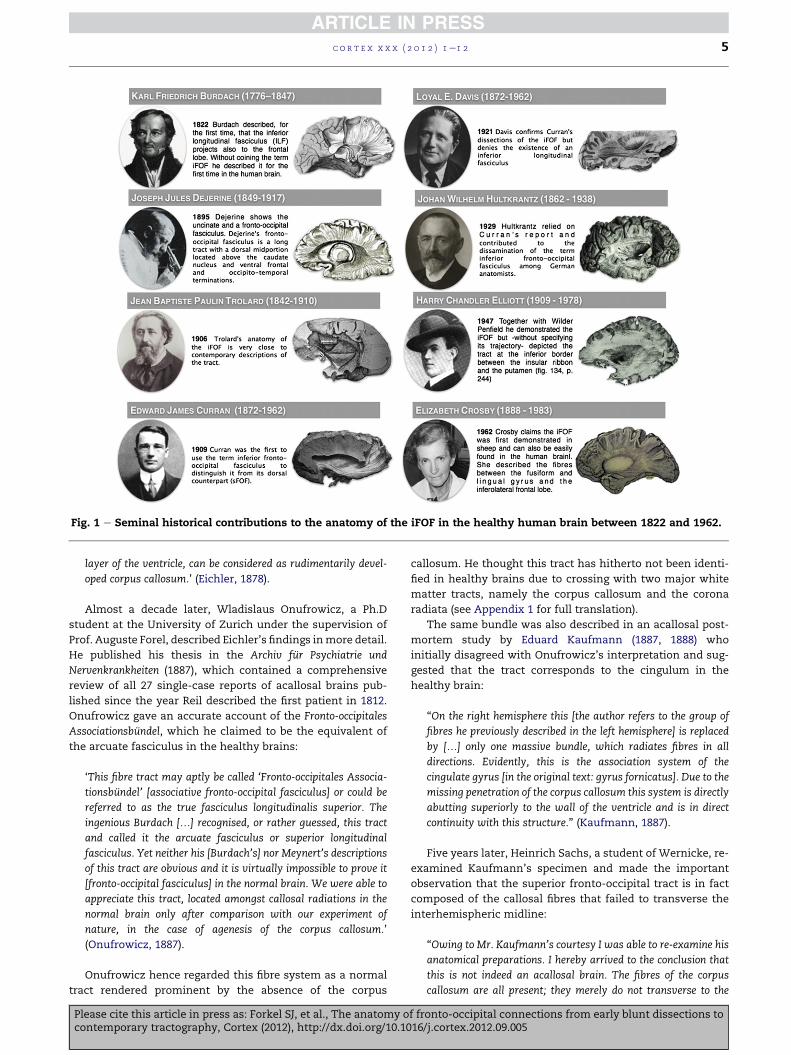
Fig. 1 e Seminal historical contributions to the anatomy of the iFOF in the healthy human brain between 1822 and 1962.
c o r t e x x x x ( 2 0 1 2 ) 1e1 2 5
layer of the ventricle, can be considered as rudimentarily devel-
oped corpus callosum.’ (Eichler, 1878).
Almost a decade later, Wladislaus Onufrowicz, a Ph.D
student at the University of Zurich under the supervision of
Prof. Auguste Forel, described Eichler’s findings inmore detail.
He published his thesis in the Archiv fur Psychiatrie und
Nervenkrankheiten (1887), which contained a comprehensive
review of all 27 single-case reports of acallosal brains pub-
lished since the year Reil described the first patient in 1812.
Onufrowicz gave an accurate account of the Fronto-occipitales
Associationsbundel, which he claimed to be the equivalent of
the arcuate fasciculus in the healthy brains:
‘This fibre tract may aptly be called ‘Fronto-occipitales Associa-
tionsbundel’ [associative fronto-occipital fasciculus] or could be
referred to as the true fasciculus longitudinalis superior. The
ingenious Burdach [.] recognised, or rather guessed, this tract
and called it the arcuate fasciculus or superior longitudinal
fasciculus. Yet neither his [Burdach’s] nor Meynert’s descriptions
of this tract are obvious and it is virtually impossible to prove it
[fronto-occipital fasciculus] in the normal brain. We were able to
appreciate this tract, located amongst callosal radiations in the
normal brain only after comparison with our experiment of
nature, in the case of agenesis of the corpus callosum.’
(Onufrowicz, 1887).
Onufrowicz hence regarded this fibre system as a normal
tract rendered prominent by the absence of the corpus
Please cite this article in press as: Forkel SJ, et al., The anatomy ocontemporary tractography, Cortex (2012), http://dx.doi.org/10.10
callosum. He thought this tract has hitherto not been identi-
fied in healthy brains due to crossing with two major white
matter tracts, namely the corpus callosum and the corona
radiata (see Appendix 1 for full translation).
The same bundle was also described in an acallosal post-
mortem study by Eduard Kaufmann (1887, 1888) who
initially disagreed with Onufrowicz’s interpretation and sug-
gested that the tract corresponds to the cingulum in the
healthy brain:
“On the right hemisphere this [the author refers to the group of
fibres he previously described in the left hemisphere] is replaced
by [.] only one massive bundle, which radiates fibres in all
directions. Evidently, this is the association system of the
cingulate gyrus [in the original text: gyrus fornicatus]. Due to the
missing penetration of the corpus callosum this system is directly
abutting superiorly to the wall of the ventricle and is in direct
continuity with this structure.” (Kaufmann, 1887).
Five years later, Heinrich Sachs, a student of Wernicke, re-
examined Kaufmann’s specimen and made the important
observation that the superior fronto-occipital tract is in fact
composed of the callosal fibres that failed to transverse the
interhemispheric midline:
“Owing to Mr. Kaufmann’s courtesy I was able to re-examine his
anatomical preparations. I hereby arrived to the conclusion that
this is not indeed an acallosal brain. The fibres of the corpus
callosum are all present; they merely do not transverse to the
f fronto-occipital connections from early blunt dissections to16/j.cortex.2012.09.005
c o r t e x x x x ( 2 0 1 2 ) 1e1 26
contralateral hemisphere but rather remain in the same hemi-
sphere and run anterior-posteriorly. Thereby producing a fronto-
occipital bundle in the ‘acallosal brain’ that is completely absent
in the healthy brain.” (Sachs, 1892).
This speculation proved to be correct much later as
demonstrated by experimental evidence in animals and
advanced investigations in humans (Rakic and Yakovlev,
1968; Loeser and Alvord, 1968; Yakovlev and Locke, 1961;
Richards et al., 2004; Paul et al., 2007).
In the neuroradiological literature the sFOF, as described in
acallosal brains, is associated with the name of Moritz Probst
(1867e1923), a German-born Austrian-educated neuropathol-
ogist and forensic psychiatrist. It is not clear when and how
Probst’s name was adopted as eponym for the sFOF. In our
review the term ‘Probst bundle’ is absent from the German
literature and it appears for the first time in the English-
printed publications of the 1960s: “[.] the composition of
the so-called Probst’s bundle (longitudinal callosal bundle)
will be noted and discussed later. [.] Its exact nature has been
the subject of much disagreement. [.] It is suggested, there-
fore, that Probst’s bundle is composed of [.] (1) septal-
hippocampal, hippocampal-septal fibres and possible other
fornix components; (2) the superior fornix of Elliot Smith; (3)
association fibres between various parts of other cortical
areas.” [Magee and Olson (with Elizabeth Crosby), 1961].
It is possible this adoptionwas in recognition of Probst’ first
microscopic description of the tract and for his claim of the
tract being uniquely present in the acallosal brain (Probst,
1901). Or, perhaps it is the result of the erroneous adoption
of the already existing eponyms in the German literature that
indicate two other tracts, one connecting to the reticular
formation that was named Probst bundle by Lewandowsky
(Peterfi, 1923; Strong and Elwyn, 1943; Truex, 1959; Holmes,
1906), the other connecting the thalamus to the temporal
lobe (Ariens-Kappers et al., 1961; Mettler, 1942).
3.2.2. The sFOF in the normal animal and human brainWhilst theexistenceof thesFOF in theacallosalbrainhasnever
been questioned, the evidence for a similar tract in the normal
brain has proved to be more difficult. In the presence of the
corpus callosum the sFOF in humans is thought to correspond
to the subcallosal bundle described in animals by Muratoff.
This tract is a system of fibres running above the caudate
nucleus at the corner formed by the internal capsule and the
corpus callosum. Muratoff consciously used the term ‘sub-
callosal bundle’ to indicate the most distinct feature of this
tract (i.e., its midportion lying beneath the corpus callosum)
without committing to a clear identification of its terminal
projections (see Appendix 2). Muratoff’s hesitation was in part
related to the crude lesionmethod he used to study the course
of this tract. His method consisted of uni- or bilateral trepa-
nation of the animal skull followed by lesioning of large
portions of the brain. After sectioning the brain he was able to
follow the trajectory of the degenerating fibres and identify
only one of the two cortical end stations (i.e., the one opposite
to the lesion).
The development of axonal tracing studies has allowed to
describe the anatomy of subcallosal fibres more precisely and
to distinguish two different components of the subcallosal
Please cite this article in press as: Forkel SJ, et al., The anatomy ocontemporary tractography, Cortex (2012), http://dx.doi.org/10.10
bundle in the monkey brain (Yakovlev and Locke, 1961;
Schmahmann and Pandya, 2006; Yeterian and Pandya, 2010).
The first group consists of fronto-striatal fibres connecting the
dorsal andmedial aspects of the frontal, parietal and occipital
lobes to the caudate nucleus. For this component the eponym
Muratoff’s bundle had been proposed by Schmahmann and
Pandya. The second group includes long association fibres
connecting the occipital and the frontal lobe, for which the
term fronto-occipital fasciculus continues to apply.
In humans, the fronto-striatal component of the sFOF was
originally suggested by Theodor Meynert who used the term
‘corona radiata of the caudate nucleus’ to indicate that these
fibres originate from the frontal lobes and project to the
caudate nucleus (1887). The idea of the fronto-occipital
fasciculus as an association bundle connecting occipital and
frontal regions, as described in the acallosal brain, was
extended to the healthy brain by Onufrowicz, Wernicke and
Dejerine.
More recently Ture et al. (1997) suggested the existence of
a third component of the subcallosal fasciculus formed by
ascending thalamo-cortical fibres.
With diffusion tensor imaging (DTI) tractography, all three
components of the sFOF have been described (Catani et al.,
2002; Wakana et al., 2004; Mori et al., 2005; Makris et al.,
2007; Catani and Thiebaut de Schotten, 2012). However, trac-
tography based on the tensor model is prone to artefactual
reconstructions mainly due to the inability to separate
crossing fibres. The development of SD methods (Dell’Acqua
and Catani, 2012) applied to tractography has in part over-
come the limitations of the tensor model by allowing the
visualization of multidirectional fibre orientations. The use of
SD tractography, for example, has been used to dissect and
quantify the three branches of the SLF (Thiebaut de Schotten
et al., 2011a, 2011b) in humans. In the next section we will
present the results of tractography dissections of the fronto-
occipital network in healthy and acallosal humans in an
attempt to replicate findings from axonal tracing and post-
mortem studies.
3.3. Original data: diffusion tractography of the healthyand acallosal brains
Two direct fronto-occipital tracts were identified using SD
tractography: an inferior pathway connecting ventro-medial
occipital and orbito-polar frontal cortex, and a dorsal
pathway running between the cingulum and the arcuate
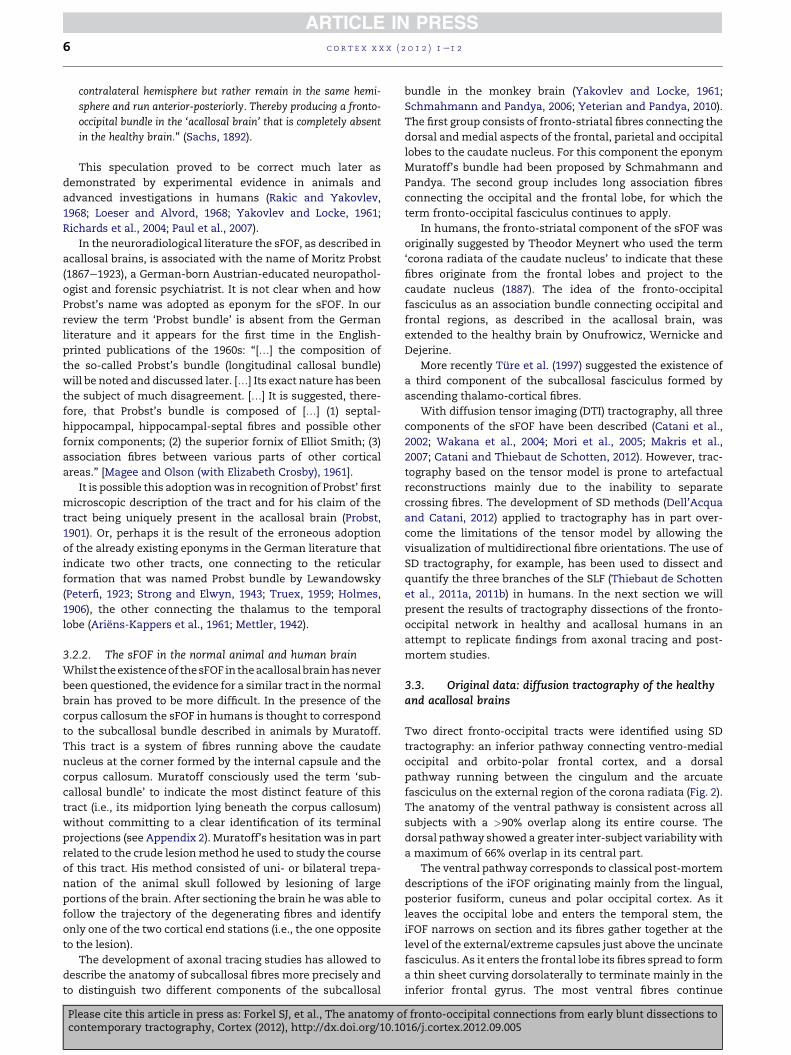
fasciculus on the external region of the corona radiata (Fig. 2).
The anatomy of the ventral pathway is consistent across all
subjects with a >90% overlap along its entire course. The
dorsal pathway showed a greater inter-subject variability with
a maximum of 66% overlap in its central part.
The ventral pathway corresponds to classical post-mortem
descriptions of the iFOF originating mainly from the lingual,
posterior fusiform, cuneus and polar occipital cortex. As it
leaves the occipital lobe and enters the temporal stem, the
iFOF narrows on section and its fibres gather together at the
level of the external/extreme capsules just above the uncinate
fasciculus. As it enters the frontal lobe its fibres spread to form
a thin sheet curving dorsolaterally to terminate mainly in the
inferior frontal gyrus. The most ventral fibres continue
f fronto-occipital connections from early blunt dissections to16/j.cortex.2012.09.005
Fig. 2 e SD tractography reconstructions and overlay percentage maps of the dorsal and ventral fronto-occipital fasciculus.
(A) 3D reconstruction of the normalised trajectory of the sFOF and the iFOF. MNI coordinates are in correspondence with the
MNI slices shown in panel B. (B) MNI-normalised overlay percentage maps of the sFOF (upper panel B, MNI 56e16) and the
iFOF (lower panel B, MNI 24 to L16).
c o r t e x x x x ( 2 0 1 2 ) 1e1 2 7
anteriorly and terminate in the medial fronto-orbital region
and frontal pole.
The dorsal pathway originates from the most anterior and
lateral regions of the occipital lobe and projects to the poste-
rior cortex of the superior and inferior frontal gyri after
running above the corpus callosum. This tract does neither
correspond to the anatomical descriptions of the sFOF nor the
subcallosal tract. Direct comparison with recent atlases of
white matter connections suggests that this tract corresponds
in part to the second branch of the superior longitudinal
fasciculus (SLF) (Schmahmann and Pandya, 2006; Thiebaut de
Schotten et al., 2011a, 2011b; Makris et al., 2008; Catani and
Thiebaut de Schotten, 2012).
Further dissections were performed to clarify the origin
and terminations of these fibres running through the sub-
callosal region. Our dissections show that the streamlines of
the ‘subcallosal fascicle of Muratoff’ connect the frontal lobe
to (i) dorsal parietal lobe, (ii) caudate nucleus and (iii) thal-
amus. None of these streamlines passing through the sub-
callosal region projected to the occipital lobe.
Please cite this article in press as: Forkel SJ, et al., The anatomy ocontemporary tractography, Cortex (2012), http://dx.doi.org/10.10
Neither the ventral nor the dorsal pathways were signifi-
cantly lateralised to either hemisphere, with the dorsal
pathway being slightly larger on the right hemisphere [iFOF:
t(29) ¼ .382, p ¼ .705; sFOF: t(29) ¼ e1.730, p ¼ .094].
Dissections of the three acallosal brains show the presence
of a major dorsal associative pathway connecting occipital,
parietal and frontal regions in each hemisphere (Fig. 3B). This
tract is separated from the cingulum, arcuate fasciculus and
iFOF (Fig. 3C). These three acallosal brains have different
degrees of agenesis. Case 1 presented with partial absence
only of the anterior body of the corpus callosum (Fig. 3A). In
this subject it is possible to visualise the callosal streamlines
coursing through the callosal remnants (genu and posterior
half of the corpus callosum). In the frontal white matter,
corresponding to the subcallosal region of a healthy brain,
a longitudinal tract connecting themedial frontal cortex to the
thalamus and basal ganglia is visualised. Cases 2 and 3 have
a more severe agenesis with callosal remnants of the sple-
nium and genu, respectively (Fig. 3A). In these two cases the
subcallosal fasciculus is a prominent tract, which resembles
f fronto-occipital connections from early blunt dissections to16/j.cortex.2012.09.005
Fig. 3 e Comparison between a healthy and three acallosal brains (1e3) with varying severity of congenital callosal agenesis
(absence of corpus callosum, ACC). (A) depicts T1-weighted structural magnetic resonance imaging (MRI) scans of the
normal callosal anatomy and acallosal brains. The corpus callosum (CC) and its remnants are delineated in red. (B) shows
the virtual in vivo dissections of the (remnants of the) corpus callosum (red) in relation to a subcallosal white matter
pathways (green). This demonstrates an increasing prominence of a subcallosal fronto-occipital fasciculus alongside the
reduced eminence of interhemispheric callosal fibres. (C) shows the corpus callosum (red) in relation to all major associative
white matter pathways, such as the arcuate fasciculus (purple), cingulum (yellow), and the ventral and dorsal fronto-
occipital fasciculus (blue, green). The subcallosal tract in the healthy brain and the sFOF in the acallosal brains is clearly
a separate tract from the arcuate, cingulum and iFOF.
c o r t e x x x x ( 2 0 1 2 ) 1e1 28
Onufrowicz’s description of the sFOF (see Appendix 1). In
these two cases the superior fronto-occipital tract connects
the frontal lobe to the parietal, temporal and occipital lobes.
Fig. 2C illustrates the tractography reconstruction of all major
association tracts in the normal and acallosal brains. The
subcallosal tract in the healthy brain and the sFOF in the
acallosal brains is clearly a separate tract from the arcuate,
cingulum and iFOF.
4. Discussion
In our study we reviewed historical literature and used trac-
tography to clarify the anatomy of the long-range connections
between frontal and occipital lobes. Four main results emerge
from our study: (i) ventral connections corresponding to clas-
sical post-mortem descriptions of the iFOF can be reliably
reconstructed in all healthy and acallosal brains; (ii) dorsal
fronto-occipital connections can be dissected in vivo in
a minority of the healthy brains using SD tractography and are
Please cite this article in press as: Forkel SJ, et al., The anatomy ocontemporary tractography, Cortex (2012), http://dx.doi.org/10.10
likely to belong to the SLF system;whereas the subcallosal tract
(or Muratoff bundle) is a complex system of projection fibres
(thalamo-frontal and fronto-striatal) without fronto-occipital
associative connections; (iii) in acallosal brains the uncrossed
callosal fibres form a large association tract that connects the
frontal toparietal, temporal andoccipital lobes. For this tractwe
propose to use the name of Onufrowicz as eponym (i.e., Onu-
frowicz’ bundle instead of Probst or SachseProbst bundle).
4.1. Ventral fronto-occipital fasciculus
Despite several post-mortem and in vivo dissection studies
have reported positive findings, the existence of the iFOF in
humans has been questioned in recent years on the basis of
axonal tracing studies in the monkey brain: ‘There is no
convincing support in the experimental literature for connections
between inferior occipital regions and the orbitofrontal or ventro-
lateral prefrontal cortices, and our own observations in the monkey
also do not document an ‘inferior FOF’’ (Schmahmann and
Pandya, 2007, p. 373).
f fronto-occipital connections from early blunt dissections to16/j.cortex.2012.09.005
c o r t e x x x x ( 2 0 1 2 ) 1e1 2 9
According to these authors the experimental literature is
not persuasive as it is primarily based on blunt dissections or
in vivo tractography, which are both prone to a number of
limitations, including generating the presence of false posi-
tives (i.e., non-existing tracts) and false negatives (i.e., absence
of truly existing tracts). The SD tractography we used to
visualise the fronto-occipital network can in part overcome
these limitations (see Dell’Acqua and Catani, 2012, for
discussion), although validation is still required. Also it is
important to consider that axonal tracing studies are limited
in exploring the connections of ventro-medial occipital
regions due to limited accessibility. For example the atlas of
Schmahmann and Pandya (2006) does not contain informa-
tion on the connections of the regions equivalent to the
human projections of the iFOF. Based on the results in
monkeys, Pandya favoured the extreme capsule as containing
long ventral association tracts rather than the external
capsule (Pandya and Kuypers, 1969). Our view is that, in
addition to the extreme capsule tract described by Schmah-
mann and Pandya, humans have a longer subset of connec-
tions reaching the most posterior occipital regions. This
enlargement in the human brain might be related to the
phylogenetic expansion of the frontal and occipital lobes
(Deacon, 1990).
Indirect evidence in support of the existence of an iFOF in
humans can be found in studies using other methods, such as
electrophysiology and functional imaging.
Rudrauf et al. (2008) employed magnetoencephalography
(MEG) to describe the activity within the ventral visual pro-
cessing tasks involving emotional material and found an
activity latency of 100 msec between early visual areas
(V2eV3eV4) and the orbitofrontal and ventro-medial
prefrontal cortex. This short time interval is highly suggestive
of a signal transmission through long-range monosynaptic
association fibres along the inferior fronto-occipital connec-
tions. Similarly, short activity latency was shown in electro-
encephalography (EEG) (Barcelo et al., 2000; ffytche and
Catani, 2005) and cortical recording (Kawasaki et al., 2001)
studies of visual percepts.
Although supportive of the existence of the iFOF, the above
methods cannot be seen as a direct anatomical validation for
blunt and in vivo virtual dissections. This short latency could,
in fact, result from a parallel processing route between the
lateral geniculate nucleus and the occipital lobe. Further
studies are also necessary to clarify whether the iFOF runs
throught the extreme capsule or extends into the external
capsule.
In conclusion, both post-mortem and multimodal in vivo
studies have consistently reported evidence for the existence
of an iFOF in humans. The lack of a similar connection in the
monkey brain, suggests that the iFOF may represent a tract
unique to the human brain (Catani, 2007).
4.2. Dorsal fronto-occipital connections
The sFOF has been extensively described in the monkey brain
using axonal tracing methods. In humans there are fewer
post-mortem studies, which deny the existence of an equiv-
alent bundle in the human brain (Meynert, 1887; Ture et al.,
1997). Tractography studies based on DTI were likewise
Please cite this article in press as: Forkel SJ, et al., The anatomy ocontemporary tractography, Cortex (2012), http://dx.doi.org/10.10
unable to visualise a dorsal fronto-occipital bundle although
the majority of studies reported direct connections between
frontal lobe and parietal regions (Catani et al., 2002; Wakana
et al., 2004; Mori et al., 2005; Makris et al., 2007). The absence
of the sFOF on tractography reconstruction could represent
a false negative due to the limitations of the tensor model.
Tractography based on deconvolution has recently been used
to show tracts, such as the SLF, that were described in the
monkey but not in DTI tractography studies (Thiebaut de
Schotten et al., 2011a, 2011b). Our dissections of the sFOF
based on SD tractography showed the presence of tracts
between occipital and frontal lobes, which are clearly distinct
from the iFOF. These tracts run lateral and dorsal to the corpus
callosumand are likely to represent longer branches of the SLF
system. Our conclusion is that even with SD tractography,
that facilitates reconstructions of pathways not visible with
DTI methods, we were not able to visualise a human equiva-
lent of the sFOF in the healthy brain. These results are in
accordance with blunt post-mortem dissections.
A second finding from our SD tractography dissections is
the identification of a subcallosal tract connecting the frontal
lobe to subcortical structures such as the caudate nucleus and
the thalamus. This projection tract corresponds to Muratoff’s
bundle that can be easily identified both in animals and
humans. The descending component of Muratoff’s bundle
belongs to an extended fronto-striatal system, which includes
tracts running outside the ‘subcallosal region’. Similarly the
thalamo-frontal component of the Muratoff’s bundle belongs
to the anterior thalamic peduncle, a much larger ascending
projection system that projects to the entire frontal cortex.
Although Muratoff’s bundle seems to have some distinct
anatomical features (e.g., composed mainly of poorly
myelinated fibres compared to other projection fibres) it
remains to be established whether it deserves to be consid-
ered as a distinct bundle. Whether the Muratoff bundle is an
entity on its own or more likely an integrated part of the
abovementioned striatal and thalamic projection systems
awaits further investigation.
4.3. Onufrowicz’s bundle in the acallosal brain
Our dissections in three acallosal brains, with varying severity
in the expression of the pathology, suggest an inverse rela-
tionship between the remnants of the corpus callosum and
the fibres of the ‘sFOF’. The volume of the ‘sFOF’ appearsmore
prominent in the most severe cases of callosal agenesis,
whereas in partial acallosal brains and in normal brains
progressively disappears (Fig. 3). Furthermore our dissections
show a clear separation between the ‘sFOF’ and the arcuate
fasciculus, which indicates that Onufrowicz’s interpretation
of the sFOF being equivalent to the arcuate/SLF, was probably
incorrect.
Recently, Schmahmann and Pandya (2006) suggested
adding the name of Sachs to that of Probst in recognition of his
insightful suggestions on the true callosal nature of the sFOF.
We suggest that if an eponym should be used, which is rather
questionable, the names of Eichler and especially Onufrowicz
should have a priority before those of Sachs and Probst. In
Appendix 1, we report the first integral translation of the
f fronto-occipital connections from early blunt dissections to16/j.cortex.2012.09.005
c o r t e x x x x ( 2 0 1 2 ) 1e1 210
paper by Onufrowicz to draw attention to his seminal contri-
bution and for future reference in the Anglophone literature.
5. Conclusion
Our in vivo dissections based on SD tractography support the
presence of a bilateral ventral pathway directly connecting
occipital and frontal lobes. This tract corresponds to the clas-
sical descriptions of the iFOF derived from blunt post-mortem
dissections and recent DTI findings in the human brain, which
has not been described in the animal brain. We also found
adorsalpathwayrunning in theoutermost regionof thecorona
radiata. This pathway was identified in only two-thirds of the
subjects and its trajectory does not correspond to the classical
descriptions of the sFOF derived fromaxonal tracing studies in
animals. It is possible that the dorsal pathway we identified is
part of the SLF, a lateral system of fibres connecting dorsal
regions of the occipital, parietal and frontal lobes. Future
studies are needed to establish whether the iFOF is a bundle
unique to humans; this includes for exampleMarchi’s staining
in the human brain and additional experiments with axonal
tracing in monkeys investigating connections from ventro-
medial occipital regions.
Acknowledgements
S.J.F. and M.C. were funded by Guy’s and St. Thomas’ Trust
Charity. F.D.A. was funded by NIHR Biomedical Research
Centre for Mental Health at South London and Maudsley NHS
Foundation Trust and Institute of Psychiatry, King’s College
London, UK British Research Council. M.T.S. was funded by
the Agence Nationale de la Recherche (ANR) [project
CAFORPFC, number ANRe09eRPDOCe004e01 and project
HMeTC, number ANRe09eEMERe006]. We gratefully
acknowledge the support of PD Dr. Jennifer Linn, Abteilung fur
Neuroradiologie Klinikum der Universitat Munchen.
Supplementary data
Supplementary data related to this article can be found at
http://dx.doi.org/10.1016/j.cortex.2012.09.005.
r e f e r e n c e s
Ariens-Kappers CU, Huber C, and Crosby E. The ComparativeAnatomy of the Nervous System of Vertebrates, Including Man. NewYork: Macmillan, 1961.
Barcelo F, Suwazono S, and Knight RT. Prefrontal modulation ofvisual processing in humans. Nature Neuroscience, 3(4):399e403, 2000.
Burdach KF. Vom Baue und Leben des Gehirns. Leipzig: Dyk, 1822.Catani M, Howard RJ, Pajevic S, and Jones DK. Virtual in vivo
interactive dissection of white matter fasciculi in the humanbrain. NeuroImage, 17(1): 77e94, 2002.
Catani M. From hodology to function. Brain, 130(3): 602e605, 2007.
Please cite this article in press as: Forkel SJ, et al., The anatomy ocontemporary tractography, Cortex (2012), http://dx.doi.org/10.10
Catani M, Allin MPG, Husain M, Pugliese L, Mesulam MM,Murray RM, et al. Symmetries in human brain languagepathways correlate with verbal recall. Proceedings of theNational Academy of Science, 104(43): 17163e17168, 2007.
Catani M and Thiebaut de Schotten M. Atlas of Human BrainConnections. Oxford: Oxford University Press, 2012.
Charpy A. Systeme nerveux (encephale). Paris: Masson et Cie,Editeurs libraires de l’academie de medecine, 1895.
Cohen L, Dehaene S, Naccache L, Leherici S, Dehaene-Lambertz G, Henaff MA, et al. The visual word form area:spatial and temporal characterization of an initial stage ofreading in normal subjects and posterior split-brain patients.Brain, 123(2): 291e307, 2000.
Commitee I.A.N. Nomina Anatomica. New York: ChurchillLivingstone, Edinburgh, 1989.
Crosby E, Humphrey T, and Lauer E. Correlative Anatomy of theNervous System. New York: Macmillan, 1962.
Curran E. A new association fibre tract in the cerebrum. Withremarks of the fibre tract dissection method of studying thebrain. Journal of Comparative Neurology and Psychology, 19(6):1e18, 1909.
Davis LE. An anatomic study of the inferior longitudinalfasciculus. Archives of Neurology and Psychiatry, 5(4): 370e381,1921.
Deacon TW. Rethinking mammalian brain evolution. Am Zool, 30:629e705, 1990.
Dell’Acqua F, Coward J, Simmons A, Murphy D, Williams S, andCatani M. Mapping crossing fibres of the human brain withspherical deconvolution: Towards an atlas for clinico-anatomical correlation studies. Proceedings International SocietyMagnetic Resonance Medicine, 17: 3562, 2009.
Dell’Acqua F, Scifo P, Rizzo G, Catani M, Simmons A, Scotti G,et al. Modified damped Richardson Lucy algorithm to reduceisotropic background effects in spherical deconvolution.NeuroImage, 49(2): 1446e1458, 2010.
Dell’Acqua F, Simmons A, Williams S, and Catani M. Canspherical deconvolution provide more information than fibreorientations? Hindrance Modulated Orientational Anisotropy(HMOA), a true-tract specific index to characterize whitematter diffusion. Human Brain Mapping, http://dx.doi.org/10.1002/hbm.22080 [Epub ahead of print], 2012.
Dell’Acqua F and Catani M. Structural human brain networks: Hottopics in diffusion tractography. Current Opinion in Neurology,25(4): 375e383, 2012.
Dejerine J. Anatomie des Centres Nerveux. Paris: Rueff et Cie,1895.
Descoteaux M, Deriche R, Knosche TR, and Anwander A.Deterministic and probabilistic tractography based oncomplex fibre orientation distributions. IEEE Transactions onMedical Imaging, 28(2): 269e286, 2009.
Ebeling U and von Cramon D. Topography of the uncinate fascicleand adjacent temporal fiber tracts. Acta Neurochirurgica,115(3e4): 143e148, 1992.
Eichler G. Ein Fall von Balkenmangel im menschlichen Gehirn.Archiv fur Psychiatrie und Nervenkrankheiten, 8(2): 355e366, 1878.
Elliott CH. Textbook of the Nervous System. A Foundation for ClinicalNeurology. Philadelphia: J.B. Lippincott Company, 1947.
Epelbaum S, Pinel P, Gaillard R, Delmaire C, Perrin M, Dupont S,et al. Pure alexia as a disconnection syndrome: New diffusionimaging evidence for an old concept. Cortex, 44(8): 962e974,2008.
Fernandez-Miranda JC, Rhoton Jr AL, Alvarez-Linera J, Kakizawa Y,Choi C, and de Oliveira EP. Three-dimensional microsurgicaland tractographic anatomy of the white matter of the humanbrain. Neurosurgery, 62(6 Suppl. 3): 989e1026, 2008.
ffytche DH and Catani M. Beyond localization: From hodology tofunction. Philosophical Transaction of the Royal Society of London,360(1456): 767e779, 2005.
f fronto-occipital connections from early blunt dissections to16/j.cortex.2012.09.005
c o r t e x x x x ( 2 0 1 2 ) 1e1 2 11
Holmes GM. Reviews: Some recent researches on the tracts of thebrain stem. Brain, 28(3e4): 556e566, 1906.
Hultkrantz W. Gehirnpraeperation mittels Zerfaserung. Anleitung zummakroskopischen Studium des Gehirns. Berlin: Verlag von JuliusSpringer, 1929.
Johnson CK, Curran EJ, and Ophth D. An historical vignette.Documenta Ophthalmologica, 89(1e2): 59e73, 1995.
Jones DK and Basser PJ. Squashing peanuts and smashingpumpkins: How noise distorts diffusion-weighted MR data.Magnetic Resonance in Medicine, 52(5): 979e993, 2004.
Kaufmann E. Ueber Mangel des Balkens im menschlichen Gehirn.Archiv fur Psychiatrie und Nervenkrankheiten, 18: 769e781, 1887.
Kaufmann E. Ueber Mangel des Balkens im menschlichen Gehirn.Archiv fur Psychiatrie und Nervenkrankheiten, 19: 229e244, 1888.
Kawasaki H, Kaufman O, Damasio H, Damasio AR, Granner M,Bakken H, et al. Single-neuron responses to emotional visualstimuli recorded in human ventral prefrontal cortex. NatureNeuroscience, 4(1): 15e16, 2001.
Kier EL, Staib LH, Davis LM, and Bronen RA. MR imaging of thetemporal stem: Anatomic dissection tractography of theuncinate fasciculus, inferior occipitofrontal fasciculus, andMeyer’s loop of the optic radiation. American Journal ofNeuroradiology, 25(5): 677e691, 2004.
Lawes IN, Barrick TR, Murugam V, Spierings N, Evans DR, Song M,et al. Atlas-based segmentation of white matter tracts of thehuman brain using diffusion tensor tractography andcomparison with classical dissection. NeuroImage, 39(1): 62e79,2008.
Leemans A, Jeurissen B, Sijbers J, and Jones DK. ExploreDTI: Agraphical toolbox for processing, analyzing, and visualizingdiffusion MR data. In 17th Annual Meeting of International Societyfor Magnetic Resonance in Medicine, Hawaii, USA, 2009: 3537.
Leemans A and Jones DK. The B-matrix must be rotated whenmotion correcting diffusion tensor imaging data. MagneticResonance in Medicine, 61(6): 1336e1349, 2009.
Loeser JD and Alvord EC. Agenesis of the corpus callosum. Brain,91(3): 553e570, 1968.
Loukas M, Shea M, Shea C, Lutter-Hoppenheim M, Zand P,Tubbs RS, et al. Jean Baptiste Paulin Trolard (1842e1910): Hislife and contributions to neuroanatomy. Journal ofNeurosurgery, 112(6): 1192e1196, 2010.
Makris N, Papadimitriou GM, Sorg S, Kennedy DN, Caviness VS,and Pandya DN. The occipitofrontal fascicle in humans: Aquantitative, in vivo, DT-MRI study. NeuroImage, 37(4):1100e1111, 2007.
Makris N, Buka SL, Biederman J, Papadimitriou GM, Hodge SM,Valera EM, et al. Attention and executive systemsabnormalities in adults with childhood ADHD: A DT-MRIstudy of connections. Cerebral Cortex, 18(5): 1210e1220, 2008.
Magee KR and Olson RN. The effect of absence of the corpuscallosum on the position of the hippocampus and on theformation of Probst’s bundle. Journal of Comparative Neurology,117: 371e382, 1961.
Martino J, Brogna C, Robles S, Vergani F, and Duffau H. Anatomicdissection of the inferior fronto-occipital fasciculus revisited inthe lights of brain stimulation data. Cortex, 46(5): 691e699, 2010.
Martino J, De Witt Hamer P, Vergani F, Brogna C, de Lucas EM,Vazquez-Barquero A, et al. Cortex-sparing fiber dissection: Animproved method for the study of white matter anatomy inthe human brain. Journal of Anatomy, 219(4): 531e541, 2011.
Mettler FA.Neuroanatomy. St. Louis: TheC.V.MosbyCompany, 1942.Meynert TH. A Clinical Treatise on Diseases of the Fore-brain Based
Upon a Study of Its Structure, Functions, and Nutrition (Sachs, BTrans.). New York: G.P. Putman’s Sons, 1887.
Mori S, Xue R, Crain B, Solaiyappan M, Chacko VP, and Zijl PC. 3Dreconstruction of axonal fibers from diffusion tensor imagingusing fiber assignment by continuous tracking (FACT). In 8thAnnual Meeting of the ISMRM, Philadelphia, PA, 1999: 320.
Please cite this article in press as: Forkel SJ, et al., The anatomy ocontemporary tractography, Cortex (2012), http://dx.doi.org/10.10
Mori S and van Zijl PC. Fiber tracking: Principles and strategies e
A technical review. NMR in Biomedicine, 15(7e8): 468e480, 2002.Mori S, Wakana S, Nagae-Poetscher LM, and Van Zijl PCM. MRI
Atlas of Human White Matter. Amsterdam: Elsevier, 2005.Muratoff W. Secundare Degenerationen nach Durchschneidung
des Balkens. Neurologisches Centralblatt, 12: 714e729, 1893.Onufrowicz W. Das balkenlose Mikrocephalengehirn Hofmann.
Ein Beitrag zur pathologischen und normalen Anatomie desmenschlichen Gehirnes. Archiv fur Psychiatrie undNervenkrankheiten, 18(2): 305e328, 1887.
Orban G, Van Essen D, and Vanduffel W. Comparative mapping ofhigher visual areas in monkeys and humans. Trends inCognitive Sciences, 8(7): 315e324, 2004.
Paul LK, Brown WS, Adolphs R, Tyszka JM, Richards LJ,Mukherjee P, et al. Agenesis of the corpus callosum: Genetic,developmental and functional aspects of connectivity. NatureReviews Neuroscience, 8(4): 287e299, 2007.
Peterfi T. Moritz Probst. Journal of Nervous & Mental Disease, 58(1):103, 1923.
Pandya DN and Kuypers HG. Cortico-cortical connections in therhesus monkey. Brain Research, 13(1): 13e36, 1969.
Probst M. Ueber den Bau des vollstaendig balkenlosenGrosshirnes sowie ueber Mikrogyrie und Heterotopie dergrauen Substanz. Archiv fur Psychiatrie und Nervenkrankheiten,34: 709e786, 1901.
Rakic P and Yakovlev PI. Development of the corpus callosum andcavum septi in man. Journal of Computational Neurology, 132(1):45e72, 1968.
Rapoport SI. Integrated phylogeny of the primate brain, withspecial reference to humans and their diseases. Brain ResearchReviews, 15(3): 267e294, 1990.
Reil JC. Die Sylvische Grube oder das Thal, das gestreifte grosseHirnganglium, dessen Kapsel und die Seitentheile des grossenGehirns. Archiv fur die Physiologie, 9: 195e208, 1809.
Reil JC. Mangel des mittleren und freien Teils des Balkens immenschlichen Gehirn. Archiv fur die Physiologie, 2: 341e344,1812.
Richards LJ, Plachez C, and Ren T. Mechanisms regulating thedevelopment of the corpus callosum and its agenesis inmouse and human. Clinical Genetics, 66(4): 276e289, 2004.
Rudrauf D, David O, Lachaux J-P, Kovach CK, Martinerie J,Renault B, et al. Rapid interactions between the ventralvisual stream and emotion-related structures rely on a two-pathway architecture. Journal of Neuroscience, 28(11):2793e2803, 2008.
Sachs H. Das Hemispharenmark des menschlichen Grosshirns. 1. DerHinterhauptlappen. Breslau Universitat psychiatrische undNervenklinik. Arbeiten. Leipzig: Thieme, 1892.
Schmahmann JD and Pandya DN. Fibre Pathways of the Brain.Oxford NY: Oxford University Press, 2006.
Schmahmann JD and Pandya DN. The complex history of thefronto-occipital fasciculus. Journal of the History of theNeurosciences, 16(4): 362e377, 2007.
Schmahmann JD, Pandya DN, Wang R, Dai G, D’Arceuil HE, deCrespigny AJ, et al. Association fibre pathways of the brain:Parallel observations from diffusion spectrum imaging andautoradiography. Brain, 130(Pt 3): 630e653, 2007.
Strong O and Elwyn A. Human Neuroanatomy. Baltimore: Williams& Wilkins, 1943.
Thiebaut de Schotten M, ffytche DH, Bizzi A, Dell’Acqua F,Allin M, Walshe M, et al. Atlasing location, asymmetry andinter-subject variability of white matter tracts in the humanbrain with MR diffusion tractography. NeuroImage, 54(1):49e59, 2011a.
Thiebaut de Schotten M, Dell’Acqua F, Forkel SJ, Simmons A,Vergani F, Murphy DGM, et al. Lateralized brain network forvisuo-spatial attention. Nature Neuroscience, 14(10): 1245e1246,2011b.
f fronto-occipital connections from early blunt dissections to16/j.cortex.2012.09.005
c o r t e x x x x ( 2 0 1 2 ) 1e1 212
Thiebaut de Schotten M, Dell’Acqua F, Valabregue R, andCatani M. Monkey to human comparative anatomy of thefrontal lobe association tracts. Cortex, 48(1): 82e96, 2012.
Trolard JB. Le faisceau longitudinal inferieur du cerveau. RevueNeurologique, 14: 440e446, 1906.
Truex RC. Strong and Elwyn’s Human Neuroanatomy. Baltimore:Williams & Wilkins, 1959.
Ture U, Yasargil MG, and Pait TG. Is there a superioroccipitofrontal fasciculus? A microsurgical anatomic study.Neurosurgery, 40(6): 1226e1232, 1997.
Umarova RM, Saur D, Schnell S, Kaller CP, Vry MS, Glauche V,et al. Structural connectivity for visuospatial attention:Significance of ventral pathway. Cerebral Cortex, 20(1):121e129, 2010.
Please cite this article in press as: Forkel SJ, et al., The anatomy ocontemporary tractography, Cortex (2012), http://dx.doi.org/10.10
Wakana S, Jiang H, Nagae-Poetscher LM, Van Zijl PCM, andMori S.Fibre tract-based atlas of human white matter anatomy.Radiology, 230(1): 77e87, 2004.
Yakovlev PI and Locke S. Limbic nuclei of thalamus andconnections of limbic cortex III. Corticocortical connections ofthe anterior cingulate gyrus, the cingulum, and the subcallosalbundle in monkey. Archives of neurology, 5(4): 364e400, 1961.
Yeterian EH, Pandya DN, Tomaiuolo F, and Petrides M. Thecortical connectivity of the prefrontal cortex in the monkeybrain. Cortex, 48(1): 58e81, 2012.
Yeterian EH and Pandya DN. Fiber pathways and corticalconnections of preoccipital areas in rhesus monkeys.Journal of Comparative Neurology, 518(18): 3725e3751,2010.
f fronto-occipital connections from early blunt dissections to16/j.cortex.2012.09.005