Embed Size (px)
Citation preview
1
SURGICAL APPROACHES TO FORAMEN MAGNUM LESIONS
2
Surgical anatomy of foramen magnum
F M - located in the occipital bone
Three parts of occipital bones : 1 – Squamous part – Contain F M 2 - Basal (clival) part – Ant. to the FM 3 - Condylar part - Connects the squamous OB and clivus Oval shaped, wider posteriorly than anteriorly Narrower anterior part sits above the odontoid process Wider posterior part transmits the medulla
3
Mean Diameters : ( Khalil Awadh et. al. 2003)
- Males - Sagittal = 37.2 ± 3.43 mm - Transverse = 31.6 ± 2.99 mm - Females - Sagittal = 34.6 ± 3.16 mm - Transverse = 29.3 ± 2.19 mm
Clivus - Thick quadrangular plate of bone that extends forward and upward, at an angle of about 45° from the FM
FM area - From lower third of clivus to the ant. arch of atlas and the odontoid process
4
Occipital condyles – - Located lateral to the anterior half of FM - Oval in shape, convex downward, face downward and laterally - Long axes directed forward and medially
Hypoglossal canal - - Transmits the hypoglossal nerve - Situated above the condyle, - Directed forward and laterally from the posterior cranial fossa.
Jugular foramen - - Situated lateral and slightly superior to the anterior half of the condyles at the posterior end of the petroclival suture
5
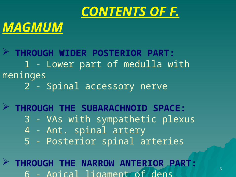
CONTENTS OF F. MAGMUM THROUGH WIDER POSTERIOR PART: 1 - Lower part of medulla with meninges 2 - Spinal accessory nerve
THROUGH THE SUBARACHNOID SPACE: 3 - VAs with sympathetic plexus 4 - Ant. spinal artery 5 - Posterior spinal arteries
THROUGH THE NARROW ANTERIOR PART: 6 - Apical ligament of dens 7 - Membrana tectoria
6
7
Choice of Surgical approaches
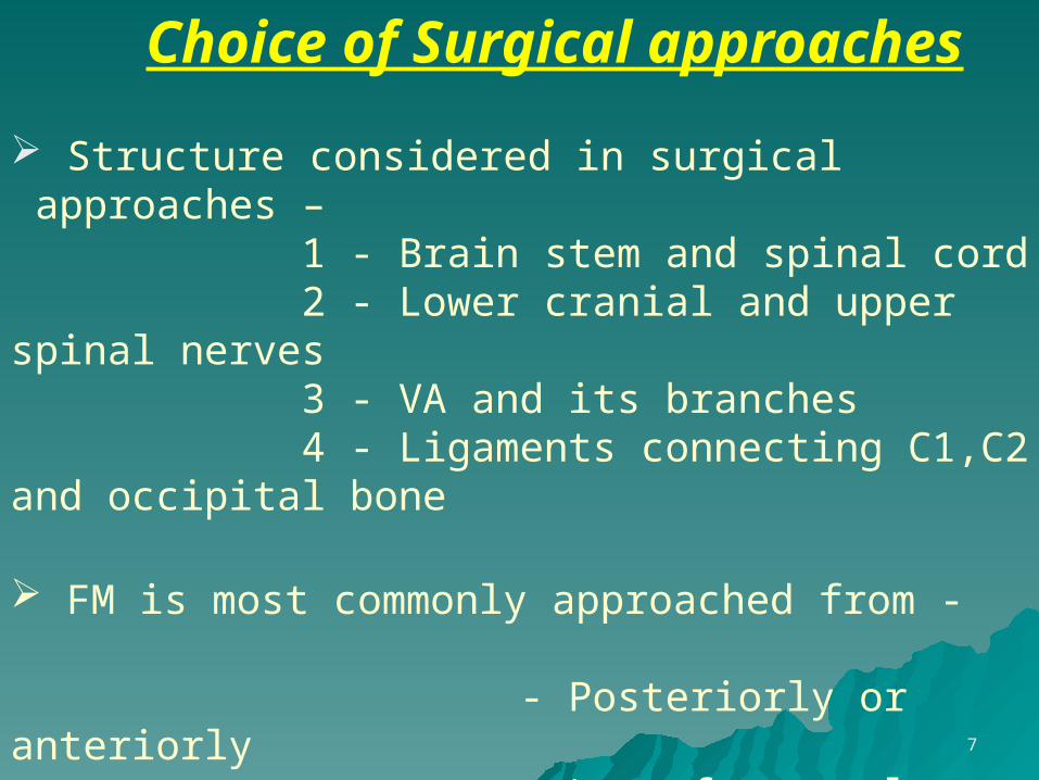
Structure considered in surgical approaches – 1 - Brain stem and spinal cord 2 - Lower cranial and upper spinal nerves 3 - VA and its branches 4 - Ligaments connecting C1,C2 and occipital bone FM is most commonly approached from - - Posteriorly or anteriorly - Less frequently from laterally
Choice depends on – 1- Location and extent of lesion 2- Size and nature of the pathology
8
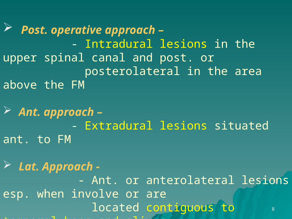
Post. operative approach – - Intradural lesions in the upper spinal canal and post. or posterolateral in the area above the FM
Ant. approach – - Extradural lesions situated ant. to FM
Lat. Approach - - Ant. or anterolateral lesions esp. when involve or are located contiguous to temporal bone and clivus
9
A - Posterior approaches
1 – SUBOCCIPITAL APPROACH : INDICATIONS: - Intradural lesions at post. or posterolateral location ADVANTAGES - Familiar to most neurosurgeons - Visualization of the VA, brainstem, cranial nerves, and tumor in a safe, simple, and rapid manner DISADVANTAGE : -Vascular injury e.g. VA and PICA - Pseudomeningocele - Not feasible to work well laterally and ant. to the spinal cord and the medulla.
10
Sub Occ. approaches
A- Three-quarter prone position.
B- Vertical midline Incision
C- S.O. craniectomy and a laminectomy of C1 and C2
D- Dural incision
E- Intradural exposure
F- Hockey-stick retro sigmoid exposure.
11
POSITION : - Prone - Sitting
STEPS : - Vertical midline or Hockey-stick skin incision
- Y-shape muscle incision - Craniectomy above the FM and a laminectomy of the axis and atlas - Dura mater opened by Y shaped incision
12
Most difficult lesions to remove are those situated ant. to
the 9th, 10th and 11th nerves and lateral medullary segment of the vertebral artery.
An attempt should be made to gently separate the rootlets and to operate through the interval between the rootlets.
13
2 - RETROSIGMOID SUBOCCIPITAL APPROACH :
INDICATION : - Intradural posterolateral lesions ADVANTAGES : - Wide view of the CP angle and of the intradural structures behind the ipsilateral lower clivus
DISADVANTAGES: - Inadequate exposure of more medial or C/L extension of lesion - Retraction on neural tissues
14
Retro sigmoid approach
- Three-quarterprone position. - Vertical paramedianincision crosses the asterion. - Superolateral margin of thecraniotomy is positioned at thejunction of the transverse and sigmoidsinuses.
15
3 - EXTREME LATERAL APPROACH : (Sen and Sekhar and AL-Mefty et al ) INDICATION : - Anterior / anterolateral lesions
PRINCIPLE : - Removal of more bone in key areas
- Exposure of VA and mobilization of extradural course from C 2 to its dural entry point
16
ADVANTAGES : - Short distance and wide surgical field - Tumor and brain stem interface under direct vision - Early proximal control of vertebral artery - Intra and extradural parts of tumor may be accessed in same sitting - Occipitocervical stabilization is possible in same sitting - May be combined with a subtemporal – infratemporal or a presigmoid approach
17
DISADVANTAGES : - Extensive soft tissue dissection - Prolong operating time - Increased postoperative pain - Possible VA and LCN injury - Requirement of experienced surgeon
Relative contraindication – - High jugular bulb
POSITION : - Lateral
STEPS: - INCISION : Horse shoe / Inverted – L / Cuvilinear
18
# Three anatomic stages # 1 - Muscular dissection 2 - Extradural dissections for mastoidectomy, s.o. craniectomy, extent of occipital condyle removal, and exposure and identification of the hypoglossal canal, jugular process, jugular tubercle, and facial nerve.
- VA exposure from f. transversarium of C 2 to dural entry point and displaced downward and medially - Tip of tr. process is preserved
3- Intradural exposure - Incision parallel to the lateral margins of the craniotomy, with base of the flap medially
19
B - Anterior Approaches 1-TRSANSORAL APPROACHES :
MODIFICATIONS : - Transpalatine approach - Labiomandibular or - Labioglossomandibular approach (exposure upto C5)
Most commonly selected anterior approach
20
INDICATION : - For most anterior extradural lesions
ADVANTAGES : - Midline exposure - Most direct route to the pathology
21
DISADVANTAGES : - Contaminated field - Frequency of CSF fistula - Pseudomeningocele - Meningitis - Depth of the operative field
POSITION: - Supine
STEPES : - Soft palate is retracted - Midline longitudinal incision over post. pharyngeal wall - Elevation of mucosa and prevertebral muscles
22
Clivus, the anterior arch of the atlas, the dens, and bodies of C2 and C3 may be removed
Clival exposure between the occipital condyles is 2-2.5 cm wide and 2.5- to 3.0-cm long
Lateral exposure limited by – 1 – Pterygoid plates 2 – Hyopoglossal canals 3 – Eustachian tubes 4 – Width b/w the VAs
23
To increase the exposure and reduce the operative depth, lip and chin may be incised vertically
Tongue and floor of the mouth may be split in the midline
After dealing with the lesion, mucosa and musculature of the tongue and floor of the mouth are re approximated
Repositioning of mandibular osteotomy

24
Transoral Approach
A- Forced opening of mouth permits the clivus to be exposed below palate.
B- Ant. view
C- Incision
D– Soft palate divided
E - Pharyngeal mucosa has been opened in the midline
F- Lt L. capitis and L. coli reflected laterally
25
2 – TRANSMAXILLARY APPROACH: Rarely used INDICATION : - Lesions extending to the upper and middle third of clivus (difficult to reach by the transoral approach) ADVANTAGES: - Also access to the sphenoid and ethmoid sinuses and the sella, and medial part of the floor of ant. fossa - Wider exposure to the clivus and upper cervical spine
26
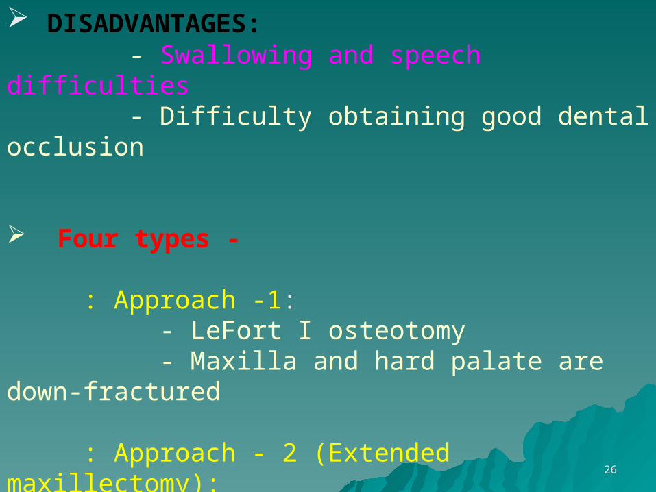
DISADVANTAGES: - Swallowing and speech difficulties - Difficulty obtaining good dental occlusion
Four types - : Approach -1: - LeFort I osteotomy - Maxilla and hard palate are down-fractured : Approach - 2 (Extended maxillectomy): - LeFort osteotomy + a midline incision of hard and soft palate and halves of the maxilla are swung laterally
27
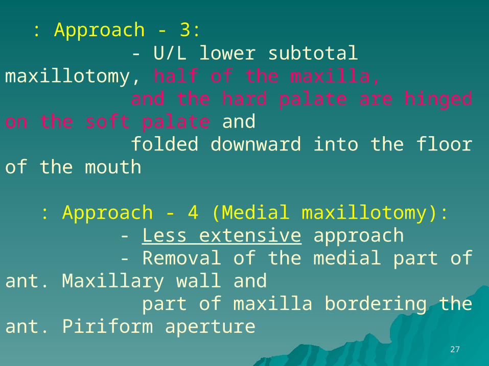
: Approach - 3: - U/L lower subtotal maxillotomy, half of the maxilla, and the hard palate are hinged on the soft palate and folded downward into the floor of the mouth
: Approach - 4 (Medial maxillotomy): - Less extensive approach - Removal of the medial part of ant. Maxillary wall and part of maxilla bordering the ant. Piriform aperture Removal of post. part of nasal septum and turbinates provide wider access to clivus and upper cervical vertebrae
28
Clival defect closure done by :
- Post. part of the mucosal flap on both sides of the nasal septum - Temporalis muscle graft
29
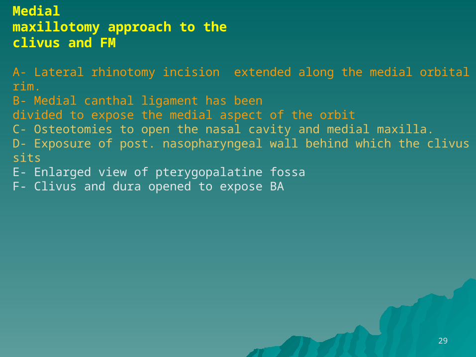
Medialmaxillotomy approach to theclivus and FM
A- Lateral rhinotomy incision extended along the medial orbital rim. B- Medial canthal ligament has beendivided to expose the medial aspect of the orbitC- Osteotomies to open the nasal cavity and medial maxilla.D- Exposure of post. nasopharyngeal wall behind which the clivus sitsE- Enlarged view of pterygopalatine fossaF- Clivus and dura opened to expose BA
30
3 - TRANSSPHENOIDAL APPROACH PRINCIPLE : - Removal of floor of the sella turcica - Extension of bony opening downward on the clivus to the inf. margin of the sphenoid sinus
INDICATION : - Biopsy or partial removal of lesions extending to the upper third of the clivus ADVANTAGES : - Low complication rate - Easy route - May be combined with TC-TB approach in removing lesions involving the clivus and FM
31
DISADVANTAGES : - Small operative field limited to sup. third of the clivus - CSF leak Endoscopic approach – - Visualization from crista galli to the FM - Exposure of entire clivus possible with 2 cm width - Lat. limit : ICAs - Used for radical resection of : # Clival chordoma # Midline clival meningioma
32
4 -TRANSCERVICAL APPROACH:
( Stevenson et al)
- Directed through the fascial planes of the neck to the region of FM. - Tracheostomy facilitates the exposure. - Selected infrequently
ADVATAGES : - Avoids opening the oropharyngeal mucosa DISADVANTGAES - Increase depth of the exposure and lenth of time - Not a direct midline exposure.

33
Trans cervical approach
A: T-shaped skin incision
B:Resectable areas
C- Exposure along the ant. border of SCM and between ECA and ICA
D- Prevertebral fascia and longus capitis and longus colli are separated in the midline
from the clivus to C3 and are retracted laterally
E and F- Ant. arch of the atlas and the odontoid process, and a 2.5-mm width of clivus
extending from the FM to the spheno-occipital synchondrosis may be removed
34
Structures that may be divided to increase the exposure : -
- Ascending pharyngeal and Sup. thyroid arteries - Stylohyoid muscle and Ant. belly of the digastric - Stylohyoid ligament and 9 th nerve - Stylopharyngeus and styloglossus Resectable areas : - Clivus - Ant. arch of the atlas - Body of the odontoid process
35
5- TRANSCRANIAL - TRANSBASAL APPROACH ( Derome et al)
Exposure even upto C2 and C3 vertebral bodies.
INDICATION:
- Ant. side of FM lesions if also involves and requires resection of ethmoid and sphenoid bones and clivus
ADVANTAGES : - Tighter closure of the dura mater is possible - Sub cranial mucosal planes can be preserved - Can be combined with another intradural approach without the high risk of infection
36
- May be combined with TB – TS route to gain access to the sella turcica - Clivus and sphenoid bone can be resected more extensively than by the transsphenoidal approach
DISADVANTAGES : - Extensive surgical trauma - Anosmia - CSF leaks - Meningitis - Pseudomeningoceles
Should not be considered for approaching a tumor strictly localized in the region of FM
37
A:Transcranial-transbasal Approach
B: Bifrontal craniotomy.Clivus is reached after resecting the post. part of floor of the antcranial fossa, upper part ofthe walls of ethmoid and sphenoid sinuses and floor of the sella. C -Orbital roof and the remainder of the cranial base are reconstructed
38
6- EXTENDED FRONTAL APPROACH :
Similar to the TC - TB approach, except that it includes an orbitofrontoethmoidal osteotomy Supraorbital ridges, and part of the orbital roofs and possibly the upper nasion, roof of the ethmoid sinuses, and the cribriform plate are removed in a single block
Extradural or combined intradural - extradural approach
39
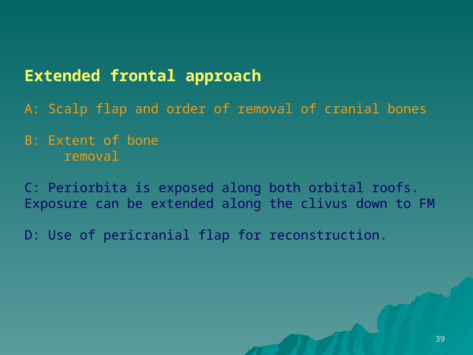
Extended frontal approach
A: Scalp flap and order of removal of cranial bones
B: Extent of bone removal
C: Periorbita is exposed along both orbital roofs. Exposure can be extended along the clivus down to FM
D: Use of pericranial flap for reconstruction.
40
C - LATERAL APPROACHES
Rarely used in combination
Directed through the temporal bone
May require repositioning of the carotid artery or facial nerve, and possibly resection of the auditory and vestibular labyrinth
INDICATION : - Intradural lesions located lateral and/or ant. of the brainstem, involving the temporal bone
41
ADVANTAGES : - Provide an avenue of exposure for lesions that involve the temporal and sphenoid bones in addition to clivus
- Also provide access to the anterior aspect of the midbrain, pons, and medulla and to the CP angle and nerves in the posterior fossa DISADVANTAGES :
- May necessitate sacrifice of the sigmoid sinus - Need of neuro-otologist in obtaining the exposure.
42
1- TRANSLABYRINTHINE APPROACH : Through a mastoidectomy and labyrinthectomy.
ADVANTAGES: - May also be combined with a retrosigmoid or a supra - and infratentorial presigmoid approach - Seventh nerve is preserved - Minimal cerebellar and brainstem retraction
43
DISADVANTAGES: - High incidence of CSF leak - Hearing is sacrificed - Reduced exposure - Longer dissection time of temporal bone
CONTRAINDICATION: - Chronic otitis media
44
2 -TRANSCOCHLEAR APPROACH:
(House and Hitselberger)
Anteromedial extension of the trans-labyrinthine approach Bone is removed up to the edge of clivus
ADVANTAGES: - Excellent exposure of clivus and both anterior and anteromedial aspect of the brain stem
DISADVANTAGES: - Hearing and seventh nerve both are sacrificed - High risk of CSF leak
45
3 - PRESIGMOID (combined supra and infra tentorial) APPROACH : Basic Principle : - Variable amounts of petrous bone dissection - Supra and infratentorial craniotomy - Division of tentorium - Vein of Labbe preserved
Reduced risk- Semicircular canals and 7th nerve are not skeletonized
46
ADVANTAGES: - Shorter working distance - Provides access from FM to dorsum sellae - Provides access to the cranial nerves III through XII and to the major arteries in the posterior circulation. - Minimal brain retraction - Provides multiple angles for dissection. - Can also be combined with a far-lateral approach
DISADVANTAGES: - Limited access to the lower petroclival region by the jugular bulb
47
4 - SUBTEMPORAL PREAURICULAR INFRATEMPORAL APPROACH :
Reaches the skull base from an anterolateral direction Directed through the infratemporal and middle fossa to the part of the ant. surface of the petrous bone ADVANTAGE: - Alternative lateral route to vascular lesions of the mid basilar artery or at the vertebrobasilar junction DISADVANTAGE: - Limited exposure of the CP angle and FM
48
5 - POSTAURICULAR TRANSTEMPORAL APPROACH Combines a transcochlear exposure with an infratemporal approach
INDICATIONS: - May be used when the pathology involves the mastoid and the infratemporal fossa and extends to the facial recess, hypotympanic area, and jugular bulb
49
ADVANTAGES: - Lower and middle clivus exposure without the neural retraction - Can be extended to the parasellar and parasphenoidal areas
DISADVANTAGES: - Hearing is sacrifised
50
Midline and far lateral approaches to foramen magnum lesions - Prof. B.S. Sharma et al Neurology India :Year : 1999 | Volume : 47 | Issue : 4 | Page : 268-71
20 patients operated in 5 yr by either post. or the far lateral approach
- Group A: (n=5)- Posterior or posterolaterally situated lesions (Approach – Midline posterior) - Group B: (n=15)- Anteriorly or anterolaterally situated lesions (Approach – Far lateral) RESULT:
- Complete neurological recovery = 14 - Mild neurological deficit = 2 - Significant neurological deficit = 1 - Death = 1 (presented late)
CONCLUSION : Far lateral approach is adequate for removal of anterior or anterolaterally situated lesions
51
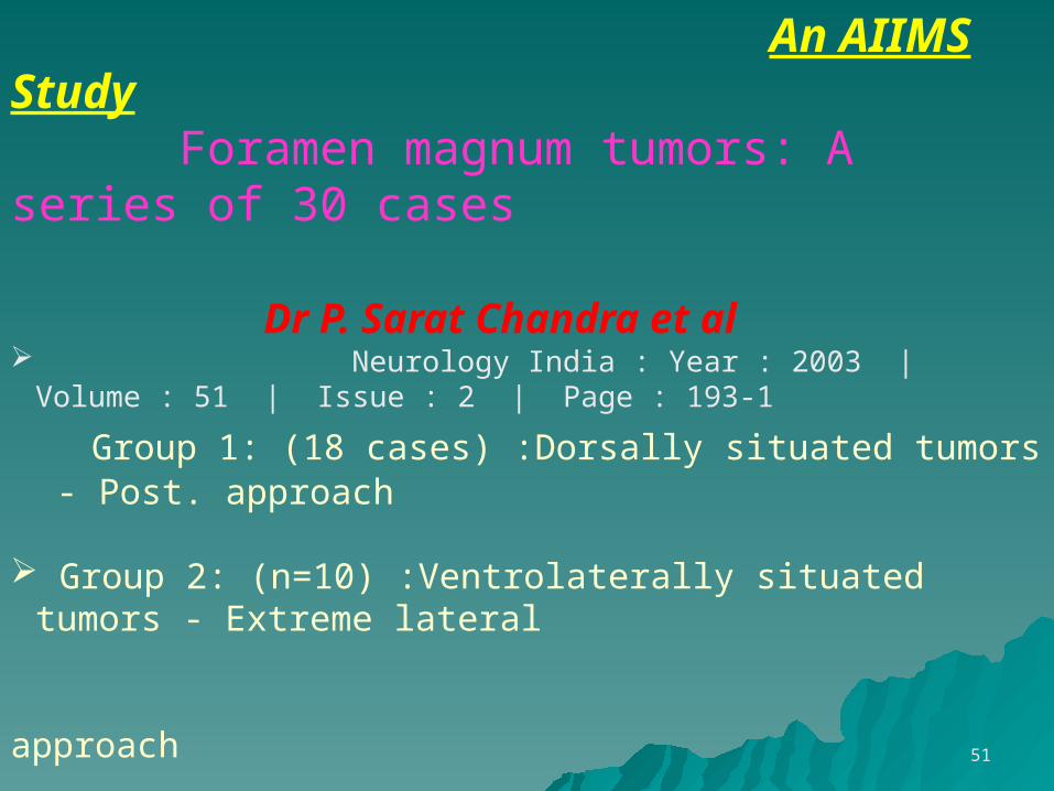
An AIIMS Study Foramen magnum tumors: A series of 30 cases Dr P. Sarat Chandra et al Neurology India : Year : 2003 | Volume : 51 | Issue : 2 | Page : 193-1
Group 1: (18 cases) :Dorsally situated tumors - Post. approach
Group 2: (n=10) :Ventrolaterally situated tumors - Extreme lateral approach
Group 3: (n=2) :Tumors were located anteriorly - Trans oral biopsy
RESULT: - Total excision of the tumor = 24 - Subtotal excision of the tumor = 6 - Death = 2 - Complications = 8 (e.g. CSF leak, meningitis, pseudomeningocele, laryngeal edema etc.)
Ext. lateral approach was satisfactory for all Group 2 cases
52
Surgical approaches: postoperative care and complications "posterolateral-far lateral transcondylar approach to the ventral foramen magnum and upper cervical spinal canal" Menezes AH.Department of Neurosurgery, University of Iowa Hospitals and Clinics, 200 Hawkins Drive, 1824 JPP, Iowa City, Iowa, 52242, USA, [email protected] Nerv Syst. 2008 Mar 26.
CONCLUSIONS: - The posterolateral transcondylar route exposure is quite satisfactory with minimal or no retraction of important neurovascular structures in the region. - Modifications of this theme can be applied as the lesions require.


![Technical Note Meningiomas del foramen magno: Reporte de ...alvarocampero.com.ar/img/pdf/8/Meningiomas del Foramen Magno 1.pdf · es mandatorio el fresado del cóndilo occipital,[2,3,5,8‑10,12,29‑32]](https://img.pdfslide.us/doc/110x75/6046007b1c6e6d708c416426/technical-note-meningiomas-del-foramen-magno-reporte-de-del-foramen-magno-1pdf.jpg)