Embed Size (px)
Citation preview
CHAPTER ONE Chapter Titlexxvi
Textbook Tour
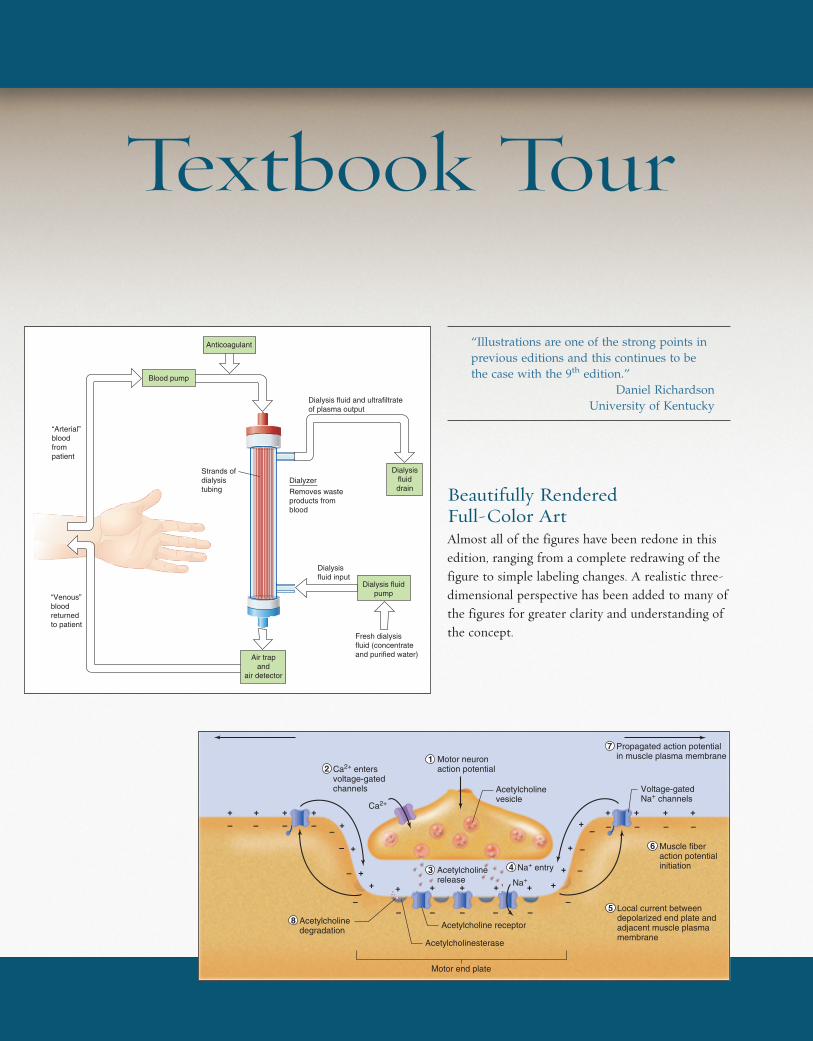
Beautifully Rendered Full-Color ArtAlmost all of the figures have been redone in thisedition, ranging from a complete redrawing of thefigure to simple labeling changes. A realistic three-dimensional perspective has been added to many ofthe figures for greater clarity and understanding ofthe concept.
“Illustrations are one of the strong points inprevious editions and this continues to bethe case with the 9th edition.”
Daniel RichardsonUniversity of Kentucky
Blood pump
Anticoagulant
Dialysisfluiddrain
Dialysis fluidpump
Air trapand
air detector
“Venous” blood returned to patient
Dialysis fluid and ultrafiltrateof plasma output
Dialysisfluid input
Fresh dialysisfluid (concentrateand purified water)
Dialyzer
Removes wasteproducts fromblood
Strands ofdialysistubing
“Arterial”bloodfrompatient
+ ++ +
+
+
+
+ + + +
+
+
++ + + +
+++
Acetylcholinerelease
Motor neuronaction potential
Muscle fiberaction potentialinitiation
Local current betweendepolarized end plate andadjacent muscle plasmamembrane
Acetylcholine receptorAcetylcholinedegradation
Acetylcholinesterase
Motor end plate
Acetylcholinevesicle
Propagated action potentialin muscle plasma membrane
Voltage-gatedNa+ channels
– – – ––
–
–– – –
–
–
–
– – – – –
– –
–
12
8
3
7
6
4
5
Na+ entry
Na+
Ca2+ entersvoltage-gatedchannels
Ca2+
wid37936_fm_i-xxxi 11/14/02 1:25 PM Page xxvi mac14 mac14:1151_Mike D.:

Contents xxvii
Color-Coded IllustrationsColor-coding is effectively used to promotelearning. For example, there are specific colorsfor the extracellular fluid, the intracellular fluid,muscle, and the lumen of the renal tubules andGl tract.
Flow DiagramsLong a hallmark of this book, extensive use of flowdiagrams has been continued in this edition. A bookmarkhas been included with your book to give a furtherexplanation.
Basement membrane
Movementof filtrate
FenestraCapillary lumen
Footprocesses
Capsular space
Filtrationslits
Cell processesPodocyte (visceral layerof Bowman's capsule) Cell body
Glomerularcapillary (cut)
Afferentarteriole
Efferentarteriole
Proximaltubule
Visceral layer(podocyte)
Parietal layer
Capillary
Renalcorpuscle
Bowman's capsule
Glomerular capillary(covered by visceral layer)
Filtrationslits
Fenestrae
Juxtaglomerularapparatus
JuxtaglomerularcellsMacula densa
Distaltubule
(c)
(b)
(a)
Substances in the blood are filtered through capillary fenestrae. The filtrate then passes across the basement membrane and through slit pores between the foot processes (also called pedicels) and enters the capsular space. From here, the filtrate is transported to the lumen of the proximal convoluted tubule.
Podocytes of Bowman’s capsule surround the capillaries. Filtration slits between the podocytes allow fluid to pass into Bowman’s capsule. The glomerulus is composed of capillary endothelium that is fenestrated. Surrounding the endothelial cells is a basement membrane.
Blood flows into the glomerulus throughthe afferent arterioles and leaves theglomerulus through the efferent arterioles. The proximal tubule exits Bowman’s capsule.
a.
b.
c.
Luminal distensionamino acids & peptides
Gastric phase stimuli:
Histaminesecretion
Enteric neural activity
Cephalic phase stimuli
Acid secretion
Parietal cell
Brain
Gastrin secretion
HCl
+
+
+
++ +
wid37936_fm_i-xxxi 11/14/02 1:26 PM Page xxvii mac14 mac14:1151_Mike D.:
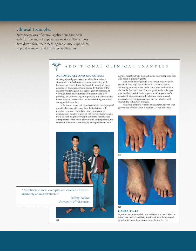
ACROMEGALY AND GIGANTISMAcromegaly and gigantism arise when there exists asituation in which chronic, excess amounts of growthhormone are secreted into the blood. In almost all cases,acromegaly and gigantism are caused by tumors of theanterior pituitary gland that secrete growth hormone atvery high rates. These tumors are typically very slowgrowing, and, if occurring after puberty, it may be decadesbefore a person realizes that there is something seriouslywrong with him or her.
If the tumor arises before puberty, when the epiphysealgrowth plates are still open, then the individual willdevelop gigantism (“pituitary giant”) and grow toextraordinary heights (Figure 11–28). Some pituitary giantshave reached heights over eight feet! If the tumor arisesafter puberty, when linear growth is no longer possible, thecondition is known as acromegaly. Such people will be of
normal height but will manifest many other symptoms thatalso occur in pituitary giants.
Even when linear growth is no longer possible (afterpuberty), very high plasma levels of GH result in thethickening of many bones in the body, most noticeably inthe hands, feet, and head. The jaw, particularly, enlarges togive the characteristic facial appearance (“prognathism”)associated with acromegaly. In addition, many internalorgans also become enlarged, and this can interfere withtheir ability to function normally.
All adults continue to make and secrete GH even aftergrowth has stopped. That is because GH has metabolic
A D D I T I O N A L C L I N I C A L E X A M P L E S
FIGURE 11–28Gigantism and acromegaly in one individual of a pair of identical
twins. Note the increased height and facial bone thickening (a),
as well as the bone thickening of hands (b) and feet (c).(a)
(c)
(b)
Clinical ExamplesNew discussions of clinical applications have beenadded at the ends of appropriate sections. The authorshave drawn from their teaching and clinical experiencesto provide students with real-life applications.
“Additional clinical examples are excellent. This isdefinitely an improvement.”
Jeffrey WalkerUniversity of Wisconsin
wid37936_fm_i-xxxi 11/14/02 1:27 PM Page xxviii mac14 mac14:1151_Mike D.:
I. ForebrainA. Cerebral hemispheres
1. Contain the cerebral cortex, which participates inperception (Chapter 7), the generation of skilledmovements (Chapter 10), reasoning, learning, andmemory (Chapter 8)
2. Contain subcortical nuclei, including those thatparticipate in coordination of skeletal muscle activity(Chapter 10)
3. Contain interconnecting fiber pathways
B. Thalamus1. Is a synaptic relay station for sensory pathways on
their way to the cerebral cortex (Chapter 7)2. Participates in control of skeletal muscle coordination
(Chapter 10)3. Plays a key role in awareness (Chapter 8)
C. Hypothalamus1. Regulates anterior pituitary gland function (Chapter 11)2. Regulates water balance (Chapter 14)3. Participates in regulation of autonomic nervous
system (Chapters 6 and 16)4. Regulates eating and drinking behavior (Chapter 16)5. Regulates reproductive system (Chapters 11 and 17)6. Reinforces certain behaviors (Chapter 8)7. Generates and regulates circadian rhythms (Chapters
1, 7, 11, and 16)8. Regulates body temperature (Chapter 16)9. Participates in generation of emotional behavior
(Chapter 8)
D. Limbic system1. Participates in generation of emotions and emotional
behavior (Chapter 8)2. Plays essential role in most kinds of learning
(Chapter 8)
II. CerebellumA. Coordinates movements, including those for posture
and balance (Chapter 10)B. Participates in some forms of learning (Chapter 8)
III. BrainstemA. Contains all the fibers passing between the spinal
cord, forebrain, and cerebellumB. Contains the reticular formation and its various
integrating centers, including those for cardiovascularand respiratory activity (Chapters 12 and 13)
C. Contains nuclei for cranial nerves III through XII
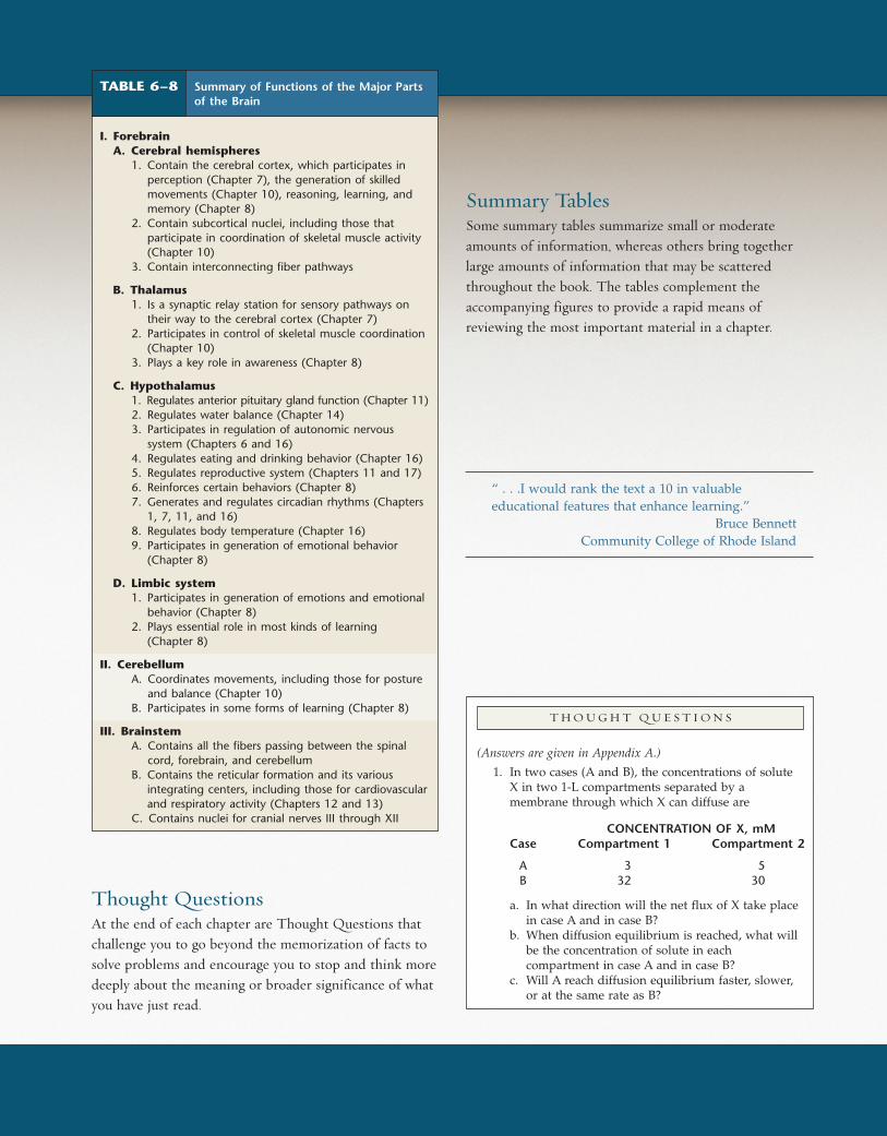
TABLE 6–8 Summary of Functions of the Major Partsof the Brain
Thought QuestionsAt the end of each chapter are Thought Questions thatchallenge you to go beyond the memorization of facts tosolve problems and encourage you to stop and think moredeeply about the meaning or broader significance of whatyou have just read.
Summary TablesSome summary tables summarize small or moderateamounts of information, whereas others bring togetherlarge amounts of information that may be scatteredthroughout the book. The tables complement theaccompanying figures to provide a rapid means ofreviewing the most important material in a chapter.
“ . . .I would rank the text a 10 in valuableeducational features that enhance learning.”
Bruce BennettCommunity College of Rhode Island
T H O U G H T Q U E S T I O N S
(Answers are given in Appendix A.)
1. In two cases (A and B), the concentrations of soluteX in two 1-L compartments separated by amembrane through which X can diffuse are
CONCENTRATION OF X, mMCase Compartment 1 Compartment 2
A 3 5B 32 30
a. In what direction will the net flux of X take placein case A and in case B?
b. When diffusion equilibrium is reached, what willbe the concentration of solute in eachcompartment in case A and in case B?
c. Will A reach diffusion equilibrium faster, slower,or at the same rate as B?
wid37936_fm_i-xxxi 11/14/02 1:28 PM Page xxix mac14 mac14:1151_Mike D.:

Supplements Tour
Active ArtStep-by-step breakdown of key illustrations allows you tosynchronize the art with your lecture presentation. Youcan also modify the art to create your own version.
Digital Content Manager CD-ROMIf you’re looking for illustrations, photographs, tables, andanimations to incorporate into your lecture presentations,handouts, or quizzes, this easy-to-use CD containshundreds of digital assets from Human Physiology.Simply click on the chapter folder, select an image, andyou’re ready to import the image into the application ofyour choice. It’s that easy.
wid37936_fm_i-xxxi 11/14/02 1:29 PM Page xxx mac14 mac14:1151_Mike D.:
Contents
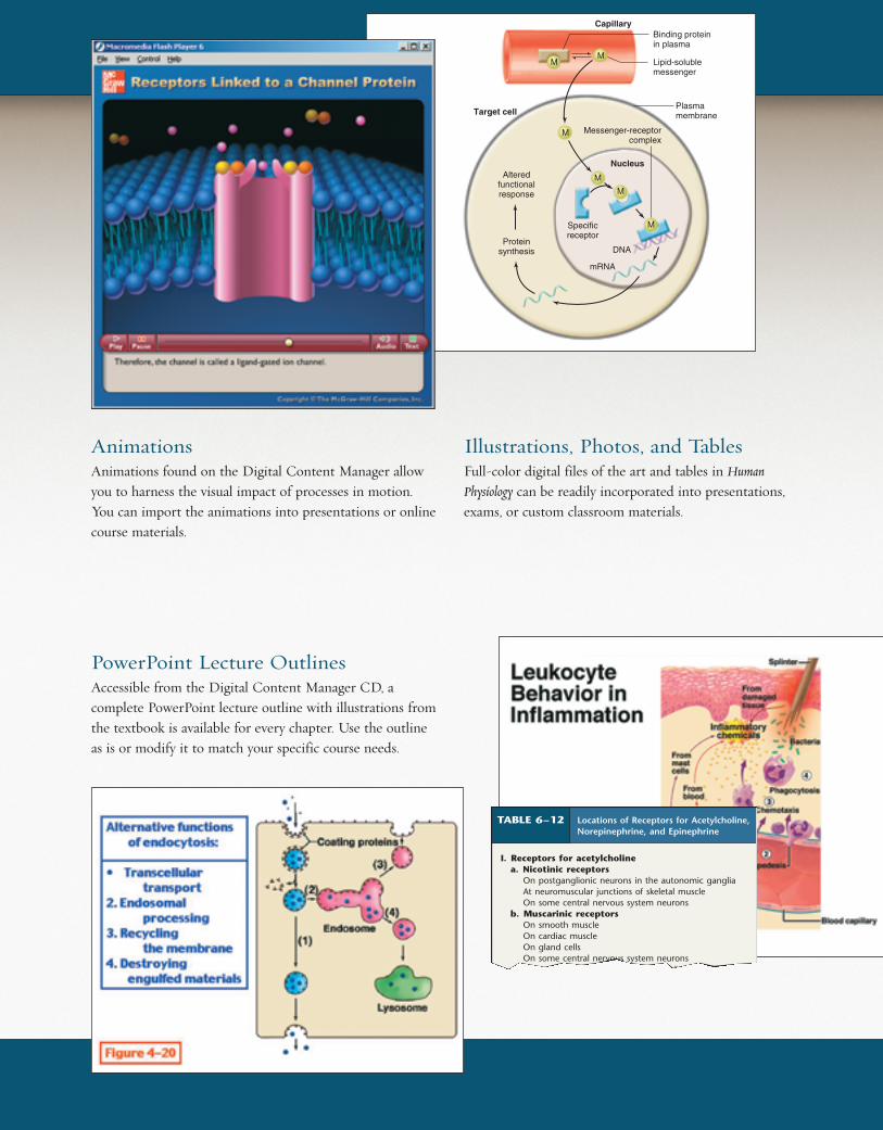
AnimationsAnimations found on the Digital Content Manager allowyou to harness the visual impact of processes in motion.You can import the animations into presentations or onlinecourse materials.
Illustrations, Photos, and TablesFull-color digital files of the art and tables in Human
Physiology can be readily incorporated into presentations,exams, or custom classroom materials.
PowerPoint Lecture OutlinesAccessible from the Digital Content Manager CD, acomplete PowerPoint lecture outline with illustrations fromthe textbook is available for every chapter. Use the outlineas is or modify it to match your specific course needs.
TABLE 6–12 Locations of Receptors for Acetylcholine,Norepinephrine, and Epinephrine
I. Receptors for acetylcholinea. Nicotinic receptors
On postganglionic neurons in the autonomic gangliaAt neuromuscular junctions of skeletal muscleOn some central nervous system neurons
b. Muscarinic receptorsOn smooth muscleOn cardiac muscleOn gland cellsOn some central nervous system neuronsOn some neurons of autonomic ganglia (although the
Lipid-solublemessenger
Binding proteinin plasma
Plasma membraneTarget cell
Nucleus
Capillary
MM
Messenger-receptorcomplex
M
M
M
MSpecificreceptor
DNA
mRNA
Proteinsynthesis
Alteredfunctionalresponse
wid37936_fm_i-xxxi 11/14/02 1:31 PM Page xxxi mac14 mac14:1151_Mike D.:
CHAPTER ONE Chapter Titlexxxii
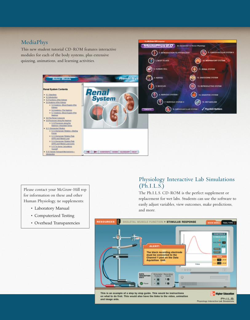
MediaPhysThis new student tutorial CD-ROM features interactivemodules for each of the body systems, plus extensivequizzing, animations, and learning activities.
Physiology Interactive Lab Simulations(Ph.I.L.S.)The Ph.I.L.S. CD-ROM is the perfect supplement orreplacement for wet labs. Students can use the software toeasily adjust variables, view outcomes, make predictions,and more.
Please contact your McGraw-Hill repfor information on these and otherHuman Physiology, 9e supplements:
• Laboratory Manual
• Computerized Testing
• Overhead Transparencies
wid37936_fm_i-xxxi 11/14/02 5:56 PM Page xxxii mac14 mac14:1151_Mike D.: