Embed Size (px)
Citation preview
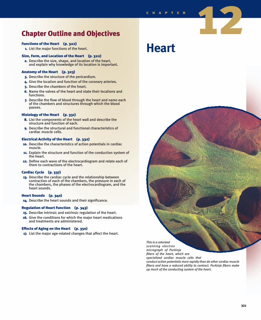
This is a colorizedscanning electronmicrograph of Purkinjefibers of the heart, which arespecialized cardiac muscle cells thatconduct action potentials more rapidly than do other cardiac musclefibers and have a reduced ability to contract. Purkinje fibers makeup much of the conducting system of the heart.
Chapter Outline and ObjectivesFunctions of the Heart (p. 322)
1. List the major functions of the heart.
Size, Form, and Location of the Heart (p. 322)2. Describe the size, shape, and location of the heart,
and explain why knowledge of its location is important.
Anatomy of the Heart (p. 323)3. Describe the structure of the pericardium.4. Give the location and function of the coronary arteries.5. Describe the chambers of the heart.6. Name the valves of the heart and state their locations and
functions.7. Describe the flow of blood through the heart and name each
of the chambers and structures through which the bloodpasses.
Histology of the Heart (p. 331)8. List the components of the heart wall and describe the
structure and function of each.9. Describe the structural and functional characteristics of
cardiac muscle cells.
Electrical Activity of the Heart (p. 332)10. Describe the characteristics of action potentials in cardiac
muscle.11. Explain the structure and function of the conduction system of
the heart.12. Define each wave of the electrocardiogram and relate each of
them to contractions of the heart.
Cardiac Cycle (p. 337)13. Describe the cardiac cycle and the relationship between
contraction of each of the chambers, the pressure in each ofthe chambers, the phases of the electrocardiogram, and theheart sounds.
Heart Sounds (p. 340)14. Describe the heart sounds and their significance.
Regulation of Heart Function (p. 343)15. Describe intrinsic and extrinsic regulation of the heart.16. Give the conditions for which the major heart medications
and treatments are administered.
Effects of Aging on the Heart (p. 350)17. List the major age-related changes that affect the heart.
12C H A P T E R
Heart
321
see43696_ch12_321-352 1:13:06 08:04 PM Page 321
*(866) 487-8889*
CONFIRMING PROOFSMASTER SETPlease markall alterations
on this set only
eople often refer to the heart as if it werethe seat of certain strong emotions. A verydetermined person may be described as
having “a lot of heart,” and a person who hasbeen disappointed romantically can be de-scribed as having a “broken heart.” A popularholiday in February not only dramatically dis-torts the heart’s anatomy, it also attaches ro-mantic emotions to it. The heart is a muscularorgan that is essential for life because itpumps blood through the body. Emotions area product of brain function, not heart function.
Fluids flow through a pipe only if theyare forced to do so. The force is commonlyproduced by a pump, which increases the pres-sure of the liquid at the pump above the pres-sure in the pipe. Thus, the liquid flows fromthe pump through the pipe from an area ofhigher pressure to an area of lower pressure. Ifthe pressure produced by the pump increases,flow of liquid through the pipe increases. If thepressure produced by the pump decreases,flow of liquid through the pipe decreases.
Like a pump that forces water to flowthrough a pipe, the heart contracts forcefullyto pump blood through the blood vesselsof the body (figure 12.1). The heart of ahealthy adult, at rest, pumps approximately5 liters (L) of blood per minute. For most peo-ple, the heart continues to pump at approxi-mately that rate for more than 75 years; and,during short periods of vigorous exercise, theamount of blood pumped per minute increasesseveral fold. If the heart loses its pumping abil-ity for even a few minutes, however, blood flow
through vessels of the pulmonary circulation(figure 12.2). The left side of the heart pumpsblood to all other tissues of the body and backto the right side of the heart through vesselsof the systemic circulation.
through the blood vessels stops, and the life ofthe individual is in danger.
The heart is actually two pumps in one.The right side of the heart pumps blood to thelungs and back to the left side of the heart
322 Chapter 12
p
Functions of the HeartFunctions of the heart include:
1. Generating blood pressure. Contractions of the heartgenerate blood pressure, which is required for bloodflow through the blood vessels.
2. Routing blood. The heart separates the pulmonary andsystemic circulations, which ensures the flow ofoxygenated blood to tissues.
3. Ensuring one-way blood flow. The valves of the heartensure a one-way flow of blood through the heart andblood vessels.
4. Regulating blood supply. Changes in the rate and forceof heart contraction match blood flow to the changingmetabolic needs of the tissues during rest, exercise, andchanges in body position.
Size, Form, and Location of the HeartThe adult heart is shaped like a blunt cone and is approximatelythe size of a closed fist. It is larger in physically active adults than inless active but otherwise healthy adults, and it generally decreasesin size after approximately age 65, especially in those who are notphysically active. The blunt, rounded point of the cone is the apex;and the larger, flat part at the opposite end of the cone is the base.
The heart is located in the thoracic cavity between the twopleural cavities, which surround the lungs. The heart, trachea,esophagus, and associated structures form a midline partition,the mediastinum (me�de-as-tı�num; see figure 1.12). The heartis surrounded by its own cavity, the pericardial cavity (peri,around � cardio, heart) (see chapter 1).
It is important for clinical reasons to know the locationand shape of the heart in the thoracic cavity. This knowledge
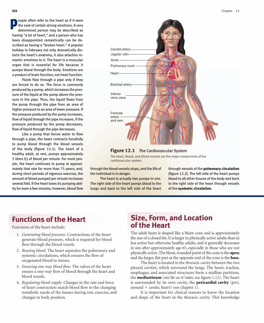
Figure 12.1 The Cardiovascular SystemThe heart, blood, and blood vessels are the major components of thecardiovascular system.
Inferiorvena cava
Femoralartery and vein
Brachial artery
Heart
Carotid artery
Jugular vein
Aorta
Pulmonary trunk
see43696_ch12_321-352 1:10:06 08:44 PM Page 322
*(866) 487-8889*
CONFIRMING PROOFSMASTER SETPlease markall alterations
on this set only

Figure 12.2 Overview of the Circulatory SystemThe circulatory system consists of the pulmonary and systemic circulations. The right side of the heart pumps blood through vessels to the lungs and back to the leftside of the heart through the pulmonary circulation. The left side of the heart pumps blood through vessels to the tissues of the body and back to the right side ofthe heart through the systemic circulation.
Tissuecapillaries
Circulation totissues of head
Circulation totissues oflower body
Systemiccirculation(to body)
Pulmonarycirculation(to lungs)
Lung
Lungcapillaries
Right side of heartLeft sideof heart
Tissuecapillaries
CO2
CO2
O2
O2
CO2
O2
allows a person to accurately place a stethoscope to hear theheart sounds, place chest leads to record an electrocardiogram(e-lek-tro-kar�de-o-gram; ECG or EKG), or administer effec-tive cardiopulmonary resuscitation (kar�de-o-pul�mo-nar-e-re-sus�i-ta-shun; CPR).
The heart lies obliquely in the mediastinum, with its basedirected posteriorly and slightly superiorly and the apex di-rected anteriorly and slightly inferiorly. The apex is also directedto the left so that approximately two-thirds of the heart’s masslies to the left of the midline of the sternum (figure 12.3). Thebase of the heart is located deep to the sternum and extends tothe level of the second intercostal space. The apex is locateddeep to the left fifth intercostal space, approximately 7–9 cen-timeters (cm) to the left of the sternum near the midclavicularline, which is a perpendicular line that extends down from themiddle of the clavicle (see figure 12.3).
Anatomy of the HeartPericardiumThe heart is surrounded by the pericardial cavity. The pericar-dial cavity is formed by the pericardium (per-i-kar�de-um),or pericardial sac, which surrounds the heart and anchors itwithin the mediastinum (see figures 12.3 and 12.4). The peri-cardium consists of two layers. The tough, fibrous connectivetissue outer layer is called the fibrous pericardium and the in-ner layer of flat epithelial cells, with a thin layer of connectivetissue, is called the serous pericardium. The portion of the serouspericardium lining the fibrous pericardium is the parietal
pericardium, whereas the portion covering the heart surface isthe visceral pericardium, or epicardium (ep-i-kar�de-um,upon the heart). The parietal and visceral pericardia are contin-uous with each other where the great vessels enter or leave theheart. The pericardial cavity, located between the visceral andparietal pericardia, is filled with a thin layer of pericardial fluidproduced by the serous pericardium. The pericardial fluid helpsreduce friction as the heart moves within the pericardial sac.
Disorders of the PericardiumPericarditis (per�i-kar-dı�tis) is an inflammation of the serous
pericardium. The cause is frequently unknown, but it can result from
infection, diseases of connective tissue, or damage due to radiation
treatment for cancer. It can be extremely painful, with sensations of pain
referred to the back and to the chest, which can be confused with the
pain of a myocardial infarction (heart attack). Pericarditis can result in a
small amount of fluid accumulation within the pericardial sac.
Cardiac tamponade (tam-po-nad�, a pack or plug) is a potentially fatal
condition in which fluid or blood accumulates in the pericardial sac. The
fluid compresses the heart from the outside. The heart is a powerful
muscle, but it relaxes passively. When it is compressed by fluid within
the pericardial sac, it cannot dilate when the cardiac muscle relaxes.
Consequently, the heart cannot fill with blood during relaxation, which
makes it impossible for it to pump. Cardiac tamponade can cause a
person to die quickly unless the fluid is removed. Causes of cardiac
tamponade include rupture of the heart wall following a myocardial
infarction, rupture of blood vessels in the pericardium after a malignant
tumor invades the area, damage to the pericardium resulting from
radiation therapy, and trauma such as occurs in a traffic accident.
Heart 323
see43696_ch12_321-352 1:10:06 10:08 AM Page 323
*(866) 487-8889*
CONFIRMING PROOFSMASTER SETPlease markall alterations
on this set only

Anterior view
Fibrous pericardium
Parietal pericardiumVisceral pericardium(or epicardium)
Pericardial cavityfilled with pericardialfluid
Serous pericardium
Pericardium
Figure 12.4 The Heart in the PericardiumThe heart is located in the pericardium, which consists of an outer fibrous pericardium and an inner serous pericardium. The serous pericardium has two parts: theparietal pericardium lines the fibrous pericardium, and the visceral pericardium (epicardium) covers the surface of the heart. The pericardial cavity, between theparietal and visceral pericardium, is filled with a small amount of pericardial fluid.
Figure 12.3 Location of the Heart in the ThoraxThe heart is located in the thoracic cavity between the lungs, deep and slightly to the left of the sternum. The base of the heart, located deep to the sternum,extends superiorly to the second intercostal space, and the apex of the heart is located deep to the fifth intercostal space, approximately 7–9 cm to the left of thesternum where the midclavicular line intersects with the fifth intercostal space (see inset).
Superior vena cava
Right lung
Right atrium
Right ventricle
Rib
Visceral pleura
Diaphragm
Aortic arch
Pulmonary trunkLeft atriumLeft lung
Left ventricle
Apex of heart
Larynx
Trachea
Pleural cavityParietal pleura
Midclavicularline
SternumApex of heart5th intercostalspace
2nd intercostal space
Anterior view
324
see43696_ch12_321-352 1:10:06 10:08 AM Page 324
*(866) 487-8889*
CONFIRMING PROOFSMASTER SETPlease markall alterations
on this set only

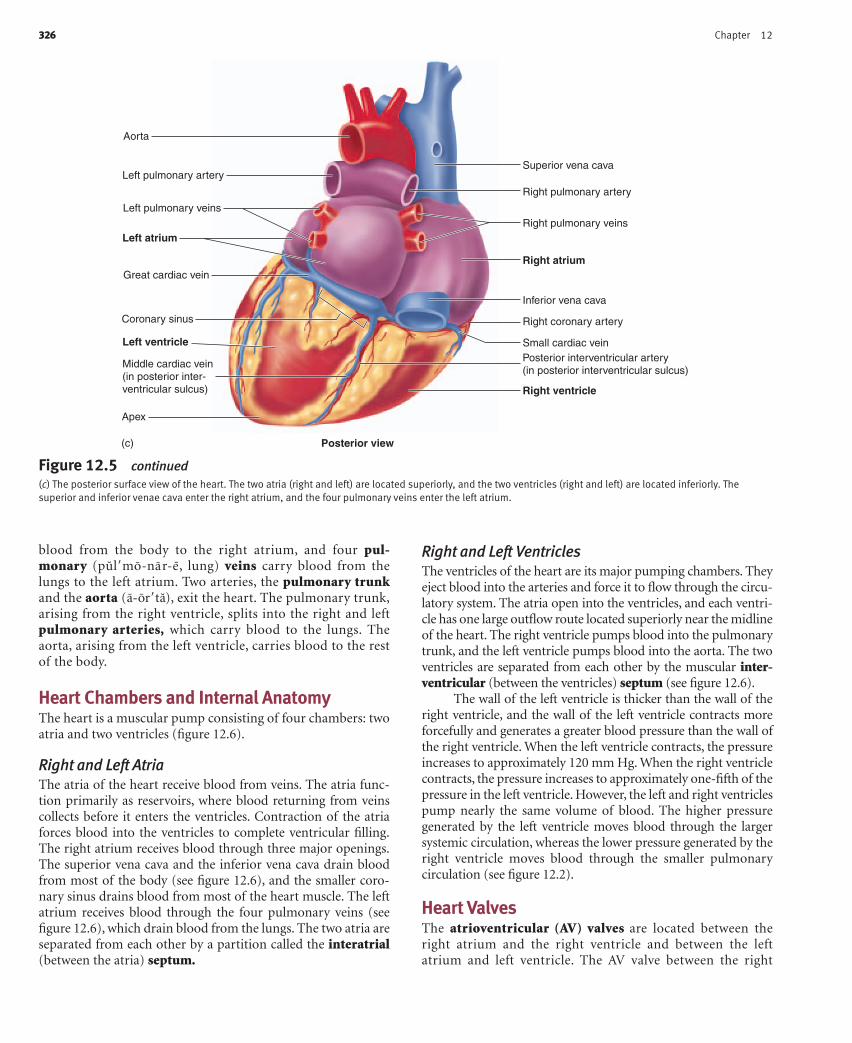
External AnatomyThe right and left atria (a�tre-a, entrance chambers; sing.atrium) are located at the base of the heart, and the right andleft ventricles (ven�tri-klz, a cavity) extend from the base ofthe heart toward the apex (figure 12.5). A coronary (kor�o-nar-e, circling like a crown) sulcus (sul�kus, ditch) extends aroundthe heart, separating the atria from the ventricles. In addition,two grooves, or sulci, which indicate the division between the
Heart 325
right and left ventricles, extend inferiorly from the coronarysulcus. The anterior interventricular sulcus extends inferi-orly from the coronary sulcus on the anterior surface of theheart, and the posterior interventricular sulcus extends infe-riorly from the coronary sulcus on the posterior surface of theheart (see figure 12.5).
Six large veins carry blood to the heart (see figure 12.5aand c): the superior vena cava and inferior vena cava carry
Anterior view
Aortic arch
Superior vena cava
Coronary sulcus
Pulmonary trunk
Left pulmonary artery
Branches of leftpulmonary artery
Left pulmonary veins
Left atrium
Great cardiac vein(in anterior interventricular sulcus)
Left ventricle
Anterior interventricular artery(in anterior interventricular sulcus)
Branches of rightpulmonary artery
Right pulmonary veins
Right atrium
Right coronary artery
Right ventricle
Inferior vena cava
(a)
Aorta
Pericardium(reflected laterally)
Pulmonary trunk
Anterior interventricular artery(in anterior interventricular sulcus)
Great cardiac vein(in anterior interventricular sulcus)
Left ventricle
Superiorvena cava
Right atrium
Right coronaryartery
Right ventricle
Smallcardiac vein
Rightmarginalartery Anterior view(b)
Figure 12.5 Anterior Surface View of the Heart(a) The anterior view of the heart. The two atria (right and left) are located superiorly, and the two ventricles (right and left) are located inferiorly. The superior andinferior venae cava enter the right atrium. The pulmonary veins enter the left atrium. The pulmonary trunk exits the right ventricle, and the aorta exits the leftventricle. (b) Photograph of the anterior surface of the heart.
see43696_ch12_321-352 12:6:05 02:51 PM Page 325
*(866) 487-8889*
CONFIRMING PROOFSMASTER SETPlease markall alterations
on this set only
326 Chapter 12
Figure 12.5 continued(c) The posterior surface view of the heart. The two atria (right and left) are located superiorly, and the two ventricles (right and left) are located inferiorly. Thesuperior and inferior venae cava enter the right atrium, and the four pulmonary veins enter the left atrium.
Posterior view
Superior vena cava
Right pulmonary artery
Right pulmonary veins
Right atrium
Inferior vena cava
Right coronary artery
Small cardiac vein
Right ventricle
Posterior interventricular artery(in posterior interventricular sulcus)
Aorta
Great cardiac vein
Left atrium
Left pulmonary veins
Left pulmonary artery
Apex
Middle cardiac vein(in posterior inter-ventricular sulcus)
Left ventricle
Coronary sinus
(c)
blood from the body to the right atrium, and four pul-monary (pul�mo-nar-e, lung) veins carry blood from thelungs to the left atrium. Two arteries, the pulmonary trunkand the aorta (a-or�ta), exit the heart. The pulmonary trunk,arising from the right ventricle, splits into the right and leftpulmonary arteries, which carry blood to the lungs. Theaorta, arising from the left ventricle, carries blood to the restof the body.
Heart Chambers and Internal AnatomyThe heart is a muscular pump consisting of four chambers: twoatria and two ventricles (figure 12.6).
Right and Left AtriaThe atria of the heart receive blood from veins. The atria func-tion primarily as reservoirs, where blood returning from veinscollects before it enters the ventricles. Contraction of the atriaforces blood into the ventricles to complete ventricular filling.The right atrium receives blood through three major openings.The superior vena cava and the inferior vena cava drain bloodfrom most of the body (see figure 12.6), and the smaller coro-nary sinus drains blood from most of the heart muscle. The leftatrium receives blood through the four pulmonary veins (seefigure 12.6), which drain blood from the lungs. The two atria areseparated from each other by a partition called the interatrial(between the atria) septum.
Right and Left VentriclesThe ventricles of the heart are its major pumping chambers. Theyeject blood into the arteries and force it to flow through the circu-latory system. The atria open into the ventricles, and each ventri-cle has one large outflow route located superiorly near the midlineof the heart. The right ventricle pumps blood into the pulmonarytrunk, and the left ventricle pumps blood into the aorta. The twoventricles are separated from each other by the muscular inter-ventricular (between the ventricles) septum (see figure 12.6).
The wall of the left ventricle is thicker than the wall of theright ventricle, and the wall of the left ventricle contracts moreforcefully and generates a greater blood pressure than the wall ofthe right ventricle. When the left ventricle contracts, the pressureincreases to approximately 120 mm Hg. When the right ventriclecontracts, the pressure increases to approximately one-fifth of thepressure in the left ventricle. However, the left and right ventriclespump nearly the same volume of blood. The higher pressuregenerated by the left ventricle moves blood through the largersystemic circulation, whereas the lower pressure generated by theright ventricle moves blood through the smaller pulmonarycirculation (see figure 12.2).
Heart ValvesThe atrioventricular (AV) valves are located between theright atrium and the right ventricle and between the leftatrium and left ventricle. The AV valve between the right
see43696_ch12_321-352 12:6:05 02:51 PM Page 326
*(866) 487-8889*
CONFIRMING PROOFSMASTER SETPlease markall alterations
on this set only

Heart 327
Anterior view
Aortic arch
Pulmonary trunk
Left pulmonary artery
Branches of rightpulmonary artery
Left pulmonary veins
Right pulmonary veins
Left atrium
Right atriumLeft ventricle
Right ventricle
Bicuspid (mitral) valve
Chordae tendineae
Papillary muscles
Papillary muscles Interventricular septum
Superior vena cava
Inferior vena cava
Pulmonary semilunar valve
Tricuspid valve
Aortic semilunar valve
Right pulmonary veins
Coronary sinus
Figure 12.6 Internal Anatomy of the HeartThe heart is cut in a frontal plane to show the internal anatomy.
atrium and the right ventricle has three cusps and is called thetricuspid valve (see figures 12.6 and 12.7a). The AV valve be-tween the left atrium and left ventricle has two cusps and iscalled the bicuspid, or mitral (resembling a bishop’s miter, atwo-pointed hat) valve (see figures 12.6 and 12.7b). Thesevalves allow blood to flow from the atria into the ventriclesbut prevent it from flowing back into the atria. When theventricles relax, the higher pressure in the atria forces the AV
valves to open and blood flows from the atria into the ventricles(figure 12.8a). In contrast, when the ventricles contract,blood flows toward the atria and causes the AV valves to close(figure 12.8b).
Each ventricle contains cone-shaped muscular pillars calledpapillary (pap�ı-lar-e, nipple- or pimple-shaped) muscles. Thesemuscles are attached by thin, strong connective tissue stringscalled chordae tendineae (kor�de ten�di-ne-e, heart strings) to the
(a) View of the tricuspid valve, the chordae tendineae, and the papillary muscles.
Anterior view
Pulmonarytrunk
Chordaetendineae
Papillarymuscles
Superiorvena cava
Ascendingaorta
Right atrium
Anterior cuspof tricuspid valve
Inferior vena cava
Figure 12.7 Heart Valves
(b) A superior view of the heart valves. Note the three cusps of each semilunar valve meeting to prevent the backflow of blood.
Pulmonarytrunk
Superior vena cava
AortaPulmonarysemilunar valve
Opening of leftcoronary artery
Bicuspid valve
Left atrium (cut open)
Opening of right coronaryarteryAortic semilunar valve
Right atrium
Superior view
see43696_ch12_321-352 1:10:06 10:08 AM Page 327
*(866) 487-8889*
CONFIRMING PROOFSMASTER SETPlease markall alterations
on this set only
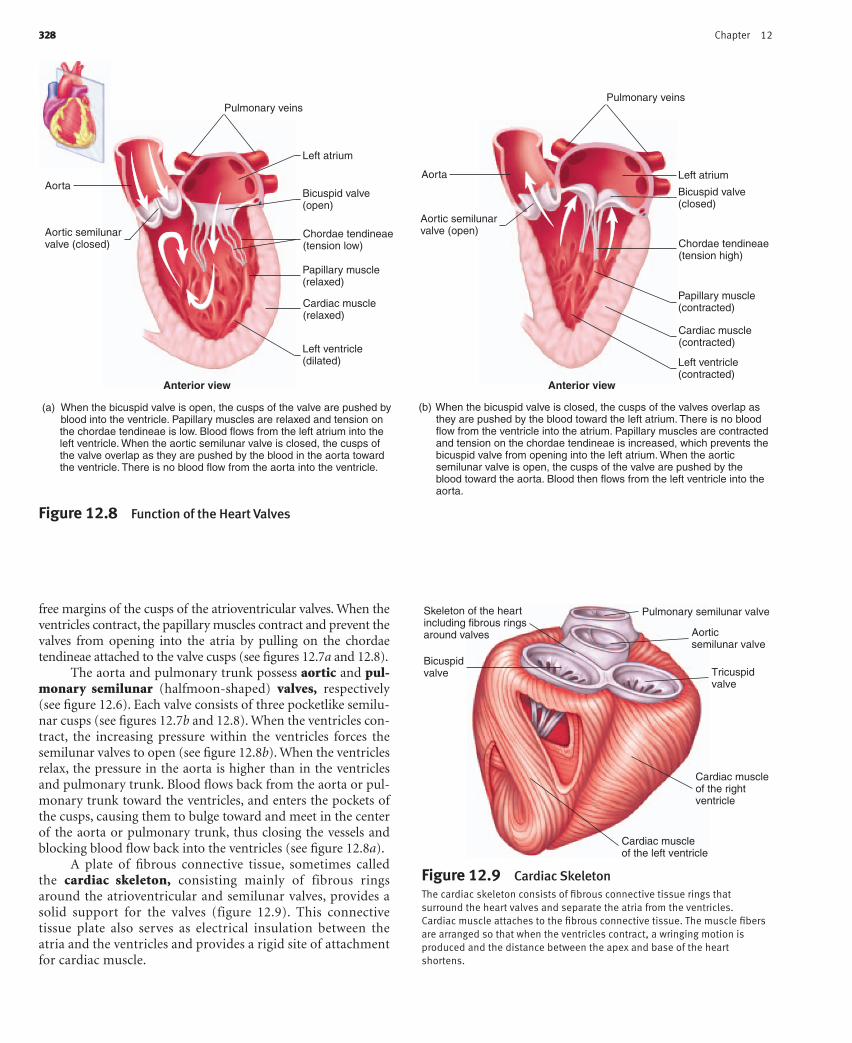
free margins of the cusps of the atrioventricular valves. When theventricles contract, the papillary muscles contract and prevent thevalves from opening into the atria by pulling on the chordaetendineae attached to the valve cusps (see figures 12.7a and 12.8).
The aorta and pulmonary trunk possess aortic and pul-monary semilunar (halfmoon-shaped) valves, respectively(see figure 12.6). Each valve consists of three pocketlike semilu-nar cusps (see figures 12.7b and 12.8). When the ventricles con-tract, the increasing pressure within the ventricles forces thesemilunar valves to open (see figure 12.8b). When the ventriclesrelax, the pressure in the aorta is higher than in the ventriclesand pulmonary trunk. Blood flows back from the aorta or pul-monary trunk toward the ventricles, and enters the pockets ofthe cusps, causing them to bulge toward and meet in the centerof the aorta or pulmonary trunk, thus closing the vessels andblocking blood flow back into the ventricles (see figure 12.8a).
A plate of fibrous connective tissue, sometimes calledthe cardiac skeleton, consisting mainly of fibrous ringsaround the atrioventricular and semilunar valves, provides asolid support for the valves (figure 12.9). This connectivetissue plate also serves as electrical insulation between theatria and the ventricles and provides a rigid site of attachmentfor cardiac muscle.
328 Chapter 12
Figure 12.8 Function of the Heart Valves
Anterior view Anterior view
Pulmonary veins
Aortic semilunarvalve (closed)
Aorta
Left atrium
Bicuspid valve(open)
Chordae tendineae(tension low)
Papillary muscle(relaxed)
Cardiac muscle(relaxed)
Left ventricle(dilated)
Pulmonary veins
Aortic semilunarvalve (open)
Left atrium
Bicuspid valve(closed)
Chordae tendineae(tension high)
Papillary muscle(contracted)
Cardiac muscle(contracted)
Left ventricle(contracted)
Aorta
(a) When the bicuspid valve is open, the cusps of the valve are pushed by blood into the ventricle. Papillary muscles are relaxed and tension on the chordae tendineae is low. Blood flows from the left atrium into the left ventricle. When the aortic semilunar valve is closed, the cusps of the valve overlap as they are pushed by the blood in the aorta toward the ventricle. There is no blood flow from the aorta into the ventricle.
(b) When the bicuspid valve is closed, the cusps of the valves overlap as they are pushed by the blood toward the left atrium. There is no blood flow from the ventricle into the atrium. Papillary muscles are contracted and tension on the chordae tendineae is increased, which prevents the bicuspid valve from opening into the left atrium. When the aortic semilunar valve is open, the cusps of the valve are pushed by the blood toward the aorta. Blood then flows from the left ventricle into the aorta.
Pulmonary semilunar valve
Aorticsemilunar valve
Tricuspidvalve
Cardiac muscleof the rightventricle
Cardiac muscleof the left ventricle
Skeleton of the heartincluding fibrous ringsaround valves
Bicuspidvalve
Figure 12.9 Cardiac SkeletonThe cardiac skeleton consists of fibrous connective tissue rings thatsurround the heart valves and separate the atria from the ventricles.Cardiac muscle attaches to the fibrous connective tissue. The muscle fibersare arranged so that when the ventricles contract, a wringing motion isproduced and the distance between the apex and base of the heartshortens.
see43696_ch12_321-352 1:10:06 10:08 AM Page 328
*(866) 487-8889*
CONFIRMING PROOFSMASTER SETPlease markall alterations
on this set only

Heart 329
Route of Blood Flow Through the HeartThe route of blood flow through the heart is depicted in figure12.10. Even though blood flow through the heart is describedfor the right and then the left side of the heart, it is important tounderstand that both atria contract at the same time, and bothventricles contract at the same time. This concept is most im-portant when the electrical activity, pressure changes, and heartsounds are considered.
Blood enters the right atrium from the systemic circulationthrough the superior and inferior venae cava, and from heartmuscle through the coronary sinus (see figure 12.10a and b). Mostof the blood flowing into the right atrium flows into the right
ventricle while the right ventricle relaxes following the previouscontraction. Before the end of ventricular relaxation, the rightatrium contracts, and enough blood is pushed from the rightatrium into the right ventricle to complete right ventricular filling.
Following right atrial contraction, the right ventricle beginsto contract. Contraction of the right ventricle pushes blood againstthe tricuspid valve, forcing it closed.After pressure within the rightventricle increases, the pulmonary semilunar valve is forced open,and blood flows into the pulmonary trunk. As the right ventriclerelaxes, its pressure falls rapidly, and pressure in the pulmonarytrunk becomes greater than in the right ventricle. The back-flowof blood forces the pulmonary semilunar valve to close.
Superiorvena cava
Inferior vena cava
Papillary muscles
Tricuspid valve
Right atrium
Pulmonary semilunarvalve
Branches of right pulmonary arteries
Branches of left pulmonary arteries
Pulmonary veins
Left atrium
Bicuspid valve
Interventricular septum
Left ventricle
Right ventricle
Pulmonary trunk
Aortic arch
Aortic semilunarvalve
Superior andinferior venacava
Rightatrium
Body tissues(systemiccirculation)
AortaLeftventricle
Leftatrium
Pulmonaryveins
Aorticsemilunarvalves
Tricuspidvalve
Bicuspidvalve
Rightventricle
Pulmonarytrunk
Pulmonaryarteries
Lung tissue(pulmonarycirculation)
Pulmonarysemilunarvalves
Coronary sinus Cardiac veins
Heart tissue
Coronary arteries
Pulmonary veins
Figure 12.10 Blood Flow Through the Heart(a) Frontal section of the heart revealing the four chambers and the direction of blood flow through the heart. (b) Diagram listing in order the structures throughwhich blood flows in the systemic and pulmonary circulations. The heart valves are indicated by circles; deoxygenated blood (blue); oxygenated blood (red ).
(a)
(b)
see43696_ch12_321-352 12:6:05 02:51 PM Page 329
*(866) 487-8889*
CONFIRMING PROOFSMASTER SETPlease markall alterations
on this set only

The pulmonary trunk branches to form the right and leftpulmonary arteries, which carry blood to the lungs, wherecarbon dioxide is released and oxygen is picked up. Blood re-turning from the lungs enters the left atrium through the fourpulmonary veins (see figure 12.10a and b). Most of the bloodflowing into the left atrium passes into the left ventricle whilethe left ventricle relaxes following the previous contraction.Before the end of ventricular relaxation, the left atrium con-tracts, and enough blood is pushed from the left atrium intothe left ventricle to complete left ventricular filling.
Following left atrial contraction, the left ventricle be-gins to contract. Contraction of the left ventricle pushesblood against the bicuspid valve, forcing it closed. After pres-sure within the left ventricle increases, the aortic semilunarvalve is forced open, and blood flows into the aorta (see figure12.10a and b). Blood flowing through the aorta is distributedto all parts of the body, except to that part of the lung sup-plied by the pulmonary blood vessels. As the left ventricle re-laxes, its pressure falls rapidly, and pressure in the aortabecomes greater than in the left ventricle. The back-flow ofblood forces the aortic semilunar valve to close.
Blood Supply to the HeartCoronary ArteriesCardiac muscle in the wall of the heart is thick and metaboli-cally very active. Two coronary arteries supply blood to the wallof the heart (figure 12.11a). The coronary arteries originate
330 Chapter 12
from the base of the aorta, just above the aortic semilunarvalves. The left coronary artery originates on the left side ofthe aorta. It has three major branches: The anterior interven-tricular artery lies in the anterior interventricular sulcus, thecircumflex artery extends around the coronary sulcus on theleft to the posterior surface of the heart, and the left marginalartery extends inferiorly along the lateral wall of the left ventri-cle from the circumflex artery. The branches of the left coronaryartery supply much of the anterior wall of the heart and most ofthe left ventricle. The right coronary artery originates on theright side of the aorta. It extends around the coronary sulcus onthe right to the posterior surface of the heart and gives rise tothe posterior interventricular artery, which lies in the poste-rior interventricular sulcus. The right marginal artery extendsinferiorly along the lateral wall of the right ventricle. The rightcoronary artery and its branches supply most of the wall of theright ventricle.
In a resting person, blood flowing through the coronaryarteries of the heart gives up approximately 70% of its oxygen. Incomparison, blood flowing through arteries to skeletal musclegives up only about 25% of its oxygen. The percentage of oxygenthe blood releases to skeletal muscle increases to 70% or moreduring exercise. The percentage of oxygen the blood releases tocardiac muscle cannot increase substantially during exercise.Cardiac muscle is therefore very dependent on an increased rateof blood flow through the coronary arteries above its restinglevel to provide an adequate oxygen supply during exercise.Blood flow into the coronary circulation is greatest during
Anterior view Anterior view
Pulmonary trunkLeft coronary artery
Left atrium
Aortic arch
Left ventricle
Aortic arch
Leftventricle
Greatcardiacvein
Coronarysinus
Posterior veinof left ventricle
Left atrium
Pulmonary trunk
Superiorvena cava
Rightatrium
Right ventricle
Smallcardiacvein
Middle cardiac vein
Intorightatrium
Superiorvena cava
Aorticsemilunarvalve
Rightatrium
Rightcoronaryartery
Posteriorinterventricularartery
Rightmarginalartery
Right ventricle
Anteriorinterventricularartery
Left marginalartery
Circumflexartery
Figure 12.11 Blood Supply to the HeartThe vessels of the anterior surface of the heart are seen directly and are a darker color, whereas the vessels of the posterior surface are seen through the heart andare a lighter color. (a) Coronary arteries supply blood to the wall of the heart. (b) Cardiac veins carry blood from the wall of the heart back to the right atrium.
(a) (b)
see43696_ch12_321-352 1:10:06 10:08 AM Page 330
*(866) 487-8889*
CONFIRMING PROOFSMASTER SETPlease markall alterations
on this set only

Heart 331
relaxation of the ventricles of the heart when contraction of thecardiac muscle does not compress the coronary arteries. Bloodflow into other arteries of the body is highest during contractionof the ventricles.
P R E D I C T �1
Predict the effect on the heart if blood flow through theanterior interventricular artery is restricted or completelyblocked (Hint: See figure 12.11a).
Cardiac VeinsThe cardiac veins drain blood from the cardiac muscle. Theirpathways are nearly parallel to the coronary arteries and mostdrain blood into the coronary sinus, a large vein located withinthe coronary sulcus on the posterior aspect of the heart. Bloodflows from the coronary sinus into the right atrium (see figure12.11b). Some small cardiac veins drain directly into the rightatrium.
Disorders of Coronary ArteriesWhen a blood clot, or thrombus (throm�bus, a clot), suddenly blocks
a coronary blood vessel, a heart attack, or coronary thrombosis
(throm�bo-sis), occurs. The area that has been cut off from its blood
supply suffers from a lack of oxygen and nutrients and dies if the blood
supply is not quickly reestablished. The region of dead heart tissue is
called an infarct (in�farkt), or myocardial infarction. If the infarct is large
enough, the heart may be unable to pump enough blood to keep the
person alive. People who are at risk for coronary thromboses can reduce
the likelihood of heart attack by taking small amounts of aspirin daily,
which inhibits thrombus formation (see chapter 11).
Aspirin is also administered to many people who are exhibiting
clear symptoms of a heart attack. In some cases, it is possible to treat
heart attacks with enzymes such as streptokinase (strep-to-kı�nas) or
tissue plasminogen (plaz-min�o-jen) activator (t-PA), which break
down blood clots. One of the enzymes is injected into the circulatory
system of a heart attack patient, where it reduces or removes the
blockage in the coronary artery. If the clot is broken down quickly, the
blood supply to cardiac muscle is reestablished, and the heart may
suffer little permanent damage.
Coronary arteries can become blocked more gradually by
atherosclerotic (ath�er-o-skler-ot�ik, athero, pasty material � sclerosis,
hardness) lesions. These thickenings in the walls of arteries can contain
deposits that are high in cholesterol and other lipids. The lesions narrow
the lumen (opening) of the arteries, thus restricting blood flow. The
ability of cardiac muscle to function is reduced when it is deprived of an
adequate blood supply. The person suffers from fatigue and often pain in
the area of the chest and usually in the left arm with the slightest exertion.
The pain is called angina pectoris (an-jı�na, pain; pek�to-ris, in the
chest).
Angioplasty (an�je -o-plas-te ) is a surgical procedure in which a
small balloon is threaded through the aorta and into a coronary artery.
After the balloon has entered a partially blocked coronary artery, it is
inflated, flattening the atherosclerotic deposits against the vessel
wall and opening the blocked blood vessel. This technique improves
the function of cardiac muscle in patients suffering from inadequate
blood flow to the cardiac muscle through the coronary arteries. Some
controversy exists about its effectiveness, at least in some patients,
because dilation of the coronary arteries can be reversed within a few
weeks or months and because blood clots can form in coronary
arteries following angioplasty. Small rotating blades and lasers are
also used to remove lesions from coronary vessels, or a small coil
device, called a stent, is placed in the vessels to hold them open
following angioplasty.
A coronary bypass is a surgical procedure that relieves the effects
of obstructions in the coronary arteries. The technique involves taking
healthy segments of blood vessels from other parts of the patient’s
body and using them to bypass, or create an alternative path around,
obstructions in the coronary arteries. The technique is common for
those who suffer from severe blockage of parts of the coronary arteries.
Histology of the HeartHeart WallThe heart wall is composed of three layers of tissue: the epi-cardium, the myocardium, and the endocardium (figure 12.12).The epicardium (ep-i-kar�de-um), also called the visceralpericardium, is a thin serous membrane forming the smoothouter surface of the heart. It consists of simple squamous
Simple squamousepithelium
Loose connectivetissue and fat
Epicardium(visceralpericardium)
Myocardium
Endocardium
Trabeculaecarneae
Figure 12.12 Heart WallPart of the wall of the heart has been removed, enlarged, and rotated so thatthe inner surface is visible. The enlarged section illustrates the epicardium(visceral pericardium), the myocardium, and the endocardium.
see43696_ch12_321-352 1:10:06 10:08 AM Page 331
*(866) 487-8889*
CONFIRMING PROOFSMASTER SETPlease markall alterations
on this set only
332 Chapter 12
mitochondria, which produce ATP at a rate rapid enough tosustain the normal energy requirements of cardiac muscle.An extensive capillary network provides an adequate oxygensupply to the cardiac muscle cells. Unlike skeletal muscle, car-diac muscle cannot develop a significant oxygen debt. Devel-opment of a large oxygen debt could result in muscularfatigue and cessation of cardiac muscle contraction.
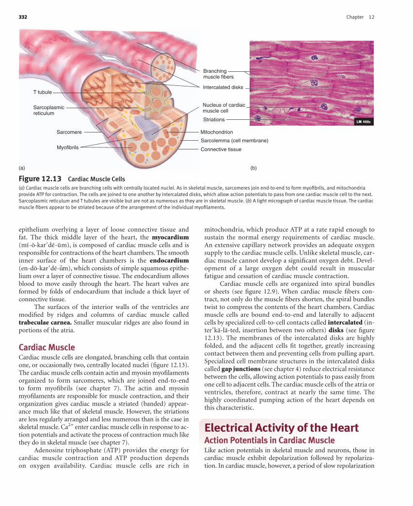
Cardiac muscle cells are organized into spiral bundlesor sheets (see figure 12.9). When cardiac muscle fibers con-tract, not only do the muscle fibers shorten, the spiral bundlestwist to compress the contents of the heart chambers. Cardiacmuscle cells are bound end-to-end and laterally to adjacentcells by specialized cell-to-cell contacts called intercalated (in-ter�ka-la-ted, insertion between two others) disks (see figure12.13). The membranes of the intercalated disks are highlyfolded, and the adjacent cells fit together, greatly increasingcontact between them and preventing cells from pulling apart.Specialized cell membrane structures in the intercalated diskscalled gap junctions (see chapter 4) reduce electrical resistancebetween the cells, allowing action potentials to pass easily fromone cell to adjacent cells. The cardiac muscle cells of the atria orventricles, therefore, contract at nearly the same time. Thehighly coordinated pumping action of the heart depends onthis characteristic.
Electrical Activity of the HeartAction Potentials in Cardiac MuscleLike action potentials in skeletal muscle and neurons, those incardiac muscle exhibit depolarization followed by repolariza-tion. In cardiac muscle, however, a period of slow repolarization
Connective tissue
Sarcolemma (cell membrane)
MitochondrionSarcomere
Sarcoplasmicreticulum
T tubule
Myofibrils
Branchingmuscle fibers
Nucleus of cardiacmuscle cell
Striations
Intercalated disks
LM 400x
Figure 12.13 Cardiac Muscle Cells(a) Cardiac muscle cells are branching cells with centrally located nuclei. As in skeletal muscle, sarcomeres join end-to-end to form myofibrils, and mitochondriaprovide ATP for contraction. The cells are joined to one another by intercalated disks, which allow action potentials to pass from one cardiac muscle cell to the next.Sarcoplasmic reticulum and T tubules are visible but are not as numerous as they are in skeletal muscle. (b) A light micrograph of cardiac muscle tissue. The cardiacmuscle fibers appear to be striated because of the arrangement of the individual myofilaments.
(a) (b)
epithelium overlying a layer of loose connective tissue andfat. The thick middle layer of the heart, the myocardium(mı-o-kar�de-um), is composed of cardiac muscle cells and isresponsible for contractions of the heart chambers. The smoothinner surface of the heart chambers is the endocardium(en-do-kar�de-um), which consists of simple squamous epithe-lium over a layer of connective tissue. The endocardium allowsblood to move easily through the heart. The heart valves areformed by folds of endocardium that include a thick layer ofconnective tissue.
The surfaces of the interior walls of the ventricles aremodified by ridges and columns of cardiac muscle calledtrabeculae carnea. Smaller muscular ridges are also found inportions of the atria.
Cardiac MuscleCardiac muscle cells are elongated, branching cells that containone, or occasionally two, centrally located nuclei (figure 12.13).The cardiac muscle cells contain actin and myosin myofilamentsorganized to form sarcomeres, which are joined end-to-endto form myofibrils (see chapter 7). The actin and myosinmyofilaments are responsible for muscle contraction, and theirorganization gives cardiac muscle a striated (banded) appear-ance much like that of skeletal muscle. However, the striationsare less regularly arranged and less numerous than is the case inskeletal muscle. Ca2+ enter cardiac muscle cells in response to ac-tion potentials and activate the process of contraction much likethey do in skeletal muscle (see chapter 7).
Adenosine triphosphate (ATP) provides the energy forcardiac muscle contraction and ATP production dependson oxygen availability. Cardiac muscle cells are rich in
see43696_ch12_321-352 1:13:06 08:04 PM Page 332
*(866) 487-8889*
CONFIRMING PROOFSMASTER SETPlease markall alterations
on this set only

Heart 333
greatly prolongs the action potential (figure 12.14). In contrastto action potentials in skeletal muscle, which take less than 2 mil-liseconds (ms) to complete, action potentials in cardiac muscletake approximately 200 to 500 ms to complete.
Unlike in skeletal muscle, action potentials in cardiacmuscle are conducted from cell to cell. The action potentialstake longer, and their rate of conduction in cardiac muscle fromcell to cell is slower than the rate of conduction of action poten-tials in single skeletal muscle cells and neurons.
In cardiac muscle, each action potential consists of a de-polarization phase followed by a rapid, but partial earlyrepolarization phase. This is followed by a longer period ofslow repolarization, called the plateau phase. At the end of theplateau phase, a more rapid final repolarization phase takesplace. During the final repolarization phase, the membranepotential achieves its maximum degree of repolarization (seefigure 12.14).
Opening and closing of membrane channels are responsiblefor the changes in the permeability of the cell membrane that pro-duce action potentials. The depolarization phase of the action po-tential results from three permeability changes. Na� channelsopen, increasing the permeability of the cell membrane to Na�.Sodium ions then diffuse into the cell, causing depolarization. Thiscauses K� channels to close quickly, decreasing the permeability ofthe cell membrane to K�. The decreased diffusion of K� out of thecell also causes depolarization. Ca2� channels slowly open, in-creasing the permeability of the cell membrane to Ca2�. Calciumions then diffuse into the cell and cause depolarization. It is notuntil the plateau phase that most of the Ca2� channels open.
Early repolarization occurs when the Na� channels closeand a small number of K� channels open. Diffusion of Na�
into the cell stops, and there is some movement of K� out of thecell. These changes in ion movement result in an early, but smallrepolarization.
Repolarizationphase
–85
1 2
0
Depolarizationphase
(mV
)
Time (ms)
1. Depolarization phase • Na+ channels open. • K+ channels begin to open.
2. Repolarization phase • Na+ channels close. • K+ channels continue to open causing repolarization. • K+ channels close at the end of repolarization and return the membrane potential to its resting value.
1. Depolarization phase • Na+ channels open. • K+ channels close. • Ca2+ channels begin to open.
2. Early repolarization and plateau phases • Na+ channels close. • Some K+ channels open, causing early repolarization. • Ca2+ channels are open, producing the plateau by slowing further repolarization.
3. Final repolarization phase • Ca2+ channels close. • Many K+ channels open.
Finalrepolarizationphase
Earlyrepolarizationphase Plateau
phase
Depolarizationphase
(mV
)
1 2
Time (ms)
500
–85
0
(a) Permeability changes due to voltage-gated channels opening and closing during an action potential in skeletal muscle:
(a) Permeability changes due to voltage-gated channels opening and closing during an action potential in cardiac muscle:
1
2 1
2
3
Process Figure 12.14 Comparison of Action Potentials in Skeletal and Cardiac Muscle(a) An action potential in skeletal muscle consists of depolarization and repolarization phases. (b) An action potential in cardiac muscle consists of depolarization,early repolarization, plateau, and final repolarization phases. Cardiac muscle does not repolarize as rapidly as skeletal muscle (indicated by the break in the curve)because of the plateau phase.
see43696_ch12_321-352 12:6:05 02:51 PM Page 333
*(866) 487-8889*
CONFIRMING PROOFSMASTER SETPlease markall alterations
on this set only

The plateau phase occurs as Ca2� channels continue toopen, and the diffusion of Ca2� into the cell counteracts the po-tential change produced by the diffusion of K� out of the cell.The plateau phase ends and final repolarization begins as theCa2� channels close, and many K� channels open. Diffusion ofCa2� into the cell decreases and diffusion of K� out of the cell in-creases. These changes cause the membrane potential to repolar-ize during the final repolarization phase.
Action potentials in cardiac muscle exhibit a refractoryperiod, like that of action potentials in skeletal muscle and inneurons. The refractory period lasts about the same length oftime as the prolonged action potential in cardiac muscle. Theprolonged action potential and refractory period allow cardiacmuscle to contract and almost complete relaxation to take placebefore another action potential can be produced. Also, the longrefractory period in cardiac muscle prevents tetanic contractionsfrom occurring, thus ensuring a rhythm of contraction andrelaxation for cardiac muscle. Therefore, action potentials incardiac muscle are different from those in skeletal muscle be-cause of the plateau phase, which makes the action potentialand its refractory period last longer.
P R E D I C T �2
Why is it important to prevent tetanic contractions in cardiacmuscle but not in skeletal muscle?
The sinoatrial (SA) (sı�no-a�tre�-al) node, which func-tions as the pacemaker of the heart, is located in the superiorwall of the right atrium and initiates the contraction of theheart. The SA node is the pacemaker because it produces actionpotentials at a faster rate than other areas of the heart. The ac-tion potential of the SA node acts as a stimulus to adjacent ar-eas of the heart. Also, the SA node action potentials havecharacteristics that are somewhat different from action poten-tials in the rest of the cardiac muscle. The SA node has a largernumber of Ca2� channels than do other areas of the heart. Assoon as the final depolarization phase of an action potential is
334 Chapter 12
completed, some Na� enter the cell through nongated chan-nels. Also, the permeability of the membrane to K� decreasesand some of the Ca2� channels open. As they open, Ca2� andsome Na� begin to diffuse into the cell and cause depolariza-tion. The depolarization stimulates additional Ca2� channelsto open. Once threshold is reached, a large number of Ca2�
channels open. Ca2� diffuse into the cell and quickly cause de-polarization. At the peak of the action potential, Ca2� channelsclose and K� channels open once again. The outward move-ment of K� causes repolarization. The cycle repeats itself whenthe K� channels begin to close once again. Ca2� channelblocking agents are drugs that slow the heart by decreasing therate of action potential production in the SA node. Ca2� chan-nel blockers decrease the rate at which Ca2� move throughCa2� channels. As a result, it takes longer for depolarization toreach threshold and the intervals between action potential pro-duction increases.
Conduction System of the HeartContraction of the atria and ventricles is coordinated by spe-cialized cardiac muscle cells in the wall of the heart that formthe conduction system of the heart (figure 12.15).
The SA node, atrioventricular node, atrioventricular bun-dle, right and left bundle branches, and Purkinje fibers comprisethe conduction system of the heart. All of the cells of the conduc-tion system have the ability to produce spontaneous actionpotentials, but at a lower rate than in the SA node. Action poten-tials originate in the SA node and spread over the right and leftatria, causing them to contract. A second area of the heart, theatrioventricular (AV) (a-tre-o-ven�trik�-u�lar) node, is locatedin the lower portion of the right atrium. When action potentialsreach the AV node, they spread slowly through it and then into abundle of specialized cardiac muscle called the atrioventricular(AV) bundle. The slow rate of action potential conduction in theAV node allows the atria to complete their contraction beforeaction potentials are delivered to the ventricles.
Left atrium
Left ventricle
Apex
Atrioventricular(AV) bundle
Atrioventricular(AV) node
Sinoatrial(SA) node1. Action potentials originate in the sinoatrial (SA)
node and travel across the wall of the atrium (arrows) from the SA node to the atrioventricular (AV) node.
2. Action potentials pass through the AV node and along the atrioventricular (AV) bundle, which extends from the AV node, through the fibrous skeleton, into the interventricular septum.
3. The AV bundle divides into right and left bundle branches, and action potentials descend to the apex of each ventricle along the bundle branches.
4. Action potentials are carried by the Purkinje fibers from the bundle branches to the ventricular walls.
Left and rightbundle branches
Purkinjefibers
1
2
3
4
Process Figure 12.15 Conduction System of the Heart
see43696_ch12_321-352 1:10:06 05:00 PM Page 334
*(866) 487-8889*
CONFIRMING PROOFSMASTER SETPlease markall alterations
on this set only
Heart 335
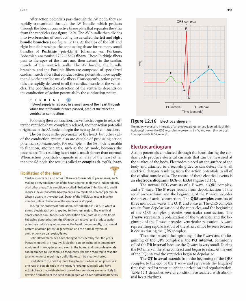
ElectrocardiogramAction potentials conducted through the heart during the car-diac cycle produce electrical currents that can be measured atthe surface of the body. Electrodes placed on the surface of thebody and attached to a recording device can detect the smallelectrical changes resulting from the action potentials in all ofthe cardiac muscle cells. The record of these electrical events isan electrocardiogram (ECG or EKG) (figure 12.16).
The normal ECG consists of a P wave, a QRS complex,and a T wave. The P wave results from depolarization of theatrial myocardium, and the beginning of the P wave precedesthe onset of atrial contraction. The QRS complex consists ofthree individual waves: the Q, R, and S waves. The QRS complexresults from depolarization of the ventricles, and the beginningof the QRS complex precedes ventricular contraction. TheT wave represents repolarization of the ventricles, and the be-ginning of the T wave precedes ventricular relaxation. A waverepresenting repolarization of the atria cannot be seen becauseit occurs during the QRS complex.
The time between the beginning of the P wave and the be-ginning of the QRS complex is the PQ interval, commonlycalled the PR interval because the Q wave is very small. Duringthe PQ interval the atria contract and begin to relax. At the endof the PQ interval the ventricles begin to depolarize.
The QT interval extends from the beginning of the QRScomplex to the end of the T wave and represents the length oftime required for ventricular depolarization and repolarization.Table 12.1 describes several conditions associated with abnor-mal heart rhythms.
Figure 12.16 ElectrocardiogramThe major waves and intervals of an electrocardiogram are labeled. Each thinhorizontal line on the ECG recording represents 1 mV, and each thin verticalline represents 0.04 second.
QRS complex
(mV
)
PQ interval QT interval
Time (seconds)
R
P
T
Q
S
After action potentials pass through the AV node, they arerapidly transmitted through the AV bundle, which projectsthrough the fibrous connective tissue plate that separates the atriafrom the ventricles (see figure 12.9). The AV bundle then dividesinto two branches of conducting tissue called the left and rightbundle branches (see figure 12.15). At the tips of the left andright bundle branches, the conducting tissue forms many smallbundles of Purkinje (pur-kın�je, Johannes von Purkinje,Bohemian anatomist, 1787–1869) fibers. These Purkinje fiberspass to the apex of the heart and then extend to the cardiacmuscle of the ventricle walls. The AV bundle, the bundlebranches, and the Purkinje fibers are composed of specializedcardiac muscle fibers that conduct action potentials more rapidlythan do other cardiac muscle fibers. Consequently, action poten-tials are rapidly delivered to all the cardiac muscle of the ventri-cles. The coordinated contraction of the ventricles depends onthe conduction of action potentials by the conduction system.
P R E D I C T �3
If blood supply is reduced in a small area of the heart throughwhich the left bundle branch passed, predict the effect onventricular contractions.
Following their contraction, the ventricles begin to relax.Af-ter the ventricles have completely relaxed, another action potentialoriginates in the SA node to begin the next cycle of contractions.
The SA node is the pacemaker of the heart, but other cellsof the conduction system also are capable of producing actionpotentials spontaneously. For example, if the SA node is unableto function, another area, such as the AV node, becomes thepacemaker. The resulting heart rate is much slower than normal.When action potentials originate in an area of the heart otherthan the SA node, the result is called an ectopic (ek-top�ik) beat.
Fibrillation of the HeartCardiac muscle can also act as if there are thousands of pacemakers, each
making a very small portion of the heart contract rapidly and independently
of all other areas. This condition is called fibrillation (f ı-bri-la�shun), and it
reduces the output of the heart to only a few milliliters of blood per minute
when it occurs in the ventricles. Death of the individual results in a few
minutes unless fibrillation of the ventricles is stopped.
To stop the process of fibrillation, defibrillation is used, in which a
strong electrical shock is applied to the chest region. The electrical
shock causes simultaneous depolarization of all cardiac muscle fibers.
Following depolarization, the SA node can recover and produce action
potentials before any other area of the heart. Consequently, the normal
pattern of action potential generation and the normal rhythm of
contraction can be reestablished.
Defibrillator machines have changed considerably over the years.
Portable models are now available that can be included in emergency
equipment in workplaces and even in the home, and nonprofessionals
can be trained to use them. Consequently, the time required to respond
to an emergency requiring a defibrillator can be greatly shorted.
Fibrillation of the heart is more likely to occur when action potentials
originate at ectopic sites in the heart. For example, people who have
ectopic beats that originate from one of their ventricles are more likely to
develop fibrillation of the heart than people who have normal heart beats.
see43696_ch12_321-352 1:10:06 05:00 PM Page 335
*(866) 487-8889*
CONFIRMING PROOFSMASTER SETPlease markall alterations
on this set only

P R E D I C T �4
Explain how the ECGs appear for a person who has a damagedleft bundle branch (see Predict 3) and for a person who hasmany ectopic beats originating from her atria.
The ECG as a Diagnostic ToolThe ECG is not a direct measurement of mechanical events in the heart,
and neither the force of contraction nor the blood pressure can be
determined from it. Each deflection in the ECG record, however,
336 Chapter 12
indicates an electrical event within the heart and correlates with a
subsequent mechanical event. Consequently, it is an extremely
valuable diagnostic tool in identifying a number of cardiac
abnormalities, particularly because it is painless, easy to record, and
does not require surgical procedures. Abnormal heart rates or rhythms,
abnormal conduction pathways such as blockages in the conduction
pathways, hypertrophy or atrophy of portions of the heart, and the
approximate location of damaged cardiac muscle can be determined
from analysis of an ECG.
Condition Symptoms Possible CausesAbnormal Heart Rhythms
Tachycardia Heart rate in excess of 100 bpm Elevated body temperature, excessive sympatheticstimulation, toxic conditions
Bradycardia Heart rate less than 60 bpm Increased stroke volume in athletes, excessive vagusnerve stimulation, nonfunctional SA node, carotidsinus syndrome
Sinus arrhythmia Heart rate varies as much as 5% during Cause not always known; occasionally caused byrespiratory cycle and up to 30% during ischemia, inflammation, or cardiac failuredeep respiration
Paroxysmal atrial tachycardia Sudden increase in heart rate to 95–150 bpm Excessive sympathetic stimulation, abnormally elevatedfor a few seconds or even for several hours; permeability of cardiac muscle to Ca2�
P waves precede every QRS complex; P wave inverted and superimposed on T wave
Atrial flutter As many as 300 P waves/min and 125 QRS Ectopic beats in the atriacomplexes/min; resulting in two or threeP waves (atrial contractions) for every QRScomplex (ventricular contraction)
Atrial fibrillation No P waves, normal QRS and T waves, irregular Ectopic beats in the atriatiming, ventricles are constantly stimulated byatria, reduced ventricle filling; increasedchance of fibrillation
Ventricular tachycardia Frequently causes fibrillation Often associated with damage to AV node or ventricular muscle
Heart Blocks
SA node block No P waves, low heart rate resulting from AV Ischemia, tissue damage resulting from infarction; node acting as the pacemaker, normal QRS cause sometimes is unknowncomplexes and T waves
AV node blocks
First-degree PQ interval greater than 0.2 s Inflammation of AV bundle
Second-degree PQ interval 0.25–0.45 s; some P waves trigger Excessive vagus nerve stimulation, AV node damageQRS complexes and others do not; examplesof 2:1, 3:1, and 3:2 P wave/QRS complex ratios
Complete heart block P wave dissociated from QRS complex, atrial Ischemia of AV node or compression of AV bundlerhythm about 100 bpm, ventricular rhythmless than 40 bpm
Premature Contractions
Premature atrial contractions Occasional shortened intervals between one Excessive smoking, lack of sleep, or too much caffeinecontraction and the succeeding contraction;frequently occurs in healthy people
Premature ventricular Prolonged QRS complex, exaggerated voltage Ectopic beat in ventricles, lack of sleep, too muchcontractions (PVCs) because only one ventricle may depolarize, coffee, irritability; occasionally occurs with
possible inverted T wave, increased probability coronary thrombosisof fibrillation
Major Cardiac ArrhythmiasTable 12.1
see43696_ch12_321-352 1:13:06 08:04 PM Page 336
*(866) 487-8889*
CONFIRMING PROOFSMASTER SETPlease markall alterations
on this set only

Heart 337
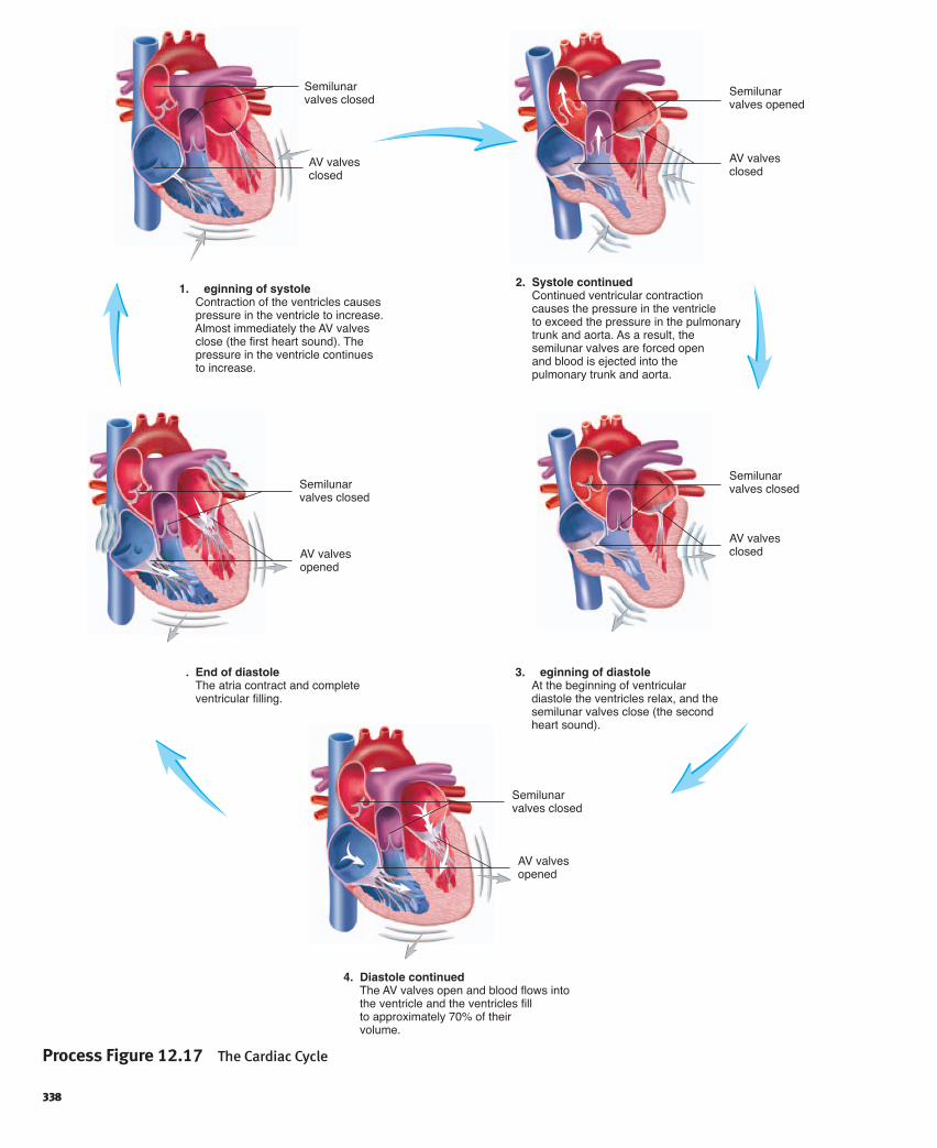
Cardiac CycleThe heart can be viewed as two separate pumps represented bythe right and left halves of the heart. Each pump consists of aprimer pump—the atrium—and a power pump—the ventri-cle. The atria act as primer pumps because they complete thefilling of the ventricles with blood, and the ventricles act aspower pumps because they produce the major force that causesblood to flow through the pulmonary and systemic circulations.The term cardiac cycle refers to the repetitive pumping processthat begins with the onset of cardiac muscle contraction andends with the beginning of the next contraction (figure 12.17).Pressure changes produced within the heart chambers as a re-sult of cardiac muscle contraction are responsible for bloodmovement because blood moves from areas of higher pressureto areas of lower pressure.
Atrial systole (sis�to-le, a contracting) refers to contrac-tion of the two atria. Ventricular systole refers to contractionof the two ventricles. Atrial diastole (dı-as�to-le, dilation)refers to relaxation of the two atria, and ventricular diastolerefers to relaxation of the two ventricles. When the termssystole and diastole are used without reference to the atria orventricles, they refer to ventricular contraction or relaxation.The ventricles contain more cardiac muscle than the atria andproduce far greater pressures, which force blood to circulatethroughout the vessels of the body.
The major events of the cardiac cycle are:
1. Systole—At the beginning of systole, contraction of theventricles pushes blood toward the atria, causing the AVvalves to close as the pressure begins to increase (seefigure 12.17, step 1). As systole continues, the increasingpressure in the ventricles exceeds the pressure in thepulmonary trunk and aorta, the semilunar valves areforced open, and blood is ejected into the pulmonarytrunk and aorta (see figure 12.17, step 2).
2. Diastole—At the beginning of ventricular diastole, thepressure in the ventricles decreases below the pressure inthe aorta and pulmonary trunk. The semilunar valvesclose and prevent blood from flowing back into theventricles (see figure 12.17, step 3).
As diastole continues, the pressure continues todecline in the ventricles until atrial pressures are greaterthan ventricular pressures. Then the AV valves open andblood flows directly from the atria into the relaxedventricles. During the previous ventricular systole, theatria were relaxed and blood collected in them. When theventricles relax and the AV valves open, blood flows intothe ventricles (see figure 12.17, step 4) and fills them toapproximately 70% of their volume.
3. At the end of ventricular diastole, the atria contractand then relax. Atrial systole forces additional blood toflow into the ventricles to complete their filling (seefigure 12.17, step 5). The semilunar valves remain closed.
Willie May Kitt is a 65-year-old man. While walking up
a short flight of stairs to his office where he works as a
bank manager, he experienced a crushing pain in his
chest and he exhibited substantial pallor. Willie fell to
the floor, lost consciousness, and then stopped
breathing. A coworker noticed the pallor, saw Willie
fall, and ran to his aid. He could detect no pulse and
decided to administer cardiopulmonary resuscitation
(CPR). Another coworker called 911 and then assisted
the first coworker. One coworker pushed down firmly
on Willie’s sternum at a rate of approximately 100
compressions per minute. After every 15 compressions,
he paused. The other coworker forced air into Willie’s
lungs by tipping Willie’s head back slightly, placing her
mouth over Willie’s mouth, and blowing air forcefully
into his mouth two times while holding his nasal
passages closed. Pushing down on the sternum
compresses the ventricles of the heart and forces
blood to flow into the aorta and pulmonary trunk.
Between compressions, blood flows into the ventricles
from the atria.
Fortunately, a fire station was only a few blocks away
and it took only about 5 minutes for emergency medical
technicians to arrive. They confirmed the lack of a
pulse and used portable equipment to record an
electrocardiogram, which indicated that the heart
was fibrillating (see Fibrillation of the Heart, p. 335).
They quickly used a portable defibrillator to apply a
strong electrical shock to Willie’s chest. Fortunately,
Willie’s heart responded by beginning to beat
rhythmically.
Willie’s heart may have first developed arrhythmia
and then ventricular fibrillation developed. Willie was
very fortunate. Most people who suffer from sudden
cessation of the pumping activity of the heart do not
survive. In Willie’s case, CPR was administered quickly
and effectively, and emergency help arrived in a very
short period of time.
Willie was transported to a hospital. His condition
could be due to a myocardial infarction (see Myocardial
Infarction, p. 348) or to some other condition. It is
important to identify the underlying cause of the
condition and treat it.
A CASE IN POINT | CardiopulmonaryResuscitation (CPR)
see43696_ch12_321-352 1:10:06 10:08 AM Page 337
*(866) 487-8889*
CONFIRMING PROOFSMASTER SETPlease markall alterations
on this set only
Semilunarvalves closed
AV valves closed
Semilunarvalves opened
AV valves closed
1. Beginning of systole Contraction of the ventricles causes pressure in the ventricle to increase. Almost immediately the AV valves close (the first heart sound). The pressure in the ventricle continues to increase.
2. Systole continued Continued ventricular contraction causes the pressure in the ventricle to exceed the pressure in the pulmonary trunk and aorta. As a result, the semilunar valves are forced open and blood is ejected into the pulmonary trunk and aorta.
3. Beginning of diastole At the beginning of ventricular diastole the ventricles relax, and the semilunar valves close (the second heart sound).
4. Diastole continued The AV valves open and blood flows into the ventricle and the ventricles fill to approximately 70% of their volume.
5. End of diastole The atria contract and complete ventricular filling.
Semilunarvalves closed
AV valves closed
Semilunarvalves closed
AV valves opened
Semilunarvalves closed
AV valves opened
Process Figure 12.17 The Cardiac Cycle
338
see43696_ch12_321-352 1:13:06 08:04 PM Page 338
*(866) 487-8889*
CONFIRMING PROOFSMASTER SETPlease markall alterations
on this set only
Heart 339
Q
S
R
T T
120
100
80
55
90
125
60
40
20
0
(mV
)P
ress
ure
(mm
Hg)
Left
vent
ricul
arvo
lum
e (m
L)"S
ound
" fr
eque
ncy
(cyc
les/
seco
nd)
First heartsound
Secondheartsound
First heartsound
Second heartsound
P
Q
S
R
P
Systole SystoleDiastole
AV valvesclose AV valves
open
Semilunarvalvesopen
Semilunarvalvesclose
AV valvesclose
Diastolicpressure
Semilunarvalves open
AV valvesopen
Semilunarvalvesclose
Systolicpressure
Systole SystoleDiastole
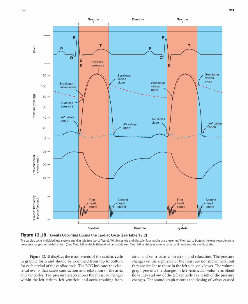
Figure 12.18 Events Occurring During the Cardiac Cycle (see Table 12.2)The cardiac cycle is divided into systole and diastole (see top of figure). Within systole and diastole, four graphs are presented. From top to bottom, the electrocardiogram;pressure changes for the left atrium (blue line), left ventricle (black line), and aorta (red line); left ventricular volume curve; and heart sounds are illustrated.
Figure 12.18 displays the main events of the cardiac cyclein graphic form and should be examined from top to bottomfor each period of the cardiac cycle. The ECG indicates the elec-trical events that cause contraction and relaxation of the atriaand ventricles. The pressure graph shows the pressure changeswithin the left atrium, left ventricle, and aorta resulting from
atrial and ventricular contraction and relaxation. The pressurechanges on the right side of the heart are not shown here, butthey are similar to those in the left side, only lower. The volumegraph presents the changes in left ventricular volume as bloodflows into and out of the left ventricle as a result of the pressurechanges. The sound graph records the closing of valves caused
see43696_ch12_321-352 12:6:05 02:51 PM Page 339
*(866) 487-8889*
CONFIRMING PROOFSMASTER SETPlease markall alterations
on this set only

Ventricular Systole Ventricular Diastole
ECG The QRS complex is completed and the The T wave is completed and the ventricles relax. ventricles are stimulated to contract. Then the P wave stimulates the atria to contract, The T wave begins. after which they relax.
Ventricular pressure Pressure increases rapidly as a result of left Ventricular pressure decreases rapidly to curve (black) ventricular contraction. When left ventricular nearly zero as the left ventricle relaxes.
pressure exceeds aortic pressure, blood pushesthe aortic semilunar valve open. Continued contraction increases ventricular pressure to a peak value of 120 mm Hg. Ventricular pressure then decreases as blood flows out of the left ventricle into the aorta.
Aortic pressure curve (red ) As ventricular contraction forces blood into Ventricular pressure decreases below aortic pressure. the aorta, pressure in the aorta increases to Blood flows back toward the left ventricle and the its highest value (120 mm Hg), called the aortic semilunar valve closes. As blood flows out ofsystolic pressure. the aorta toward the body, elastic recoil of the aorta
prevents a sudden decrease in pressure. Justbefore the aortic semilunar valve opens, pressure in the aorta decreases to its lowestvalue (80 mm Hg), called the diastolic pressure.
Atrial pressure curve (blue) Atrial pressure increases slightly as contraction After the bicuspid valve opens, pressure of the left ventricle pushes blood through the decreases slightly as blood flows into the aorta and toward the left atrium. After closure of left ventricle. At the end of ventricular the bicuspid valve, pressure drops in the left diastole, contraction of the left atrium atrium as it relaxes, then increases as blood flows increases the pressure slightly.into the left atrium from the four pulmonary veins.
Volume graph Blood pushes the aortic semilunar valve open, Blood flows from the left atrium into the leftblood is ejected from the left ventricle, ventricle, accounting for 70% of ventricular and ventricular volume decreases. filling. Near the end of ventricular diastole, contraction
of the left atrium pushes blood into the left ventricle, completing ventricular filling.
Sound graph As contraction of the ventricles pushes As blood flows back toward the heart, the blood toward the atria, the AV valves close, semilunar valves close, preventing the flow preventing the flow of blood into the atria of blood into the ventricles and producing and producing the first heart sound. the second heart sound.
Summary of Events of the Cardiac Cycle for the Left Atrium and Ventricle (see figure 12.18)Table 12.2
by blood flow. See figure 12.17 for illustration of the valves andblood flow and table 12.2 for a summary of the events occurringduring each period.
P R E D I C T �5
Predict the effect of a leaky (incompetent) aortic semilunarvalve on the volume of blood in the left ventricle just beforeventricular contraction. Predict the effect of a severelynarrowed opening through the aortic semilunar valves on theamount of work the heart must do to pump the normal volumeof blood into the aorta during each beat of the heart.
The Consequences of an Incompetent Bicuspid ValveIncompetent valves do not close completely and therefore they leak
when they are supposed to be closed. Incompetent valves allow blood to
flow in the reverse direction. For example, an incomepetent bicuspid
valve allows blood to flow from the left ventrical to the left atrium during
ventricular systole. This reduces the amount of blood pumped into the
340 Chapter 12
aorta. It also dramatically increases the blood pressure in the left atrium
and in the pulmonary veins during ventricular systole. During diastole,
the excess blood pumped into the atrium once again flows into the
ventricle along with the blood that normally flows from the lungs to the
left atrium. Therefore, the volume of blood entering the left ventricle is
greater than normal. The increased filling of the left ventricle gradually
causes it to hypertrophy and can lead to heart failure. The increased
pressure in the pulmonary veins can cause edema in the lungs.
Heart SoundsA stethoscope (steth�o-skop, stetho, the chest) was originally de-veloped to listen to the sounds of the lungs and heart and is nowused to listen to other sounds of the body (figure 12.19). Thereare two main heart sounds. The first heart sound can be repre-sented by the syllable lubb, and the second heart sound can berepresented by dupp. The first heart sound has a lower pitch than
see43696_ch12_321-352 1:13:06 08:04 PM Page 340
*(866) 487-8889*
CONFIRMING PROOFSMASTER SETPlease markall alterations
on this set only

Heart 341
the second. The first heart sound occurs at the beginning of ven-tricular systole and results from closure of the AV valves (see fig-ure 12.17, step 1 and figure 12.18). The second heart soundoccurs at the beginning of ventricular diastole and results fromclosure of the semilunar valves (see figure 12.17, step 3 and figure12.18). The valves usually do not make sounds when they open.
Clinically, ventricular systole occurs between the first andsecond heart sounds. Ventricular diastole occurs between thesecond heart sound and the first heart sound of the next beat.Because ventricular diastole lasts longer than ventricular sys-tole, there is less time between the first and second heart soundsthan between the second heart sound and the first heart soundof the next beat.
P R E D I C T �6
Compare the rate of blood flow out of the ventricles betweenthe first and second heart sounds of the same beat with the rateof blood flow out of the ventricles between the second heartsound of one beat and the first heart sound of the next beat.
Abnormal heart sounds called murmurs are usually a re-sult of faulty valves. For example, an incompetent valve fails toclose tightly and blood leaks through the valve when it is closed.A murmur caused by an incompetent valve makes a swishingsound immediately after closure of the valve. For example, an in-competent bicuspid valve results in a swishing sound immediatelyafter the first heart sound.
When the opening of a valve is narrowed, or stenosed(sten�ozd, a narrowing), a swishing sound precedes closure ofthe stenosed valve. For example, when the bicuspid valve isstenosed, a swishing sound precedes the first heart sound.
P R E D I C T �7
If normal heart sounds are represented by lubb–dupp,lubb–dupp, what does a heart sound represented bylubb–duppshhh, lubb–duppshhh represent? Whatdoes lubb–shhhdupp, lubb–shhhdupp represent (assumethat shhh represents a swishing sound)?
Bicuspidvalve
Tricuspidvalve
Pulmonarysemilunar valve
Outline ofheart
Aorticsemilunar valve
Figure 12.19 Location of the Heart Valves in the ThoraxSurface markings of the heart in the male. The positions of the four heartvalves are indicated by blue ellipses, and the sites where the sounds of thevalves are best heard with the stethoscope are indicated by pink circles.
Speedy Beat is a 70-year-old man. He and his daughter
Normal were getting out of the car at one of his favorite
restaurants where they planned to have dinner. Before
Speedy could get completely out of the car he became
dizzy. He exhibited substantial pallor and experienced
chest pains. Normal checked her father’s pulse, which
was close to 180–200 bpm and irregular. She helped
him back into the car and drove to the emergency room
at a nearby hospital. There, it was determined that
Speedy’s blood pressure was low even though his heart
rate was rapid. Speedy was previously diagnosed as
suffering from paroxysmal atrial tachycardia and he
regularly takes a calcium channel blocking agent to
control it.
The term paroxysmal means that the cause of
Speedy’s tachycardia is not known, but it results from
rapid and ectopic beats that originate in the atria. The
heart rate is irregular because the rate at which action
potentials responsible for atrial contractions occur is
greater than the rate at which the ventricles can contract.
Consequently, not every atrial contraction is followed by
a ventricular contraction. Speedy’s blood pressure is low
because the heart rate is so fast that there is little time
for blood to fill the rapidly contracting chambers of the
heart between the contractions. Consequently, Speedy’s
stroke volume and cardiac output are low. Chest pains
result because the heart muscle is working hard, but
blood flow to cardiac muscle through coronary vessels is
reduced so that the heart muscle suffers from an
inadequate supply of oxygen (ischemia).
Once Speedy’s heart rate and rhythm were stabilized,
the amount of calcium channel blocking agent
prescribed for him was adjusted and he was released
from the hospital the next day. Another drug used to
treat this condition along with calcium channel blocking
agent is digoxin, which has the overall effect of
increasing the force and slowing the rate of cardiac
muscle contraction.
A CASE IN POINT | Paroxysmal AtrialTachycardia
see43696_ch12_321-352 1:10:06 10:08 AM Page 341
*(866) 487-8889*
CONFIRMING PROOFSMASTER SETPlease markall alterations
on this set only

Heart DiseasesInflammation of Heart TissuesEndocarditis (en�do-kar-dı�tis) is inflamma-tion of the endocardium. It affects thevalves more severely than other areas of theheart and may lead to deposition of scar tis-sue, causing valves to become stenosed orincompetent.
Pericarditis (per�i-kar-dı�tis) is inflam-mation of the pericardium. Pericarditis canresult from bacterial or viral infections andcan be extremely painful.
Cardiomyopathy (kar�de-o-mı-op�a-the )is a disease of the myocardium of unknowncause or occurring secondarily to otherdiseases. It may develop secondarily toinfections or from exposure to toxins includ-ing alcohol, drugs, or environmental pollu-tants. It may also result from nutritionaldisorders or other systemic diseases such asleukemia. In most cases the cause is un-known. The most common form of cardiomy-opathy results in weakened cardiac muscle,which causes all of the chambers of theheart to dilate or enlarge. This can eventuallylead to congestive heart failure.
Rheumatic (roo-mat�ik) heart disease canresult from a streptococcal (strep�to-kok�al)infection in young people. Toxin produced bythe bacteria can cause an immune reactioncalled rheumatic fever approximately 2–4weeks after the infection. The immune reac-tion can cause inflammation of the endo-cardium, called rheumatic endocarditis. Theinflamed valves, especially the bicuspidvalve, can become stenosed or incompetent.The effective treatment of streptococcal infec-tions with antibiotics has reduced the fre-quency of rheumatic heart disease.
Reduced Blood Flow to Cardiac MuscleCoronary heart disease reduces theamount of blood that the coronary arteriesare able to deliver to the myocardium. Thereduction in blood flow can damage the myo-cardium. The degree of damage depends onthe size of the arteries involved, whetherocclusion (blockage) is partial or complete,
and whether occlusion is gradual or sud-den. As the walls of the arteries thicken andharden with age, the volume of blood theycan supply to the heart muscle declines,and the ability of the heart to pump blooddecreases. Inadequate blood flow to theheart muscle can result in angina pectoris,which is a poorly localized sensation ofpain in the region of the chest, left arm, andleft shoulder.
Degenerative changes in the artery wallcan cause the inside surface of the artery tobecome roughened. The chance of plateletaggregation increases at the rough surface,which increases the chance of coronarythrombosis (throm-bo�sis), the formation ofa blood clot in a coronary vessel. Inade-quate blood flow can cause an infarct(in�farkt), an area of damaged cardiac tis-sue. A heart attack is often referred to as acoronary thrombosis or a myocardial infarc-tion. The outcome of coronary thrombosisdepends on the extent of the damage toheart muscle caused by inadequate bloodflow and whether other blood vessels cansupply enough blood to maintain the func-tion of the heart. Death can occur swiftly ifthe infarct is large; if the infarct is small, theheart can continue to function. In somecases, the infarct weakens the wall of theheart, and the wall ruptures; but in mostcases scar tissue replaces damaged cardiacmuscle in the area of the infarct.
People who survive infarctions oftenlead fairly normal lives if they take precau-tions. Most cases call for moderate exer-cise, adequate rest, a disciplined diet, andreduced stress. Small doses of aspirin andtreatments, including drugs, to reduce ele-vated blood pressure appear to provideprotection against the development of myo-cardial infarcts.
Congenital Conditions Affectingthe HeartCongenital (occuring at birth) heart diseaseis heart disease present at birth and isthe result of abnormal development of theheart. The following are common congenitaldefects:
A septal defect is a hole in a septum be-tween the left and right sides of the heart.The hole may be in the interatrial or inter-ventricular septum. These defects allowblood to flow from one side of the heart tothe other and, as a consequence, greatly re-duce the pumping effectiveness of theheart.
Patent (to lie open) ductus arteriosus(duk�tus arter�e-o-sus) results when a bloodvessel called the ductus arteriosus, whichis present in the fetus, fails to close afterbirth. The ductus arteriosus extends be-tween the pulmonary trunk and the aorta. Itallows blood to pass from the pulmonarytrunk to the aorta, thus bypassing thelungs. This is normal before birth becausethe lungs are not functioning. If the ductusarteriosus fails to close after birth, how-ever, blood flows in the opposite direction,from the aorta to the pulmonary trunk. As aconsequence, blood flows through thelungs under a higher pressure and dam-ages them. In addition, the amount of workrequired of the left ventricle to maintain anadequate systemic blood pressure in-creases.
Stenosis (ste-no�sis) of the heart valvesis a narrowed opening through one or moreof the heart valves. In aortic or pulmonarysemilunar valve stenosis, the workload ofthe heart is increased because the ventri-cles must contract with a much greaterforce to pump blood from the ventricles.Stenosis of the bicuspid valve prevents theflow of blood into the left ventricle, causingblood to back up in the left atrium and thelungs, resulting in edema in the lungs.Stenosis of the tricuspid valve causesblood to back up in the right atrium andsystemic veins, causing edema in the pe-riphery.
Cyanosis (sı-a-no�sis, cyan, blue � osis,condition of ) is a symptom of inadequateheart function in babies suffering from con-genital heart disease. The term “blue baby”is sometimes used to refer to infants withcyanosis. The blueness of the skin iscaused by low oxygen levels in the blood inperipheral blood vessels.
Clinical Focus Conditions and Diseases Affecting the Heart
342
see43696_ch12_321-352 1:13:06 08:04 PM Page 342
*(866) 487-8889*
CONFIRMING PROOFSMASTER SETPlease markall alterations
on this set only

Heart 343
Regulation of Heart FunctionCardiac output (CO) is the volume of blood pumped by eitherventricle of the heart each minute. Cardiac output can be calcu-lated by multiplying the stroke volume times the heart rate.Stroke volume (SV) is the volume of blood pumped per ventri-cle each time the heart contracts, and the heart rate (HR) is thenumber of times the heart contracts each minute.
CO � SV � HR
(mL/min) (mL/beat) (beats/min)
Under resting conditions, the heart rate is approximately72 beats/min (or bpm) and the stroke volume is approximately70 mL/beat. Consequently, the cardiac output is slightly morethan 5 L/min:
CO � SV � HR
� 70 mL/beat � 72 bpm
� 5040 mL/min (approximately 5 L/min)
The heart rate and the stroke volume vary considerablyamong people. Athletes tend to have a larger stroke volume andlower heart rate at rest because exercise has increased the size oftheir hearts. Nonathletes are more likely to have a higher heartrate and lower stroke volume. During exercise the heart in anonathlete can increase to 190 bpm and the stroke volume canincrease to 115 mL/beat. Therefore, the cardiac output increasesto approximately 22 L/min:
CO � SV � HR
� 115 mL/beat � 190 bpm
� 21,850 mL/min (approximately 22 L/min)
This produces a cardiac output that is several times greater thanthe cardiac output under resting conditions. Athletes can in-crease their cardiac output to a greater degree than nonathletes.
The control mechanisms that modify the stroke volume andthe heart rate are classified as intrinsic and extrinsic mechanisms.
Intrinsic Regulation of the HeartIntrinsic regulation of the heart refers to mechanisms con-tained within the heart itself. The force of contraction pro-duced by cardiac muscle is related to the degree of stretch ofcardiac muscle fibers. The amount of blood in the ventriclesat the end of ventricular diastole determines the degree towhich cardiac muscle fibers are stretched. Venous return isthe amount of blood that returns to the heart, and the degreeto which the ventricular walls are stretched at the end of di-astole is called preload. If venous return increases, the heartfills to a greater volume and stretches the cardiac musclefibers, producing an increased preload. In response to the in-creased preload, cardiac muscle fibers contract with a greaterforce. The greater force of contraction causes an increasedvolume of blood to be ejected from the heart, resulting inan increased stroke volume. As venous return increases,resulting in an increased preload, cardiac output increases.Conversely, if venous return decreases, resulting in a decreased
preload, the cardiac output decreases. The relationshipbetween preload and stroke volume is called Starling’s law ofthe heart.
Because venous return is influenced by many conditions,Starling’s law of the heart has a major influence on cardiac out-put. For example, muscular activity during exercise causes in-creased venous return, resulting in an increased preload, strokevolume, and cardiac output. This is beneficial because an in-creased cardiac output is needed during exercise to supply oxy-gen to exercising skeletal muscles.
Afterload refers to the pressure against which the ven-tricles must pump blood. People suffering from hypertensionhave an increased afterload because they have an elevatedaortic pressure during contraction of the ventricles. Theheart must do more work to pump blood from the left ventri-cle into the aorta, which increases the workload on the heartand can eventually lead to heart failure. A reduced afterloaddecreases the work the heart must do. People who have alower blood pressure have a reduced afterload and developheart failure less often than people who have hypertension.The afterload, however, influences cardiac output less thanpreload influences it. The afterload must increase substan-tially before it decreases the volume of blood pumped by ahealthy heart.
Consequences of Heart FailureAlthough heart failure can occur in young people, it usually results from a
progressive weakening of the heart muscle in elderly people. A failing
heart gradually enlarges and eventually fails. In heart failure, the heart is
not capable of pumping all the blood that is returned to it because
further stretching of the cardiac muscle fibers does not increase the
stroke volume of the heart. Consequently, blood backs up in the veins.
For example, heart failure that affects the right ventricle is called right
heart failure and causes blood to back up in the veins that return blood
from systemic vessels to the heart. Filling of the veins with blood causes
edema, especially in the legs and feet. Edema results from the
accumulation of fluid in tissues outside of blood vessels. Heart failure
that affects the left ventricle is called left heart failure and causes blood
to back up in the veins that return blood from the lungs to the heart.
Filling of these veins causes edema in the lungs, which makes breathing
difficult.
Extrinsic Regulation of the HeartExtrinsic regulation refers to mechanisms external to theheart, such as either hormonal or nervous regulation (figure12.20). Nervous influences are carried through the auto-nomic nervous system. Both sympathetic and parasympa-thetic nerve fibers innervate the heart, and have a major effecton the SA node. Stimulation by sympathetic nerve fiberscauses the heart rate and the stroke volume to increase,whereas stimulation by parasympathetic nerve fibers causesthe heart rate to decrease.
The baroreceptor (bar�o-re-sep�ter, baro, pressure) reflexplays an important role in regulating the function of the heart.Baroreceptors are stretch receptors that monitor blood pressurein the aorta and in the wall of the internal carotid arteries, which
see43696_ch12_321-352 1:10:06 10:08 AM Page 343
*(866) 487-8889*
CONFIRMING PROOFSMASTER SETPlease markall alterations
on this set only
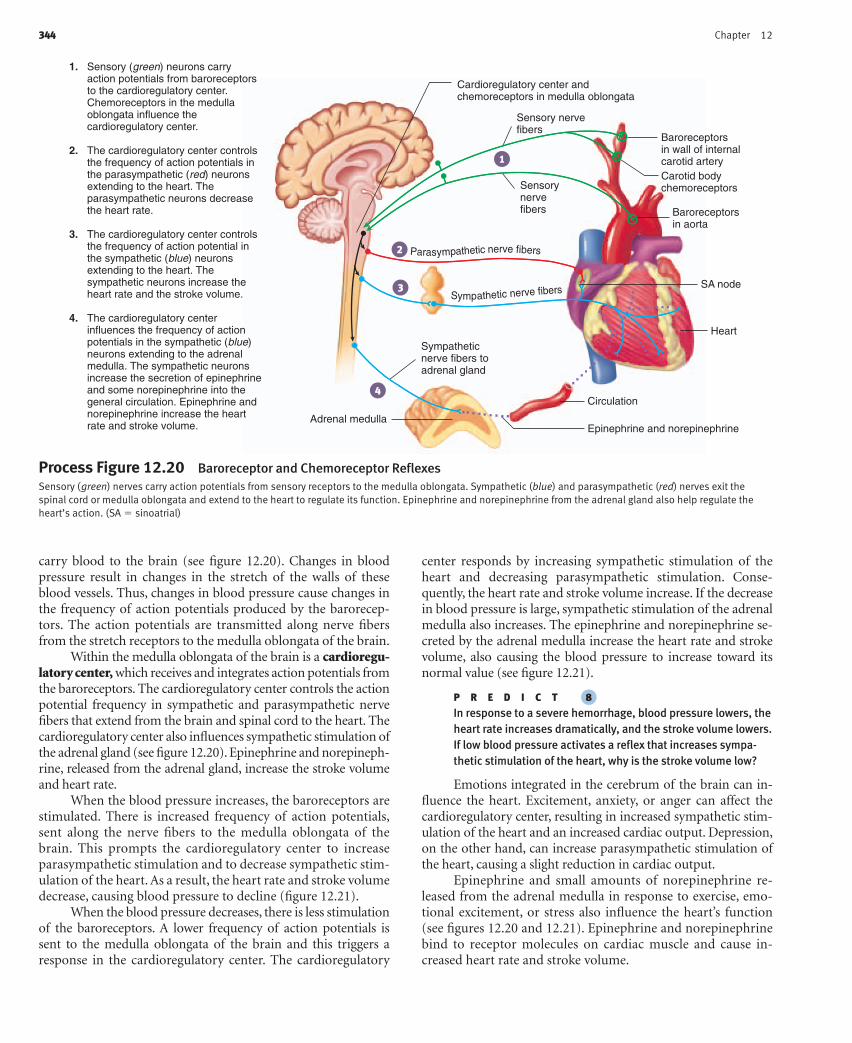
344 Chapter 12
1
2
3
4
Sensory nervefibers
Baroreceptorsin wall of internalcarotid artery
Baroreceptorsin aorta
Carotid bodychemoreceptors
SA node
Heart
Circulation
Epinephrine and norepinephrineAdrenal medulla
Sympatheticnerve fibers toadrenal gland
Sensorynervefibers
Parasympathetic nerve fibers
Sympathetic nerve fibers
Cardioregulatory center andchemoreceptors in medulla oblongata
Sensory (green) neurons carry action potentials from baroreceptors to the cardioregulatory center. Chemoreceptors in the medulla oblongata influence the cardioregulatory center.
The cardioregulatory center controls the frequency of action potentials in the parasympathetic (red) neurons extending to the heart. The parasympathetic neurons decrease the heart rate.
The cardioregulatory center controls the frequency of action potential in the sympathetic (blue) neurons extending to the heart. The sympathetic neurons increase the heart rate and the stroke volume.
The cardioregulatory center influences the frequency of action potentials in the sympathetic (blue) neurons extending to the adrenal medulla. The sympathetic neurons increase the secretion of epinephrine and some norepinephrine into the general circulation. Epinephrine and norepinephrine increase the heart rate and stroke volume.
1.
2.
3.
4.
Process Figure 12.20 Baroreceptor and Chemoreceptor ReflexesSensory (green) nerves carry action potentials from sensory receptors to the medulla oblongata. Sympathetic (blue) and parasympathetic (red) nerves exit thespinal cord or medulla oblongata and extend to the heart to regulate its function. Epinephrine and norepinephrine from the adrenal gland also help regulate theheart’s action. (SA � sinoatrial)
carry blood to the brain (see figure 12.20). Changes in bloodpressure result in changes in the stretch of the walls of theseblood vessels. Thus, changes in blood pressure cause changes inthe frequency of action potentials produced by the barorecep-tors. The action potentials are transmitted along nerve fibersfrom the stretch receptors to the medulla oblongata of the brain.
Within the medulla oblongata of the brain is a cardioregu-latory center, which receives and integrates action potentials fromthe baroreceptors. The cardioregulatory center controls the actionpotential frequency in sympathetic and parasympathetic nervefibers that extend from the brain and spinal cord to the heart. Thecardioregulatory center also influences sympathetic stimulation ofthe adrenal gland (see figure 12.20). Epinephrine and norepineph-rine, released from the adrenal gland, increase the stroke volumeand heart rate.
When the blood pressure increases, the baroreceptors arestimulated. There is increased frequency of action potentials,sent along the nerve fibers to the medulla oblongata of thebrain. This prompts the cardioregulatory center to increaseparasympathetic stimulation and to decrease sympathetic stim-ulation of the heart. As a result, the heart rate and stroke volumedecrease, causing blood pressure to decline (figure 12.21).
When the blood pressure decreases, there is less stimulationof the baroreceptors. A lower frequency of action potentials issent to the medulla oblongata of the brain and this triggers aresponse in the cardioregulatory center. The cardioregulatory
center responds by increasing sympathetic stimulation of theheart and decreasing parasympathetic stimulation. Conse-quently, the heart rate and stroke volume increase. If the decreasein blood pressure is large, sympathetic stimulation of the adrenalmedulla also increases. The epinephrine and norepinephrine se-creted by the adrenal medulla increase the heart rate and strokevolume, also causing the blood pressure to increase toward itsnormal value (see figure 12.21).
P R E D I C T �8
In response to a severe hemorrhage, blood pressure lowers, theheart rate increases dramatically, and the stroke volume lowers.If low blood pressure activates a reflex that increases sympa-thetic stimulation of the heart, why is the stroke volume low?
Emotions integrated in the cerebrum of the brain can in-fluence the heart. Excitement, anxiety, or anger can affect thecardioregulatory center, resulting in increased sympathetic stim-ulation of the heart and an increased cardiac output. Depression,on the other hand, can increase parasympathetic stimulation ofthe heart, causing a slight reduction in cardiac output.
Epinephrine and small amounts of norepinephrine re-leased from the adrenal medulla in response to exercise, emo-tional excitement, or stress also influence the heart’s function(see figures 12.20 and 12.21). Epinephrine and norepinephrinebind to receptor molecules on cardiac muscle and cause in-creased heart rate and stroke volume.
see43696_ch12_321-352 1:10:06 05:00 PM Page 344
*(866) 487-8889*
CONFIRMING PROOFSMASTER SETPlease markall alterations
on this set only

Exercise and Cardiac OutputDuring exercise, the cardiac output of the heart increases, resulting in
increased delivery of blood to skeletal muscles. Cardiac output increases
because of the increased heart rate and stroke volume that result from
increased sympathetic stimulation of the heart and from the effects of
epinephrine and norepinephrine on cardiac muscle. Starling’s law of the
heart also contributes to the increase in stroke volume during exercise.
Blood vessels in exercising skeletal muscles dilate, which increases
blood flow to the muscle tissue. The dilation of the blood vessels also
increases venous return to the heart because the rate of blood flow from
exercising skeletal muscle through the veins is greatly increased. As
venous return increases, preload increases, and cardiac muscle is
stretched. Consequently, the muscle contracts more forcefully (Starling’s
law) and stroke volume increases.
The medulla oblongata of the brain also containschemoreceptors that are sensitive to changes in pH and carbondioxide levels (see figure 12.20). A decrease in pH, often causedby an increase in carbon dioxide, results in sympathetic stimu-lation of the heart (figure 12.22).
Changes in the extracellular concentration of K�, Ca2�,and Na�, which influence other electrically excitable tissues,also affect cardiac muscle function. Excess extracellular K�
cause the heart rate and stroke volume to decrease. If the ex-tracellular K� concentration increases further, normal con-duction of action potentials through cardiac muscle isblocked, and death can result. An excess of extracellular Ca2�
causes the heart to contract arrhythmically. Reduced extra-cellular Ca2� cause both the heart rate and stroke volume todecrease.
• Increased heart rate and stroke volume result from the changed ANS stimulation of the heart.
• Increased heart rate and stroke volume result from the increased release of epinephrine and norepinephrine from the adrenal medulla.
The blood pressure decreases because of the decreased cardiac output resulting from the
decreased heart rate and stroke volume.
• Decreased heart rate and stroke volume result from the changed ANS stimulation of the heart.
• Decreased heart rate and stroke volume result from the decreased release of epinephrine and norepinephrine from the adrenal medulla.
A sudden decrease in blood pressure is detected by the baroreceptors in the internal carotid
arteries and aorta, which affects the baroreceptor reflex.
The cardioregulatory center decreases parasympathetic stimulation of the heart and
increases sympathetic stimulation of the heart and adrenal medulla.
The blood pressure increases because of the increased cardiac output resulting from the
increased heart rate and stroke volume.
A sudden increase in blood pressure is detected by the baroreceptors in the internal carotid
arteries and aorta, which affect the baroreceptor reflex.
The cardioregulatory center increases parasympathetic stimulation of the heart and
decreases sympathetic stimulation of the heart and adrenal medulla.
Blo
od p
ress
ure
(nor
mal
ran
ge)
Blood pressurehomeostasisis maintained.
Blo
od p
ress
ure
(nor
mal
ran
ge)
Follow the green arrows whenblood pressure increases.
Follow the red arrows when blood pressure decreases.
Start here
Homeostasis Figure 12.21 Baroreceptor ReflexThe baroreceptor reflex maintains homeostasis in response to changes in blood pressure. (ANS � autonomic nervous system)
Heart 345
see43696_ch12_321-352 1:10:06 10:08 AM Page 345
*(866) 487-8889*
CONFIRMING PROOFSMASTER SETPlease markall alterations
on this set only

Heart MedicationsDigitalisDigitalis (dij�i-tal�is, refers to fingerlikeflowers), an extract of foxglove plants,slows and strengthens contractions of theheart muscle. This drug is frequently givento people who suffer from heart failure, al-though it can also be used to treat atrialtachycardia.
NitroglycerinDuring exercise, when the heart rate andstroke volume are increased, dilation ofblood vessels in the exercising skeletalmuscles and constriction in most otherblood vessels results in an increased ve-nous return to the heart and an increasedpreload. Nitroglycerin (nı-tro-glis�er-in)causes dilation of all of the veins and arter-ies without an increase in heart rate orstroke volume. When all blood vessels di-late, a greater volume of blood pools in thedilated blood vessels, causing a decreasein the venous return to the heart. The re-duced preload causes cardiac output to de-crease, resulting in a decreased amount ofwork performed by the heart. Nitroglycerinis frequently given to people who sufferfrom coronary artery disease, which re-stricts coronary blood flow. The decreasedwork performed by the heart reduces theamount of oxygen required by the cardiacmuscle. In addition, dilation of coronaryarteries can increase blood flow to cardiacmuscle. Consequently, the heart does notsuffer from a lack of oxygen, and anginapectoris does not develop.
Beta-Adrenergic Blocking AgentsBeta-adrenergic (ba-ta ad-re-ner�jik) block-ing agents reduce the rate and strength ofcardiac muscle contractions, thus reducingthe oxygen demand of the heart. They bindto receptors for norepinephrine and epi-nephrine and prevent these substancesfrom having their normal effects. Beta-adrenergic blocking agents are often usedto treat people who suffer from rapid heartrates, certain types of arrhythmias, and hy-pertension.
Calcium Channel BlockersCalcium channel blockers reduce the rate atwhich Ca2� diffuse into cardiac muscle
cells and smooth muscle cells. Because theaction potentials that produce cardiac mus-cle contractions depend in part on the flowof Ca2� into the cardiac muscle cells, theCa2� channel blockers can be used tocontrol the force of heart contractions andreduce arrhythmia, tachycardia, and hyper-tension. Because entry of Ca2� to smoothmuscle cells causes contraction, Ca2�
channel blockers cause dilation of bloodvessels. They dilate coronary blood vesselsand increase blood flow to cardiac muscle.Consequently, they can be used to treatangina pectoris.
Antihypertensive AgentsSeveral drugs are used specifically to treathypertension. These drugs reduce bloodpressure and therefore reduce the workrequired by the heart to pump blood. In ad-dition, the reduction of blood pressure re-duces the risk of heart attacks and strokes.Medications used to treat hypertension in-clude drugs that reduce the activity of thesympathetic division, those that dilate ar-teries and veins, those that increase urineproduction (diuretics), and those thatblock the conversion of angiotensin I to an-giotensin II (see chapter 13).
AnticoagulantsAnticoagulants (an�te-ko-ag�u-lants) pre-vent clot formation in persons with damageto heart valves or blood vessels or inpersons who have had a myocardialinfarction. Aspirin functions as a weak anti-coagulant by inhibiting the synthesis ofprostaglandins in platelets, which in turnreduces clot formation. Some data suggestthat taking a small dose of aspirin regularlyreduces the chance of a heart attack. Ap-proximately one baby aspirin each day maybenefit those who are likely to experience acoronary thrombosis.
InstrumentsArtificial PacemakerAn artificial pacemaker is an instrumentplaced beneath the skin that is equippedwith an electrode that extends to the heart.An artificial pacemaker provides an electri-cal stimulus to the heart at a set frequency.Artificial pacemakers are used in patients inwhom the natural pacemaker of the heart
does not produce a heart rate high enoughto sustain normal physical activity. Modernelectronics has made it possible to designartificial pacemakers that can increase theheart rate as physical activity increases. Inaddition, special artificial pacemakers candefibrillate the heart if it becomes arrhyth-mic. It is likely that rapid development ofelectronics for artificial pacemakers will fur-ther increase the degree to which the pace-makers can regulate the heart.
Heart–Lung MachineA heart=lung machine serves as a tempo-rary substitute for the patient’s heart andlungs. It pumps blood throughout the bodyand oxygenates and removes carbon diox-ide from the blood. It has made possiblemany surgeries on the heart and lungs.
Surgical ProceduresHeart Valve Replacement or RepairHeart valve replacement or repair is a surgi-cal procedure performed on those whohave diseased valves that are so deformedand scarred from conditions such as endo-carditis that the valves are severely incom-petent or stenosed. Substitute valves madeof synthetic materials such as plastic orDacron are effective; valves transplantedfrom pigs are also used.
Heart TransplantsHeart transplants are possible when theimmune characteristics of a donor and therecipient are closely matched. The heart ofa recently deceased donor is transplantedto the recipient, and the diseased heart ofthe recipient is removed. People who havereceived heart transplants must remain ondrugs that suppress their immune re-sponses for the rest of their lives. Unlessthey do so, their immune system rejects thetransplanted heart.
Artificial HeartsArtificial hearts have been used on an ex-perimental basis to extend the lives of indi-viduals until an acceptable transplant can befound or to replace the heart permanently.The technology currently available for artifi-cial hearts has not yet reached the point atwhich a high quality of life can be achievedwith a permanent artificial heart.
Clinical Focus Treatment and Prevention of Heart Disease
346
see43696_ch12_321-352 1:6:06 11:09 AM Page 346
*(866) 487-8889*
CONFIRMING PROOFSMASTER SETPlease markall alterations
on this set only

Prevention of Heart DiseaseProper nutrition is important in reducingthe risk of heart disease. A recommendeddiet is low in fats, especially saturated fatsand cholesterol, and low in refined sugar.Diets should be high in fiber, whole grains,fruits, and vegetables. Total food intakeshould be limited to avoid obesity, andsodium chloride intake should be re-duced.
Tobacco and excessive use of alcoholshould be avoided. Smoking increases the
risk of heart disease by at least 10-fold, andexcessive use of alcohol also substantiallyincreases the risk of heart disease. How-ever, moderate use of alcohol may reducethe risk of heart disease.
Chronic stress, frequent emotionalupsets, and a lack of physical exercisecan increase the risk of cardiovasculardisease. Remedies include relaxationtechniques and aerobic exercise pro-grams involving gradual increases in dura-tion and difficulty in activities such as
swimming, walking, jogging, or aerobicdancing.
Hypertension is an abnormally highsystemic blood pressure. Hypertensionaffects approximately one-fifth of the popu-lation. Regular blood pressure measure-ments are important because hypertensiondoes not produce obvious symptoms. If hy-pertension cannot be controlled by diet andexercise, it is important to treat the conditionwith prescribed drugs. The cause of hyper-tension in the majority of cases is unknown.
Clinical Focus (continued)
Blo
od p
H(n
orm
al r
ange)
Start hereBlood pHhomeostasisis maintained.
• Increased heart rate and stroke volume result from the changed ANS stimulation of the heart.
• Increased heart rate and stroke volume result from the increased release of epinephrine and norepinephrine from the adrenal medulla.
A decrease in blood pH (caused by an increase in blood CO2) results from decreased blood flow to the lungs. The decreased blood flow results from
the decreased cardiac output caused by decreased heart rate and stroke volume.
• Decreased heart rate and stroke volume result from the changed ANS stimulation of the heart.
• Decreased heart rate and stroke volume result from the decreased release of epinephrine and norepinephrine from the adrenal medulla.
A decrease in blood pH (often caused by an increase in blood CO2) is detected by
chemoreceptors in the medulla oblongata, which affects the chemoreceptor reflex.
The cardioregulatory center decreases parasympathetic stimulation of the heart and
increases sympathetic stimulation of the heart and adrenal medulla.
An increase in blood pH (caused by a decrease in blood CO2) results from increased blood flow to the lungs. The increased blood flow results from the increased cardiac output caused by
increased heart rate and stroke volume.
An increase in blood pH (often caused by a decrease in blood CO2) is detected by
chemoreceptors in the medulla oblongata, which affects the chemoreceptor reflex.
The cardioregulatory center increases parasympathetic stimulation of the heart and
decreases sympatheticstimulation of the heart and adrenal medulla.
Blo
od p
H(n
orm
al r
ange)
Follow the green arrows whenblood pH increases.
Follow the red arrows when blood pH decreases.
Homeostasis Figure 12.22 Chemoreceptor Reflex-pHThe chemoreceptor reflex maintains homeostasis in response to changes in blood concentrations of CO2 and H� (or pH). (ANS � autonomic nervous system)
347
see43696_ch12_321-352 1:10:06 05:00 PM Page 347
*(866) 487-8889*
CONFIRMING PROOFSMASTER SETPlease markall alterations
on this set only

Systems PathologyMyocardial Infarction
Mr. P. was an overweight, out-of-shape executive who regularly con-sumed food with a high fat content and smoked. He viewed his job asfrustrating because he was frequently confronted with stressful dead-lines. He had not had a physical examination for several years so hewas not aware that his blood pressure was high. One evening, Mr. P.was walking to his car after work when he began to feel pain in hischest that also radiated down his left arm. Shortly after the onset ofpain he became out of breath, developed marked pallor, becamedizzy, and had to lie down on the sidewalk. The pain in his chest andarm was poorly localized, but intense and he became anxious andthen disoriented. Mr. P. lost consciousness, although he did not stopbreathing. After a short delay, one of his coworkers noticed him andcalled for help. When paramedics arrived, they determined that Mr. P’sblood pressure was low and he exhibited arrhythmia and tachycardia.The paramedics transmitted the electrocardiogram they took to aphysician by way of their electronic communication system, and theydiscussed Mr. P.’s symptoms with the physician who was at the hospi-tal. The paramedics were directed to administer oxygen and medica-tion to control arrythmias and to transport him to the hospital. At thehospital, tissue plasminogen activator (t-PA) was administered, whichimproved blood flow to the damaged area of the heart by activatingplasminogen, which dissolves blood clots. Blood levels of enzymessuch as creatine phosphokinase increased in Mr. P.’s blood over thenext few days, which confirmed that damage to cardiac muscle re-sulted from an infarction.
In the hospital, Mr. P. began to experience shortness of breathbecause of pulmonary edema, and after a few days in the hospital hedeveloped pneumonia. He was treated for pneumonia and graduallyimproved over the next few weeks. An angiogram (an�je-o-gram,angeion, a vessel � gramma, a writing) (figure A), which is an imagingtechnique used to visualize the coronary arteries, was performed sev-eral days after Mr. P.’s infarction. The angiogram indicated that Mr. P.suffered from a permanent reduction in a significant part of the lateralwall of his left ventricle and that neither angioplasty nor bypasssurgery was necessary. Mr. P. has some other restrictions to blood flowin his coronary arteries.
Background Information
Mr. P. experienced a myocardial infarction. A thrombosis in one of thebranches of the left coronary artery reduced blood supply to the lateralwall of the left ventricle, resulting in ischemia of the left ventricle wall.That t-PA was an effective treatment is consistent with the conclusionthat the infarction was due to a thrombosis. An ischemic area of theheart wall was not able to contract normally and, therefore, the pumping
effectiveness of the heart was dramatically reduced. The reducedpumping capacity of the heart was responsible for the low blood pres-sure, which caused the blood flow to his brain to decrease resulting inconfusion, disorientation, and possibly unconsciousness.
Low blood pressure, increasing blood carbon dioxide levels,pain, and anxiousness increased sympathetic stimulation of the heartand adrenal glands. Increased sympathetic stimulation of the adrenalmedulla resulted in the release of epinephrine from the adrenalmedulla. Increased parasympathetic stimulation of the heart resultedfrom pain sensations. The heart rate was periodically arrhythmic be-cause of the combined effects of parasympathetic stimulation, epi-nephrine and norepinephrine from the adrenal gland, andsympathetic stimulation. In addition, ectopic beats were produced bythe ischemic areas of the left ventricle.
Pulmonary edema resulted from the increased pressure in thepulmonary veins because of the reduced ability of the left ventricle topump blood. The edema allowed bacteria to infect the lungs andcause pneumonia.
Occluded coronaryartery
Figure A AngiogramAn angiogram is a picture of a blood vessel. It is usually obtained by placinga catheter into a blood vessel and injecting a dye that can be detected withx-rays. Note the partially occluded (blocked) coronary blood vessel in thisangiogram, which has been computer-enhanced to show colors.
348
see43696_ch12_321-352 1:10:06 10:08 AM Page 348
*(866) 487-8889*
CONFIRMING PROOFSMASTER SETPlease markall alterations
on this set only

The heart began to beat rhythmically in response to medicationbecause the infarction did not damage the conducting system of theheart, which is an indication that there were no permanent arrhyth-mias. Permanent arrhythmias are indications of damage done to car-diac muscle cells specialized to conduct action potentials in the heart.
Analysis of the electrocardiogram, blood pressure measure-ments, and the angiogram indicated that the infarction, in this case,was located on the left side of Mr P.’s heart.
Mr. P.’s physician made it very clear to him that he was lucky tohave survived a myocardial infarction and recommended a weight-loss program and a low-sodium and low-fat diet. Mr. P.’s physicianalso recommended that Mr. P. regularly take a small amount of aspirinand stop smoking. He explained that Mr. P. would have to take med-ication for high blood pressure if his blood pressure did not decreasein response to the recommended changes. After a period of recovery,the physician recommended an aerobic exercise program and sug-gested that Mr. P. seek ways to reduce the stress associated with his
job. Mr. P. followed the doctor’s recommendations, and after severalmonths he began to feel better than he had in years and his bloodpressure was normal.
P R E D I C T �9
Severe ischemia in the wall of a ventricle can result in thedeath of cardiac muscle cells. Inflammation around thenecrotic tissue results and macrophages invade the necrotictissue and phagocytize dead cells. At the same time, bloodvessels and connective tissue grow into the necrotic area andbegin to deposit connective tissue to replace the necrotictissue. A person who entered the hospital at about the sametime with a very similar condition to Mr. P.’s was recovering.After about a week, his blood pressure suddenly decreased tovery low levels and he died within a very short time. Uponautopsy, a large amount of blood was found in the pericardialsac, and the wall of the left ventricle was ruptured. Explain.
System InteractionsIntegumentary Pallor of the skin resulted from intense vasoconstriction of peripheral blood vessels, including those in the skin.
Muscular Reduced skeletal muscle activity required for activities such as walking result because of the effect of a lack of blood flow to the brain and because blood is shunted from blood vessels that supply skeletal muscles to those that supply theheart and brain.
Nervous Decreased blood flow to the brain, decreased blood pressure, and pain because of ischemia of heart muscle result in increased sympathetic and parasympathetic stimulation of the heart. Loss of consciousness occurs when the blood flow tothe brain decreases enough to result in too little oxygen to maintain normal brain function, especially in the reticularactivating system.
Endocrine When blood pressure decreases to low values, antidiuretic hormone (ADH) is released from the posterior pituitarygland and renin, released from the kidney, activates the renin–angiotensin–aldosterone mechanism. ADH, secreted in largeamounts, and angiotensin II cause vasoconstriction of peripheral blood vessels. ADH and aldosterone act on the kidneys toretain water and ions. An increased blood volume increases venous return, which results in an increased stroke volume ofthe heart and an increase in blood pressure unless damage to the heart is very severe.
Lymphatic or Immune White blood cells, including macrophages, move to the area of cardiac muscle damage and phagocytize any dead cardiac muscle cells.
Respiratory Decreased blood pressure results in a decreased blood flow to the lungs. The decrease in gas exchange results in increased blood carbon dioxide levels, acidosis, and decreased blood oxygen levels. Initially, respiration becomes deep andlabored because of the elevated carbon dioxide levels, decreased blood pH, and depressed oxygen levels. If the bloodoxygen levels decrease too much the person loses consciousness. Pulmonary edema can result when the pumpingeffectiveness of the left ventricle is substantially reduced.
Digestive Intense sympathetic stimulation decreases blood flow to the digestive system to very low levels, which often resultsin increased nausea and vomiting.
Urinary Blood flow to the kidney decreases dramatically in response to sympathetic stimulation. If the kidney becomesischemic, damage to the kidney tubules can occur, resulting in the development of acute renal failure. Acute renal failureresults in reduced urine production. Increased blood urea nitrogen, increased blood levels of K�, and edema are indicationsthat the kidneys cannot eliminate waste products and excess water. If damage is not too great, the period of reduced urineproduction may last up to 3 weeks, and then the rate of urine production slowly returns to normal as the kidney tubulesheal.
Effects of Myocardial Infarctions on Other SystemsSystems Interactions:
349
see43696_ch12_321-352 1:13:06 08:04 PM Page 349
*(866) 487-8889*
CONFIRMING PROOFSMASTER SETPlease markall alterations
on this set only

S U M M A R Y
Functions of the Heart (p. 322)
The heart functions include1. The heart generates blood pressure.2. The heart routes blood through the systemic and pulmonary
circulation.3. The pumping action of the heart and the valves of the heart ensure a
one-way flow of blood through the heart and blood vessels.4. The heart helps regulate blood supply to tissues.
Size, Form, and Location of the Heart (p. 322)
The heart is approximately the size of a fist and is located in the pericardialcavity.
Anatomy of the Heart (p. 323)
Pericardium 1. The pericardial sac consists of a fibrous and serous pericardium. The
fibrous pericardium is lined by the parietal pericardium.2. The outer surface of the heart is lined by the visceral pericardium
(epicardium).3. Between the visceral and parietal pericardium is the pericardial
cavity, which is filled with pericardial fluid.
External Anatomy1. Atria are separated externally from the ventricles by the coronary
sulcus. The right and left ventricles are separated externally by theinterventricular sulci.
2. The inferior and superior venae cava enter the right atrium. Thefour pulmonary veins enter the left atrium.
3. The pulmonary trunk exits the right ventricle, and the aorta exitsthe left ventricle.
Heart Chambers and Internal Anatomy1. There are four chambers in the heart. The left and right atria receive
blood from veins and function mainly as reservoirs. Contraction ofthe atria completes ventricular filling.
2. The atria are separated internally from each other by the interatrialseptum.
3. The ventricles are the main pumping chambers of the heart. Theright ventricle pumps blood into the pulmonary trunk and the leftventricle, which has a thicker wall, and pumps blood into the aorta.
4. The ventricles are separated internally by the interventricular septum.
Heart Valves1. The heart valves ensure one-way flow of blood.2. The tricuspid valve (three cusps) separates the right atrium and
right ventricle, and the bicuspid valve (two cusps) separates the leftatrium and left ventricle.
3. The papillary muscles attach by the chordae tendineae to the cuspsof the tricuspid and bicuspid valves and adjust tension on the valves.
4. The aorta and pulmonary trunk are separated from the ventricles bythe semilunar valves.
5. The skeleton of the heart is a plate of fibrous connective tissue thatseparates the atria from the ventricles, acts as an electrical barrierbetween the atria and ventricles, and supports the valves of theheart.
Route of Blood Flow Through the Heart1. The left and right sides of the heart can be considered separate
pumps.2. Blood flows from the systemic vessels to the right atrium and from
the right atrium to the right ventricle. From the right ventricleblood flows to the pulmonary trunk and from the pulmonary trunk
Body temperature affects metabolism in the heart like itaffects other tissues. Elevated body temperature increases theheart rate, and reduced body temperature slows the heart rate.For example, during fever the heart rate is usually elevated.During heart surgery the body temperature is sometimes inten-tionally lowered to slow the heart rate and metabolism.
Effects of Aging on the HeartGradual changes in the function of the heart are associated withaging. These changes are minor under resting conditions, butbecome more obvious during exercise and in response to age-related diseases.
By age 70 cardiac output often decreases by approximatelyone-third. Because of the decrease in the reserve strength of theheart, many elderly people are limited in their ability to respondto emergencies, infections, blood loss, or stress.
Hypertrophy (enlargement) of the left ventricle is a com-mon age-related change. This appears to result from a gradualincrease in the pressure in the aorta (afterload) against whichthe left ventricle must pump. The increased aortic pressure re-sults from a gradual decrease in the elasticity of the aorta, andthere is an increased stiffness of the cardiac muscle. The en-larged left ventricle has a reduced ability to pump blood outof the left ventricle. This can cause an increase in left atrialpressure, which can result in increased pulmonary edema.
350 Chapter 12
Consequently, there is an increased tendency for people to feelout of breath when they exercise strenuously.
Aging cardiac muscle requires a greater amount of time tocontract and relax. Thus, there is a decrease in the maximumheart rate. Both the resting and maximum cardiac outputslowly decrease as people age and, by 85 years of age, the cardiacoutput is decreased by 30–60%.
Age-related changes in the connective tissue of the heartvalves occur. The connective tissue becomes less flexible, andcalcium deposits develop in the valves. As a result, there is an in-creased tendency for the aortic semilunar valve to becomestenosed or incompetent.
There is an age-related increase in cardiac arrhythmiasas a consequence of a decrease in the number of cardiac cellsin the SA node and because of the replacement of cells of theAV bundle.
The development of coronary artery disease and heartfailure also are age-related. Approximately 10% of elderly peo-ple over age 80 have heart failure, and a major contributing fac-tor is coronary heart disease. Advanced age, malnutrition,chronic infections, toxins, severe anemias, hyperthyroidism,and hereditary factors can lead to heart failure.
Exercise has many beneficial effects on the heart. Regularaerobic exercise improves the functional capacity of the heart atall ages, providing there are no conditions that cause the in-creased workload of the heart to be harmful.
see43696_ch12_321-352 12:6:05 02:51 PM Page 350
*(866) 487-8889*
CONFIRMING PROOFSMASTER SETPlease markall alterations
on this set only

to the lungs. From the lungs blood flows through the pulmonaryveins to the left atrium, and from the left atrium blood flows to theleft ventricle. From the left ventricle blood flows into the aorta andthen through the systemic vessels.
Blood Supply to the Heart1. The left and right coronary arteries originate from the base of the
aorta and supply the heart.2. The left coronary artery has three major branches: the anterior
interventricular, the circumflex, and the left marginal arteries.3. The right coronary artery has two major branches: the posterior
interventricular and the right marginal arteries.4. Blood returns from heart tissue through cardiac veins to the
coronary sinus and into the right atrium. Small cardiac veins alsoreturn blood directly to the right atrium.
Histology of the Heart (p. 331)
Heart WallThe heart wall consists of the outer epicardium, the middle myocardium,and the inner endocardium.
Cardiac Muscle1. Cardiac muscle is striated and depends on ATP for energy. It
depends on aerobic metabolism.2. Cardiac muscle cells are joined by intercalated disks that allow
action potentials to be propagated throughout the heart.
Electrical Activity of the Heart (p. 332)
Action Potentials in Cardiac Muscle1. Action potentials in cardiac muscle are prolonged compared with
those in skeletal muscle and have a depolarization phase, an earlyrepolarization phase, a plateau phase, and a final repolarization phase.
2. The depolarization is due mainly to opening of the voltage-gatedNa� channels, and the plateau phase is due to opened voltage-gatedCa2� channels. Repolarization at the end of the plateau phase is dueto the opening of K� channels for a brief period.
3. The prolonged action potential in cardiac muscle ensures thatcontraction and relaxation occurs and prevents tetany in cardiacmuscle.
4. The SA node located in the upper wall of the right atrium is thenormal pacemaker of the heart and cells of the SA node have morevoltage-gated Ca2� channels than other areas of the heart.
Conduction System of the Heart1. The conduction system of the heart is made up of specialized
cardiac muscle cells.2. The SA node produces action potentials that are propagated over
the atria to the AV node.3. The AV node and atrioventricular bundle conduct action potentials
to the ventricles.4. The right and left bundle branches conduct action potentials from
the atrioventricular bundle through Purkinje fibers to theventricular muscle.
5. An ectopic beat results from an action potential that originates in anarea of the heart other than the SA node.
Electrocardiogram1. The ECG is a record of electrical events within the heart.2. The ECG can be used to detect abnormal heart rates or rhythms,
conduction pathways, hypertrophy or atrophy of the heart, and theapproximate location of damaged cardiac muscle.
3. The normal ECG consists of a P wave (atrial depolarization), a QRScomplex (ventricular depolarization), and a T wave (ventricularrepolarization).
4. Atrial contraction occurs during the PQ interval, and the ventriclescontract and relax during the QT interval.
Cardiac Cycle (p. 337)
1. Atrial systole is contraction of the atria, and ventricular systole iscontraction of the ventricles. Atrial diastole is relaxation of the atria,and ventricular diastole is relaxation of the ventricles.
2. During ventricular systole, the AV valves close, pressure increases inthe ventricles, the semilunar valves are forced to open, and bloodflows into the aorta and pulmonary trunk.
3. At the beginning of ventricular diastole, pressure in the ventriclesdecreases. The semilunar valves close to prevent backflow of bloodfrom the aorta and pulmonary trunk into the ventricles.
4. When the pressure in the ventricles is low enough, the AV valvesopen and blood flows from the atria into the ventricles.
5. During atrial systole, the atria contract and complete filling of theventricles.
Heart Sounds (p. 340)
1. The first heart sound results from closure of the AV valves.The second heart sound results from closure of the semilunarvalves.
2. Abnormal heart sounds are called murmurs. They can result fromincompetent (leaky) valves or stenosed (narrowed) valves.
Regulation of Heart Function (p. 343)
Cardiac output (volume of blood pumped per ventricle per minute) isequal to the stroke volume (volume of blood ejected per beat) times theheart rate (beats per minute).
Intrinsic Regulation of the Heart1. Intrinsic regulation refers to regulation that is contained in the heart.2. As venous return to the heart increases, the heart wall is
stretched, and the increased stretch of the ventricular walls is calledpreload.
3. An increase in preload causes the cardiac output to increase becausestroke volume increases (Starling’s law of the heart).
4. Afterload is the pressure against which the ventricles must pumpblood.
Extrinsic Regulation of the Heart1. Extrinsic regulation refers to nervous and hormonal mechanisms.2. Sympathetic stimulation increases stroke volume and heart rate;
parasympathetic stimulation decreases heart rate.3. The baroreceptor reflex detects changes in blood pressure and
causes a decrease in heart rate and stroke volume in response to asudden increase in blood pressure or an increase in heart rateand stroke volume in response to a sudden decrease in bloodpressure.
4. Emotions influence heart function by increasing sympatheticstimulation of the heart in response to exercise, excitement, anxiety,or anger and by increasing parasympathetic stimulation in responseto depression.
5. Alterations in body fluid levels of carbon dioxide, pH, and ionconcentrations, as well as changes in body temperature, influenceheart function.
Effects of Aging on the Heart (p. 350)
The following age-related changes are common1. By age 70 cardiac output often decreases by one-third.2. Hypertrophy of the left ventricle can cause pulmonary edema.3. Decrease in the maximum heart rate by 30–60% by age 85 leads to a
decrease in cardiac output.4. Aortic semilunar valves can become stenotic or incompetent.5. Coronary artery disease and congestive heart failure can develop.6. Aerobic exercise improves the functional capacity of the heart at all ages.
Heart 351
see43696_ch12_321-352 12:6:05 02:51 PM Page 351
*(866) 487-8889*
CONFIRMING PROOFSMASTER SETPlease markall alterations
on this set only

R E V I E W A N D C O M P R E H E N S I O N
1. Describe the size and location of the heart, including its base and apex.
2. Describe the structure and function of the pericardium.
3. What chambers make up the left and right side of the heart? Whatare their functions?
4. Describe the structure and location of the tricuspid, bicuspid, andsemilunar valves. What is the function of these valves?
5. What are the functions of the atria and ventricles?
6. Starting in the right atrium, describe the flow of blood through theheart.
7. Describe the vessels that supply blood to the cardiac muscle.
8. Define coronary thrombosis and infarct. How do atheroscleroticlesions affect the heart?
9. Describe the three layers of the heart. Which of the three layers ismost important in causing contractions of the heart?
10. Describe the structure of cardiac muscle cells, including thestructure and function of intercalated disks.
11. Describe the events that result in an action potential in cardiac muscle.
12. Explain how cardiac muscle cells in the SA node produce actionpotentials spontaneously and why the SA node is the pacemaker ofthe heart.
13. What is the function of the conduction system of the heart? Startingwith the SA node, describe the route taken by an action potential asit goes through the conduction system of the heart.
14. Explain the electrical events that generate each portion of theelectrocardiogram. How do they relate to contraction events?
15. What contraction and relaxation events occur during the P-Qinterval and the Q-T interval of the electrocardiogram?
16. Define cardiac cycle, systole, and diastole.
17. Describe blood flow and the opening and closing of heart valvesduring the cardiac cycle.
18. Describe the pressure changes that occur in the left atrium, leftventricle, and aorta during ventricular systole and diastole (seefigure 12.18).
19. What events cause the first and second heart sounds?
20. Define murmur. Describe how either an incompetent or a stenosedvalve can cause a murmur.
21. Define cardiac output, stroke volume, and heart rate.
22. What is Starling’s law of the heart? What effect does an increase or adecrease in venous return have on cardiac output?
23. Describe the effect of parasympathetic and sympathetic stimulationon heart rate and stroke volume.
24. How does the nervous system detect and respond to the following:a. A decrease in blood pressureb. An increase in blood pressure
25. What is the effect of epinephrine on the heart rate and strokevolume?
26. Explain how emotions affect heart function.
27. What effects do the following have on cardiac output:a. Decrease in blood pHb. Increase in blood carbon dioxide
28. How do changes in body temperature influence the heart rate?
29. List the common age-related heart diseases that develop in elderlypeople.
352 Chapter 12
1. A friend tells you that her son had an ECG, and it revealed that hehas a slight heart murmur. Should you be convinced that he has aheart murmur? Explain.
2. Predict the effect on Starling’s law of the heart if theparasympathetic (vagus) nerves to the heart are cut.
3. Predict the effect on heart rate if the sensory nerve fibers from thebaroreceptors are cut.
4. An experiment is performed on a dog in which the arterial bloodpressure in the aorta is monitored before and after the commoncarotid arteries are clamped (at time A). Explain the change inarterial blood pressure (Hint: Baroreceptors are located in theinternal carotid arteries, which are superior to the site of clampingof the common carotid arteries).
5. Predict the consequences on the heart if a person took a large doseof a drug that blocks Ca2� channels.
6. What happens to cardiac output following the ingestion of a largeamount of fluid?
7. At rest, the cardiac output of athletes and nonathletes can be equal,but the heart rate of athletes is lower than that of nonathletes. Atmaximum exertion, the maximum heart rate of athletes andnonathletes can be equal, but the cardiac output of athletes is greaterthan that of nonathletes. Explain.
8. Explain why the walls of the ventricles are thicker than those of theatria.
9. Predict the effect of having an incompetent aortic semilunar valveon ventricular and aortic pressure during ventricular systole anddiastole.
Answers in Appendix D
C R I T I C A L T H I N K I N G
Visit this textbook’s website at www.mhhe.com/seeleyess6 forpractice quizzes, animations, interactive learning exercises, andother study tools.
see43696_ch12_321-352 1:10:06 10:08 AM Page 352
*(866) 487-8889*
CONFIRMING PROOFSMASTER SETPlease markall alterations
on this set only