Embed Size (px)
Citation preview

8/9/2019 Tesis Biosensors based on carbon nanotube field effect transistors
http://slidepdf.com/reader/full/tesis-biosensors-based-on-carbon-nanotube-field-effect-transistors 1/239
IVERSITAT ROVIRA I VIRGILI
OSENSORS BASED ON CARBON NANOTUBE FIELD EFFECT TRANSISTORS (CNTFETs) FOR DETECTING
THOGENIC MICROORGANISMS
quel Amanda Villamizar Gallardo
BN:978-84-692-9761-2/DL:T-203-2010

8/9/2019 Tesis Biosensors based on carbon nanotube field effect transistors
http://slidepdf.com/reader/full/tesis-biosensors-based-on-carbon-nanotube-field-effect-transistors 2/239
IVERSITAT ROVIRA I VIRGILI
OSENSORS BASED ON CARBON NANOTUBE FIELD EFFECT TRANSISTORS (CNTFETs) FOR DETECTING
THOGENIC MICROORGANISMS
quel Amanda Villamizar Gallardo
BN:978-84-692-9761-2/DL:T-203-2010
8/9/2019 Tesis Biosensors based on carbon nanotube field effect transistors
http://slidepdf.com/reader/full/tesis-biosensors-based-on-carbon-nanotube-field-effect-transistors 3/239
Biosensors based on carbon nanotube field effect
transistors (CNTFETs) for detecting pathogenic
microorganisms
A dissertation presented by
Raquel Amanda Villamizar Gallardo
to receive the degree of Doctor with the mention of Doctor Europeus
by the Universitat Rovira i Virgili
UNIVERSITAT ROVIRA I VIRGILI
Department of Analytical and Organic Chemistry
Tarragona, Spain. 2009
IVERSITAT ROVIRA I VIRGILI
OSENSORS BASED ON CARBON NANOTUBE FIELD EFFECT TRANSISTORS (CNTFETs) FOR DETECTING
THOGENIC MICROORGANISMS
quel Amanda Villamizar Gallardo
BN:978-84-692-9761-2/DL:T-203-2010

8/9/2019 Tesis Biosensors based on carbon nanotube field effect transistors
http://slidepdf.com/reader/full/tesis-biosensors-based-on-carbon-nanotube-field-effect-transistors 4/239
IVERSITAT ROVIRA I VIRGILI
OSENSORS BASED ON CARBON NANOTUBE FIELD EFFECT TRANSISTORS (CNTFETs) FOR DETECTING
THOGENIC MICROORGANISMS
quel Amanda Villamizar Gallardo
BN:978-84-692-9761-2/DL:T-203-2010

8/9/2019 Tesis Biosensors based on carbon nanotube field effect transistors
http://slidepdf.com/reader/full/tesis-biosensors-based-on-carbon-nanotube-field-effect-transistors 5/239
Biosensors based on carbon nanotube field effect
transistors (CNTFETs) for detecting pathogenic
microorganisms
Supervised by: Dr. Alicia Maroto
Prof. F. Xavier Rius
UNIVERSITAT ROVIRA I VIRGILI
Tarragona, Spain. 2009
IVERSITAT ROVIRA I VIRGILI
OSENSORS BASED ON CARBON NANOTUBE FIELD EFFECT TRANSISTORS (CNTFETs) FOR DETECTING
THOGENIC MICROORGANISMS
quel Amanda Villamizar Gallardo
BN:978-84-692-9761-2/DL:T-203-2010

8/9/2019 Tesis Biosensors based on carbon nanotube field effect transistors
http://slidepdf.com/reader/full/tesis-biosensors-based-on-carbon-nanotube-field-effect-transistors 6/239
IVERSITAT ROVIRA I VIRGILI
OSENSORS BASED ON CARBON NANOTUBE FIELD EFFECT TRANSISTORS (CNTFETs) FOR DETECTING
THOGENIC MICROORGANISMS
quel Amanda Villamizar Gallardo
BN:978-84-692-9761-2/DL:T-203-2010

8/9/2019 Tesis Biosensors based on carbon nanotube field effect transistors
http://slidepdf.com/reader/full/tesis-biosensors-based-on-carbon-nanotube-field-effect-transistors 7/239
DEPARTAMENT DE QUÍMICA ANALÍTICAI QUÍMICA ORGÀNICA
Campus SesceladesMarcel·lí Domingo, s/n43007 TarragonaTel. 34 977 55 81 37Fax 34 977 55 95 63e-mail: [email protected]
Dr. Alicia Maroto, Lecturer at the École Supérieure de Chimie Organique etMinérale (ESCOM), Compiègne, France and Dr. F. Xavier Rius, Professor at the
Department of Analytical Chemistry and Organic Chemistry at the Universitat Rovira
i Virgili,
CERTIFY:
The Doctoral Thesis entitled: “Biosensors based on carbon nanotube field effect
transistors (CNTFETs) for detecting pathogenic microorganisms”, submitted
by Raquel Amanda Villamizar Gallardo to obtain the Doctor degree with
European mention by the Universitat Rovira i Virgili, has been carried out under our
supervision, at the Department of Analytical Chemistry and Organic Chemistry at
the Universitat Rovira i Virgili, and all the results reported in this thesis were
obtained from experiments performed by the above mentioned student.
Tarragona, November, 2009
Dr. Alicia Maroto Prof. F. Xavier Rius
IVERSITAT ROVIRA I VIRGILI
OSENSORS BASED ON CARBON NANOTUBE FIELD EFFECT TRANSISTORS (CNTFETs) FOR DETECTING
THOGENIC MICROORGANISMS
quel Amanda Villamizar Gallardo
BN:978-84-692-9761-2/DL:T-203-2010

8/9/2019 Tesis Biosensors based on carbon nanotube field effect transistors
http://slidepdf.com/reader/full/tesis-biosensors-based-on-carbon-nanotube-field-effect-transistors 8/239
IVERSITAT ROVIRA I VIRGILI
OSENSORS BASED ON CARBON NANOTUBE FIELD EFFECT TRANSISTORS (CNTFETs) FOR DETECTING
THOGENIC MICROORGANISMS
quel Amanda Villamizar Gallardo
BN:978-84-692-9761-2/DL:T-203-2010

8/9/2019 Tesis Biosensors based on carbon nanotube field effect transistors
http://slidepdf.com/reader/full/tesis-biosensors-based-on-carbon-nanotube-field-effect-transistors 9/239
Acknowledgements
I would like to thank to my supervisors, Dr. Alicia Maroto and Dr. F. Xavier Rius,
who supported this work with valuable ideas and helpful discussions. I especially
appreciate Dr. Xavier Rius for giving me the opportunity to develop the PhD through
the collective agreement between Universitat Rovira i Virgili and the University of
Pamplona, Colombia. Furthermore, I would like to thank the Catalan AGAUR for
funding the mobility fellowship that I enjoyed.
I wish to thank the old and new members of the Chemometrics, Qualimetrics and
Nanosensors research group who shared these four years with me not only in theoffice but also with some parts of my research. I would also like to thank the PhD
students of the research group, especially Nestor for his kind help and friendship.
Moreover, I wish to thank my master´s friends Hamdi and especially Rupis; we
spent very special moments together. It was great meeting you.
Special gratitude goes to Professor Martin Dressel for granting me the opportunity
to join his Institute at the Stuttgart University. I also wish to thank the rest of themembers of the Institute especially Bruno, Julia, Martin, Agni, Gabi and Eva for their
genteel help and support during this period and for making my stay there very
comfortable. Danke vielmals!!!
I would like to express my gratitude to Tere for her kind help during these years at
the lab and also to Mercè, Mariana and Lucas for their technical support in
microscopy.
My deepest gratitude is reserved for my family, especially to my mother Amparo, for
always being there when I needed her, for her prayers and unrestricted love. I
would like to especially thank my husband, Oscar, for his infinite love, unconditional
support and encouragement during each day and each step of my life. Finally, I
want to express all my love for my son Ozy, his “agus” has been the main
motivation to finish this stage of my professional life.
IVERSITAT ROVIRA I VIRGILI
OSENSORS BASED ON CARBON NANOTUBE FIELD EFFECT TRANSISTORS (CNTFETs) FOR DETECTING
THOGENIC MICROORGANISMS
quel Amanda Villamizar Gallardo
BN:978-84-692-9761-2/DL:T-203-2010

8/9/2019 Tesis Biosensors based on carbon nanotube field effect transistors
http://slidepdf.com/reader/full/tesis-biosensors-based-on-carbon-nanotube-field-effect-transistors 10/239
IVERSITAT ROVIRA I VIRGILI
OSENSORS BASED ON CARBON NANOTUBE FIELD EFFECT TRANSISTORS (CNTFETs) FOR DETECTING
THOGENIC MICROORGANISMS
quel Amanda Villamizar Gallardo
BN:978-84-692-9761-2/DL:T-203-2010

8/9/2019 Tesis Biosensors based on carbon nanotube field effect transistors
http://slidepdf.com/reader/full/tesis-biosensors-based-on-carbon-nanotube-field-effect-transistors 11/239
“To my dear husband Oscar and my divine force Ozy”
IVERSITAT ROVIRA I VIRGILI
OSENSORS BASED ON CARBON NANOTUBE FIELD EFFECT TRANSISTORS (CNTFETs) FOR DETECTING
THOGENIC MICROORGANISMS
quel Amanda Villamizar Gallardo
BN:978-84-692-9761-2/DL:T-203-2010

8/9/2019 Tesis Biosensors based on carbon nanotube field effect transistors
http://slidepdf.com/reader/full/tesis-biosensors-based-on-carbon-nanotube-field-effect-transistors 12/239
IVERSITAT ROVIRA I VIRGILI
OSENSORS BASED ON CARBON NANOTUBE FIELD EFFECT TRANSISTORS (CNTFETs) FOR DETECTING
THOGENIC MICROORGANISMS
quel Amanda Villamizar Gallardo
BN:978-84-692-9761-2/DL:T-203-2010

8/9/2019 Tesis Biosensors based on carbon nanotube field effect transistors
http://slidepdf.com/reader/full/tesis-biosensors-based-on-carbon-nanotube-field-effect-transistors 13/239
Table of contents
Summary 1
Resumen 5
Chapter 1 Introduction 9
1.1 Background 11
1.2 Objectives 23
1.3 References 25
Chapter 2 Scientific bases 29
2.1 Transducer: Carbon Nanotubes (CNTs) 31
2.1.1 Definition, structure and types of CNTs 31
2.1.2 Synthesis of carbon nanotubes 33
2.1.3 Electrical properties and potential applications 34
2.2 Recognition element: Antibodies 37
2.2.1 Immobilization methods 39
2.2.1.1 Adsorption of the IgG 39
2.2.1.2 IgG-binding through bacterial proteins 40
2.2.1.3 Blocking agents 43
2.3 Devices based on Carbon Nanotube Field effect transistors
(CNTFETs) 45
2.3.1 Biosensing applications of the CNTFETs 51
2.3.1.1 Bacteria 54
2.3.1.2 Yeast 55
2.3.1.3 Moulds 56
2.4 References 58
Chapter 3 Experimental development of the biofunctionalized devices 67
3.1 Introduction 69
3.2 Apparatus, materials, and reagents 70
IVERSITAT ROVIRA I VIRGILI
OSENSORS BASED ON CARBON NANOTUBE FIELD EFFECT TRANSISTORS (CNTFETs) FOR DETECTING
THOGENIC MICROORGANISMS
quel Amanda Villamizar Gallardo
BN:978-84-692-9761-2/DL:T-203-2010

8/9/2019 Tesis Biosensors based on carbon nanotube field effect transistors
http://slidepdf.com/reader/full/tesis-biosensors-based-on-carbon-nanotube-field-effect-transistors 14/239
3.2.1 Cells preparation 70
3.2.1.1 Apparatus 70
3.2.1.2 Strains 70
3.2.1.2.1 Bacteria 70
3.2.1.2.2 Yeast 70
3.2.1.2.3 Moulds 70
3.2.1.3 Media 71
3.2.2 Synthesis of the CNTs 71
3.2.2.1 Apparatus 71
3.2.2.2 Materials 71
3.2.2.3 Reagents 713.2.3 Development of the CNTFETs 72
3.2.3.1 Materials 72
3.2.4 Functionalization of the CNTFETs 72
3.2.4.1 Proteins 72
3.2.4.2 Reagents 72
3.2.5 Characterization of the CNTFETs 73
3.2.5.1 Electrical characterization 733.2.5.2 Microscopy characterization 73
3.2.5.3 Ellipsometry characterization 73
3.3 Procedures 73
3.3.1 Preparation of the pathogenic microorganisms 73
3.3.1.1 Bacteria 73
3.3.1.2 Yeast 74
3.3.1.3 Moulds 75
3.3.2 Synthesis of the SWCNTs 76
3.3.3 Development of the CNTFETs 77
3.3.4 Functionalization process 78
3.3.4.1 Direct adsorption of IgG onto the CNTs 78
3.3.4.2 IgG-binding through the protein G 78
3.3.5 Characterization of the biofunctionalized CNTFET 80
3.3.5.1 Spectroscopic Ellipsometry characterization 80
3.3.5.2 Electrical characterization 81
IVERSITAT ROVIRA I VIRGILI
OSENSORS BASED ON CARBON NANOTUBE FIELD EFFECT TRANSISTORS (CNTFETs) FOR DETECTING
THOGENIC MICROORGANISMS
quel Amanda Villamizar Gallardo
BN:978-84-692-9761-2/DL:T-203-2010

8/9/2019 Tesis Biosensors based on carbon nanotube field effect transistors
http://slidepdf.com/reader/full/tesis-biosensors-based-on-carbon-nanotube-field-effect-transistors 15/239
3.3.5.3 Microscopic characterization 82
3.4 References 84
Chapter 4 CNTFET for pathogen bacteria determination 87
4.1 Introduction 89
4.2 Article: “Fast detection of Salmonella Infantis with carbon nanotube
field effect transistors”. 94
4.3 Supplementary experimental section 110
4.3.1 Microbiological test 110
4.3.2 Covalent immobilization of the antibody 111
4.3.3 Non-covalent immobilization of the antibody 113
4.3.4 Response time of the sensor 114
4.4 Complementary conclusions 116
4.5 References 117
Chapter 5 CNTFET for pathogenic yeast determination 121
5.1 Introduction 123
5.2 Article: “Improved detection of Candida albicans with carbon nanotubes
field effect transistors”. 126
5.3 Supplementary experimental section 147
5.3.1 Microbiological test 147
5.3.2 Selectivity test 148
5.4 Complementary Conclusions 150
5.5 References 151
IVERSITAT ROVIRA I VIRGILI
OSENSORS BASED ON CARBON NANOTUBE FIELD EFFECT TRANSISTORS (CNTFETs) FOR DETECTING
THOGENIC MICROORGANISMS
quel Amanda Villamizar Gallardo
BN:978-84-692-9761-2/DL:T-203-2010

8/9/2019 Tesis Biosensors based on carbon nanotube field effect transistors
http://slidepdf.com/reader/full/tesis-biosensors-based-on-carbon-nanotube-field-effect-transistors 16/239
Chapter 6 Improvement of CNT based FETs to detect microorganisms 153
6.1 Introduction 155
6.2 Article: “Morphological and electrical characteristics of biofunctionalizedlayers on carbon nanotubes”. 157
6.3 Supplementary experimental section 174
6.3.1 Effect of the temperature on the adsorption of the protein G 174
6.3.2 Effect of the temperature on the protein G - anti-Aspergillus
antibody interaction 175
6.3.3 Non-specific binding test 176
6.3.4 Further control experiments 177
6.4 Complementary Conclusions 179
6.5 References 180
Chapter 7 Application of CNTFETs to the analysis of real samples 181
7.1 Introduction 183
7.2 Article: “Rapid detection of Aspergillus flavus in rice using biofunctio-
nalized carbon nanotubes field effect transistors” 186
7.3 Supplementary experimental section 204
7.3.1 Microbiological test 204
7.4 Complementary conclusions 205
7.5 References 206
Chapter 8 Conclusions 207
8.1 General conclusions 209
8.2 Acquisition of attributes and skills 212
IVERSITAT ROVIRA I VIRGILI
OSENSORS BASED ON CARBON NANOTUBE FIELD EFFECT TRANSISTORS (CNTFETs) FOR DETECTING
THOGENIC MICROORGANISMS
quel Amanda Villamizar Gallardo
BN:978-84-692-9761-2/DL:T-203-2010
8/9/2019 Tesis Biosensors based on carbon nanotube field effect transistors
http://slidepdf.com/reader/full/tesis-biosensors-based-on-carbon-nanotube-field-effect-transistors 17/239
Annexes 213
Annex 1 Glossary 215
Annex 2 Contributions to the scientific literature 217
Scientific articles 217
Congresses contributions 218
Annex 3 Popularization of science through the general information
media 220
Interviews, Local Newspapers, National Newspapers, National
Web sites and International Web sites 220
IVERSITAT ROVIRA I VIRGILI
OSENSORS BASED ON CARBON NANOTUBE FIELD EFFECT TRANSISTORS (CNTFETs) FOR DETECTING
THOGENIC MICROORGANISMS
quel Amanda Villamizar Gallardo
BN:978-84-692-9761-2/DL:T-203-2010

8/9/2019 Tesis Biosensors based on carbon nanotube field effect transistors
http://slidepdf.com/reader/full/tesis-biosensors-based-on-carbon-nanotube-field-effect-transistors 18/239
IVERSITAT ROVIRA I VIRGILI
OSENSORS BASED ON CARBON NANOTUBE FIELD EFFECT TRANSISTORS (CNTFETs) FOR DETECTING
THOGENIC MICROORGANISMS
quel Amanda Villamizar Gallardo
BN:978-84-692-9761-2/DL:T-203-2010

8/9/2019 Tesis Biosensors based on carbon nanotube field effect transistors
http://slidepdf.com/reader/full/tesis-biosensors-based-on-carbon-nanotube-field-effect-transistors 19/239
Summary
Microorganisms are present in a variety of sources, including food, water, animals,
environment as well as the human body. They can be harmless or harmful. Thelatter is also called pathogenic and their detection is extremely important due to
health and safety reasons.
It is well known that food contaminated with bacteria can produce a number of
foodborne diseases. As a consequence, thousands of euros are invested each year
in medical treatments trying to keep the population healthy. There are more than
250 known foodborne diseases. For example, outbreaks of salmonellosis haveincreased in many countries in the last decades being Salmonella Infantis one of
the most important etiological agents associated with this enteric disease.
Moreover, due to the wide distribution of the microorganisms, they can also
contaminate foods in the field as well as during the storage stage. In that sense,
filamentous fungi are one of the etiological agents responsible for most post-harvest
food spoilage producing quality losses and economic devaluation.
On the other hand, the invasive fungal infections due to yeast have risen
considerably in recent years. Candidiasis is the so-called disease produced by
Candida albicans. This is an opportunistic infection that affects
immunocompromised patients requiring costly treatment with advanced medicine.
Several methods have been proposed so far to detect pathogenic microorganisms.
Conventional culture is highly selective and sensitive but they also require several
days to yield the results. To simplify and automate the identification of both bacteria
and fungi rapid biochemical kits have been developed. Although the results
obtained with these kits are comparable to the traditional biochemical tests they
also need 1 or 2 days to obtain results. Enzyme-linked immunosorbent assays
(ELISA) can be applied for the direct identification of pathogenic microorganisms in
real samples. This immuno-based method has been widely used in both food and
the medical sector with high sensitivity. Nevertheless, the main disadvantage of this
method is that it can also be time-consuming because a pre-enrichment of the
IVERSITAT ROVIRA I VIRGILI
OSENSORS BASED ON CARBON NANOTUBE FIELD EFFECT TRANSISTORS (CNTFETs) FOR DETECTING
THOGENIC MICROORGANISMS
quel Amanda Villamizar Gallardo
BN:978-84-692-9761-2/DL:T-203-2010
8/9/2019 Tesis Biosensors based on carbon nanotube field effect transistors
http://slidepdf.com/reader/full/tesis-biosensors-based-on-carbon-nanotube-field-effect-transistors 20/239
sample is often required in order to achieve low limits of detection. As a
consequence, many researchers have addressed their efforts towards the
development of alternative methods to allow the rapid detection of pathogens.
Molecular biology-based methods, specifically polymerase chain reaction (PCR)
and real-time PCR are nowadays the most common tools used for pathogen
detection. They are highly sensitive and allow the quantification of the target. In
addition, microarray platforms of DNA have been developed in order to analyse
hundreds of targets simultaneously. However, this technique is costly and reagent-
consuming.
The introduction of biosensors has brought new alternatives in pathogenic
detection. Biosensors are the most used tools in pathogenic detection after PCR,
culture methods and ELISA. They provide rapid results after the sample has been
taken. However, their real application lies in achieving selectivities and sensitivities
comparable to the established methods and at low cost.
Since carbon nanotubes (CNTs) were discovered by Iijima, many papers havereported their unique electronic and optical properties which, together with their
size, make these nanostructures interesting materials in the development of
biosensing platforms. Their very high capacity for charge transfer between
heterogeneous phases makes them suitable as components in electrochemical
sensors. The electrical conductivity of the CNTs is highly sensitive to changes in
their chemical environment and, as a result, they have been successfully applied in
the study of molecular recognition processes.
An approach for the direct electrical detection of biomolecules integrates CNTs as
transducer elements within a field-effect transistor (FET) configuration. The main
advantages of this kind of configuration lies in that the conducting channel is usually
located on the surface of the substrate and as a result, they are extremely sensitive
to any change in the surrounding environment. Moreover, CNTFET devices can
operate at room temperature and in ambient conditions.
IVERSITAT ROVIRA I VIRGILI
OSENSORS BASED ON CARBON NANOTUBE FIELD EFFECT TRANSISTORS (CNTFETs) FOR DETECTING
THOGENIC MICROORGANISMS
quel Amanda Villamizar Gallardo
BN:978-84-692-9761-2/DL:T-203-2010

8/9/2019 Tesis Biosensors based on carbon nanotube field effect transistors
http://slidepdf.com/reader/full/tesis-biosensors-based-on-carbon-nanotube-field-effect-transistors 21/239
At the beginning of this research (2006) electrochemical CNTFETs based on single
walled carbon nanotubes had not been applied to detect bacteria or fungi. Only the
interaction between CNTs and bacteria had been explored, but without sensing
purposes. Therefore, this thesis reports the first CNTFET devices applied to the
detection of pathogenic microorganisms. First, the background and the introduction
containing the state of the art are presented covering relevant investigations made
in the last years. Next, the main analytical methods are described. These
descriptions involve detailed information of all procedures, analytical tools and
materials used throughout this research work.
In the following chapters, the application of the CNTFETs for the determination ofbacteria, yeast and moulds is presented throughout the scientific articles published
along the development of the thesis. Briefly, the first device developed was applied
to the detection of Salmonella Infantis in a simple matrix (0.85 % saline solution)
and it was proven for first time, that this kind of sensor was able to detect, at least,
100 cfu/mL of the bacteria in just one hour with high selectivity. Subsequently, we
enlarged the application field to other types of microorganisms: Candida albicans.
In this study we improved not only the detection limit of the devices to 50 cfu/mL butalso we proved the selectivity of the CNTFETs against possible interference that
can be present in real samples like serum proteins. Finally, the devices were
applied to the detection of the mould Aspegillus flavus in real samples. In this assay
the response time was 30 minutes and a high sensitivity (10 µg of A. flavus / 25 g of
rice) was obtained.
As the final chapters, general conclusions extracted from the overall work and
annexes are reported. It can be stated that nanomaterials displaying extraordinary
properties like carbon nanotubes can be combined with biological entities to obtain
highly sensitive and selective biosensors able to detect bacteria, yeasts and moulds
in a very short time. In future work, other performance parameters such as, long
term stability, robustness and reusability must be studied further and contrasted
with standard methods before thinking of the commercialization of the devices.
IVERSITAT ROVIRA I VIRGILI
OSENSORS BASED ON CARBON NANOTUBE FIELD EFFECT TRANSISTORS (CNTFETs) FOR DETECTING
THOGENIC MICROORGANISMS
quel Amanda Villamizar Gallardo
BN:978-84-692-9761-2/DL:T-203-2010

8/9/2019 Tesis Biosensors based on carbon nanotube field effect transistors
http://slidepdf.com/reader/full/tesis-biosensors-based-on-carbon-nanotube-field-effect-transistors 22/239
IVERSITAT ROVIRA I VIRGILI
OSENSORS BASED ON CARBON NANOTUBE FIELD EFFECT TRANSISTORS (CNTFETs) FOR DETECTING
THOGENIC MICROORGANISMS
quel Amanda Villamizar Gallardo
BN:978-84-692-9761-2/DL:T-203-2010

8/9/2019 Tesis Biosensors based on carbon nanotube field effect transistors
http://slidepdf.com/reader/full/tesis-biosensors-based-on-carbon-nanotube-field-effect-transistors 23/239
Resumen
Los microorganismos están presentes en una gran variedad de orígenes,
incluyendo alimentos, agua, animales, medio ambiente también como en el propiocuerpo humano. Estos pueden ser beneficiosos o perjudiciales. Los
microorganismos perjudiciales reciben el nombre de patógenos y su detección es
de gran importancia por razones de salud y seguridad.
Es bien conocido que los alimentos contaminados con bacterias pueden producir
cierto número de enfermedades. Como consecuencia de esto, miles de euros se
invierten cada año en tratamientos médicos para mantener la salud de la población.
Existen más de 250 enfermedades transmitidas por alimentos. En las últimas
décadas se ha incrementado por ejemplo, la incidencia de brotes de salmonelosis
en muchos países, siendo Salmonella Infantis uno de los agentes etiológicos más
importantes asociados con la producción de esta enfermedad entérica. Debido a la
amplia distribución de los microorganismos, estos pueden llegar también a
contaminar alimentos durante su cultivo como durante la fase de almacenamiento.
En este sentido, los hongos filamentosos son en gran parte los agentes etiológicos
responsables del deterioro de alimentos después de la cosecha produciendo
pérdidas en la calidad y devaluación económica.
Por otra parte, las infecciones fúngicas invasivas producidas por levaduras han
aumentado considerablemente en los últimos años. Candidiasis, es la enfermedad
producida por Candida albicans. Esta es una de las infecciones más comunes que
afectan pacientes inmunocomprometidos requiriendo tratamientos de elevado
coste.
Se han propuesto varios métodos hasta la fecha para la detección de
microorganismos patógenos. El cultivo es el método de referencia utilizado para la
detección y cuantificación de bacterias. Tiene la ventaja de ser altamente selectivo
y sensible pero tiene el inconveniente de requerir varios días para obtener un
resultado. Para simplificar y automatizar la identificación de bacterias y hongos se
han desarrollado kits bioquímicos rápidos. Aunque los resultados obtenidos
IVERSITAT ROVIRA I VIRGILI
OSENSORS BASED ON CARBON NANOTUBE FIELD EFFECT TRANSISTORS (CNTFETs) FOR DETECTING
THOGENIC MICROORGANISMS
quel Amanda Villamizar Gallardo
BN:978-84-692-9761-2/DL:T-203-2010

8/9/2019 Tesis Biosensors based on carbon nanotube field effect transistors
http://slidepdf.com/reader/full/tesis-biosensors-based-on-carbon-nanotube-field-effect-transistors 24/239
usando esta clase de kits son comparables a las pruebas bioquímicas
tradicionales, también 1 o 2 días son requeridos para la obtención de resultados.
El enzimoinmunoensayo (“Enzyme Linked Immunosorbent Assay”, ELISA) es un
método immunológico de gran sensibilidad que se utiliza ampliamente para
detectar y cuantificar microorganismos patógenos, tanto en el sector médico como
en el alimentario. Sin embargo, su principal desventaja es que a veces el tiempo
de análisis puede aumentar considerablemente, específicamente cuando se
realizan etapas de pre-enriquecimiento de la muestra para disminuir el límite de
detección. Como consecuencia, muchos investigadores han dirigido sus esfuerzos
hacia el desarrollo de métodos más rápidos.
Los métodos basados en el uso de la biología molecular, específicamente la
reacción en cadena de la polimerasa (PCR) y la PCR en tiempo real, son hoy en
día las herramientas más comúnmente usadas para la detección de patógenos.
Estas técnicas son altamente sensibles y permiten la cuantificación del patógeno.
Adicionalmente, se han desarrollado chips con plataformas de DNA para analizar
cientos de patógenos simultáneamente. Sin embargo, esta técnica es costosa y
requiere el uso de muchos reactivos.
La introducción de los biosensores ha contribuído a generar nuevas alternativas
para la detección de patógenos. Los biosensores son las herramientas más
usadas en la detección de patógenos después de la PCR, los métodos
convencionales y el ELISA. Tienen la ventaja de proporcionar respuestas rápidas
entre la toma de muestra y la obtención de los resultados. No obstante, el reto
para su aplicación en muestras reales radica en alcanzar selectividades y
sensibilidades comparables a los métodos convencionales ya establecidos y a un
costo económico reducido.
Desde que Iijima descubrió los nanotubos de carbono (CNTs) se han publicado
numerosos trabajos sobre sus excelentes propiedades electrónicas y ópticas, las
cuales, en conjunción con su tamaño, hacen de estas nanoestructuras materiales
interesantes en el desarrollo de plataformas de biodetección. Los CNTs presentan
una gran capacidad de transferencia de carga entre estructuras heterogéneas. Ello
IVERSITAT ROVIRA I VIRGILI
OSENSORS BASED ON CARBON NANOTUBE FIELD EFFECT TRANSISTORS (CNTFETs) FOR DETECTING
THOGENIC MICROORGANISMS
quel Amanda Villamizar Gallardo
BN:978-84-692-9761-2/DL:T-203-2010

8/9/2019 Tesis Biosensors based on carbon nanotube field effect transistors
http://slidepdf.com/reader/full/tesis-biosensors-based-on-carbon-nanotube-field-effect-transistors 25/239
les confiere una gran utilidad en la elaboración de sensores de tipo electroquímico.
Su conductividad eléctrica varía de forma muy acusada con cambios en su
ambiente químico y, como resultado, se han aplicado con éxito en el estudio de
procesos de reconocimiento molecular.
Una metodología para la detección directa de biomoléculas integra los CNTs como
elementos transductores dentro de una configuración de transistor de efecto campo
(FET). Las principales ventajas de esta clase de configuraciones radican en que el
canal conductor se localiza sobre la superficie del substrato y, como resultado, es
altamente sensible a cualquier cambio en el medio ambiente. Además, los
CNTFETs pueden operar a temperatura y, humedad ambientales.
Al inicio de esta tesis (2006), todavía no se habían aplicado los CNTFETs basados
en nanotubos de carbono monocapa a la detección de bacterias y hongos. Sólo se
había estudiado la interacción entre los CNTs y bacterias, pero sin el objetivo de
detección. Por tanto, esta tesis aporta los primeros CNTFETs aplicados a la
detección de microorganismos patógenos. En primer lugar, se presentan los
antecedentes y la introducción, donde se realiza una revisión crítica y actualizadade los métodos e investigaciones más relevantes para detectar microorganismos
patógenos. Posteriormente, se incluye un capítulo con la información detallada de
todos los procedimientos experimentales, herramientas analíticas y materiales
utilizados a lo largo del trabajo de investigación.
En los siguientes capítulos, se presenta la aplicación de CNTFETs en la
determinación de bacterias, mohos y levaduras mediante artículos científicos
publicados a lo largo del desarrollo de la tesis. Brevemente, el primer dispositivo
desarrollado se aplicó a la detección de Salmonella Infantis en una matriz simple
(solución salina 0.85 %) y se comprobó por primera vez que esta clase de sensores
eran capaces de detectar al menos 100 ufc/mL de la bacteria en tan solo una hora
con alta selectividad. Seguidamente, se amplió el campo de aplicación a otro tipo
de microorganismo, Candida albicans. En este estudio, se mejoró no sólo el límite
de detección de los dispositivos a 50 ufc/mL sino que también se mejoró la
selectividad de los CNTFETs frente a posibles interferentes que pueden estar
IVERSITAT ROVIRA I VIRGILI
OSENSORS BASED ON CARBON NANOTUBE FIELD EFFECT TRANSISTORS (CNTFETs) FOR DETECTING
THOGENIC MICROORGANISMS
quel Amanda Villamizar Gallardo
BN:978-84-692-9761-2/DL:T-203-2010

8/9/2019 Tesis Biosensors based on carbon nanotube field effect transistors
http://slidepdf.com/reader/full/tesis-biosensors-based-on-carbon-nanotube-field-effect-transistors 26/239
presentes en muestras reales, tales como proteínas séricas. Finalmente, se
aplicaron los dispositivos a la detección del moho Aspergillus flavus en muestras
reales. En este ensayo, el tiempo de respuesta fue de 30 minutos y se obtuvo una
buena sensiblidad (10 µg de A. flavus / 25 g de arroz).
Como parte final de la tesis, se presentan las conclusiones generales extraídas a lo
largo del trabajo completo junto con los anexos. Puede concluirse que, gracias a
las propiedades únicas de los nanotubos de carbono, dichos nanomateriales
pueden combinarse con entidades biológicas (como los anticuerpos) para obtener
biosensores altamente sensibles y selectivos capaces de detectar bacterias,
levaduras y mohos en un tiempo de análisis muy reducido. Como trabajo futuro, sedeberán estudiar otros parámetros de calidad de los dispositivos tales como la
estabilidad a lo largo del tiempo, la robustez o su reutilización con el fin de
contrastarlos con los métodos estándar antes de poder iniciar la comercialización
de este tipo de sensores.
IVERSITAT ROVIRA I VIRGILI
OSENSORS BASED ON CARBON NANOTUBE FIELD EFFECT TRANSISTORS (CNTFETs) FOR DETECTING
THOGENIC MICROORGANISMS
quel Amanda Villamizar Gallardo
BN:978-84-692-9761-2/DL:T-203-2010

8/9/2019 Tesis Biosensors based on carbon nanotube field effect transistors
http://slidepdf.com/reader/full/tesis-biosensors-based-on-carbon-nanotube-field-effect-transistors 27/239
Chapter 1
Introduction
IVERSITAT ROVIRA I VIRGILI
OSENSORS BASED ON CARBON NANOTUBE FIELD EFFECT TRANSISTORS (CNTFETs) FOR DETECTING
THOGENIC MICROORGANISMS
quel Amanda Villamizar Gallardo
BN:978-84-692-9761-2/DL:T-203-2010

8/9/2019 Tesis Biosensors based on carbon nanotube field effect transistors
http://slidepdf.com/reader/full/tesis-biosensors-based-on-carbon-nanotube-field-effect-transistors 28/239
IVERSITAT ROVIRA I VIRGILI
OSENSORS BASED ON CARBON NANOTUBE FIELD EFFECT TRANSISTORS (CNTFETs) FOR DETECTING
THOGENIC MICROORGANISMS
quel Amanda Villamizar Gallardo
BN:978-84-692-9761-2/DL:T-203-2010
8/9/2019 Tesis Biosensors based on carbon nanotube field effect transistors
http://slidepdf.com/reader/full/tesis-biosensors-based-on-carbon-nanotube-field-effect-transistors 29/239
1.1. Background
The incidence of diseases caused by foodborne pathogens and opportunistic
yeasts and moulds has increased in the last years. Enteric diseases are normally
related to bad consumer habits and bad manufacturing processing. Nowadays,
fast-food is replacing traditional meals. A high volume of food is being processed
every day and distributed to thousands of consumers and if it is contaminated, there
exists a high potential risk of mass epidemics. In Spain (July, 2005), a batch of
contaminated pre-cooked chicken resulted in a Salmonella outbreak causing 2500
sick people and at least one death by salmonellosis (Lazcka, O. et al, 2007). This
bacterium can be found in a variety of sources, including food, water, animals andthe environment causing serious morbidity, especially among infants and
immunocompromised people (Marcus, R. 2008).
On the other hand, the incidence of invasive fungal infections due to yeast and
mould has risen considerably in recent years. Mycoses produced by opportunistic
fungi constitutes one of the most important infections in human beings. These fungi
can be levaduriform or filamentous. Levaduriform fungi involves etiologic agents,such as; Candida spp., Cryptococcus neoformans , Trichosporon spp. and
Saccharomyces spp.(Pontón, J. and del Palacio, A. 2007) where the main pathogen
is Candida albicans . This yeast is the fourth leading cause of bloodstream infections
in the USA; moreover, it causes life-threatening systemic infections in premature
infants, surgical patients, chemotherapy patients, as well as other patients with
weakened immune systems with a mortality rate of over 30 %. The strong
prevalence of this yeast has been related to the excessive use of antifungal
treatments. As a consequence, this yeast has developed drug resistance (Asleson,
C.M. et al, 2001).
Filamentous fungi are becoming recognised as important human pathogens but
they are also responsible for most post-harvest food spoilage. Aspergillus flavus
follows Candida as the second most frequent fungi in opportunistic mycoses. In
addition it is one of the most significant fungi that contributes to the spoilage of grain
producing quality losses during storage and therefore, it is responsible for the
IVERSITAT ROVIRA I VIRGILI
OSENSORS BASED ON CARBON NANOTUBE FIELD EFFECT TRANSISTORS (CNTFETs) FOR DETECTING
THOGENIC MICROORGANISMS
quel Amanda Villamizar Gallardo
BN:978-84-692-9761-2/DL:T-203-2010

8/9/2019 Tesis Biosensors based on carbon nanotube field effect transistors
http://slidepdf.com/reader/full/tesis-biosensors-based-on-carbon-nanotube-field-effect-transistors 30/239
economic devaluation of the grain due to mycotoxin contamination (Gordon, S. H. et
al, 1999).
Several methods have been proposed so far to detect pathogenic microorganisms.
The conventional culture is a standard method used in both the food and medical
sectors. It relies on specific microbiological media to isolate and enumerate viable
cells. They contain both specific substrates and inhibitors that only allow the growth
of the targeted strains (Lazcka, O. et al, 2007). They are very sensitive and
inexpensive and can give both qualitative and quantitative information regarding the
microorganisms present in a sample. However, the main disadvantage of traditional
methods is that they are labour intensive and require several days to give resultswhile the microorganism is able to multiply and produce visible colonies (de Boer,
E. and Beumer, R.R. 1999). Moreover, biochemical tests are normally required to
confirm the species of the microorganism.
Rapid biochemical kits have been developed to simplify and automate the
identification of individual microorganisms. They are available on the market for
both bacteria and fungi (e.g. API systems, BBL-Crystal) and the results obtainedare comparable to the traditional biochemical tests (de Boer, E. and Beumer, R.R .
1999). However, an incubation period of 24 to 48 h is necessary previous to the
interpretation of the biochemical reactions.
Since conventional cultures are time consuming, chromogenic and fluorogenic
substrates have been incorporated into the culture media as an alternative method
that allow both enumeration and identification of the microorganism directly on the
media avoiding further biochemical tests. This method is based on the ability of the
microorganism to produce specific enzymes or metabolites able to react with the
fluorogenic or chromogenic substrates. As a result, different colour colonies are
generated making it possible to differentiate between genera and even species (de
Boer, E. and Beumer, R.R. 1999.; Eraso, E. et al, 2006). Nevertheless, a
disadvantage is that some substrates are expensive. On the other hand, in some
media there exists the possibility of fluorescence diffusion over the agar surface
making difficult the colony differentiation. In addition false positive outcomes can be
IVERSITAT ROVIRA I VIRGILI
OSENSORS BASED ON CARBON NANOTUBE FIELD EFFECT TRANSISTORS (CNTFETs) FOR DETECTING
THOGENIC MICROORGANISMS
quel Amanda Villamizar Gallardo
BN:978-84-692-9761-2/DL:T-203-2010

8/9/2019 Tesis Biosensors based on carbon nanotube field effect transistors
http://slidepdf.com/reader/full/tesis-biosensors-based-on-carbon-nanotube-field-effect-transistors 31/239
obtained due to the presence of the specific enzymes in raw foods(de Boer, E. and
Beumer, R.R. 1999). Figure 1-1 shows the conventional and rapid methods used to
identify Candida spp.
AC. albicans
C. tropicalis
C. krusei
A B
C
AC. albicans
C. tropicalis
C. krusei
C. albicans
C. tropicalis
C. krusei
A B
C
Figure 1-1. A). Conventional culture (Candida in blood agar). B). Conventional culture using
chromogenic medium (BBLCHROMagar that allows the presumptive identification of different
species of Candida ). C). Rapid biochemical test (API 20 C AUX for identification of yeast).
Methods based on immunoassays have been developed in order to obtain a direct
identification of the microorganism present in a sample. In 1971, Engvall and
Perlmann reported the first application of the enzyme-linked immunosorbent assays
(ELISA) for detecting IgG in rabbit serum with alkaline phosphatase as the reported
label (Lequin, R. M. 2005). Since then, this method has been widely used in both
food and the medical sectors. It relies on the specific binding of an antibody to an
antigen. This method combines the specificity of antibodies which are usually
immobilized on solid supports, like polystyrene tubes or microtiter plates, with the
sensitivity of conjugated enzymes (Lazcka, O. et al, 2007). By using antibody-
enzyme conjugates it is possible to measure the antigen or antibody concentration
(figure 1-2 shows a scheme of the different ELISA assays). It normally has a limit of
detection for pathogens ranging from 103 to 105 cfu/mL. However, by using pre-
enrichment procedures of the sample, it has been possible to detect up to 2 cfu/mL
(Kumar, S. et al, 2008). Therefore, the main disadvantage of this method is that, at
IVERSITAT ROVIRA I VIRGILI
OSENSORS BASED ON CARBON NANOTUBE FIELD EFFECT TRANSISTORS (CNTFETs) FOR DETECTING
THOGENIC MICROORGANISMS
quel Amanda Villamizar Gallardo
BN:978-84-692-9761-2/DL:T-203-2010

8/9/2019 Tesis Biosensors based on carbon nanotube field effect transistors
http://slidepdf.com/reader/full/tesis-biosensors-based-on-carbon-nanotube-field-effect-transistors 32/239
least 16-24 h of pre-enrichment are required to reach a low limit of detection. In
addition, this is an indirect method that requires the use of specific labels.
Figure 1-2. Diagram of the different types of ELISA. (Adapted from
http://www.genwaybio.com/gw_file.php?fid=6056)
Molecular biology-based methods for detection and characterisation of pathogens
have been developed in the last decades. Polymerase chain reaction (PCR) is,
according to Lazcka et al, (Lazcka, O. et al, 2007), the most common tool used for
pathogen detection. It was developed in the mid-80’s by Mullis et al (Mullis, K. et al,
1986), and is based on the isolation, amplification and quantification of a short DNA
sequence including the targeted pathogen’s genetic material in just a few hours
(Lazcka, O. et al, 2007). Nowadays, microarray platforms of DNA allow the
analysis of thousands of targets at the same time with high sensitivity and specificity
(Mullis, K. et al, 1986). PCR is without a doubt highly specific and has been proven
to be accurate. Nevertheless, it is not a widely used test due to the difficulty to
establish a comparison between the results obtained throughout the different
investigations performed (Leaw, S.N. et al, 2007). Moreover, this technique is alsocostly and time-consuming due to the pre-processing steps of culturing bacterial
cells and extracting the DNA before the amplification procedure (Steven, H. and
Martin, L. 2007). Real time-PCR allows results to be obtained in a short time;
however, this mechanism is based on the fluorescent emission. Therefore, specific
dyes are required to label samples (Lazcka, O. et al, 2007). Figure 1-3 shows a
representative scheme of the PCR process.
IVERSITAT ROVIRA I VIRGILI
OSENSORS BASED ON CARBON NANOTUBE FIELD EFFECT TRANSISTORS (CNTFETs) FOR DETECTING
THOGENIC MICROORGANISMS
quel Amanda Villamizar Gallardo
BN:978-84-692-9761-2/DL:T-203-2010

8/9/2019 Tesis Biosensors based on carbon nanotube field effect transistors
http://slidepdf.com/reader/full/tesis-biosensors-based-on-carbon-nanotube-field-effect-transistors 33/239
Step 1. Denaturation (1 minute at 94 ºC)
Step 2. Annealing (45 seconds at 45 ºC)
Step 3. Extension ( 2 minutes at 72 ºC)
Step 1. Denaturation (1 minute at 94 ºC)
Step 2. Annealing (45 seconds at 45 ºC)
Step 3. Extension ( 2 minutes at 72 ºC)
Figure 1-3. Principle of the PCR. Denaturation, annealing and extension steps are repeated
for at least 35 cycles and about 68 billion copies of the selected gene can be obtained(Adapted from reference http://users.ugent.be/~avierstr/principles/pcr.html).
Since 1962 when the technology of biosensors started to expand, there have been
many attempts to develop simple and reliable devices with analytical applications.
Biosensors are the method most used to detect pathogenic microorganisms after
PCR, conventional methods and ELISA (Lazcka, O. et al, 2007). Mass of sensors
is a kind of biosensor frequently based on ultrasonic technology that utilizes filmsdeposited on the surface of the piezoelectric device to sense chemicals and
biological analytes. This is the case of the quartz crystal microbalance (QCM). On
the other hand, microcantilevers have also been developed to detect the presence
of bacteria and fungi. These sensors are based on the recording of resonance
frequency changes as a consequence of mass changes on the transducer surface.
They can also detect changes in the deflection, quality factor (Q-factor), and
amplitude due to adsorption or changes in the environment (Yan, X. et al, 2006).
Normally, piezoelectric detectors are used to determine the change in the frequency
while the deflection of the cantilevers can be detected using optical detectors
(Gautshchi, G. 2002).
In order to be used as biosensors, mass sensors must be functionalised with
adequate molecular receptors, such as; antibodies, enzymes or peptides allowing
the identification of specific targets. For instance, the use of antibody-modified-
microcantilevers has made it possible to reach sensitivities on the order of
IVERSITAT ROVIRA I VIRGILI
OSENSORS BASED ON CARBON NANOTUBE FIELD EFFECT TRANSISTORS (CNTFETs) FOR DETECTING
THOGENIC MICROORGANISMS
quel Amanda Villamizar Gallardo
BN:978-84-692-9761-2/DL:T-203-2010

8/9/2019 Tesis Biosensors based on carbon nanotube field effect transistors
http://slidepdf.com/reader/full/tesis-biosensors-based-on-carbon-nanotube-field-effect-transistors 34/239
attograms where a single bacteria has been detected (IIic, B. et al, 2001). Figure 1-
4 shows a scheme of a functionalised cantilever and the immobilized bacteria. On
the other hand, the use of microcantilevers permits observing microbial growth
process in situ. Nugaeva et al (Nugaeva, N. et al, 2005), functionalised
microcantilevers with proteins like concanavaline A (con A), fibronectine (Fn) or
anti-Aspergillus niger antibodies to monitor the growth of Saccharomyces cerevisiae
and the mould A. niger respectively. Only 4 hours were required to observe the
germination process.
(c)
(b)
(c)
(b)
Figure 1-4. Scheme of a functionalised cantilever showing (a) antibody immobilization (b)
SEM image of a single E. coli O157:H7 cell bound to the immobilized antibody layer on top ofthe oscillator (Adapted from IIic, B. et al, 2001).
Mass sensors have several advantages over other sensor technologies, including
small sized, high sensitivity, short response times and the possibility to build
microarrays to detect multiple microorganisms on a single chip. However, the main
disadvantage of these devices, is, for example, in the case of the piezoelectric
detectors; the level of intrinsic noise reduces the resolution and the sensitivity. In
addition, if optical detectors are employed, the main disadvantage is the amount of
time required for the calibration of the equipment. Moreover, the measurement can
be affected by the optical density when the analysis is carried out in liquid samples
(Carrascosa, L.G. et al, 2006). Finally, adsorption phenomena often hinder high
levels of selectivity and the instruments required are rather costly.
Another type of biosensor employed to detect pathogenic microorganisms, in this
case frequently used in a homogeneous format, is based on the use of
IVERSITAT ROVIRA I VIRGILI
OSENSORS BASED ON CARBON NANOTUBE FIELD EFFECT TRANSISTORS (CNTFETs) FOR DETECTING
THOGENIC MICROORGANISMS
quel Amanda Villamizar Gallardo
BN:978-84-692-9761-2/DL:T-203-2010

8/9/2019 Tesis Biosensors based on carbon nanotube field effect transistors
http://slidepdf.com/reader/full/tesis-biosensors-based-on-carbon-nanotube-field-effect-transistors 35/239
nanoparticles (NPs). NPs can be defined as small clusters of atoms with a diameter
ranging from 5 to 100 nm., containing approximately 20 to 15000 atoms. They can
be classified based on the electrical characteristics of the semiconductor or metallic
material used (Liu, W.T. 2006). The development of NPs has been extensively
pursued in recent years due to their unique optical, electronic, and magnetic
properties (Yang, H. et al, 2008). Metallic NPs involve, among others, gold and
silver nanoparticles. These NPs have absorption throughout most of the visible
region. Colloidal gold spheres have a characteristic red colour, while silver spheres
are yellow. The colour is related to the surface plasmon oscillation; a process that
involves a collective oscillation of the electrons in the conduction bands. The
oscillation is usually in the visible region for gold and silver giving rise to the strongsurface plasmon resonance absorption. The resonance condition is determined by
the shape, size and dielectric constants of both the metal and the surrounding
material (Eustis, S. and El-Sayed, M. A. 2006).
The optical properties of the noble metal NPs lead to many uses, such as sensing
and imaging techniques. It is well known that gold NPs display a red colour when
they are well-dispersed in solution. However, a blue colour appears when they are
aggregated (Eustis, S. and El-Sayed, M. A. 2006). Different sensors have taken
advantage of this phenomenon. This detection system of gold NPs offers several
advantages; like the use of visible light instead of using flourescent light emission
and excitation, reducing the cost (Liu, W.T. 2006). In addition, the optical properties
of the gold NPs do not undergo change when they are conjugated to biomolecules
and the biomolecules attached to their surface preserve their biological activity
(Hernández, D. et al, 2002) being useful as labels when optical techniques are used
as the detection system.
Silver NPs have received considerable attention due to their attractive
physicochemical properties (Elechiguerra, J.L. et al, 2005). These NPs compared
to other metal NPs exhibit a larger area for adsorption which makes them more
sensitive and more suitable for the detection of microorganisms. In addition they
also display a surface plasmon resonance (SPR) band which enables following the
detection process by observing the change in the spectral position of the SPR band.
Using this approach, Kalele et al (Kalele, S.A. et al, 2006) used silver nanoshells
IVERSITAT ROVIRA I VIRGILI
OSENSORS BASED ON CARBON NANOTUBE FIELD EFFECT TRANSISTORS (CNTFETs) FOR DETECTING
THOGENIC MICROORGANISMS
quel Amanda Villamizar Gallardo
BN:978-84-692-9761-2/DL:T-203-2010

8/9/2019 Tesis Biosensors based on carbon nanotube field effect transistors
http://slidepdf.com/reader/full/tesis-biosensors-based-on-carbon-nanotube-field-effect-transistors 36/239
functionalized with anti-E.coli antibodies to detect this bacterium in drinking water.
Figure 1-5 shows a schematic representation of the functionalisation and detection
process. The technique allowed determining in less than a minute; small (1 to 5
cells/mL) or high (109 cells/mL) concentrations of the bacteria were seen by
observing the reduction in the SPR-band intensity.
Figure 1-5. Diagram of the functionalised silver nanoshells for the detection of E. coli by
Kalele et al (Kalele, S.A. et al, 2006).
On the other hand, semiconductor NPs like quantum dots (QDs) have been the
subject of intensive investigation because of their unique photoluminescent
properties and potential applications. QDs are small three-dimensional groupings
of atoms in which the electron motion is “confined” by potential barriers in all three
dimensions. In QDs there are discreet electronic energy levels in which the spacing
of these electronic energy levels can be precisely chosen through the variation of
their size. The electronic and optical properties of a semiconductor arise primarily
through the quantum mechanical scattering of the valence electrons by the atomic
cores (Alivisatos, P. 2004). QDs have significant advantages over traditional
fluorescent dyes, including better stability, stronger fluorescent intensity, and
different colours, that are adjusted by controlling the size of the dots. They display a
high photostability and, therefore, they exhibit more resistance to photobleaching
than organic dyes. QDs with different sizes have a wide absorption spectrum and a
narrow emission spectrum. This allows simultaneous detection of multiple
microorganisms (Rotem, E. et al, 2006).
IVERSITAT ROVIRA I VIRGILI
OSENSORS BASED ON CARBON NANOTUBE FIELD EFFECT TRANSISTORS (CNTFETs) FOR DETECTING
THOGENIC MICROORGANISMS
quel Amanda Villamizar Gallardo
BN:978-84-692-9761-2/DL:T-203-2010

8/9/2019 Tesis Biosensors based on carbon nanotube field effect transistors
http://slidepdf.com/reader/full/tesis-biosensors-based-on-carbon-nanotube-field-effect-transistors 37/239
Liu Y et al (Liu, Y. et al, 2007), reported the use of CdSe/ZnS core/shell dendron
nanocrystals functionalised with antibodies against Escherichia coli O157:H7 and
hepatitis B that were able to detect 2.3 cfu/mL for E. coli O157: H7 and 5 ng/mL for
the hepatitis B surface Ag (HBsAg). The detection process was carried out in only
30 minutes and no pre-treatment of the sample was required.
Magnetic nanoparticles coupled with QDs have also been used in bacterial
detection. In this method, magnetic beads coated with anti-E. coli O157 antibodies
were employed to selectively capture the target bacteria, and biotin-conjugated anti-
E. coli antibodies were added to form sandwich immuno-complexes. After magnetic
separation, the immuno-complexes were labeled with QDs via biotin-streptavidinconjugation. This was followed by a fluorescent measurement. The fluorescent
emission was proportional to the initial cell concentration of E. coli O157:H7 in the
range of 103-107 cfu/mL in less than 2 hours (Su, X.L. and Li, Y. 2004). However,
nanoparticles have certain limitations. It is necessary to ‘fine tune’ the synthesis
methodology to produce homogeneous sized nanostructures. Moreover, their small
size and large surface area can easily lead to particle-particle aggregation, making
physical handling of nanoparticles difficult in liquid and dry forms (Mohanraj, V.J.and Chen, Y. 2006). Additionally, in many cases, NPs only allow the detection but
not the quantification of a microbial population.
Electrochemical sensors have also been developed with the aim of detecting
microorganisms. These devices are mainly based on the observation of current or
potential changes due to interactions occurring at the sensor sample matrix
interface. Techniques are generally classified according to the observed parameter:
current (amperometric), potential (potentiometric) or impedance (impedimetric)
(Lazcka, O. et al, 2007). Chen et al (Chen, H. et al, 2005), used an electrochemical
impedance biosensor for the rapid detection of Saccharomyces cerevisiae. The
yeast cells were immobilized on a gold surface modified with an alkanethiolate
SAM. A linear relationship between the electron-transfer resistance and the
logarithmic value of yeast concentrations was found in the range between 102 and
108 cfu/mL. Dungchai et al (Dungchai, W. et al, 2008), developed an
electrochemical metalloimmunoassay based on a copper-enhanced gold
IVERSITAT ROVIRA I VIRGILI
OSENSORS BASED ON CARBON NANOTUBE FIELD EFFECT TRANSISTORS (CNTFETs) FOR DETECTING
THOGENIC MICROORGANISMS
quel Amanda Villamizar Gallardo
BN:978-84-692-9761-2/DL:T-203-2010

8/9/2019 Tesis Biosensors based on carbon nanotube field effect transistors
http://slidepdf.com/reader/full/tesis-biosensors-based-on-carbon-nanotube-field-effect-transistors 38/239
nanoparticle label for S. typhi determination in real samples. The detection limit
enhanced by the authors was 98.9 cfu/mL. Obuchowska A (Obuchowska A. 2008),
used cyclical voltammetry to quantify Micrococcus luteus , Clostridium sporogenes
and E. coli JM105 in the exponential and stationary growth phases following
adsorption of cellular components by screen-printed carbon electrodes (SPCEs).
The detection limit of the devices had a range of 103 to 106 cfu/mL with a detection
time of 15 minutes per measurement using lysate bacterial samples. The device
can be miniaturized and is adaptable to a lab-on-a-chip device. Compared to
optical methods, electrochemistry can be applied to turbid samples and the capital
cost of equipment is much lower. On the other hand, electrochemical methods
usually display slightly more limited selectivity and sensitivity than their opticalcounterparts (Lazcka, O. et al, 2007).
At the beginning of this research (2006) CNTFETs based on single walled carbon
nanotubes had not been applied to detect bacteria or fungi. Only the interaction
between CNTs and bacteria had been explored (Huang, T. S. et al, 2004.; Elkin T.
et al, 2005.; Lin, Y. et al, 2006) but without sensing purposes. Figure 1-6 shows an
image of bacteria entrapped in a network of SWCNTs.
Figure 1-6. SEM images of E. coli cells bound with immuno-SWNT species. There was the
possibility of occasionally finding two cells bound together by the nanotubes (Elkin, T. et al,
2005).
IVERSITAT ROVIRA I VIRGILI
OSENSORS BASED ON CARBON NANOTUBE FIELD EFFECT TRANSISTORS (CNTFETs) FOR DETECTING
THOGENIC MICROORGANISMS
quel Amanda Villamizar Gallardo
BN:978-84-692-9761-2/DL:T-203-2010

8/9/2019 Tesis Biosensors based on carbon nanotube field effect transistors
http://slidepdf.com/reader/full/tesis-biosensors-based-on-carbon-nanotube-field-effect-transistors 39/239
Nevertheless, simultaneously to our investigation So et al (So, H.M., et al, 2008),
reported the use of an array of carbon nanotube field effect transistors (CNTFET)
functionalised with RNA-aptamer to detect E.coli in less than 20 minutes. Even if
the detection time is low, authors suggest the increase in the number of arrays or
the use of a network with a large sensing area in order to obtain similar results to
those obtained using conventional methods. Therefore, it is clear that many
attempts are still required to improve the performance parameters of these devices
to obtain reliable and useful biosensors for the market.
IVERSITAT ROVIRA I VIRGILI
OSENSORS BASED ON CARBON NANOTUBE FIELD EFFECT TRANSISTORS (CNTFETs) FOR DETECTING
THOGENIC MICROORGANISMS
quel Amanda Villamizar Gallardo
BN:978-84-692-9761-2/DL:T-203-2010

8/9/2019 Tesis Biosensors based on carbon nanotube field effect transistors
http://slidepdf.com/reader/full/tesis-biosensors-based-on-carbon-nanotube-field-effect-transistors 40/239
IVERSITAT ROVIRA I VIRGILI
OSENSORS BASED ON CARBON NANOTUBE FIELD EFFECT TRANSISTORS (CNTFETs) FOR DETECTING
THOGENIC MICROORGANISMS
quel Amanda Villamizar Gallardo
BN:978-84-692-9761-2/DL:T-203-2010

8/9/2019 Tesis Biosensors based on carbon nanotube field effect transistors
http://slidepdf.com/reader/full/tesis-biosensors-based-on-carbon-nanotube-field-effect-transistors 41/239
1.2. Objectives
The general objective of this thesis is the development of electrochemical
biosensors for microorganisms in which CNTs are used as the transducer elements.
The antigen-antibody interaction is used as the molecular recognition mechanism
where the antibodies act as the sensing layers able to detect both pathogenic
bacteria and fungi. The molecular recognition mechanism and the ability of the
CNTs to transduce the presence of microorganisms make labels unnecessary.
The scope of the thesis covers the area from the synthesis of the CNTs to the
exposure of different microorganisms. Therefore, the following specific objectives
have been aimed at in this research:
Functionalise the devices with specific molecular recognition able to selectively
detect Salmonella Infantis, Candida albicans and Aspergillus flavus in a sample and
avoid the non-specific binding of undesired molecules by means of blocking agents.
Detect bacteria, yeasts and moulds in both known and unknown samples and
determine their presence by means of the electrical and microscopy
characterisation.
Explore the capability of CNTFET-biosensors to reach selectivity and sensitivity
comparable to current detection methods of pathogenic microorganisms.
Improve the performance parameters of the available methods for the detection of
microorganisms; thus, providing an alternative method that can be used in the
reliable determination of pathogens in real samples.
The main added value of this thesis is to explore the ability of the CNTs to
transduce the presence of pathogenic microorganisms into an electrical signal that
can be measured. In this way, this thesis is the first attempt to provide an
alternative method for the sensitive and selective detection of pathogenic
microorganisms in a short time without any kind of labels; thereby, improving the
performance parameters of the traditional existent methods.
IVERSITAT ROVIRA I VIRGILI
OSENSORS BASED ON CARBON NANOTUBE FIELD EFFECT TRANSISTORS (CNTFETs) FOR DETECTING
THOGENIC MICROORGANISMS
quel Amanda Villamizar Gallardo
BN:978-84-692-9761-2/DL:T-203-2010

8/9/2019 Tesis Biosensors based on carbon nanotube field effect transistors
http://slidepdf.com/reader/full/tesis-biosensors-based-on-carbon-nanotube-field-effect-transistors 42/239
IVERSITAT ROVIRA I VIRGILI
OSENSORS BASED ON CARBON NANOTUBE FIELD EFFECT TRANSISTORS (CNTFETs) FOR DETECTING
THOGENIC MICROORGANISMS
quel Amanda Villamizar Gallardo
BN:978-84-692-9761-2/DL:T-203-2010

8/9/2019 Tesis Biosensors based on carbon nanotube field effect transistors
http://slidepdf.com/reader/full/tesis-biosensors-based-on-carbon-nanotube-field-effect-transistors 43/239
1.3. References
Alivisatos, P. Nature Biotechnology. 2004. 22, 47-52.
Asleson, C.M., Bensen, E.S., Gale, C. A., Melms, A.S., Kurischko, C., Berman, J.
Journal of Molecular and Cellular Biology. 2001. 21, 1272–1284.
Carrascosa, L.G., Moreno, M., Álvarez, M., Lechuga, L.M. Trends in Analytical
Chemistry. 2006. 25, 3, 196-206.
Chen, H., Heng, C.K., Puiu, P.D., Zhou, X.D., Lee, A.C., Lim, T.M., Tan, S.N.
Analytica Chimica Acta. 2005. 554, 52–59.
de Boer E. and Beumer, R.R. International Journal of Food Microbiology. 1999. 50,
119–130.
Dungchai, W., Siangproh, W., Chaicumpa., W., Tongtawe, P., Chailapakul, P.
Talanta. 2008. 77, 727–732.
Elechiguerra, J.L., Burt, J.L., Morones, J.R., Camacho, A., Gao, X., Lara, H.H.,
Yacaman, M.J. Journal of Nanobiotechnology. 2005. 3,6.
Elkin, T., Jiang, X., Taylor, S., Lin, Y., Gu, L., Yang, H., Brown, J., Collins, S., Sun,
Y. ChemBioChem. 2005, 6, 640 –643.
Eraso, E., Moragues, M.D., Villar-Vidal, M., Sahand, I. H., González, N., Pontón, J.,
Quindós, G. Journal of Clinical Microbiology. 2006. 44, 3340-3345.
Eustis, S. and El-Sayed, M. A. Chemical Society Reviews. 2006. 35, 209–217.
G. Gautshchi, Piezoelectric Sensorics, Springer-Verlag, Berlin, 2002.
Gordon, S.H., Jones, R.W., McClelland, J.F., Wicklow, D.T., Greene, R.V. Journal
of Agricultural and Food Chemistry. 1999. 47, 5267-5272.
Huang, T.S., Tzeng, Y., Liu, Y.K., Chen, Y.C., Walker, K.R., Guntupalli, R., Liu, C.
Diamond and Related Materials. 2004. 13, 1098-1102.
IVERSITAT ROVIRA I VIRGILI
OSENSORS BASED ON CARBON NANOTUBE FIELD EFFECT TRANSISTORS (CNTFETs) FOR DETECTING
THOGENIC MICROORGANISMS
quel Amanda Villamizar Gallardo
BN:978-84-692-9761-2/DL:T-203-2010
8/9/2019 Tesis Biosensors based on carbon nanotube field effect transistors
http://slidepdf.com/reader/full/tesis-biosensors-based-on-carbon-nanotube-field-effect-transistors 44/239
IIic, B., Czaplewski, D., Zalatutdinov, M., Craighead, H.G., Neuzil, P., Campagnolo,
C., Batt, C. Journal of Vaccum Science and Technology B. 2001. 19, 2825-2828.
Kalele, S.A., Kundu, A.A., Gosavi, S.W., Deobagkar, D.N., Deobagkar, D.D.,Kulkarni, S.K. Small. 2006. 2, 335-338.
Kumar, S., Balakrishna, K., Batra, H. Biomedical and Environmental Sciences.
2008. 21, 137-143.
Lazcka, O., Del Campo, F.J., Muñoz, F.X. Biosensors and Bioelectronics. 2007.
22, 1205–1217.
Leaw, S.N., Chang, H.C., Barton, R., Bouchara, J.P., Chang, T.C. Journal of
Clinical Microbiology. 2007. 45, 2220 – 2229.
Lequin, R. M. Clinical Chemistry. 2005. 51, 2415–2418.
Lin, Y., Jiang, X., Elkin, T., Fernando, K.A., Gu, L., Taylor, S., Yang, H., Jones, E.,
Wang, W., Sun, Y.P. Journal of Nanoscience and Nanotechnology. 2006. 6, 868–
871.
Liu, W.T. Journal of Bioscience and Bioenginnering. 2006. 102, 1-7.
Liu, Y., Brandon, R., Cate, M., Peng. X., Stony, R., Johnson, M. Analytical
Chemistry. 2007. 79, 8796-8802.
Marcus, R. Current Opinion in Pediatrics. 2008. 20, 79–84.
Mohanraj, V.J. and Chen, Y. Tropical Journal of Pharmaceutical Research. 2006.
5, 561-573.
Mullis, K., Faloona, F., Scharf, S., Saiki, R., Horn, G., Erlich, H. Cold Spring Harbor
Symposia on Quantitative Biology. 1986. 51, 263–273.
Nugaeva, N., Gfeller, K.Y., Backmann, N., Lang, H.P., Duggelin, M., Hegner, M.
Biosensors and Bioelectronics. 2005. 21, 849–856.
Obuchowska, A. Analytical and Bioanalytical Chemistry. 2008. 390, 1361-1371.
IVERSITAT ROVIRA I VIRGILI
OSENSORS BASED ON CARBON NANOTUBE FIELD EFFECT TRANSISTORS (CNTFETs) FOR DETECTING
THOGENIC MICROORGANISMS
quel Amanda Villamizar Gallardo
BN:978-84-692-9761-2/DL:T-203-2010

8/9/2019 Tesis Biosensors based on carbon nanotube field effect transistors
http://slidepdf.com/reader/full/tesis-biosensors-based-on-carbon-nanotube-field-effect-transistors 45/239
Pontón, J. and del Palacio, A. Revista Iberoamericana de Micología. 2007. 24,
181-186.
Rotem, E., McKinstry, M., Hwang, J., Oppenheim, A.B., Fekete, R. A., Giulian, G.,Merril, C., Nagashima, K., Adhya, S. Proceedings of the National Academic of
Science. 2006. 103, 4841-4845.
So, H.M., Park, D.W., Jeon, E.K., Kim, Y.H., Kim, S.K., Lee, C.K., Choi, S.Y., Kim,
S.C., Chang, H., Lee, J.O. Small. 2008. 4, 197–201.
Steven, H. and Martin, L. Applied Microbiology and Biotechnology. 2007. 76, 513-
519.
Su, X.L. and Li, Y. Analytical Chemistry. 2004. 76, 4806-4810.
Yan, X., Ji, F.H., Thundat, T. Current Analytical Chemistry. 2006. 2, 297-307.
Yang, H., Li, H.P., Jiang, X.P. Microfluidics and Nanofluidics. 2008. 5, 571-583.
http://www.genwaybio.com/gw_file.php?fid=6056. Last accessed in 18th, March,
2009.
http://users.ugent.be/~avierstr/principles/pcr.html.
Last accessed in 18th, March,
2009.
IVERSITAT ROVIRA I VIRGILI
OSENSORS BASED ON CARBON NANOTUBE FIELD EFFECT TRANSISTORS (CNTFETs) FOR DETECTING
THOGENIC MICROORGANISMS
quel Amanda Villamizar Gallardo
BN:978-84-692-9761-2/DL:T-203-2010

8/9/2019 Tesis Biosensors based on carbon nanotube field effect transistors
http://slidepdf.com/reader/full/tesis-biosensors-based-on-carbon-nanotube-field-effect-transistors 46/239
IVERSITAT ROVIRA I VIRGILI
OSENSORS BASED ON CARBON NANOTUBE FIELD EFFECT TRANSISTORS (CNTFETs) FOR DETECTING
THOGENIC MICROORGANISMS
quel Amanda Villamizar Gallardo
BN:978-84-692-9761-2/DL:T-203-2010

8/9/2019 Tesis Biosensors based on carbon nanotube field effect transistors
http://slidepdf.com/reader/full/tesis-biosensors-based-on-carbon-nanotube-field-effect-transistors 47/239
Chapter 2
Scientific bases
IVERSITAT ROVIRA I VIRGILI
OSENSORS BASED ON CARBON NANOTUBE FIELD EFFECT TRANSISTORS (CNTFETs) FOR DETECTING
THOGENIC MICROORGANISMS
quel Amanda Villamizar Gallardo
BN:978-84-692-9761-2/DL:T-203-2010

8/9/2019 Tesis Biosensors based on carbon nanotube field effect transistors
http://slidepdf.com/reader/full/tesis-biosensors-based-on-carbon-nanotube-field-effect-transistors 48/239
IVERSITAT ROVIRA I VIRGILI
OSENSORS BASED ON CARBON NANOTUBE FIELD EFFECT TRANSISTORS (CNTFETs) FOR DETECTING
THOGENIC MICROORGANISMS
quel Amanda Villamizar Gallardo
BN:978-84-692-9761-2/DL:T-203-2010

8/9/2019 Tesis Biosensors based on carbon nanotube field effect transistors
http://slidepdf.com/reader/full/tesis-biosensors-based-on-carbon-nanotube-field-effect-transistors 49/239
The main aim of this chapter is to explain the fundamental concepts of the building
blocks that take part in the biosensors developed in this thesis; from the molecular
recognition element to the transducer. First, section 2.1 introduces the transducer
element: the carbon nanotubes (CNT). In this section, the properties of CNT, their
synthesis and their integration in field-effect transistors (FET) are explained. Section
2.2. defines the antibodies, the recognition element of our biosensors. Special
attention is paid to their structure and to their physical adsorption on hydrophobic
surfaces. Finally, section 2.3. describes the kind of pathogens that have been
detected with our devices: bacteria, yeasts and fungi.
2.1. Transducer: Carbon Nanotubes (CNTs)
2.1.1. Definition, structure and types of CNTs
CNTs, discovered by Iijima, S. (Iijima, S. 1991), can be described as a graphene
sheet rolled up into a nanoscale-tube (Dresselhaus, M.S. et al, 1996). A single
sheet generates single-walled carbon nanotubes (SWCNTs) while multiple sheetsproduce multi-walled carbon nanotubes (MWCNTs) (see figure 2-1). SWCNTs
have a minimum diameter of about 0.4 nm to 2 nm and and lengths up to 1.5 cm
have been reported (Huang, S. et al, 2004). Such dimensions give rise to aspect
ratios (length/diameter) of over ten million (Raffaellea, R.P. et al, 2005). SWCNTs
can be either metallic or semiconducting (Fischer, J.E. 2006).
Noriaki Hamada and colleagues, at the NEC Laboratory in Tsukuba have calculateddispersion relations for small-diameter nanotubes showing that about one-third of
small-diameter nanotubes are metallic, while the rest are semiconducting,
depending on their diameter and chiral angle. By contrast, MWCNTs have
diameters from 10 to 200 nm and lengths up to hundreds of microns with an
adjacent shell separation of 0.34 nm (Khare, R., and Bose, S. 2005). They usually
display a metallic character.
IVERSITAT ROVIRA I VIRGILI
OSENSORS BASED ON CARBON NANOTUBE FIELD EFFECT TRANSISTORS (CNTFETs) FOR DETECTING
THOGENIC MICROORGANISMS
quel Amanda Villamizar Gallardo
BN:978-84-692-9761-2/DL:T-203-2010

8/9/2019 Tesis Biosensors based on carbon nanotube field effect transistors
http://slidepdf.com/reader/full/tesis-biosensors-based-on-carbon-nanotube-field-effect-transistors 50/239
Figure 2-1. Schematic representation of the CNTs. A single-walled carbon nanotube (left)
and a multi-walled carbon nanotube (right)
The atomic structure of a SWCNT is conventionally described by a pair of integers
(n ,m) denoting the relative position Ch =n a1+m a2 of the pair of atoms on a graphene
strip which, when rolled onto each other, form a tube (a1 and a2 are unit vectors of
the hexagonal honeycomb lattice). The chiral vector Ch uniquely defines a particular
(n ,m ) tube, as well as its chiral angle (ϴ), which is the angle between Ch and a1
(Charlier, J.C. and Roche, X.B. 2007). It can vary between 0 and 30º, which allow
obtaining three possible configurations of CNTs (figure 2-2). Armchair nanotubes
formed when n = m and the chiral angle is 30°. Zigzag nanotubes formed when
either n or m is zero and the chiral angle is 0°. All other nanotubes, with chiral
angles intermediate between 0° and 30°, are known as chiral nanotubes.
A. B.
Figure 2-2. A). Graphene honeycomb network with lattice vectors a1 and a2. The chiral
vector Ch =5a1+3a2 represents a possible wrapping of the two-dimensional graphene sheet
IVERSITAT ROVIRA I VIRGILI
OSENSORS BASED ON CARBON NANOTUBE FIELD EFFECT TRANSISTORS (CNTFETs) FOR DETECTING
THOGENIC MICROORGANISMS
quel Amanda Villamizar Gallardo
BN:978-84-692-9761-2/DL:T-203-2010

8/9/2019 Tesis Biosensors based on carbon nanotube field effect transistors
http://slidepdf.com/reader/full/tesis-biosensors-based-on-carbon-nanotube-field-effect-transistors 51/239
into a tubular form. The direction perpendicular to Ch is the tube axis. The chiral angle ϴ is
defined by the Ch vector and the a1 zigzag direction of the graphene lattice. B). Three
different configurations of CNTs by rolling up a graphene sheet (12,0) zigzag, (6,6) armchair
and (6,4) chiral nanotubes (Adapted from Charlier, J.C. and Roche, X.B. 2007).
A few published reports on measurements of the SWCNTs show that the armchair
tubes are metallic (Cowley, J. M. et al, 1997.; Rao, A. M. et al, 1997), the zigzag
tubes are semiconducting and chiral tubes are semiconducting or metallic,
depending on the diameter of the tube and on the wrapping angle (Wildöer, J.W.G.
et al, 1998). Both, diameter of the tube and the wrapping angle at the same time
depend on the indices n and m . In general, if n – m = 3q , where q is an integer, thenanotube is metallic, whereas for n - m ≠ 3q it is semiconducting (Heller, I. et al,
2006). However because of the growth process of the CNTs is stochastic with
respect to wrapping indices, one third of the tubes will be metallic (Fischer, J.E.
2006).
2.1.2. Synthesis of carbon nanotubes
Three methods are usually employed to synthesise CNTs. They involve arch-
discharge, laser ablation and chemical vapour deposition (CVD). In this thesis we
have utilized CVD as technique to produce the CNTs which are the transducer
element in our biosensors. CVD method employs a carbon source (carbon
monoxide, acetylene, methane, etc) which is decomposed in an oven by heating at
temperatures in the range of 500 to 1100º. The carbon released by the
decomposition of the gas, is deposited onto the surface of the catalyst particles
(usually Ni, Fe and Co) which act as seeds to nucleate the growth of CNT
(Trojanowicz, M. 2006). Depending on the operating conditions (temperature,
catalyst, flow rate of gases, size of the particles) SWCNTs or MWCNTs can be
obtained. Usually, SWCNTs are synthesized at higher temperatures (800-1100 ºC)
than MWCNTs (Bourgoin, J.P. et al, 2006). CVD allows continuous fabrication, and
may be the most favourable method for scale up and production (Poole, C.P. and
IVERSITAT ROVIRA I VIRGILI
OSENSORS BASED ON CARBON NANOTUBE FIELD EFFECT TRANSISTORS (CNTFETs) FOR DETECTING
THOGENIC MICROORGANISMS
quel Amanda Villamizar Gallardo
BN:978-84-692-9761-2/DL:T-203-2010

8/9/2019 Tesis Biosensors based on carbon nanotube field effect transistors
http://slidepdf.com/reader/full/tesis-biosensors-based-on-carbon-nanotube-field-effect-transistors 52/239
Owens, F.J. 2003). Nevertheless, the control of the size of the catalyst particles in
order to produce tubes with high purity and low diameter is still a challenge.
2.1.3. Electrical properties
The CNTs properties are also related to the electronic structure of the graphene
sheet. It contains sp2-hybridized carbon atoms. Three out of the four outer-shell
electrons of these carbons participate in bonding with neighbour carbons while the
fourth electron is in a p -orbital perpendicular to the hexagonal lattice. In a flat
graphene sheet, these p-orbital electrons are distributed in the valence (π) and
conduction (π*) bands, providing a semi-metallic character owing to their
theoretically zero bandgap (Kim, S. N. et al, 2007).
The band with high energy is named conduction band while the band with low
energy is the valence band . According to the energy band model, it is possible to
distinguish between metallic conductors, semiconductors and isolators by
considering whether conductivity and valence band are separated by a gap (the
band gap ) or whether they overlap. To move an electron from the valence band to
the conduction band it is required energy, defined as E g . In the case of isolating
materials this E g is higher than ~5 eV (Gründler, P. 2006) while in semiconducting
nanotubes E g is lower but has a dependence on the diameter. Consequently,
smaller diameter nanotubes have larger band gaps (in a range between 0.3 to 0.8
eV for SWCNTs with diameters of 1 to 3 nm) (Heller, I. et al, 2006). In the case of
metallic tubes there is no gap between the valence band and the conduction band.
Additionally to the classification of CNTs in metallic or semiconducting, anotherimportant parameter is the position of the Fermy energy (E F ) with respect to the
charge neutrality point (CNP). For an undoped C NT the E F coincides with the CNP
(E F = 0). Electron (n) or hole (p) doping produce a shift of the E F up or downwards. If
the doping induced Fermi level shifts are larger than the energy separation between
the one-dimensional subbands, a semiconducting CNT is turned into a metallic one
(Krüger, M. et al, 2001).
IVERSITAT ROVIRA I VIRGILI
OSENSORS BASED ON CARBON NANOTUBE FIELD EFFECT TRANSISTORS (CNTFETs) FOR DETECTING
THOGENIC MICROORGANISMS
quel Amanda Villamizar Gallardo
BN:978-84-692-9761-2/DL:T-203-2010

8/9/2019 Tesis Biosensors based on carbon nanotube field effect transistors
http://slidepdf.com/reader/full/tesis-biosensors-based-on-carbon-nanotube-field-effect-transistors 53/239
Some of the electrical properties of the CNTs which made them extraordinary
nanomaterials with many potential applications include:
• CNTs are quasi-ballistic carrier transport (Javey, A. et al. , 2003.; Saito, R.
et al, 1998) showing a great potential as building blocks of nano and micro-
electronic devices.
• CNTs have remarkable conductivity and high specific surface area (up to
1600 m2 /g (Baughman, R.H. et al, 1999).
• Due to symmetric structure of the conduction and valence bands, CNTs can
be used to make complementary circuits (Pourfath, M. et al, 2009).
• The tunability of the band-gap with the tube diameter make CNTs suitablecandidates for opto-electronic devices, especially for infra-red (IR)
applications due to the relatively narrow band gap (Freitag, M. et al., 2003).
• The presence of electrons and holes in semiconducting CNTs favours the
development of transistors with high density of on-state current (Javey, A.
et al, 2004).
• Individual SWCNTs can be used as nanoelectrodes for electrochemistry,
yielding enhanced mass transport and high current densities equivalent tosub-10 nm hemispherical electrodes (Heller, I. et al, 2005).
• Networks of CNTs have potential applications as transparent and
conductive coatings, with properties comparable to indium tin oxide (ITO),
which is the current industry material standard (Hu, L. et al, 2004).
• Hu (Hu, L. et al, 2004) and Lay (Lay, M. D. et al, 2004) have shown that a
carbon nanotube network containing both metallic and semiconducting
nanotubes can operate as the conducting channel in a field effect transistor(FET) configuration and that their conductance can be tuned by
electrostatic interaction with a solid-state gate or an electrolyte gate.
• Devices based on single tube have mobilities as high as 100,000 cm2 /Vs,
(Duerkop, T. et al, 2004), current carrying capacities of 109 A/cm2 (Yao, Z.
et al, 2000) and ON/OFF current ratios as large as 105 (Javey, A. et al,
2002).
IVERSITAT ROVIRA I VIRGILI
OSENSORS BASED ON CARBON NANOTUBE FIELD EFFECT TRANSISTORS (CNTFETs) FOR DETECTING
THOGENIC MICROORGANISMS
quel Amanda Villamizar Gallardo
BN:978-84-692-9761-2/DL:T-203-2010

8/9/2019 Tesis Biosensors based on carbon nanotube field effect transistors
http://slidepdf.com/reader/full/tesis-biosensors-based-on-carbon-nanotube-field-effect-transistors 54/239
CNTs can be used in biosensing applications due to they have every atom on the
surface and exposed to the environment. As a consequence, even small changes
in the charge environment can affect strongly their electrical properties. Therefore,
they have the potential for very high detection sensitivity through the depletion or
accumulation of charge carriers, caused by the binding of charged biomolecules at
the surface (Allen, B.L. 2007).
IVERSITAT ROVIRA I VIRGILI
OSENSORS BASED ON CARBON NANOTUBE FIELD EFFECT TRANSISTORS (CNTFETs) FOR DETECTING
THOGENIC MICROORGANISMS
quel Amanda Villamizar Gallardo
BN:978-84-692-9761-2/DL:T-203-2010

8/9/2019 Tesis Biosensors based on carbon nanotube field effect transistors
http://slidepdf.com/reader/full/tesis-biosensors-based-on-carbon-nanotube-field-effect-transistors 55/239
2.2. Recognition element: Antibodies
Several research works have been reported regarding to the use of carbon
nanotubes for the detection of biomolecules (Lin, Y. et al, 2004.; O`Connor, M., et
al, 2004.; Pantarotto, D. et al, 2003.; Okuno, J. et al, 2007). The detection
mechanism is mainly based on the antigen-antibody interaction. Antibodies also
called immunoglobulins, are glycoproteins with a molecular weight ranges from 140
KDa to 970 KDa, secreted by specialized B lymphocytes known as plasma cells.
They are synthesized by an animal in response to the presence of a foreign
substance, the antigen. Antibodies display an extraordinary specificity and binding
affinity for a given antigen being useful as analytical reagents in diagnostics, inenvironmental and food tests (immunoassays and immunosensors), and in
separations (immunoaffinity chromatography) (Giacomelli, C.E. 2006). In this thesis
immunoglobulin G (IgG) antibodies have been immobilized over the surface of the
CNTs. They act as the recognition elements of our devices being able to detect
different pathogenic microorganisms.
The structure of an immunoglobulin G presents three protein domains. Two of thedomains are identical and form the arms of the Y-shape. They are also known as
“Fab” domains in which each arm contains a site that can bind to an antigen,
making IgG bivalent molecules. The third domain forms the base of the Y and it is
known as “Fc” (fragment that cristallizes). The Fab segments are linked to the Fc by
the hinge region, which varies in length and flexibility in the different antibody
classes and isotypes (Vermeer, A.W.P. and Norde, W. 2000). This segment allows
lateral and rotational movement of the two antigen binding domains. The two heavy-
chain polypeptides in the Y structure are identical with about 55 KDa, and the
carboxy-terminal regions of the two chain fold together to make the Fc domain. The
two light chains are also identical with a molecular weight of about 25 KDa. All,
polypeptide chains are held together by disulfide bridges and non-covalent bonds
(Voet, P. et al, 2008) (see figure 2-3). According to the number of Y-like units and
the type of heavy chain polypeptide, antibodies have been divided into five classes;
IgG, IgM, IgA, IgE and IgD (Liljas, A. et al, 2009).
IVERSITAT ROVIRA I VIRGILI
OSENSORS BASED ON CARBON NANOTUBE FIELD EFFECT TRANSISTORS (CNTFETs) FOR DETECTING
THOGENIC MICROORGANISMS
quel Amanda Villamizar Gallardo
BN:978-84-692-9761-2/DL:T-203-2010

8/9/2019 Tesis Biosensors based on carbon nanotube field effect transistors
http://slidepdf.com/reader/full/tesis-biosensors-based-on-carbon-nanotube-field-effect-transistors 56/239
Figure 2-3. Schematic structure of the immunoglobulin G (IgG).
According to the specificity of an antibody against a given antigen they can be
classified in two groups. The first one involves polyclonal antibodies. They are
produced by immunization of an animal (often rabbit or goat) and then, they are
extracted from the animal antiserum and purified to the desired level. The
production process is relatively inexpensive, universal and large quantities of an
antibody can be isolated from a single extraction. Polyclonal antibodies bind to
multiple epitopes of a given antigen.
The second group includes monoclonal antibodies. They are produced by means
of a more complex process involving the fusion of sensitized B lymphocytes with a
myeloma cell (malignant cancer cell) preparation. The complex lymphocytes B-
myeloma cell is implanted in the animal and then they are harvested and purified. A
powerful feature of monoclonal antibodies is that the immortal nature of the
hybridoma allows for its continual reculturing, thus providing a constant source of
monoclonal antibodies. Monoclonal antibodies bind to only one epitope of a given
antigen, being more specific than polyclonal antibodies (Voet, P. et al, 2008).
IVERSITAT ROVIRA I VIRGILI
OSENSORS BASED ON CARBON NANOTUBE FIELD EFFECT TRANSISTORS (CNTFETs) FOR DETECTING
THOGENIC MICROORGANISMS
quel Amanda Villamizar Gallardo
BN:978-84-692-9761-2/DL:T-203-2010
8/9/2019 Tesis Biosensors based on carbon nanotube field effect transistors
http://slidepdf.com/reader/full/tesis-biosensors-based-on-carbon-nanotube-field-effect-transistors 57/239
2.2.1. Immobilization methods
The immobilization of immunoglobulin G (IgG) on CNTs is an important step in the
development of immunosensors. The attachment involves covalent and non-
covalent process. The first one provides stability but reduces the desirable
electronic properties of SWNTs. By contrast, non-covalent immobilization preserves
the primary structures of the SWNTs along with their electrical properties (Allen, B.
L. et al, 2007). Therefore, in this thesis we have used non-covalent process to
immobilize the IgG antibodies over the CNTs.
2.2.1.1. Adsorption of the IgG
Theoretically, the non-specific adsorption of proteins involving IgG, onto the
nanotube surface is attributed to supramolecular interactions. Chen et al (Chen, J.
et al, 2003), proven that after one hour of incubation proteins such as IgG, BSA and
avidine were adsorbed onto the CNTs. This spontaneous adsorption has been
attributed to hydrophobic interactions (Allen, B.L. et al, 2007) and it has also been
associated with the affinity of carbon nanotubes by amino groups, as demonstrated
by Bradley et al (Bradley, K. et al, 2004). Moreover, intermolecular interactionsinvolving aromatic amino acids (i.e., histidine and tryptophan) in the polypeptide
chains of the proteins can also contribute to the observed affinity of the peptides to
carbon nanotubes (Wang, S. et al, 2003).
In the adsorption process the degree of hydrophobicity of the external surface of the
protein play a key role in their immobilization. As a rule the more hydrophobic the
protein is, the stronger is its affinity for a surface (figure 2-4). In addition, the more
hydrophobic the surface is, the stronger adsorption is promoted (Vermeer, A.W.P.
et al, 2000).
IVERSITAT ROVIRA I VIRGILI
OSENSORS BASED ON CARBON NANOTUBE FIELD EFFECT TRANSISTORS (CNTFETs) FOR DETECTING
THOGENIC MICROORGANISMS
quel Amanda Villamizar Gallardo
BN:978-84-692-9761-2/DL:T-203-2010

8/9/2019 Tesis Biosensors based on carbon nanotube field effect transistors
http://slidepdf.com/reader/full/tesis-biosensors-based-on-carbon-nanotube-field-effect-transistors 58/239
A. B.A. B.
S-S
Figure 2-4. Molecular structure of a murine monoclonal antibody. A). Fab frgment B). Fc
fragment in which can be clearly appreciated the disulphide bonds. The hydrophobicaminoacids in both fragments are shown in the dark blue color. The sequences were
obtained from the National Center of Biotechnology Information (NCBI).
2.2.1.2. IgG-binding through bacterial proteins
Due to the fact that adsorption is mainly a non-covalent process, the protein is
orientated onto the CNTs surface in such a way that acquires the lowest energy.
This fact does not always assure that the Fab distal ends are orientated towards the
most favourable interaction with the antigens. Therefore, to achieve a stable and
orientated attachment of immunoglobulin molecules on a solid surface without
disrupting their biological function (i.e. selectivity and specificity) it is required a
method that allows to anchor only the Fc region of the immunoglobulin leaving the
Fab domain available to attach the antigen.
An approach to find out a well-defined antibody surface is the use of bacterial
proteins like protein A or protein G. They are found in the cell wall of some species
of Staphylococcus and Streptococcus respectively. It has been suggested that
these bacteria can evade the immune response by coating with host antibodies,
thereby increasing their pathogenicity (Sloan, D.J. and Hellinga, H.W. 1999). Both,
protein A and protein G display high specificity for the Fc domain of the IgG (Oh,
B.K. et al, 2004). Protein A has been immobilized on a surface of CNTs (Chen, R.J.
et al, 2003.; Byon, H.R. and Choi, H.C., 2006). Nevertheless, a drawback is that it
IVERSITAT ROVIRA I VIRGILI
OSENSORS BASED ON CARBON NANOTUBE FIELD EFFECT TRANSISTORS (CNTFETs) FOR DETECTING
THOGENIC MICROORGANISMS
quel Amanda Villamizar Gallardo
BN:978-84-692-9761-2/DL:T-203-2010

8/9/2019 Tesis Biosensors based on carbon nanotube field effect transistors
http://slidepdf.com/reader/full/tesis-biosensors-based-on-carbon-nanotube-field-effect-transistors 59/239
lacks of Fc reactivity with IgG3, a subclass constituting approximately 8 % of total
IgG (Bjorck, L. and Kronvall, G. 1984).
By contrast, protein G, has been demonstrated to be more generally IgG binding. It
has different and separate sites to bind IgG. B1 is one of the domains of protein G
with 56-residue that folds into a four-stranded β-sheet and one α-helix (figure 2-5 )
It has two separate IgG-binding sites on its surface, each interacting respectively
with specific, independent sites on the Fab or Fc fragments of the antibody. A B1-
Fc complex has shown that the Fc-binding site is mediated primarily by side-chain
contacts between the two proteins. This interaction is predominantly polar rather
than hydrophobic in character formed by a double “knobs-into-holes” interaction inwhich a knob from the B1 protrudes into a hole in the Fc region, and vice versa
(figure 2-6). (Sloan, D.J. and Hellinga, H.W. 1999).
A. B.A. B.
Figure 2-5. Molecular structure of the protein G. A). Secondary structure of the protein G
showing four β-sheets and an α-helix. B). Hydrophobic aminoacids present in the protein
(dark blue color). The sequence was obtained from the National Center of Biotechnology
Information (NCBI).
IVERSITAT ROVIRA I VIRGILI
OSENSORS BASED ON CARBON NANOTUBE FIELD EFFECT TRANSISTORS (CNTFETs) FOR DETECTING
THOGENIC MICROORGANISMS
quel Amanda Villamizar Gallardo
BN:978-84-692-9761-2/DL:T-203-2010

8/9/2019 Tesis Biosensors based on carbon nanotube field effect transistors
http://slidepdf.com/reader/full/tesis-biosensors-based-on-carbon-nanotube-field-effect-transistors 60/239
Figure 2-6. B1-Fc complex interaction, highlighting the position of the knobs and holes on
the both proteins (Sloan, D.J. and Hellinga, H.W. 1999).
Nevertheless, protein G displays affinity Fab-binding sites of the IgG and it is also
reactive with human albumin protein. As a result, genetic engineering has been
used to produce molecules that retain their affinity only for the constant fraction “Fc“
of the IgG, assuring a well orientation process of the antibodies (Lee, J.M. et al,
2007). In our research we used recombinant protein G that allows increasing the
density of binding sites available for the antigen and therefore the sensitivity of thebiosensors devices (figure 2-7).
IgG-antibodies
ProteinG
A.
B.
Figure 2-7. Schematic representation of the antibody immobilization process. A) Random
adsorption of the IgG on a top of CNT. B) IgG binding through the protein G.
IVERSITAT ROVIRA I VIRGILI
OSENSORS BASED ON CARBON NANOTUBE FIELD EFFECT TRANSISTORS (CNTFETs) FOR DETECTING
THOGENIC MICROORGANISMS
quel Amanda Villamizar Gallardo
BN:978-84-692-9761-2/DL:T-203-2010

8/9/2019 Tesis Biosensors based on carbon nanotube field effect transistors
http://slidepdf.com/reader/full/tesis-biosensors-based-on-carbon-nanotube-field-effect-transistors 61/239
2.2.1.3. Blocking agents
SWCNTs have all their carbon atoms at the surface; therefore they trend to interact
with other compounds including proteins. In order to produce selective biosensors
based on SWCNTs it is necessary to avoid the non-specific binding (NSB) of
possible interferences present in the real samples. It can be achieved on one hand
by using specific molecular receptors (in our case IgG antibodies) that only
recognize the pathogen of interest avoiding cross-reactivity. On the other hand, it
can be enhanced covering the gaps of the CNTs walls left unprotected by the
adsorbed protein with specific blocking agents. Many molecules have been
reported in the literature that are used for protecting various surfaces from proteinsin mechanisms such as steric repulsion, hydration, and solvent structuring,
employing π -π stacking or hydrophobic interactions (Zhao, Y. and Stoddart, J.F.
2009).
The most commonly used to protect carbon nanotubes are surfactants like Tween
20 (Chen, R.J. et al, 2004.; Star, A. et al, 2003.; Shim, M. et al, 2002), polymers like
polyethylenglycol (PEG), polyethylene oxide (PEO) (Tuncel, A. 2000), polyethyleneglycol methacrylate (Beyer, M. et al, 2006.; Chen, E.S. et al, 1998.; Martínez, M.T.
et al, 2009) and gelatins like fish gelatin or fish skin gelatin (Sanchez, Z.C. et al,
2009).
Both, Tween 20 and PEG display protein-resistant properties. Tween 20 contains a
long alkyl chain of twelve carbons (dodecyl chain), with twenty ethylene glycol
groups and three terminal hydroxy (-OH) (Chi, Y.S. et al, 2007). Lin et al (Lin, Y. et
al, 2004) showed that a Tween 20 coated semiconducting nanotube device did not
show any changes in conductance upon exposure to various types and
concentrations of protein solutions. They suggested that this surfactant formed a
nearly uniform layer on the nanotube surface by favorable hydrophobic interactions.
In the case of PEG the mechanism of NSB is less understood. It seems that a
brush-induced steric repulsion preventing contact between proteins and the
underlying surface, and the hydration shells around the PEG moieties, can be the
IVERSITAT ROVIRA I VIRGILI
OSENSORS BASED ON CARBON NANOTUBE FIELD EFFECT TRANSISTORS (CNTFETs) FOR DETECTING
THOGENIC MICROORGANISMS
quel Amanda Villamizar Gallardo
BN:978-84-692-9761-2/DL:T-203-2010

8/9/2019 Tesis Biosensors based on carbon nanotube field effect transistors
http://slidepdf.com/reader/full/tesis-biosensors-based-on-carbon-nanotube-field-effect-transistors 62/239
two dominating mechanisms (Marie, R. 2006). Martínez et al (Martínez, M.T. et al,
2009) showed that a multifunctional polymer consisting of polyethylene glycol and
methacrylate groups as well as N-succinimidyl esters interacted hydrophobically
with the CNT through an aliphatic backbone. At the same time this compound
allows the covalent bond between CNTs and DNA.
The use of gelatin as blocking agent has been reported by Piao M.H.M. et al (Piao,
M.H.M. et al, 2008) whose blocked their polyclonal antibody modified PVC-COOH
electrodes with a PBS solution containing Tween 20 and getalin at 0.8 % (PBSTG)
to avoid nonspecific binding bisphenol A (BPA). The frequency response was
tested in both the absence and the presence of BPA. They did not observe changesin the background solution without BPA, indicating that there was not non-specific
adsorption. Sánchez et al (Sanchez, Z.C. et al, 2009) used the same solution to
protect CNTFETs against the non-specific binding of the same xenobiotic
compound. They found that their CNTFETs were effectively protected against the
NSB of the BPA and even of the small interferences like fluoranthene,
pentacloronitrobenzene and malathionhe on the sidewalls of the CNTs. However
the mechanism of interaction between this blocking agent and the CNTs is stillunknown.
IVERSITAT ROVIRA I VIRGILI
OSENSORS BASED ON CARBON NANOTUBE FIELD EFFECT TRANSISTORS (CNTFETs) FOR DETECTING
THOGENIC MICROORGANISMS
quel Amanda Villamizar Gallardo
BN:978-84-692-9761-2/DL:T-203-2010

8/9/2019 Tesis Biosensors based on carbon nanotube field effect transistors
http://slidepdf.com/reader/full/tesis-biosensors-based-on-carbon-nanotube-field-effect-transistors 63/239
2.3. Devices based on Carbon Nanotube Field effect transistors (CNTFETs)
The development of CNTFETs was independently reported by the Dekker group
(Tans, S.J. et al, 1998) and the Avouris group at IBM (Martel, R. et al, 1998) in
1998. They are electronic devices that use CNTs as conducting channels and
display high sensitivity towards local chemical environments (Kauffmanab, D.R. and
Star, A. 2008).
The structure of a FET consists of two metal electrodes called source and drain
connected to the conducting channel. Moreover, a third electrode called gate is
often used (figure 2-8). The gate can be used to electrostatically induce chargesonto the tube. In semiconducting CNTs, a negative bias on the gate induces
positive charges (holes) and a positive bias induces negative charges (electrons)
(McEuen, P. 2003).
SiO2 (Insulating)
Silicon substrate.
Source Drain
Vsd Isd
Vgate
Gate
Conducting
channel
Figure 2-8. Schematic representation of a field effect transistor based on CNTs.
Based on the conducting channel two types of CNTFETs can be obtained. The first
one is based on a single wall carbon nanotube (figure 2-9A). Dai and co-workers
(Kong, J. et al, 2000) were the first to report the use of SWCNT to detect NO 2 and
NH3, where the resistance of a single semiconducting SWCNT was observed to
IVERSITAT ROVIRA I VIRGILI
OSENSORS BASED ON CARBON NANOTUBE FIELD EFFECT TRANSISTORS (CNTFETs) FOR DETECTING
THOGENIC MICROORGANISMS
quel Amanda Villamizar Gallardo
BN:978-84-692-9761-2/DL:T-203-2010

8/9/2019 Tesis Biosensors based on carbon nanotube field effect transistors
http://slidepdf.com/reader/full/tesis-biosensors-based-on-carbon-nanotube-field-effect-transistors 64/239
increase or decrease by 2–3 orders of magnitude upon exposure to NO2 or NH3,
respectively. This change was attributed to shifts in the valence band with respect
to the Fermi level. FETs based on a single semiconducting tube are highly effective
for specialized use with a relation ON/OFF of several orders of magnitude, being
very sensitive for biosensing purpose. However, they are also impractical for mass
production due to great variability in SWCNT (chirality and diameter) resulting in
irregular device function (Vichchulada, P. et al, 2007). Moreover, the Schottky
barrier may vary substantially from device to device.
The second type is based on a network of CNTs (figure 2-9B) in which the device
operation depends on the density of the nanotubes (Hecht, D.S. et al, 2006.;Kocabas, C. et al, 2005.; Javey, A. et al, 2003.; Izard, N. et al, 2008). Although they
are less sensitive than FETs based on a single nanotube, offer better reproducibility
and manufacturability (Grüner, G. 2006). In these devices each CNT has its own
threshold voltage and on–off ratio due to natural variations in CNT synthesis. Thus,
the electronic characteristics of the devices depend on the sum of individual CNT
thresholds and on–off ratios (Lin, A. et al, 2009). For a dense network, conductive
nanotubes can act to screen the gate voltage, thus decreasing the on/off ratio of thedevice (Hu, L. et al, 2004).
B.A.
GateSi
SiO2
GateSi
SiO2
Si
SiO 2
GateGateSi
SiO2
GateSi
SiO2
Si
SiO 2
GateGateSi
SiO2
GateSi
SiO2
Si
SiO 2
Gate
DDSS
GateSi
SiO2
GateSi
SiO2
Si
SiO 2
GateGateSi
SiO2
GateSi
SiO2
Si
SiO 2
GateGateSi
SiO2
GateSi
SiO2
Si
SiO 2
Gate
DDSS
Figure 2-9. Field effect transistor. A) Based on a CNT. The inset shows an AFM image of a
typical device. B) Based on a network of CNTs. The inset shows an ESEM image of a
network of CNTs synthesized by CVD.
IVERSITAT ROVIRA I VIRGILI
OSENSORS BASED ON CARBON NANOTUBE FIELD EFFECT TRANSISTORS (CNTFETs) FOR DETECTING
THOGENIC MICROORGANISMS
quel Amanda Villamizar Gallardo
BN:978-84-692-9761-2/DL:T-203-2010
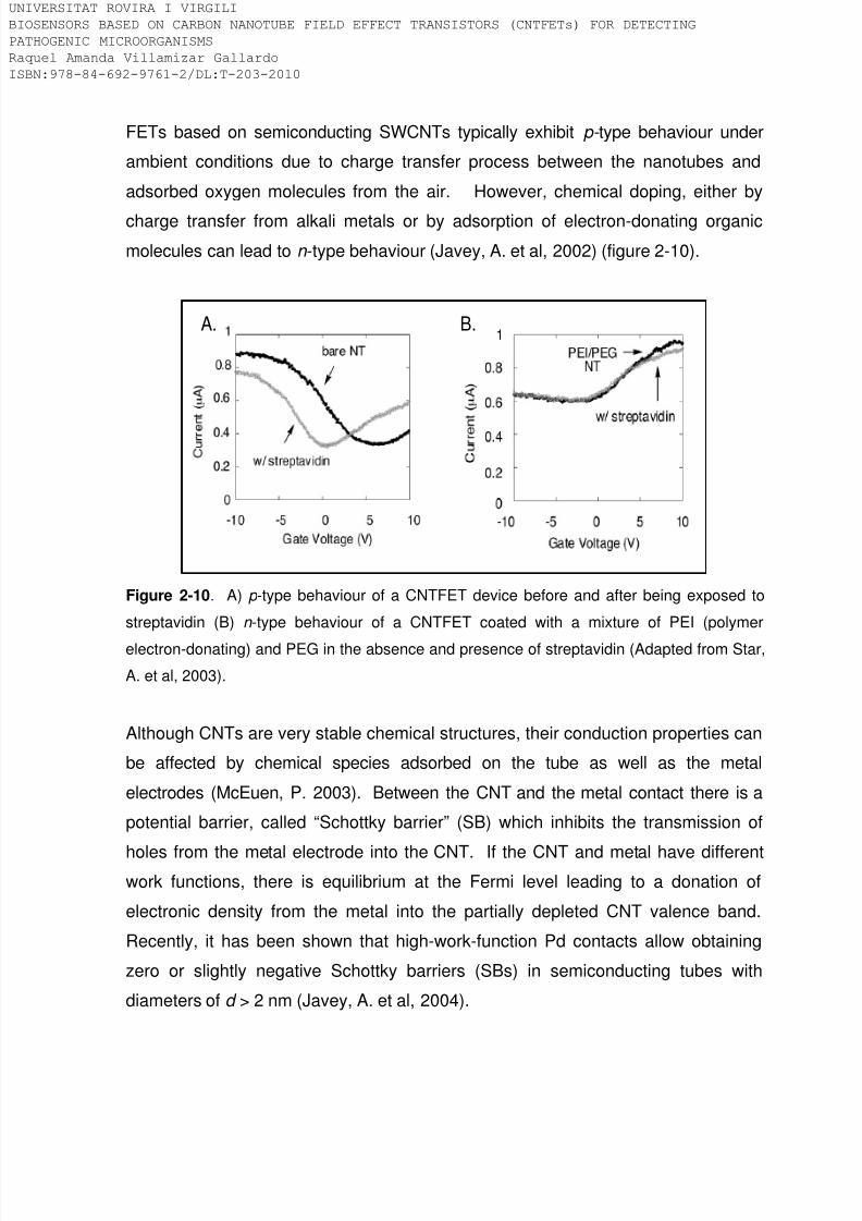
8/9/2019 Tesis Biosensors based on carbon nanotube field effect transistors
http://slidepdf.com/reader/full/tesis-biosensors-based-on-carbon-nanotube-field-effect-transistors 65/239
FETs based on semiconducting SWCNTs typically exhibit p- type behaviour under
ambient conditions due to charge transfer process between the nanotubes and
adsorbed oxygen molecules from the air. However, chemical doping, either by
charge transfer from alkali metals or by adsorption of electron-donating organic
molecules can lead to n -type behaviour (Javey, A. et al, 2002) (figure 2-10).
A. B.
Figure 2-10. A) p -type behaviour of a CNTFET device before and after being exposed to
streptavidin (B) n -type behaviour of a CNTFET coated with a mixture of PEI (polymer
electron-donating) and PEG in the absence and presence of streptavidin (Adapted from Star,
A. et al, 2003).
Although CNTs are very stable chemical structures, their conduction properties can
be affected by chemical species adsorbed on the tube as well as the metal
electrodes (McEuen, P. 2003). Between the CNT and the metal contact there is a
potential barrier, called “Schottky barrier” (SB) which inhibits the transmission of
holes from the metal electrode into the CNT. If the CNT and metal have differentwork functions, there is equilibrium at the Fermi level leading to a donation of
electronic density from the metal into the partially depleted CNT valence band.
Recently, it has been shown that high-work-function Pd contacts allow obtaining
zero or slightly negative Schottky barriers (SBs) in semiconducting tubes with
diameters of d > 2 nm (Javey, A. et al, 2004).
IVERSITAT ROVIRA I VIRGILI
OSENSORS BASED ON CARBON NANOTUBE FIELD EFFECT TRANSISTORS (CNTFETs) FOR DETECTING
THOGENIC MICROORGANISMS
quel Amanda Villamizar Gallardo
BN:978-84-692-9761-2/DL:T-203-2010

8/9/2019 Tesis Biosensors based on carbon nanotube field effect transistors
http://slidepdf.com/reader/full/tesis-biosensors-based-on-carbon-nanotube-field-effect-transistors 66/239
Towards very positive gate voltages, the SB at the CNT – metal electrode inhibits
the transport of holes from the metal electrode into the CNT valence band, therefore
the measured current is small and the device is said to be in the ‘‘OFF-state’’.
Towards more negative gate voltages the SB decrease and increase hole transport
into the CNT, the device is said to turn on, the current increase and the device is
said to be in the ‘‘ON-state’’. Sweeping the gate voltage at a constant Vsd generates
a current (or conductance increase) versus gate voltage Vg curve, I –Vg called a
transistor transfer characteristic (Kauffmanab, D.R. and Star, A. 2008).
In a dense network involving both metallic and semiconducting nanotubes, the
conductance of the off state, will be dominated by the conductance of the metallictubes. Assuming the same conductance for metallic and negatively biased
semiconducting nanotubes, a ratio of the metallic to semiconducting tubes of 1 to 2
would suggest a modulation (on/off conductance ratio) of 3. However, in low density
networks, percolation issues can also tend to increase the on/off ratio. For a sample
with very few pathways from source to drain, there is a high probability that there
will exist no continuous all-metallic tube paths, therefore yielding a slight off current,
and an on/off ratio several orders of magnitude higher than the on/off ratio for athick film (Hu, L. et al, 2004).
Figure 2-11 shows the gate dependence of conductance for a metallic and a
semiconducting SWNT. Dotted curve shows that there is not gate dependence of
conductance, indicating the presence of exclusively metallic tubes, while in a solid
line shows a strong gate dependence of conductance of at least 4 orders of
magnitude. This is representative of transport through purely semiconducting
tubes. Balasubramanian et al (Balasubramanian, K. et al, 2004) report that a strong
hysteresis is observed due to the electrical characterization is performed under
ambient conditions. They explain that this phenomenon can be associated to
oxygen-related charge traps either in the SiO2 or to the presence of SiO2 surface-
bound water molecules.
IVERSITAT ROVIRA I VIRGILI
OSENSORS BASED ON CARBON NANOTUBE FIELD EFFECT TRANSISTORS (CNTFETs) FOR DETECTING
THOGENIC MICROORGANISMS
quel Amanda Villamizar Gallardo
BN:978-84-692-9761-2/DL:T-203-2010

8/9/2019 Tesis Biosensors based on carbon nanotube field effect transistors
http://slidepdf.com/reader/full/tesis-biosensors-based-on-carbon-nanotube-field-effect-transistors 67/239
On state
Off state
Figure 2-11. Gate dependence of conductance for a metallic SWNT (dotted line) and for a
semiconducting SWNT (solid line), measured at room temperature. The gate voltage scan
start at the point marked with the solid circle and stopped at the point marked with the solid
square. A drain source voltage (Vsd) of 1 mV is used for measuring the conductance of the
metallic SWNT and 100 mV for the semiconducting SWNT (Adapted from Balasubramanian,
K. et al, 2004).
CNTFETs can be operated in two gate configurations. The first one, involve
CNTFETs back-gated, which was applied in the present research work and was
represented in figure 2-8 and 2-9. In this configuration the capacitor is a SiO 2 layer
located between the n or p -doped Si substrate and the CNTs. The carbon
nanotubes are connected to the source and drain electrodes. The third electrode
“gate” is the so-called back-gate electrode since it is placed at the back of the
device producing the final structure of a transistor. When a voltage is appliedthrough the back-gate electrode, it makes possible to modulate the field strength
across the insulating SiO2, therefore the density of mobile charges in
semiconducting SWCNTs are modified. As a consequence the conductivity of the
device undergoes changes that can be measured and recorded. It is important to
highlight that if the gate is placed in close contact with the CNTs the
transconductance of the device can increase (Krüger, M. et al, 2001).
IVERSITAT ROVIRA I VIRGILI
OSENSORS BASED ON CARBON NANOTUBE FIELD EFFECT TRANSISTORS (CNTFETs) FOR DETECTING
THOGENIC MICROORGANISMS
quel Amanda Villamizar Gallardo
BN:978-84-692-9761-2/DL:T-203-2010

8/9/2019 Tesis Biosensors based on carbon nanotube field effect transistors
http://slidepdf.com/reader/full/tesis-biosensors-based-on-carbon-nanotube-field-effect-transistors 68/239
This kind of configuration permits two important functions. On one hand, due to the
CNTs are in close contact with the sample, the electrical signal generated can be
easily related to the presence of the analyte. The generated field can be measured
as a change in the resistance in the source-to-drain current. On the other hand, in
this configuration the gate electrode is directly integrated into the device being
unnecessary an external electrode (Cid, C., 2009).
The second type of configuration include CNTFETs electrolyte gated. Krüger et al
(Krüger, M. et al, 2001) and Rosenblatt et al (Rosenblatt, S. et al, 2002) have
shown that SWCNT-based field-effect transistors (FETs) can be electrolytically
gated with high sensitivity. In this configuration a reference electrode (usually Pt) is
placed in contact with a solution containing the analyte which at the same time is in
contact with the SWCNTs (figure 2-12). The electrostatic potential difference
between the solution and the CNTs is controlled through a gate voltage and the
potential at the metal-liquid interface. Because the reaction is often carried out in
buffer solution, biomolecules can reach the metal surface of the electrode altering
the redox conditions at the metal-liquid interface. To overcome this problem
Ag/AgCl reference electrode with a porous glass frit can be used ensuring that the
redox conditions at the metal-liquid interface are well controlled and therefore,
achieving artifact-free measurements (Minot, E.D. et al, 2007).
It has been found that the electrolyte-gated configuration increase the sensitivity of
the CNTFETs devices, nevertheless the main disadvantage is related to the
miniaturizing the reference electrode and/or conditions of the electrolyte when using
Pt electrodes (Cid, C., 2009).
IVERSITAT ROVIRA I VIRGILI
OSENSORS BASED ON CARBON NANOTUBE FIELD EFFECT TRANSISTORS (CNTFETs) FOR DETECTING
THOGENIC MICROORGANISMS
quel Amanda Villamizar Gallardo
BN:978-84-692-9761-2/DL:T-203-2010

8/9/2019 Tesis Biosensors based on carbon nanotube field effect transistors
http://slidepdf.com/reader/full/tesis-biosensors-based-on-carbon-nanotube-field-effect-transistors 69/239
Figure 2-12. Transfer characteristics of a CNTFET in air (solid line), using the bottom gate,
and in water (dotted line), using the liquid gate (Bradley, K. et al, 2004).
2.3.1. Biosensing applications of the CNTFETs
Different strategies can be applied to detect an analyte. In the case of FET based
on CNTs the current flows through their tubular structure, therefore, any change in
the surrounding environment modify the device characteristics being easily
quantified (Grüner, G. 2006). When a molecule is adsorbed on the device, two
main phenomena can take place; the first one involves a charge transfer process in
which molecules provide or withdraw electrons to the tube. The second one is a
scattering process, in which the adsorbed molecules act as scattering source(Grüner, G. 2006). Electrical measurements can help to distinguish between these
two effects. If there is a charge transfer process, the threshold voltage becomes
more positive (electron acceptor) or more negative (electron donor). In contrast, if a
scattering mechanism occurs the overall conductance drops (Allen, B.L. et al, 2007)
(figure 2-13).
IVERSITAT ROVIRA I VIRGILI
OSENSORS BASED ON CARBON NANOTUBE FIELD EFFECT TRANSISTORS (CNTFETs) FOR DETECTING
THOGENIC MICROORGANISMS
quel Amanda Villamizar Gallardo
BN:978-84-692-9761-2/DL:T-203-2010

8/9/2019 Tesis Biosensors based on carbon nanotube field effect transistors
http://slidepdf.com/reader/full/tesis-biosensors-based-on-carbon-nanotube-field-effect-transistors 70/239
Figure 2-13. Change in the transistor device characteristics. a) Electron transfer from a
protein to the tube b) Potential scattering (Gruner, G. 2006).
The detection of biological molecules using CNTs is a rapidly growing field.
SWCNTs possess diameter comparable to large biological molecules, giving them
the size compatibility necessary to ‘‘wire into’’ the system of interest. The surface
area of individual SWCNTs is about 1600 m2g-1, the highest value for any known
material allowing absorption process (Cinke, M. et al, 2002). They also have very
high surface-to-weight ratio (~300 m2g-1) making them accessible to both
electrochemistry and immobilization of biomolecules (Grüner, G. 2006).
Furthermore, the low charge carrier density of SWCNTs is directly comparable to
the surface charge density of proteins, which intuitively makes SWCNTs well suited
for electronic detection that relies on electrostatic interactions with analyte
biomolecules (Heller, I. et al, 2008).
CNTFETs have been used to detect glucose (Besteman, K. et al, 2003),
xenocompounds (Sanchez, Z.C. et al, 2009), proteins like IgG (Cid, C. et al, 2008),
DNA (Gui, E.L., 2007.; Star, A., 2006.; Martínez, M. T. et al, 2009), viruses
(Dastagir, T., 2007) among others. Nevertheless, untill 2006 nobody had been
detected bacteria or higher cells using CNTFETs. Only the interaction between
bacteria and CNTs had been previously studied.
IVERSITAT ROVIRA I VIRGILI
OSENSORS BASED ON CARBON NANOTUBE FIELD EFFECT TRANSISTORS (CNTFETs) FOR DETECTING
THOGENIC MICROORGANISMS
quel Amanda Villamizar Gallardo
BN:978-84-692-9761-2/DL:T-203-2010

8/9/2019 Tesis Biosensors based on carbon nanotube field effect transistors
http://slidepdf.com/reader/full/tesis-biosensors-based-on-carbon-nanotube-field-effect-transistors 71/239
For instance, Huang et al (Huang, T. S. et al, 2004) proven that anti-Salmonella and
anti-S. aureus antibodies immobilized onto the CNTs allowed to detect
concentrations up to 108 cfu/mL of these two bacteria. Gu et al (Gu, L. et al, 2005),
reported the use of SWCNTs functionalized with galactose to detect E.coli. The
complex Gal-SWCNT was mixed with 5x10⁸ cfu/mL of the bacteria during one hour.
After that, the complex Gal-SWCNT-E.coli was observed in the SEM and the results
showed a strong interaction between the complex and the bacteria. Elkin et al
(Elkin, T. et al, 2005) used immuno-carbon nanotubes functionalized with BSA and
anti-E.coli O157:H7 antibody. The complex SWCNT-BSA-antibody was finally
exposed to ~109 cfu/mL of the bacteria. The antigen-antibody interaction was
proven through the confocal microscopy.
Lin et al (Lin, Y. et al, 2006), used the complex MWCNT synthesized by CVD using
as catalyst xilene a precursor of the ferrocene. The resulting MWCNTs, containing
residues of ferromagnetic catalyst, were coated with BSA and anti-E.coli antibody
and were exposed to E.coli O157:H7 at room temperature for 1 hour. By means of
immunomagnetic separation the authors were able to detect ~ 40 cfu/100 µL of the
bacteria in pure culture. Simultaneously to the publication of our first paper, So et al (So, H. et al, 2008) have also published an electrochemical sensor based on
CNTFETs to detect the bacteria E.coli . They functionalized an array of CNTFETs
with aptamers as the recognition element and were able to detect up to 93 cfu/mL
of E. coli in water in less than 20 minutes.
In this research work, we tried to develop selective and sensitive biosensors able to
detect different types of pathogenic microorganisms including bacteria, moulds and
yeast with high relevance in both the food and medical sectors. Moreover, different
blocking agents were used in order to protect the sidewalls of the CNTs from the
non-specific binding of possible interference present in real samples. In this way,
our devices were applied for biosensing purpose in simple and complex matriz. A
brief explanation follows about the structure and antigenic determinants of bacteria,
yeasts and moulds are given.
IVERSITAT ROVIRA I VIRGILI
OSENSORS BASED ON CARBON NANOTUBE FIELD EFFECT TRANSISTORS (CNTFETs) FOR DETECTING
THOGENIC MICROORGANISMS
quel Amanda Villamizar Gallardo
BN:978-84-692-9761-2/DL:T-203-2010

8/9/2019 Tesis Biosensors based on carbon nanotube field effect transistors
http://slidepdf.com/reader/full/tesis-biosensors-based-on-carbon-nanotube-field-effect-transistors 72/239
2.3.1.1. Bacteria
They are unicellular organisms that can be both pathogenic and non-pathogenic
with diameters having a range of 0.2 µm y 5 µm. Bacteria can be classified
according to the composition of their cell wall in Gram positive, Gram negative and
alcohol resistant. Gram positive bacteria, possess a cell wall with high content of
peptidoglucane (about 90 %) and low amounts of polymers like teichoic acids
(figure 2-14). Certain teichoic acids are intimately associated with lipids; therefore
they are called lipotheichoic acids (Madigan, M.T. et al, 2000). Both teichoic and
lipoteichoic acids are responsible for the antigenic properties of these organisms.
By contrast, in Gram negative bacteria the amount of peptidoglucane is lower thanGram positive (about 10 %). In addition, they present an external membrane
composed of lipopolysaccharides (LPS) (figure 1-14). These LPS contain lateral
polysaccharides known as somatic antigen (O) which vary depending on the type of
bacteria. The lipid portion of the LPS, referred to as lipid A is associated with the
toxic properties of these organisms.
As can be seen, gram positive and gram negative bacteria are antigenicallydifferent; therefore, antibodies should not display cross-reactivity in the presence of
a mixture of both types of microorganisms. Furthermore, bacteria can present
surface structures such as, flagellum or capsules, which also determine their
antigenicity. Flagellums are composed by the flagellar protein called flagellin; by
contrast capsules are formed by polysaccharides containing glycoproteins and
different polysaccharides. This structure represents and plays an important role in
the attachment of the pathogenic microorganism to their host. Therefore, flagellar
antigen (H) or capsular antigen (K) can be recognized by the antibodies
respectively.
IVERSITAT ROVIRA I VIRGILI
OSENSORS BASED ON CARBON NANOTUBE FIELD EFFECT TRANSISTORS (CNTFETs) FOR DETECTING
THOGENIC MICROORGANISMS
quel Amanda Villamizar Gallardo
BN:978-84-692-9761-2/DL:T-203-2010
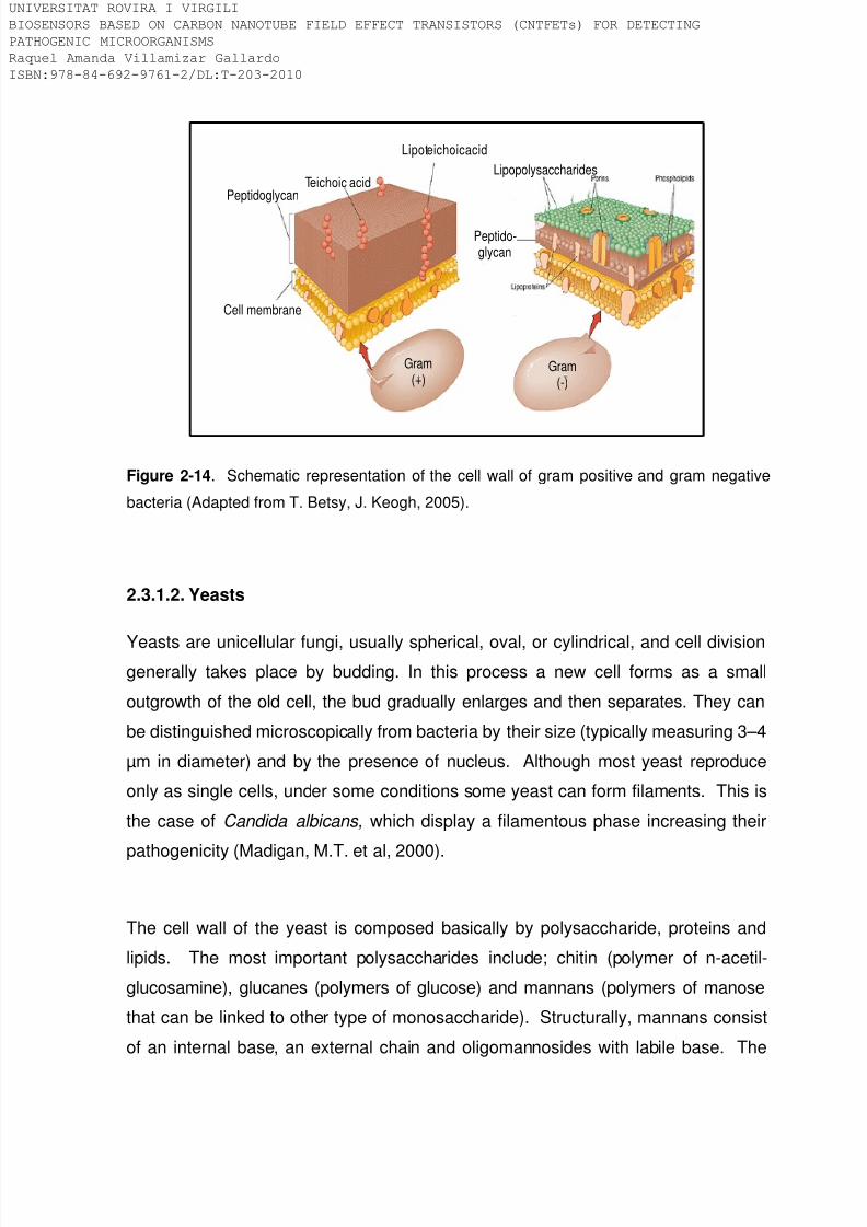
8/9/2019 Tesis Biosensors based on carbon nanotube field effect transistors
http://slidepdf.com/reader/full/tesis-biosensors-based-on-carbon-nanotube-field-effect-transistors 73/239
Gram(+)
Gram(-)
Cell membrane
PeptidoglycanTeichoic acid
Lipoteichoicacid
Lipopolysaccharides
Peptido-glycan
Figure 2-14. Schematic representation of the cell wall of gram positive and gram negative
bacteria (Adapted from T. Betsy, J. Keogh, 2005).
2.3.1.2. Yeasts
Yeasts are unicellular fungi, usually spherical, oval, or cylindrical, and cell divisiongenerally takes place by budding. In this process a new cell forms as a small
outgrowth of the old cell, the bud gradually enlarges and then separates. They can
be distinguished microscopically from bacteria by their size (typically measuring 3–4
µm in diameter) and by the presence of nucleus. Although most yeast reproduce
only as single cells, under some conditions some yeast can form filaments. This is
the case of Candida albicans, which display a filamentous phase increasing their
pathogenicity (Madigan, M.T. et al, 2000).
The cell wall of the yeast is composed basically by polysaccharide, proteins and
lipids. The most important polysaccharides include; chitin (polymer of n-acetil-
glucosamine), glucanes (polymers of glucose) and mannans (polymers of manose
that can be linked to other type of monosaccharide). Structurally, mannans consist
of an internal base, an external chain and oligomannosides with labile base. The
IVERSITAT ROVIRA I VIRGILI
OSENSORS BASED ON CARBON NANOTUBE FIELD EFFECT TRANSISTORS (CNTFETs) FOR DETECTING
THOGENIC MICROORGANISMS
quel Amanda Villamizar Gallardo
BN:978-84-692-9761-2/DL:T-203-2010

8/9/2019 Tesis Biosensors based on carbon nanotube field effect transistors
http://slidepdf.com/reader/full/tesis-biosensors-based-on-carbon-nanotube-field-effect-transistors 74/239
external region of the chain determines the antigenic specificity. Figure 2-15 shows
a schematic representation of a yeast cell.
Manno-
proteins
Vacuoles
Cell wallNucleus
Plasmamembrane
Figure 2-15. Schematic representation of the morphological structure of yeast.
2.3.1.3. Moulds
They are filamentous fungi, widespread in nature and are frequently found on stale
bread, cheese or fruit. A single filament is called hypha. It usually grows together
across a surface and forms a compact structure called a mycelium. From the
fungal mycelium, other hyphal branches may reach the air producing aerial
branches spores called conidia (figure 2-16). They are asexual spores and are
responsible of the final color for the fungi which may be black, blue-green, red,yellow or brown. As well as in yeasts, the cell wall of the moulds is generally
composed of 80-90 % polysaccharide, with proteins, lipids, polyphosphates and
inorganic ions. Mannans and galactomannas are often responsible for the
immunological response against moulds and yeast (McGinnis, M.R. and Borgers,
M. 1989).
IVERSITAT ROVIRA I VIRGILI
OSENSORS BASED ON CARBON NANOTUBE FIELD EFFECT TRANSISTORS (CNTFETs) FOR DETECTING
THOGENIC MICROORGANISMS
quel Amanda Villamizar Gallardo
BN:978-84-692-9761-2/DL:T-203-2010

8/9/2019 Tesis Biosensors based on carbon nanotube field effect transistors
http://slidepdf.com/reader/full/tesis-biosensors-based-on-carbon-nanotube-field-effect-transistors 75/239
These fungi can cause disease through three main mechanisms. The first involves
allergic reactions after being exposed to specific fungal species. Aspergillus spp, is
commonly associated with hypersensitivity causing asthma. A second fungal
disease mechanism is the production of mycotoxins. The best known examples are
the aflatoxins produced by A. flavus, a fungus frequent in grains. The mycotoxin can
induce tumors in animals. The third fungal disease-produced mechanims is through
an overgrow of a fungus on or in the body, producing mycosis which can vary from
superficial to serius life-threatening diseases (Madigan, M.T. et al, 2000).
Figure 2-16. Diagram of the different structures a mould displays along its life cycle
(Madigan, M.T. et al, 2000).
IVERSITAT ROVIRA I VIRGILI
OSENSORS BASED ON CARBON NANOTUBE FIELD EFFECT TRANSISTORS (CNTFETs) FOR DETECTING
THOGENIC MICROORGANISMS
quel Amanda Villamizar Gallardo
BN:978-84-692-9761-2/DL:T-203-2010

8/9/2019 Tesis Biosensors based on carbon nanotube field effect transistors
http://slidepdf.com/reader/full/tesis-biosensors-based-on-carbon-nanotube-field-effect-transistors 76/239
2.4. References
Allen, B.L., Kichambare, P.D., Star, A. Advanced Materials. 2007, 19, 1439–1451.
Balasubramanian, K., Sordan, R., Burghard, M., Kern, K. Nano Letters. 2004. 4,
827-830.
Baughman, R.H., Cui, C., Zakhidov, A.A., Iqbal, Z., Barisci, J.N., Spinks, G.M.,
Wallace, G.G., Mazzoldi, A., De Rossi, D., Rinzler, A.G., Jaschinski, O., Roth, S.,
Kertesz, M. Science. 1999. 284, 1340–1344.
Besteman, K., Lee, J. O., Wiertz, F.G.M., Heering, H.A. , Dekker, C. Nano Letters.
2003. 3,727-730.
T. Betsy, J. Keogh, Microbiology Demystified, McGraw-Hill Publishing, New York,
2005.
Beyer, M., Felgenhauer, T., Bischoff, F. R., Breitling, F., Stadler, V. Biomaterials.2006. 27, 3505-3514.
Bjorck, L. and Kronvall, G. The Journal of Immunology. 1984. 133, 969-974.
J.P. Bourgoin, A. Loiseau, J.F. Nierengarten, Nanoscience, Springer, Berlin. 2006.
Bradley, K., Briman, M., Star, A., Grüner, G. Nano Letters. 2004. 4, 253-256.
Byon, H.R., Choi, H.C. Journal of American Chemical Society. 2006. 128, 2188-
2189.
Cinke, M., Li, J., Chen, B., Cassell, A., Delzeit, L., Han, J., Meyyappan, M.
Chemical Physics Letters. 2002, 365, 69–74.
Charlier, J.C., Roche, X.B. Reviews of Modern Physics. 2007. 79, 677-732.
IVERSITAT ROVIRA I VIRGILI
OSENSORS BASED ON CARBON NANOTUBE FIELD EFFECT TRANSISTORS (CNTFETs) FOR DETECTING
THOGENIC MICROORGANISMS
quel Amanda Villamizar Gallardo
BN:978-84-692-9761-2/DL:T-203-2010

8/9/2019 Tesis Biosensors based on carbon nanotube field effect transistors
http://slidepdf.com/reader/full/tesis-biosensors-based-on-carbon-nanotube-field-effect-transistors 77/239
Chen, R.J., Bangsaruntip, S., Drouvalakis, K.A., Shi, N.W., Shim, M., Li, Y., Kim,
W., Utz, P.J., Dai, H. Proceedings of the National Academy of Sciences. 2003.
100, 4984-4989.
Chen, E. S.; Chen, E. C. M. Bioelectrochemistry and Bioenergetics. 1998. 46, 15-
19.
Chen, R. J., Choi, H. C., Bangsaruntip, S., Yenilmez, E., Tang, X., Wang, Q.,
Chang, Y. L., Dai, H. Journal of American Chemical Society. 2004. 126, 1563-
1568.
Chi, Y.S., Byon, H.R., Choi, H.C., Choi, I. S. ChemBioChem. 2007. 8, 1380-1387.
Cid, C., Riu, J., Maroto, A., Rius, F.X. Analyst. 2008. 133, 1005–1008.
Cid C. 2009. Doctoral Thesis. Sensors based on carbon nanotube field-effect
transistors and molecular recognition approaches. Department of Analytical and
Organic Chemistry. Universitat Rovira I Virgilli.
Cowley, J.M., Nikolaev, P., Thess, A., Smalley, R.E. Chemical Physics Letters.
1997. 265, 379–384.
Dastagir, T., Forzani, E.S., Zhang, R., Amlani, I., Nagahara, L.A., Tsui, R., Tao, N.
Analyst. 2007.132, 738-740.
M.S. Dresselhaus, G. Dresselhaus, P.C. Eklund, Science of Fullerenes and Carbon
Nanotubes, Academic Press, New York, 1996.
Duerkop, T., Getty, S., Cobas, E., Fuhrer, M. Nano Letters. 2004. 4, 35-39.
Elkin, T., Jiang, X., Taylor, S., Lin, Y., Gu, L., Yang, H., Brown, J., Collins, S., Sun,
Y. ChemBioChem. 2005. 6, 640-643.
IVERSITAT ROVIRA I VIRGILI
OSENSORS BASED ON CARBON NANOTUBE FIELD EFFECT TRANSISTORS (CNTFETs) FOR DETECTING
THOGENIC MICROORGANISMS
quel Amanda Villamizar Gallardo
BN:978-84-692-9761-2/DL:T-203-2010

8/9/2019 Tesis Biosensors based on carbon nanotube field effect transistors
http://slidepdf.com/reader/full/tesis-biosensors-based-on-carbon-nanotube-field-effect-transistors 78/239
J.E. Fischer, Nanotubes and Nanofibers, Taylor & Francis Group, LLC, New York,
2006.
Freitag, M., Martin, Y., Misewich, J.A., Martel, R., Avouris Ph. Nano Letters. 2003.
3, 1067–1071.
Giacomelli, C.E. Encyclopedia of Surface and Colloid Science. 2006. DOI
10.1081/E-ESCS-120000092.
P. Gründler, Chemical Sensors, Springer-Verlag, Berlin, 2006.
Grüner, G. Analytical and Bioanalytical Chemistry. 2006. 384, 322-335.
Gui, E.L., Li, L.J., Zhang, K., Xu, Y., Dong, X., Ho, X., Lee, P.S., Kasim, J., Shen,
Z.X., Rogers, J.A., Mhaisalkar, S.G. Journal of the American Chemistry Society.
2007.129, 14427-14432.
Gu, L., Elkin, T., Jiang, X., Li, H., Lin, Y., Qu, L., Tzeng, T.R.J., Joseph, R., Sun,Y.P. Chemical Communications. 2005. 874-876.
Hecht, D.S., Ramirez, R.J.A., Briman, M., Artukovic, E., Chichak, K.S., Stoddart,
J.F., Grüner, G. Nano Letters. 2006. 6, 2031–2036.
Heller, I., Kong, J., Heering, H.A., Williams, K.A., Lemay, S.G., Dekker, C. Nano
Letters. 2005. 5, 137-142.
Heller, I., Kong, J., Williams, K.A., Dekker, C., Lemay, S.G. Journal of the American
Chemical Society. 2006. 128, 7353-7359.
Heller,I., Janssens, A.M., Mannik, J., Minot, E.D., Lemay, S.G., Dekker, C. Nano
Letters. 2008. 8, 591-595.
Hu, L., Hecht, D.S., Gruner, G. Nano Letters. 2004. 4, 2513-2517.
IVERSITAT ROVIRA I VIRGILI
OSENSORS BASED ON CARBON NANOTUBE FIELD EFFECT TRANSISTORS (CNTFETs) FOR DETECTING
THOGENIC MICROORGANISMS
quel Amanda Villamizar Gallardo
BN:978-84-692-9761-2/DL:T-203-2010

8/9/2019 Tesis Biosensors based on carbon nanotube field effect transistors
http://slidepdf.com/reader/full/tesis-biosensors-based-on-carbon-nanotube-field-effect-transistors 79/239
Huang, S., Woodson, M., Smalley, R., Liu, J. Nano Letters. 2004. 4, 1025–1028.
Iijima, S. Nature.1991. 354, 56.
Izard, N., Kazaoui, S., Hata, K., Okazaki, T., Saito, T., Iijima, S., Minami, N. Applied
Physics Letters. 2008. 92, 243112.
Javey, A., Kim, H., Brink, M., Wang, Q., Ural, A., Guo, J., McIntyre, P., McEuen, P.,
Lundstrom, M., Dai, H. Journal of Nature Materials. 2002. 1, 241-246.
Javey, A., Guo, J., Wang,Q., Lundstrom, M., Dai, H. Nature. 2003. 424, 7.
Javey, A., Guo, J., Farmer, D.B., Wang, Q., Wang, D.W., Gordon, R.G., Lundstrom,
M., Dai, H. Nano Letters. 2004. 4, 447-450.
Kauffmanab, D.R. and Star, A. Chemical Society Reviews. 2008. 37, 1197–1206.
Khare, R. and Bose, S. Journal of Minerals & Materials Characterization &Engineering. 2005. 4, 31-46.
Kim, S.N., Rusling, J.F., Papadimitrakopoulos, F. Advanced Materials. 2007. 19,
3214-3228.
Kong, J., Franklin, N.R., Zhou, C.W., Chapline, M.G., Peng, S., Cho, K.J., Dai, H.J.
Science. 2000. 287, 622–625.
Kocabas, C., Hur, S.H., Gaur, A., Meitl, M.A., Shim, M., Rogers, J.A. Small. 2005.
1,1110-1116.
Krüger, M., Buitelaar, M.R., Nussbaumer, T., Schönenberger, C., Forro, L. Applied
Physcis Letters. 2001. 78, 1291-1293.
Lay, M.D., Novak, J.P., Snow, E.S. Nano Letters. 2004. 4, 603-606.
IVERSITAT ROVIRA I VIRGILI
OSENSORS BASED ON CARBON NANOTUBE FIELD EFFECT TRANSISTORS (CNTFETs) FOR DETECTING
THOGENIC MICROORGANISMS
quel Amanda Villamizar Gallardo
BN:978-84-692-9761-2/DL:T-203-2010

8/9/2019 Tesis Biosensors based on carbon nanotube field effect transistors
http://slidepdf.com/reader/full/tesis-biosensors-based-on-carbon-nanotube-field-effect-transistors 80/239
Lee, J.M., Park, H.K., Jung, Y., Kim, J.K., Jung, S.O., Chung, B.H. Analytical
chemistry. 2007. 79, 2680-2687.
A. Liljas, L. Liljas, J. Pirkur, G, Lindblom, P. Nissen, M. Kjeldgaard, Textbook of
Structural Biology, World Scientific Publishing Co. Pte. Ltd, Singapore, 2009.
Lin, A., Patil, N., Ryu, K., Badmaev, A., De Arco, L., Zhou,C., Mitra, S., Wong, H.S.
IEEE Transactions on Nanotechnology. 2009. 8,1.
Lin, Y., Allard, L.F., Sun, Y.P. Journal of Physical Chemistry B. 2008. 108, 3760-
3764.
Lin, Y., Taylor, S., Li, H., Fernando, K.A., Qu, L., Wang, W., Gu, L., Zhou, B., Sun,
Y.P. Journal of Materials Chemistry. 2004. 14, 527-541.
Lin, Y., Jiang, X., Elkin, T., Fernando, K.A., Gu, L., Taylor, S., Yang, H., Jones, E.,
Wang, W., Sun, Y.P. Journal of Nanoscience and Nanotechnology. 2006. 6, 868-
871.
M.T. Madigan, J.M. Martinko, J. Parker, Brock Biology of Microorganisms. Ninth
ed., Prentice Hall, Inc, New Jersey, 2000.
Martínez, M.T., Tseng, Y.C., Ormategui, N., Loinaz, I., Eritja, R., Bokor, J. Nano
Letters. 2009. 9, 530-536.
Marie, R., Beech, J.P., Vörös, J., Tegenfeldt, J.O., Höök, F. Langmuir. 2006. 22,
10103-10108.
Martel, R., Schmidt, T., Shea, H.R., Hertel, T., Avouris, P. Applied Physics Letters.
1998. 73, 2447.
McEuen, P. Single wall carbon nanotubes. Physics World. 2000.31-36
IVERSITAT ROVIRA I VIRGILI
OSENSORS BASED ON CARBON NANOTUBE FIELD EFFECT TRANSISTORS (CNTFETs) FOR DETECTING
THOGENIC MICROORGANISMS
quel Amanda Villamizar Gallardo
BN:978-84-692-9761-2/DL:T-203-2010

8/9/2019 Tesis Biosensors based on carbon nanotube field effect transistors
http://slidepdf.com/reader/full/tesis-biosensors-based-on-carbon-nanotube-field-effect-transistors 81/239
M.R. McGinnis, M. Borgers, Current Topics in Medical Mycology, Springer-Verlag,
New York. 1989.
Minot, E.D., Janssens, A.M., Heller, I., Heering, H.A., Dekker, C., Lemay, S.G.Applied Physical Letters. 2007. 91, 093507.
O`Connor, M., Kim, S.N., Killard, A.J., Foster, R.J., Smyth, M.R.,
Papadimitrakopoulos, F., Rusling, J.F. Analyst. 2004. 129, 1176-1180.
Oh, B.K., Lee, W., Kim, Y.K., Lee, W.H., Choi, J.W. Journal of Biotechnology.
2004. 111, 1-8.
Okuno, J., Maehashi, K., Kerman, K., Takamura, Y., Matsumoto, K., Tamiya, E.
Biosensors and Bioelectronics. 2007. 22, 2377-2381.
Pantarotto, D., Partidos, C.D., Graff, R., Hoebeke, J., Briand, J.P., Prato, M.,
Bianco, A. Journal of the American Chemical Society. 2003. 125, 6160-6164.
Piao, M.H.M., Noh, M.A. Rahman, H., Won, M.S., Shim, Y.B. Electroanalysis. 2008.
20, 30–37.
C.P. Poole, F.J. Owens, Introduction to Nanotechnology, Wiley-Interscience,
Hoboken, New Jersey , 2003.
Pourfath, M., Kosina, H., Selberherr, S. Downloaded on June 4, 2009 from IEEE
Xplore.
Raffaellea, R.P., Landia, B.J., Harrisb, J.D., Baileyb, S.G., Hepp, A.F. Materials
Science and Engineering B. 2005. 116, 233–243.
Rao, A. M., Richter, E., Bandow, S., Chase, B., Eklund, P.C., Williams, K.A., Fang,
S., Subbaswamy, K.R., Menon, M., Thess, A., Smalley, R.E., Dresselhaus, G.,
Dresselhaus, M. S. Science. 1997. 275, 187–190.
IVERSITAT ROVIRA I VIRGILI
OSENSORS BASED ON CARBON NANOTUBE FIELD EFFECT TRANSISTORS (CNTFETs) FOR DETECTING
THOGENIC MICROORGANISMS
quel Amanda Villamizar Gallardo
BN:978-84-692-9761-2/DL:T-203-2010
8/9/2019 Tesis Biosensors based on carbon nanotube field effect transistors
http://slidepdf.com/reader/full/tesis-biosensors-based-on-carbon-nanotube-field-effect-transistors 82/239
Rosenblatt, S., Yaish, Y., Park, J., Gore, J., Sazonova, V., McEuen, P. L. Nano
Letters. 2002. 2, 869-872.
R. Saito, G., Dresselhaus, M.S. Dresselhaus, Physical Properties of Carbon
Nanotubes, Imperial College Press, London, 1998.
Sánchez-Acevedo, Z.C., Riu, J., Rius, F. X. Biosensors and Bioelectronics. 2009.
24, 2842–2846.
Shim, M., Kam, N.W.S., Chen, R.J., Li, Y., Dai, H. Nano Letters. 2002. 2, 285-288.
Sloan, D.J. and Hellinga, H.W. Protein Science. 1999. 8, 1643–1648.
So, H.M., Park, D. W., Jeon, E. K., Kim, Y. H., Kim, S.K., Lee, C.K., Choi, S. Y, Kim,
S.C., Chang, H., Lee, J.O. Small. 2008. 4, 197-201.
Star, A., Gabriel, J.C.P., Bradley, K., Grüner, G. Nano Letters. 2003. 3, 459-463.
Star, A., Tu, E., Niemann, J., Gabriel, J.C.P., Joiner, C.S., Valcke, C. Proceedings
of the National Academy of Sciences. 2006. 103, 921-926.
Tans, S.J., Devoret, M.H., Dai, H., Thess, A., Smalley, R.E., Geerligs, L.J., Dekker,
C. Nature. 1997. 386, 474.
Trojanowicz, M. Trends in Analytical Chemistry. 2006. 25, 480-489.
Tuncel, A. Colloid and Polymer Science. 2000, 278, 1126-1138.
Vermeer, A.W.P., Norde, W., van Amerongen, A. Journal of Biophysics. 2000. 79,
2150-2154.
Vermeer, A.W.P. and Norde, W. Journal of Biophysics. 2000. 78, 394-404.
IVERSITAT ROVIRA I VIRGILI
OSENSORS BASED ON CARBON NANOTUBE FIELD EFFECT TRANSISTORS (CNTFETs) FOR DETECTING
THOGENIC MICROORGANISMS
quel Amanda Villamizar Gallardo
BN:978-84-692-9761-2/DL:T-203-2010

8/9/2019 Tesis Biosensors based on carbon nanotube field effect transistors
http://slidepdf.com/reader/full/tesis-biosensors-based-on-carbon-nanotube-field-effect-transistors 83/239
Vichchulada, P., Zhang, Q., Lay, M.D. Analyst. 2007. 132, 719–723.
P. Voet, J. Voet, C. Pratt, Principles of Biochemistry, third ed., John Wiley & Sons,
INC, 2008.
Wang, S., Humphreys, E. S., Chung, S.Y., Delduco, D. F., Lustig, S. R., Wang, H.,
Parker, K. N., Rizzo, N. W., Subramoney, S., Chiang, Y.M., Jagota, A. Nature
Materials. 2003. 2, 196.
Wildöer, J.W.G., Venema, L.C., Rinzler, A.G., Smalley, R.E., Dekker, C. Nature.
1998. 391, 59.
Yao, Z., Kane, C.L., Dekker, C. Physics Reviews Letters. 2000. 84, 2941-2944.
Zhao, Y.L. and Stoddart, J.F. Accounts of Chemical Research. 2009. 42, 1161-
1171.
IVERSITAT ROVIRA I VIRGILI
OSENSORS BASED ON CARBON NANOTUBE FIELD EFFECT TRANSISTORS (CNTFETs) FOR DETECTING
THOGENIC MICROORGANISMS
quel Amanda Villamizar Gallardo
BN:978-84-692-9761-2/DL:T-203-2010

8/9/2019 Tesis Biosensors based on carbon nanotube field effect transistors
http://slidepdf.com/reader/full/tesis-biosensors-based-on-carbon-nanotube-field-effect-transistors 84/239
IVERSITAT ROVIRA I VIRGILI
OSENSORS BASED ON CARBON NANOTUBE FIELD EFFECT TRANSISTORS (CNTFETs) FOR DETECTING
THOGENIC MICROORGANISMS
quel Amanda Villamizar Gallardo
BN:978-84-692-9761-2/DL:T-203-2010

8/9/2019 Tesis Biosensors based on carbon nanotube field effect transistors
http://slidepdf.com/reader/full/tesis-biosensors-based-on-carbon-nanotube-field-effect-transistors 85/239
Chapter 3
Experimental development of the biofunctionalized
devices
IVERSITAT ROVIRA I VIRGILI
OSENSORS BASED ON CARBON NANOTUBE FIELD EFFECT TRANSISTORS (CNTFETs) FOR DETECTING
THOGENIC MICROORGANISMS
quel Amanda Villamizar Gallardo
BN:978-84-692-9761-2/DL:T-203-2010

8/9/2019 Tesis Biosensors based on carbon nanotube field effect transistors
http://slidepdf.com/reader/full/tesis-biosensors-based-on-carbon-nanotube-field-effect-transistors 86/239
IVERSITAT ROVIRA I VIRGILI
OSENSORS BASED ON CARBON NANOTUBE FIELD EFFECT TRANSISTORS (CNTFETs) FOR DETECTING
THOGENIC MICROORGANISMS
quel Amanda Villamizar Gallardo
BN:978-84-692-9761-2/DL:T-203-2010

8/9/2019 Tesis Biosensors based on carbon nanotube field effect transistors
http://slidepdf.com/reader/full/tesis-biosensors-based-on-carbon-nanotube-field-effect-transistors 87/239
3.1. Introduction
This chapter presents a detailed description of the materials, reagents, equipment
and procedures used for the development of the CNTFET sensors. The objective is
to give a general idea concerning the manufacture of the biosensors, from the
synthesis of the carbon nanotubes to the functionalization and detection process as
well as the preparation of the pathogenic microorganisms detected are explained
along with the characterization techniques of the devices. Finally, the aim of this
chapter is to supply a complete protocol that can be reproduced and therefore be
used in practice for future research.
IVERSITAT ROVIRA I VIRGILI
OSENSORS BASED ON CARBON NANOTUBE FIELD EFFECT TRANSISTORS (CNTFETs) FOR DETECTING
THOGENIC MICROORGANISMS
quel Amanda Villamizar Gallardo
BN:978-84-692-9761-2/DL:T-203-2010

8/9/2019 Tesis Biosensors based on carbon nanotube field effect transistors
http://slidepdf.com/reader/full/tesis-biosensors-based-on-carbon-nanotube-field-effect-transistors 88/239
3.2. Apparatus, materials, and reagents
3.2.1. Cells preparation
3.2.1.1. Apparatus
• Autoclave (Selecta)
• Mini orbital shaker (Stuart)
• Biosafety chamber, Telstar Technologies, S.L. Model Bio II A (Izasa)
• Memmert incubator (Afora)
3.2.1.2. Strains
3.2.1.2.1. Bacteria
• Salmonella Infantis S34/11(Faculty of Medicine and Health Sciences at the
Universitat Rovira i Virgili)
• Shigella sonnei CECT 413(Faculty of Medicine and Health Sciences at the
Universitat Rovira i Virgili)
• Streptococcus pyogenes CECT 985T(Faculty of Medicine and HealthSciences at the Universitat Rovira i Virgili)
3.2.1.2.2. Yeast
• C. albicans CECT 1001 (Spanish Type Culture Collection, Valencia,Spain)
• Saccharomyces cerevisiae CECT 1443 (Spanish Type Culture Collection,
Valencia,Spain)
• C. albidus CECT 11978 (Spanish Type Culture Collection, Valencia,Spain)
3.2.1.2.3. Moulds
• Aspergillus flavus CECT 2684 (Spanish Type Culture Collection,
Valencia,Spain)
• Penicillium chrysogenum CECT 2307 (Spanish Type Culture Collection,
Valencia,Spain)
IVERSITAT ROVIRA I VIRGILI
OSENSORS BASED ON CARBON NANOTUBE FIELD EFFECT TRANSISTORS (CNTFETs) FOR DETECTING
THOGENIC MICROORGANISMS
quel Amanda Villamizar Gallardo
BN:978-84-692-9761-2/DL:T-203-2010

8/9/2019 Tesis Biosensors based on carbon nanotube field effect transistors
http://slidepdf.com/reader/full/tesis-biosensors-based-on-carbon-nanotube-field-effect-transistors 89/239
• Fusarium oxysporum CECT 2154 (Spanish Type Culture Collection,
Valencia,Spain)
3.2.1.3. Media
• Trypticase soy agar (TSA), Breckton-Dickinson (Comercial Belles S.A.)
• Trypticase soy broth (TSB), Beckton-Dickinson (Comercial Belles S.A.)
• Sabouraud Dextrosa Agar (SDA), Oxoid
• Oxytetracycline, gentamicine yeast agar (OGY), Oxoid
• Brain heart infusion broth (BHI), Scharlau Chemie Microbiology (Comercial
Belles S.A.)
• Buffered peptone water, Scharlau Chemie Microbiology (Comercial Belles
S.A.)
3.2.2. Synthesis of the CNTs
3.2.2.1. Apparatus
• Horizontal split tube furnace HST 12/600 (Carbolite)
• Quartz tubular reactor (4 cm x 120 cm) (Afora)• Ultrasonic bath 100 W (Selecta)
• Spin coater WS-400B-6NPP/LITE (Laurell Technologies Corporation).
• Mass controllers for methane and hydrogen including PC software
(Bronkhorst, Ruurlo)
3.2.2.2. Materials
Substrates of Si/SiO2. 500 nm of SiO2 grown thermally over type n-type lowresistivity Si. Dimensions: 0.5 x 0.5 cm (D+T Microelectrónica, National
Microelectronics Centre).
3.2.2.3. Reagents
• Methane C-45 (Carburos metálicos)
• Hydrogen C-50 (Carburos metálicos)
• Argon C-50 (Carburos metálicos)
IVERSITAT ROVIRA I VIRGILI
OSENSORS BASED ON CARBON NANOTUBE FIELD EFFECT TRANSISTORS (CNTFETs) FOR DETECTING
THOGENIC MICROORGANISMS
quel Amanda Villamizar Gallardo
BN:978-84-692-9761-2/DL:T-203-2010

8/9/2019 Tesis Biosensors based on carbon nanotube field effect transistors
http://slidepdf.com/reader/full/tesis-biosensors-based-on-carbon-nanotube-field-effect-transistors 90/239
• Nitrogen C-50 (Carburos metálicos)
• Iron nitrate (III) nonahydrated 99.99 % (Sigma)
• Acetone 99.5 % (Sigma)
• Isopropanol 99.5 % (Sigma)
• Deionized and charcoal-treated water (18.2 MΩ·cm specific resistance)
obtained with Milli-Q PLUS reagent-grade water system (Millipore).
3.2.3. Development of the CNTFETs
3.2.3.1. Materials
Silver ink Electrodag ® 1415 M (Acheson Industries, Scheemdam,Netherlands)
3.2.4. Functionalization of the CNTFETs
3.2.4.1. Proteins
• IgG anti-Salmonella OBT0958 (i.e. a rabbit polyclonal antibody for
Salmonella “O” & “H” antigens (4–5 mg/mL)), Oxford Biotechnology Ltd.(Oxford, UK)
• IgG anti-Candida , 1750-5070 (i.e. a monoclonal mouse anti-C. albicans (2.8
mg/mL)), Oxford Biotechnology Ltd (Oxford, U.K.).
• IgG anti-Aspergillus 0071-1300 (i.e. a polyclonal rabbit anti-Aspergillus (1
mg/mL)), Oxford Biotechnology Ltd. (Oxford, U.K.)
• Protein G from Streptococcus sp. recombinant, expressed in Escherichia
coli (1 mg/mL) (Sigma).
3.2.4.2. Reagents
• Dulbecco phosphate buffered saline solution (PBS), 150 mM, pH: 7.1 - 7.5
(Sigma)
• Tween 20 (Sigma)
• Gelatine from cold water fish skin (Sigma)
IVERSITAT ROVIRA I VIRGILI
OSENSORS BASED ON CARBON NANOTUBE FIELD EFFECT TRANSISTORS (CNTFETs) FOR DETECTING
THOGENIC MICROORGANISMS
quel Amanda Villamizar Gallardo
BN:978-84-692-9761-2/DL:T-203-2010

8/9/2019 Tesis Biosensors based on carbon nanotube field effect transistors
http://slidepdf.com/reader/full/tesis-biosensors-based-on-carbon-nanotube-field-effect-transistors 91/239
• Deionized and charcoal-treated water (18.2 MΩ·cm specific resistance)
obtained with Milli-Q PLUS reagent-grade water system (Millipore)
3.2.5. Characterization of the CNTFETs
3.2.5.1. Electrical characterization
• Precision semiconductor parameter analyser Agilent 4157A (Agilent
Technologies)
• MP 1008 Manual probe station (Wentworth Laboratories)
• Faraday box (SIRM)
• Anti-vibration table (SIRM)
3.2.5.2. Microscopy characterization
• Scanning electron microscope (SEM), JSM 6400 (Jeol, Tokyo, Japan)
• Environmental-Scanning electron microscope (ESEM), Quanta 600, (FEI,
Hillsboro, OR, USA)
• Atomic force microscope (AFM), PicoPlus (Molecular Imaging, Tempe, AZ,
USA)
3.2.5.3. Ellipsometry characterization
• Woollam VASE spectroscopic ellipsometer (J.A. Woollam Co. Lincoln,
USA)
• Software WVASE32 (J.A. Woollam Co. Lincoln, USA)
3.3. Procedures
3.3.1. Preparation of the pathogenic microorganisms
3.3.1.1. Bacteria
• Take a sample of the bacteria from the initial culture stored at -20 ºC and
reactivate it by inoculating in 10 mL of sterile TSB at 37 ºC for 12 h.
IVERSITAT ROVIRA I VIRGILI
OSENSORS BASED ON CARBON NANOTUBE FIELD EFFECT TRANSISTORS (CNTFETs) FOR DETECTING
THOGENIC MICROORGANISMS
quel Amanda Villamizar Gallardo
BN:978-84-692-9761-2/DL:T-203-2010

8/9/2019 Tesis Biosensors based on carbon nanotube field effect transistors
http://slidepdf.com/reader/full/tesis-biosensors-based-on-carbon-nanotube-field-effect-transistors 92/239
• Then, pelletize the sample in a centrifuge at 6000 rpm for 15 minutes and
discard the supernatant.
• Resuspend the pellet in 10 mL of sterile 0.85 % saline solution and
enumerate the bacteria contained in the saline solution by plating serial
dilutions on TSA plates and incubating at 37 ºC for 24–48 h (figure 3-1).
• Prepare additional dilutions in sterile 0.85 % saline solution to produce the
desired final sample concentration of each strain.
Figure 3-1. Cell culture preparation of Salmonella Infantis.
3.3.1.2. Yeast
• Rehydrate the stock strains with sterile water and subculture at least three
times on SDA and incubated at 35 ºC for 48 h to test their viability.
• Take isolated colonies and resuspend them on sterile 0.85 % saline
solution.
• Enumerate the yeast contained in the saline solution by plating serial
dilutions on SDA plates and incubating at 35 ºC for 24–48 h (Chang, H.C.
et al, 2000) (figure 3-2).
• Prepare additional dilutions in sterile 0.85 % saline solution to produce thedesired final sample concentrations of each strain.
•
IVERSITAT ROVIRA I VIRGILI
OSENSORS BASED ON CARBON NANOTUBE FIELD EFFECT TRANSISTORS (CNTFETs) FOR DETECTING
THOGENIC MICROORGANISMS
quel Amanda Villamizar Gallardo
BN:978-84-692-9761-2/DL:T-203-2010

8/9/2019 Tesis Biosensors based on carbon nanotube field effect transistors
http://slidepdf.com/reader/full/tesis-biosensors-based-on-carbon-nanotube-field-effect-transistors 93/239
Figure 3-2. Cell culture preparation of Candida albicans .
3.3.1.3. Moulds
• Rehydrate the stock strains with sterile water and plate them in SDA at 30
ºC for 5-7 days (Hao, W. et al, 2008) to check their viability.
• Wash the spores from the surface of the medium with PBS solution.
• Take flaks of 100 mL of BHI, inoculate them with 1 mL of the spore
suspension obtained before and incubate at 30 ºC, 120 rpm for 7 days
(Tsai G.J. and Yu S.C., 1999) (figure 3-3).
• Harvest the cultures and separate the mycelium by filtrating with Whatman
No. 5 filter paper (Hetherington, S.V. 1994).
• Collect the mycelium, wash it with sterile distilled water and dry in the oven
at 105 ºC until stable weight (De Vos, M.M. and Nelis, H.J. 2003).
• Dilute the previous samples in PBS (w/v) to produce the desired final
sample concentration of each strain.
Figure 3-3. Cell culture preparation of Aspergillus flavus .
IVERSITAT ROVIRA I VIRGILI
OSENSORS BASED ON CARBON NANOTUBE FIELD EFFECT TRANSISTORS (CNTFETs) FOR DETECTING
THOGENIC MICROORGANISMS
quel Amanda Villamizar Gallardo
BN:978-84-692-9761-2/DL:T-203-2010

8/9/2019 Tesis Biosensors based on carbon nanotube field effect transistors
http://slidepdf.com/reader/full/tesis-biosensors-based-on-carbon-nanotube-field-effect-transistors 94/239
3.3.2. Synthesis of the SWCNTs
• Prepare the catalyst solution by dissolving iron nitrate in isopropanol up to a
concentration of 100 μg/mL.
• Sonicate the solution in an ultrasonic bath, for 10-15 minutes.
• Clean the Si/SiO2 substrate by making successive immersions in acetone,
isopropanol, and deionized water in the ultrasonic bath. Each immersion
should last about 5 minutes.
• Dry with nitrogen.
• Spin-coat three times 20 μL of the catalyst solution over the Si/SiO2 at 3000
rpm and leave it spinning until the solvent is completely evaporated (about
30 seconds).
• Place the Si/SiO2 substrates into the quartz reactor and switch on the oven
at 900 ºC.
• Introduce an argon current of 1000 sccm until the maximum temperature is
reached. After that, close the argon flow and simultaneously introduce 600
sccm of methane and 200 sccm of hydrogen for 20 minutes (figure 3-4).
• Finally, close the methane and hydrogen flows while argon flow is
introduced again into the quartz reactor until the system cools down toroom temperature (Cid C. 2009).
SubstrateCatalystnanoparticle
Nanotubegrowth
Gas inlet
Oven /900 ºC
Quartz tube
Gas outlet
Ar CH4 H2
Figure 3-4. Schematic representation of the chemical vapour deposition (CVD)
process used for the synthesis of SWCNT at 900 ºC. The zoom shows the growth
mechanism of the nanostructures.
IVERSITAT ROVIRA I VIRGILI
OSENSORS BASED ON CARBON NANOTUBE FIELD EFFECT TRANSISTORS (CNTFETs) FOR DETECTING
THOGENIC MICROORGANISMS
quel Amanda Villamizar Gallardo
BN:978-84-692-9761-2/DL:T-203-2010

8/9/2019 Tesis Biosensors based on carbon nanotube field effect transistors
http://slidepdf.com/reader/full/tesis-biosensors-based-on-carbon-nanotube-field-effect-transistors 95/239
3.3.3. Development of the CNTFETs
As it has been explained in chapter 2, the structure of a FET consists of two metal
electrodes named source and drain connected to the semiconducting channel
forming a transistor. Perpendicular to this structure, we find a capacitor made of the
semiconducting nantubes, the SiO2 dielectric layer and the semiconducting n -type
doped Si layer. Different methods have been used to develop the sensor.
Photolithography is one of the common methods used to grafting the electrodes.
Although this method generates well reproducible devices it is also expensive.
Therefore, in the present research we use a homemade screen printing process.
This process allows obtaining cheap devices in a very short time.
The process involve the use of a conductive ink, Electrodag® 1415 M, made of
silver and 4-methylpentan-2-one. By using a home-made designed silicone mask,
the silver ink is deposited manually over the Si/SiO2 substrate containing the as-
grown network of SWCNTs. The mask allows printing the metallic electrodes at the
desired spots (figure 3-5). Subsequently in order to fix the electrodes to the surface,
the chips are cured in an oven at 150 ºC for 10 min (Cid C. 2009).
1 mm
Figure 3-5. SEM image of the home-made screen-printed electrodes. The size of the
electrodes is about 200 µm2 and the distance between the source and drain is approximately
500 µm. The zoom shows an E-SEM image of the typical network of SWCNTs obtained after
a CVD process.
IVERSITAT ROVIRA I VIRGILI
OSENSORS BASED ON CARBON NANOTUBE FIELD EFFECT TRANSISTORS (CNTFETs) FOR DETECTING
THOGENIC MICROORGANISMS
quel Amanda Villamizar Gallardo
BN:978-84-692-9761-2/DL:T-203-2010

8/9/2019 Tesis Biosensors based on carbon nanotube field effect transistors
http://slidepdf.com/reader/full/tesis-biosensors-based-on-carbon-nanotube-field-effect-transistors 96/239
3.3.4. Functionalization process
In the development of our sensor devices, two methods have been used to
immobilize the molecular receptors (antibodies) onto the transducers (SWCNTs).
3.3.4.1. Direct adsorption of IgG onto the CNTs
The direct adsorption of immunoglobulin on a surface is a non-covalent process
governed mainly by hydrophobic interactions between antibodies and the solid
surface (Chen, R.J. et al, 2003). Since the adsorption is a random process, it can
leave free gaps in the surface. Therefore, in order to avoid the non-specific binding
of other proteins or possible interferences on the SWCNTs a solution of Tween 20
is used as blocking agent (Chi, Y.S. et al, 2007). The functionalization process iscarried out as follows:
• Dilute the commercial antibody up to the desired concentration (i.e. 10
µg/mL) in phosphate buffered saline solution (PBS) (15 mM, pH=7.4).
• Immerse the CNTFET obtained before in the prepared antibody solution for
1 hour at 37 ºC to have the antibodies adsorbed onto the SWCNTs (Lin, Y.
et al, 2006).• Rinse with PBS (15 mM, pH=7.4), deionized water and dry with nitrogen
(IIic, B., et al, 2000).
• Immerse the CNTFET previously coated with the antibodies in a solution
containing Tween 20 at 0.5 % for 2 hours (figure 3-6). Subsequently, rinse
with deionized water and dry with nitrogen (So, H.M. et al, 2005).
• After this process, the devices are ready to use to detect the
microorganism
1
3.3.4.2. IgG binding through the protein G
In order to obtain well-orientated antibodies linked to the SWCNT surface, bacterial
proteins like protein G can be used. They are found in the cell wall of most species
of Staphylococcus and Streptococcus and display high specificity for the Fc domain
1 CNTFETs with the IgG directly adsorbed were used to detect the bacteria Salmonella Infantis and the
yeast Candida albicans .
IVERSITAT ROVIRA I VIRGILI
OSENSORS BASED ON CARBON NANOTUBE FIELD EFFECT TRANSISTORS (CNTFETs) FOR DETECTING
THOGENIC MICROORGANISMS
quel Amanda Villamizar Gallardo
BN:978-84-692-9761-2/DL:T-203-2010

8/9/2019 Tesis Biosensors based on carbon nanotube field effect transistors
http://slidepdf.com/reader/full/tesis-biosensors-based-on-carbon-nanotube-field-effect-transistors 97/239
of the IgG, thus leaving the Fab region available for detecting the target (Oh, B.K. et
al, 2004). This functionalization process involves the following steps:
• Dilute the commercial protein G up to the desired concentration (e.g. 5µg/mL) in phosphate buffered saline solution (PBS) (15 mM, pH=7.4).
• Immerse the CNTFET in the prepared protein G solution for 30 minutes at
37 ºC to have the protein adsorbed over the SWCNT (Lin, Y. et al, 2006).
• Rinse with PBS (15 mM, pH=7.4), deionized water and dry with nitrogen
(IIic, B. et al, 2000).
• Immerse the CNTFET previously coated with protein G in a PBSTG solution
containing 1.5 % of Tween 20 and 2 % of gelatin for 3 hours in order toavoid the non-specific adsorption of the antibodies onto the transducer
(Piao, M.H. et al, 2008) and to protect the SWCNTs from the interferences
of the sample matrix (figure 3-6).
• Rinse again with deionized water and dry with nitrogen (IIic, B. et al, 2000)
and immerse the CNTFET previously coated with protein G and PBSTG in
an antibody solution (8 µg/mL) for 1 hour at 37 ºC to have the proteins
attached on a top of the protein G.
• Finally, the devices are ready to detect the microorganism2.
Figure 3-6. Schematic representation of the functionalization process. A) Direct
adsorption of the IgG antibodies onto the SWCNTs B). Immobilization of the IgG
antibodies by using protein G.
2
CNTFETs with the IgG adsorbed onto protein G was used to detect the mould Aspergillus flavus.
IVERSITAT ROVIRA I VIRGILI
OSENSORS BASED ON CARBON NANOTUBE FIELD EFFECT TRANSISTORS (CNTFETs) FOR DETECTING
THOGENIC MICROORGANISMS
quel Amanda Villamizar Gallardo
BN:978-84-692-9761-2/DL:T-203-2010
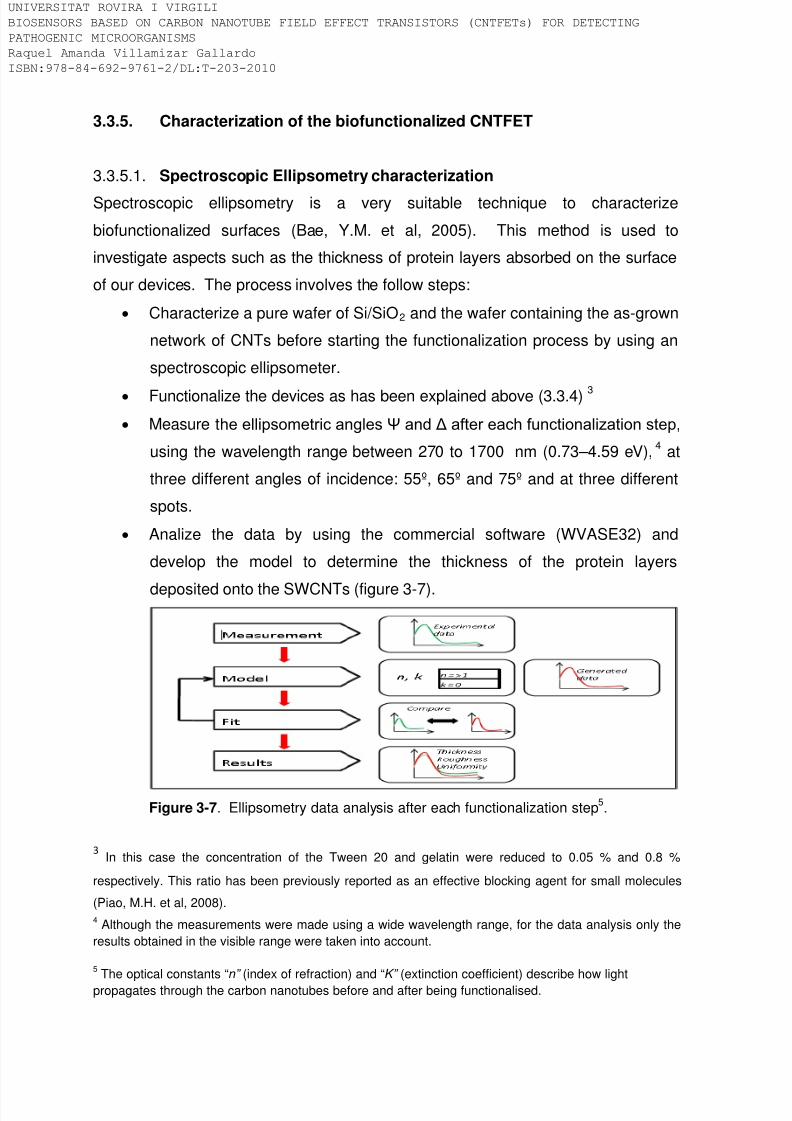
8/9/2019 Tesis Biosensors based on carbon nanotube field effect transistors
http://slidepdf.com/reader/full/tesis-biosensors-based-on-carbon-nanotube-field-effect-transistors 98/239
3.3.5. Characterization of the biofunctionalized CNTFET
3.3.5.1. Spectroscopic Ellipsometry characterization
Spectroscopic ellipsometry is a very suitable technique to characterize
biofunctionalized surfaces (Bae, Y.M. et al, 2005). This method is used to
investigate aspects such as the thickness of protein layers absorbed on the surface
of our devices. The process involves the follow steps:
• Characterize a pure wafer of Si/SiO2 and the wafer containing the as-grown
network of CNTs before starting the functionalization process by using an
spectroscopic ellipsometer.
• Functionalize the devices as has been explained above (3.3.4) 3
• Measure the ellipsometric angles Ψ and Δ after each functionalization step,
using the wavelength range between 270 to 1700 nm (0.73–4.59 eV),
4
• Analize the data by using the commercial software (WVASE32) and
develop the model to determine the thickness of the protein layers
deposited onto the SWCNTs (figure 3-7).
at
three different angles of incidence: 55º, 65º and 75º and at three different
spots.
Figure 3-7. Ellipsometry data analysis after each functionalization step5
3 In this case the concentration of the Tween 20 and gelatin were reduced to 0.05 % and 0.8 %
respectively. This ratio has been previously reported as an effective blocking agent for small molecules
(Piao, M.H. et al, 2008).
.
4 Although the measurements were made using a wide wavelength range, for the data analysis only theresults obtained in the visible range were taken into account.
5 The optical constants “n” (index of refraction) and “K” (extinction coefficient) describe how lightpropagates through the carbon nanotubes before and after being functionalised.
IVERSITAT ROVIRA I VIRGILI
OSENSORS BASED ON CARBON NANOTUBE FIELD EFFECT TRANSISTORS (CNTFETs) FOR DETECTING
THOGENIC MICROORGANISMS
quel Amanda Villamizar Gallardo
BN:978-84-692-9761-2/DL:T-203-2010
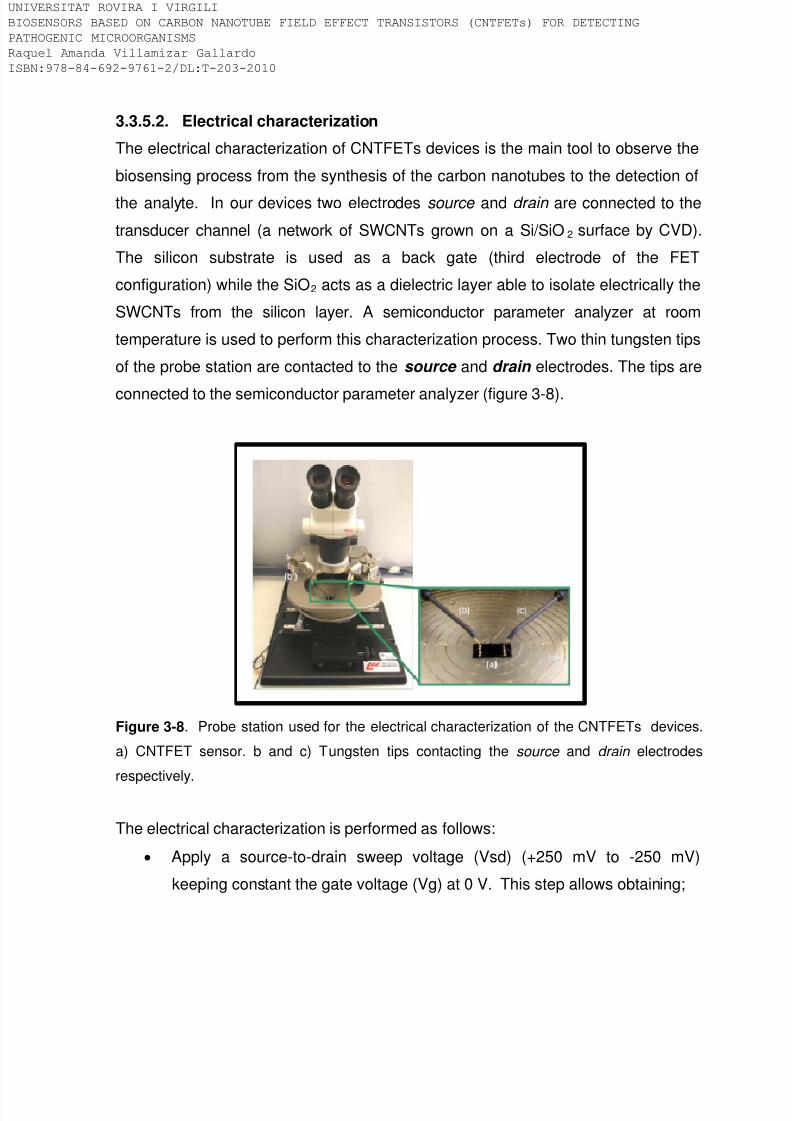
8/9/2019 Tesis Biosensors based on carbon nanotube field effect transistors
http://slidepdf.com/reader/full/tesis-biosensors-based-on-carbon-nanotube-field-effect-transistors 99/239
3.3.5.2. Electrical characterization
The electrical characterization of CNTFETs devices is the main tool to observe the
biosensing process from the synthesis of the carbon nanotubes to the detection of
the analyte. In our devices two electrodes source and drain are connected to the
transducer channel (a network of SWCNTs grown on a Si/SiO2 surface by CVD).
The silicon substrate is used as a back gate (third electrode of the FET
configuration) while the SiO2 acts as a dielectric layer able to isolate electrically the
SWCNTs from the silicon layer. A semiconductor parameter analyzer at room
temperature is used to perform this characterization process. Two thin tungsten tips
of the probe station are contacted to the source and drain electrodes. The tips are
connected to the semiconductor parameter analyzer (figure 3-8).
Figure 3-8. Probe station used for the electrical characterization of the CNTFETs devices.
a) CNTFET sensor. b and c) Tungsten tips contacting the source and drain electrodes
respectively.
The electrical characterization is performed as follows:
• Apply a source-to-drain sweep voltage (Vsd) (+250 mV to -250 mV)
keeping constant the gate voltage (Vg) at 0 V. This step allows obtaining;
IVERSITAT ROVIRA I VIRGILI
OSENSORS BASED ON CARBON NANOTUBE FIELD EFFECT TRANSISTORS (CNTFETs) FOR DETECTING
THOGENIC MICROORGANISMS
quel Amanda Villamizar Gallardo
BN:978-84-692-9761-2/DL:T-203-2010
8/9/2019 Tesis Biosensors based on carbon nanotube field effect transistors
http://slidepdf.com/reader/full/tesis-biosensors-based-on-carbon-nanotube-field-effect-transistors 100/239
a) the resistance of the channel (R = V / I), b) the optimum value of Vsd 6,
c) the source-to-gate current (“gate leakage”) at Vg = 0 V 7
• Register the electrical current while sweeping the Vg
(Cid C. 2009).8
. From this curve, we
can know whether the CNT network is semi-conducting or metallic. This
curve, i.e. also known as the “device characteristics” or “gate dependence”,
is also registered after each functionalisation step and after exposing the
functionalised devices to the pathogenic microorganisms.
3.3.5.3. Microscopic characterization
The CNTFET are microscopically characterized with Atomic Force Microscopy
(AFM), Scanning Electron Microscopy (SEM) and Environmental Scanning ElectronMicroscopy (ESEM). AFM is a well known technique that provides direct images of
the surface topography (Kuhlmeier, D. et al, 2003) without special sample
preparation. This technique allows to mesure the height of the CNTs. In this way, it
is possible: 1) to verify that the synthesized CNTs are indeed single-walled carbon
nanotubes; and 2) to follow the functionalisation steps by measuring the height after
each functionalisation. AFM is also a contactless technique being possible to reuse
the samples after their analysis. The disadvantage of AFM is that it is quite slow.Moreover, it is impossible to obtain an overall image of the whole sample. This
technique was used in this research to obtain surface images from 1µm x 1µm to 20
µm x 20µm.
SEM and ESEM are techniques that allow obtaining images of a sample surface
with a high range of magnification by scanning it with a high-energy beam. These
electrons interact with the sample producing signals that contain information about
6 In our devices the optimum Vsd is the one that provides electrical current values of a few µA and is
usually found between 100 mV and 250 mV.
7 Devices with a gate leakage higher than 1 nA cannot be used. The gate leakage is sometimes due tophysical damage in the isolating layer (SiO2) but also it can be due to a high density of the as-grownSWCNTs. To overcome this problem, the network of the SWCNTs containing the source and drainelectrodes is isolated by cutting the surface around the electrodes.
8 The best value of Vg is the one that produces the highest source-drain current values but also, the one
that keeps the capacity to maximize the modulation of the signal.
IVERSITAT ROVIRA I VIRGILI
OSENSORS BASED ON CARBON NANOTUBE FIELD EFFECT TRANSISTORS (CNTFETs) FOR DETECTING
THOGENIC MICROORGANISMS
quel Amanda Villamizar Gallardo
BN:978-84-692-9761-2/DL:T-203-2010

8/9/2019 Tesis Biosensors based on carbon nanotube field effect transistors
http://slidepdf.com/reader/full/tesis-biosensors-based-on-carbon-nanotube-field-effect-transistors 101/239
its topography and composition. The most common method used to obtain the
images is by using the secondary electron imaging mode, which can produce very
high-resolution images of a sample surface, revealing details about 1 to 5 nm in
size. Furthermore they provide large depth field. SEM often requires sample pre-
treatment being useful only for electrically conductive samples. Moreover, the
sample must be dehydrated due to it only operate in high or low vacuum. By
contrast in E-SEM electrically or non-electrically conductive samples can be
analysed. In addition, E-SEM allows imaging in high vacuum, low vacuum and
environmental conditions. Both techniques, SEM and ESEM do not allow
calculating the height of the CNTs and the potential applied often burn the CNTs.
Therefore, the samples cannot be reused.
These techniques are used in this research to known the density of the CNT
networks and to verify the presence of antibodies and microorganisms on the CNT
networks. The voltage used vary from 15 kV for the as-grown carbon nanotubes to
1 or 2 kV for the carbon nanotubes after being coated with the antibodies and after
interaction with the microorganisms. The working distance used is in a range of 8
mm to 10 mm. We did not metalize the sample because a same device wasobserved several times after each functionalization step.
IVERSITAT ROVIRA I VIRGILI
OSENSORS BASED ON CARBON NANOTUBE FIELD EFFECT TRANSISTORS (CNTFETs) FOR DETECTING
THOGENIC MICROORGANISMS
quel Amanda Villamizar Gallardo
BN:978-84-692-9761-2/DL:T-203-2010

8/9/2019 Tesis Biosensors based on carbon nanotube field effect transistors
http://slidepdf.com/reader/full/tesis-biosensors-based-on-carbon-nanotube-field-effect-transistors 102/239
3.4. References
Bae, Y.M., Oh, B.K., Lee, W., Lee, W.H., Choi, J.W. Biosensors and Bioelectronics.
2005. 21,103-110.
Cid, C. 2009. Doctoral Thesis. Sensors based on carbon nanotube field-effect
transistors and molecular recognition approaches. Department of Analytical and
Organic Chemistry. Universitat Rovira i Virgili.
Chang, H.C., Chang, J.J., Huang, A.H., Chang, T.C. Journal of Clinical
Microbiology. 2000. 38, 971–976.
Chen, R.J., Bangsaruntip, S., Drouvalakis, K.A., Kam, N.W., Shim, M., Li, Y., Kim,
W., Utz, P.J., Dai, H. Proceedings of the National Academy of Sciences. 2003.
100, 4984–4989.
Chi, Y.S., Byon, H.R., Choi, H.C., Choi, I. S. ChemBioChem. 2007. 8, 1380 – 1387.
De Vos, M.M. and Nelis, H.J. Journal of Microbiological Methods. 2003. 55, 557–
564.
Hao, W., Pan, Y.X., Ding, Y.Q., Xiao, S., Yin, K., Wang, Y.D., Qiu, L.W., Zhang,
Q.L., Woo, P.C.Y., Lau, S.K.P., Yuen, K.Y., Che, X.Y. Clinical and Vaccine
Immunology. 2008. 15, 194–202.
Hetherington, S.V., Henwick, S., Parham, D.M., Patrick, C.C. Clinical and
Diagnostic Laboratory Immunology. 1994. 1, 63-67.
Kuhlmeier, D., Rodda, E., Kolarik, L.O., Furlong, D.N., Bilitewski, U. Biosensorsand Bioelectronics. 2003.18, 925-936.
IIic, B., Czaplewski, D., Craighead, G. Applied Physics Letters. 2000. 77, 450–452.
Lin, Y., Jiang, X., Elkin, T., Fernando, K.A., Gu, L., Taylor, S., Yang, H., Jones, E.,
Wang, W., Sun, Y.P. Journal of Nanoscience and Nanotechnology. 2006. 6, 868–
871.
IVERSITAT ROVIRA I VIRGILI
OSENSORS BASED ON CARBON NANOTUBE FIELD EFFECT TRANSISTORS (CNTFETs) FOR DETECTING
THOGENIC MICROORGANISMS
quel Amanda Villamizar Gallardo
BN:978-84-692-9761-2/DL:T-203-2010

8/9/2019 Tesis Biosensors based on carbon nanotube field effect transistors
http://slidepdf.com/reader/full/tesis-biosensors-based-on-carbon-nanotube-field-effect-transistors 103/239
Oh, B.K., Lee, W., Kim, Y.K., Lee, W.H., Choi, J.W. Journal of Biotechnology.
2004. 111, 1-8.
Piao, M.H., Noh, M.H., Rahman, M.A., Won, M.S., Shim, Y.B. Electroanalysis.2008. 20, 30–37.
So, H.M., Won, K., Kim, Y.H., Kim, B.K., Ryu, B.H., Na, P.S., Kim, H., Lee, J.O.
Journal of the American Chemical Society. 2005. 127, 11906–11907.
Tsai, G.J. and Yu, S.C. International Journal of Food Microbiology. 1999. 50,
181–189.
IVERSITAT ROVIRA I VIRGILI
OSENSORS BASED ON CARBON NANOTUBE FIELD EFFECT TRANSISTORS (CNTFETs) FOR DETECTING
THOGENIC MICROORGANISMS
quel Amanda Villamizar Gallardo
BN:978-84-692-9761-2/DL:T-203-2010

8/9/2019 Tesis Biosensors based on carbon nanotube field effect transistors
http://slidepdf.com/reader/full/tesis-biosensors-based-on-carbon-nanotube-field-effect-transistors 104/239
IVERSITAT ROVIRA I VIRGILI
OSENSORS BASED ON CARBON NANOTUBE FIELD EFFECT TRANSISTORS (CNTFETs) FOR DETECTING
THOGENIC MICROORGANISMS
quel Amanda Villamizar Gallardo
BN:978-84-692-9761-2/DL:T-203-2010

8/9/2019 Tesis Biosensors based on carbon nanotube field effect transistors
http://slidepdf.com/reader/full/tesis-biosensors-based-on-carbon-nanotube-field-effect-transistors 105/239
Chapter 4
CNTFET for pathogenic bacteria determination
IVERSITAT ROVIRA I VIRGILI
OSENSORS BASED ON CARBON NANOTUBE FIELD EFFECT TRANSISTORS (CNTFETs) FOR DETECTING
THOGENIC MICROORGANISMS
quel Amanda Villamizar Gallardo
BN:978-84-692-9761-2/DL:T-203-2010

8/9/2019 Tesis Biosensors based on carbon nanotube field effect transistors
http://slidepdf.com/reader/full/tesis-biosensors-based-on-carbon-nanotube-field-effect-transistors 106/239
IVERSITAT ROVIRA I VIRGILI
OSENSORS BASED ON CARBON NANOTUBE FIELD EFFECT TRANSISTORS (CNTFETs) FOR DETECTING
THOGENIC MICROORGANISMS
quel Amanda Villamizar Gallardo
BN:978-84-692-9761-2/DL:T-203-2010

8/9/2019 Tesis Biosensors based on carbon nanotube field effect transistors
http://slidepdf.com/reader/full/tesis-biosensors-based-on-carbon-nanotube-field-effect-transistors 107/239
4.1. Introduction
The growing incidence of pathogenic bacteria in foods is a considerable public
health problem around the world. For that reason, regulatory agencies such as theFood and Drug Administration (FDA) have established control quality programs in
order to avoid pathogens in foods. Hazard analysis critical control point (HACCP) is
a preventive management program from the FDA applied in the food industry to
take corrective actions during the food manufacturing process thus reducing
possible microbial contamination in the final
product(http://www.fda.gov/Food/FoodSafety/HazardAnalysis
CriticalControlPointsHACCP/default.htm).
A great deal of pathogenic bacteria has been related to the generation of foodborne
diseases. Some of the most significant pathogens include Salmonella spp.,
Escherichia coli , Staphylococcus aureus , Campylobacter jejuni and Bacillus cereus
among others (Taux, R.V. 1997). Salmonellosis is an infectious disease produced
by Salmonella that is an important pathogen in both developed and developing
countries.
Many analytical tests for pathogenic bacteria including Salmonella are available in
the market. Current methods for detecting contamination of foodstuffs with
Salmonella consist of conventional culture techniques, biochemical test, immune-
based assays, and nucleic-acid based methods (Taitt, C.R. et al, 2004). The
culturing and plating method is the oldest bacterial detection technique and remains
the standard detection method. Selective media can be used to detect specific
bacteria species. They usually contain inhibitors (compounds that stop or delay the
growth of non-targeted strains) or particular substrates that only the targeted
bacteria is able to degrade producing specific colours characteristic of each specie.
The final detection and enumeration is often performed by ocular inspection
(Lazcka, O. et al, 2007). However, conventional methods require long analysis time
and in some cases pre-enrichment are required to recover and detect the pathogen
(Koyuncu, S. and Haggblom, P. 2009). Moreover, additional biochemical test are
often required to identify the species.
IVERSITAT ROVIRA I VIRGILI
OSENSORS BASED ON CARBON NANOTUBE FIELD EFFECT TRANSISTORS (CNTFETs) FOR DETECTING
THOGENIC MICROORGANISMS
quel Amanda Villamizar Gallardo
BN:978-84-692-9761-2/DL:T-203-2010

8/9/2019 Tesis Biosensors based on carbon nanotube field effect transistors
http://slidepdf.com/reader/full/tesis-biosensors-based-on-carbon-nanotube-field-effect-transistors 108/239
Biochemical testing uses the enzymatic machinery of microorganisms to degrade
specific compounds present in a media. The test often requires between 4 to 24 h
of incubation time and fresh cultures to obtain reliable results. Wilson, G. (Wilson,
G. 2004) developed a biochemical screening protocol for Salmonella and Shigella
that uses rapid enzymatic reactions. It had a sensitivity of 100 % and a specificity of
94 %. This researcher reported the presumptive presence of Salmonella 3 h after
colony isolation. However, further testing on other types of media is recommended
by the author.
Immuno-assays rely on the specific binding of an antibody to an antigen (de Boer,
E. and Beumer, R.R. 1999) Commercial immunoassays for microorganisms likethe enzyme linked-immunosorbent assay (ELISA) combine the specificity of
antibodies and the sensitivity of antibodies or antigens coupled to an enzyme
achieving sensitivities and specificities of 100 % (Janyapoon, K. et al, 2000).
However, the need of specific labels in the case of the typical sandwich assay
makes the method labour intensive.
The sensitivity in conventional ELISA test can be defined as the proportion of true
positives correctly identified [agreed positives/(agreed positives+false negatives) x
100 %] while specificity can defined as the proportion of true negatives correctly
identified [agreed negatives/(false positives+agreed negatives) x 100 %] (Abass,
E.M. et al, 2006).
Nucleic-acid methods are based on the extraction and amplification of target
DNA/RNA with specifics probe using the well known polymerase chain reaction
(PCR) (Scheu, P.M. et al, 1998). To improve the sensitivity and reduce the timeanalysis of pathogens. Eyigör A et al, (Eyigör, A. et al, 2007) designed a PCR-
ELISA method to detect Salmonella DNA from selective primary enrichment culture
of chicken intestine. The detection limit of PCR-ELISA for pure culture of
Salmonella.Enteritidis 64 K was 50 cfu/mL and for intestinal samples artificially
contaminated with Salmonella Enteritidis 64 K was found to be 70 cfu/mL.
Sensitivity and specificity of the assay was determined as 100 %
IVERSITAT ROVIRA I VIRGILI
OSENSORS BASED ON CARBON NANOTUBE FIELD EFFECT TRANSISTORS (CNTFETs) FOR DETECTING
THOGENIC MICROORGANISMS
quel Amanda Villamizar Gallardo
BN:978-84-692-9761-2/DL:T-203-2010

8/9/2019 Tesis Biosensors based on carbon nanotube field effect transistors
http://slidepdf.com/reader/full/tesis-biosensors-based-on-carbon-nanotube-field-effect-transistors 109/239
The immunocapture and the use of PCR is other approach to enhance the
pathogen detection process (Jeníková, G. et al, 2000.; Leon-Velarde, C.G. et al,
2009.; Liébana, S et al, 2009). Liébana et al (Liébana, S et al, 2009), reported the
use of magnetic beads coated with antibodies against Salmonella coupled to an
amplification of the genetic material by PCR and the use of electrochemical
magneto-genosensing to capture, concentrate and detect Salmonella in skim milk
samples. They obtained a limit of detection of 1 cfu/mL in 3.5 h without any
pretreatment. Furthermore, the authors stated that if the skim milk is pre-enriched
for 6 h, the method is able to selectively detect as low as 0.04 cfu/mL (1 cfu / 25 g
of milk) of Salmonella .
In the last decades researches have been trying to develop devices able to made
analysis in situ and in real time. The importance of this kind of elements comes from
their high specificity and sensitivity, which allow the detection of a broad spectrum
of analytes in complex sample matrices (food, blood, serum, urine, etc). Biosensors
are devices that are composed by a biological (protein, nucleic acid) or biomimetic
recognition element (peptide, aptamer) in close contact with a transducer.
Depending on the detection system the biosensors can be classified in optical,
mass, electrochemical and thermal sensors. In addition, biosensors can also be
classified as direct, in which the analyte of interest is detected by the sensor, or
indirect, in which a preliminary biochemical reaction takes place and the products of
that reaction are then detected by the sensor (Ivnitski, D. et al, 1999).
Electrochemical biosensors are devices based on the observation of an electrical
measurement (current, impedance, potential) due to chemical interactions occurringat the sensor sample matrix interface (Lazcka, O. et al, 2007). Rishpon et al
(Rishpon, J. et al, 1992) described a rapid and sensitive method based on an
enzyme-tagged immuno-electrochemical assay to detect pathogenic bacteria. In
these devices antibodies are immobilized on disposable carbon filter disc electrodes
and are used to capture antigens in test solutions. The system was able to detect
as low as 10 cell/mL in less than 10 minutes of Staphylococcus aureus and
Escherichia coli . Nevertheless, this method requires like the conventional ELISA
IVERSITAT ROVIRA I VIRGILI
OSENSORS BASED ON CARBON NANOTUBE FIELD EFFECT TRANSISTORS (CNTFETs) FOR DETECTING
THOGENIC MICROORGANISMS
quel Amanda Villamizar Gallardo
BN:978-84-692-9761-2/DL:T-203-2010

8/9/2019 Tesis Biosensors based on carbon nanotube field effect transistors
http://slidepdf.com/reader/full/tesis-biosensors-based-on-carbon-nanotube-field-effect-transistors 110/239
the use of an enzyme-labeled antibody and specific substrates to obtain the
potentiometric signal.
Shabani et al (Shabani, A. et al, 2008) have recently proposed a novel methodbased on a bacteriophage-modified microarray for the specific and direct detection
of E. coli. The system use bacteriophage T4 as recognition receptors immobilized
covalently onto functionalized screen-printed carbon electrode (SPE) microarrays.
Impedance measurements were recorded to detect the binding of E. coli to the T4
phages. The researchers stated that using this method it is possible to determine 20
cfu/mL. In addition, because phage lyse only living bacteria, the method can
differentiate living from dead bacteria. Delibato et al (Delibato, E. et al, 2009)
reported the use of an immunosensor array, based on a 96-well electrochemical
plate coupled with immunomagnetic beads (ELIME array), for the detection of
Salmonella in meat samples. The immunosensor array was able to detect a low
number of Salmonella cells (1-10 cfu/25 g) after only 6 h of incubation in a pre-
enrichment broth. Barreiros, M (Barreiros, M. et al, 2009) developed and
electrochemical impedance spectroscopy (EIS) to detect pathogenic E. coli
O157:H7 bacteria via a label free immunoassay-based detection method. Sensors
were able to detect very low concentration of E. coli with limits of detection as low
as 10-100 cfu/mL.
Recently, in our research group, Zelada-Guillén et al (Zelada-Guillén, G. et al,
2009) reported a potentiometric aptasensor based on carbon nanotube for the
detection of only 1 cfu/mL of Salmonella Typhi in real time. Therefore,
electrochemical biosensors are particularly interesting because they are usually
inexpensive, are well adapted to miniaturization, and can provide disposable-typechips for field applications.
This chapter presents the first attempt to develop an electrochemical biosensor
based on carbon nanotube field effect transistors (CNTFET) to detect pathogenic
bacteria and it is thoroughly explained in the published article presented below.
Briefly, the biosensor consists of a network of CNTs, previously synthesized by
CVD, that are used as the transducer element. Antibodies are the molecular
IVERSITAT ROVIRA I VIRGILI
OSENSORS BASED ON CARBON NANOTUBE FIELD EFFECT TRANSISTORS (CNTFETs) FOR DETECTING
THOGENIC MICROORGANISMS
quel Amanda Villamizar Gallardo
BN:978-84-692-9761-2/DL:T-203-2010

8/9/2019 Tesis Biosensors based on carbon nanotube field effect transistors
http://slidepdf.com/reader/full/tesis-biosensors-based-on-carbon-nanotube-field-effect-transistors 111/239
receptor able to detect selectively the bacteria Salmonella Infantis. These
antibodies are adsorbed onto the sidewalls of the CNTs by means of hydrophobic
interactions allowing the detection of the bacteria and at the same time preserving
the electrical properties of the transducer. Non-specific interactions from the sample
components with the CNTs are avoided by using a blocking agent. The developed
sensor was characterized by using both electrical and microscopic techniques.The
direct detection system together with the excellent electrical properties of the CNTs
produces a label-free and sensitive biosensor.
Furthermore, this chapter supplies an additional experimental session that compiles
other tests performed that were not included in the published paper. Finally the
complementary conclusions and the references are reported.
IVERSITAT ROVIRA I VIRGILI
OSENSORS BASED ON CARBON NANOTUBE FIELD EFFECT TRANSISTORS (CNTFETs) FOR DETECTING
THOGENIC MICROORGANISMS
quel Amanda Villamizar Gallardo
BN:978-84-692-9761-2/DL:T-203-2010

8/9/2019 Tesis Biosensors based on carbon nanotube field effect transistors
http://slidepdf.com/reader/full/tesis-biosensors-based-on-carbon-nanotube-field-effect-transistors 112/239
4.2. Article:
“Fast detection of Salmonella Infantis with carbon
nanotube field effect transistors”. Biosensors and
Bioelectronics. 2008. 24, 279-283.
Raquel A. VillamizarτŦ, Alicia Maroto*Ŧ, F. Xavier RiusŦ, Isabel Inza ŧ and Maria J.
Figueras ŧ
Ŧ Department of Analytical and Organic Chemistry. Universitat Rovira i Virgili.
Marcel.lí. Domingo, s/n. 43007. Tarragona, Spain.
ŧ Department of Basic Medical Sciences. Faculty of Medicine and Health Sciences.
Universitat Rovira i Virgili. Reus. Spain.
τ On leave from the Department of Microbiology. University of Pamplona. Colombia.
IVERSITAT ROVIRA I VIRGILI
OSENSORS BASED ON CARBON NANOTUBE FIELD EFFECT TRANSISTORS (CNTFETs) FOR DETECTING
THOGENIC MICROORGANISMS
quel Amanda Villamizar Gallardo
BN:978-84-692-9761-2/DL:T-203-2010

8/9/2019 Tesis Biosensors based on carbon nanotube field effect transistors
http://slidepdf.com/reader/full/tesis-biosensors-based-on-carbon-nanotube-field-effect-transistors 113/239
ABSTRACT
In this paper we report a fast, sensitive and label-free biosensor for the selective
determination of Salmonella Infantis. It is based on a field-effect transistor (FET) inwhich a network of single-walled carbon nantotubes (SWCNTs) acts as the
conductor channel. Anti-Salmonella antibodies were adsorbed onto the SWCNTs
and subsequently the SWCNTs were protected with Tween 20 to prevent the non
specific binding of other bacteria or proteins. Our FET devices were exposed to
increasing concentrations of S . Infantis and were able to detect at least 100 cfu/mL
in 1 hour. To evaluate the selectivity of our FET devices, Streptococcus pyogenes
and Shigella sonnei were tested as potential competing bacteria for Salmonella . At
a concentration of 500 cfu/mL, neither Streptococcus nor Shigella interfered with
the detection of Salmonella . Therefore, these devices could be used as useful label-
free platforms to detect S . Infantis and, by using the suitable antibody, other
bacteria or viruses.
KEYWORDS: Biosensor, Field-Effect Transistor, Carbon nanotubes, Bacteria,
Salmonella
INTRODUCTION
Salmonella enterica subsp. enterica serotype Infantis (S . Infantis) is a rod-shaped
bacterium that is found in the bacterial flora of living reptiles and amphibians. It is
also very common in poultry, red meat, raw egg shells, unpasteurized milk and their
dairy products. S . Infantis can cause gastroenteritis and can be transmitted through
the ingestion of contaminated food or water (Boyle et al., 2007; Plym et al., 2006;
Shahada et al., 2006). It is one of the most significant pathogenic microorganisms infood-borne infections in humans due to its multidrug resistance. For this reason,
there is a widespread need to protect public health by developing fast and reliable
techniques to detect it at low concentrations in food and water.
Conventional culturing methods are reliable but very time consuming (i.e. typically
requiring 3-7 days) and other methods such as the polymerase chain reaction
(PCR) or enzyme-linked immunosorbant assays (ELISA) require between 8-48h.
Some biosensors have been developed for the label-free detection of pathogens,
IVERSITAT ROVIRA I VIRGILI
OSENSORS BASED ON CARBON NANOTUBE FIELD EFFECT TRANSISTORS (CNTFETs) FOR DETECTING
THOGENIC MICROORGANISMS
quel Amanda Villamizar Gallardo
BN:978-84-692-9761-2/DL:T-203-2010

8/9/2019 Tesis Biosensors based on carbon nanotube field effect transistors
http://slidepdf.com/reader/full/tesis-biosensors-based-on-carbon-nanotube-field-effect-transistors 114/239
although they still have limits of detection (LODs) of about 103 cfu/mL (Taylor et al.,
2006).
Single-walled carbon nanotubes (SWCNTs) have outstanding electrical andmechanical properties. Tans et al (Tans et al., 1998) reported the first Field Effect
Transistor (FET) based on a single SWCNT and Kong et al (Kong et al., 2000) used
it to develop the first Carbon Nanotube Field Effect Transistor (CNTFET) chemical
sensor. The functionalization of SWCNTs gives rise to the development of selective
CNTFET biosensors based on the principles of molecular recognition. Star et al
used a CNTFET based on a single SWCNT to detect streptavidin by means of the
biotin-streptavidin interaction (Star et al., 2003). Byon et al detected protein-protein
interactions at 1pM concentration with a highly sensitive FET based on networks of
SWCNTs (Byon et al., 2006). CNTFETs functionalized with aptamers have been
applied to detect either thrombin (So et al., 2005) or immunoglobulin E (Maehashi et
al., 2006).
So far CNTFETs have not been applied to detect bacteria or higher organisms.
Only the interaction between CNTs and bacteria has already been studied. Huang
et al (Huang et al., 2004) adsorbed anti-Salmonella antibodies on CNTs and thenexposed it to 108 cfu/mL of Salmonella typhimurium for 1 hour. The bacteria linked
to the CNTs were observed with scanning electron microscopy (SEM). Elkin et al
(Elkin et al., 2005) prepared immuno-carbon nanotubes by functionalising SWCNTs
with albumin serum bovine (BSA) and anti-E.coli antibodies. The interaction
between the immuno-SWNTs and E. coli cells was evidenced both by SEM and the
use of a green fluorescent protein secondary antibody as a marker.
Lin et al (Lin et al., 2006) used anti-E.coli antibodies to functionalize multi-walled
carbon nanotubes (MWCNTs) with encapsulated ferromagnectic elements. After the
immunomagnetic separation of the MWCNTs, they were able to selectively detect
400 cfu/mL of E.coli .
The antigen-antibody interaction implemented as the sensing layer together with the
capability of CNTs to transduce the charge transfer by using FETs can be further
exploited to detect cells. In this study, we report the development of fast and
IVERSITAT ROVIRA I VIRGILI
OSENSORS BASED ON CARBON NANOTUBE FIELD EFFECT TRANSISTORS (CNTFETs) FOR DETECTING
THOGENIC MICROORGANISMS
quel Amanda Villamizar Gallardo
BN:978-84-692-9761-2/DL:T-203-2010

8/9/2019 Tesis Biosensors based on carbon nanotube field effect transistors
http://slidepdf.com/reader/full/tesis-biosensors-based-on-carbon-nanotube-field-effect-transistors 115/239
sensitive CNTFET to selectively detect at least 100 cfu/mL of S. Infantis in one
hour. The recognition layer of the CNTFET consists of the anti-Salmonella
antibodies adsorbed onto the CNTs. Tween 20 was used to avoid the non-specific
binding (NSB) of other bacteria or proteins. The molecular recognition mechanism
used and the ability of the CNTs to transduce the presence of S . Infantis makes
labels unnecessary.
EXPERIMENTAL SECTION
Materials
Anti-Salmonella (i.e. a rabbit polyclonal antibody for Salmonella “O” & “H” antigens(4-5mg/mL), was purchased from Oxford Biotechnology Ltd. (Oxford, U.K). It was
dissolved in phosphate buffer saline (PBS) to a final concentration of 10 ppm
(pH=7.2) and stored at -20 ºC until use. Tween 20 and PBS were obtained from
Sigma-Aldrich. Trypticase soy agar (TSA) and trypticase soy broth (TSB) were
provided by Beckton-Dickinson and prepared according to their specifications.
Silver ink was provided by Acheson industries.
Bacterial preparation
Stocks of strains of S . Infantis S34/11, Shigella sonnei (S. sonnei ) CECT 413 and
Streptococcus pyogenes (S. pyogenes ) CECT 985T were obtained from the Faculty
of Medicine and Health Sciences at the Universitat Rovira i Virgili. They were stored
at -20 ºC with glycerol. S. Infantis was reactivated by inoculating the bacteria in
10 mL of sterile TSB at 37 ºC for 12 hours. Then, the sample was pelletized in a
centrifuge at 6000 rpm for 15 minutes and the supernatant was discarded. The
pellet was resuspended in 10 mL of sterile 0.85 % saline solution. The bacteria
contained in the saline solution were enumerated by plating 107-fold serial dilutions
on TSA plates and incubating at 37 ºC for 24-48 hours. Further dilutions in sterile
0.85 % saline solution were made to produce the final samples at 100, 300 and 500
cfu/mL of S . Infantis. The same procedure was followed to prepare 500 cfu/mL of S.
sonnei and S. pyogenes .
IVERSITAT ROVIRA I VIRGILI
OSENSORS BASED ON CARBON NANOTUBE FIELD EFFECT TRANSISTORS (CNTFETs) FOR DETECTING
THOGENIC MICROORGANISMS
quel Amanda Villamizar Gallardo
BN:978-84-692-9761-2/DL:T-203-2010
8/9/2019 Tesis Biosensors based on carbon nanotube field effect transistors
http://slidepdf.com/reader/full/tesis-biosensors-based-on-carbon-nanotube-field-effect-transistors 116/239
Apparatus
Atomic force microscopy (AFM), Pico Plus (Molecular Imaging) in tapping mode
was used to obtain the images of the networks of CNTs. Scanning electronmicroscopy (SEM), JSM 6400 (Jeol) was used to obtain the images of the
functionalization process of the CNTs. SEM images were obtained by applying a
voltage of 5 KV at a working distance of 8 mm. Electrical measurements were made
using a 4157A Agilent semiconductor parameter analyzer and a Wentworth
Laboratories MP1008 probe station.
Development of the CNTFETs
The CNTs networks were synthesized on top of a 500 nm layer of silicon dioxide
thermally grown on highly n-type doped silicon chips (total area 0,5 x 0,5 cm2) by
chemical vapour deposition (CVD). A solution of 100 ppm iron nitrate in isopropanol
was used as the catalyst. The CVD was performed at 900 ºC for 20 minutes with
600 sccm of methane and 200 sccm of hydrogen. AFM (Figure 1A) shows that a
dense network of SWCNTs with an average height of 1.5 nm was obtained after the
CVD. The source and drain electrodes of the CNTFETs were screen-printed with
silver ink.
The gap between both electrodes was 0.5 mm and the size of the electrodes was
200 μm x 200 μm. The gate electrode was an aluminium layer on the back side of
Si. The CNTFETs were electrically characterized by recording the current vs. the
gate voltage. To obtain the instrumental variability, we carried out all the electrical
measurements three times and we plotted the mean value and the range of the
measurements.
Functionalization of the CNTFETs
The CNTFETs were functionalised as follows. First, the CNTFETs were incubated
for 1 hour at 37 ºC in a 10 ppm solution of anti-Salmonella antibodies in PBS
solution 15 mM (Lin et al., 2006; Bradley et al., 2004). Then, the CNTFETs were
rinsed with PBS solution and distilled water, dried with nitrogen (IIic et al., 2000)
and immersed for 2 hours in a solution of 0.5 % Tween 20 dissolved in PBS 15 mM
IVERSITAT ROVIRA I VIRGILI
OSENSORS BASED ON CARBON NANOTUBE FIELD EFFECT TRANSISTORS (CNTFETs) FOR DETECTING
THOGENIC MICROORGANISMS
quel Amanda Villamizar Gallardo
BN:978-84-692-9761-2/DL:T-203-2010

8/9/2019 Tesis Biosensors based on carbon nanotube field effect transistors
http://slidepdf.com/reader/full/tesis-biosensors-based-on-carbon-nanotube-field-effect-transistors 117/239
(So et al., 2005). The CNTFETs were again thoroughly rinsed with distilled water,
dried with nitrogen and ready to be used to detect S . Infantis. Figure 1A shows a
scheme of the CNTFET and Figure 1B shows the interaction of S. Infantis with the
antibodies adsorbed onto the CNTs.
Figure 1. A) Experimental set up for detecting S. Infantis with a network of CNTFETsfunctionalized with anti-Salmonella antibodies. B) Antigen-antibody interaction of S. Infantis
with a SWCNT functionalized with anti-Salmonella antibodies and protected with Tween 20.
Detection of S . Infantis
The functionalized CNTFETs were exposed to increasing concentrations of S.
Infantis (i.e. first to 100, then to 300 and finally to 500 cfu/mL) . For each
concentration, the CNTFETs were immersed for 1 hour at 37 ºC, rinsed thoroughly
with distilled water, dried with nitrogen and electrically characterized. For each
concentration we scanned the area between the electrodes (500 μm2) with SEM to
count the number of bacteria attached to the CNTs.
Selectivity of the CNTFETs
Selectivity was checked in the presence of two bacteria: S. pyogenes and S .
sonnei . A functionalized CNTFET was first immersed in a bacteria solution
containing 500 cfu/mL of S. pyogenes for 1 hour at 37 ºC, thoroughly rinsed with
IVERSITAT ROVIRA I VIRGILI
OSENSORS BASED ON CARBON NANOTUBE FIELD EFFECT TRANSISTORS (CNTFETs) FOR DETECTING
THOGENIC MICROORGANISMS
quel Amanda Villamizar Gallardo
BN:978-84-692-9761-2/DL:T-203-2010

8/9/2019 Tesis Biosensors based on carbon nanotube field effect transistors
http://slidepdf.com/reader/full/tesis-biosensors-based-on-carbon-nanotube-field-effect-transistors 118/239
distilled water, dried with nitrogen and electrically characterized. Subsequently it
was exposed to 500 cfu/mL S. Infantis under the same conditions mentioned above.
This procedure was also followed for 500 cfu/mL of S. sonnei. All these experiments
were confirmed microscopically with SEM.
RESULTS AND DISCUSSION
The CNTFETs were electrically characterized after each functionalization step in
dry conditions by measuring three times the dependence of the source-drain
current, I, on the back gate voltage, Vg, in the range +10 V to -10 V (Bradley et al.,
2004). The bias voltage, Vsd, was fixed at 250 mV. Figure 2 shows how each
functionalization step affects the electrical current of a typical CNTFET. Eachelectrical current plotted corresponds to the mean value and range of the replicates.
The range of the measured electrical current was in all cases less than 10 % of the
electrical current measured. A p -type behaviour was observed for the as grown
networks due to the electron withdrawal of adsorbed oxygen molecules from the air
(Star et al., 2003).
0
1
2
3
4
5
6
7
-10 -5 0 5 10
I ( μ A )
Vg(V)
0
1
2
3
4
5
6
7
-10 -5 0 5 10
I ( μ A )
Vg(V)
Figure 2. Gate voltage dependence of the source-drain current of a typical CNTFET after
each functionalization step. Pristine CNTs (----); anti-Salmonella antibodies adsorbed on the
CNTs ( and CNTs covered with anti-Salmonella antibodies and Tween 20) (––)
Anti-Salmonella antibodies were adsorbed on the carbon nanotubes. This
phenomenon has been attributed to several origins, among them, the size
IVERSITAT ROVIRA I VIRGILI
OSENSORS BASED ON CARBON NANOTUBE FIELD EFFECT TRANSISTORS (CNTFETs) FOR DETECTING
THOGENIC MICROORGANISMS
quel Amanda Villamizar Gallardo
BN:978-84-692-9761-2/DL:T-203-2010

8/9/2019 Tesis Biosensors based on carbon nanotube field effect transistors
http://slidepdf.com/reader/full/tesis-biosensors-based-on-carbon-nanotube-field-effect-transistors 119/239
compatibility between the two entities (Shim et al., 2002) and the role of
hydrophobic interactions (Chen et al., 2003). We observed through the SEM that
one hour was enough to immobilize anti-Salmonella antibodies onto the side walls
of CNTs. We took SEM images of a CNTFET just after the antibodies had been
immobilised and the CNTFET had been submerged in PBS solution for 24 hours.
We observed a uniform layer of antibodies, suggesting that they were strongly
adsorbed on the CNTs since the density of antibodies adsorbed did not decrease
24 hours later. The electrical current of the device decreased after the adsorption of
anti-Salmonella due to the charge transfer process in which the antibodies provide
electrons to the CNTs (Grüner et al., 2006). These electrons are provided by the
amine groups of aminoacids with base-containing residues (i.e. arginine, histidine
and lysine) that are located at the external envelope of the protein. Each adsorbed
amine donates 0.04 electrons to the nanotubes giving rise to a decrease of the
electrical current and to a shift in the threshold voltage of the semiconducting CNTs
(Bradley et al., 2004).
The CNTFET electrical current decreased slightly after the adsorption of Tween 20,
as previously observed by So et al (So et al., 2005). Tween 20 is a surfactant withlinear aliphatic chains that adsorbs on the walls of CNTs by hydrophobic
interactions and protects them against the non-specific binding (NSB) of proteins
(Byon et al., 2006; So et al., 2005; Chen et al., 2003) or bacteria. In our case,
Tween 20 covers the gaps of the CNTs walls left unprotected by the adsorbed anti-
Salmonella protein. These functionalized devices were subsequently exposed to
solutions of S. Infantis for 1 hour at 37 ºC. Figure 3 shows a SEM image of a FET
device exposed to Salmonella . Notice that the immobilization of anti- Salmonella antibodies can be observed as bright dots on the SWCNTs. The bacteria are linked
to the CNTs through antigen-antibody interaction.
IVERSITAT ROVIRA I VIRGILI
OSENSORS BASED ON CARBON NANOTUBE FIELD EFFECT TRANSISTORS (CNTFETs) FOR DETECTING
THOGENIC MICROORGANISMS
quel Amanda Villamizar Gallardo
BN:978-84-692-9761-2/DL:T-203-2010

8/9/2019 Tesis Biosensors based on carbon nanotube field effect transistors
http://slidepdf.com/reader/full/tesis-biosensors-based-on-carbon-nanotube-field-effect-transistors 120/239
Figure 3. SEM image of a typical functionalized device after exposure to 500 cfu/mL of
bacteria for 1 hour at 37 ºC. S. Infantis is linked to the CNTs network through antigen-
antibody interactions. The small dots along the carbon nanotubes, some of them much
brighter, are the antibodies adsorbed onto the CNTs.
The functionalized CNTFETs were exposed to increasing concentrations of S.
Infantis. For each concentration, the CNTFETs were immersed for 1 hour at 37 ºC,
rinsed thoroughly with distilled water, dried with nitrogen and electrically
characterized by measuring three times the I-V characteristics and by plotting the
mean value and range of the measurement values. Figure 4 shows the I-V
characteristics (for a bias=0.25 V) of a functionalized CNTFET before exposure to
S. Infantis and after exposure to 100, 300 and 500 cfu/mL. The inset in Figure 4
shows that the adsorption of S. Infantis significantly decreases the electrical current
at negative gate voltages (i.e. from a 45 % decrease for 100 cfu/mL to an 80 %
decrease for 500 cfu/mL). The error bars (that correspond to the range of the
electrical current measured for the three replicates) show that the variability of the
electrical current was lower than 10 %. The decrease of the electrical current isprobably due to the antibodies interacting with the somatic O antigen (O-antigen)
and the flagellar H antigen (H-antigen) present in the outer membrane and the
flagellums of S. Infantis, respectively.
The O-antigen consists of a large polymer of repeating oligosaccharide units giving
the major antigen specificity of the organism, because the monosaccharide units
vary among species and even among strains. The H-antigen is composed of a
single globular protein, flagellin (Johnson et al., 1967; Wang et al., 2003). In a
IVERSITAT ROVIRA I VIRGILI
OSENSORS BASED ON CARBON NANOTUBE FIELD EFFECT TRANSISTORS (CNTFETs) FOR DETECTING
THOGENIC MICROORGANISMS
quel Amanda Villamizar Gallardo
BN:978-84-692-9761-2/DL:T-203-2010

8/9/2019 Tesis Biosensors based on carbon nanotube field effect transistors
http://slidepdf.com/reader/full/tesis-biosensors-based-on-carbon-nanotube-field-effect-transistors 121/239
similar way as with the adsorption of the antibody, either the charge transfer mainly
from the amino groups of the bacterial proteins or the distortion of the CNTs could
be the reason for the conductance decrease (Star et al., 2003; Grüner et al., 2006)
The inset in figure 4 shows that the higher the concentration of Salmonella is, the
more the electrical current decreases. Since the bacteria were prepared in sterile
0.85 % NaCl solution, we checked the possible effect of the interferences due to the
matrix components, i.e. if the decrease of the electrical current could also be due to
the presence of NaCl. As a control experiment, we exposed the devices to a 0.85 %
NaCl solution for 1 hour at 37 ºC and no significant change of the electrical current
was observed. The measured response of the devices exposed to Salmonella was
stable for at least twenty-four hours, i.e. the electrical current measured showed nodrift and a variability lower than 10 %.
0
1
2
3
-10 -5 0 5 10
0
1
2
3
-
Vg(V)
I ( μ
A )
0
1
2
3
0 100 200 300 400 500
cfu/mL
I ( μ A )
0 100 200 300 400 500
cfu/mL
I ( μ A )
0
1
2
3
-10 -5 0 5 10
0
1
2
3
-
Vg(V)
I ( μ
A )
0
1
2
3
0 100 200 300 400 500
cfu/mL
I ( μ A )
0 100 200 300 400 500
cfu/mL
I ( μ A )
Figure 4. Gate voltage dependence of the source-drain current of a typical CNTFET before
exposure to S. Infantis (–) and after exposure to 100 cfu/mL (----); 300 cfu/mL (-X-) and 500
cfu/mL (-◊-) of S. Infantis. Each electrical current plotted corresponds to the mean value of
three replicates. The inset shows the behaviour of the source-drain current vs. increasing
bacteria concentration (cfu/mL) at Vg=- 10 V. The error bars correspond to the range of
electrical current measured for the three replicates.
SEM confirmed that the number of bacteria attached to the CNTs was proportional
to the concentration of Salmonella in solution. A functionalized CNTFET was
exposed to different bacteria concentrations for one hour each, thoroughly rinsed
IVERSITAT ROVIRA I VIRGILI
OSENSORS BASED ON CARBON NANOTUBE FIELD EFFECT TRANSISTORS (CNTFETs) FOR DETECTING
THOGENIC MICROORGANISMS
quel Amanda Villamizar Gallardo
BN:978-84-692-9761-2/DL:T-203-2010

8/9/2019 Tesis Biosensors based on carbon nanotube field effect transistors
http://slidepdf.com/reader/full/tesis-biosensors-based-on-carbon-nanotube-field-effect-transistors 122/239
with distilled water and dried with nitrogen. For each concentration we scanned the
area between the electrodes (500 μm2) with SEM to count the number of bacteria
attached to the CNTs. We counted 25, 36 and 63 bacteria after exposing the
CNTFET to 100, 300 and 500 cfu/mL, respectively.
As a control experiment, we submerged CNTFET devices in Tween 20 without
having adsorbed the anti-Salmonella antibodies on the CNTs. These devices were
subsequently exposed to 500 cfu/mL S. Infantis for 1 hour at 37 ºC. Figure 5 shows
that the electrical current changed slightly after the device was exposed to 500
cfu/mL of S. Infantis. The electrical current plotted corresponds to the mean value of
the three measurements. The error bars of the inset (that correspond to the range
of the current values) show that the slight change of the electrical current is due to
the variability of the electrical current. In this way we proved that Tween 20 was
effectively protecting the CNTs against the NSB of bacteria and that the decrease in
the electrical current was only due to antigen-antibody interaction.
0
1
2
3
- 10 - 5 0 5 10
Vg(V)
-
I ( μ A ) 0
1
2
3
I ( μ A )
Withoutbacteria
500 cfu/mLSalmonella
0
1
2
3
- 10 - 5 0 5 10
Vg(V)
-
I ( μ A ) 0
1
2
3
I ( μ A )
Withoutbacteria
500 cfu/mLSalmonella
Figure 5. Electrical behaviour of a Tween 20 modified-CNTFET before exposure to S .
Infantis(–) and after exposure to 500 cfu/mL (-X-) of S . Infantis. Each electrical current plotted
corresponds to the mean value of three replicates. The inset shows the behaviour of the
source-drain current at Vg=-10 V. The error bars correspond to the range of the electrical
current measured for the three replicates.
IVERSITAT ROVIRA I VIRGILI
OSENSORS BASED ON CARBON NANOTUBE FIELD EFFECT TRANSISTORS (CNTFETs) FOR DETECTING
THOGENIC MICROORGANISMS
quel Amanda Villamizar Gallardo
BN:978-84-692-9761-2/DL:T-203-2010

8/9/2019 Tesis Biosensors based on carbon nanotube field effect transistors
http://slidepdf.com/reader/full/tesis-biosensors-based-on-carbon-nanotube-field-effect-transistors 123/239
The effectiveness of Tween 20 against the NSB was also checked with SEM. A
CNTFET (functionalized with Tween 20 but not with anti-Salmonella ) was exposed
to 100, 300 and 500 cfu/mL S . Infantis for one hour each, thoroughly rinsed with
distilled water and dried with nitrogen. For each concentration we scanned the area
between the electrodes (500 μm2) with SEM. Only one bacterium was bound when
the device was exposed to 100 cfu/mL of S . Infantis whereas two bacteria were
observed when it was exposed to 300 and 500 cfu/mL of S. Infantis.
Selectivity of our sensors devices was checked in the presence of two bacteria: S.
pyogenes and S . sonnei . These pathogens are normal part of animal flora and can
be found together with Salmonella in contaminated food or water. S. pyogenes
should not have any cross-reactions with anti-Salmonella antibody because it is a
Gram positive bacterium that is totally different from S. Infantis. On the other hand,
S. sonnei is a Gram negative bacterium that is morphologically and antigenically
similar to S. Infantis. However, no cross-reaction was observed with S. sonnei . A
functionalized CNTFET was first immersed in a bacteria solution containing 500
cfu/mL of S. pyogenes for 1 hour at 37 ºC, thoroughly rinsed with distilled water,
dried with nitrogen and electrically characterized. Subsequently it was exposed to
500 cfu/mL S. Infantis under the same conditions mentioned above. This procedure
was also followed with another functionalized CNTFET for 500 cfu/mL of S. sonnei.
Figures 6A and 6B show that the electrical current changes slightly after the
CNTFETs are exposed either to S. pyogenes or S. sonnei whereas it decreases
between 50-60 % after exposure to S. Infantis. The slight changes of the electrical
current after exposing the devices to S. pyogenes and S. sonnei are due to the
variability of the electrical current.
IVERSITAT ROVIRA I VIRGILI
OSENSORS BASED ON CARBON NANOTUBE FIELD EFFECT TRANSISTORS (CNTFETs) FOR DETECTING
THOGENIC MICROORGANISMS
quel Amanda Villamizar Gallardo
BN:978-84-692-9761-2/DL:T-203-2010
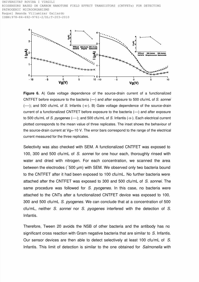
8/9/2019 Tesis Biosensors based on carbon nanotube field effect transistors
http://slidepdf.com/reader/full/tesis-biosensors-based-on-carbon-nanotube-field-effect-transistors 124/239
Figure 6. A) Gate voltage dependence of the source-drain current of a functionalized
CNTFET before exposure to the bacteria (––) and after exposure to 500 cfu/mL of S. sonnei
(----); and 500 cfu/mL of S. Infantis (-x-). B) Gate voltage dependence of the source-drain
current of a functionalized CNTFET before exposure to the bacteria (––) and after exposure
to 500 cfu/mL of S. pyogenes (----); and 500 cfu/mL of S. Infantis (-x-). Each electrical current
plotted corresponds to the mean value of three replicates. The inset shows the behaviour of
the source-drain current at Vg=-10 V. The error bars correspond to the range of the electrical
current measured for the three replicates.
Selectivity was also checked with SEM. A functionalized CNTFET was exposed to100, 300 and 500 cfu/mL of S. sonnei for one hour each, thoroughly rinsed with
water and dried with nitrogen. For each concentration, we scanned the area
between the electrodes (~500 μm) with SEM. We observed only two bacteria bound
to the CNTFET after it had been exposed to 100 cfu/mL. No further bacteria were
attached after the CNTFET was exposed to 300 and 500 cfu/mL of S. sonnei . The
same procedure was followed for S. pyogenes . In this case, no bacteria were
attached to the CNTs after a functionalized CNTFET device was exposed to 100,
300 and 500 cfu/mL S. pyogenes . We can conclude that at a concentration of 500
cfu/mL, neither S. sonnei nor S. pyogenes interfered with the detection of S .
Infantis.
Therefore, Tween 20 avoids the NSB of other bacteria and the antibody has no
significant cross reaction with Gram negative bacteria that are similar to S. Infantis.
Our sensor devices are then able to detect selectively at least 100 cfu/mL of S .
Infantis. This limit of detection is similar to the one obtained for Salmonella with
1
2
3
4
- 10 - 5 0 5 10
0
1
2
3
4
- 10 - 5
Vg(V)
I ( μ A )
0
1
2
3
4
Without
bacteria
I ( μ A )
500 cfu/mL
Shigella
500 cfu/mL
Salmonella
1
2
3
4
- 10 - 5 0 5 10
0
1
2
3
4
- 10 - 5
Vg(V)
I ( μ A )
0
1
2
3
4
Without
bacteria
I ( μ A )
500 cfu/mL
Shigella
500 cfu/mL
Salmonella
0
1
2
3
4
-10 5 0 5 10
0
1
2
3
4
- -
Vg(V)
I ( μ A ) 0
1
2
3
4
I ( μ A )
Withoutbacteria
500 cfu/mLStreptococcus
500 cfu/mLSalmonella
/ /
0
1
2
3
4
-10 5 0 5 10
0
1
2
3
4
- -
Vg(V)
I ( μ A ) 0
1
2
3
4
I ( μ A )
Withoutbacteria
500 cfu/mLStreptococcus
500 cfu/mLSalmonella
/ /
IVERSITAT ROVIRA I VIRGILI
OSENSORS BASED ON CARBON NANOTUBE FIELD EFFECT TRANSISTORS (CNTFETs) FOR DETECTING
THOGENIC MICROORGANISMS
quel Amanda Villamizar Gallardo
BN:978-84-692-9761-2/DL:T-203-2010

8/9/2019 Tesis Biosensors based on carbon nanotube field effect transistors
http://slidepdf.com/reader/full/tesis-biosensors-based-on-carbon-nanotube-field-effect-transistors 125/239
other type of recently developed biosensors based on quartz crystal microbalance
(Su et al., 2005), piezoelectric transducers (Olsen et al., 2006) and surface plasmon
resonance (Oh et al., 2004).
CONCLUSIONS
We have developed a fast, sensitive and label-free biosensor for the selective
detection of S. Infantis. It is based on the transduction power of CNTFETs
combined with the recognition capacity of the antigen-antibody interaction. It can
detect at least 100 cfu/mL of S. Infantis in just one hour. This is clearly better than
the time responses of the current techniques used for detecting this pathogen. This
biosensor, then, could be used as a useful label-free platform to detect otherpathogenic bacteria, viruses or eukaryotic cells like moulds and yeast by using an
adequate molecular receptor. The performance of these devices will be evaluated
by applying them to other strains of Salmonella and also to food products
contaminated with Salmonella .
ACKNOWLEDGMENTS
We thank the Spanish Ministry of Education and Science, MEC, for supporting thework through the project grants NAN2004-09306-C05-05 and CTQ2007-67570.
RAV also acknowledges the University of Pamplona and the Universitat Rovira i
Virgili for providing economic support. A. Maroto also would like to thank the
Spanish Ministry of Science and Technology for providing her Juan de la Cierva
contract.
REFERENCES
Boyle, E.C., Bishop, J.L., Grassl, G.A., Finlay, B.B., 2007. Journal of Bacteriology.
189, 1489-1495.
Bradley, K., Briman, M., Star, A., Gruner, G., 2004. Nano Letters. 4, 253-256.
Byon, H. R., Choi, H. C., 2006. Journal of the American Chemical Soci ety. 128,
2188-2189.
IVERSITAT ROVIRA I VIRGILI
OSENSORS BASED ON CARBON NANOTUBE FIELD EFFECT TRANSISTORS (CNTFETs) FOR DETECTING
THOGENIC MICROORGANISMS
quel Amanda Villamizar Gallardo
BN:978-84-692-9761-2/DL:T-203-2010
8/9/2019 Tesis Biosensors based on carbon nanotube field effect transistors
http://slidepdf.com/reader/full/tesis-biosensors-based-on-carbon-nanotube-field-effect-transistors 126/239
Chen, R. J., Bangsaruntip, S., Drouvalakis, K. A., Kam, N. W., Shim, M., Li, Y., Kim,
W., Utz, P. J., Dai, H., 2003. Proceedings of the National Academy of Sciences of
the United States of America. 100, 4984-4989.
Elkin T., Jiang X., Taylor S., Lin Y., Gu L., Yang H., Brown J., Collins S., Sun Y.,
2005. ChemBioChem. 6, 640 –643.
Grüner, G., 2006. Analytical and Bioanalytical Chemistry. 384, 322-335.
Huang, T. S., Tzeng, Y., Liu, Y. K., Chen, Y. C., Walker, K. R., Guntupalli, R., Liu,
C., 2004. Diamond and Related Materials. 13, 1098-1102.
IIic, B., Czaplewski, D., Craighead, G., 2000. Applied Physics Letters. 77, 450-452.
Johnson, E. M., 1967. Journal of Bacteriology. 94, 2018-2021.
Kong, J., Franklin, N. R., Zhou, C., Chapline, M. G., Peng, S., Cho, K., Dai, H.,
2000. Science. 287, 622-625.
Lin, Y., Jiang, X., Elkin, T., Fernando, K.A., Gu, L., Taylor, S., Yang, H., Jones, E.,
Wang, W., Sun, Y.P., 2006. Journal of Nanoscience and Nanotechnology. 6, 868-871.
Maehashi, K., Katsura, T., Kerma, K., Takamura, Y., Matsumoto, K., Tamiya, E.,
2006. Analitycal Chemistry. 79, 782-787.
Oh, B.K., Kim, Y.K., Park, Lee W.H., Choi J.W., 2004. Biosensors and
Bioelectronics. 19, 1497-1504.
Olsen, E.V., Sorokulova, I.B., Petrenko V.A., Chen, I.H., Barbaree, J.M., Vodyanoy,
V.J., 2006. Biosensors and Bioelectronics. 21, 1434-1442.
Plym, F. L., Wierup, M., 2006. Revue Scientifique et Technique. 25, 541-554.
Shahada, F., Chuma, T., Tobata, T., Okamoto, K., Sueyoshi, M., Takase, K., 2006.
International Journal of Antimicrobial Agents. 28, 302–307.
IVERSITAT ROVIRA I VIRGILI
OSENSORS BASED ON CARBON NANOTUBE FIELD EFFECT TRANSISTORS (CNTFETs) FOR DETECTING
THOGENIC MICROORGANISMS
quel Amanda Villamizar Gallardo
BN:978-84-692-9761-2/DL:T-203-2010

8/9/2019 Tesis Biosensors based on carbon nanotube field effect transistors
http://slidepdf.com/reader/full/tesis-biosensors-based-on-carbon-nanotube-field-effect-transistors 127/239
Shim, M., Kam, N. W. S., Chen, R. J., Li, Y., Dai, H., 2002. Nano Letters. 2, 285-
288.
So, H. M., Won, K., Kim, Y. H., Kim, B. K., Ryu, B. H., Na, P. S., Kim, H., Lee, J. O.,2005. Journal of the American Chemical Society. 127, 11906-11907.
Star, A., Gabriel, J. C., Bradley, K., Gruner, G., 2003. Nano Letters. 3, 459-463.
Su, X.L., Li, Y., 2005. Biosensors and Bioelectronics. 21, 840-848.
Tans, S. J., Verschueren, A. R., Dekker, C., 1998. Nature. 393, 49-52.
Taylor, A. D., Ladd J., Yu Q., Chen S., Homola J., Jiang S., 2006. Biosensors andBioelectronics. 22, 752–758.
Wang, L., Rothemund, D., Curd, H., Reeves, P.R., 2003. Journal of Bacteriology.
185, 2936–2943.
IVERSITAT ROVIRA I VIRGILI
OSENSORS BASED ON CARBON NANOTUBE FIELD EFFECT TRANSISTORS (CNTFETs) FOR DETECTING
THOGENIC MICROORGANISMS
quel Amanda Villamizar Gallardo
BN:978-84-692-9761-2/DL:T-203-2010

8/9/2019 Tesis Biosensors based on carbon nanotube field effect transistors
http://slidepdf.com/reader/full/tesis-biosensors-based-on-carbon-nanotube-field-effect-transistors 128/239
4.3. Supplementary experimental section
4.3.1. Microbiological test
Microbiological cultures were performed in order to confirm the purity of Salmonella
strain. Figure 4-1A shows an image of the typical growth of Salmonella in xylose
lysine desoxycholate agar (XLD agar). It has red appearance due to the indicator
phenol red with a pH of approximately 7.4. This media contains a small amount of
sodium desoxycholate that provide the selectivity. Moreover, it contains xylose, a
sugar that it is fermented by practically all enterics except for the shigellae, and this
property enables the differentiation of Shigella species.
Salmonella , is able to ferment the sugar xylose producing acid. It causes a
descense in the pH making that the phenol red indicator changes the color from red
to yellow. Shigella cannot do this and therefore remain red. After xylose is
fermented, Salmonella decarboxylate lysine, increasing the pH once again to
alkaline and therefore, the final colonies appeare red colored. At this pH Salmonella
species can produce hydrogen sulphide from the reduction of thiosulphate. This is
indicated by ferric ammonium citrate producing black or black-centred colonies.
Black centre allowed differentiating from Shigella .Salmonella was also observed by
using SEM. It can be seen in figure 4-1B the rod-shape cells with a range from 0.7
to 1.5 µm in diameter and from 2 to 5 µm in length.
2 µm
A. B.
Figure 4-1. A) Typical grow of Salmonella Infantis in XLD agar. B) SEM image of the
bacteria.
IVERSITAT ROVIRA I VIRGILI
OSENSORS BASED ON CARBON NANOTUBE FIELD EFFECT TRANSISTORS (CNTFETs) FOR DETECTING
THOGENIC MICROORGANISMS
quel Amanda Villamizar Gallardo
BN:978-84-692-9761-2/DL:T-203-2010

8/9/2019 Tesis Biosensors based on carbon nanotube field effect transistors
http://slidepdf.com/reader/full/tesis-biosensors-based-on-carbon-nanotube-field-effect-transistors 129/239
4.3.2. Covalent immobilization of the antibody
We initially applied a covalent process in order to immobilize the molecular receptor
on a polymer deposited on top of the previously grown SWCNTs. Thefunctionalization process was as follow: First, the as-grown SWCNTs were
submerged in a 10 % solution of PEI in water for four hours at room temperature (rt)
and rinsed thoroughly with water. Then, the devices were immersed during 2 hours
at rt in a 2.5 % solution of glutaraldehyde (GA) in water. GA was then used as a
crosslinker to covalently bind the anti-Salmonella antibodies to the PEI. One of the
aldehyde groups of GA reacts with the amine groups of the PEI to form imine
groups. After that, the devices were submerged in a 10 µL/mL solution of IgG in
PBS (15 mM, pH 7.4) to link the antibodies to the CNTs through the free aldehyde
group of the GA. Subsequently, the devices were immersed for 1 hour in glycine 0.1
M at pH=7 to block the remaining free aldehyde groups of the GA that did not react
with the antibodies (Yakovleva, J. et al, 2002.; Su, X. et al, 1999). The CNTFETs
were again thoroughly rinsed with distilled water, dried with nitrogen and ready to
be used to detect Salmonella Infantis. Figure 4-2 shows a schematic
representation of the whole functionalisation process and figure 4-3 shows an
image taken from the SEM of a functionalised device after exposure to S . Infantis.
CNTFET
PEI 4h/rt GA 2h/rt
Anti-Salmonella1h/37ºC
NH2
Glycin e 1h/rt
Salmonella
1h/37ºC
NH2 NH2
NH2
Figure 4-2. Experimental set up for detecting S. Infantis. The CNTs represent the network
used as transducer elements. Anti-Salmonella antibodies are covalently linked to the PEI
IVERSITAT ROVIRA I VIRGILI
OSENSORS BASED ON CARBON NANOTUBE FIELD EFFECT TRANSISTORS (CNTFETs) FOR DETECTING
THOGENIC MICROORGANISMS
quel Amanda Villamizar Gallardo
BN:978-84-692-9761-2/DL:T-203-2010

8/9/2019 Tesis Biosensors based on carbon nanotube field effect transistors
http://slidepdf.com/reader/full/tesis-biosensors-based-on-carbon-nanotube-field-effect-transistors 130/239
through the crossliker glutaraldehyde. The bacteria are detected by means of the antigen-
antibody interaction.
Figure 4-3. SEM image of a functionalized CNTFET after being exposed to 1000 cfu/mL of
S . Infantis. The bright dot seems to be groups of anti-Salmonella antibodies aligned on top of
the SWCNTs.
The CNTFETs devices were electrically characterized after each functionalisation
step in dry conditions by measuring three times the dependence of the source-drain
current, I, on the back gate voltage, Vg, in the range +10 V to -10 V (Bradley, K. et
al., 2004). The bias voltage, Vsd, was fixed at 250 mV. Figure 4-4 shows how each
functionalisation step affects the electrical current of a typical CNTFET. A n -type
behaviour was observed for the as grown networks after being coated with PEI. It
is probably due to the electron donating property of amine groups of the polymer
that can lead to a change from p - to n -type behaviour (Javey, A. et al, 2002).
The electrical current decreases significantly after GA was linked to the PEI as well
as when the antibody and glycine were bond to the system. Finally, when the
device was exposed to the bacteria the electrical current became nearly zero. This
is probably due to a deformation of the sidewalls of the CNTs taking into account
that the bacteria are almost two thousand times larger than the transducer (CNTs).
As a result, it can be concluded that the electrical properties of the devices were
dramatically affected along the functionalization process hindering the final
detection of the bacteria.
IVERSITAT ROVIRA I VIRGILI
OSENSORS BASED ON CARBON NANOTUBE FIELD EFFECT TRANSISTORS (CNTFETs) FOR DETECTING
THOGENIC MICROORGANISMS
quel Amanda Villamizar Gallardo
BN:978-84-692-9761-2/DL:T-203-2010

8/9/2019 Tesis Biosensors based on carbon nanotube field effect transistors
http://slidepdf.com/reader/full/tesis-biosensors-based-on-carbon-nanotube-field-effect-transistors 131/239
Figure 4-4. Gate voltage dependence of the source-drain current of a typical CNTFET after
each functionalization step. As-grown CNTs coated with PEI (––); glutaraldehyde (– –), anti-
Salmonella antibodies (……), glycine (----); and after exposure to 100 cfu/mL of S. Infantis (– .
–) .
4.3.2. Non-covalent immobilization of the antibody
It is well known that the electrical properties of the CNTs can be affected due tocovalent functionalization process. Therefore, we decided to apply a non-covalent
adsorption of the molecular receptors on the transducer.
Anti-Salmonella antibodies were absorbed onto the CNTs after 1 hour of incubation
at 37 ºC. As it was previously commented in chapter 2, the non-specific adsorption
of proteins involving IgG onto the nanotube surface is attributed to hydrophobic
interactions (Allen, B. L. et al, 2007), the affinity of carbon nanotubes by amino
groups (Bradley, K. et al, 2004) and intermolecular interactions involving aromaticamino acids (i.e., histidine and tryptophan) in the polypeptide chains of the proteins
(Wang, S. et al, 2003).
Figure 4-5 shows AFM images of the as-grown CNTs before and after being coated
with the antibodies. It can be clearly appreciated that the height of the as-grown
CNTs is in a range of about 1.6 nm to 1.8 nm. After the antibodies are adsorbed
the height of the nanotubes increase to about 8 nm to 10 nm. IgG structure is based
IVERSITAT ROVIRA I VIRGILI
OSENSORS BASED ON CARBON NANOTUBE FIELD EFFECT TRANSISTORS (CNTFETs) FOR DETECTING
THOGENIC MICROORGANISMS
quel Amanda Villamizar Gallardo
BN:978-84-692-9761-2/DL:T-203-2010
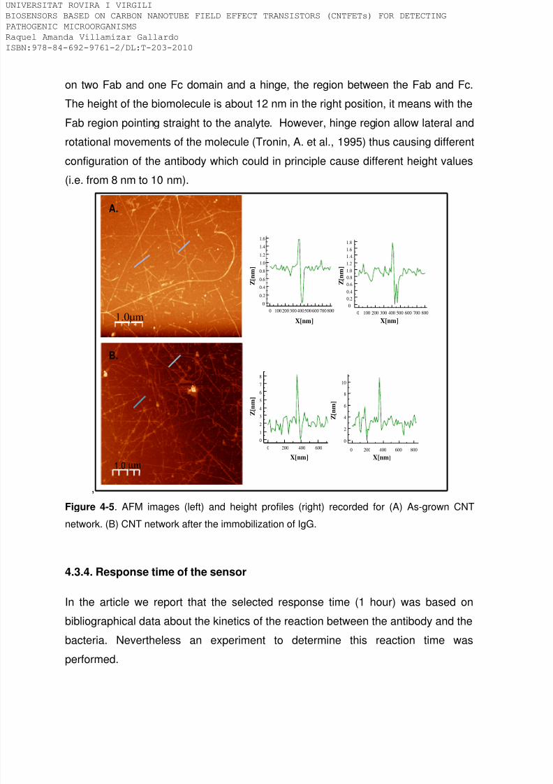
8/9/2019 Tesis Biosensors based on carbon nanotube field effect transistors
http://slidepdf.com/reader/full/tesis-biosensors-based-on-carbon-nanotube-field-effect-transistors 132/239
on two Fab and one Fc domain and a hinge, the region between the Fab and Fc.
The height of the biomolecule is about 12 nm in the right position, it means with the
Fab region pointing straight to the analyte. However, hinge region allow lateral and
rotational movements of the molecule (Tronin, A. et al., 1995) thus causing different
configuration of the antibody which could in principle cause different height values
(i.e. from 8 nm to 10 nm).
,
1.0µm
1.0 µm
8007006005004003002001000
1.6
1.4
1.2
1.0
0.8
0.6
0.4
0.2
0
X[nm]
Z [ n m ]
8007006005004003002001000
1.8
1.6
1.4.
1.2
1.0
0.8
0.6
0.4
0.2
0
X[nm]
Z [ n m ]
8006004002000
8
7
6
5
4
3
2
1
0
X[nm]
Z
[ n m ]
8006004002000
10
8
6
4
2
0
X[nm]
Z [ n m ]
A.
B.
Figure 4-5. AFM images (left) and height profiles (right) recorded for (A) As-grown CNT
network. (B) CNT network after the immobilization of IgG.
4.3.4. Response time of the sensor
In the article we report that the selected response time (1 hour) was based on
bibliographical data about the kinetics of the reaction between the antibody and the
bacteria. Nevertheless an experiment to determine this reaction time was
performed.
IVERSITAT ROVIRA I VIRGILI
OSENSORS BASED ON CARBON NANOTUBE FIELD EFFECT TRANSISTORS (CNTFETs) FOR DETECTING
THOGENIC MICROORGANISMS
quel Amanda Villamizar Gallardo
BN:978-84-692-9761-2/DL:T-203-2010

8/9/2019 Tesis Biosensors based on carbon nanotube field effect transistors
http://slidepdf.com/reader/full/tesis-biosensors-based-on-carbon-nanotube-field-effect-transistors 133/239
For this purpose, CNTFET devices were immersed in a solution containing the
lowest assayed concentration of S. Infantis (100 cfu/mL) at 37 ºC. Every 15 minutes
we rinsed the devices thoroughly with water, dried it and measured the electrical
current (see Figure 4-6)After each measurement the devices were again
submerged in the solution containing 100 cfu/mL S . Infantis for another 15 minutes
and electrically characterized. The highest decrease for the current intensity was
obtained between 30 and 60 minutes. The current decreased further from 60 to 75
minutes but not significantly. Therefore, the suitable reaction time and, therefore,
the response time of our devices was 60 minutes.
I ( μ A )
0
1
2
3
0 15 30 45 60 75Time (minutes)
I ( μ A )
0
1
2
3
0 15 30 45 60 75Time (minutes)
Figure 4-6. Source-drain current (at Vg=-10 V) obtained for a functionalized device after
exposure to 100 cfu/mL of S . Infantis for 75 minutes. The source-drain current was measured
every 15 minutes. Each electrical current plotted and the error bars correspond respectively
to the mean value and range of three replicates measured in repeatability conditions.
IVERSITAT ROVIRA I VIRGILI
OSENSORS BASED ON CARBON NANOTUBE FIELD EFFECT TRANSISTORS (CNTFETs) FOR DETECTING
THOGENIC MICROORGANISMS
quel Amanda Villamizar Gallardo
BN:978-84-692-9761-2/DL:T-203-2010

8/9/2019 Tesis Biosensors based on carbon nanotube field effect transistors
http://slidepdf.com/reader/full/tesis-biosensors-based-on-carbon-nanotube-field-effect-transistors 134/239
4.4. Complementary conclusions
Microscopy analysis showed that antibodies were irreversibly and uniformly
adsorbed on top of the CNTs in only one hour. Although the orientation of theantibodies was not specifically addressed, most of them must be correctly oriented
in the experimental conditions used, making it possible to detect, at least, 100
cfu/mL of S. Infantis.
The use of PEI to immobilise antibodies dramatically affected the electrical
properties of the transducer. PEI changed the p-type behaviour to n-type behaviour
and, more importantly, the four functionalization steps significantly decreased the
conductance of the CNT networks. As a result, the low conductance of thefunctionalized CNTFET hindered their use as valuable devices to detect the
presence of Salmonella.
By contrast, the direct adsorption of the antibodies on the CNT is a more feasible
process since it preserved the electrical properties of the CNTs making it possible
to determine from 100 to 500 cfu/mL of Salmonella.
Our devices are direct biosensors like those reported by Shabani et al (Shabani, Aet al, 2008) label-free and easy to manufacture compared to the electrochemical
device developed by Rishpon et al (Rishpon, J et al, 1992). However, the limit of
detection and the response time is lower compared to those obtained by Barreiros
et al (Barreiros, M et al, 2009) and Zelada-Guillén et al (Zelada-Guillén, G et al,
2009).
Therefore, further research is required in order to produce efficient biodevices with
optimal performance parameters. It is probable that an enhanced orientation of the
antibodies would increase the sensitivity of the devices. On ther hand, due to the
proteic nature of the molecular receptor it is difficult to regenerate the sensors. To
solve this problem, other type of receptors like aptamers or peptides could be
assayed. In addition the improved biosensors must be tested with real samples.
IVERSITAT ROVIRA I VIRGILI
OSENSORS BASED ON CARBON NANOTUBE FIELD EFFECT TRANSISTORS (CNTFETs) FOR DETECTING
THOGENIC MICROORGANISMS
quel Amanda Villamizar Gallardo
BN:978-84-692-9761-2/DL:T-203-2010

8/9/2019 Tesis Biosensors based on carbon nanotube field effect transistors
http://slidepdf.com/reader/full/tesis-biosensors-based-on-carbon-nanotube-field-effect-transistors 135/239
4.5. References
Abass, E.M., Mansour, D., Mutasim, M., Hussein, M., Harith, A. Journal of Medical
Microbiology. 2006. 55, 1193-1196.
Allen, B. L., Kichambare, P. D., Star, A. Advanced Materials. 2007.19, 1439–1451.
Barreiros, M., Sporera, C., Sanvicensc, N., Pascuald, N., Errachida, A., Martineza,
E., Marcod, M.-P., Teixeirae, V., Samiter, J. Procedia Chemistry. 2009. 1, 1291–
1294.
Bradley, K., Briman, M., Star, A., Gruner, G. Nano Letters. 2004. 4, 253-256.
Delibato, E., Volpe, G., Romanazzo, D., De Medici, D., Toti, L., Moscone, D.,
Palleschi, G. Journal of Agricultural and Food Chemistry. 2009. 57, 7200–7204.
de Boer, E. and Beumer, R.R. International Journal of Food Microbiology. 1999.
50,119 –130.
Eyigör, A., Goncagül, G., Carli, K.T. Journal of Environmental Biology. 2007. 1, 45-
49.
Ivnitski, D., Hamid, I.A., Atanasov, P., Wilkins, E. Biosensors and Bioelectronics.
1999. 14, 599–624.
Janyapoon, K., Korbsrisate, S., Thamapa, H., Thongmin, S., Kanjanahareutai, K.,Wongpredee, N., Sarasombath, S. Clinical and Diagnostic Laboratory Immunology.
2000. 7, 977–979.
Javey, A., Kim, H., Brink, M., Wang, Q., Ural, A., Guo, J., McIntyre, P., McEuen, P.,
Lundstrom, M., Dai, H. Journal of Nature Materials. 2002. 1, 241-246.
IVERSITAT ROVIRA I VIRGILI
OSENSORS BASED ON CARBON NANOTUBE FIELD EFFECT TRANSISTORS (CNTFETs) FOR DETECTING
THOGENIC MICROORGANISMS
quel Amanda Villamizar Gallardo
BN:978-84-692-9761-2/DL:T-203-2010

8/9/2019 Tesis Biosensors based on carbon nanotube field effect transistors
http://slidepdf.com/reader/full/tesis-biosensors-based-on-carbon-nanotube-field-effect-transistors 136/239
Jeníková, G., Pazlarová, J., Demnerová, K. International Microbiology. 2000. 3,
225–229.
Koyuncu, S. and Haggblom, P. BMC Veterinary Research. 2009. 5,6.
Lazcka, O., Del Campob, F.J., Muñoz, F.X. Biosensors and Bioelectronics. 2007.
22, 1205–1217.
Leon-Velarde, C.G., Zosherafatein, L., Odumeru, J.A. Journal of Microbiological
Methods. 2009. 79, 13-17.
Liébana, S., Lermo, A., Campoy, S., Barbé, J., Alegret, S., Pividori, M.I. Analytical
Chemistry. 2009. 81, 5812–5820.
J. Rishpon, Y. Gezundhajt, L. Soussan, L.R. Margalit, E. Hadas, Biosensor design
and application, Oxford University Press, Oxford, 1992.
Shabani, A., Zourob, M., Allain, B., Marquette, C.A., Lawrence, M.F., Mandeville, R.Analytical Chemistry. 2008. 80, 9475–9482.
Scheu, P.M., Berghof, K., Stahl, U. Food Microbiology. 1998. 15, 13–31.
Su, X., Chew, F.T., Li, S.F.Y. Analytical Biochemistry. 1999. 273, 66 -72.
Taux, R.V. Emerging Infectious Diseases. 1997. 4, 425-434.
Taitt, C.R., Shubin, Y.S., Angel, E., Ligler, F.S. Applied and Environmental
Microbiology. 2004. 70, 152–158.
Tronin, A., Dubrovsky, T., Nicolini, C. Langmuir. 1995. 11, 385–389.
IVERSITAT ROVIRA I VIRGILI
OSENSORS BASED ON CARBON NANOTUBE FIELD EFFECT TRANSISTORS (CNTFETs) FOR DETECTING
THOGENIC MICROORGANISMS
quel Amanda Villamizar Gallardo
BN:978-84-692-9761-2/DL:T-203-2010

8/9/2019 Tesis Biosensors based on carbon nanotube field effect transistors
http://slidepdf.com/reader/full/tesis-biosensors-based-on-carbon-nanotube-field-effect-transistors 137/239
Wang, S., Humphreys, E. S., Chung, S.Y., Delduco, D. F., Lustig, S. R., Wang, H.,
Parker, K. N., Rizzo, N. W., Subramoney, S., Chiang, Y.M., Jagota, A. Nature
Materials. 2003. 2, 196.
Wilson, G. Journal of Clinical Microbiology. 2004. 42, 4821–4823.
Yakovleva, J., Davidsson, R., Bengtsson, M., Laurell, T., Emneus, J. Analytical
Chemistry. 2002. 74, 2994-3004.
Zelada-Guilllén, G.A., Riu, J., Düzgün, A., Rius, F.X. Angewandte Chemie
International Edition. 2009. 48, 7334 –7337.
IVERSITAT ROVIRA I VIRGILI
OSENSORS BASED ON CARBON NANOTUBE FIELD EFFECT TRANSISTORS (CNTFETs) FOR DETECTING
THOGENIC MICROORGANISMS
quel Amanda Villamizar Gallardo
BN:978-84-692-9761-2/DL:T-203-2010

8/9/2019 Tesis Biosensors based on carbon nanotube field effect transistors
http://slidepdf.com/reader/full/tesis-biosensors-based-on-carbon-nanotube-field-effect-transistors 138/239
IVERSITAT ROVIRA I VIRGILI
OSENSORS BASED ON CARBON NANOTUBE FIELD EFFECT TRANSISTORS (CNTFETs) FOR DETECTING
THOGENIC MICROORGANISMS
quel Amanda Villamizar Gallardo
BN:978-84-692-9761-2/DL:T-203-2010

8/9/2019 Tesis Biosensors based on carbon nanotube field effect transistors
http://slidepdf.com/reader/full/tesis-biosensors-based-on-carbon-nanotube-field-effect-transistors 139/239
Chapter 5
CNTFET for pathogenic yeast determination
IVERSITAT ROVIRA I VIRGILI
OSENSORS BASED ON CARBON NANOTUBE FIELD EFFECT TRANSISTORS (CNTFETs) FOR DETECTING
THOGENIC MICROORGANISMS
quel Amanda Villamizar Gallardo
BN:978-84-692-9761-2/DL:T-203-2010

8/9/2019 Tesis Biosensors based on carbon nanotube field effect transistors
http://slidepdf.com/reader/full/tesis-biosensors-based-on-carbon-nanotube-field-effect-transistors 140/239
IVERSITAT ROVIRA I VIRGILI
OSENSORS BASED ON CARBON NANOTUBE FIELD EFFECT TRANSISTORS (CNTFETs) FOR DETECTING
THOGENIC MICROORGANISMS
quel Amanda Villamizar Gallardo
BN:978-84-692-9761-2/DL:T-203-2010

8/9/2019 Tesis Biosensors based on carbon nanotube field effect transistors
http://slidepdf.com/reader/full/tesis-biosensors-based-on-carbon-nanotube-field-effect-transistors 141/239
5.1. Introduction
Mycosis is one of the most common pathology that affects human beings. Candida
albicans is an opportunistic fungi, ubiquitously distributed on the body microflora,able to produce both superficial and systemic infections (Trama, J.P. et al., 2005).
Candidiasis is the so-called disease produced by Candida. In Spain the invasive
candidiasis is the most important systemic mycosis and the main factor related with
its production is the drug´s abuse (Pontón, J and del Palacio, A., 2007). Vaginal
candidiasis causes 20–25 % of infectious vaginitis cases, ranked second among the
bacterial vaginosis (Trama, J.P. et al., 2005).
Identification of C . albicans in clinical microbiology is usually made by usingstandard culture media (Cooke,V.M. et al, 2002.; Munin, E., 2007). Depending on
the species, medium and detection system, the pathogen is detected within 24 to 72
hours. However, current culture methodologies have shown the occurrence of up to
35 % false results (Horvath, L.L. et al, 2005). As a consequence improved media
based on chromogenic compounds have been developed (Chunchanur, S.K. et al,
2009). Although the culture is the standard method; it requires well trained
technician and several days to obtain a reliable result.
The use of commercial kits like peptide nucleic acid fluorescent in situ hybridization
(PNA-FISH) allows identifying Candida species isolated from blood cultures in 3
hours. However, an additional incubation for 24 hours is required to final
determination of the yeast. Therefore, this confirmation method is time and reagent
consuming.
PCR-based methods are also used to detect yeast (Wellinghausen, N. et al, 2009.;
Maaroufi, F. et al, 2003.; McMullan, R. et al, 2008). Maaroufi et al ( Maaroufi, F. et
al, 2003) described a rapid and reproducible PCR assay for quantification of the
Candida albicans ribosomal DNA (rDNA) in clinical blood samples. The assay for C.
albicans exhibited a low limit of detection (5 cfu/mL of blood) and an excellent
reproducibility (96 to 99 %).
IVERSITAT ROVIRA I VIRGILI
OSENSORS BASED ON CARBON NANOTUBE FIELD EFFECT TRANSISTORS (CNTFETs) FOR DETECTING
THOGENIC MICROORGANISMS
quel Amanda Villamizar Gallardo
BN:978-84-692-9761-2/DL:T-203-2010

8/9/2019 Tesis Biosensors based on carbon nanotube field effect transistors
http://slidepdf.com/reader/full/tesis-biosensors-based-on-carbon-nanotube-field-effect-transistors 142/239
McMullan et al. proposed a method for early detection and identification of Candida .
After manual extraction of DNA from serum, PCR was performed. This method
shortens the procedure time up to 6 hours with similar accuracy outcome compared
to conventional methods. However, processing requires multiple steps, and it may
take longer to apply this method in clinical practice.
Immuno-based methods have also been reported for the detection of Candida
(Moudni, E.B. et al, 1998.; Marcilla, A. et al, 1999.; Matsui, H et al, 2009). Matsui et
al , developed an immunochromatographic method that enables the detection of
vaginal Candida spp. The immunochromatographic strip was prepared with anti-
Candida mannan polyclonal antibody. With this technique it is possible to detect asfew as 104 cfu/mL of Candida spp. in about 30 minutes without using expensive
equipment or skill. Overall, the sensitivity, specificity, positive predictive value, and
negative predictive value of this method appeared to be 80.3, 99.3, 98.0, and 92.0
%, respectively.
Sensors have been recently introduced as alternative method to detect this
pathogen (Chen, H. et al, 2005.; Mulero, R. et al, 2009). Mulero et al , describe asolid-state micro-scale pore in which a steady electrolyte current flow through the
pore. Changes in the current were taken when individual yeasts travel through the
pore. With this method was possible to detect about 200 cells/mL of Candida
albicans in few seconds being possible to distinguish individual cells by their
structural properties. In order to achieve a clinically applicable technique pre-
processing of the sample would be necessary to reduce or eliminate potentially
pore clogging particles from sample such as red blood cells, white blood cells, and
platelets.
Nevertheless many efforts are still required to develop a method with improved
performance parameters like rapidness, sensitivity and high selectivity. This
chapter reports the first carbon nanotube field effect transistor (CNTFET) applied to
the detection of a yeast. To improve the performance parameters of our devices, a
monoclonal antibody highly specific for C. albicans was used as molecular receptor.
CNTs were the transducer element able to detect the presence of at least 50 cfu/mL
IVERSITAT ROVIRA I VIRGILI
OSENSORS BASED ON CARBON NANOTUBE FIELD EFFECT TRANSISTORS (CNTFETs) FOR DETECTING
THOGENIC MICROORGANISMS
quel Amanda Villamizar Gallardo
BN:978-84-692-9761-2/DL:T-203-2010

8/9/2019 Tesis Biosensors based on carbon nanotube field effect transistors
http://slidepdf.com/reader/full/tesis-biosensors-based-on-carbon-nanotube-field-effect-transistors 143/239
of the yeast in only one hour. Possible interferences from the sample like proteins
were avoided by using Tween 20 at 0.5 % as a protecting polymer that covers the
CNTs. This biosensor is stable, easy to manipulate and provides an inexpensive
option to in vitro screening of yeast.
Finally, this chapter provides additional experimental information to those included
in the published paper, general conclusions and references.
IVERSITAT ROVIRA I VIRGILI
OSENSORS BASED ON CARBON NANOTUBE FIELD EFFECT TRANSISTORS (CNTFETs) FOR DETECTING
THOGENIC MICROORGANISMS
quel Amanda Villamizar Gallardo
BN:978-84-692-9761-2/DL:T-203-2010

8/9/2019 Tesis Biosensors based on carbon nanotube field effect transistors
http://slidepdf.com/reader/full/tesis-biosensors-based-on-carbon-nanotube-field-effect-transistors 144/239
5.2. Article:
“Improved detection of Candida albicans with carbon
nanotubes field effect transistors”. Sensors and
Actuators, B: Chemical. 2009.136, 451-457.
Raquel A. Villamizarτ, Alicia Maroto* F. Xavier Rius
Department of Analytical and Organic Chemistry. Universitat Rovira i Virgili
Marcel.lí. Domingo, s/n. 43007. Tarragona, Spain.
τ On leave from the Department of Microbiology. University of Pamplona. Km 1, vía
Bucaramanga, N. de S. Colombia.
IVERSITAT ROVIRA I VIRGILI
OSENSORS BASED ON CARBON NANOTUBE FIELD EFFECT TRANSISTORS (CNTFETs) FOR DETECTING
THOGENIC MICROORGANISMS
quel Amanda Villamizar Gallardo
BN:978-84-692-9761-2/DL:T-203-2010

8/9/2019 Tesis Biosensors based on carbon nanotube field effect transistors
http://slidepdf.com/reader/full/tesis-biosensors-based-on-carbon-nanotube-field-effect-transistors 145/239
Abstract
A biosensor for detecting Candida albicans has been developed to improve the
performance characteristics of the currently available methods. It is based on afield-effect transistor (FET) in which a network of single-walled carbon nantotubes
(SWCNTs) acts as the conductor channel. Monoclonal anti-Candida antibodies
were adsorbed onto the SWCNT to provide specific binding sites for fungal
antigens. Tween 20 at 0.5 % was used as a blocking agent to prevent the non-
specific binding of other yeasts or proteins. Our FET devices were exposed to
increasing concentrations of C. albicans and were able to detect at least 50 cfu/mL
in only 1 hour. The sensor response remained stable for more than ten days. To
evaluate the selectivity of our FET devices, Cryptococcus albidus and
Saccharomyces cerevisiae were tested as potential competing yeasts for Candida
albicans . The results showed that biosensors based on a carbon nanotube field
effect transistor are useful tools to detect pathogenic yeasts like Candida albicans
at low concentrations displaying high selectivity.
Keywords: Biosensor, Field-Effect Transistor, Carbon nanotubes, Yeast, Candida
albicans.
Introduction
Candida albicans (C. albicans ) is a yeast-like fungal organism that can make the
morphologic transition from yeast to hyphal, thereby increasing its pathogenicity [1].
It can be found on human skin, in the digestive tract, mouth, throat, oesophagus,
vagina and other mucus membranes causing a wide spectrum of diseases from
simple cutaneous mycosis to complicated cases of candidiasis. Candidiasis is
mainly an endogenous infection produced by the overgrowth of the human body’s
own fungi. In some cases, it can also be acquired from exogenous sources like
catheters or prosthetic devices, person-to-person transmission or vertical
transmission [2]. This yeast can affect especially patients with immunodeficient,
neoplasya, diabetes, lymphomas among others.
IVERSITAT ROVIRA I VIRGILI
OSENSORS BASED ON CARBON NANOTUBE FIELD EFFECT TRANSISTORS (CNTFETs) FOR DETECTING
THOGENIC MICROORGANISMS
quel Amanda Villamizar Gallardo
BN:978-84-692-9761-2/DL:T-203-2010

8/9/2019 Tesis Biosensors based on carbon nanotube field effect transistors
http://slidepdf.com/reader/full/tesis-biosensors-based-on-carbon-nanotube-field-effect-transistors 146/239
Conventional diagnoses of Candida involve methods such as germ tube or
colorimetric tests. Nevertheless, up to 5 % of the C. albicans isolates have been
reported as germ tube negative [3]. Blood cultures for Candida species generally
display low sensitivity and require additional tests to determine marker antigens.
Some traditional culture media allows bacterial growth, thus reducing the recovery
rate of target yeast from the samples [4]. Selective chromogenic agar medium has
also been used to detect yeast with medical importance but they are time-
consuming [5]. Serological tests for Candida involve different techniques such as
counterimmunoelectrophoresis and radioimmunoassay. However they display
variable sensitivity and lack of specificity, with up to 40 % false positive and
negative rates [6]. Enzyme-linked immunosorbent assay (ELISA) is highly sensitive
but requires the use of labels. Molecular biology techniques such as PCR and
hybridization assays are reliable and highly sensitive but they are also reagent and
time-consuming [7].
Mass sensors [8-11] and electrochemical sensors [12] have also been developed
to detect both pathogenic and non-pathogenic yeasts and moulds. In spite of the
emerging methods for detecting fungi, the detection range is still quite high (ca. 10
2
cfu/mL). This means that a simple, easy-to-perform and sensitive assay for
detecting yeasts like Candida is still not available.
Single-walled carbon nanotubes (SWCNT) are one-dimensional nanostructures with
all the carbon atoms on the surface, allowing optimal interaction with biomolecules
in their chemical environment [13]. Dekker’s group in 1998 reported the first carbon
nanotube field effect transistor (CNTFET) [14]. CNTFETs with a selective coating
have become a suitable system for biosensing applications because they combine
the principles of molecular recognition through the recognition layer with the
transduction capabilities of the carbon nanotubes. They have been used to detect
DNA [15-17], proteins like thrombin and IgE and IgG by means of antibodies and
aptamers as molecular receptors [18-20], viruses by means of peptide nucleic acids
[21] and bacteria [22, 23].
IVERSITAT ROVIRA I VIRGILI
OSENSORS BASED ON CARBON NANOTUBE FIELD EFFECT TRANSISTORS (CNTFETs) FOR DETECTING
THOGENIC MICROORGANISMS
quel Amanda Villamizar Gallardo
BN:978-84-692-9761-2/DL:T-203-2010

8/9/2019 Tesis Biosensors based on carbon nanotube field effect transistors
http://slidepdf.com/reader/full/tesis-biosensors-based-on-carbon-nanotube-field-effect-transistors 147/239
So far, CNTFETs have not been used to detect eukaryotic cells. It is therefore
worthwhile to broaden the application field to include these organisms. Here, we
report the development of a sensitive CNTFET for the fast detection of C. albicans .
As a recognition element, we adsorbed onto the SWCNTs monoclonal anti-Candida
antibodies, which were able to provide specific binding sites for fungal membrane
antigens. Tween 20 was used as a blocking agent to prevent the non-specific
binding (NSB) of other yeasts [18, 24, 25]. In the presence of competing yeasts like
Cryptococcus albidus the sensor showed high selectivity. Our devices were able to
detect 50 cfu/mL with a time response of one hour, eliminating the need for a
special labelling process. The operational performance parameters of our devices
represent a clear advantage over traditional as well as some current methods usedin the detection of pathogenic yeasts.
Experimental
Antibodies and biochemicals
Anti-Candida (i.e. a monoclonal mouse anti-Candida albicans (2.8 mg/mL) was
purchased from Oxford Biotechnology Ltd. (Oxford, U.K.). It was dissolved in
phosphate buffer saline (PBS) to a final concentration of 10 ppm (pH=7.2) and
stored at -20 ºC until use. Tween 20 and PBS were obtained from Sigma-Aldrich.
Sabouraud Dextrosa Agar (SDA) was provided by Oxoid and prepared according to
their specifications. Silver ink Electrodag 1415M was provided by Acheson
industries (Europe) Ltd (Scheemda, The Netherlands).
Yeast preparation
Lyophilized strains of C. albicans CECT 1001, Saccharomyces cerevisiae (S.
cerevisiae ) CECT 1443 and Cryptococcus albidus (C. albidus ) CECT 11978 were
obtained from the Spanish Type Culture Collection (CECT, Valencia, Spain). They
were rehydrated with sterile water, subcultured at least three times on SDA and
incubated at 35 ºC for 48 hours to test their viability. Isolated colonies were
resuspended on sterile 0.85 % saline solution. The yeast cells contained in the
saline solution were enumerated by plating 108-fold serial dilutions on SDA plates
and incubating at 35 ºC for 24-48 hours [26]. Further dilutions in sterile 0.85 %
IVERSITAT ROVIRA I VIRGILI
OSENSORS BASED ON CARBON NANOTUBE FIELD EFFECT TRANSISTORS (CNTFETs) FOR DETECTING
THOGENIC MICROORGANISMS
quel Amanda Villamizar Gallardo
BN:978-84-692-9761-2/DL:T-203-2010
8/9/2019 Tesis Biosensors based on carbon nanotube field effect transistors
http://slidepdf.com/reader/full/tesis-biosensors-based-on-carbon-nanotube-field-effect-transistors 148/239
saline solution were performed to produce the final samples at 50, 200 and 400
cfu/mL of C . albicans . The same procedure was followed to prepare 50, 200 and
400 cfu/mL of S. cerevisiae and C. albidus.
Apparatus
Atomic force microscopy (AFM), Pico Plus (Molecular Imaging) was used in tapping
mode to characterize the SWCNTs before and after they were functionalised with
the antibodies. Scanning electron microscopy (SEM), JSM 6400 (Jeol) was used to
take images of the as-grown networks of SWCNTs and the functionalisation
process. SEM images were obtained by applying a voltage of 1kV at a working
distance of 8 mm. Electrical measurements were taken using a 4157A Agilentsemiconductor parameter analyzer and a Wentworth Laboratories MP1008 probe
station.
Development of the CNTFETs
The SWCNT networks were synthesized on top of a 500 nm layer of silicon dioxide
thermally grown on highly doped n-type silicon chips (total area 0.5 cm x 0.5 cm)
using chemical vapour deposition (CVD). A solution of 100ppm iron nitrate inisopropanol was used as the catalyst. The CVD was performed at 900 ºC for 20
minutes with 600 sccm of methane and 200 sccm of hydrogen. Figure 1 A-1 shows
that a dense network of SWCNTs was obtained after the CVD. The average height
of the SWCNTs (obtained from AFM) was about 1.5 nm. The source and drain
electrodes of the CNTFETs were screen-printed with silver ink. The gap between
both electrodes was 0.5 mm and the size of the electrodes was 200 μm x 200 μm.
The gate electrode was an aluminium layer on the back side of Si. The CNTFETswere electrically characterised by recording the current vs. the gate voltage. To
obtain the instrumental variability, we carried out all the electrical characterization
measurements three times and we plotted the mean value and the range value of
the measurements.
IVERSITAT ROVIRA I VIRGILI
OSENSORS BASED ON CARBON NANOTUBE FIELD EFFECT TRANSISTORS (CNTFETs) FOR DETECTING
THOGENIC MICROORGANISMS
quel Amanda Villamizar Gallardo
BN:978-84-692-9761-2/DL:T-203-2010

8/9/2019 Tesis Biosensors based on carbon nanotube field effect transistors
http://slidepdf.com/reader/full/tesis-biosensors-based-on-carbon-nanotube-field-effect-transistors 149/239
Functionalisation of the CNTFETs
The functionalisation protocol was performed similarly to that previously described
for the detection of Salmonella [22]. The CNTFETs were incubated for one hour at37 ºC in a 5 ppm solution of anti-Candida antibodies in 15 mM PBS solution. The
CNTFETs were then rinsed with PBS solution and distilled water, dried with
nitrogen [27] and immersed for 2 hours in a solution of 0.5 % Tween 20 dissolved in
15 mM PBS [18]. The CNTFETs were again thoroughly rinsed with distilled water,
dried with nitrogen and ready to be used to detect C . albicans . Figure 1 shows the
experimental set up for detecting C. albicans using a functionalised CNTFET.
Figure 1A-1 shows a CNTFET functionalised with the monoclonal anti-Candida antibodies. The average height of the antibodies (obtained from AFM) was about 9
nm. Figure 1B-1 shows that, as is well known, the yeasts do not retain their shape
after being dried and having a high vacuum applied in the SEM.
Figure 1. A) Schematic structure of the CNTFET device. A1) SEM image of a typical
network of SWCNTs obtained by CVD and coated with anti-Candida antibodies. B) Antigen-
antibody interaction of C. albicans with a SWCNT functionalised with anti-Candida antibodies
and protected with Tween 20. B2) A SEM image of a functionalised CNTFET after exposure
to C. albicans.
IVERSITAT ROVIRA I VIRGILI
OSENSORS BASED ON CARBON NANOTUBE FIELD EFFECT TRANSISTORS (CNTFETs) FOR DETECTING
THOGENIC MICROORGANISMS
quel Amanda Villamizar Gallardo
BN:978-84-692-9761-2/DL:T-203-2010

8/9/2019 Tesis Biosensors based on carbon nanotube field effect transistors
http://slidepdf.com/reader/full/tesis-biosensors-based-on-carbon-nanotube-field-effect-transistors 150/239
Detection of C. albicans
The functionalised CNTFETs were exposed to increasing concentrations of C.
albicans (i.e. 50, 200 and 400 cfu/mL). For each concentration, the CNTFETs wereimmersed for one hour at 37 ºC, rinsed thoroughly with distilled water, dried with
nitrogen and electrically characterised. For each concentration we scanned the area
between the electrodes (∼500 µm2) with SEM to count the number of yeasts
attached to the SWCNTs.
Determination of the response time
A functionalised CNTFET was immersed in a solution containing 50 cfu/mL of C.albicans at 37 ºC for one hour. Every 15 minutes the CNTFET was thoroughly
rinsed with water, dried with nitrogen, electrically characterised, and again
submerged in the solution containing 50 cfu/mL of C. albicans for another 15
minutes.
Selectivity of the CNTFETs
Selectivity was checked in the presence of two yeasts: C. albidus and S.cerevisiae . A functionalised CNTFET was first immersed in a yeast solution
containing 400 cfu/mL of C. albidus for one hour at 37 ºC, thoroughly rinsed with
distilled water, dried with nitrogen and electrically characterised. It was
subsequently exposed to 400 cfu/mL of C. albicans under the same conditions
mentioned above. The same procedure was followed for 400 cfu/mL of S.
cerevisiae. All these experiments were confirmed microscopically with SEM.
Interference assay
Our sensor devices were also proven against possible interferences that can be
found out in real samples of diseased patients. We took a functionalised CNTFET
and it was exposed to different concentrations of BSA obtained from Sigma-Aldrich,
(i.e. 1mg/L, 10 mg/L, 100 mg/L, 1000 mg/L and 10000 mg/L) for 1 hour each. The
devices were washed with a solution of PBS and distilled water after exposure to
each concentration, then were dried with nitrogen and electrically characterized.
IVERSITAT ROVIRA I VIRGILI
OSENSORS BASED ON CARBON NANOTUBE FIELD EFFECT TRANSISTORS (CNTFETs) FOR DETECTING
THOGENIC MICROORGANISMS
quel Amanda Villamizar Gallardo
BN:978-84-692-9761-2/DL:T-203-2010

8/9/2019 Tesis Biosensors based on carbon nanotube field effect transistors
http://slidepdf.com/reader/full/tesis-biosensors-based-on-carbon-nanotube-field-effect-transistors 151/239
Results and discussion
The electrical behaviour of the CNTFETs was monitored after each functionalisation
step. We measured the dependence of the source-drain current, I, on the backgate voltage, Vg, in the +5 V to -5 V range at room temperature in dry conditions
three times. The bias voltage, Vsd, was fixed at 250 mV. Figure 2 shows how each
functionalisation step affects the electrical current of a typical CNTFET. A p-type
behaviour was observed for the pristine networks due to the electron withdrawal of
adsorbed oxygen molecules from the air which coincides with that found by Star et
al [28].
0
1
2
3
4
5
6
-5 -3 -1 1 3 5
0
1
2
3
4
5
6
Pristine CNTs Tween 20Anti-Candida
antibody
I ( µ A )
0
1
2
3
4
5
6
Pristine CNTs Tween 20Anti-Candida
antibody
I ( µ A )
0
1
2
3
4
5
6
-5 -3 -1 1 3 5
0
1
2
3
4
5
6
-5 -3 -1 1 3 5
0
1
2
3
4
5
6
Pristine CNTs Tween 20Anti-Candida
antibody
I ( µ A )
0
1
2
3
4
5
6
Pristine CNTs Tween 20Anti-Candida
antibody
I ( µ A )
Figure 2. Gate voltage dependence of the source-drain current of a typical CNTFET after
each functionalisation step. Pristine CNTs (----); anti- Candida antibodies adsorbed on the
CNTs ( X ) and CNTs covered with anti-Candida antibodies and Tween 20 (––).The inset
shows the behaviour of the source-drain current after each functionalisation step at Vg= -5 V.
Anti-Candida antibodies were adsorbed on the CNTs. Although the mechanism of
non-specific adsorption of proteins onto SWCNTs is still unknown [29], it is thought
that supramolecular interactions between the aromatic amino acids (i.e., histidine
and tryptophan) present in the polypeptide chains of the proteins and the SWCNTs
may help to clarify the affinity of the peptides towards this nanostructure [30]. This
phenomenon also has been attributed to size compatibility between the two entities
IVERSITAT ROVIRA I VIRGILI
OSENSORS BASED ON CARBON NANOTUBE FIELD EFFECT TRANSISTORS (CNTFETs) FOR DETECTING
THOGENIC MICROORGANISMS
quel Amanda Villamizar Gallardo
BN:978-84-692-9761-2/DL:T-203-2010

8/9/2019 Tesis Biosensors based on carbon nanotube field effect transistors
http://slidepdf.com/reader/full/tesis-biosensors-based-on-carbon-nanotube-field-effect-transistors 152/239
[31]. We observed through the AFM that 1 h was enough to immobilize anti-
Candida antibodies onto the sidewalls of SWCNTs.
We took AFM images of a CNTFET just after the antibodies had been immobilizedand the CNTFET had been submerged in PBS solution for 24 h. We found small
points non-uniformly distributed onto the sidewalls of the SWCNTs after one hour,
which remained attached after 24 h, suggesting that they were strongly adsorbed.
The absorbed anti-Candida antibodies caused a change in the electrical properties
of the device, which may be explained by a charge transfer process in which the
aminoacids with base-containing residues (i.e. arginine, histidine and lysine) located
on the external envelope of the protein provide electrons. According to Bradley K et
al . [32] each adsorbed amine donates 0.04 electrons to the nanotubes giving rise to
a decrease in the electrical current and to a shift in the threshold voltage of the
semiconducting CNTs.
The CNTFET electrical current decreased slightly after the adsorption of Tween 20,
as previously observed by So et al. [18]. Tween 20 is a surfactant with linear
aliphatic chains that adsorbs on the walls of SWCNTs through hydrophobicinteractions and protects them against the non-specific binding (NSB) of proteins
[18, 24, 25] and bacteria [22, 23]. In this study, Tween 20 covered the gaps on the
walls of the CNTs that were left unprotected by the adsorbed anti-Candida protein
and also reduce the aggregation of the hydrophobic domains present in the
polypeptide chain of the anti-Candida IgG. Since the yeast was prepared in sterile
0.85 % NaCl solution, we tested whether the electrical current might be affected by
this matrix. We exposed the devices to a 0.85 % NaCl solution for 1 h at 37 ºC and
no significant change in the electrical current was observed.
A functionalised CNTFET was later taken to determine the response time of our
devices. We immersed it in a solution containing 50 cfu/mL of C. albicans at 37 ºC
for 15 minutes and then thoroughly rinsed it with water, dried with nitrogen and
measured the electrical current. After each measurement the devices were again
submerged in the solution containing 50 cfu/mL for another 15 minutes and
electrically characterised. Figure 3 shows that the highest decrease (i.e. about 34
IVERSITAT ROVIRA I VIRGILI
OSENSORS BASED ON CARBON NANOTUBE FIELD EFFECT TRANSISTORS (CNTFETs) FOR DETECTING
THOGENIC MICROORGANISMS
quel Amanda Villamizar Gallardo
BN:978-84-692-9761-2/DL:T-203-2010
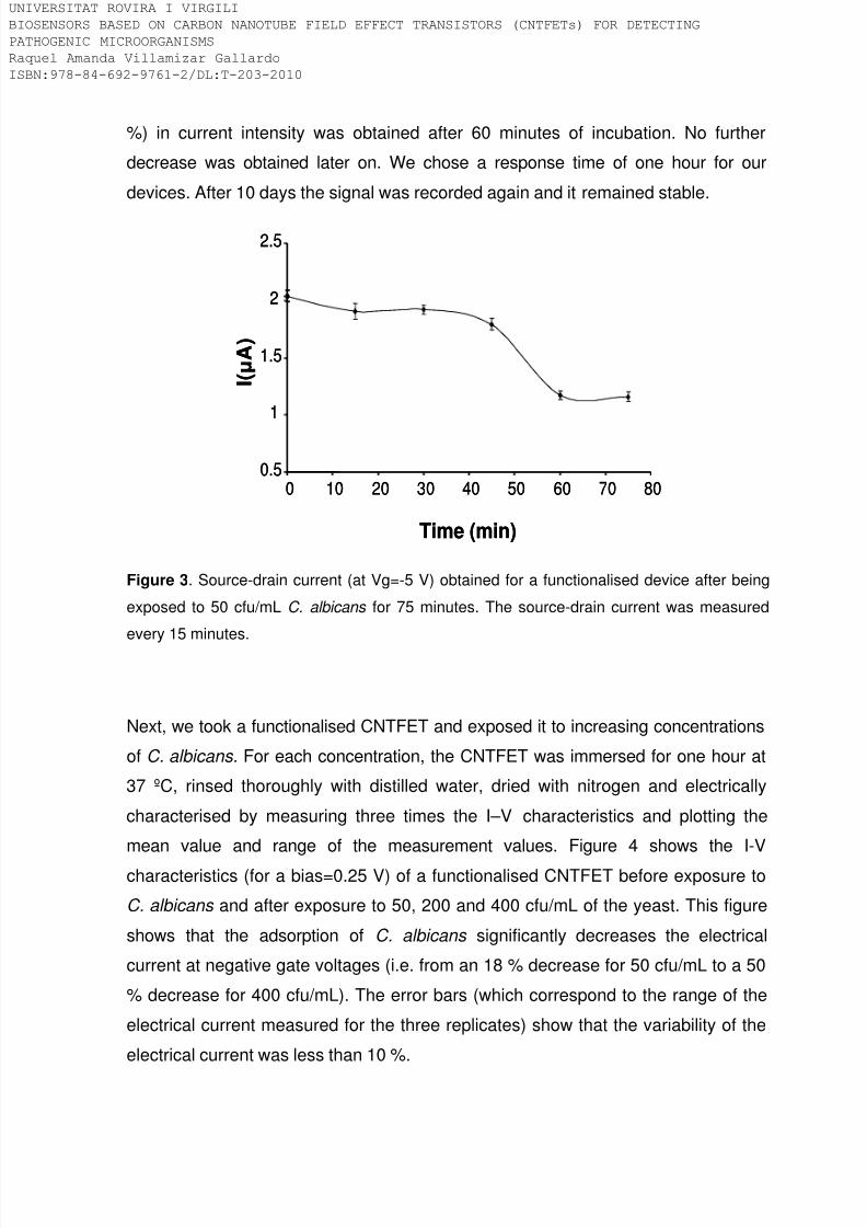
8/9/2019 Tesis Biosensors based on carbon nanotube field effect transistors
http://slidepdf.com/reader/full/tesis-biosensors-based-on-carbon-nanotube-field-effect-transistors 153/239
%) in current intensity was obtained after 60 minutes of incubation. No further
decrease was obtained later on. We chose a response time of one hour for our
devices. After 10 days the signal was recorded again and it remained stable.
Time (min)
I ( μ A )
0.5
1
1.5
2
2.5
0 10 20 30 40 50 60 70 80
Time (min)
I ( μ A )
0.5
1
1.5
2
2.5
0 10 20 30 40 50 60 70 80
Figure 3. Source-drain current (at Vg=-5 V) obtained for a functionalised device after being
exposed to 50 cfu/mL C. albicans for 75 minutes. The source-drain current was measured
every 15 minutes.
Next, we took a functionalised CNTFET and exposed it to increasing concentrations
of C. albicans. For each concentration, the CNTFET was immersed for one hour at
37 ºC, rinsed thoroughly with distilled water, dried with nitrogen and electrically
characterised by measuring three times the I–V characteristics and plotting the
mean value and range of the measurement values. Figure 4 shows the I-V
characteristics (for a bias=0.25 V) of a functionalised CNTFET before exposure to
C. albicans and after exposure to 50, 200 and 400 cfu/mL of the yeast. This figure
shows that the adsorption of C. albicans significantly decreases the electrical
current at negative gate voltages (i.e. from an 18 % decrease for 50 cfu/mL to a 50
% decrease for 400 cfu/mL). The error bars (which correspond to the range of the
electrical current measured for the three replicates) show that the variability of the
electrical current was less than 10 %.
IVERSITAT ROVIRA I VIRGILI
OSENSORS BASED ON CARBON NANOTUBE FIELD EFFECT TRANSISTORS (CNTFETs) FOR DETECTING
THOGENIC MICROORGANISMS
quel Amanda Villamizar Gallardo
BN:978-84-692-9761-2/DL:T-203-2010

8/9/2019 Tesis Biosensors based on carbon nanotube field effect transistors
http://slidepdf.com/reader/full/tesis-biosensors-based-on-carbon-nanotube-field-effect-transistors 154/239
0
0.5
1
1.5
2
-5 -3 -1 1 3 5
I ( μ A )
Vg(V)
0
0.5
1
1.5
2
-5 -3 -1 1 3 5
0
0.5
1
1.5
2
-5 -3 -1 1 3 5
I ( μ A )
Vg(V)
0
0.5
1
1.5
2
50cfu/mL
200cfu/mL
400cfu/mL
I ( µ A )
0
0.5
1
1.5
2
I ( µ A )
0
0.5
1
1.5
2
-5 -3 -1 1 3 5
I ( μ A )
Vg(V)
0
0.5
1
1.5
2
-5 -3 -1 1 3 5
0
0.5
1
1.5
2
-5 -3 -1 1 3 5
I ( μ A )
Vg(V)
Without yeast
Figure 4. Gate voltage dependence of the source-drain current of a typical CNTFET before
exposure to C. albicans (––) and after exposure to 50 cfu/mL (----); 200 cfu/mL ( X ) and 400
cfu/mL ( ◊ ) of C. albicans . The inset shows the behaviour of the source-drain current at Vg=
-5 V.
The exposure of the devices to C. albicans decreased significantly the conductance
of the functionalised CNTFET. Two mechanisms can explain this decrease. Thefirst one is a possible distortion of the CNTs as a consequence of the bound yeast
[28, 33]. The second one is the charge transfer process in which the analyte
provide electrons to the SWCNTs [30, 33]. In this way, we assume that once anti-
Candida antibodies interact with the antigen “mannan” present in the outer cell wall
layer of C. albicans there is a charge transfer process. Mannans are molecules
composed by homopolymers of D-mannose, which are linked to proteins by N-
glycosidic bonds to asparagine residues and by O-glycosidic, alkali-labile linkages
to threonine or serine residues [1]. Therefore, the charge transfer process could be
mainly due to electron donating from the amino groups of the mannoproteins
present in the cell wall of the yeast to the CNTs. The inset in figure 4 shows that
the higher the concentration of Candida , the more the electrical current decreases.
SEM confirmed that the number of yeasts attached to the CNTs was proportional to
the concentration of Candida in the solution. A functionalised CNTFET was
exposed to different yeast concentrations for 1 h each, thoroughly rinsed with
distilled water and dried with nitrogen. For each concentration we scanned the area
IVERSITAT ROVIRA I VIRGILI
OSENSORS BASED ON CARBON NANOTUBE FIELD EFFECT TRANSISTORS (CNTFETs) FOR DETECTING
THOGENIC MICROORGANISMS
quel Amanda Villamizar Gallardo
BN:978-84-692-9761-2/DL:T-203-2010
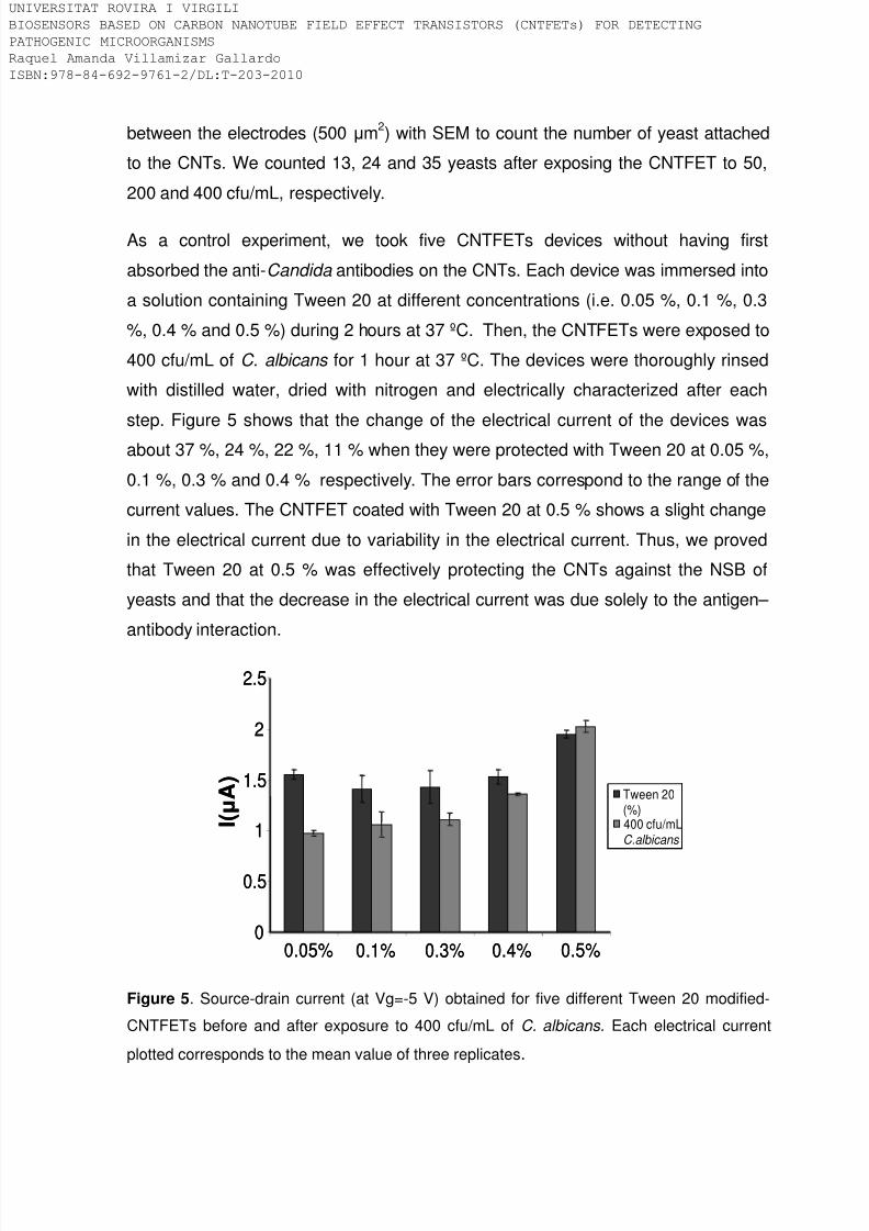
8/9/2019 Tesis Biosensors based on carbon nanotube field effect transistors
http://slidepdf.com/reader/full/tesis-biosensors-based-on-carbon-nanotube-field-effect-transistors 155/239
between the electrodes (500 μm2) with SEM to count the number of yeast attached
to the CNTs. We counted 13, 24 and 35 yeasts after exposing the CNTFET to 50,
200 and 400 cfu/mL, respectively.
As a control experiment, we took five CNTFETs devices without having first
absorbed the anti-Candida antibodies on the CNTs. Each device was immersed into
a solution containing Tween 20 at different concentrations (i.e. 0.05 %, 0.1 %, 0.3
%, 0.4 % and 0.5 %) during 2 hours at 37 ºC. Then, the CNTFETs were exposed to
400 cfu/mL of C. albicans for 1 hour at 37 ºC. The devices were thoroughly rinsed
with distilled water, dried with nitrogen and electrically characterized after each
step. Figure 5 shows that the change of the electrical current of the devices was
about 37 %, 24 %, 22 %, 11 % when they were protected with Tween 20 at 0.05 %,
0.1 %, 0.3 % and 0.4 % respectively. The error bars correspond to the range of the
current values. The CNTFET coated with Tween 20 at 0.5 % shows a slight change
in the electrical current due to variability in the electrical current. Thus, we proved
that Tween 20 at 0.5 % was effectively protecting the CNTs against the NSB of
yeasts and that the decrease in the electrical current was due solely to the antigen–
antibody interaction.
0
0.5
1
1.5
2
2.5
Tween 20(%)400 cfu/mLC.albicans
0.05% 0.1% 0.3% 0.4% 0.5%
I ( μ A )
0
0.5
1
1.5
2
2.5
Tween 20(%)400 cfu/mLC.albicans
0.05% 0.1% 0.3% 0.4% 0.5%
I ( μ A )
Figure 5. Source-drain current (at Vg=-5 V) obtained for five different Tween 20 modified-
CNTFETs before and after exposure to 400 cfu/mL of C. albicans. Each electrical current
plotted corresponds to the mean value of three replicates.
IVERSITAT ROVIRA I VIRGILI
OSENSORS BASED ON CARBON NANOTUBE FIELD EFFECT TRANSISTORS (CNTFETs) FOR DETECTING
THOGENIC MICROORGANISMS
quel Amanda Villamizar Gallardo
BN:978-84-692-9761-2/DL:T-203-2010

8/9/2019 Tesis Biosensors based on carbon nanotube field effect transistors
http://slidepdf.com/reader/full/tesis-biosensors-based-on-carbon-nanotube-field-effect-transistors 156/239
The effectiveness of Tween 20 at 0.5 % against NSB was also checked using SEM.
A CNTFET (functionalised with Tween 20 but not with anti-Candida ) was exposed
to 50, 200 and 400 cfu/mL C. albicans for 1 h each, thoroughly rinsed with distilled
water and dried with nitrogen. For each concentration we scanned the area
between the electrodes (500 μm2) with SEM. No yeasts were bound when the
device was exposed to 50 cfu/mL of C. albicans whereas 2 yeasts were observed
when it was exposed to 200 and 400 cfu/mL of C. albicans.
Selectivity of our devices was tested in the presence of two yeasts: C. albidus and
S. cerevisiae. C. albidus is an emerging pathogen, ubiquitously found in the
environment infecting and causing human disease as well as Candida [34].
However, it should not have any cross reaction with the anti-Candida antibody
because it is antigenically different from Candida . On the other hand, S. cerevisiae
is an emerging opportunist pathogen and can be found along with C. albicans in
clinical cases like cutaneous or vaginal infections [35]. The mannoproteins of C.
albicans share several general features with the mannoproteins of S. cerevisiae [1]
as they are antigenically similar.
A functionalised CNTFET was first immersed in a yeast solution containing 400cfu/mL of C. albidus for one hour at 37 ºC, thoroughly rinsed with distilled water,
dried with nitrogen and electrically characterised. The device was then exposed to
400 cfu/mL of C. albicans under the same conditions mentioned above. We follow
the same procedure with another functionalised CNTFET for 400 cfu/mL of S.
cerevisiae . Figure 6A shows that the electrical current did not change after
exposing the CNTFET to 400 cfu/mL of C. albidus whereas it decreased by about
30 % after exposure to C. albicans. Figure 6B shows that the electrical currentchanged slightly (i.e. 10 %) after exposing the CNTFET to 400 cfu/mL of S.
cerevisiae, whereas it decreased in the range of about 36 % after exposure to C.
albicans . The change in the electrical current after exposing the devices to S.
cerevisiae are probably due to the antigenic similarity between the two yeasts,
which may cause a slight cross-reactivity of the commercially available antibody.
The electrical current plotted corresponds to the mean value of the three
IVERSITAT ROVIRA I VIRGILI
OSENSORS BASED ON CARBON NANOTUBE FIELD EFFECT TRANSISTORS (CNTFETs) FOR DETECTING
THOGENIC MICROORGANISMS
quel Amanda Villamizar Gallardo
BN:978-84-692-9761-2/DL:T-203-2010

8/9/2019 Tesis Biosensors based on carbon nanotube field effect transistors
http://slidepdf.com/reader/full/tesis-biosensors-based-on-carbon-nanotube-field-effect-transistors 157/239
measurements. The error bars of the inset correspond to the range of the current
values.
Selectivity was also checked with SEM. A functionalised CNTFET was exposed to50, 200 and 400 cfu/mL of C. albidus for 1 h each, thoroughly rinsed with water and
dried with nitrogen. For each concentration, we scanned the area between the
electrodes (500 μm2) with SEM. We did not observe any yeast after the device had
been exposed to 50 cfu/mL whereas only one yeast was attached after the
CNTFET was exposed to 200 and 400 cfu/mL of C. albidus . The same procedure
was followed for S. cerevisiae . In this case, we observed 2, 5 and 7 yeasts after the
device had been exposed to 50, 200 and 400 cfu/mL of S. cerevisiae, respectively
(thus showing, the slight cross-reaction of the antibody with S. cerevisiae ). With C.
albidus the sensor displayed high selectivity; however, due to the antigenic
similarity between C. albicans and S. cerevisiae there was a slight cross-reactivity
of the commercially available antibody. Therefore, a more specific antibody against
C. albicans would be able to overcome this problem.
Figure 6. A) Gate voltage dependence of the source-drain current of a functionalised
CNTFET before exposure to the bacteria (––) and after exposure to 400 cfu/mL of C. albidus
(----); and 400 cfu/mL of C. albicans ( X ). B) Gate voltage dependence of the source-drain
current of a functionalised CNTFET before exposure to the yeast (––) and after exposure to
400 cfu/mL of S. cerevisiae (----); and 400 cfu/mL of C. albicans ( X ). The inset shows the
behaviour of the source-drain current after each functionalisation step at Vg= -5 V.
IVERSITAT ROVIRA I VIRGILI
OSENSORS BASED ON CARBON NANOTUBE FIELD EFFECT TRANSISTORS (CNTFETs) FOR DETECTING
THOGENIC MICROORGANISMS
quel Amanda Villamizar Gallardo
BN:978-84-692-9761-2/DL:T-203-2010
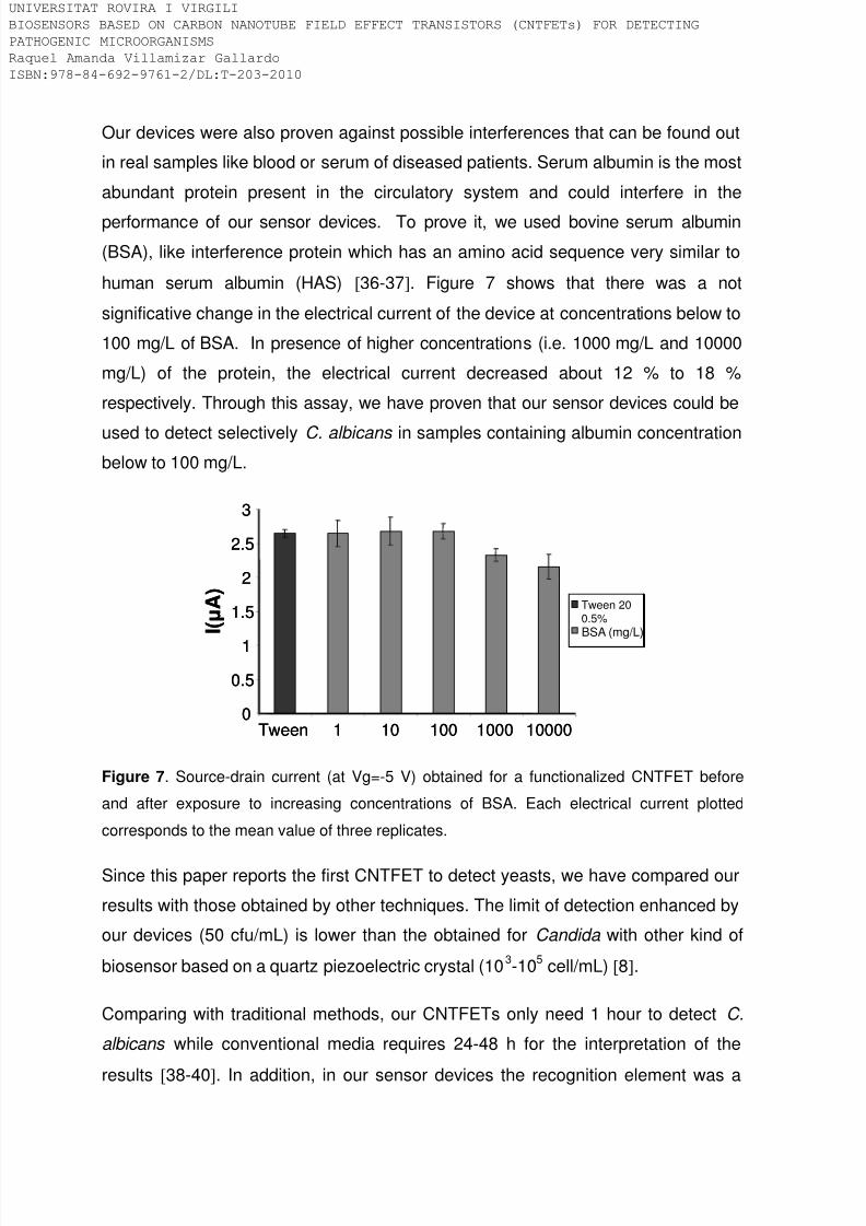
8/9/2019 Tesis Biosensors based on carbon nanotube field effect transistors
http://slidepdf.com/reader/full/tesis-biosensors-based-on-carbon-nanotube-field-effect-transistors 158/239
Our devices were also proven against possible interferences that can be found out
in real samples like blood or serum of diseased patients. Serum albumin is the most
abundant protein present in the circulatory system and could interfere in the
performance of our sensor devices. To prove it, we used bovine serum albumin
(BSA), like interference protein which has an amino acid sequence very similar to
human serum albumin (HAS) [36-37]. Figure 7 shows that there was a not
significative change in the electrical current of the device at concentrations below to
100 mg/L of BSA. In presence of higher concentrations (i.e. 1000 mg/L and 10000
mg/L) of the protein, the electrical current decreased about 12 % to 18 %
respectively. Through this assay, we have proven that our sensor devices could be
used to detect selectively C. albicans in samples containing albumin concentrationbelow to 100 mg/L.
0
0.5
1
1.5
2
2.5
3
Tween 1 10 100 1000 10000
I ( μ A )
Tween 200.5%
BSA (mg/L)
0
0.5
1
1.5
2
2.5
3
Tween 1 10 100 1000 10000
I ( μ A )
Tween 200.5%
BSA (mg/L)
Figure 7. Source-drain current (at Vg=-5 V) obtained for a functionalized CNTFET before
and after exposure to increasing concentrations of BSA. Each electrical current plotted
corresponds to the mean value of three replicates.
Since this paper reports the first CNTFET to detect yeasts, we have compared our
results with those obtained by other techniques. The limit of detection enhanced by
our devices (50 cfu/mL) is lower than the obtained for Candida with other kind of
biosensor based on a quartz piezoelectric crystal (103-105 cell/mL) [8].
Comparing with traditional methods, our CNTFETs only need 1 hour to detect C.
albicans while conventional media requires 24-48 h for the interpretation of the
results [38-40]. In addition, in our sensor devices the recognition element was a
IVERSITAT ROVIRA I VIRGILI
OSENSORS BASED ON CARBON NANOTUBE FIELD EFFECT TRANSISTORS (CNTFETs) FOR DETECTING
THOGENIC MICROORGANISMS
quel Amanda Villamizar Gallardo
BN:978-84-692-9761-2/DL:T-203-2010

8/9/2019 Tesis Biosensors based on carbon nanotube field effect transistors
http://slidepdf.com/reader/full/tesis-biosensors-based-on-carbon-nanotube-field-effect-transistors 159/239
specific antibody anti-C.albicans displaying high selectivity against other yeasts
both antigenically similar and different and tween 20 avoids effectively the non-
specific binding of the yeast or proteins. This is an advantage comparing for
instance with chromogenic culture media, which contain chromogenic or fluorogenic
substrates for a rapid identification of C. albicans on the basis of colony colour.
Although some of them present high sensitivity and specificity (97 % and 92 %
respectively), false positive results are frequently obtained [38, 40].
CONCLUSIONS
In this paper we have shown that carbon nanotube field effect transistors
(CNTFETs) functionalised with monoclonal anti-Candida antibodies can be used to
detect 50 cfu/mL of the yeast in only one hour. The selectivity of the sensor was
determined by exposing the devices to C. albidus and S. cerevisiae.
The results suggest that CNTFETs biosensors might be a valuable screening tool
addressed to future routine analysis of real samples like blood, serum, infected
tissues, vaginal smears, swabs samples of diseased patients. On the other hand, it
is also possible to detect in human milk clinical cases of mastitis caused by C.
albicans and using an adequate molecular receptor other pathogenic Candida
species can be detected in samples like mastitic bovine milk. In addition, our
biosensor could help to obtain an early diagnostic of sick patients which would
improve the administration of adequate drugs and treatments.
ACKNOWLEDGMENTS
We thank the Spanish Ministry of Education and Science, MEC, for supporting the
work through the project grants NAN2004-09306-C05-05 and CTQ2007-67570.
RAV also acknowledges the University of Pamplona and the Universitat Rovira i
Virgili for providing economic support. A. Maroto also would like to thank the
Spanish Ministry of Science and Technology for providing her Juan de la Cierva
contract.
IVERSITAT ROVIRA I VIRGILI
OSENSORS BASED ON CARBON NANOTUBE FIELD EFFECT TRANSISTORS (CNTFETs) FOR DETECTING
THOGENIC MICROORGANISMS
quel Amanda Villamizar Gallardo
BN:978-84-692-9761-2/DL:T-203-2010

8/9/2019 Tesis Biosensors based on carbon nanotube field effect transistors
http://slidepdf.com/reader/full/tesis-biosensors-based-on-carbon-nanotube-field-effect-transistors 160/239
References
[1]. J. P. Martinez, M.L. Gil, J.L. López-Ribot, W.L.Chaffin, Serologic Response to
Cell Wall Mannoproteins and Proteins of Candida albicans, Clin. Microbiol. Rev, 11,(1998) 121–141.
[2]. R. A. Cavalheiro, F. Fortes, J. Borecký, V. C. Faustinoni, A. Z. Schreiber, A.
E.Vercesi, Respiration, oxidative phosphorylation, and uncoupling protein in
Candida albican, Braz. J. Biol. Biomed. Res, 37 (2004) 1455-1461.
[3]. I. F. Salkin, G. A. Land, N. J. Hurd, P.R. Goldson, M. R. McGinnis, Evaluation
of YeastIdent and Uni-Yeast-Tek Yeast Identification Systems, J. Clin. Microbiol, 25(1987) 624-627.
[4]. J. F. Okulicz, R. G. Rivard, N. G. Conger, M. X. Nguyen, D. R. Hospenthal,
Primary isolation of Candida species from urine specimens using chromogenic
medium, Mycoses, 51(2007)141–146.
[5]. E. Eraso, M. D. Moragues, M. Villar-Vidal, I. H. Sahand, N. González, J.
Pontón, G. Quindós, Evaluation of the New Chromogenic Medium Candida ID 2for Isolation nd Identification of Candida albicans and Other Medically Important
Candida Species, J. Clin. Microbiol, 44 (2006) 3340-3345.
[6]. W.M. Scheld, R.S. Brown, S.A. Harding, M.A. Sande, Detection of Circulating
Antigen in Experimental Candida albicans Endocarditis by an Enzyme-Linked
Immunosorbent Assay, J. Clin. Microbiol, 12 (1980) 679-683.
[7]. H. Xiang, L. Xiong, X. Liu, Z.Tu, Rapid simultaneous detection and identificationof six species Candida using polymerase chain reaction and reverse line
hybridization assay, J. Microbiol. Methods, 69 (2007) 282–287.
[8]. H. Muramatsu, K. Kajiwara, E. Tamiya, I. Karube, Piezoelectric immunosensor
for the detection of Candida albicans microbes, Anal. Chim. Acta, 188 (1986) 257-
261.
IVERSITAT ROVIRA I VIRGILI
OSENSORS BASED ON CARBON NANOTUBE FIELD EFFECT TRANSISTORS (CNTFETs) FOR DETECTING
THOGENIC MICROORGANISMS
quel Amanda Villamizar Gallardo
BN:978-84-692-9761-2/DL:T-203-2010
8/9/2019 Tesis Biosensors based on carbon nanotube field effect transistors
http://slidepdf.com/reader/full/tesis-biosensors-based-on-carbon-nanotube-field-effect-transistors 161/239
[9]. N. Nugaeva, K.Y. Gfeller, N. Backmann, H.P.Lang, M. Duggelin, M. Hegner,
Micromechanical cantilever array sensors for selective fungal immobilization and
fast growth detection, Biosens. Bioelectron, 21 (2005) 849–856.
[10]. J.W. Yi, W.Y. Shih, R. Mutharasan, W. H. Shih, In situ cell detection using
piezoelectric lead zirconate titanate-stainless steel cantilevers, J. Appl. Phys, 93
(2002) 619-625.
[11]. F. L. Dickert and O. Hayden, Bioimprinting of Polymers and Sol-Gel Phases
Selective Detection of Yeasts with Imprinted Polymers, Anal. Chem, 74 (2002)
1302-1306.
[12]. H. Chen, C.K. Heng, P.D. Puiu, X.D. Zhou, A.C. Lee, T.M. Lim, S.N. Tan,
Detection of Saccharomyces cerevisiae immobilized on self-assembled monolayer
(SAM) of alkanethiolate using electrochemical impedance spectroscopy, Anal.
Chim. Acta, 554 (2005) 52–59.
[13].Heller, I., A.M. Janssens, J. Männik, E. D. Minot, S.G. Lemay, C. Dekker,
Identifying the Mechanism of Biosensing with Carbon Nanotube Transistors, Nano
Lett, 8 (2008) 591-595.
[14]. S. J. Tans, A. R. Verschueren, C. Dekker, Room-temperature transistor based
on a single carbon nanotube, Nature, 393 (1998) 49-52.
[15]. E.L. Gui, L.J. Li, K. Zhang, Y. Xu, X. Dong, X. Ho, P.S. Lee, J. Kasim, Z.X.
Shen, J.A. Rogers, S.G. Mhaisalkar, DNA Sensing by Field-Effect Transistors
Based on Networks of Carbon Nanotubes, JACS, 129 (2007) 14427-14432.
[16]. A. Star, E. Tu, J. Niemann, J.C.P. Gabriel, C.S. Joiner, C. Valcke, Label-free
detection of DNA hybridization using carbon nanotube network field-effect
transistors, PNAS, 103 (2006) 921-926.
[17]. H. M. So, K. Won, Y. H. Kim, B. K. Kim, B. H. Ryu, P. S. Na, H. Kim, A. Star,
E. Tu, E. J. Niemann, J.C.P. Gabriel, C.S. Joiner, C. Valcke, Label-free detection of
IVERSITAT ROVIRA I VIRGILI
OSENSORS BASED ON CARBON NANOTUBE FIELD EFFECT TRANSISTORS (CNTFETs) FOR DETECTING
THOGENIC MICROORGANISMS
quel Amanda Villamizar Gallardo
BN:978-84-692-9761-2/DL:T-203-2010

8/9/2019 Tesis Biosensors based on carbon nanotube field effect transistors
http://slidepdf.com/reader/full/tesis-biosensors-based-on-carbon-nanotube-field-effect-transistors 162/239
DNA hybridization using carbon nanotube network field-effect transistors, PNAS,
103 (2006) 921-926.
[18]. H. M. So, K. Won, Y. H. Kim, B. K. Kim, B. H. Ryu, P. S. Na, H. Kim, O.J. Lee,Single-Walled Carbon Nanotube Biosensors Using Aptamers as Molecular
Recognition Elements, JACS, 127(2005) 11906-11907.
[19]. K. Maehashi, T. Katsura, K. Kerman, Y. Takamura, K. Matsumoto, Tamiya, E,
Label-Free Protein Biosensor Based on Aptamer-Modified Carbon Nanotube Field-
Effect Transistors, Anal. Chem, 79 (2007) 782-787.
[20]. C. Cid, J. Riu, A. Maroto, F.X. Rius, Carbon nanotube field effect transistorsfor the fast and selective detection of human immunoglobulin G., Analyst, doi:
10.1039/b805301b,( 2008).
[21]. T. Dastagir, E.S. Forzani, R. Zhang, I. Amlani, L. A. Nagahara, R. Tsui, N.
Tao, Electrical detection of hepatitis C virus RNA on single wall carbon nanotube-
field effect transistors, Analyst, 132 (2007) 738-740.
[22]. R.A. Villamizar, A. Maroto, F.X. Rius, I. Inza, M.J. Figueras, Fast detection ofSalmonella Infantis with carbon nanotube field effect transistors, Biosens.
Bioelectrons, 24 (2008) 279-283.
[23]. H.M. So, D. W. Park, E. K. Jeon, Y. H. Kim, S.K. Kim, C.K. Lee, S. Y. Choi,
S.C. Kim, H. Chang, J.O. Lee, Detection and Titer Estimation of Escherichia coli
Using Aptamer-Functionalized Single-Walled Carbon-Nanotube Field-Effect
Transistors, Small, 4 (2008) 197-201.
[24]. H. R. Byon and H.C. Choi, Network Single-Walled Carbon Nanotube-Field
Effect Transistors (SWNT-FETs) with Increased Schottky Contact Area for Highly
Sensitive Biosensor Applications, JACS, 128 (2006) 2188-2189.
[25]. R. J. Chen, S. Bangsaruntip, K. A. Drouvalakis, N. W. Kam, M. Shim, Y. Li, W.
Kim, P. J. Utz, H. Dai, Noncovalent functionalization of carbon nanotubes for highly
specific electronic biosensors, PNAS, 100 (2003) 4984-4989.
IVERSITAT ROVIRA I VIRGILI
OSENSORS BASED ON CARBON NANOTUBE FIELD EFFECT TRANSISTORS (CNTFETs) FOR DETECTING
THOGENIC MICROORGANISMS
quel Amanda Villamizar Gallardo
BN:978-84-692-9761-2/DL:T-203-2010
8/9/2019 Tesis Biosensors based on carbon nanotube field effect transistors
http://slidepdf.com/reader/full/tesis-biosensors-based-on-carbon-nanotube-field-effect-transistors 163/239
[26]. H. C. Chang, J. J. Chang, A. H. Huang, T. C. Chang, Evaluation of a
Capacitance Method for Direct Antifungal Susceptibility Testing of Yeasts in
Positive Blood Cultures, J. Clin. Microbiol, 38 (2000) 971-976.
[27]. B. IIic, D. Czaplewski, G. Craighead, Mechanical resonant immunospecific
biological detector, Appl. Phys. Lett, 77 (2000) 450–452.
[28]. A. Star, J.C. Gabriel, K. Bradley, G. Gruner, Electronic Detection of Specific
Protein Binding Using Nanotube FET Devices, Nano Lett, 3 (2003) 459–463.
[29]. K. Teker, E. Wickstrom, B. Panchapakesan, Biomolecular Tuning of Electronic
Transport Properties of Carbon Nanotubes via Antibody Functionalization, IEEESens. J, 6 (2006) 1422-1428.
[30]. B. L. Allen, P. D. Kichambare, A. Star, Carbon Nanotube Field-Effect-
Transistor Based Biosensors, Adv. Mater, 19 (2007) 1439–1451.
[31]. M. Shim, N.W.S. Kam, R.J. Chen, Y. Li, H. Dai, Functionalization of Carbon
Nanotubes for Biocompatibility and Biomolecular Recognition, Nano Lett, 2(2002)
285–288.
[32]. K. Bradley, M. Briman, A. Star, G. Gruner, Charge Transfer from Adsorbed
Proteins, Nano Lett, 4 (2004) 253–256.
[33]. G. Grüner, Carbon Nanotube Transistors for Biosensing Applications, Anal.
Bioanal. Chem, 384 (2006) 322–335.
[34] J. Schutzbach, H. Ankel, I. Brockhausen, Synthesis of cell envelope
glycoproteins of Cryptococcus laurentii, Carbohydr.Res, 342 (2007) 881–893.
[35]. A.L. Goldstein and J.H. McCusker, Development of Saccharomyces cerevisiae
as a Model Pathogen: A System for the Genetic Identification of Gene Products
Required for Survival in the Mammalian Host Environment, Genetics, 159 (2001)
499–513.
IVERSITAT ROVIRA I VIRGILI
OSENSORS BASED ON CARBON NANOTUBE FIELD EFFECT TRANSISTORS (CNTFETs) FOR DETECTING
THOGENIC MICROORGANISMS
quel Amanda Villamizar Gallardo
BN:978-84-692-9761-2/DL:T-203-2010

8/9/2019 Tesis Biosensors based on carbon nanotube field effect transistors
http://slidepdf.com/reader/full/tesis-biosensors-based-on-carbon-nanotube-field-effect-transistors 164/239
[36]. S. Aoyagi, M. Dohi, N. Kato, M. Kudo, S. Iida, M. Tozu, N. Sanada. Structure
analysis of immobilized-bovine serum albumin by means of TOF-SIMS, e-J. Surf.
Sci. Nanotech, 4 (2006) 614-618.
[37]. N. E. Kadi, N. Taulier, J. Y. Le Huerou, M. Gindre, W. Urbach, I. Nwigwe, P. C.
Kahn, M. Waks, Unfolding and Refolding of Bovine Serum Albumin at Acid pH:
Ultrasound and Structural Studies, Biophys. J, 91(2006), 3397–3404.
[38]. C.D. Cárdenes, A.J. Carrillo, A. Ariasa, C. Rodríguez, A. Torres, A. Sierra,
M.P. Arévalo, Comparative evaluation of four commercial tests for presumptive
identification of Candida albicans, J. Microbiol. Methods, 59 (2004) 293– 297.
[39]. M. Ilkit, S. Hilmioglu, M. Tasbakan, S. Aydemir, Evaluation of Albicans ID2 and
Biggy agar for the isolation and direct identification of vaginal yeast isolates, J. Med.
Microbiol, 56 (2007) 762–765.
IVERSITAT ROVIRA I VIRGILI
OSENSORS BASED ON CARBON NANOTUBE FIELD EFFECT TRANSISTORS (CNTFETs) FOR DETECTING
THOGENIC MICROORGANISMS
quel Amanda Villamizar Gallardo
BN:978-84-692-9761-2/DL:T-203-2010

8/9/2019 Tesis Biosensors based on carbon nanotube field effect transistors
http://slidepdf.com/reader/full/tesis-biosensors-based-on-carbon-nanotube-field-effect-transistors 165/239
5.3. Supplementary experimental section
5.3.1. Microbiological test
Microbiological cultures were performed in order to confirm the viability of Candida
albicans strains. Figure 5-1A shows an image of the typical growth of Candida
albicans in sabouraud agar. This media is used for cultivation of pathogenic and
nonpathogenic fungi. The low pH of approximately 5.6 is favorable for the growth of
fungi, especially dermatophytes, and slightly inhibitory to contaminating bacteria in
clinical specimens (Chung, K.J. and Bennett, J.E. 1992). The typical grow of
Candida albicans is characterized for white colonies, circular and soft. Only 24 h at
30 ºC were required to observe the first typical colonies of the yeast over the media.It showed the viability of the lyophilized reference strain. Moreover, in order to
preserve the culture pure, the colonies were plated on oxytetracycline gentamicine
yeast agar (OGY). The oxytetracycline is used as the selective agent suppressing
the bacterial growth.
Additionally to the culture plate, a sample was analyzed using SEM. It can be seen
in figure 5-1B typical budding yeast (blastoconidia), ovals, with a diameter from 2 to4 µm.
Figure 5-1. A) Typical grow of Candida albicans in sabouraud agar. B) SEM image
of the yeast.
IVERSITAT ROVIRA I VIRGILI
OSENSORS BASED ON CARBON NANOTUBE FIELD EFFECT TRANSISTORS (CNTFETs) FOR DETECTING
THOGENIC MICROORGANISMS
quel Amanda Villamizar Gallardo
BN:978-84-692-9761-2/DL:T-203-2010
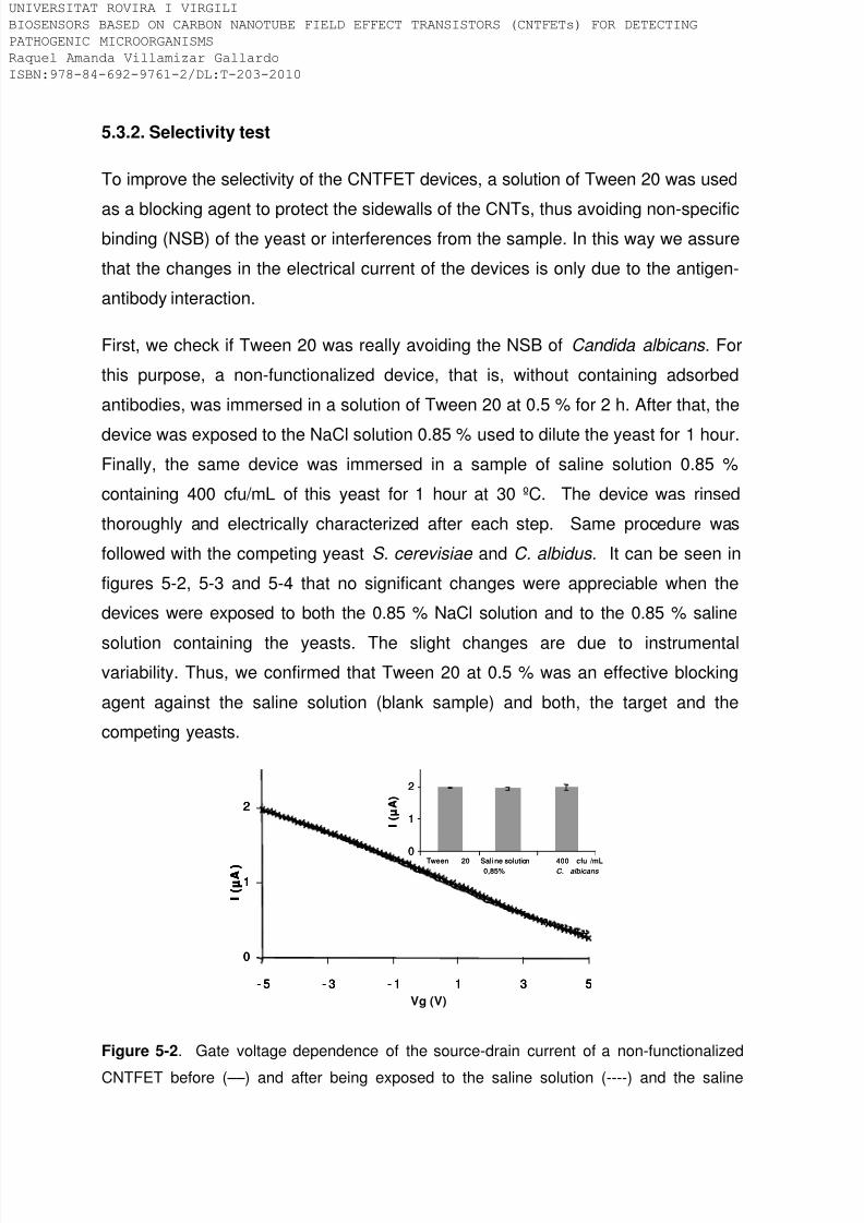
8/9/2019 Tesis Biosensors based on carbon nanotube field effect transistors
http://slidepdf.com/reader/full/tesis-biosensors-based-on-carbon-nanotube-field-effect-transistors 166/239
5.3.2. Selectivity test
To improve the selectivity of the CNTFET devices, a solution of Tween 20 was used
as a blocking agent to protect the sidewalls of the CNTs, thus avoiding non-specificbinding (NSB) of the yeast or interferences from the sample. In this way we assure
that the changes in the electrical current of the devices is only due to the antigen-
antibody interaction.
First, we check if Tween 20 was really avoiding the NSB of Candida albicans . For
this purpose, a non-functionalized device, that is, without containing adsorbed
antibodies, was immersed in a solution of Tween 20 at 0.5 % for 2 h. After that, the
device was exposed to the NaCl solution 0.85 % used to dilute the yeast for 1 hour.Finally, the same device was immersed in a sample of saline solution 0.85 %
containing 400 cfu/mL of this yeast for 1 hour at 30 ºC. The device was rinsed
thoroughly and electrically characterized after each step. Same procedure was
followed with the competing yeast S. cerevisiae and C. albidus. It can be seen in
figures 5-2, 5-3 and 5-4 that no significant changes were appreciable when the
devices were exposed to both the 0.85 % NaCl solution and to the 0.85 % saline
solution containing the yeasts. The slight changes are due to instrumentalvariability. Thus, we confirmed that Tween 20 at 0.5 % was an effective blocking
agent against the saline solution (blank sample) and both, the target and the
competing yeasts.
0
1
2
Tween 20 400 cfu /mL
C. albicans
Sali ne solution
0,85%
I ( µ A )
0
1
2
- 5 - 3 - 1 1 3 5
Vg (V)
I ( µ A )
0
1
2
Tween 20 400 cfu /mL
C. albicans
Sali ne solution
0,85%
I ( µ A )
0
1
2
- 5 - 3 - 1 1 3 5
I ( µ A )
0
1
2
- 5 - 3 - 1 1 3 5
I ( µ A )
Figure 5-2. Gate voltage dependence of the source-drain current of a non-functionalized
CNTFET before (––) and after being exposed to the saline solution (----) and the saline
IVERSITAT ROVIRA I VIRGILI
OSENSORS BASED ON CARBON NANOTUBE FIELD EFFECT TRANSISTORS (CNTFETs) FOR DETECTING
THOGENIC MICROORGANISMS
quel Amanda Villamizar Gallardo
BN:978-84-692-9761-2/DL:T-203-2010

8/9/2019 Tesis Biosensors based on carbon nanotube field effect transistors
http://slidepdf.com/reader/full/tesis-biosensors-based-on-carbon-nanotube-field-effect-transistors 167/239
solution containing 400 cfu/mL of C. albicans ( X ). The inset shows the behaviour of the
source-drain current after each functionalisation step at Vg= -5 V.
0
1
2
-5 -3 -1 1 3 5
I ( µ A )
Vg (V)
0
1
2
Saline solution0,85%
Tween 20 400 cfu/mLS. c erevisiae
I ( µ A )
0
1
2
-5 -3 -1 1 3 5
I ( µ A )
0
1
2
-5 -3 -1 1 3 5
I ( µ A )
Vg (V)
0
1
2
Saline solution0,85%
Tween 20 400 cfu/mLS. c erevisiae
I ( µ A )
Figure 5-3. Gate voltage dependence of the source-drain current of a non-functionalized
CNTFET before (––) and after being exposed to the saline solution (----) and to the saline
solution containing 400 cfu/mL of S. cerevisiae ( X ). The inset shows the behaviour of the
source-drain current after each functionalisation step at Vg= -5 V.
0
0,5
1
1,5
-5 -3 -1 1 3 5
Vg (V)
I ( μ A ) 0
0,5
1
1,5
Tween 20 Saline solution
0,85%
400 cfu/mL
C. albidus
I ( μ A )
Figure 5-4. Gate voltage dependence of the source-drain current of a non-functionalized
CNTFET before (––) and after being exposed to the saline solution (----) and to the saline
solution containing 400 cfu/mL of C. albidus ( X ). The inset shows the behaviour of the
source-drain current after each functionalisation step at Vg= -5 V.
IVERSITAT ROVIRA I VIRGILI
OSENSORS BASED ON CARBON NANOTUBE FIELD EFFECT TRANSISTORS (CNTFETs) FOR DETECTING
THOGENIC MICROORGANISMS
quel Amanda Villamizar Gallardo
BN:978-84-692-9761-2/DL:T-203-2010

8/9/2019 Tesis Biosensors based on carbon nanotube field effect transistors
http://slidepdf.com/reader/full/tesis-biosensors-based-on-carbon-nanotube-field-effect-transistors 168/239
5.4. Complementary Conclusions
This study shows another application of the CNTFETs in the biosensing of higher,
more complexed organism like yeast. An effective blocking agent (Tween 20) wasused to avoid non-specific binding, thus the change in the electrical current was due
only to antigen-antibody interaction.
We proved that our sensor devices could be used to detect C. albicans selectively
in samples containing albumin concentrations below 0.1 g/L. However, the
concentration of albumin in a normal serum sample is about 35 to 55 g/L.
Therefore, it is necessary to dilute the sample (as is also required in conventional
clinical assays) and use another type of blocking agent that can protect thesidewalls more effectively against proteins present in real samples. An option that
could have been explored is the use of PBS containing Tween 20 at 0,05 % and
gelatin 0,8 % reported by Sanchez -Acevedo et al , (Sanchez Acevedo, Z.C. et al,
2009) as an effective blocking agent for small molecules.
We developed an improved biosensor that represents an alternative method than
can be used to detect yeast with medical importance in short time. Neverthelessuntil today, molecular biology based methods like PCR in real time is the most
sensitive assay to detect this pathogenic yeast. Our devices must be proven with
real sample and must be validated with traditional methods, thus establishing the
possibility of using them on the market. Moreover, our devices allow us to make a
qualitative detection. This is mainly due to the CNTFETs present different
sensitivities because the synthesis process of the CNT is irreproducible generating
networks with diverses densities and electrical behaviours.
IVERSITAT ROVIRA I VIRGILI
OSENSORS BASED ON CARBON NANOTUBE FIELD EFFECT TRANSISTORS (CNTFETs) FOR DETECTING
THOGENIC MICROORGANISMS
quel Amanda Villamizar Gallardo
BN:978-84-692-9761-2/DL:T-203-2010

8/9/2019 Tesis Biosensors based on carbon nanotube field effect transistors
http://slidepdf.com/reader/full/tesis-biosensors-based-on-carbon-nanotube-field-effect-transistors 169/239
5.5. References
Chen, H., Heng, C.K., Puiu, P.D., Zhou, X.D., Lee, A.C., Lim, T.M., Tan, S.N.
Analytica Chimica Acta. 2005. 554, 52–59.
Chunchanur, S.K., Nadgir, S.D., Halesh, L.H., Patil, B.S., Kausar, Y.,
Chandrasekhar, M.R. Indian Journal of Pathology and Microbiology. 2009. 52,
501-504.
K.J. Chung, J. E. Bennett, Medical Mycology, Lea & Febiger, Philadelphia, 1992
Cooke, V.M., Miles, R.J., Price, R.G., Midgley, G., Khamri, W., Richardson, A.C.
Applied and Environmental Microbiology. 2002. 68, 3622-3627.
Horvath, L.L., George, B.J., Murray, C.K., Harrison, L.S., Hospenthal, D.R. Journal
of Clinical of Microbiology. 2005. 42, 115-118.
Maaroufi, Y., Heymans, C., De Bruyne, J.M., Duchateau, V., Rodriguez-Villalobos,
H., Aoun, M., Crokaert, F. Journal of Clinical Microbiology. 2003. 41, 3293–3298.
Marcilla, A., Monteagudo, C., Mormeneo, S., Sentandreu, R. Microbiology. 1999.
145, 695–701.
Matsui, H., Hanaki, H., Takahashi, K., Yokoyama, A., Nakae, T., Sunakawa, K.,
Omura, S. Clinical and Vaccine Immunology. 2009. 16, 1366–1368.
McMullan, R., Metwally, L., Coyle, P.V., Hedderwick, S., McCloskey, B., O'Neill,
H.J., Patterson, C.C., Thompson, G., Webb, C.H., Hay, R.J. Clinical Infectious
Diseases. 2008. 46, 890-896.
Moudni, B.L., Rodier, M.H., Daniault,G., Jacquemin, J.L. Clinical Diagnostic of
Laboratory Immunology. 1998. 5: 823–825.
IVERSITAT ROVIRA I VIRGILI
OSENSORS BASED ON CARBON NANOTUBE FIELD EFFECT TRANSISTORS (CNTFETs) FOR DETECTING
THOGENIC MICROORGANISMS
quel Amanda Villamizar Gallardo
BN:978-84-692-9761-2/DL:T-203-2010

8/9/2019 Tesis Biosensors based on carbon nanotube field effect transistors
http://slidepdf.com/reader/full/tesis-biosensors-based-on-carbon-nanotube-field-effect-transistors 170/239
Mulero, R., Lee, D.H., Kutzler, M.A., Jacobson, J.M., Kim, M.J. Sensors . 2009. 9,
1590-1598.
Munin, E., Giroldo, L.M., Alves, L.P., Silva, M. Journal of Photochemistry andPhotobiology B: Biology. 2007. 88, 16–20.
Pontón, J. and del Palacio, A. Revista Iberoamericana de Micología. 2007. 24,
181-186.
Sánchez-Acevedo, Z.C., Riu, J., Rius, F. X. Biosensors and Bioelectronics. 2009.
24, 2842–2846.
Trama, J.P., Mordechai, E., Adelson, M.E. Molecular and Cellular Probes. 2005. 19,
145–152.
Wellinghausen, N., Siegel, D., Winter, J., Geber, S. Journal of Medical
Microbiology. 2009. 58, 1106-1111.
IVERSITAT ROVIRA I VIRGILI
OSENSORS BASED ON CARBON NANOTUBE FIELD EFFECT TRANSISTORS (CNTFETs) FOR DETECTING
THOGENIC MICROORGANISMS
quel Amanda Villamizar Gallardo
BN:978-84-692-9761-2/DL:T-203-2010

8/9/2019 Tesis Biosensors based on carbon nanotube field effect transistors
http://slidepdf.com/reader/full/tesis-biosensors-based-on-carbon-nanotube-field-effect-transistors 171/239
Chapter 6
Improvement of CNT based FETs to detect
microorganisms
IVERSITAT ROVIRA I VIRGILI
OSENSORS BASED ON CARBON NANOTUBE FIELD EFFECT TRANSISTORS (CNTFETs) FOR DETECTING
THOGENIC MICROORGANISMS
quel Amanda Villamizar Gallardo
BN:978-84-692-9761-2/DL:T-203-2010

8/9/2019 Tesis Biosensors based on carbon nanotube field effect transistors
http://slidepdf.com/reader/full/tesis-biosensors-based-on-carbon-nanotube-field-effect-transistors 172/239
IVERSITAT ROVIRA I VIRGILI
OSENSORS BASED ON CARBON NANOTUBE FIELD EFFECT TRANSISTORS (CNTFETs) FOR DETECTING
THOGENIC MICROORGANISMS
quel Amanda Villamizar Gallardo
BN:978-84-692-9761-2/DL:T-203-2010

8/9/2019 Tesis Biosensors based on carbon nanotube field effect transistors
http://slidepdf.com/reader/full/tesis-biosensors-based-on-carbon-nanotube-field-effect-transistors 173/239
6.1. Introduction
There are a number of parameters used to characterize the performance of
biosensors. Among the most relevant are the specificity, sensitivity, stability, limit ofdetection, time response, life time or recovery time. In the development of an
immunosensor the specificity and sensitivity depend greatly on the kind of antibody
and its immobilization method (Corry, B. et al, 2003). The denaturation or the
conformational changes an antibody can undergo during or after its immobilization
is one of the main reasons in the reduction of the immunosensor sensitivity. These
types of changes are usually associated with the non-specific adsorption of
antibodies on solid supports (Jung, Y. et al, 2008).
A judicious approach to improve the sensitivity of an immunosensor would therefore
be to increase the density of the binding sites on the sensor surface. This density
mainly depends on the adequate orientation of the bioreceptor (Van Regenmortel,
M.H.V. et al, 2002). Several research groups have tried to control the orientation of
antibodies. One of the most frequent techniques is the use of bacterial proteins
which can bind to the Fc constant fraction of antibodies (Bae, Y.M. et al, 2005.;
Kanno, S. et al, 2000). As a result, the antigen binding sites of the immunoglobulinsmaximize the access to the target.
Additionally to the binding specificity of the bacterial protein for the antibodies, the
most important property is their ability to remove the active antibody layer and
replace it with a different one under relatively similar conditions. As a result, a
surface modified with bacterial proteins can act as a generic platform to build
immunosensors, where the selectivity can change by using different antibodies as
receptors. Moreover, the sensor surface can also be regenerated by desorbing the
antibody from the binding protein, process easier to achieve than trying to break the
Ag/Ab interaction. Therefore, the use of bacterial proteins as linkers generate more
reproducible surfaces where the antibody can be replaced each time without the
use of aggressive conditions thus preserving the surface or biomolecule
configuration (Van Regenmortel, M.H.V. et al, 2002).
IVERSITAT ROVIRA I VIRGILI
OSENSORS BASED ON CARBON NANOTUBE FIELD EFFECT TRANSISTORS (CNTFETs) FOR DETECTING
THOGENIC MICROORGANISMS
quel Amanda Villamizar Gallardo
BN:978-84-692-9761-2/DL:T-203-2010
8/9/2019 Tesis Biosensors based on carbon nanotube field effect transistors
http://slidepdf.com/reader/full/tesis-biosensors-based-on-carbon-nanotube-field-effect-transistors 174/239
Nevertheless, the use of linkers between the transducer (CNTs) and the analyte
affect the electrical behaviour of the CNTFETs devices. For this reason, an optimal
concentration of the protein should be used in order to reduce side effects (electron
donating or scattering effects) from the protein to the CNTs, thus preserving the
electrical characteristics of the devices for the antigen-antibody interaction.
In this chapter we describe the morphology and electrical characteristics of protein
layers non-covalently adsorbed onto an irregular surface of a network of carbon
nanotubes (CNT). The specific adsorption of antibodies through the recombinant
protein G, previously adsorbed on the CNTs surface, provides enhanced properties
of the device, such as a high degree of IgG molecular orientation and adjustable
molecular density of the IgG monolayer. Spectroscopy ellipsometry is used to
investigate aspects of the structures of the protein layers such as their thickness.
Together with atomic force microscopy, these two complementary characterization
techniques provide the overall morphology of the functionalized layer system.
Furthermore, electrical characterization is performed in order to determine the
adsorption dynamics of the proteins.
Finally, this chapter provides supplementary experimental information to thatincluded in the published paper, general conclusions and references.
IVERSITAT ROVIRA I VIRGILI
OSENSORS BASED ON CARBON NANOTUBE FIELD EFFECT TRANSISTORS (CNTFETs) FOR DETECTING
THOGENIC MICROORGANISMS
quel Amanda Villamizar Gallardo
BN:978-84-692-9761-2/DL:T-203-2010
8/9/2019 Tesis Biosensors based on carbon nanotube field effect transistors
http://slidepdf.com/reader/full/tesis-biosensors-based-on-carbon-nanotube-field-effect-transistors 175/239
6.2. Article:
“Morphological and electrical characteristics of
biofunctionalized layers on carbon nanotubes”.
Biosensors and Bioelectronics, 2009.25, 161-166
Raquel A. Villamizar,1τ Julia Braun,2 Bruno Gompf,*2 Martin Dressel,2 and F. Xavier
Rius1
1 Department of Analytical and Organic Chemistry. Universitat Rovira i Virgili.
Marcel.lí. Domingo, s/n. 43007. Tarragona, Spain.
2 Physikalisches Institut, Universität Stuttgart, Pfaffenwaldring 57, 70550 Stuttgart,
Germany.
τ On leave from the Department of Microbiology. University of Pamplona. Km 1, Vía
Bucaramanga, N. de S. Colombia.
IVERSITAT ROVIRA I VIRGILI
OSENSORS BASED ON CARBON NANOTUBE FIELD EFFECT TRANSISTORS (CNTFETs) FOR DETECTING
THOGENIC MICROORGANISMS
quel Amanda Villamizar Gallardo
BN:978-84-692-9761-2/DL:T-203-2010

8/9/2019 Tesis Biosensors based on carbon nanotube field effect transistors
http://slidepdf.com/reader/full/tesis-biosensors-based-on-carbon-nanotube-field-effect-transistors 176/239
ABSTRACT
In this study we have investigated the morphology and electrical characteristics of
protein layers non-covalently adsorbed onto an irregular network of carbonnanotubes (CNT). The layer-system presents a prototype for an ion-sensitive field-
effect transistor based on CNT-networks. The complementary characterization
techniques AFM and ellipsometry give the overall morphology of the functionalized
layer system and in combination with concentration dependent measurements a
detailed image of the adsorption dynamics. The advantage of CNT-based FETs is
their huge surface area, which make them extremely sensitive even to weak
adsorption processes. The here presented comparative investigations cleary show
that significant changes in the transport properties of the CNTs occur much below
one monolayer. This sensitivity is an important condition for the future development
of efficient biodevices with optimal performance parameters for the detection of
pathogenic microorganisms.
KEYWORDS: Biosensor, Carbon nanotubes, AFM, spectroscopy ellipsometry,
proteins.
INTRODUCTION
Biosensors based on carbon nanotubes functionalized with antibodies have been
widely studied (Allen et al., 2007; Kim et al., 2008; Sanchez et al., 2008; Cid et al.,
2008; Villamizar et al., 2009). However, a critical point for the development of this
kind of sensors is the stable immobilization of the biomolecules to the CNT surface
(Cosnier 1999; Corry et al., 2003; Bae et al., 2005; Chung et al., 2006). The
immobilization process should on the one hand not influence the biological activity
of the proteins, on the other hand it should preserve the electrical properties of the
CNT. Developing immobilization processes, which fullfill both conditions is an
important step towards efficient biodevices for advanced analytical, environmental
or medical applications. Carbon nanotubes (CNTs) are one-dimesional
nanostructures with a large pure carbon surface, allowing optimal interaction with
biomolecules in their natural environment (Heller et al., 2008). They can be
IVERSITAT ROVIRA I VIRGILI
OSENSORS BASED ON CARBON NANOTUBE FIELD EFFECT TRANSISTORS (CNTFETs) FOR DETECTING
THOGENIC MICROORGANISMS
quel Amanda Villamizar Gallardo
BN:978-84-692-9761-2/DL:T-203-2010

8/9/2019 Tesis Biosensors based on carbon nanotube field effect transistors
http://slidepdf.com/reader/full/tesis-biosensors-based-on-carbon-nanotube-field-effect-transistors 177/239
functionalized in two ways, leading to covalent or non-covalent binding sides (Agüí
et al., 2008). The first one, provide higher stability. However, the activity of the
immobilized biomolecules may be affected due to steric hindrance by covalent
binding (Wang and Lin., 2008). In addition, the electrical properties of the CNTs can
be changed (Vaseashta et al., 2005). The second one, involve the formation of
supramolecular interactions between the molecular receptor and the transducer.
The advantage of the non-covalent immobilization is that it preserves the sp2-
hybridization of the C-atoms and therefore the electronic properties of the CNTs.
The disadvantage of this method is that it can lead to ramdonly immobilized
biomolecules producing loss of the activity and therefore, less-efficient biodevices.
Many approaches to find out an adequate immobilization of antibodies on differentsurfaces have been made. The use of bacterial proteins like protein A and protein G
seems to be a good way to obtain well orientated antibodies.
These proteins have affinity for the constant fraction “Fc” of immunoglubulins
assuring full antigen-binding capacity. Therefore, they can be used as cross-linking
elements between the CNT and the immunoglobulins. Chen et al (Chen et al.,
20003) have proven that protein A is absorbed onto the CNTs through hydrophobicinteraction after one hour of incubation, while Byon et al (Byon et al., 2006) have
shown that physics absorption of protein A onto CNTs provide an anchor point to
the IgGs, producing changes in the electrical current of the CNTs. However, a
disadvantage of protein A is its present affinity to a narrow spectrum of IgGs and
that it also can bind to the Fab fragment of the human IgM (Aybay 2003) which
reduces the specificity.
In this study, we report the characteristics of the immobilization process of
immunoglubulin G against Aspergillus spp. The antibody has been adsorbed on
protein G which had been previously adsorbed on top of a single wall carbon
nanotube (SWCNTs) network. Protein G has at least 2 molecule binding sites for
IgG and the equilibrium constant of the reactions between protein G and rabbit
polyclonal IgG is in a range of 1x10¹⁰ and 7x10¹⁰ M-1 (Åkrestöm et al.,1986). In
addition, the protein G used in our assays is a recombinant protein, produced by
Streptococcus spp., genetically truncated, which retains its affinity only for the
IVERSITAT ROVIRA I VIRGILI
OSENSORS BASED ON CARBON NANOTUBE FIELD EFFECT TRANSISTORS (CNTFETs) FOR DETECTING
THOGENIC MICROORGANISMS
quel Amanda Villamizar Gallardo
BN:978-84-692-9761-2/DL:T-203-2010

8/9/2019 Tesis Biosensors based on carbon nanotube field effect transistors
http://slidepdf.com/reader/full/tesis-biosensors-based-on-carbon-nanotube-field-effect-transistors 178/239
constant fraction “Fc“ of the IgG and lacks Fab-binding sites, assuring a well
oritentation process of the antibodies.
The adsorption process of the proteins preserves the electrical properties of the
CNTs (Byon et al., 2006). In addition, the CNTs acts as transducer elements within
a field effect transistor (FETs) configuration. This enables the development of
selective biosensors based on the principles of molecular recognition. It is shown
that only a small amount of proteins attached to the target give rise to measureable
electrical signals in the carbon-nanotube FETs. Therefore, this paper is to our
knowledge, the first report about morphological and electrical characteristics of
protein-modified carbon nanotubes grown by chemical vapour deposition on SiO2 surfaces.
The morphology of the layer system was studied by Atomic Force Microscopy
(AFM) and spectroscopic ellipsometry (SE), two complementary techniques which
together give detailed information on the layered structure (i.e. the thickness and
roughness of the individual layers as well as their optical properties). Whereas AFM
is a well established technique to investigate protein interaction with different kindsof surfaces (Kuhlmeier et al., 2003; Ouerghi et al., 2002; Lin et al., 1990), only
recently it was shown that SE is also a powerfull tool to investigate protein
modiefied surfaces (Bae et al., 2005; Yu et al., 2005; Kozma et al., 2008).
However, these studies have been performed on flat surfaces like gold or glass. In
this study we demonstrate that SE is also a usefull technique to characterize
immobilized proteins on an irregular CNT network. Additionally the effect of protein
adsorption on CNTs was studied by electrical measurements.
MATERIALS AND METHODS
Antibodies and biochemicals
Anti-Aspergillus (i.e. a polyclonal rabbit anti-Aspergillus (1 mg/mL) was purchased
from Oxford Biotechnology Ltd (Oxford, U.K.). Protein G′ from Streptococcus spp.
IVERSITAT ROVIRA I VIRGILI
OSENSORS BASED ON CARBON NANOTUBE FIELD EFFECT TRANSISTORS (CNTFETs) FOR DETECTING
THOGENIC MICROORGANISMS
quel Amanda Villamizar Gallardo
BN:978-84-692-9761-2/DL:T-203-2010
8/9/2019 Tesis Biosensors based on carbon nanotube field effect transistors
http://slidepdf.com/reader/full/tesis-biosensors-based-on-carbon-nanotube-field-effect-transistors 179/239
recombinant, expressed in Escherichia coli (1 mg/mL), PBS containing Tween 20
pH 7.4 and gelatine from cold water fish skin were obtained from Sigma-Aldrich.
Synthesis of the SWNTs and development of the CNT-FETs
The CNT network were synthesized by a CVD process previously described
(Villamizar et al., 2008) on top of a 500 nm thick SiO2 layer thermally grown on
highly n-doped silicon wafers (total area 0.5 cm x 0.5 cm). Briefly, in the process a
solution of 100 ppm iron nitrate in isopropanol is used as catalyst. The CVD process
itself is performed at 900 ºC for 20 minutes with 600 sccm of methane and 200
sccm of hydrogen. After preparation source and drain electrodes are screen-printed
with silver ink, and as gate electrode an aluminium layer on the back side of waferis used. In the so obtained field effect transistors (FETs) the CNTs act as the
transducer elements. The gap between source and drain was about 0.5 mm and the
size of the electrodes was 200 μm x 200 μm.
Functionalization process
The CNT-FET structures obtained in this way were immersed into a solution of
protein G at 5 μL/mL for 1 hour at 37 ºC and after that for 3 hours in a solution ofPBS containing Tween 20 at 0.05 % and gelatin 0.8 % (PBSTG). Protein G is a
monomeric globular biomolecule, that consists of four stranded β-sheet traversed
by α-helix containing nearly 600 amino acid residues (Aybay 2003). It adsorbes on
the CNTs in a non-specific way via supramolecular interactions, mainly hydrophobic
interactions between the aromatic amino acids (i.e., histidine and tryptophan)
present in the protein and the CNTs. On the other hand, PBSTG solution is
absorbed onto the gaps of the CNTs not covered by protein G to avoid the non-
specific absorption of the IgG onto the transducer. It has been proven that Tween
20 is an ideal coating material for hydrophobic surfaces. It contains a long alkyl
chain of twelve carbons, which can efficiently interact with other hydrophobic
surfaces (in our case CNTs) through twenty ethylene glycol groups and three
terminal hydroxy (-OH) groups (Chi et al., 2007). Finally we exposed the samples to
10 μL/mL solution of anti-Aspergillus antibody for one additional hour at 37 ºC. After
each functionalization step, the samples were thoroughly rinsed with PBS solution,
IVERSITAT ROVIRA I VIRGILI
OSENSORS BASED ON CARBON NANOTUBE FIELD EFFECT TRANSISTORS (CNTFETs) FOR DETECTING
THOGENIC MICROORGANISMS
quel Amanda Villamizar Gallardo
BN:978-84-692-9761-2/DL:T-203-2010

8/9/2019 Tesis Biosensors based on carbon nanotube field effect transistors
http://slidepdf.com/reader/full/tesis-biosensors-based-on-carbon-nanotube-field-effect-transistors 180/239
distilled water and dried with nitrogen. Figure 1 shows a scheme of the
functionalization process.
GateSi
SiO2
GateSi
SiO2
Si
SiO 2
Network of C NTs
Protein G PBS+Tween +Gelatin (PBSTG)
Anti-Aspergillus antibody
GateSi
SiO2
GateSi
SiO2
Si
SiO 2
GateSi
SiO2
GateSi
SiO2
Si
SiO 2
GateSi
SiO2
GateSi
SiO2
Si
SiO 2
Figure 1. Schematic representation of the non-covalent functionalization process of the
CNTs with protein G and IgG. The PBSTG solution avoids the NSB of the proteins and
protein G enables the antibodies to be appropriately oriented.
Spectroscopic Ellipsometry
All ellipsometric measurements were carried out at room temperature with a
Woollam VASE spectroscopic ellipsometer in the wavelength range between 270 to
1700 nm (0.73–4.59 eV) and at three different angles of incidence: 55º, 65º and
75º. To account for inhomogeneities in the films the ellipsometric angles Ψ and Δ
were measured on each sample at three different spots. Data analysis was
performed by the commercial software (WVASE32 from J.A. Woollam Co. Lincoln).Prior to the immobilization process the pure wafer and the wafer with the CNT
network on top were characterized. Subsequently the ellipsometric angles were
recorded after each immobilization step shown in Fig.1
Microscopic characterization
Tapping mode Atomic force microscopy (Pico Plus) was used for the topographic
characterization of the CNT network before and after the functionalization process.
IVERSITAT ROVIRA I VIRGILI
OSENSORS BASED ON CARBON NANOTUBE FIELD EFFECT TRANSISTORS (CNTFETs) FOR DETECTING
THOGENIC MICROORGANISMS
quel Amanda Villamizar Gallardo
BN:978-84-692-9761-2/DL:T-203-2010

8/9/2019 Tesis Biosensors based on carbon nanotube field effect transistors
http://slidepdf.com/reader/full/tesis-biosensors-based-on-carbon-nanotube-field-effect-transistors 181/239
The root mean square roughness (RMS) determined by the software software
WSxM 4.0, Develop 13 (Horcas et al., 2007) was utilized for the characterisation of
surface irregularities and height distributions.
Electrical characterization
Electrical measurements were carried out using a 4157A Agilent semiconductor
parameter analyzer and a Wentworth Laboratories MP1008 probe station. The
device characteristics, i.e. the dependence of the source–drain current in
dependence of the gate voltage Vg was measured in the range +5 V to −5 V for
different steps of the functionalized transistor. The bias voltage, V sd was fixed at
250 mV (Villamizar et al., 2009).
RESULTS AND DISCUSSION
Spectroscopy Ellipsometry
Prior to the functionalization process the bare Si/SiO2 substrates were measured
and the obtained parameters were then kept constant in the further fit procedures.
The optical properties of single wall carbon nanotubes depend not only on the ratiobetween conducting and semiconducting CNTs, but are also influenced by the
density of the network (Fanchini et al., 2006). It turned out that the here used CNT
network with a relative low density appears transparent in the investigated
frequency range. Therefore it could be modelled by a Cauchy layer with the two
independent fit parameters thickness d and refractive index n . For protein layers it is
known that they are transparent in the visible. Modelling them as Cauchy layers we
obtained refractive indices for protein G and IgG in good agreement to literaturevalues (Tronin et al., 1995). One representative stack is shown in Table 1. Layer 0
and 1 represents the Si/SiO2 substrate. The optical constants for these two
materials were taken from literature (Herzinger et al., 1998). Layer 2 represents the
CNT network grown on SiO2. For this layer we obtainded a refractive index of 1.45
and an effective thickness of 18 nm. Layer 3 is the Cauchy layer representing the
protein G with a refractive index of 1.3 and an effective thickness of 2.1 nm. The
next functionalizing step, the PBSTG treatment, did not lead to any measurable
changes in the ellipsometric angles. It seems to absorb only into the voids of the
IVERSITAT ROVIRA I VIRGILI
OSENSORS BASED ON CARBON NANOTUBE FIELD EFFECT TRANSISTORS (CNTFETs) FOR DETECTING
THOGENIC MICROORGANISMS
quel Amanda Villamizar Gallardo
BN:978-84-692-9761-2/DL:T-203-2010

8/9/2019 Tesis Biosensors based on carbon nanotube field effect transistors
http://slidepdf.com/reader/full/tesis-biosensors-based-on-carbon-nanotube-field-effect-transistors 182/239
CNT network where it is invisible for ellipsometry due to the nearly identical optical
properties. Therefore this step was not considered as an additional layer. Finally,
the IgG linked through the Fc region to the protein G was modelled with the top
Cauchy layer with the two parameters refractive index 1.3 and effective thickness
5.0 nm. In Figure 2 the changes in the measured ellipsometric angles Ψ and Δ
cased by the different functionalization steps are shown.
4. Cauchy (IgG) 5.0 nm
3. Cauchy (Protein G) 2.1 nm
2. Cau chy (As-grown CNTs) 18.0 n m
1. SiO2 490.0 nm
0. Si 0.50 mm
Table 1. Stack structure used to model the thicknesses of the protein G layer onto the as-
grown carbon nanotubes and the thicknesses of IgG on the protein G layer. With this model
the measured ellipsometric data could be described with MSE values below 4.
Figure 2: Changes in the measured ellipsometric angles Ψ and Δ caused by the different
functionalization steps.
IVERSITAT ROVIRA I VIRGILI
OSENSORS BASED ON CARBON NANOTUBE FIELD EFFECT TRANSISTORS (CNTFETs) FOR DETECTING
THOGENIC MICROORGANISMS
quel Amanda Villamizar Gallardo
BN:978-84-692-9761-2/DL:T-203-2010

8/9/2019 Tesis Biosensors based on carbon nanotube field effect transistors
http://slidepdf.com/reader/full/tesis-biosensors-based-on-carbon-nanotube-field-effect-transistors 183/239
After the development of a reliable model the thickness of the IgG layers as a
function of the IgG concentration was studied. Immunoglobulin G has a height of
about 12 nm and a width of 4 nm. Due to its non-spherical structure the thickness of
a monolayer should depend on the orientation of the biomolecule on the substrate
(Tronin et al., 1995) and the minimum value should be 4 nm (Bae et al., 2005). For
our investigations a previously functionalised sample with protein G and coated with
a solution of PBSTG was exposed to ascending concentrations of IgG (i.e. 0.02
μg/mL, 0.2 μg/mL, 2 μg/mL and 20 μg/mL). Each sample was incubated for
overnight to the solution at 37 ºC, rinsed thoroughly with distilled water, dried with
nitrogen and then characterized by ellipsometry.
Figure 3 shows the thickness as a function of the concentration of IgG. As expected
the thickness increases rapidly up to about a concentration of 5 μg/mL and then
tends to saturate. The maximum thickness obtained in this series of experiments is
slightly lower than the one shown in the model in Table 1. This difference is most
probably due to dehydration effects which can lead to structural changes in the
proteins. We attributed the thickness change to successive steps of washing and
drying during the exposition to different protein concentrations which could produceconformational changes in the protein and thereby to a reduced thickness.
Additional biochemical tests are required to obtain information about the adhesive
stability of the protein on the surface. From the saturation behaviour of the
concentration dependence it can be concluded that a IgG concentration between 8
μg/mL and 12 μg/mL forms almost one monolayer of IgG on a previously attached
protein G layer on a CNT network.
IVERSITAT ROVIRA I VIRGILI
OSENSORS BASED ON CARBON NANOTUBE FIELD EFFECT TRANSISTORS (CNTFETs) FOR DETECTING
THOGENIC MICROORGANISMS
quel Amanda Villamizar Gallardo
BN:978-84-692-9761-2/DL:T-203-2010

8/9/2019 Tesis Biosensors based on carbon nanotube field effect transistors
http://slidepdf.com/reader/full/tesis-biosensors-based-on-carbon-nanotube-field-effect-transistors 184/239
0
1
2
3
4
5
0 5 10 15 20
Concentration of IgG (µg/mL)
T h i c k n e s s ( n m
)
0
1
2
5 10 15 20
Figure 3. Change in the thickness as a function of the IgG concentration applied to the
protein G layer simulated by SE.
AFM analysis
The AFM characterization was performed on dry samples which in principle can
lead to some structural changes. All AFM images were taken on an area of 5 μm by
5 μm and for all images the RMS roughness was calculated. For the RMS
roughness of the non-functionalised CNT network we obtained 2.7 nm, which is
much smaller than the effective thickness obtained by ellipsometry. The protein G
layer on top of the CNT network with an RMS roughness of 2.2 nm flattens the
surface a little bit. A clear hint, that it is a dense packed layer. As expected from the
ellipsometric results, the PBSTG solution has nearly no influence on the surface
roughness. Finally, the dependence of the amount of absorbed IgG on the surface
roughness was studied. The IgG protein structure has three domains, two Fab and
one Fc domain and a hinge, the region between the Fab and Fc, which in principle
can lead to lateral and rotational movements of the two antigen binding domains
(Tronin et al., 1995).
The different configuration of the antibody could in principle cause different height
values; however, the high affinity of the protein G-IgG complex should force the
biomolecules all in one orientation. The measured RMS roughness with increasing
IVERSITAT ROVIRA I VIRGILI
OSENSORS BASED ON CARBON NANOTUBE FIELD EFFECT TRANSISTORS (CNTFETs) FOR DETECTING
THOGENIC MICROORGANISMS
quel Amanda Villamizar Gallardo
BN:978-84-692-9761-2/DL:T-203-2010

8/9/2019 Tesis Biosensors based on carbon nanotube field effect transistors
http://slidepdf.com/reader/full/tesis-biosensors-based-on-carbon-nanotube-field-effect-transistors 185/239
antibody concentration (0.02, 0.2, 2 and 20 μg/mL) was 2.5, 2.7, 2.8 and 2.8 nm,
respectively and therefore gives clear evidence for this assumption.
At higher concentrations of the antibody (i.e. 20 μg/mL) the surface roughnessbecame constant in accordance with the observed saturation behaviour of the film
thickness measured by ellipsometry and in agreement to similar results reported by
Bae et al. Additionally, we proved that one hour was enough to immobilize the
protein since it remains strongly adsorbed on the CNTs after the successive
washing and drying process.
In Fig 4 AFM images recorded after each funtionalization step are shown. It can be
clearly appreciated that the height of the nanotube increases after theimmobilization of protein G on the carbon nanotubes (step B) and after the further
immobilization of IgG onto the protein G (step C). The presence of PBSTG does not
affect the height of the nanotubes, therefore, it has not been considered in the
figure.
A B C
1µm 1µm 1µm
0 5 0 1 0 0 1 5 0 20 0 2 5 0 30 0 35 0
0
0.2
0.4
0.6
0.8
1
1.2
1.4
1.6
X[nm]
Z [ n m ]
0 5 0 1 0 0 1 5 0 20 0 2 50 3 00 3 5 0 4 00
0
2
4
6
8
10
12
14
16
18
X[nm]
Z [ n m ]
1
2
3
4
5
6
7
8
Z [ n m ]
0 5 0 1 0 0 15 0 2 00 2 5 0 30 0 3 50 4 0 0
0
X[nm]
Figure 4. AFM images (above) and height profile (below) recorded after each funtionalization
step. A) As-grown CNT network. B) CNT network after the immobilization of protein G onto
the as-grown nanotubes. C) CNT network after the immobilization of IgG onto the previously
immobilized protein G layer.
IVERSITAT ROVIRA I VIRGILI
OSENSORS BASED ON CARBON NANOTUBE FIELD EFFECT TRANSISTORS (CNTFETs) FOR DETECTING
THOGENIC MICROORGANISMS
quel Amanda Villamizar Gallardo
BN:978-84-692-9761-2/DL:T-203-2010
8/9/2019 Tesis Biosensors based on carbon nanotube field effect transistors
http://slidepdf.com/reader/full/tesis-biosensors-based-on-carbon-nanotube-field-effect-transistors 186/239
Electrical measurements
For the electrical characterization the dependence of the source-drain current, Isd,
on the gate voltage, V g, in the range +/-5 V was measured at room temperatureunder dry conditions. Figure 5A shows how each functionalisation step affects the
electrical current of a typical CNTFET. The absorbed proteins cause a change in
the electrical properties of the device. It has been proven that when a CNT interact
with an analyte two possible mechanisms can take place. The first one is a charge
transfer process in which the analyte provide electrons to the CNTs. In the second
one, the analyte acts as an additional scatterer at the CNT surface. By using
electrical measurements it is possible to distinguish between both effects. In the
case of charge transfer, the threshold voltage becomes more positive (electron
acceptor) or more negative (electron donor). By contrast, if an additional scattering
mechanism occurs the overall conductance drops (Allen et al, 2007). Both
mechanisms are present in our stack. Figure 5A shows that after protein G
adsorption a general decrease in the conductance is observed (scattering),
whereas the binding of IgG clearly leads also to a shift of the threshold (charge
transfer).
This doping process is presumable due to amino acids with base-containing
residues (i.e. arginine, histidine and lysine) present in the protein that provide
electrons to the CNTs (Bradley 2004). The PBSTG process, also barely visible in
the AFM and ellipsometry data, causes additional scattering and thereby a slight
decrease in the electrical current of the devices as previously observed by So et al.
Recently it was shown that with CNT-based FETs it is possible to detect specific
protein-protein interactions between the complex protein A-IgG at pMconcentrations (Byon et al., 2006). Control experiments for the different
functionalization steps were performed in order to check if the cleaning process
could affect the electrical current. The absence of any significant change in the
device characteristics after the cleaning step proved that the change was only due
to the absorbed proteins and the components of the PBSTG solution onto the
carbon nanotubes.
IVERSITAT ROVIRA I VIRGILI
OSENSORS BASED ON CARBON NANOTUBE FIELD EFFECT TRANSISTORS (CNTFETs) FOR DETECTING
THOGENIC MICROORGANISMS
quel Amanda Villamizar Gallardo
BN:978-84-692-9761-2/DL:T-203-2010

8/9/2019 Tesis Biosensors based on carbon nanotube field effect transistors
http://slidepdf.com/reader/full/tesis-biosensors-based-on-carbon-nanotube-field-effect-transistors 187/239
To test the performance of our device we finally studied the effect of increasing
concentrations of IgG on the I-V characteristics. Therefore a protein G modified-
CNTFET was immersed in a solution containing 0.02 μg/mL of IgG at 37 ºC for 15
minutes. The CNTFET was thoroughly rinsed with water, dried with nitrogen, and
then electrically characterised. This process was repeated for increasing
concentration on the same device. Figure 5B shows that even the lowest
concentration of 0.2 μg/mL is enough to produce a change in the electrical current
of the device (about 32 %) in only 15 minutes of incubation. CNTs are extremely
sensitive to surface modification due the the fact that they consist only of surface;
there are no bulk C-atoms. Figure 5B also clearly shows that saturation in the
electrical behaviour sets in at around 4 μg/mL, prior to the formation of amonolayer.
A.
1
2
3
4
5
As-grown
CNTs
Protein G PBSTG
I ( µ A )
0IgG
B.
0,5
1
1,5
2
Concentration of IgG (µg/mL)
I ( µ A )
20
0
5 10 150
Vg (V)
0
1
2
3
4
5
-5 -3 -1 1 3 5
I ( µ A )
Figure 5. A). Source-drain current vs. gate voltage of a typical CNTFET after each
functionalization step. As-grown CNTs (—); protein G adsorbed on the CNTs (— —); CNTs
coated with protein G and PBSTG solution (- - - -); and anti-Aspergillus antibodies linked to
the protein G previously absorbed on the CNTs (……).The inset shows the behaviour of the
IVERSITAT ROVIRA I VIRGILI
OSENSORS BASED ON CARBON NANOTUBE FIELD EFFECT TRANSISTORS (CNTFETs) FOR DETECTING
THOGENIC MICROORGANISMS
quel Amanda Villamizar Gallardo
BN:978-84-692-9761-2/DL:T-203-2010

8/9/2019 Tesis Biosensors based on carbon nanotube field effect transistors
http://slidepdf.com/reader/full/tesis-biosensors-based-on-carbon-nanotube-field-effect-transistors 188/239
source-drain current after each functionalization step at V g= −5 V. B). Source-drain current
(at Vg= -5 V) obtained for a protein G-coated CNTFET after being exposed to ascending
concentrations of IgG.
CONCLUSIONS
In summary, we have presented the first comprehensive study on CNT-based FET
biosensors where topography and transport properties were linked to a consistent
image of the detection mechanism. The different complementary characterization
techniques AFM and ellipsometry provide the overall morphology of the layer
system. In combination with concentration dependent measurements a detailedimage of the adsorption dynamics can be achieved. Functionalising the CNT
network with 5 μg/mL of protein G enables the building of almost one monolayer
IgG at concentrations above about 8 μg/mL. Once the two available binding sites of
the protein G layer are filled no more IgG can be attached to the system. The
thickness as well as the RMS roughness saturates. Compared to conventional
ChemFETs, CNT-based FETs have the advantage of a much larger surface.
Therefore not only charge transfer processes, which will be weak in the case of
non-covalent bonding sites, can contribute to the detection mechanism, but also
surface scattering. How sensitive this detection mechanism can be, is demonstrated
in our study.
The comparative investigations clearly show that significant changes in the
transport properties of the CNTs occur much below one monolayer. At least an
incubation time of 12 hours are required to obtain a film that can be measured byspectroscopic ellipsometry, while 15 minutes are enough to detect a signal in the I-
V characteristic of the CNT-based FET. The here presented results are the basis for
the future development of CNT-based FETs for the detection of micotoxigenic
mould Aspergillus flavus in real samples.
IVERSITAT ROVIRA I VIRGILI
OSENSORS BASED ON CARBON NANOTUBE FIELD EFFECT TRANSISTORS (CNTFETs) FOR DETECTING
THOGENIC MICROORGANISMS
quel Amanda Villamizar Gallardo
BN:978-84-692-9761-2/DL:T-203-2010

8/9/2019 Tesis Biosensors based on carbon nanotube field effect transistors
http://slidepdf.com/reader/full/tesis-biosensors-based-on-carbon-nanotube-field-effect-transistors 189/239
ACKNOWLEDGMENTS
RV would like to thank the Catalan AGAUR for providing a mobility fellowship. RV
is also very grateful for assistance and helpful discussions with Martin Hövel duringtheir stay at the 1.Physics Institute, Universität Stuttgart, Germany.
REFERENCES
Agüí, L., Yañez, P., Pingarrón, J.M., 2008. Analytica Chimica Acta. 622, 11–47.
Akerstroms, B., Bjorck, L., 1986. Journal of Biological Chemistry. 261 (22), 10240-
10247.
Allen, B. L., Kichambare, P. D., Star, A., 2007. Advanced Materials. 19, 1439–
1451.
Aybay, C., 2003. Immunology Letters. 85, 231-235.
Bae, Y.M., Oh, B.K., Lee, W., Lee, W.H., Choi, J.W., 2005. Biosensors and
Bioelectronics. 21,103-110.
Bradley, K., Briman, M., Star, A., Gruner, G., 2004. Nano Letters. 4(2), 253-256.
Byon, H. R., Choi, H.C., 2006. Journal of American Chemical Society. 128, 2188-
2189.
Chen, R.J., Bangsaruntip, S., Drouvalakis, K.A., Shi, N.W., Shim, M., Li, Y., Kim,
W., Utz, P.J., Dai, H., 2003. Proceedings of the Natural Academy of Sciences. 100
(9) 4984-4989.
Chi, Y.S., Byon, H.R., Choi, H.C., Choi, I. S., 2007. ChemBioChem. 8, 1380 –
1387.
Chung, J.W., Park, J.M., Bernhardt, R., Pyun, J.C., 2006. Journal of Biotechnology.
126, 325–333.
Cid, C., Riu, J., Maroto, A., Rius, F. X., 2008. Analyst. 133, 1005 – 1008.
Corry, B., Uilk, J., Crawley, C., 2003. Analytica Chimica Acta. 496, 103–116.
IVERSITAT ROVIRA I VIRGILI
OSENSORS BASED ON CARBON NANOTUBE FIELD EFFECT TRANSISTORS (CNTFETs) FOR DETECTING
THOGENIC MICROORGANISMS
quel Amanda Villamizar Gallardo
BN:978-84-692-9761-2/DL:T-203-2010
8/9/2019 Tesis Biosensors based on carbon nanotube field effect transistors
http://slidepdf.com/reader/full/tesis-biosensors-based-on-carbon-nanotube-field-effect-transistors 190/239
Cosnier, S., 1999. Biosensors and Bioelectronics. 14, 443–456.
Fanchini, G., Unalan, H.E., Chhowalla, M., 2006. Applied Physics Letters. 88,
19191.
Gruner, G., 2006. Analytical Bioanalytical Chemistry. 384, 322–335.
Heller, I., Janssens, A.M., Männik, J., Minot, E.D., Lemay, S.G., Dekker, C., 2008.
Nano Letters. 8, 591–595.
Herzinger, C.M., Johs, B., McGahan, W.A., Woollam, J.A., Paulson, W., 1998.
Journal of Applied Physics. 83, 3323.
Horcas, I., Fernández, R., Gómez, J. M., Colchero, J., 2007. Review of Scientific
Instruments. 78, 013705.
Kim, J. P., Lee, B. Y., Hong, S., Sim, S.J., 2008. Analytical Biochemistry. 381,
193–198.
Kozma, P., Nagy, N., Kurunczi, S., Petrik, P., Hámori, A., Muskotál, A.,
Vonderviszt, F., Fried, M., Bársony, I., 2008. Physica Status Solidi (c). 5(5), 1427–1430.
Kuhlmeier, D., Rodda, E., Kolarik, L.O., Furlong, D.N., Bilitewski, U., 2003.
Biosensors and Bioelectronics. 18, 925-936.
Lin, J.N., Drake, B., Lea, A.S., Hansma, P.K., Andrade, J. D., 1990. Langmuir. 6,
509-511.
Martensson, J., Arwin, H., 1995. Langmuir. 11, 963-968.
Ouerghi, O., Touhami, A., Othmane, A., Ouada, H., Martelet, B., Fretigny, C.,
Renault, N.R., 2002. Biomolecular Engineering. 19, 183-188.
Sánchez, S., Roldán, M., Pérez, S., Fàbregas, E., 2008. Anaytical Chemistry. 80,
6508–6514.
IVERSITAT ROVIRA I VIRGILI
OSENSORS BASED ON CARBON NANOTUBE FIELD EFFECT TRANSISTORS (CNTFETs) FOR DETECTING
THOGENIC MICROORGANISMS
quel Amanda Villamizar Gallardo
BN:978-84-692-9761-2/DL:T-203-2010

8/9/2019 Tesis Biosensors based on carbon nanotube field effect transistors
http://slidepdf.com/reader/full/tesis-biosensors-based-on-carbon-nanotube-field-effect-transistors 191/239
Sánchez, Z.C., Riu,J., Rius, F. X., 2009. Biosensors and Bioelectronics. 24, 2842-
2846.
Star, A., Gabriel, J.C., Bradley, K., Gruner, G., 2003. Nano Letters. 3, 459–463.
Tronin, A., Dubrovsky, T and Nicolini, C., 1995. Langmuir. 11 (2), 385-389.
Vaseashta, A., Malinowska, D.D., 2005. Science and Technology of Advanced
Materials. 6, 312-318.
Villamizar, R.A., Maroto, A., Rius, F. X. I. Inza., M.J. Figueras., 2008. Biosensors
and Bioelectronics. 24, 279-283.
Villamizar, R.A., Maroto, A., Rius, F.X., 2009. Sensors and Actuators B: Chemical.
136 (2), 451-457.
Wang, J., Lin, Y., 2008. Trends Analytical Chemistry. 27(7), 619–626.
Woollam, J.A., Johs, B., Herzinger, C.M., Hilfiker, J., Synowicki, R., Bungay, C.L.,
1999. Critical Reviews of Optical Science and Technology. Vol. CR72, 3-28.
Yu, Y., Jin, G., 2005. Journal of Colloid and Interface Science. 283, 477–481.
IVERSITAT ROVIRA I VIRGILI
OSENSORS BASED ON CARBON NANOTUBE FIELD EFFECT TRANSISTORS (CNTFETs) FOR DETECTING
THOGENIC MICROORGANISMS
quel Amanda Villamizar Gallardo
BN:978-84-692-9761-2/DL:T-203-2010

8/9/2019 Tesis Biosensors based on carbon nanotube field effect transistors
http://slidepdf.com/reader/full/tesis-biosensors-based-on-carbon-nanotube-field-effect-transistors 192/239
6.3. Supplementary experimental section
6.3.1. Effect of the temperature on the adsorption of the protein G
Protein adsorption has been extensively investigated because it is an important
aspect for the improvement of many applications including the design of biosensors.
The adsorption is the simplest method of antibody immobilization involving
hydrophilic, hydrophobic or both types of interactions between the antibody and the
substrate (Jung, Y. et al, 2008). Nevertheless, it can be affected by several
parameters like pH, temperature or protein concentration among others (Kondo, A.
and Fukuda, H. 1998).
In this section we studied the effect of the temperature on the adsorption of protein
G onto the CNTs. We took three different CNTFET devices and each of them was
immersed in a solution containing 5 µg/mL of protein G. Then, they were incubated
at 4 ºC, room temperature and 37 ºC respectively for 90 minutes. Subsequently, the
devices were dried with PBS and distilled water and electrically characterized each
30 minutes. Figures 6-1 and show that protein G is adsorbed after 30 minutes of
incubation causing a strong change in the electrical current of the device at all
temperatures assayed (i.e 38 % at 4 ºC, 35 % at room temperature and 44 % at 37
ºC).
Figure 6-1. Source-drain current (at Vg=-5 V) obtained for three different functionalized
devices after being exposed to 5 µg/mL of protein G for 90 minutes at 4 ºC (—), room
IVERSITAT ROVIRA I VIRGILI
OSENSORS BASED ON CARBON NANOTUBE FIELD EFFECT TRANSISTORS (CNTFETs) FOR DETECTING
THOGENIC MICROORGANISMS
quel Amanda Villamizar Gallardo
BN:978-84-692-9761-2/DL:T-203-2010

8/9/2019 Tesis Biosensors based on carbon nanotube field effect transistors
http://slidepdf.com/reader/full/tesis-biosensors-based-on-carbon-nanotube-field-effect-transistors 193/239
temperature (- - -); and 37 ºC (- - -). The source-drain current was measured every 30
minutes. Each electrical current plotted corresponds to the mean value and range of three
replicates.
6.3.2. Effect of the temperature on the protein G - anti-Aspergillus antibody
interaction
The antigen-antibody interaction is a well known process. In this section we studied
the effect of the temperature on the interaction between the protein G and the anti-
Aspergillus antibodies. We took three different protein G-functionalized devices
previously coated with PBSTG and exposed them to a solution containing 10 µg/mL
of IgG. Then, the devices were incubated at 4 ºC, room temperature and 37 ºCrespectively for 30 minutes. After that, they were dried with PBS and distilled water
and electrically characterized. Figures 6-2 shows that the binding of the IgG cause
a change in the electrical current (i.e. 11 % at 4 ºC, 18 % at room temperature and
29 % at 37 ºC) leading to a shift of the threshold voltage due to a charge transfer
process. It can be seen that at low temperature and at room temperature it is
required more than 30 minutes to produce a change similar to those obtained at 37
ºC. As a consequence we choose 37 ºC to perform all incubation process including
the adsorption of the protein-G.
A. B. C.
Figure 6-2. Source-drain current vs. gate voltage of three different protein G-modified
CNTFETs before (—) and after being exposed to anti-Aspergillus antibodies (— —). A)
device exposed to the IgG at 4 ºC; B) device exposed to the IgG at room temperature; C)
device exposed to the IgG at 37 ºC.
IVERSITAT ROVIRA I VIRGILI
OSENSORS BASED ON CARBON NANOTUBE FIELD EFFECT TRANSISTORS (CNTFETs) FOR DETECTING
THOGENIC MICROORGANISMS
quel Amanda Villamizar Gallardo
BN:978-84-692-9761-2/DL:T-203-2010
8/9/2019 Tesis Biosensors based on carbon nanotube field effect transistors
http://slidepdf.com/reader/full/tesis-biosensors-based-on-carbon-nanotube-field-effect-transistors 194/239
6.3.3. Non-specific binding test
To improve the selectivity of the CNTFET devices, a solution of Tween 20 at
different concentrations was used in order to find out the optimal blocking agent toprotect the sidewalls of the CNTs, thus avoiding non-specific binding (NSB) of
proteins and other interferences from the sample. In this way we assure that the
changes in the electrical current of the devices is only due to the antigen-antibody
interaction.
We took three different CNTFETs devices without having adsorbed protein G on the
SWCNTs. Each device was immersed into a solution containing Tween 20 at
different concentration (i.e. 0.5 %, 1 %, 2 %) during 2 hours at 37 ºC.Subsequently, the CNTFETs were exposed to a solution containing 10 µg/mL of
anti-Aspergillus antibody for 30 minutes at 37 ºC. The devices were thoroughly
rinsed with distilled water, dried with nitrogen and electrically characterized after
each step. Figure 6-3 shows that the electrical current changed significantly (i.e.
about 66 %, 22 % and 14 %) when devices were protected with Tween 20 at 0.5 %,
1 % and 2 % respectively.
Therefore, we modified a fourth device with a solution of PBS containing Tween 20
at 0.05 % and gelatine at 0.8 % (PBSTG) as effective blocking agent for small
molecules (Piao, M.H. et al, 2008). It can be seen in Figure 3 that the PBSTG
solution effectively protects the carbon nanotubes from the non-selective binding.
The slight change in the electrical current of the fourth device is within the random
variability of the electrical current. The error bars correspond to the range of the
current values. In this way, we proved that PBSTG was effectively protecting the
SWCNTs against the NSB of the antibody and other proteins.
IVERSITAT ROVIRA I VIRGILI
OSENSORS BASED ON CARBON NANOTUBE FIELD EFFECT TRANSISTORS (CNTFETs) FOR DETECTING
THOGENIC MICROORGANISMS
quel Amanda Villamizar Gallardo
BN:978-84-692-9761-2/DL:T-203-2010

8/9/2019 Tesis Biosensors based on carbon nanotube field effect transistors
http://slidepdf.com/reader/full/tesis-biosensors-based-on-carbon-nanotube-field-effect-transistors 195/239
0
0.5
1
1.5
2
2.5
Tween 0.5% Tween 1% Tween 2% PBSTG
I ( µ A )
0
0.5
1
1.5
2
2.5
Tween 0.5% Tween 1% Tween 2% PBSTG
I ( µ A )
Figure 6-3. Source-drain current (at V g=−5 V) obtained for four different CNTFETs coated
with different blocking agents before and after exposure to 10 µg/mL of IgG . Each electrical
current plotted corresponds to the mean value of three replicates.
6.3.4. Further control experiments
The mechanism of the protein adsorption onto the sidewalls of the carbon
nanotubes and their effects on the electrical properties of the CNTFETs has been
well studied (Bradley, K. et al, 2004). Nevertheless, we assessed that the rinsingprocess with PBS and/or distilled water does not affect the device characteristics
and that the change in the electrical current is only due to the protein adsorption.
We took a CNTFET to perform the control experiments for the different
functionalization steps. The device was electrically characterized in dry conditions
at room temperature.
Step 1: As-grown carbon nanotubes before and after being rinsed with distilled
water and dried with nitrogen.
Step 2: As-grown carbon nanotubes just after being functionalised with 5 µg/mL
protein G and after being rinsed with PBS and distilled water and dried with
nitrogen.
Step 3: As-grown carbon nanotubes functionalised with 5 µg/mL of protein G just
after being covered with PBSTG solution and after being rinsed with distilled water
and dried with nitrogen.
IVERSITAT ROVIRA I VIRGILI
OSENSORS BASED ON CARBON NANOTUBE FIELD EFFECT TRANSISTORS (CNTFETs) FOR DETECTING
THOGENIC MICROORGANISMS
quel Amanda Villamizar Gallardo
BN:978-84-692-9761-2/DL:T-203-2010

8/9/2019 Tesis Biosensors based on carbon nanotube field effect transistors
http://slidepdf.com/reader/full/tesis-biosensors-based-on-carbon-nanotube-field-effect-transistors 196/239
Step 4: As-grown carbon nanotubes functionalised with 5 µg/mL protein G coated
with PBSTG just after being exposed to 10 µg/mL of IgG and after being rinsed with
distilled water and dried with nitrogen.
Figure 6-4 shows that not significant modifications occurred in the electrical
characteristics of the device after the rinsing process. Therefore, these results
proved that the changes in electrical current were only due to the protein adsorption
onto the carbon nanotubes.
Figure 6-4. Source-drain current vs. gate voltage of a typical CNTFET after each
functionalization step. As-grown CNTs (——); protein G adsorbed on the CNTs ( — —); CNTs
coated with protein G and PBSTG solution ( ) and anti-Aspergillus antibodies linked to
the protein G previously absorbed on the CNTs (- - - -). The change in the electrical current
after the rinsing process is always represented by (……).
IVERSITAT ROVIRA I VIRGILI
OSENSORS BASED ON CARBON NANOTUBE FIELD EFFECT TRANSISTORS (CNTFETs) FOR DETECTING
THOGENIC MICROORGANISMS
quel Amanda Villamizar Gallardo
BN:978-84-692-9761-2/DL:T-203-2010

8/9/2019 Tesis Biosensors based on carbon nanotube field effect transistors
http://slidepdf.com/reader/full/tesis-biosensors-based-on-carbon-nanotube-field-effect-transistors 197/239
6.4. Complementary conclusions
Combining the results obtained by spectroscopy ellipsometry and atomic force
microscopy for the protein immobilization process, enables obtaining detailedinformation about the protein layers, thus contributing to the understanding the
surface characteristics of the biosensor.
Numerous studies have proven that properly oriented antibodies, exhibit higher
antigen binding capacities compared to those randomly oriented. In our study the
orientation of the antibody was controlled by using a recombinant protein G non-
covalently adsorbed on the CNT. This allowed the improvement of the surface
density of binding sites over the surface of the sensor and therefore their sensitivity.
Based on the results of the effect of the temperature on the protein adsorption and
the protein-protein interaction, it can be concluded that the adsorption of the protein
G is not significantly influenced by the temperature, causing considerable changes
in the electrical properties of the devices at all temperatures tested. By contrast, the
kinetics of the interaction between protein G – anti- Aspergillus antibody is clearly
slower at low temperatures. Therefore, a temperature close to that of the humanbody is ideal to carry out these types of interactions.
Regarding the electrical characterization, it can be concluded that our devices are
highly sensitive and selective. Low concentrations of the proteins are enough to
produce a change in the characteristics of the devices. In addition, these changes
are due only to the interaction between the protein G and the anti-Aspergillus
antibody. This enabled proving the protective effect of the PBSTG solution on the
sidewalls of the CNTs.
IVERSITAT ROVIRA I VIRGILI
OSENSORS BASED ON CARBON NANOTUBE FIELD EFFECT TRANSISTORS (CNTFETs) FOR DETECTING
THOGENIC MICROORGANISMS
quel Amanda Villamizar Gallardo
BN:978-84-692-9761-2/DL:T-203-2010

8/9/2019 Tesis Biosensors based on carbon nanotube field effect transistors
http://slidepdf.com/reader/full/tesis-biosensors-based-on-carbon-nanotube-field-effect-transistors 198/239
6.5. References
Bae, Y.M., Oh, B.K., Lee, W., Lee, W.H., Choi, J.W. Biosensors and Bioelectronics.
2005. 21, 103–110.
Bradley, K., Briman, M., Star, A., Grüner, G. Nanoletters. 2004. 4, 253-256.
Corry, B., Uilk, J., Crawley, C. Analytica Chimica Acta. 2003. 496,103–116.
Jung,Y., Young, J., Chung, B.H. Analyst. 2008. 133, 697–701.
Kanno, S., Yanagida, Y., Haruyama, T., Kobatake, E., Aizawa, M. Journal of
Biotechnology. 2000. 76, 207–214.
Kondo, A. and Fukuda, H. Journal of Colloid and Interface Science. 1998. 198, 34-
41.
Piao, M.H., Noh, B.H., Rahman, M.A., Won, M.S., Shim, Y.B. Electroanalysis.
2008. 20, 30–37.
M.H.V. Van Regenmortel, D. Altschuh, G. Schreiber, Biomolecular Sensors. Taylor
& Francis Inc, London, 2002.
IVERSITAT ROVIRA I VIRGILI
OSENSORS BASED ON CARBON NANOTUBE FIELD EFFECT TRANSISTORS (CNTFETs) FOR DETECTING
THOGENIC MICROORGANISMS
quel Amanda Villamizar Gallardo
BN:978-84-692-9761-2/DL:T-203-2010

8/9/2019 Tesis Biosensors based on carbon nanotube field effect transistors
http://slidepdf.com/reader/full/tesis-biosensors-based-on-carbon-nanotube-field-effect-transistors 199/239
Chapter 7
Application of CNTFETs to the analysis of real samples
IVERSITAT ROVIRA I VIRGILI
OSENSORS BASED ON CARBON NANOTUBE FIELD EFFECT TRANSISTORS (CNTFETs) FOR DETECTING
THOGENIC MICROORGANISMS
quel Amanda Villamizar Gallardo
BN:978-84-692-9761-2/DL:T-203-2010

8/9/2019 Tesis Biosensors based on carbon nanotube field effect transistors
http://slidepdf.com/reader/full/tesis-biosensors-based-on-carbon-nanotube-field-effect-transistors 200/239
IVERSITAT ROVIRA I VIRGILI
OSENSORS BASED ON CARBON NANOTUBE FIELD EFFECT TRANSISTORS (CNTFETs) FOR DETECTING
THOGENIC MICROORGANISMS
quel Amanda Villamizar Gallardo
BN:978-84-692-9761-2/DL:T-203-2010

8/9/2019 Tesis Biosensors based on carbon nanotube field effect transistors
http://slidepdf.com/reader/full/tesis-biosensors-based-on-carbon-nanotube-field-effect-transistors 201/239
7.1. Introduction
Yeast and moulds are ubiquitously distributed microorganisms that can be found in
diverse environments. They have the capacity to grow in a variety of substratesbeing tolerant to extreme conditions such as low pH, low water activity (aw)*
Logrieco, A
, and
low temperature. Foods are a very suitable culture media for fungal growth because
they have essential nutrients (Dantigny, P. et al, 2005). Therefore, food safety is
one of the main aspects which have led to an increasing interest regarding
contamination of foods and feed with toxigenic fungi, mainly Aspergillus, Penicillium
and Fusarium genera as well as their mycotoxins ( . et al, 2005) which
can be a hazard for human health.
Monitoring of agricultural commodities, foods and animal feed for the presence of
fungal contamination include conventional culture and taxonomic identification at
morphological level. However, this method is time-consuming (about one week to
obtain results) and expert mycologists are needed (Konietzny, U. and Greiner, R.
2003).
Enzyme-linked immunosorben assays (ELISA) are also widely used to detect the
presence of moulds and mycotoxins in foods with high sensitivity (Tsai, G.J. and
Yu, S.C., 1999.; Yong, R.K. and Cousin, M.A., 2001). This technique is based on
the detection of extracellular antigens secreted by moulds or mycelial antigens.
Yong R.K. and Cousin, M.A. (Yong, R.K. and Cousin, M.A., 2001), reported an
ELISA to detect aflatoxigenic moulds in naturally and artificially contaminated maize
and peanut samples. Both samples were inoculated with 102 spores/mL of A.
parasiticus and incubated at 15 ºC and 22 ºC for several days. A. parasiticus
antigens were detected in maize with a detection limit beween 50 cfu/g to 4.9x10⁵
cfu/g at 15 ºC and between 50 cfu/g to 3.6 x10⁵ cfu/g at 21 ºC. In peanuts, the
detection limit was in a range <100 cfu/g to 3x10³ cfu/g at 15 ºC and <100 cfu/g to
7.2x10⁶ cfu/g at 21 ºC. The selectivity was tested with several moulds and yeast
and the results showed more than 50 % cross-reactivity with different Aspergillus
*aw is the amount of avalable water in a food for the microbial growth. It is measures in a scale from 0 to
1, where the pure water has an activity of 1.
IVERSITAT ROVIRA I VIRGILI
OSENSORS BASED ON CARBON NANOTUBE FIELD EFFECT TRANSISTORS (CNTFETs) FOR DETECTING
THOGENIC MICROORGANISMS
quel Amanda Villamizar Gallardo
BN:978-84-692-9761-2/DL:T-203-2010

8/9/2019 Tesis Biosensors based on carbon nanotube field effect transistors
http://slidepdf.com/reader/full/tesis-biosensors-based-on-carbon-nanotube-field-effect-transistors 202/239
species. Although this method allows the detection of mould at an early stage of
mould growth, it is reagent and instrumental consuming. Another disadvantage of
ELISA assays it that requires a stable source of antibody to ensure continuity of
analytical performance and stability (Logrieco, A. et al, 2005).
PCR-based methods to detect mycotoxigenic fungi have provided alternatives to
standard methods. The polymersa chain reaction (PCR) amplifies DNA sequences
of defined length. The process involves 25 to 40 repetitive cycles of heat
denaturation, primer annealing and enzymatic primer extension generating about 32
billion of copies of the DNA sequence. Nowadays, this technique has been
improved being possible to perform real time detection. Suanthie, Y. et al,
(Suanthie, Y. et al, 2009) developed a multiplex real-time PCR assay to detect and
quantify important mycotoxigenic species of Aspergillus , Penicillium and Fusarium.
Authors recommend this assay for monitoring fungal growth in distiller’s grain (DG)
because fungal DNA was quantifiable one day after inoculation with the fungi when
no signs of them were visible with a microscope. The linear range of detection was
1 pg to 10 ng of DNA. Both PCR and real-time PCR are accurate and sensitive.
However, the second method is less time time-consuming because it avoids
analyzing the reaction product by techniques such as southern blotting and agarose
gel electrophoresis (Yong, R.K. and Cousin, M.A., 2001). This technique can be
applied to the screening of agricultural commodities for the absence of mycotoxin
producer before or even after processing (Konietzny, U. and Greiner, R. 2003).
Electrometric methods are applied to detect fungus. Indirect conductance
measurement of carbon dioxide produced by yeasts and molds can be performed
and correlated with the amount of fungi present in a sample. Although it is sensitive(10 cfu/mL), it takes about 48 h obtain results (Foong-Cunningham, S.C. et al,
2009).
Mass and electrochemical sensors have also been introduced to detect moulds and
yeast. However, electrochemical sensors have not been used to detect specifically
mycotoxigenic moulds. Most of the research has been focused towards the
detection of mycotoxins (Parker, C.O. et al, 2009.; Tan, Y. et al, 2009). Tan, Y. et al,
IVERSITAT ROVIRA I VIRGILI
OSENSORS BASED ON CARBON NANOTUBE FIELD EFFECT TRANSISTORS (CNTFETs) FOR DETECTING
THOGENIC MICROORGANISMS
quel Amanda Villamizar Gallardo
BN:978-84-692-9761-2/DL:T-203-2010
8/9/2019 Tesis Biosensors based on carbon nanotube field effect transistors
http://slidepdf.com/reader/full/tesis-biosensors-based-on-carbon-nanotube-field-effect-transistors 203/239
reported the indirect electrochemical monitoring of the interaction of a mycotoxin
with an immobilized recognition system. At the optimal conditions, the working
range of the electrochemical immunosensor was from 0.1 to 10 ng/mL with a
detection limit of 0.06 ng/mL with good recoveries in spiked rice samples. This
sensor is highly sensitive; nevertheless a complex procedure including the use of
labeled-antibodies is required.
In this chapter we report the first electrochemical biosensor based on carbon
nanotubes field effect transistors for the rapid and label-free detection of A. flavus in
contaminated milled-rice. The developed devices were previously characterized in
order to improve the immobilization of the molecular recognition element onto thetransducer. The adsorbed protein G onto the carbon nanotubes walls enables the
IgG anti-Aspergillus antibodies to be well orientated and therefore to display full
antigen binding capacity for fungal antigens. An optimized solution of Tween 20 and
gelatine was used as effective blocking agent to prevent the non-specific binding of
the antibodies and other moulds onto the CNTs but also to protect the transducer
against the interferences present in the rice samples. We proved that our devices
are useful tools for detecting mycotoxigenic moulds at low concentrations in realsamples with high selectivity.
Finally, this chapter supplies supplementary experimental information to that
included in the published paper, complementary conclusions and references.
IVERSITAT ROVIRA I VIRGILI
OSENSORS BASED ON CARBON NANOTUBE FIELD EFFECT TRANSISTORS (CNTFETs) FOR DETECTING
THOGENIC MICROORGANISMS
quel Amanda Villamizar Gallardo
BN:978-84-692-9761-2/DL:T-203-2010

8/9/2019 Tesis Biosensors based on carbon nanotube field effect transistors
http://slidepdf.com/reader/full/tesis-biosensors-based-on-carbon-nanotube-field-effect-transistors 204/239
7.2. Article:
“Rapid detection of Aspergillus flavus in rice using
biofunctionalized carbon nanotube field effect
transistors”. Submitted to Analyst, 2009.
Raquel A. Villamizar 1τ, Alicia Maroto 1,2 and F. Xavier Rius1*
1 Department of Analytical and Organic Chemistry. Universitat Rovira i Virgili.
Marcel.lí. Domingo, s/n. 43007. Tarragona, Spain.
2 École Supérieure de Chimie Organique et Minérale (ESCOM), 1 allée du réseau
Jean-Marie Buckmaster, 60200 Compiègne, France
τ On leave from the Department of Microbiology. University of Pamplona. Km 1, Vía
Bucaramanga, N. de S. Colombia.
IVERSITAT ROVIRA I VIRGILI
OSENSORS BASED ON CARBON NANOTUBE FIELD EFFECT TRANSISTORS (CNTFETs) FOR DETECTING
THOGENIC MICROORGANISMS
quel Amanda Villamizar Gallardo
BN:978-84-692-9761-2/DL:T-203-2010

8/9/2019 Tesis Biosensors based on carbon nanotube field effect transistors
http://slidepdf.com/reader/full/tesis-biosensors-based-on-carbon-nanotube-field-effect-transistors 205/239
Summary
In the present study we have used carbon nanotube field effect transistors that have
been functionalised with protein G and IgG to detect A. flavus in contaminatedmilled-rice. The adsorbed protein G on the carbon nanotubes walls enables the IgG
anti-Aspergillus antibodies to be well oriented and therefore to display full antigen
binding capacity for fungal antigens. A solution of Tween 20 and gelatine was used
as an effective blocking agent to prevent the non-specific binding of the antibodies
and other moulds and also to protect the transducer against the interferences
present in the rice samples. Our FET devices were able to detect at least 10 µg/g of
A. flavus in only 30 minutes. To evaluate the selectivity of our biosensors, Fusarium
oxysporum and Penicillium chrysogenum were tested as potential competing
moulds for A. flavus . We have proven that our devices are high selective tools for
detecting mycotoxigenic moulds at low concentrations in real samples.
1. Introduction
Aspergillus flavus is a mycotoxigenic and filamentous mould widely distributed in
nature on a variety of food and agricultural products. It causes a wide range of
diseases, ranging from hypersensitive reactions to invasive infections associated
with angioinvasion. A. flavus is after A. fumigatus , the second leading cause of
invasive and non-invasive aspergillosis1. In addition it is one of the most significant
fungi in the spoilage of grain during storage and is therefore responsible for the
economic devaluation of the grain through mycotoxin contamination2. Rice is one
of the most important staple foods for a large part of the world's human population.
It can grow in different agro-climatic conditions and is vulnerable to Aspergillus
infection in the field as well as in storage.
It is difficult to develop a general method for detecting all mycotoxins because they
are metabolites that display different chemical structures. A good approach,
therefore, is to detect the mycotoxigenic moulds in the early stages of growth before
they can produce mycotoxins3. Conventional methods for identifying and detecting
mycotoxigenic fungi in rice and foods use cultures in different media or
immunological methods4. Viable plate count method uses specific media which
IVERSITAT ROVIRA I VIRGILI
OSENSORS BASED ON CARBON NANOTUBE FIELD EFFECT TRANSISTORS (CNTFETs) FOR DETECTING
THOGENIC MICROORGANISMS
quel Amanda Villamizar Gallardo
BN:978-84-692-9761-2/DL:T-203-2010

8/9/2019 Tesis Biosensors based on carbon nanotube field effect transistors
http://slidepdf.com/reader/full/tesis-biosensors-based-on-carbon-nanotube-field-effect-transistors 206/239
allow the moulds to grow over a surface. A high mould count indicates possible
aflatoxin contamination3. Immunological methods rely on the specific binding of an
antibody to an antigen5. Several studies have reported using antibodies to detect
microbial toxins 6-7.
Other methods currently available either analyze the electrical properties (e.g. the
impedance) of the contaminated food substratum or they directly detect fungal
metabolites such as chitin, ergosterol or ATP8. PCR-based methods are specific
and have been used to detect aflatoxigenic strains of A. flavus 9-10. However, most of
these methods are time consuming and require trained personal.
Another methodology uses electronic noise to detect volatiles and odours producedby the fungi11-14. These results can subsequently be correlated with certain
parameters such as mycelia development on cereal grain or mycotoxin
production11. This methodology is able to distinguish between infected and non-
infected samples and can sometimes even distinguish between non-mycotoxigenic
and mycotoxigenic fungi. However, the methodology is time consuming because
the devices have to be previously trained to avoid false positive or false negative
results. Moreover, the information obtained is often qualitative.
To overcome this problem, mass and electrochemical sensors have been
developed to quickly detect and quantify fungi15-17. Nugaeva et al. 16 used protein
modified-microcantilevers to detect in situ the growth process of Aspergillus niger
and Saccharomyces cerevisiae. After four hours of incubation the cantilever
detected the presence of the fungi and was able to distinguish between alive and
dead cells. Despite the emerging methods, some performance parameters such as
sensitivity and selectivity must be improved.
Single-walled carbon nanotubes (SWCNTs) are one-dimensional nanostructures
that display remarkable physical and mechanical properties. Moreover, they can be
incorporated into a field effect transistor (CNTFET) to make a biosensor with
improved performance parameters18-25. In this study we used a new electrochemical
biosensor based on carbon nanotube field effect transistors to quickly and
selectively detect Aspergillus flavus in rice samples. In order to improve the
IVERSITAT ROVIRA I VIRGILI
OSENSORS BASED ON CARBON NANOTUBE FIELD EFFECT TRANSISTORS (CNTFETs) FOR DETECTING
THOGENIC MICROORGANISMS
quel Amanda Villamizar Gallardo
BN:978-84-692-9761-2/DL:T-203-2010

8/9/2019 Tesis Biosensors based on carbon nanotube field effect transistors
http://slidepdf.com/reader/full/tesis-biosensors-based-on-carbon-nanotube-field-effect-transistors 207/239
sensitivity of our devices we have used protein G; this allows the proper orientation
of the polyclonal anti-Aspergillus antibodies and, therefore, optimal antigen binding.
Thus, anti-Aspergillus antibodies are able to provide two specific binding sites for
fungal membrane antigens. A phosphate buffer (PBS) solution containing Tween 20
and gelatine was used as a blocking agent to prevent the non-specific binding
(NSB)26 of the antibodies and other moulds and also to protect the transducer
against the interferences present in rice samples. The sensor showed high
selectivity in the presence of competing moulds. Our devices are label-free and able
to detect 10 µg/g of A. flavus with a time response of 30 minutes. All these
performance parameters are clearly better than those of the methods currently used
to detect mycotoxigenic fungi.
2. Materials and methods
2.1. Proteins and biochemicals
Anti-Aspergillus (i.e. a polyclonal rabbit anti-Aspergillus (1 mg/mL) was purchased
from Oxford Biotechnology Ltd. (Oxford, U.K.). It was dissolved in phosphate buffer
saline (PBS) to a final concentration of 8 µg/mL (pH=7.2) and stored at -20 ºC until
use. Protein G from Streptococcus spp. recombinant, expressed in Escherichia coli
(1 mg/mL), phosphate buffered saline (PBS, pH 7.4), Tween 20 and gelatin from
cold water fish skin were obtained from Sigma-Aldrich. Sabouraud dextrosa agar
(SDA) was provided by Oxoid and brain heart infusion broth (BHI) and buffered
peptone water were obtained from Scharlau Chemie Microbiology and prepared
according to their specifications.
2.2. Apparatus
A mini orbital shaker Stuart was used to prepare the fungal biomass. An
environmental-scanning electron microscope (ESEM), Quanta 600, (FEI, Hillsboro,
OR, USA) was used to take images of the as-grown networks of SWCNTs and the
functionalization process. Electrical measurements were taken using a 4157A
Agilent semiconductor parameter analyzer and a Wentworth Laboratories MP1008
probe station.
IVERSITAT ROVIRA I VIRGILI
OSENSORS BASED ON CARBON NANOTUBE FIELD EFFECT TRANSISTORS (CNTFETs) FOR DETECTING
THOGENIC MICROORGANISMS
quel Amanda Villamizar Gallardo
BN:978-84-692-9761-2/DL:T-203-2010

8/9/2019 Tesis Biosensors based on carbon nanotube field effect transistors
http://slidepdf.com/reader/full/tesis-biosensors-based-on-carbon-nanotube-field-effect-transistors 208/239
2.3. Fungal growth and preparation of fungal biomass
Lyophilized strains of Aspergillus flavus CECT 2684, Penicillium chrysogenum (P.
chrysogenum ) CECT 2307 and Fusarium oxysporum (F. oxysporum) CECT 2154were obtained from the Spanish Type Culture Collection (CECT, Valencia, Spain).
They were rehydrated with sterile water, subcultured at least three times on SDA
and incubated at 30 ºC for 7 days to form single colonies 7. The spores from the
subcultured media were washed from the surface with a PBS solution.
Subsequently, flasks containing 100 mL of BHI were inoculated with 1 mL of the
spore suspension obtained before and were incubated at 30 ºC and agitated at 120
rpm for 7 days 6. The cultures were harvested and the mycelium was separated by
filtration through Whatman No. 5 filter paper27. The mycelium was collected,
washed with sterile distilled water and dried in the oven at 105 ºC until its weight
was stable28. Then, it was diluted in PBS (w/v) to produce the final samples of A.
flavus at 10 ng/mL, 100 ng/mL and 1000 ng/mL. The same procedure was followed
for P. crysogenum and F. oxysporum.
2.4. Rice sample preparation
Milled-rice samples were obtained from Arrosaires del Delta de l´Ebre, Tarragona,
Spain. 25 g of the sample was dissolved in 225 mL of 0.1 % sterile buffered
peptone water and sterilized by autoclaving at 121 ºC for 15 minutes. The rice
sample was then shaken and diluted in the same solvent for up to 105 fold. After
that, the resulting samples were artificially contaminated by spiking 1 mL of A.
flavus at 10, 100 or 1000 ng/mL in 9 mL of the diluted rice sample. In this way, the
final concentration of A. flavus in the diluted rice samples was 1, 10 or 100 ng/mL.
The same procedure was used to contaminate the diluted rice samples with 1, 10 or
100 ng/mL of P. chrysogenum and F. oxysporum 6.
2.5. Development and functionalization of the CNTFETs.
The SWCNT networks were synthesized on a 500 nm layer of silicon dioxide
thermally grown on highly doped n-type silicon chips (total area 0.5 cm x 0.5 cm)
using chemical vapour deposition (CVD). This process has been described
elsewhere24. To obtain the field effect transistor configuration, source and drain
IVERSITAT ROVIRA I VIRGILI
OSENSORS BASED ON CARBON NANOTUBE FIELD EFFECT TRANSISTORS (CNTFETs) FOR DETECTING
THOGENIC MICROORGANISMS
quel Amanda Villamizar Gallardo
BN:978-84-692-9761-2/DL:T-203-2010
8/9/2019 Tesis Biosensors based on carbon nanotube field effect transistors
http://slidepdf.com/reader/full/tesis-biosensors-based-on-carbon-nanotube-field-effect-transistors 209/239
electrodes were screen-printed with silver ink over the synthesized SWCNTs. The
gap between both electrodes was 0.5 mm and the size of the electrodes was 200
μm x 200 μm. The gate electrode was an aluminium layer on the back side of Si.
The CNTFETs were electrically characterized by recording the current vs. the gate
voltage. To obtain the instrumental variability, we took all the electrical
characterisation measurements three times and plotted the mean value and the
range value of the measurements.
The functionalization protocol was similar to that applied in a previous work where
we characterized the biofunctionalized layer deposited on the SWCNTs29. Briefly,
CNTFETs were incubated for 30 minutes at 37 ºC in a 5 µg/mL solution of protein
G. This is a small globular cell surface protein produced by Streptococcus spp. It is
composed of two or three nearly identical domains of 55 amino acids each. The
protein G used in our assays is genetically truncated, which means that retains its
affinity for the constant fraction “Fc” of the IgG, but lacks the Fab-binding sites.
Subsequently, the devices were immersed for 3 hours in a solution of Tween 20
and gelatine (PBSTG). The amount of Tween 20 and gelatine was optimised to
avoid the NSB of the anti-Aspergillus antibodies and, more importantly, to avoid thatthe components of the rice samples could interfere with the determination of A.
flavus . Next, the CNTFETs were immersed in an 8 µg/mL solution of anti-
Aspergillus antibodies for 30 minutes at 37 ºC. Finally, the CNTFETs were again
thoroughly rinsed with distilled water and ready to be used to detect A. flavus . After
each functionalization step the devices were dried with nitrogen and electrically
characterized. Figure 1 shows the experimental process used to functionalize the
CNTFET for the detection of A. flavus.
IVERSITAT ROVIRA I VIRGILI
OSENSORS BASED ON CARBON NANOTUBE FIELD EFFECT TRANSISTORS (CNTFETs) FOR DETECTING
THOGENIC MICROORGANISMS
quel Amanda Villamizar Gallardo
BN:978-84-692-9761-2/DL:T-203-2010

8/9/2019 Tesis Biosensors based on carbon nanotube field effect transistors
http://slidepdf.com/reader/full/tesis-biosensors-based-on-carbon-nanotube-field-effect-transistors 210/239
Protein G
IgG
PBSTGGateSi
SiO2
GateSi
SiO2
Si
SiO2
Gate GateSi
SiO2SiSiO2Si
SiO2
Gate GateSiO2
GateSiO2SiO2
Gate
GateSiO2
GateSiO2SiO 2
GateA. flavus
Network of CNTs
S D S S
S
D D
D
GateSiO2
GateSiO2SiO2
Gate
S D
Protein G
IgG
PBSTGGateSi
SiO2
GateSi
SiO2
Si
SiO2
GateGateSi
SiO2
GateSi
SiO2
Si
SiO2
Gate GateSi
SiO2SiSiO2Si
SiO2
Gate GateSiO2
GateSiO2SiO2
Gate
GateSiO2
GateSiO2SiO 2
GateA. flavus
Network of CNTs
S D S S
S
D D
D
GateSiO2
GateSiO2SiO2
Gate
S D
Figure 1. Functionalization process to detect A. flavus with CNTFETs. Protein G enables the
antibodies to be appropriately orientated while PBSTG avoids the NSB of biomolecules onthe transducer. A. flavus is detected by means of the antigen-antibody interaction.
2.6. Optimisation of the PBSTG and the dilution factor of rice
Several CNTFET devices were incubated with protein G. Each device was then
incubated with a different concentration of Tween 20 and gelatine for 3 hours. After
this, the devices were immersed in an 8 µg/mL solution of anti- Aspergillus
antibodies for 30 minutes at 37 ºC. Finally, the devices were exposed for 1 hour tothe diluted rice samples: first to the 105-fold diluted rice sample and finally to the 10-
fold diluted rice sample.
2.7. Determination of the response time
A functionalized CNTFET (protected with 1.5 % Tween and 2 % gelatine) was
immersed at 37 ºC for 1 hour in a 103-fold diluted rice solution contaminated with 1
ng/mL of A. flavus . Every 15 minutes the CNTFET was thoroughly rinsed withwater, dried with nitrogen, electrically characterized, and submerged again in the
solution containing 1 ng/mL of A. flavus for another 15 minutes.
2.8. Detection of A. flavus
Another functionalised CNTFET (protected with 1.5 % Tween and 2 % gelatine)
was exposed to 103-fold diluted rice samples spiked with increasing concentrations
of A. flavus (i.e. 1, 10 and 100 ng/mL). For each concentration, the CNTFETs were
IVERSITAT ROVIRA I VIRGILI
OSENSORS BASED ON CARBON NANOTUBE FIELD EFFECT TRANSISTORS (CNTFETs) FOR DETECTING
THOGENIC MICROORGANISMS
quel Amanda Villamizar Gallardo
BN:978-84-692-9761-2/DL:T-203-2010

8/9/2019 Tesis Biosensors based on carbon nanotube field effect transistors
http://slidepdf.com/reader/full/tesis-biosensors-based-on-carbon-nanotube-field-effect-transistors 211/239
immersed for 30 minutes at 37 ºC, rinsed thoroughly with distilled water, dried with
nitrogen and electrically characterized. The presence of the moulds in the
recognition layer of the biosensor was confirmed microscopically with ESEM.
2.9. Selectivity of the CNTFETs
Selectivity was checked in the presence of two mycotoxigenic mould P.
chrysogenum and F. oxysporum . A functionalised CNTFET (protected with 1.5 %
Tween and 2 % gelatine) was exposed to rice samples contaminated with 100
ng/mL of P. chrysogenum, for 30 minutes at 37 ºC, thoroughly rinsed with distilled
water, dried with nitrogen and electrically characterized. It was subsequently
exposed to 100 ng/mL of A. flavus under the same conditions mentioned above.The same procedure was followed for F. oxysporum. The presence of the A. flavus
and the interference moulds was also confirmed microscopically with ESEM.
3. Results and discussion
3.1. Electrical characterization of the functionalised CNTFETs.
The electrical behaviour of the CNTFETs was monitored after each functionalizationstep. We measured the dependence of the source-drain current, I, three times on
the back gate voltage, Vg, in the +5 V to -5 V range at room temperature in dry
conditions. The bias voltage, Vsd, was fixed at 250 mV. The change in the electrical
current as a consequence of the attached proteins has been already studied 29.
3.2. Optimisation of the PBSTG and the dilution factor of rice
It is essential to prevent the non-specific binding (NSB) of the antibodies on the
CNTs and, even more important, to protect the CNTs against possible interferences
from the rice samples. Avoiding the NSB of the anti-Aspergillus antibodies allows
having all the antibodies with the correct orientation. In this way, the sensitivity of
the devices is improved because all the antibodies are able to provide two antigen
binding sites for A.flavus . Most importantly, preventing the NSB of the rice
compounds onto the CNTs ensures that the electrical characteristics of the devices
will only respond to the presence of A. flavus . Therefore, we ensure that the devices
IVERSITAT ROVIRA I VIRGILI
OSENSORS BASED ON CARBON NANOTUBE FIELD EFFECT TRANSISTORS (CNTFETs) FOR DETECTING
THOGENIC MICROORGANISMS
quel Amanda Villamizar Gallardo
BN:978-84-692-9761-2/DL:T-203-2010

8/9/2019 Tesis Biosensors based on carbon nanotube field effect transistors
http://slidepdf.com/reader/full/tesis-biosensors-based-on-carbon-nanotube-field-effect-transistors 212/239
will be selective to A. flavus as long as the antibody does not have cross-reactions
with other fungi.
Sánchez-Acevedo et al30
prevented the NSB of small molecules by protecting theCNTs with a Tween 20 at 0.05 % and gelatine at 0.8 % diluted in phosphate buffer
solution (PBSTG). Figure 2 shows the results obtained for a CNTFET device
protected with this solution and after being exposed to a solution of anti-Aspergillus
antibodies. It can be seen that in fact the PBSTG effectively protects the sidewalls
of the CNTs against the NSB of the proteins. However, we also have to protect the
CNTs against the effect of the matrix sample to prevent the rice compounds from
interfering with the detection of A. flavus . Figure 4 shows that increasing the
percentage of gelatine to 2 % and Tween to 1.5 % allows A.flavus to be detected in
103-fold diluted rice samples. The change in the electrical current when the devices
are exposed to higher dilution is attributed to the nature of the sample (i.e. the
presence of polysaccharides such as starch). As a result, a 103-fold dilution of
milled rice was chosen as the medium to be artificially contaminated with A. flavus
assuring that the matrix sample would not affect the electrical behaviour of devices
protected with 2 % of gelatine and 1.5 % of Tween 20.
0
1
2
-5 -3 -1 1 3 5
I ( µ A )
Vg (V)
1
1,5
2
PBSTG IgG
I ( µ A )
0
1
2
-5 -3 -1 1 3 5
I ( µ A )
Vg (V)
1
1,5
2
PBSTG IgG
I ( µ A )
Figure 2. Source-drain current vs. gate voltage of a CNTFET coated with PBSTG (——) and
after being exposed to a solution of anti-Aspergillus antibodies (—x—). The inset shows the
behaviour of the source-drain current after each functionalization step at V g = −5 V.
IVERSITAT ROVIRA I VIRGILI
OSENSORS BASED ON CARBON NANOTUBE FIELD EFFECT TRANSISTORS (CNTFETs) FOR DETECTING
THOGENIC MICROORGANISMS
quel Amanda Villamizar Gallardo
BN:978-84-692-9761-2/DL:T-203-2010

8/9/2019 Tesis Biosensors based on carbon nanotube field effect transistors
http://slidepdf.com/reader/full/tesis-biosensors-based-on-carbon-nanotube-field-effect-transistors 213/239
0
0,5
1
1,5
Protein G PBSTG IgG 105-fold 104-fold 103-fold 102-fold
Figure 3. Source-drain current (at Vg=-5 V) obtained for three functionalized CNTFET
coated with different concentrations of PBSTG and after being exposed to different dilutionsof milled rice. Each electrical current plotted corresponds to the mean value of three
replicates.
3.3. Time response
The time response was then optimised by immersing functionalized CNTFETs
(protected with 2 % gelatine and 1.5 % Tween 20) in a 103-fold milled rice solution
containing 1 ng/mL of A. flavus at 37 ºC for 1 hour. Every 15 minutes, the CNTFETswere thoroughly rinsed with water, dried with nitrogen and electrically characterized.
Figure 4 shows that the highest decrease (i.e. about 12 %) in current intensity was
obtained after 30 minutes of incubation. No further decrease was obtained. We
chose a response time of 30 minutes for our devices.
1,5
2
2,5
3
0 15 30 45 60
Time (minutes)
Figure 4. Source-drain current (at V g =−5 V) obtained for a functionalised device after being
exposed to 1 ng/mL of A. flavus for 15, 30, 45 and 60 minutes.
IVERSITAT ROVIRA I VIRGILI
OSENSORS BASED ON CARBON NANOTUBE FIELD EFFECT TRANSISTORS (CNTFETs) FOR DETECTING
THOGENIC MICROORGANISMS
quel Amanda Villamizar Gallardo
BN:978-84-692-9761-2/DL:T-203-2010

8/9/2019 Tesis Biosensors based on carbon nanotube field effect transistors
http://slidepdf.com/reader/full/tesis-biosensors-based-on-carbon-nanotube-field-effect-transistors 214/239
3.5. Detection of A. flavus mycelia with CNTFETs in milled rice.
A functionalised CNTFET (protected with 2 % gelatine and 1.5 % Tween 20) was
then exposed to 103
-fold diluted milled rice artificially contaminated with 1, 10 and100 ng/mL of A. flavus . For each concentration, the CNTFET was immersed for 30
minutes at 37 ºC, rinsed thoroughly with distilled water, dried with nitrogen and
electrically characterised by measuring the I–V characteristics three times and
plotting the mean value and range of the measurement values.
Figure 5 shows the I-V characteristics (for a Vsd=0.25 V) of a functionalised
CNTFET before and after exposure to 1, 10 and 100 ng/mL of the mould. It can be
seen that the adsorption of A. flavus decreases the conductance of the devices (i.e.from a 9 % decrease for 1 ng/mL to a 30 % decrease for 100 ng/mL). This may be
because the antigen-antibody interaction causes a deformation on the SWCNTs
and thus a scattering effect. The anti-Aspergillus antibody is able to recognize the
antigen galactomannan (GM). It is the major cell wall component present in
Aspergillus species and is made up of a main chain of mannose with galactose side
groups 31.
0
0.5
1
1.5
2
-5 -3 -1 1 3 5
Vg(V)
I ( µ A ) 1
1.5
2
Withoutmould
1ng/mL
10ng/mL
100ng/mL
I ( µ A )
0
0.5
1
1.5
2
-5 -3 -1 1 3 5
Vg(V)
I ( µ A ) 1
1.5
2
Withoutmould
1ng/mL
10ng/mL
100ng/mL
I ( µ A )
1
1.5
2
Withoutmould
1ng/mL
10ng/mL
100ng/mL
I ( µ A )
Figure 5. Gate voltage dependence of the source-drain current of a typical functionalized
CNTFET before exposure to A. flavus (—) and after exposure to 1 ng/mL (- - - -); 10 ng/mL
(—x—) and 100 ng/mL (—ο —) of milled rice spiked with A. flavus . The inset shows the
behaviour of the source-drain current after each functionalization step at V g = −5 V.
IVERSITAT ROVIRA I VIRGILI
OSENSORS BASED ON CARBON NANOTUBE FIELD EFFECT TRANSISTORS (CNTFETs) FOR DETECTING
THOGENIC MICROORGANISMS
quel Amanda Villamizar Gallardo
BN:978-84-692-9761-2/DL:T-203-2010

8/9/2019 Tesis Biosensors based on carbon nanotube field effect transistors
http://slidepdf.com/reader/full/tesis-biosensors-based-on-carbon-nanotube-field-effect-transistors 215/239
SEM confirmed that the mould was attached to the SWCNTs. Figure 6 shows an
image of a functionalized device after being exposed to A. flavus. We observed that
regardless the mycelia filtration process, some spores remained attached. A. flavus
has hyphas that varies in length and that is rough with conidia globose to
subglobose varying from 3.5 to 4.5 µm in diameter. GM antigen is produced at an
early stage in the metabolic activation of resting conidia, but it is increasingly
expressed during the swollen-conidium and hypha stages. Since moulds are
primarily found as asexual spores or dried mycelia on food, the anti-Aspergillus
antibody used in our assays could recognize the GM in both conidia and hyphas
structures 27
Figure 6. A SEM image of a functionalized CNTFET after exposure to A. flavus. The mould
is linked to the SWCNT network through antigen–antibody interaction.
3.6. Selectivity of the CNTFETs.
The selectivity of our devices was tested in the presence of two molds: P.chrysogenum and F. oxysporum. Under favourable environmental conditions, these
mycotoxigenic moulds can be found together with A. flavus causing disease in
grains during plant growth or after harvest in storage. Chemical analyses of the cell
wall of Fusarium oxysporum have shown that these hyphal walls are composed of
(N -acetyl)-glucosamine, glucose, mannose, galactose, uronic acid, and proteins32.
Thus, although this mould shares some general features with A. flavus , it should not
have any cross reaction with the anti-Aspergillus antibody. By contrast the GM is
IVERSITAT ROVIRA I VIRGILI
OSENSORS BASED ON CARBON NANOTUBE FIELD EFFECT TRANSISTORS (CNTFETs) FOR DETECTING
THOGENIC MICROORGANISMS
quel Amanda Villamizar Gallardo
BN:978-84-692-9761-2/DL:T-203-2010

8/9/2019 Tesis Biosensors based on carbon nanotube field effect transistors
http://slidepdf.com/reader/full/tesis-biosensors-based-on-carbon-nanotube-field-effect-transistors 216/239
present in the cell wall of most Penicillium and Aspergillus species33. Therefore,
because these two fungi present similar antigenic epitopes, cross-reactivity would
occur with the antibody.
A functionalised CNTFET was first immersed in a rice solution containing 100
ng/mL of P. chrysogenum for 30 minutes at 37 ºC, thoroughly rinsed with distilled
water, dried with nitrogen and electrically characterized. The device was then
exposed to 100 ng/mL of A. flavus under the same conditions as mentioned above.
We follow the same procedure with another functionalized CNTFET for 100 ng/mL
of F. oxysporum . Figure 7 shows that the electrical current changes slightly after the
CNTFET was exposed to F. oxysporum whereas it decreases about 16 % after
exposure to A. flavus . The slight changes of the electrical current after exposing the
devices to F. oxysporum are due to the variability of the electrical current. By
contrast, when the devices are exposed to P. chrysogenum , the electrical current
decreases about 3.5 % whereas it decreases by about 20 % after exposure to A.
flavus .
The change in the electrical current after exposing the devices to P. chrysogenum is
probably due to the immunological similarity or the identity of the galactomannans inboth moulds. Therefore, the anti-Aspergillus antibody is able to recognize cross-
reacting epitopes on other fungal cell walls that are causing a slight cross-reactivity
with the commercially available antibody, as has also been observed by De Vos et
al 28. The electrical current plotted corresponds to the mean value of the three
measurements. The error bars of the inset correspond to the range of the current
values.
IVERSITAT ROVIRA I VIRGILI
OSENSORS BASED ON CARBON NANOTUBE FIELD EFFECT TRANSISTORS (CNTFETs) FOR DETECTING
THOGENIC MICROORGANISMS
quel Amanda Villamizar Gallardo
BN:978-84-692-9761-2/DL:T-203-2010

8/9/2019 Tesis Biosensors based on carbon nanotube field effect transistors
http://slidepdf.com/reader/full/tesis-biosensors-based-on-carbon-nanotube-field-effect-transistors 217/239
1
0
1
2
-5 -3 -1 1 3 5
I ( µ A )
Vg (V)
100 ng/mL
F. oxysporum
Without
mould
100 ng/mL
A. flavus
I ( µ A )
1,5
2
0
1
2
-5 -3 -1 1 3 5
I ( µ A )
Vg (V)
1
1,5
2
2,5
Withoutmould 100 ng/mLP. chrysogenum ,
100 ng/mL
A. flavus
I ( µ A )
1
0
1
2
-5 -3 -1 1 3 5
I ( µ A )
Vg (V)
100 ng/mL
F. oxysporum
Without
mould
100 ng/mL
A. flavus
I ( µ A )
1,5
2
0
1
2
-5 -3 -1 1 3 5
I ( µ A )
Vg (V)
0
1
2
-5 -3 -1 1 3 5
I ( µ A )
Vg (V)
1
1,5
2
2,5
Withoutmould 100 ng/mLP. chrysogenum ,
100 ng/mL
A. flavus
I ( µ A )
Figure 7. A). Gate voltage dependence of the source-drain current of a functionalised
CNTFET before exposure to the mould (––) and after exposure to 100 ng/mL of F.
oxysporum ( X ); and 100 ng/mL of A. flavus (----) . B) Gate voltage dependence of the
source-drain current of a functionalized CNTFET before exposure to the mold (––) and after
exposure to 100 ng/mL of P. chrysogenum ( X ); and 100 ng/mL of A. flavus (----). Each
electrical current plotted corresponds to the mean value of two replicates. The inset shows
the behaviour of the source-drain current at Vg=-5 V. The error bars correspond to the
electrical current obtained for the three replicates.
Selectivity was also checked with SEM. A functionalised CNTFET was exposed to
1, 10 and 100 ng/mL of F. oxysporum for 30 minutes each, thoroughly rinsed with
water and dried with nitrogen. For each concentration, we scanned the area
between the electrodes (500 μm2) with SEM. We did not observe either conidia nor
mycelia when the device was exposed to the different mould concentrations. The
same procedure was followed for P. chrysogenum . In this case, we did not observe
any conidia or mycelia when the device had been exposed to 1 ng/mL and 10ng/mL, whereas 3 conidia were observed when it was exposed to 100 ng/mL P.
chrysogenum, (thus showing, a slight cross-reaction between the antibody and P.
chrysogenum ). With F. oxysporum the sensor displayed high selectivity; however,
due to the antigenic similarity between P. chrysogenum and A. flavus there was a
slight cross-reactivity of the commercially available antibody. Therefore, a
monoclonal antibody against A. flavus would be able to overcome this problem.
IVERSITAT ROVIRA I VIRGILI
OSENSORS BASED ON CARBON NANOTUBE FIELD EFFECT TRANSISTORS (CNTFETs) FOR DETECTING
THOGENIC MICROORGANISMS
quel Amanda Villamizar Gallardo
BN:978-84-692-9761-2/DL:T-203-2010

8/9/2019 Tesis Biosensors based on carbon nanotube field effect transistors
http://slidepdf.com/reader/full/tesis-biosensors-based-on-carbon-nanotube-field-effect-transistors 218/239
Our CNTFETs are then quite fast, selective and with a minimum detectable level of
only 10 µg/g. This value was obtained from the lowest amount of mould detected by
the CNTFET (1 ng/mL) and by taking into account all the dilution factors to prepare
the artificially contaminated 103-fold diluted rice samples (section 2.4). The devices
we have developed in this study, have improved some performance parameters like
selectivity and sensitivity if we compare with other biosensors applied in the
detection of mycotoxigenic moulds. For instance, Paolesse et al. (2006) used an
electronic nose and were able to discriminate between non-infected and infected
samples with two Penicillium chrysogenum and Fusarium verticillioides . Although
this sensor can detect the early stages of grain spoilage by fungi, the detection
process is qualitative. Moreover, the sensor cannot distinguish between differentstrains of fungi because they produce similar volatile compounds, thus reducing the
selectivity of the device. Finally, our CNTFETs only need 30 minutes to detect A.
flavus whereas conventional media require about 6 days to interpret the results4.
4. Conclusions
We have developed a CNTFET device that uses the recognition ability of the
antibodies and the transduction capacity of carbon nanotubes to detect A. flavus .Under adequate conditions (protein concentrations, pH, temperature) the device
could selectively detect at least 10 µg/g of A. flavus in milled rice. This is probably
because the use of protein G permits the IgG antibodies to be properly oriented.
The transduction power of the carbon nanotubes makes a special labelling process
unnecessary; as a result, no additional reagent is required, thereby reducing costs.
The sensor devices only measure the change caused by the antigen-antibody
interaction; therefore, the system displays high selectivity and, since it is wellprotected, it is not affected by possible interferences from the raw food. Using
suitable molecular receptors, this type of device would be highly sensitive and could
detect any mycotoxigenic mould in grains and foods in a short time.
Acknowledgments
We thank the Spanish Ministry of Education and Science, MEC, for supporting this
study with the project grant CTQ2007-67570. RAV acknowledges the Universitat
IVERSITAT ROVIRA I VIRGILI
OSENSORS BASED ON CARBON NANOTUBE FIELD EFFECT TRANSISTORS (CNTFETs) FOR DETECTING
THOGENIC MICROORGANISMS
quel Amanda Villamizar Gallardo
BN:978-84-692-9761-2/DL:T-203-2010

8/9/2019 Tesis Biosensors based on carbon nanotube field effect transistors
http://slidepdf.com/reader/full/tesis-biosensors-based-on-carbon-nanotube-field-effect-transistors 219/239
Rovira i Virgili for providing economic support. RAV would also like to thank
Veronica Beltran from Arrossaires S.A. del Delta de l'Ebre for providing the rice
samples.
References
1. M. T. Hedayati, A. C. Pasqualotto, P. A. Warn, P. Bowyer, D. W. Denning,
Microbiology ., 2007, 153, 1677–1692.
2. S. H. Gordon, R. W. Jones, J. F. McClelland, D. T. Wicklow, R. V. J.
Greene, J, Agr Food Chem ., 1999, 47, 5267-5272.
3. R.K. Yong and M.A. Cousin, Int J Food Microbiol., 2001, 65 , 27–38.
4. K.R.N. Reddy, C.S. Reddy, K. Muralidharan, Food Microbiol ., 2009, 26, 2731.
5. E. de Boer and R.R. Beumer, Int J Food Microbiol., 1999, 50, 119–130 .
6. G.J. Tsai and S.C. Yu, Int J Food Microbiol., 1999, 50, 181–189.W. Hao,
Y.X. Pan, Y.Q. Ding, S. Xiao, K. Yin, Y.D. Wang, L.W. Qiu, Q.L. Zhang,
P.C.Y. Woo, S.K.P. Lau, K.Y. Yuen, X.Y. Che, Clin Vaccine Immunol.,
2008, 15, 2. 194–202.
7. H. Girardin, Sci. Des Aliments., 1997, 17, 3–19.8. A. González, T. González, C. Vázquez, B. Patiño, Food Addit Contam .,
2008, 25, 6,758–764.
9. G.E.O. Midorikawa, M.R.R. Pinheiro, B.S. Vidigal, M.C. Arruda, F.F., Costa,
G.J. Pappas, S.G. Ribeiro, F. Freire, R.N.G. Miller. Lett Appl Microbiol.,
2008. 47,12-18.
10. R. Paolesse, A. Alimelli, E. Martinelli, C. Di Natale, A. D’Amicob, M.G.
D’Egidio, G. Aureli, A. Ricelli, C. Fanelli. Sensor Actuator B Chem., 2006,119, 425–430.
11. G. Keshri and N. Magan, J Appl Microbiol., 2000, 89, 825-833.
12. B.P.J. de Lacy Costello, R.J. Ewen, H. Gunson, N.M. Ratcliffe, P.S.
Sivanand, P.T.N. Spencer. Meas Sci Tech ., 2003. 14, 397-409.
13. M. Falasconi, E. Gobbi, M. Pardo, M. Della Torre, A. Bresciani, G.
Sberveglieri. Sensor Actuator B Chem., 2005, 108, 250–257.
14. H. Muramatsu, K. Kajiwara, E. Tamiya, I. Karube. Anal Chim Acta., 1986,
188, 257-261.
IVERSITAT ROVIRA I VIRGILI
OSENSORS BASED ON CARBON NANOTUBE FIELD EFFECT TRANSISTORS (CNTFETs) FOR DETECTING
THOGENIC MICROORGANISMS
quel Amanda Villamizar Gallardo
BN:978-84-692-9761-2/DL:T-203-2010

8/9/2019 Tesis Biosensors based on carbon nanotube field effect transistors
http://slidepdf.com/reader/full/tesis-biosensors-based-on-carbon-nanotube-field-effect-transistors 220/239
15. N. Nugaeva, K.Y. Gfeller, N. Backmann, H.P. Lang, M. Duggelin, M.
Hegner. Biosens & Bioelectron ., 2005, 21, 849–856.
16. H. Chen, C.K. Heng, P.D. Puiu, X.D. Zhou, A.C. Lee, T.M. Lim, S.N. Tan,
Anal Chim Acta ., 2005, 554, 52–59.
17. E.L. Gui, L.J. Li, K. Zhang, Y. Xu, X. Dong, X. Ho, P.S. Lee, J. Kasim, Z.X.
Shen, J.A. Rogers, S.G. Mhaisalkar, JACS., 2007, 129, 14427-14432.
18. A. Star, E. Tu, J. Niemann, J.C.P. Gabriel, C.S. Joiner, C. Valcke, PNAS.,
2006, 103, 921-926.
19. H. M. So, K. Won, Y. H. Kim, B. K. Kim, B. H. Ryu, P. S. Na, H. Kim, J.O.
Lee, JACS., 2005, 127, 11906-11907.
20. K. Maehashi, T. Katsura, K. Kerman, Y. Takamura, K. Matsumoto, E.Tamiya, Anal Chem ., 2007, 79, 782-787.
21. T. Dastagir, E.S. Forzani, R. Zhang, I. Amlani, L. A. Nagahara, R. Tsui, N.
Tao, Analyst ., 2007, 132, 738-740.
22. H.M. So, D. W. Park, E. K. Jeon, Y. H. Kim, S.K. Kim, C.K. Lee, S. Y. Choi,
S.C. Kim, H. Chang, J.O. Lee, Small., 2008, 4, 197-201.
23. R.A. Villamizar, A. Maroto, F. X. Rius, I. Inza., M.J. Figueras, Biosens &
Bioelectron., 2008, 24, 279-283.24. R.A. Villamizar, A. Maroto, F.X. Rius, Sensor Actuator B Chem., 2009, 136,
451-457.
25. M.H.Piao, M.H. Noh, M.A. Rahman, M.S. Won, Y.B. Shim, Electroanalysis .,
2008, 20, 30–37.
26. S.V. Hetherington, S. Henwick, D.M. Parham, C.C. Patrick, Clin Diagn Lab
Immunol., 1994, 1, 63-67.
27. M.M. De Vos, H.J. Nelis, J Microbiol Meth ., 2003, 55, 557–564.
28. R.A. Villamizar, J. Braun, B. Gompf, M. Dressel, F. X. Rius, 2009. Biosens
& Bioelectron., 2009, 25, 161-166.
29. Z.C. Sánchez-Acevedo, J. Riu, F. X. Rius, Biosens & Bioelectron., 2009,
24, 2842–2846.
30. M. Bernard, J.P. Latgé, Med Mycol., 2001, 39, 9–17.
31. E.A.M. Schoffelmeer, F.M. Klis, J.H. Sietsma, B.J.C. Cornelissen, Fungal
Genet Biol ., 1999, 27, 275–282.
IVERSITAT ROVIRA I VIRGILI
OSENSORS BASED ON CARBON NANOTUBE FIELD EFFECT TRANSISTORS (CNTFETs) FOR DETECTING
THOGENIC MICROORGANISMS
quel Amanda Villamizar Gallardo
BN:978-84-692-9761-2/DL:T-203-2010

8/9/2019 Tesis Biosensors based on carbon nanotube field effect transistors
http://slidepdf.com/reader/full/tesis-biosensors-based-on-carbon-nanotube-field-effect-transistors 221/239
32. J.P. Latgé, H. Kobayashi, J.P. Debeaupuis, M. Diaquin, J. Sarfati, J.M.
Wieruszeski, E. Parra, J.P. Bouchara, B. Fournet, Infect Immun., 1994, 62 ,
5424–33.
IVERSITAT ROVIRA I VIRGILI
OSENSORS BASED ON CARBON NANOTUBE FIELD EFFECT TRANSISTORS (CNTFETs) FOR DETECTING
THOGENIC MICROORGANISMS
quel Amanda Villamizar Gallardo
BN:978-84-692-9761-2/DL:T-203-2010

8/9/2019 Tesis Biosensors based on carbon nanotube field effect transistors
http://slidepdf.com/reader/full/tesis-biosensors-based-on-carbon-nanotube-field-effect-transistors 222/239
7.3. Supplementary experimental section
7.3.1. Microbiological test
Microbiological cultures were performed in order to confirm the viability of
Aspergillus flavus strain. Figure 7-1A shows an image of the typical growth of the
mould in sabouraud agar. As it was previously described in chapter 5, section
5.3.1. this media is used to cultivate pathogenic and nonpathogenic fungi. The
typical growth of A. flavus is characterized for granular colonies, often with radial
grooves, yellow at first but quickly becoming green when ageing. After 48 h at 30 ºC
we can observe the first typical colonies of the mould over the media. It shows the
viability of the lyophilized reference strain. Moreover, in order to preserve theculture pure, the colonies were plated on oxytetracycline gentamicine yeast agar
(OGY).
Additionally to the culture plate, a sample was analyzed using SEM. It can be seen
in figure 7-1B the conidiophores, whith lengths in the range 400-850 μm, are mostly
hyaline and coarsely roughened. The spores also called conidiospores, are
typically colorless, globose to echinulate and approximately 3-6 μm in diameter.
Figure 7-1. A) Typical grow of Aspergillus flavus on sabouraud agar. B) SEM image of the
mould showing a) conidiophore; b). vesicle (it is a swollen conidiophore) ; c). conidiospores.
IVERSITAT ROVIRA I VIRGILI
OSENSORS BASED ON CARBON NANOTUBE FIELD EFFECT TRANSISTORS (CNTFETs) FOR DETECTING
THOGENIC MICROORGANISMS
quel Amanda Villamizar Gallardo
BN:978-84-692-9761-2/DL:T-203-2010

8/9/2019 Tesis Biosensors based on carbon nanotube field effect transistors
http://slidepdf.com/reader/full/tesis-biosensors-based-on-carbon-nanotube-field-effect-transistors 223/239
7.4. Complementary conclusions
The detection of microorganisms in real food samples remains a huge challenge
due to the complexity of the matrix. The presence of proteins, polysaccharides, aswell as the presence of a wide range of organic compounds can interfere in the
device measurements. For rice samples, we have shown that diluting and using an
effective blocking agent is enough to avoid interference. However, a nalysis of
mycotoxigenic moulds by using CNTFETs in more complex matrix like foods with a
great deal of fats (i.e. the peanut) would require certain supplementary sample pre-
processing.
Analysis of foods requires the dilution process of the sample. The main objective ofthis step is to avoid interferences in the performance of the assays due to the
complexity of the foods. However, when samples are diluted the limit of detection
(LOD) decreases. CNTFETs applied to the detection of mycotoxigenic fungi in
distilled water has a LOD of about 1ng/mL which is better than the LOD obtained
using ELISA assays (1 µg/mL). However, when CNTFET are applied to a real
sample the LOD increases 103-fold to ~10 µg/g. Therefore, the use of more
effective blocking agents could help to overcome interference from the sample,
therefore improving the LOD of the CNTFETs.
Finally, it is well known that nowadays there is an increasing interest in the
development of fast, accurate, sensitive, user friendly and cost-effective biosensors
for the detection of mycotoxigenic moulds in the food industry. The devices
developed here, seem to go in the right direction to create a method able to provide
some of the above mentioned characteristics.
IVERSITAT ROVIRA I VIRGILI
OSENSORS BASED ON CARBON NANOTUBE FIELD EFFECT TRANSISTORS (CNTFETs) FOR DETECTING
THOGENIC MICROORGANISMS
quel Amanda Villamizar Gallardo
BN:978-84-692-9761-2/DL:T-203-2010

8/9/2019 Tesis Biosensors based on carbon nanotube field effect transistors
http://slidepdf.com/reader/full/tesis-biosensors-based-on-carbon-nanotube-field-effect-transistors 224/239
7.5. References
Dantigny, P., Guilmart, A., Bensoussan, M. International Journal of Food
Microbiology. 2005. 100, 187–196.
Foong-Cunningham, S.C., Vasavada, P.C., Deak, T., Fung, D.Y.C. ARI The
Bulletin of the Istanbul Technical University. Volume 54, number 4.
http://www.ari.itu.edu.tr/previous_articles5/c.pdf. Dated of access; September,
2009.
Konietzny, U. and Greiner, R. Brazilian Journal of Microbiology. 2003. 34, 283,
300.
Logrieco, A., Arrigan, D.W.M., Brengel-Pesce, K., Siciliano, P., Tothill, I. Food
Additives and Contaminants. 2005. 22, 335-344.
Parker, C.O., Lanyon, Y.H., Manning, M., Arrigan, D.W.M., Tothill, I.E. Analytical
Chemistry. 2009. 81, 5291–5298.
Tan, Y., Chu, X., Shen, G.L., Yu, R.Q. Analytical Biochemistry. 2009. 387, 82-86.
Tsai, G.J. and Yu, S.C. International Journal of Food Microbiology. 1999. 50,181–
189.
Suanthie, Y., Cousin, M.A., Woloshuk, C.P. Journal of Stored Products Research.
2009. 45, 139–145.
Yong, R.K. and Cousin, M.A. International Journal of Food Microbiology. 2001. 65,
27–38.
IVERSITAT ROVIRA I VIRGILI
OSENSORS BASED ON CARBON NANOTUBE FIELD EFFECT TRANSISTORS (CNTFETs) FOR DETECTING
THOGENIC MICROORGANISMS
quel Amanda Villamizar Gallardo
BN:978-84-692-9761-2/DL:T-203-2010

8/9/2019 Tesis Biosensors based on carbon nanotube field effect transistors
http://slidepdf.com/reader/full/tesis-biosensors-based-on-carbon-nanotube-field-effect-transistors 225/239
Chapter 8
Conclusions
IVERSITAT ROVIRA I VIRGILI
OSENSORS BASED ON CARBON NANOTUBE FIELD EFFECT TRANSISTORS (CNTFETs) FOR DETECTING
THOGENIC MICROORGANISMS
quel Amanda Villamizar Gallardo
BN:978-84-692-9761-2/DL:T-203-2010

8/9/2019 Tesis Biosensors based on carbon nanotube field effect transistors
http://slidepdf.com/reader/full/tesis-biosensors-based-on-carbon-nanotube-field-effect-transistors 226/239
IVERSITAT ROVIRA I VIRGILI
OSENSORS BASED ON CARBON NANOTUBE FIELD EFFECT TRANSISTORS (CNTFETs) FOR DETECTING
THOGENIC MICROORGANISMS
quel Amanda Villamizar Gallardo
BN:978-84-692-9761-2/DL:T-203-2010

8/9/2019 Tesis Biosensors based on carbon nanotube field effect transistors
http://slidepdf.com/reader/full/tesis-biosensors-based-on-carbon-nanotube-field-effect-transistors 227/239
This chapter contains the general conclusions extracted from the overall research
work. Furthermore, a set of attributes and skills acquired throughout the four years
of doctoral research are also presented.
8.1. General conclusions
The present thesis reports the first electrochemical devices based on carbon
nanotube field effect transistors applied to the detection of pathogenic
microorganisms which can be of high relevance in the food and medical sectors.
Experimental results proved that nanomaterials displaying extraordinary propertieslike carbon nanotubes can be combined with biological compounds to obtain highly
sensitive and selective biosensors able to detect bacteria, yeasts and moulds in a
very short time.
The small diameter, high aspect ratio and capacity of CNTs to transfer charges from
heterogeneous phases make them ideal nanomaterials that can be incorporated in
a configuration of field effect transistors, where they also act as the conducting
channel. In this architecture the CNTs, acting as transducer element, are directlyexposed to their immediate chemical environment, increasing in this way, the
sensitivity with respect to the classical FETs. Taking advantage of this FET
architecture, specific IgG antibodies were adsorbed as receptors on the surface of
the carbon nanotubes to detect different pathogenic microorganisms.
In the first two studies converning the detection of Salmonella Infantis and Candida
albicans there was not a forced orientation control of the antibodies over the CNTs.However, in the last study concerning Aspergillus flavus , the orientation of the
molecular receptor was forced by adsorbing a recombinant-protein G over the
carbon nanotubes previous to the interaction with the IgG. This protein provided
stable and permanent anchoring for the antibodies through polar knobs-into-holes
interaction. The results enabled the confirmation that the antibodies were well
orientated providing high surface density of binding sites for the antigen.
IVERSITAT ROVIRA I VIRGILI
OSENSORS BASED ON CARBON NANOTUBE FIELD EFFECT TRANSISTORS (CNTFETs) FOR DETECTING
THOGENIC MICROORGANISMS
quel Amanda Villamizar Gallardo
BN:978-84-692-9761-2/DL:T-203-2010

8/9/2019 Tesis Biosensors based on carbon nanotube field effect transistors
http://slidepdf.com/reader/full/tesis-biosensors-based-on-carbon-nanotube-field-effect-transistors 228/239
The functionalized CNTFETs devices were characterized by both optical and
electrical techniques. Scanning electron microscopy (SEM), environmental-
scanning electron microscopy (ESEM) and Atomic Force Microscopy (AFM)
provided valuable information regarding to the dimensions of the as-grown
nanotubes as well as the change of these nanostructures through the different
functionalization steps. By means of these techniques it was possible to confirm
that IgG antibodies as well as protein G were strongly bound to the surface of the
CNTs only after one hour of incubation time. Moreover, spectroscopic ellipsometry
(SE) was used to make the first study about morphological characteristics of
irregular surfaces like protein-modified carbon nanotubes and established the ideal
conditions to obtain monolayers deposited over these nanostructures.
The electrical characterization of the CNTFET enabled the confirmation of the
performance of the devices. Furthermore, the molecular recognition process
between the antibody-microorganism was followed by recording the gate voltage
dependence. The conducting channel of the CNTFET was a network of CNTs. The
results showed a variation of the gate voltage dependence among the devices. This
phenomenon can be attributed to different chiralities of CNTs as well as to thevariation of the density of the CNT network among the devices with resistance
ranging from 100 kΩ to 5 MΩ. As a consequence, the sensitivity was variable.
For all the devices, an overall decrease of the conductance was observed after
each functionalization step. This decrease is due to the charge transfer process of
the proteins adsorbed on the CNTs while the mechanism associated to the antigen-
antibody interaction can be attributed to a scattering effect.
The development of the CNTFET devices was quite a simple process. CVD was
used as a technique that allows obtaining networks of CNTs with good electrical
behaviour in short time. However, it is still a challenge to optimize the process since
the CNTs have different diameters and chiralities. Moreover, the CVD is not a
completely reproducible process producing a variation in the density of the CNT
network among the devices. The configuration of field effect transistors was
obtained by depositing silver microelectrodes through a home-made silicon mask.
IVERSITAT ROVIRA I VIRGILI
OSENSORS BASED ON CARBON NANOTUBE FIELD EFFECT TRANSISTORS (CNTFETs) FOR DETECTING
THOGENIC MICROORGANISMS
quel Amanda Villamizar Gallardo
BN:978-84-692-9761-2/DL:T-203-2010

8/9/2019 Tesis Biosensors based on carbon nanotube field effect transistors
http://slidepdf.com/reader/full/tesis-biosensors-based-on-carbon-nanotube-field-effect-transistors 229/239
Compared to lithography, it is a simple, fast and inexpensive technique for electrode
fabrication.
The regeneration of the sensor is one of the critical points of our devices. This is
mainly due to two reasons: the first one, is related to the proteic nature of our
molecular receptors which can undergo a denaturation process through time.
However, it could be overcome by using other types synthetic receptors like
aptamers or peptides which are more stable. The second one is due to the silver
screen-printed electrodes. They were easily removed after being exposed to the
regeneration solution. Therefore, a different deposition method of the electrodes
like photolithography should be applied. However, this could increase themanufacturing cost.
In summary, the main challenge of this thesis was to develop CNTFETs devices
that could be used to detect pathogenic microorganisms with improved performance
parameters like sensitivity, selectivity and response time. Analyzing bacteria was
the beginning in a simple matrix (0.85 % saline solution) and it was proven for the
first time that CNTFET devices were able to detect, at least, 100 cfu/mL ofSalmonella Infantis in just one hour with high selectivity. Subsequently, the
application field was expanded to other type of microorganism, Candida albicans.
In this study the detection limit of the devices was improved to 50 cfu/mL, but also
the selectivity of the CNTFETs against possible interferences that can be present in
real samples like serum proteins was proven. As a result, it was found that an
effective blocking agent against small molecules was needed in order to enhance
the selectivity of the devices.
Finally, taking into account the previous results, the devices were optimized, well
characterized and tested in the detection of the mould Aspegillus flavus in real
samples. In this assay the response time of the devices was 30 minutes and good
sensitivity (10 µg of A. flavus / 25 g of rice) was obtained. However, other
performance parameters such as long term stability, robustness and reusability
must be studied further and contrasted with standard methods before to ultimately
be used in the commercialization of the device.
IVERSITAT ROVIRA I VIRGILI
OSENSORS BASED ON CARBON NANOTUBE FIELD EFFECT TRANSISTORS (CNTFETs) FOR DETECTING
THOGENIC MICROORGANISMS
quel Amanda Villamizar Gallardo
BN:978-84-692-9761-2/DL:T-203-2010
8/9/2019 Tesis Biosensors based on carbon nanotube field effect transistors
http://slidepdf.com/reader/full/tesis-biosensors-based-on-carbon-nanotube-field-effect-transistors 230/239
8.2. Acquisition of attributes and skills
The development of the doctoral thesis provided a great deal of personal and
professional experience in addition to the acquisition of a series of skills. A
perspective was acquired on key topics in Nanoscience and Nanotechnology during
the study of my master degree. It was structured through lectures and practical
class sessions that offered a broad overview of the concepts needed to understand
this growing field. Moreover, it gave me a large opportunity to ask questions and
discuss these topics with mates. Additionally, the research activity provided
important tips to help in the management of this current research and future
projects.
Furthermore, the ability to master the bibliographical resources, extract the relevant
information from them and use them as supporting material to discuss and compare
experimental results with those obtained by others researchers was also adhered to
the gamut of experience. Also acquired the skill to work in a research group and, to
communicate, both orally and in a written form in English was enhanced. Moreover,
to be able to develop presentations and to write articles published in scientific journals under ethical principles of originality and truth was also achieved.
As part of personal experiences an internship benefited this research for three
months in Germany. It was an extraordinary opportunity to know people from
different countries allowing me to share and learn other cultures aspects but even
more important to perform a part of my research work in the Physical Institute of the
University of Stuttgart. I learned about spectroscopy characterization techniques
along with the ability to present the knowledge obtained in the field of carbon
nanotube-based biosensors by means of scientific lectures and discussions.
Finally, during these last four years, I was also working with a high level research
group together with people of different nationalities, thoughts and backgrounds. It
was a great occasion to share knowledge which was very useful in the development
of this research.
IVERSITAT ROVIRA I VIRGILI
OSENSORS BASED ON CARBON NANOTUBE FIELD EFFECT TRANSISTORS (CNTFETs) FOR DETECTING
THOGENIC MICROORGANISMS
quel Amanda Villamizar Gallardo
BN:978-84-692-9761-2/DL:T-203-2010

8/9/2019 Tesis Biosensors based on carbon nanotube field effect transistors
http://slidepdf.com/reader/full/tesis-biosensors-based-on-carbon-nanotube-field-effect-transistors 231/239
Annexes
IVERSITAT ROVIRA I VIRGILI
OSENSORS BASED ON CARBON NANOTUBE FIELD EFFECT TRANSISTORS (CNTFETs) FOR DETECTING
THOGENIC MICROORGANISMS
quel Amanda Villamizar Gallardo
BN:978-84-692-9761-2/DL:T-203-2010

8/9/2019 Tesis Biosensors based on carbon nanotube field effect transistors
http://slidepdf.com/reader/full/tesis-biosensors-based-on-carbon-nanotube-field-effect-transistors 232/239
IVERSITAT ROVIRA I VIRGILI
OSENSORS BASED ON CARBON NANOTUBE FIELD EFFECT TRANSISTORS (CNTFETs) FOR DETECTING
THOGENIC MICROORGANISMS
quel Amanda Villamizar Gallardo
BN:978-84-692-9761-2/DL:T-203-2010

8/9/2019 Tesis Biosensors based on carbon nanotube field effect transistors
http://slidepdf.com/reader/full/tesis-biosensors-based-on-carbon-nanotube-field-effect-transistors 233/239
Annex 1: Glossary
Ab: Antibody
AFM: Atomic force microscopy
Ag: Antigen
BSA: Bovine serum albumin
BHI: Brain-heart infusion broth
CB: Conduction band
CFU: Colony forming unit
CNT: Carbon nanotube
CNTFET: Carbon nanotube field-effect transistorCVD: Chemical vapour deposition
DOS: Density of states
E F: Fermy energy
Eg : Energy required to move an electron from the valence band to the conduction
band
ELISA: Enzyme-linked immunosorbent assay
E-SEM: Environmental scanning electron microscopyeV: Electron volt
Fab: Fragment antigen binding
Fc: Constant fraction of the antibodies
FDA: Food and drug administration
FET: Field-effect transistor
GA: Glutaraldehyde
IgG: Immunoglobulin G
I: Electrical current
KDa: Kilodalton
LPS: Lipopolysaccharides
MOSFET: Metal-oxide-semiconductor field-effect transistor
MWCNT: Multi-walled carbon nanotube
NSB: Non-specific binding
NPs: Nanoparticles
OGY:Oxytetracycline, gentamicine yeast agar
IVERSITAT ROVIRA I VIRGILI
OSENSORS BASED ON CARBON NANOTUBE FIELD EFFECT TRANSISTORS (CNTFETs) FOR DETECTING
THOGENIC MICROORGANISMS
quel Amanda Villamizar Gallardo
BN:978-84-692-9761-2/DL:T-203-2010

8/9/2019 Tesis Biosensors based on carbon nanotube field effect transistors
http://slidepdf.com/reader/full/tesis-biosensors-based-on-carbon-nanotube-field-effect-transistors 234/239
PEG: Polyetylenglycol
PEI: Polietylenimine
PBS: Phosphate buffer solution
PBSTG: Phosphate buffer solution containing Tween 20 and gelatine
PCR: Polimerase chain reaction
QDs: Quantum Dots
rpm: Revolutions per minute
sccm: Standard cubic centimeters per minute
SAM: Self-assembly monolayer
SB: Schottky barrier
SDA: Sabouraud Dextrose Agar SEM: Scanning electron microscopy
SE: Spectroscopic ellipsometry
SWCNT: Single-walled carbon nanotube
TSA: Trypticase soy agar
TSB: Trypticase soy broth
Vg: Gate-to-source voltage
Vsd: Source-to-drain voltage
IVERSITAT ROVIRA I VIRGILI
OSENSORS BASED ON CARBON NANOTUBE FIELD EFFECT TRANSISTORS (CNTFETs) FOR DETECTING
THOGENIC MICROORGANISMS
quel Amanda Villamizar Gallardo
BN:978-84-692-9761-2/DL:T-203-2010

8/9/2019 Tesis Biosensors based on carbon nanotube field effect transistors
http://slidepdf.com/reader/full/tesis-biosensors-based-on-carbon-nanotube-field-effect-transistors 235/239
Annex 2: Contributions to the scientific literature
Scientific articles
Authors: Raquel A. Villamizar, Alicia Maroto, F. Xavier Rius, Isabel Inza, Maria J.
Figueras
Year: 2008
Title: Fast detection of Salmonella Infantis with carbon nanotube field effect
transistors
Journal: Biosensors and Bioelectronics
Volume: 24Pages: 279-283
Authors: Raquel A. Villamizar, Alicia Maroto, F. Xavier Rius
Year: 2009
Title: Improved detection of Candida albicans with carbon nanotube field effect
transistors
Journal: Sensors and Actuators BVolume: 136
Pages: 451-457
Authors: Raquel A. Villamizar, Julia Braun, Bruno Gompf, Martin Dressel, F. Xavier
Rius
Year: 2009
Title: Morphological and electrical characteristics of biofunctionalized layers on
carbon nanotubes
Journal: Biosensors and Bioelectronics
Volume: 25
Pages: 161-166
Authors: Raquel A. Villamizar, Alicia Maroto, F. Xavier Rius
Year: 2009
IVERSITAT ROVIRA I VIRGILI
OSENSORS BASED ON CARBON NANOTUBE FIELD EFFECT TRANSISTORS (CNTFETs) FOR DETECTING
THOGENIC MICROORGANISMS
quel Amanda Villamizar Gallardo
BN:978-84-692-9761-2/DL:T-203-2010

8/9/2019 Tesis Biosensors based on carbon nanotube field effect transistors
http://slidepdf.com/reader/full/tesis-biosensors-based-on-carbon-nanotube-field-effect-transistors 236/239
Title: Rapid detection of Aspergillus flavus in rice using biofunctionalized carbon
nanotubes field effect transistors
Journal: Submitted to Analyst.
Congresses contributions
Contribution Type: Oral presentation
Authors: Raquel A. Villamizar, Alicia Maroto, F. Xavier Rius
Year: 2007
Title: Determination of Salmonella with carbon nanotube field effect transistors
Congress: 4th Nanospain WorkshopPlace: Sevilla (Spain)
Contribution Type: Poster
Authors: Raquel A. Villamizar, Alicia Maroto, F. Xavier Rius
Year: 2007
Title: Fast, selective and label free sensor based on carbon nanotubes field effect
transistor for Salmonella spp. determinationCongress: I Workshop Nanociencia y Nanotecnología Analíticas
Place: Cordoba (Spain)
Contribution Type: Poster
Authors: Raquel A. Villamizar, Alicia Maroto, F. Xavier Rius
Year: 2008
Title: Improved detection of Candida albicans with carbon nanotube field effect
transistors
Congress: II Workshop Nanociencia y Nanotecnología Analíticas
Place: Tarragona (Spain)
Contribution Type: Poster
Authors: Raquel A. Villamizar, Julia Braun, Bruno Gompf, Martin Dressel, F. Xavier
Rius
Year: 2009
IVERSITAT ROVIRA I VIRGILI
OSENSORS BASED ON CARBON NANOTUBE FIELD EFFECT TRANSISTORS (CNTFETs) FOR DETECTING
THOGENIC MICROORGANISMS
quel Amanda Villamizar Gallardo
BN:978-84-692-9761-2/DL:T-203-2010

8/9/2019 Tesis Biosensors based on carbon nanotube field effect transistors
http://slidepdf.com/reader/full/tesis-biosensors-based-on-carbon-nanotube-field-effect-transistors 237/239
Title: Morphological and electrical characteristics of biofunctionalized layers on
carbon nanotubes
Congress: III Workshop Nanociencia y Nanotecnología Analíticas
Place: Oviedo (Spain)
Contribution Type: Poster
Authors: Raquel A. Villamizar, Alicia Maroto, F. Xavier Rius
Year: 2009
Title: Rapid detection of Aspergillus flavus in rice using biofunctionalized carbon
nanotubes field effect transistors
Congress: III Workshop Nanociencia y Nanotecnología AnalíticasPlace: Oviedo (Spain)
IVERSITAT ROVIRA I VIRGILI
OSENSORS BASED ON CARBON NANOTUBE FIELD EFFECT TRANSISTORS (CNTFETs) FOR DETECTING
THOGENIC MICROORGANISMS
quel Amanda Villamizar Gallardo
BN:978-84-692-9761-2/DL:T-203-2010

8/9/2019 Tesis Biosensors based on carbon nanotube field effect transistors
http://slidepdf.com/reader/full/tesis-biosensors-based-on-carbon-nanotube-field-effect-transistors 238/239
Annex 3: Popularization of science through the general information media
Interviews
Radio Nacional: Radio Regional de Murcia, España
Local TV Channel: Canal CataláTV
(http://www.quimica.urv.es/quimio/nanosensors/)
Local Newspapers
Title: Científics de la URV desenvolupen un sensor que detecta el “Candida
albicans” en una hora
Source: Diarimés
National Newspapers
Title: Un biosensor que detecta en una hora el hongo responsable de la
candidiasis
Published by: El Mundo.es
Information available at:
http://www.elmundo.es/elmundosalud/2009/05/13/tecnologiamedica/1242208279.html/
National Web sites
Title: Las cándidas se pueden ver ya con transistores
Published by: Servicio de Información y Noticias Científicas (SINC)
Information available at: http://www.plataformasinc.es/index.php/esl/Noticias/Las-
candidas-se-pueden-ver-ya-con-transistores/
Title: Transistors used to detect Candida
Published by: Science Centric
Information available at: http://www.sciencecentric.com/news/article.php?
q=09050833-transistors-used-detect-candida/
Title: Investigadores de la URV desarrollan un biosensor para detectar el hongo
"Candida albicans"
IVERSITAT ROVIRA I VIRGILI
OSENSORS BASED ON CARBON NANOTUBE FIELD EFFECT TRANSISTORS (CNTFETs) FOR DETECTING
THOGENIC MICROORGANISMS
quel Amanda Villamizar Gallardo
BN:978-84-692-9761-2/DL:T-203-2010

8/9/2019 Tesis Biosensors based on carbon nanotube field effect transistors
http://slidepdf.com/reader/full/tesis-biosensors-based-on-carbon-nanotube-field-effect-transistors 239/239
Published by: Universitat Rovira I Virgili
Information available at: http://www.urv.cat/es_noticies/161/investigadores-de-la-
urv-desarrollan-un-biosensor-para-detectar-el-hongo-candida-albicans/
International Web sites
Title: Sexually Transmitted Infections: Transistors Used To Detect Fungus Candida
Albicans
IVERSITAT ROVIRA I VIRGILI
OSENSORS BASED ON CARBON NANOTUBE FIELD EFFECT TRANSISTORS (CNTFETs) FOR DETECTING
THOGENIC MICROORGANISMS
quel Amanda Villamizar Gallardo
BN:978-84-692-9761-2/DL:T-203-2010


![Towards Multi-Scale Modeling of Carbon Nanotube Transistors · Jav03]. From a scientific perspective, carbon nanotube electronics offers a model system in which to explore and understand](https://img.pdfslide.us/doc/110x75/5f34c40fd157a632270d20e6/towards-multi-scale-modeling-of-carbon-nanotube-jav03-from-a-scientific-perspective.jpg)

![Multiwalled carbon nanotube hybrids derived from ... · promising applications as electronics, catalysts, biosensors, imaging and therapeutics [22-24]. Recent advances have revealed](https://img.pdfslide.us/doc/110x75/604ddaed4ffc9d04883ce623/multiwalled-carbon-nanotube-hybrids-derived-from-promising-applications-as-electronics.jpg)