Embed Size (px)
Citation preview

Updates on New and Emerging Therapies for Neuroendocrine Tumors
Featuring Consultation With:
Jonathan Strosberg, MDAssociate ProfessorSection Head, Neuroendocrine Tumor Program
Chair, GI Research CommiteeChair, Scientific Review CommiteeMoffitt Cancer CenterTampa, FL
Faculty ReviewerRenuka Iyer, MDAssociate ProfessorSection Chief, GI Medical OncologyCo-Director Liver and Pancreas Tumor Center
Roswell Park Cancer InstituteBuffalo, NY
A CME-Certified Activity
OncologyR E V I E W S
Targeted

Oncology Reviews: Updates on New and Emerging Therapies for Neuroendocrine Tumors
Jonathan Strosberg, MDConsulting Author Associate Professor Section Head, Neuroendocrine Tumor Program Chair, GI Research Committee Chair, Scientific Review Committee Moffitt Cancer Center Tampa, FL
Renuka Iyer, MDFaculty Reviewer Associate Professor Section Chief, GI Medical Oncology Co-Director Liver and Pancreas Tumor Center Roswell Park Cancer Institute Buffalo, NY
Editorial SupportKaren Chiu, MDHitt Medical Writing, LLC
Needs StatementNeuroendocrine tumors are recognized as a very heterogeneous family of malignancies, originating from neuroendocrine cells throughout the endocrine system—scattered throughout the gastrointestinal tract as well as other organ systems. Most NETs are regarded as sporadic, although some occur as the result of inherited genetic syndromes. While many well-differentiated NETs are relatively indolent malignancies, some can behave aggressively. Treatment approaches vary across the different subtypes of neuroendocrine tumors as well as between individual patients, depending on multiple factors including symptoms (functional versus nonfunctional tumors), primary site, differentiation and grade, and rate of tumor growth. Surgery for primary and metastatic tumors is common, and there are a variety of systemic approaches that include somatostatin analogues (SSAs), interferon alpha, chemotherapy, and targeted therapies. The field of neuroendocrine tumors has been evolving with several new drug approvals in recent years, allowing for a more personalized approach.
Collectively, the incidence of NET diagnosis has been on the rise, possibly related, at least in part, to increased scans and endoscopies. Key gaps for clinicians involved in the treatment of patients with neuroendocrine tumors have been identified based on expert opinions, surveys of practicing oncologists, emerging data for novel treatment options, and ongoing clinical studies investigating novel treatment regimens. Practice needs include early detection of disease symptoms, appropriate staging and tumor grading techniques, and recognition of multidisciplinary approaches for treatment of metastatic disease.
Target AudienceMedical oncologists, surgical oncologists, radiation oncologists, hem-oncs, and other allied health professionals.
Release date: June 25, 2015. Expiration date: June 24, 2016.
ObjectivesUpon completion of this activity participants will be able to:1. Assess emerging treatment options for neuroendocrine tumors based on evaluation of efficacy and safety, as well as patient characteristics and tumor subtypes.2. Evaluate data on new agents and approaches in development for neuroendocrine tumors
AccreditationCMEThis activity has been planned and implemented in accordance with the accreditation requirements and policies of the Accreditation Council for Continuing Medical Education (ACCME) through the joint providership of the University of Kentucky College of Medicine, and ArcMesa Educators. The University of Kentucky College of Medicine is accredited by the ACCME to provide continuing medical education for physicians.
The University of Kentucky College of Medicine designates this enduring material for a maximum of 1.50 AMA PRA Category 1 Credit(s)™. Physicians should only claim credit commensurate with the extent of their participation in the activity.
The University of Kentucky College of Medicine presents this activity for educational purposes only. Participants are expected to utilize their own expertise and judgment while engaged in the practice of medicine. The content of the activity is provided solely by opinion leaders who have been selected because of their recognized expertise in their field.
ACGME CompetenciesMedical knowledge
Estimated time to complete activity: 1.5 hours
Faculty DisclosureJonathan Strosberg, MD has relevant financial relationships with commercial interests as follows:Grant/Research/Clinical Trial Support: Novartis [Neuroendocrine tumors (Octreotide, everolimus)]Research Funds: speaker's bureau: Pfizer [Neuroendocrine tumors (Sunitinib)]; speaker feeConsultant/Advisory Boards: Ipsen [Neuroendocrine tumors (Lanreotide)] Honoraria: Novartis [Neuroendocrine tumors (Octreotide, everolimus)] No (other) authors, planners, or content reviewers have any relevant financial relationships to disclose. No other planners or content reviewers will discuss off-label use of a product.
Content review confirmed that the content was developed in a fair, balanced manner, free from commercial bias. Disclosure of a relationship is not intended to suggest or condone commercial bias in any presentation, but it is made to provide participants with information that might be of potential importance to their evaluation of a presentation.
AcknowledgementThis activity is jointly provided by the University of Kentucky College of Medicine and ArcMesa Educators.Supported by an unrestricted educational grant from Ipsen Biopharmaceuticals, Inc.

Oncology Reviews 3
Updates on New and Emerging Therapies for Neuroendocrine Tumors
Background
Neuroendocrine tumors (NETs) comprise a heterogeneous group of neoplasms that arise from endocrine cells located throughout many different organ systems. The most com-mon tumors originate from tissues derived from the embryonic gut, which is divided into the foregut (lung, thymus, stomach, proximal duodenum), midgut (small intestine, appendix, proximal colon), and hindgut (distal colon, rectum). Other sites include the parathyroid, thyroid, adrenal, and pituitary glands, as well as the pancreas.1,2 NETs are usually characterized as slow-growing compared with epithelial malignancies, although poorly differentiated tumors are highly aggressive.1,3 Most cases of NETs are sporadic, with potential risk factors largely unknown, but some cases can arise as part of inher-ited genetic syndromes, including multiple endocrine neoplasia (MEN) types 1 and 2, Von Hippel-Lindau disease, tuberous sclerosis, and neurofibromatosis.2,4
EpidemiologyAccording to an analysis of the Surveillance, Epidemiology, and End Results (SEER) da-tabase, the estimated age-adjusted incidence of NETs in the United States in 2004 was about 5.25 cases per 100,000 people, and the prevalence was estimated at about 103,000 cases or 35 per 100,000 people.3 The analysis also showed a rising incidence in reported NETs over time across all primary sites, likely from improvements in disease recognition and increased use of scans and other diagnostic tools, especially since many tumors are found incidentally.3 Data show that there has been significant improvement in survival for patients with metastatic disease. While some improvements in survival may be related to earlier diagnosis, much of the improvement may be attributable to the introduction of somatostatin analogs in 1987.3 The somatostatin analogs, octreotide and lanreotide, were initially used for palliation of hormonal symptoms such as the carcinoid syndrome.5 It is now known that they also act to inhibit tumor growth in well-differentiated tumors of the gastroenteropancreatic tract.6,7
Clinical FeaturesThe clinical presentation of NETs can vary, because tumors arise in many different pri-mary sites and some tumors secrete peptide hormones and amines, which can give rise to symptoms such as flushing diarrhea, wheezing, or hypoglycemia. Similar to many other cancers, gastrointestinal (GI) NETs, which do not secrete hormones, present with symptoms such as abdominal pain, nausea, vomiting, weight loss, or jaundice.4 Bowel obstruction or ischemia are also common presenting features of small bowel NETs.4,8 Often, NETs are detected incidentally on screening endoscopies or on scans performed for other indications, such as renal stones.4
Functional NETs are those that present with clinical features of hormone hypersecre-tion. Carcinoid syndrome occurs almost exclusively in patients with small bowel NETs metastatic to the liver. It results from release of amines such as serotonin, tachykinins, and other vasoactive substances that cause flushing, diarrhea, abdominal pain, bronchocon-striction, and carcinoid heart disease.9-10 Carcinoid syndrome is relatively uncommon in patients with locoregional intestinal tumors, because vasoactive amines are metabolized by the liver; however, in cases of liver metastases, or more rarely, in retroperitoneal dis-ease, the amines are released into the systemic circulation, making patients more likely to become symptomatic.1,9 Other functional tumors result in clinical presentations unique to the hormone that is hypersecreted (TABLE 1). For example, gastrin-producing tumors (gastrinomas) can cause gastroesophageal reflux, diarrhea, and multiple bleeding peptic ulcers, while insulinomas of the pancreas produce symptoms of hypoglycemia, confusion, sweating, weakness, and syncope.4,11
Evaluation and DiagnosisBecause of the slow-growing nature and nonspecific symptoms of many NETs, diagnosis is often delayed by many years. Indeed, symptoms such as diarrhea and intermittent ab-
OncologyR E V I E W S
Targeted
Office Center at Princeton MeadowsBldg. 300 • Plainsboro, NJ 08536(609) 716-7777
Copyright © 2015 Intellisphere, LLC. All rights reserved.
www.TargetedOnc.com
Editorial & Production
President of HealthcareSpecialty Group andOncology Specialty GroupMike Hennessy, Jr
Senior Vice President, Operations and Clinical AffairsJeff D. Prescott, PharmD, RPh
Senior Managing EditorSandra Kear
Quality Assurance Editor David Allikas
Art DirectorGwendolyn Salas
Operations & Finance
Group Director, Circulation and ProductionJohn Burke
Director of OperationsThomas J. Kanzler
ControllerJonathan Fisher, CPA
Assistant ControllerLeah Babitz, CPA
Corporate
Chairman and CEOMike Hennessy
Vice Chairman Jack Lepping
PresidentTighe Blazier
Chief Financial OfficerNeil Glasser, CPA/CFE
Executive Vice President and General ManagerJohn C. Maglione
Vice President, Human Resources Rich Weisman
Vice President, Executive Creative DirectorJeff Brown
HealthcareCommunications
Targeted

4 Oncology Reviews
dominal pain are often attributed to irritable bowel syndrome.4,12 Thus, accurate and timely diagnosis of NETs remains a challenge.
For patients who present with a hormonal syndrome, evalu-ation of relevant hormonal markers is important. For example, patients who complain of diarrhea and/or flushing should under-go measurement of 24-hour urinary 5-hydroxyindoleacetic acid (5-HIAA), a metabolite of serotonin. Avoidance of serotonin-rich foods during the urine collection is important to maximize the specificity of the test.1,4,13
A variety of imaging modalities are available for primary de-tection of the tumor and for assessing the extent of disease. Mul-tiphasic computed tomography (CT) scans are often the imaging modality of choice for identifying primary sites such as the small intestine, lung, or pancreas, and for characterization of liver me-tastases.14-15 Magnetic resonance imaging (MRI) scans can be even more sensitive for detection of small liver lesions.16 For well-differ-entiated tumors, radiolabeled somatostatin receptor scintigraphy is often an important tool for full-body staging and for assessment of somatostatin-receptor expression.4,17,18 Emerging positron emis-sion tomography (PET) scan technologies, such as gallium-68 Dot-atate scans (FIGURE 1), offer even higher diagnostic sensitivity and image resolution.19 Other modalities, such as colonoscopy, en-doscopic ultrasound (EUS), esophagogastroduodenoscopy (EGD), and bronchoscopy, can be used based on the suspected site of the primary tumor.1
Finally, confirmation of diagnosis should always be sought in
tissue histology, usually from surgical or diagnostic biopsies. Im-munohistochemistry for chromogranin A (CgA) and synaptophy-sin, which are considered pan-neuroendocrine markers, should be performed. Neuron-specific enolase (NSE) and CD56 are also often positive, though less specific for NETs. Tumor grade should be as-sessed by measuring mitotic rate as well as Ki-67 index, a marker of cell proliferation.2,5,6
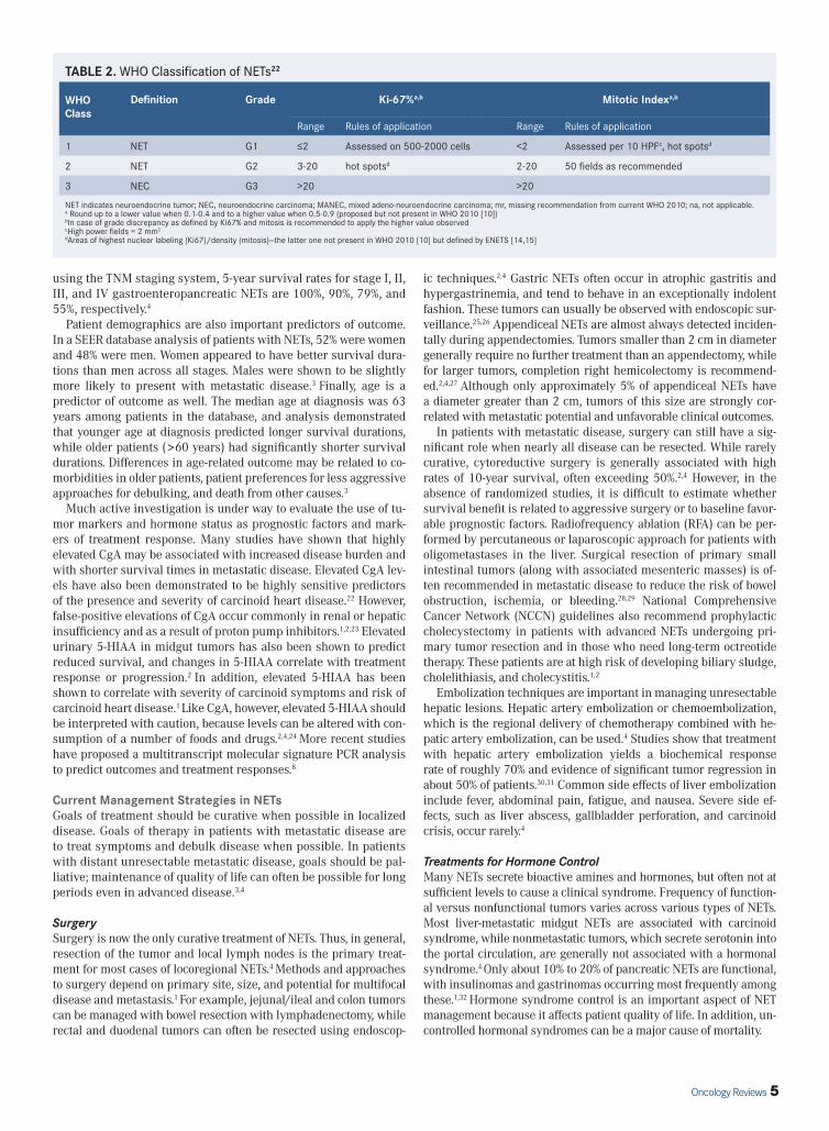
ClassificationBecause of their heterogeneity as a group, NETs have historically been classified in a number of ways, including by primary site, embryologic origin, malignant potential, and functionality.4 Tumor-node-metastasis (TNM) staging classifications have been intro-duced by several guideline organizations, including the American Joint Committee on Cancer (AJCC) and the European Neuroendo-crine Tumor Society (ENETS). A grading classification introduced by the World Health Organization (WHO) distinguishes between well-differentiated low-grade (G1) tumors, well-differentiated intermedi-ate (G2) tumors, and poorly differentiated high-grade (G3) tumors (TABLE 2). This classification implies that there is a concordance between tumor differentiation (morphology) and tumor grade; how-ever there are exceptions in the form of well-differentiated tumors with Ki-67 index >20% (high-grade).20
Many studies have shown that tumor differentiation and grade cor-relate with outcomes, with more aggressive course, worse prognosis, poor differentiation, and higher grade.2 For example, in one analysis only 20% of G1 tumors were found to have distant metastases at di-agnosis, while 50% of poorly differentiated tumors had distant metas-tases at diagnosis.3 In addition, survival is significantly affected by tumor grade: the median overall survival (OS) in G1 and G2 NETs are 124 and 64 months, respectively, while patients with high-grade can-cers experience median OS of 10 months.3 Thus, all pathology report-ing should indicate tumor differentiation, mitotic rate, Ki-67 index, and assigned WHO classification.
Prognostic FactorsClassification by tumor differentiation and grade, as in the cur-rent WHO classification system, gives significant prognostic in-formation. In addition, other factors can affect survival in patients with NETs. Likely, primary tumor site is one of the most important predictors of outcome.1,3,21 For example, among patients with local-ized disease, colon, rectal, and appendiceal tumors are associated with the highest 5-year survival rates, ranging from 85% to 90%.3
However, among patients with metastatic tumors, small bowel pri-mary site is associated with the most favorable prognosis. Tumor burden and presence of metastases at diagnosis are also predic-tive of negative outcome, with significantly decreased survival in patients who present with evidence of regional or distant disease;
TABLE 1. Symptoms Associated With Various NET Types1,2,4,6
Syndrome Clinical Symptoms Site Hormones
Carcinoid Flushing, diarrhea, wheezing Jejurium, ileum, cecum (midgut) Serotonin, substance P, NKA, VIP, PP
Glucagonoma Dermatitis (Sweet’s Syndrome), weight loss, diabetes, stomatitis, diarrhea
Pancreas Glucagon
Gastrinoma/Zollinger-Ellison Syndrome
Severe peptic ulcerations, diarrhea Pancreas, duodenum Gastrin
Insulinoma/Hypoglycemia
Confusion, sweating, dizziness, weakness, syncope, relief with eating
Pancreas Insulin
VIPoma Profuse diarrhea, constipation, abdominal pain Pancreas VIP
NKA indicates neurokinin A; PP, pancreatic polypeptide; VIP, vasoactive intestinal polypeptide.
FIGURE 1. A 35-year-old female presenting with recurrent pain in abdomen and multiple hepatic space-occupying lesions on ultrasound. Fine-needle aspiration cytology from liver lesions demonstrated metastatic neuroendocrine tumor (NET).
From Sharma P, Singh H, Bal C, and Kumar R. PET/CT imaging of neuroendocrine tumors with Gallium-labeled somatostatin analogues: An overview and single institutional experience from India. Indian J Nucl Med. 2014;29(1):2-12.
Oncology Reviews 5
using the TNM staging system, 5-year survival rates for stage I, II, III, and IV gastroenteropancreatic NETs are 100%, 90%, 79%, and 55%, respectively.4
Patient demographics are also important predictors of outcome. In a SEER database analysis of patients with NETs, 52% were women and 48% were men. Women appeared to have better survival dura-tions than men across all stages. Males were shown to be slightly more likely to present with metastatic disease.3 Finally, age is a predictor of outcome as well. The median age at diagnosis was 63 years among patients in the database, and analysis demonstrated that younger age at diagnosis predicted longer survival durations, while older patients (>60 years) had significantly shorter survival durations. Differences in age-related outcome may be related to co-morbidities in older patients, patient preferences for less aggressive approaches for debulking, and death from other causes.3
Much active investigation is under way to evaluate the use of tu-mor markers and hormone status as prognostic factors and mark-ers of treatment response. Many studies have shown that highly elevated CgA may be associated with increased disease burden and with shorter survival times in metastatic disease. Elevated CgA lev-els have also been demonstrated to be highly sensitive predictors of the presence and severity of carcinoid heart disease.22 However, false-positive elevations of CgA occur commonly in renal or hepatic insufficiency and as a result of proton pump inhibitors.1,2,23 Elevated urinary 5-HIAA in midgut tumors has also been shown to predict reduced survival, and changes in 5-HIAA correlate with treatment response or progression.2 In addition, elevated 5-HIAA has been shown to correlate with severity of carcinoid symptoms and risk of carcinoid heart disease.1 Like CgA, however, elevated 5-HIAA should be interpreted with caution, because levels can be altered with con-sumption of a number of foods and drugs.2,4,24 More recent studies have proposed a multitranscript molecular signature PCR analysis to predict outcomes and treatment responses.8
Current Management Strategies in NETsGoals of treatment should be curative when possible in localized disease. Goals of therapy in patients with metastatic disease are to treat symptoms and debulk disease when possible. In patients with distant unresectable metastatic disease, goals should be pal-liative; maintenance of quality of life can often be possible for long periods even in advanced disease.3,4
SurgerySurgery is now the only curative treatment of NETs. Thus, in general, resection of the tumor and local lymph nodes is the primary treat-ment for most cases of locoregional NETs.4 Methods and approaches to surgery depend on primary site, size, and potential for multifocal disease and metastasis.1 For example, jejunal/ileal and colon tumors can be managed with bowel resection with lymphadenectomy, while rectal and duodenal tumors can often be resected using endoscop-
ic techniques.2,4 Gastric NETs often occur in atrophic gastritis and hypergastrinemia, and tend to behave in an exceptionally indolent fashion. These tumors can usually be observed with endoscopic sur-veillance.25,26 Appendiceal NETs are almost always detected inciden-tally during appendectomies. Tumors smaller than 2 cm in diameter generally require no further treatment than an appendectomy, while for larger tumors, completion right hemicolectomy is recommend-ed.2,4,27 Although only approximately 5% of appendiceal NETs have a diameter greater than 2 cm, tumors of this size are strongly cor-related with metastatic potential and unfavorable clinical outcomes.
In patients with metastatic disease, surgery can still have a sig-nificant role when nearly all disease can be resected. While rarely curative, cytoreductive surgery is generally associated with high rates of 10-year survival, often exceeding 50%.2,4 However, in the absence of randomized studies, it is difficult to estimate whether survival benefit is related to aggressive surgery or to baseline favor-able prognostic factors. Radiofrequency ablation (RFA) can be per-formed by percutaneous or laparoscopic approach for patients with oligometastases in the liver. Surgical resection of primary small intestinal tumors (along with associated mesenteric masses) is of-ten recommended in metastatic disease to reduce the risk of bowel obstruction, ischemia, or bleeding.28,29 National Comprehensive Cancer Network (NCCN) guidelines also recommend prophylactic cholecystectomy in patients with advanced NETs undergoing pri-mary tumor resection and in those who need long-term octreotide therapy. These patients are at high risk of developing biliary sludge, cholelithiasis, and cholecystitis.1,2
Embolization techniques are important in managing unresectable hepatic lesions. Hepatic artery embolization or chemoembolization, which is the regional delivery of chemotherapy combined with he-patic artery embolization, can be used.4 Studies show that treatment with hepatic artery embolization yields a biochemical response rate of roughly 70% and evidence of significant tumor regression in about 50% of patients.30,31 Common side effects of liver embolization include fever, abdominal pain, fatigue, and nausea. Severe side ef-fects, such as liver abscess, gallbladder perforation, and carcinoid crisis, occur rarely.4
Treatments for Hormone ControlMany NETs secrete bioactive amines and hormones, but often not at sufficient levels to cause a clinical syndrome. Frequency of function-al versus nonfunctional tumors varies across various types of NETs. Most liver-metastatic midgut NETs are associated with carcinoid syndrome, while nonmetastatic tumors, which secrete serotonin into the portal circulation, are generally not associated with a hormonal syndrome.4 Only about 10% to 20% of pancreatic NETs are functional, with insulinomas and gastrinomas occurring most frequently among these.1,32 Hormone syndrome control is an important aspect of NET management because it affects patient quality of life. In addition, un-controlled hormonal syndromes can be a major cause of mortality.
TABLE 2. WHO Classification of NETs22
WHO Class
Definition Grade Ki-67%a,b Mitotic Indexa,b
Range Rules of application Range Rules of application
1 NET G1 ≤2 Assessed on 500-2000 cells <2 Assessed per 10 HPFc, hot spotsd
2 NET G2 3-20 hot spotsd 2-20 50 fields as recommended
3 NEC G3 >20 >20
NET indicates neuroendocrine tumor; NEC, neuroendocrine carcinoma; MANEC, mixed adeno-neuroendocrine carcinoma; mr, missing recommendation from current WHO 2010; na, not applicable.a Round up to a lower value when 0.1-0.4 and to a higher value when 0.5-0.9 (proposed but not present in WHO 2010 [10])bIn case of grade discrepancy as defined by Ki67% and mitosis is recommended to apply the higher value observedcHigh power fields = 2 mm2
dAreas of highest nuclear labeling (Ki67)/density (mitosis)—the latter one not present in WHO 2010 [10] but defined by ENETS [14,15]
6 Oncology Reviews
SurgerySurgical resection, as discussed previously, is one strategy for hor-mone control. In locoregional disease, surgical resection has a cu-rative goal, while in advanced disease it has a primarily palliative role. Debulking surgery can be used to manage symptoms from hormone secretion. Because tumors with hepatic metastases are more likely to manifest hormone-related symptoms, management of hepatic metastases with a variety of techniques can have a sig-nificant impact on patient quality of life, including surgical re-section, hepatic artery embolization or chemoembolization, and RFA. For example, in one prospective trial of RFA, 92% of treated patients reported at least partial symptom relief, and 70% had sig-nificant or complete relief within 1 week of procedure.33 Similarly, one benefit of hepatic embolization techniques is symptom relief, with 70% to 90% of patients experiencing improvement.4
An important consideration when considering surgery in patients with NETs is the need to prevent carcinoid crisis, especially in pa-tients with functional tumors, carcinoid syndrome, and hepatic me-tastases. Carcinoid crisis can be provoked by anesthesia, surgical or endoscopic interventions, and manipulation of the tumor.1 Current guidelines suggest administering prophylactic short-acting octreo-tide therapy before the induction of anesthesia, and then continuing during surgery.2,4, 25
Somatostatin AnalogsNatural somatostatin acts as a general inhibitor of endocrine func-tion. In the GI tract, it inhibits intestinal motility, exocrine secretions, and absorption of nutrients and ions. It also inhibits the secretion of other hormones such as serotonin, gastrin, or glucagon.1,34 Because of the very short half-life of natural somatostatin, somatostatin ana-logs (SSAs) with prolonged duration of action were developed for hormonal symptoms control. The commercially available SSAs are octreotide and lanreotide. Octreotide selectively binds to somatosta-tin receptor subtypes (SSTR) 2 and, to a lesser extent, SSTR 3 and SSTR 5.35 Its chemical structure differs from that of somatostatin, resulting in a longer half-life of 2 hours (immediate release formula-tion).36,37 Lanreotide has a binding profile similar to that of octreo-tide, but it has a high affinity for SSTR 2 and 5 and a reduced binding affinity for SSTR 1, 3, and 4.36,38 Both lanreotide and octreotide are recommended by the NCCN guidelines for control of symptoms as-sociated with metastatic carcinoid tumors.2
Octreotide was approved by the US Food and Drug Administra-tion (FDA) in 1987 for relief of flushing or diarrhea associated with
metastatic carcinoid tumors.1,5 It is also active for palliation of rare syndromes produced by vasoactive intestinal peptide (VIP) and glucagon.4,39 Octreotide is more potent than somatostatin at inhibi-tion of growth hormone, glucagon, and insulin than somatostatin. It suppresses LH response to GnRH. It also decreases splanchnic blood flow and inhibits release of serotonin, gastrin, vasoactive intestinal peptide, secretin, motilin, and pancreatic polypeptide.37
Lanreotide is approved outside of the United States for control of symptoms.38 In the phase III multinational ELECT trial, 115 pa-tients with carcinoid syndrome were randomly assigned to receive lanreotide or placebo. The primary endpoint (percentage of days rescue octreotide was required) between lanreotide and placebo was 34% compared with 49%. The results seen were deemed impor-tant enough to warrant use of lanreotide for symptom control.2 GH inhibition produced by lanreotide is believed to be due to activity at SSTR 2 and 5. The primary effect of lanreotide is reduction of GH and/or IGF-1 levels. It also inhibits the basal secretion of motilin, gastric inhibitory peptide, and pancreatic polypeptide; however, it has no significant effect on the secretion of secretin. Postprandial secretion of pancreatic polypeptide, gastrin, and cholecystokinin are inhibited.38
Long-acting formulations of SSAs, administered every 4 weeks, are now the standard of care for management of symptoms in NETs, because they have been shown to have comparable or bet-ter efficacy than shorter-acting agents.4,40 Sandostatin LAR Depot (octreotide acetate for injectable suspension) is a long-acting dos-age form of octreotide. It is injected intramuscularly once every 4 weeks.37 Somatuline Depot (lanreotide) is an octapeptide analog of natural somatostatin. Lanreotide is injected subcutaneously every 4 weeks in the superior external quadrant of the buttock. It is avail-able in single-use, prefilled syringes.38
The side effects of SSAs are typically mild and include nausea, cramping, loose stools, steatorrhea, pain, or erythema at the injec-tion site. In addition, biliary sludge and cholelithiasis occur in up to 50% of patients, although only 1% to 3% will become symptomatic and require cholecystectomy.1,4
InterferonsUse of interferon-α (IFN-α) for control of carcinoid syndrome predates SSAs. Because of its side-effect profile, which includes flu-like symptoms, IFN-α is rarely prescribed as a first-line agent.41 However, it can be added to an SSA for treatment of refractory carcinoid syndrome.1,42
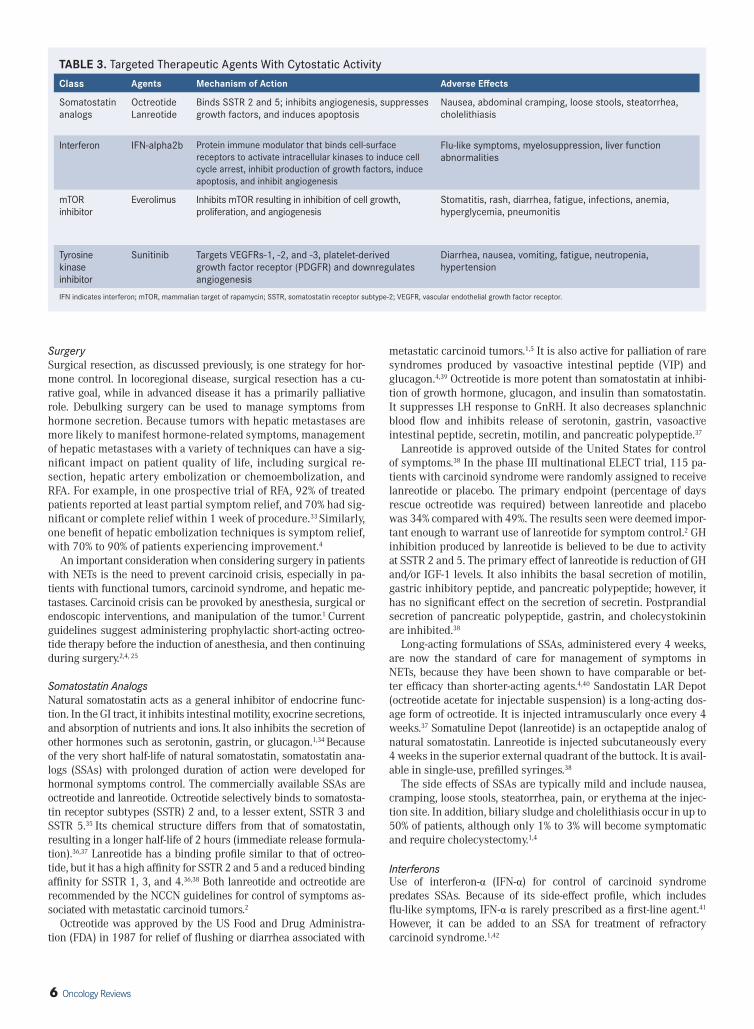
TABLE 3. Targeted Therapeutic Agents With Cytostatic Activity
Class Agents Mechanism of Action Adverse Effects
Somatostatin analogs
OctreotideLanreotide
Binds SSTR 2 and 5; inhibits angiogenesis, suppresses growth factors, and induces apoptosis
Nausea, abdominal cramping, loose stools, steatorrhea, cholelithiasis
Interferon IFN-alpha2b Protein immune modulator that binds cell-surface receptors to activate intracellular kinases to induce cell cycle arrest, inhibit production of growth factors, induce apoptosis, and inhibit angiogenesis
Flu-like symptoms, myelosuppression, liver function abnormalities
mTOR inhibitor
Everolimus Inhibits mTOR resulting in inhibition of cell growth, proliferation, and angiogenesis
Stomatitis, rash, diarrhea, fatigue, infections, anemia, hyperglycemia, pneumonitis
Tyrosine kinase inhibitor
Sunitinib Targets VEGFRs-1, -2, and -3, platelet-derived growth factor receptor (PDGFR) and downregulates angiogenesis
Diarrhea, nausea, vomiting, fatigue, neutropenia, hypertension
IFN indicates interferon; mTOR, mammalian target of rapamycin; SSTR, somatostatin receptor subtype-2; VEGFR, vascular endothelial growth factor receptor.
Oncology Reviews 7
Hormone-Specific TreatmentsAgents targeting symptoms caused by specific hormones are also effective. For example, proton pump inhibitors are the treatment of choice for symptomatic control of Zollinger-Ellison syndrome caused by gastrinomas.43 Diazoxide is used in insulinomas to help control symptoms of hypoglycemia.4
Treatments With Antitumor Activity (TABLE 3)Somatostatin AnalogsSSAs, historically used for symptom control, are now known to have cytostatic effects on NET cells. Both direct and indirect mechanisms of action have been proposed, including induction of intracellular phosphotyrosine phosphatases, suppression of cytokines and growth factors, and inhibition of angiogenesis.33,44 Lanreotide and octreotide have been studied for the treatment of neuroendocrine tumors.
PROMID Trial. This double-blind, placebo-controlled phase III trial examined the effect of octreotide LAR 30 mg in midgut NETs in 85 patients who were treatment-naïve. Patients were randomly as-signed to octreotide LAR 30 mg or placebo in monthly intervals until tumor progression or death. A total of 67 tumor progressions and 16 deaths occurred prior to analysis. Median time to tumor progression was 14.3 months and 6 months in the octreotide LAR and placebo groups, respectively (P = .000072). Stable disease was observed after 6 months of treatment in 66.7% and 37.2% of patients in the octreotide LAR and placebo groups, respectively. The HR for overall survival was 0.81 (95% CI, 0.30-2.18). The most favorable outcomes were seen in patients with resected primary tumor and low hepatic tumor load. No difference in overall survival was observed, possible because of the small study size and the low number of deaths ob-served (7 and 9 in the octreotide and placebo groups, respectively).6
CLARINET Trial. The randomized, double-blind, placebo-controlled CLARINET trial evaluated long-acting lanreotide in 204 patients with advanced, grade 1 or 2 (Ki-67 <10%) nonfunctioning somatostatin-receptor–positive gastroenteropancreatic NETs. Patients received lan-reotide 120 mg or placebo once every 28 days for 96 weeks. Lanreo-tide was associated with significantly improved progression-free sur-vival compared with placebo (median not reached vs median of 18.0 months; P <.001). At 24 months, estimated rates of progression-free survival were 65.1% and 33.0% in the lanreotide and placebo groups, respectively. There was no impact in QOL with lanreotide treatment. No significant difference was observed in OS, possibly due to the low number of deaths and because crossover was allowed in the study. The benefit was noted irrespective of tumor grade or tumor volume.7
Both octreotide and lanreotide are recommended by the NCCN for tumor control in NETs of the gastroenteropancreatic tract.2 Lanreo-tide was approved by the US FDA for the treatment of metastatic gastrointestinal and pancreatic neuroendocrine tumors to improve progression-free survival in 2014.
InterferonsAnother potential antitumor agent is IFN-α; it is a protein im-mune modulator that acts on cell-surface receptors to activate in-tracellular kinases to induce cell cycle arrest, inhibit production of growth factors, induce apoptosis, and inhibit angiogenesis.4,34
Several small, randomized studies have demonstrated prolonged time to progression with the combination of a somatostatin ana-log with IFN-α versus SSA monotherapy; however, the absence of large studies and the side-effect profile of IFN-α have limited its use.34,45,46 According to NCCN guidelines, use of IFN-α 2b is an option (category 3 recommendation) for use in progressive NETs of GI origin.2
Systemic ChemotherapySystemic chemotherapy is another option in the treatment of advanced or progressive disease. However, in G1 nonpancreatic NETs, treatment with chemotherapy typically results in poor response rates of <15% with no increase in OS.2,12 Thus, NCCN guidelines recommend systemic chemotherapy in advanced well-differentiated nonpacreatic tumors only if no other treatment options are available.2 In poorly differentiated tumors, however, response rates of 42% to 67% using platinum-based regimens such as cisplatin/etoposide have been reported.4,47,48 Accordingly, systemic chemotherapy is first-line treatment for these cases.2
Other chemotherapy regimens under investigation include temo-zolomide alone or in combination with capecitabine and with or without bevacizumab.12
Pancreatic NETs are substantially more responsive to chemother-apy than intestinal NETs. Streptozotocin-based regimens (combined with fluorouracil [5-FU] or doxorubicin) are standard. However, these treatment regimens are often limited by toxicity, and there is some controversy about their true efficacy, with recent studies finding 30% to 40% response rates in treated patients compared with >60% re-sponse rates in older studies.29,49 More recently, regimens consisting of temozolomide with or without capecitabine have been associated with response rates in the 30% to 70% range.50,51 However, random-ized trials evaluating temozolomide-based regimens have not been completed, and high-level data are lacking.29
EverolimusThe mammalian target of rapamycin (mTOR) has become a po-tential therapeutic target because studies have shown that abnor-malities within the pathway, which stimulate cell growth, prolif-eration, and angiogenesis, may play a role in the pathogenesis of NETs. Everolimus is a novel agent that inhibits mTOR.52 Phase II studies confirmed its antitumor activity in pancreatic NETs. Sub-sequently, in the RADIANT-3 study, a phase III trial of 410 pa-tients with advanced low-/intermediate-grade, progressive pan-creatic NETs, patients treated with everolimus had median PFS of 11 months compared with 4.6 months in patients who received placebo.49 The most common adverse effects, primarily grade 1 or 2, included stomatitis (64%), rash (49%), diarrhea (34%), fatigue (31%), and infections (23%). More severe grade 3 or 4 effects were anemia (6%) and hyperglycemia (5%).49 The results of RADIANT-3 prompted FDA approval of everolimus for treatment of advanced progressive pancreatic NETs.
The RADIANT-2 trial compared treatment with everolimus plus octreotide versus placebo plus octreotide in 429 patients with ad-vanced carcinoid tumors who had a history of carcinoid syndrome. Although an improvement in median PFS, from 11.3 months on the control arm to 16.4 months on the experimental arm, was observed on central radiographic review, the trial fell short of statistical signifi-cance (actual P value of .026 vs target value of .024). Discrepancies between central and local radiographic review may have led to loss of statistical significance. No differences in OS were seen in this study. However, the study was not powered for this endpoint, and crossover from placebo to everolimus may have diminished any trends toward an improvement in survival.53 Results are expected from the RADI-ANT-4 trial, which is comparing everolimus to placebo in patients with nonfunctional NETs of the GI tract and lungs.54
SunitinibSunitinib is an orally available tyrosine kinase inhibitor (TKI) that targets the intracellular domain of VEGF receptors.4 A phase III, randomized, double-blind, placebo-controlled trial showed im-provement in PFS (11.4 months) in the sunitinib-treated group compared with the placebo group (5.5 months). Response rate was

8 Oncology Reviews
9.3% in the sunitinib group (compared with 0% in the placebo group), and 63% of treated patients demonstrated stable disease. Adverse ef-fects were primarily grade 1 or 2, including diarrhea, nausea, vomit-ing, and fatigue, and the most common grade 3 or 4 adverse effects were neutropenia and hypertension.55 The results of this study led to FDA approval of sunitinib in 2011 for treatment of progressive, well-differentiated, pancreatic NETs in patients with unresectable, locally advanced, or metastatic disease. VEGF-receptor–targeting TKIs continue to be investigated for the treatment of nonpancreatic NETs; potential agents include pazopanib and axitinib.34
BevacizumabBevacizumab, an antibody that binds and inhibits VEGF, is thought to be a agent with a relatively favorable toxicity profile. Based on a promising randomized, phase II trial, a multicenter phase III trial compared bevacizumab with INF-α in patients with carcinoid tu-mors that were either progressive or considered high risk based on primary tumor location and tumor grade. This study did not demonstrate improved PFS with bevacizumab compared with in-terferon.56,57 Results of a clinical study comparing bevacizumab plus everolimus versus everolimus monotherapy in pancreatic NETs are pending.58
Radiolabeled Somatostatin AnalogsAn emerging treatment option consists of radiolabeled somatosta-tin analogs. This form of systemic radiation, also known as peptide receptor radiotherapy (PRRT), enables targeted delivery of radio-activity to somatostatin-receptor–expressing NETs.4 In a study of patients with advanced NETs treated with Lu-177 labeled octreo-tate, complete, partial, or minor tumor response rates were 2%, 28%, and 16%, respectively, and 20% of patients had stable disease. Treated patients also showed significantly longer median OS from time of diagnosis compared with historical controls.59 However, PRRT is available primarily in Europe, and is still only investi-gational in the US. The most common adverse effects are nausea, vomiting, and abdominal pain after administration, but these are usually easily managed with supportive care. Grade 3 or 4 adverse effects, including myelosuppression, nephrotoxicity, and hepatic toxicity, are rare. Cases of leukemia or myelodysplastic syndrome (MDS) have rarely developed as a long-term consequence of treat-ment.4,56 The international NETTER trial, a phase III study compar-ing Lu-177-octreotate with high-dose octreotide in patients with progressive midgut carcinoid tumors, has recently completed ac-crual, and represents the first randomized, phase III study evalu-ating a radiolabeled SSA.60
Other Novel AgentsTelotristat etiprate is a novel agent under investigation for control of carcinoid syndrome; it inhibits tryptophan hydroxylase activity, which is involved in serotonin synthesis, thus reducing serum se-rotonin levels. A phase III trial is in progress to assess the efficacy and safety of telotristat compared with placebo in patients with refractory carcinoid syndrome despite SSAs.34
Recent studies show that there is often coexpression of soma-tostatin and dopamine receptors (DRs) on NET cell lines, suggest-ing that the 2 receptors may dimerize. Chimeric somatostatin mol-ecules that inhibit both SSTR 2 and DR have thus been developed. Although initial results have largely been negative, there is ongoing research to develop new chimeric molecules.53
Guidelines for Management of Neuroendocrine TumorsGuidelines have been released in recent years to guide manage-ment of NETs. These include the NCCN guidelines (2015), Europe-an Society for Medical Oncology (ESMO) guidelines (2012), North
American Neuroendocrine Tumors Society (NANETS) guidelines (2010 and updated in 2013), and the UK/Ireland Neuroendocrine Tumour Society guidelines (2012).2,4,12,61 While they have differ-ent approaches to NETs, the guidelines reflect several general guiding principles:
1. Surgery should be pursued for cure in locoregional disease.2. In advanced disease, cytoreductive resection should still be
considered in the large majority of disease if >90% can be safely resected. Clinicians should also consider resection of primary intestinal tumors, even in the setting of metastases.
3. Therapy with an SSA should be offered to all patients with car-cinoid syndrome and considered as the first option for patients with unresectable, well or moderately differentiated, locally advanced or metastatic gastroenteropancreatic neuroendo-crine tumors to improve progression-free survival.
4. Systemic options beyond SSAs are limited in nonpancreatic NETs. Liver-directed therapy should be considered for patients with liver-predominant metastases.
5. Systemic treatment options beyond SSAs consist of everolim-us, sunitinib, and systemic chemotherapy for pancreatic NETs.
6. While US guidelines are generally silent on the role of radiola-beled SSAs because of lack of availability, European guidelines generally advocate consideration of these agents for manage-ment of progressive somatostatin-receptor–expressing (Oc-treoScan-positive) tumors.
7. Platinum-based regimens such as carboplatin-etoposide are generally recommended for poorly differentiated neuroendo-crine cancers.
8. Patients should always be considered for a clinical trial.
Challenges in the Management of Neuroendocrine TumorsSurveillance ChallengesAlthough most NETs are slow growing, the natural history of vari-ous types of NETs can vary widely; thus, the optimal surveillance strategy (including follow-up interval/frequency, duration, im-aging, and biochemical testing) for NETs is not standardized. In general, according to NCCN guidelines, surveillance of resected lo-coregional disease within the first year should include CT/MRI and biochemical testing as clinically indicated; intervals range from every 3 to 12 months. Beyond 1 to 3 years, surveillance is usually conducted every 6 to 12 months using imaging and biochemical testing as clinically indicated. In most types of NETs, NCCN recom-mends following to 10 years. In some NET types, follow-up schedule and choice of imaging vary with primary tumor size; for example, in rectal NETs, tumors <1 cm do not warrant any follow-up, and tumors of 1 to 2 cm should be followed with MRI or EUS at 6 and 12 months, and then as needed. Likewise, gastric tumors ≤2 cm at diagnosis can be monitored with esophagogastroduodenoscopy (EGD), while larger tumors should be monitored with CT/MRI.2 According to ESMO guidelines, tumor grade should be considered when determining surveillance strategy: resected NET G1/2 tu-mors should be imaged every 3 to 6 months, and G3 tumors should be monitored every 2 to 3 months within the first year.12
Somatostatin receptor imaging (OctreoScan or Gallium-68-PET) and use of biochemical markers are additional tools that can be used for surveillance. ESMO recommends repeat somatostatin re-ceptor imaging every 18 to 24 months for surveillance, but only if there is proven expression of SSTR 2 on tumor cells.2,5 The role of tumor markers for surveillance is controversial. CgA is the most commonly used marker for historical reasons but is considered a category 3 recommendation (no consensus) by the NCCN.2 Other markers that can be used to monitor patients include NSE, neuroki-nin A, and pancreastatin.53

Oncology Reviews 9
Treatment ChallengesRefractory Carcinoid SyndromeRefractory carcinoid syndrome or tachyphylaxis is seen in many pa-tients treated with SSAs. Several mechanisms of resistance have been proposed, including downregulation of SSTRs, alterations in expres-sion pattern of SSTRs (less type 2 and 5, which are main targets of available SSAs), internalization of SSTR 2, development of mutated forms of SSTRs, and development of antibodies against SSAs.41 So far, there are no reliably effective treatments for resistant patients. How-ever, several retrospective studies indicate benefit associated with above-label dosage or frequency of SSA injections. The role of telotri-stat, a peripheral serotonin inhibitor, is yet to be defined.4,34
Carcinoid CrisisCarcinoid crisis is caused by an acute release of serotonin and oth-er vasoactive peptides, often provoked by anesthesia, surgery, and intraoperative manipulation of the tumor, chemotherapy, hepatic artery embolization, and hepatic ablation procedures. Clinical presentation is characterized by severe flushing, bronchoconstric-tion, and hemodynamic instability.1,4 Prevention of carcinoid crisis is essential in patients with carcinoid syndrome, and even those already using long-acting SSAs require prophylactic, short-acting SSAs before and during procedures.1,4 In the event of carcinoid crisis, short-acting octreotide as bolus then intravenous infusion should be administered. Patients with nonfunctional tumors do not need prophylaxis, but octreotide should be available, especial-ly in patients with small bowel NETs undergoing an intervention.4
Carcinoid Heart DiseaseCarcinoid heart disease (CHD) is a rare but serious complication of carcinoid syndrome, occurring most commonly in patients with extremely high levels of circulating serotonin.1,4 The condi-tion manifests as valvular fibrosis, most commonly right-sided (tricuspid and pulmonic valves most commonly affected), lead-ing to valve regurgitation or stenosis, and eventually right-sided heart failure.62 Diagnosis is established by echocardiography, with valvular findings pathognomonic for CHD; N-terminal of prohor-mone brain natriuretic peptide (NT-proBNP) can serve as a serum marker of CHD.4,.59
The presence of CHD confers worse prognosis: 3-year survival in patients with CHD is 31% compared with 68% in patients with car-cinoid syndrome but no CHD.1,4 The most common cause of death in patients with CHD (50%) is right-sided heart failure. Although most patients present with signs and symptoms of heart failure, some remain asymptomatic.59,63 Because of the significantly decreased survival of patients with CHD, there is a need to establish guide-lines for screening, as well as initiation of earlier or more aggressive intervention. ENETS recommends mandatory annual echocardiog-raphy screening of patients with carcinoid syndrome, while other guidelines only recommend echocardiograms in patients with signs or symptoms of carcinoid heart disease.64
Cardiac valve surgery (typically replacement of tricuspid and pulmonic valves) is the only definitive treatment that provides symptomatic improvement and increased OS in CHD.59
Clinical Use/Development of Newer AgentsNETs are a heterogenous group of neoplasms, and consensus pan-els have developed guidelines for trial endpoints that have taken into consideration both the unique needs of this patient population and also the requirements for FDA approval. However, much work remains to be done in sequencing and patient selection. Under-standing of resistance mechanisms and interplay between path-ways known to drive carcinogenesis is vital. Immune oncology is a rapidly developing field, and as yet not well studied in this disease.
The mechanism of SSA activity as an antitumor agent is also not well understood and requires further study in the context of tumor burden and other known prognostic factors.65
REFERENCES1. Gastrointestinal Carcinoid Tumors Treatment (PDQ®). National Cancer Institute. http://
www.cancer.gov/cancertopics/pdq/treatment/gastrointestinalcarcinoid/HealthProfes-
sional. Updated February 25, 2015. Accessed April 27, 2015.
2. National Comprehensive Cancer Network. Clinical Practice Guidelines in Oncology
(NCCN Guidelines®). Neuroendocrine Tumors. Version 1.2015. http://www.nccn.org/
professionals/physician_gls/pdf/neuroendocrine.pdf. Published November 11, 2014.
Accessed April 27, 2015.
3. Yao YC, Hassan M, Phan A, et al. One hundred years after “carcinoid”: epidemiology of
and prognostic factors for neuroendocrine tumors in 35,825 cases in the United States. J
Clin Oncol. 2008;26:3063-3072.
4. Ramage JK, Ahmed A, Ardill J, et al. Guidelines for the management of gastroenteropan-
creatic neuroendocrine (including carcinoid) tumours (NETs). Gut. 2012;61:6-32.
5. Kvols LK, Moertel CG, O’Connell MJ, et al. Treatment of the malignant carcinoid syn-
drome. Evaluation of a long-acting somatostatin analogue. N Engl J Med. 1986;315:663-
666.
6. Rinke A, Muller HH, Schade-Brittinger C, et al. Placebo-controlled, double-blind prospec-
tive, randomized study on the effect of octreotide LAR in the control of tumor growth
in patients with metastatic neuroendocrine midgut tumors: a report from the PROMID
study group. J Clin Oncol. 2009;27:4656-4663.
7. Caplin ME, Pavel M, Cwikla JB, et al. Lanreotide in metastatic enteropancreatic neuroen-
docrine tumors. N Engl J Med. 2014;371(3):224-233.
8. Eckhauser FE, Argenta LC, Strodel WE, et al. Mesenteric angiopathy, intestinal gangrene,
and midgut carcinoids. Surgery. 1981;90:720-728.
9. Vinik AI, Silva MP, Woltering EA, et al. Biochemical testing for neuroendocrine tumors.
Pancreas. 2009;38:876-889.
10. Matuchansky C, Launay JM. Serotonin, catecholamines, and spontaneous midgut
carcinoid flush: plasma studies from flushing and nonflushing sites. Gastroenterology.
1995;108:743-751.
11. Oberg K, Knigge U, Kwekkboom D, et al. Neuroendocrine gastro-entero-pancreatic
tumors: ESMO clinical practice guidelines for diagnosis, treatment and follow-up. Ann
Oncol. 2012;23(suppl 7):vii124-vii130.
12. Mamikunian G, Vinik A, O’Dorisio TM, Woltering EA, Go VLW. Neuroendocrine Tumors:
A Comprehensive Guide to Diagnosis and Management, Fourth Edition. Inglewood, CA:
Inter Science Institute; 2009.
13. O’Toole D, Grossman A, Gross D, et al. ENETS consensus guidelines for the standards of
care in neuroendocrine tumors: biochemical markers. Neuroendocrinology. 2009;90:194-
202.
14. Paulson EK, McDermott VG, Keogan MT, et al. Carcinoid metastases to the liver: role of
triple-phase helical CT. Radiology. 1998;206:143-150.
15. Legmann P, Vignaux O, Dousset B, et al. Pancreatic tumors: comparison of dual-phase
helical CT and endoscopic sonography. AJR Am J Roentgenol. 1998;170:1315-1322.
16. Dromain C, de Baere T, Lumbroso J, et al. Detection of liver metastases from endocrine
tumors: a prospective comparison of somatostatin receptor scintigraphy, computed
tomography, and magnetic resonance imaging. J Clin Oncol. 2005;23:70-78.
17. Krenning EP, Bakker WH, Kooij PP, et al. Somatostatin receptor scintigraphy with indi-
um-111-DTPA-D-Phe-1-octreotide in man: metabolism, dosimetry and comparison with
iodine-123-Tyr-3-octreotide. J Nucl Med. 1992;33:652-658.
18. Krenning EP, Kwekkboom DJ, Bakker WH, et al. Somatostatin receptor scintigraphy with
[111In-DTPA-D-Phe1]- and [123I-Tyr3]-octreotide: the Rotterdam experience with more
than 1000 patients. Eur J Nucl Med. 1993;20:716-731.
19. Koopmans KP, Neels OC, Kema IP, et al. Improved staging of patients with carcinoid and
islet cell tumors with 18F-dihydroxy-phenyl-alanine and 11C-5-hydroxy-tryptophan
positron emission tomography. J Clin Oncol. 2008;26:1489-1495.
20. Rindi G, Petrone G, Inzani F. The 2010 WHO classification of digestive neuroendo-
crine neoplasms: a critical appraisal four years after its introduction. Endocr Pathol.
2014;25(2):186-192.
21. Maggard MA, O’Connell JB, Ko CY. Updated population-based review of carcinoid tu-
mors. Ann Surg. 2004;240:117-122.
22. Korse CM, Taal BG, de Groot CA, Bakker RH, Bonfrer JM. Chromogranin-A and N-
10 Oncology Reviews
terminal pro-brain natriuretic peptide: an excellent pair of biomarkers for diagnostics in
patients with neuroendocrine tumor. J Clin Oncol. 27(26):4293-4299.
23. Duque M, Modlin IM, Gupta A, Saif MW. Biomarkers in neuroendocrine tumors. J Pan-
creas. 2013;14(4):372-376.
24. Campana D, Nori F, Piscitelli L, et al. Chromogranin A: is it a useful marker of neuroen-
docrine tumors? J Clin Oncol. 2007;25:1967-1973.
25. Kulke MH, Anthony LB, Bushnell DL, et al. NANETS treatment guidelines: well-differen-
tiated neuroendocrine tumors of the stomach and pancreas. Pancreas. 2010;39:735-752.
26. Schindl M, Kaserer K, Niederle B. Treatment of gastric neuroendocrine tumors: the
necessity of a type-adapted treatment. Arch Surg. 2001;136:49-54.
27. Boudreaux JP, Klimstra DS, Hassan MM, et al. The NANETS consensus guideline for the
diagnosis and management of neuroendocrine tumors: well-differentiated neuroendo-
crine tumors of the jejunum, ileum, appendix, and cecum. Pancreas. 2010;39:753-766.
28. Sarmiento JM, Heywood G, Rubin J, et al. Surgical treatment of neuroendocrine metasta-
ses to the liver: a plea for resection to increase survival. J Am Coll Surg. 2003;197:29-37.
29. Hellman P, Ladjevardi S, Skogseid B, et al. Radiofrequency tissue ablation using cooled
tip for liver metastases of endocrine tumors. World J Surg. 2002;26:1052-1056.
30. Strosberg JR, Choi J, Cantor AB, Kvols LK. Selective hepatic artery embolization for
treatment of patients with metastatic carcinoid and pancreatic endocrine tumors. Cancer
Control. 2006;13:72-78.
31. Gupta S, Yao JC, Ahrar K, et al. Hepatic artery embolization and chemoembolization for
treatment of patients with metastatic carcinoid tumors: the M.D. Anderson experience.
Cancer J. 2003;9:261-267.
32. McKenna LR, Edil BH. Update on pancreatic neuroendocrine tumors. Gland Surg.
2014;3(4):258-275.
33. Mazzaglia PJ, Berber E, Milas M, et al. Laparoscopic radiofrequency ablation of neuroen-
docrine liver metastases: a 10-year experience evaluating predictors of survival. Surgery.
2007;142(1):10-19.
34. Reichlin S. Somatostatin. N Engl J Med. 1983;309:1495-1501.
35. Maurer R, Reubi JC. Somatostatin receptors. JAMA. 1985;253:2741.
36. Öberg KE. Gastrointestinal neuroendocrine tumors. Ann Oncol. 2010 Oct;21
Suppl7:vii72-80.
37. Sandostatin LAR Depot [package insert]. East Hanover, NJ: Novartis Pharmaceuticals
Corporation; 2014. Available at: http://www.pharma.us.novartis.com/product/pi/pdf/
sandostatin_lar.pdf. Accessed May 27, 2015.
38. Somatuline Depot [package insert]. Basking Ridge, NJ: Ipsen Biopharmaceuticals,
Inc. Revised 12/2014. Available at: http://www.accessdata.fda.gov/drugsatfda_docs/
label/2014/022074s011lbl.pdf. Accessed May 27, 2015.
39. Maton PN. Use of octreotide acetate for control of symptoms in patients with islet cell
tumors. World J Surg. 1993;17:504-510.
40. Alonso-Gordoa T, Capdevila J, Grande E. GEP-NETs update: biotherapy for neuroendo-
crine tumours. Eur J Endocrinol. 2015;172(1):R31-R46.
41. Moertel CG, Rubin J, Kvols LK. Therapy of metastatic carcinoid tumor and the malignant
carcinoid syndrome with recombinant leukocyte A interferon. J Clin Oncol. 1989;7:865-
868.
42. Janson ET, Oberg K. Long-term management of the carcinoid syndrome. Treatment with
octreotide alone and in combination with α-interferon. Acta Oncol. 1993;32:225-229.
43. Jensen RT. Gastrinomas: advances in diagnosis and management. Neuroendocrinology.
2004;80(suppl 1):23-27.
44. Toumpanakis C, Caplin ME. Update on the role of somatostatin analogs for the treat-
ment of patients with gastroenteropancreatic neuroendocrine tumors. Semin Oncol.
2013;40(1):56-68.
45. Kolby L, Persson G, Franzen S, Ahren B. Randomized clinical trial of the effect of inter-
feron α on survival in patients with disseminated midgut carcinoid tumours. Br J Surg.
2003;90:687-693.
46. Arnold R, Rinke A, Klose KJ, et al. Octreotide versus octreotide plus interferon-α in
endocrine gastroenteropancreatic tumors: a randomized trial. Clin Gastroenterol Hepatol.
2005;3:761- 771.
47. Moertel CG, Kvols LK, O’Connell MJ, Rubin J. Treatment of neuroendocrine carcinomas
with combined etoposide and cisplatin. Evidence of major therapeutic activity in the
anaplastic variants of these neoplasms. Cancer. 1991;68:227-232.
48. Mitry E, Baudin E, Ducreux M, et al. Treatment of poorly differentiated neuroendocrine
tumours with etoposide and cisplatin. Br J Cancer. 1999;81:1351-1355.
49. Kouvaraki MA, Ajani JA, Hoff P, et al. Fluorouracil, doxorubicin, and streptozocin in the
treatment of patients with locally advanced and metastatic pancreatic endocrine carci-
nomas. J Clin Oncol. 2004;22:4762-4771.
50. Strosberg JR, Fine RL, Choi J, et al. First-line chemotherapy with capecitabine and
temozolomide in patients with metastatic pancreatic endocrine carcinomas. Cancer.
2011;117:268-275.
51. Fine RL, Gulati AP, Krantz BA, et al. Capecitabine and temozolomide (CAPTEM) for meta-
static, well-differentiated neuroendocrine cancers: The Pancreas Center at Columbia
University experience. Cancer Chemother Pharmacol. 2013;71:663-670.
52. Yao JC, Shah MH, Ito T, et al. Everolimus for advanced pancreatic neuroendocrine tu-
mors. N Engl J Med. 2011;364(6):514-523.
53. Pavel ME, Hainsworth JD, Baudin E, et al. Everolimus plus octrreotide long-acting repeat-
able for the treatment of advanced neuroendocrine tumours associated with carcinoid
syndrome (RADIANT-2): a randomised, placebo-controlled, phase 3 study. Lancet.
2011;378(9808):2005-2012.
54. National Institutes of Health. Everolimus plus best supportive care vs placebo plus best
supportive care in the treatment of patients with advanced neuroendocrine tumors (GI
or lung origin) (RADIANT-4). ClinicalTrials.gov web site: https://clinicaltrials.gov/ct2/
show/NCT01524783. Published December 22, 2011. Updated January 28, 2015. Ac-
cessed May 14, 2015.
55. Raymond E, Dahan L, Raoul JL, et al. Sunitinib malate for the treatment of pancreatic
neuroendocrine tumors. N Engl J Med. 2011;364(6):501-513.
56. Abdel-Rahman O, Fouad M. Bevacizumab-based combination therapy for advanced
gastroenteropancreatic neuroendocrine neoplasms (GEP-NENs): a systematic review of
literature. J Cancer Res Clin Oncol. 2015;141(2):295-305.
57. Yao JC, Phan AT, Hoff PM, et al. Targeting vascular endothelial growth factor in advanced
carcinoid tumor: a random assignment phase II study of depot octreotide with bevaci-
zumab and pegylated interferon alpha-2b. J Clin Oncol. 2008;26(8):1316-1323.
58. National Institutes of Health. Everolimus and octreotide acetate with or without beva-
cizumab in treating patients with locally advanced or metastatic pancreatic neuroen-
docrine tumors that cannot be removed by surgery. ClinicalTrials.gov web site:https://
clinicaltrials.gov/ct2/show/NCT01229943. Published October 27, 2010. Updated April
22, 2015. Accessed May 14, 2015.
59. Kwekkboom DJ, de Herder WW, Kam BL, et al. Treatment with the radiolabeled soma-
tostatin analog [177 Lu-DOTA 0, Tyr3] octreotate: toxicity, efficacy, and survival. J Clin
Oncol. 2008;26(13):2124-2130.
60. Sierra ML, Kwekkboom D, Mariani MF, Bodei L, Santoro P, Krenning EP. NETTER-1:
First pivotal phase III study evaluating 177-Lu-DOTATATE in midgut neuroendocrine
tumours. Presented at: ESMO; September 2014; Madrid, Spain.
61. Kunz KL, Reidy-Lagunes D, Anthony LB, et al. Consensus guidelines for the manage-
ment and treatment of neuroendocrine tumors. Pancreas. 2013;42(4):557-577.
62. Dobson R, Burgess MI, Pritchard DM, Cuthbertson DJ. The clinical presentation and
management of carcinoid heart disease. Int J Cardiol. 2014;173(1):29-32.
63. Zuetenhorst JM, Taal BG. Carcinoid heart disease. N Engl J Med. 2003;348:2359-2361;
author reply 2359-2361.
64. Eriksson B, Kloppel G, Krenning E, et al. Consensus guidelines for the management of
patients with digestive neuroendocrine tumors- well-differentiated jejunal-ileal tumor/
carcinoma. Neuroendocrinology. 2008;87(1):8-19.
65. Zhou C, Zhang J, Zheng Y, Zhu Z. Pancreatic neuroendocrine tumors: a comprehensive
review. Int J Cancer. 2012;131(5):1013-1022.

Oncology Reviews 11
Updates on New and Emerging Therapies for Neuroendocrine TumorsHow to Obtain CME Credit:
1. Read article in its entirety.2. Upon completion, go to www.CECentral.com/getcredit.3. Enter activity code MEN16056.4. Login or register for a free account.5. Complete posttest and evaluation.6. Get credit. A printable certificate will be issued. A passing score of 70% is required.
OncologyR E V I E W S
Targeted
Notes