Embed Size (px)
Citation preview
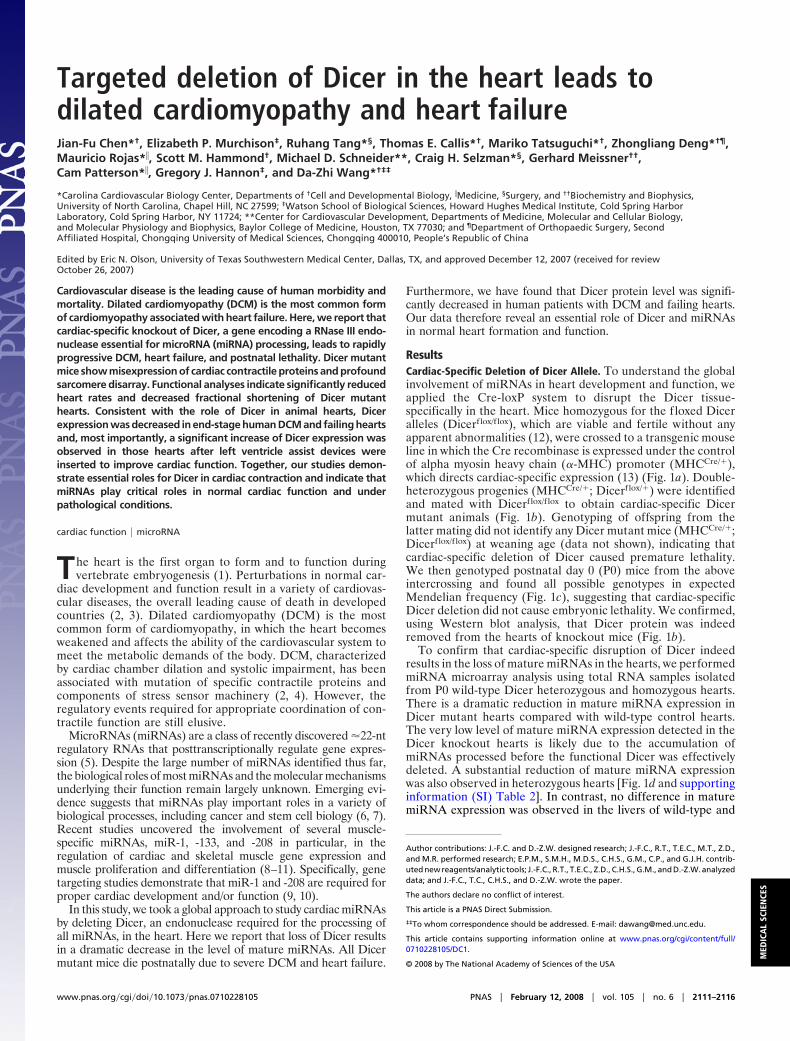
Targeted deletion of Dicer in the heart leads todilated cardiomyopathy and heart failureJian-Fu Chen*†, Elizabeth P. Murchison‡, Ruhang Tang*§, Thomas E. Callis*†, Mariko Tatsuguchi*†, Zhongliang Deng*†¶,Mauricio Rojas*�, Scott M. Hammond†, Michael D. Schneider**, Craig H. Selzman*§, Gerhard Meissner††,Cam Patterson*�, Gregory J. Hannon‡, and Da-Zhi Wang*†‡‡
*Carolina Cardiovascular Biology Center, Departments of †Cell and Developmental Biology, �Medicine, §Surgery, and ††Biochemistry and Biophysics,University of North Carolina, Chapel Hill, NC 27599; ‡Watson School of Biological Sciences, Howard Hughes Medical Institute, Cold Spring HarborLaboratory, Cold Spring Harbor, NY 11724; **Center for Cardiovascular Development, Departments of Medicine, Molecular and Cellular Biology,and Molecular Physiology and Biophysics, Baylor College of Medicine, Houston, TX 77030; and ¶Department of Orthopaedic Surgery, SecondAffiliated Hospital, Chongqing University of Medical Sciences, Chongqing 400010, People’s Republic of China
Edited by Eric N. Olson, University of Texas Southwestern Medical Center, Dallas, TX, and approved December 12, 2007 (received for reviewOctober 26, 2007)
Cardiovascular disease is the leading cause of human morbidity andmortality. Dilated cardiomyopathy (DCM) is the most common formof cardiomyopathy associated with heart failure. Here, we report thatcardiac-specific knockout of Dicer, a gene encoding a RNase III endo-nuclease essential for microRNA (miRNA) processing, leads to rapidlyprogressive DCM, heart failure, and postnatal lethality. Dicer mutantmice show misexpression of cardiac contractile proteins and profoundsarcomere disarray. Functional analyses indicate significantly reducedheart rates and decreased fractional shortening of Dicer mutanthearts. Consistent with the role of Dicer in animal hearts, Dicerexpression was decreased in end-stage human DCM and failing heartsand, most importantly, a significant increase of Dicer expression wasobserved in those hearts after left ventricle assist devices wereinserted to improve cardiac function. Together, our studies demon-strate essential roles for Dicer in cardiac contraction and indicate thatmiRNAs play critical roles in normal cardiac function and underpathological conditions.
cardiac function � microRNA
The heart is the first organ to form and to function duringvertebrate embryogenesis (1). Perturbations in normal car-
diac development and function result in a variety of cardiovas-cular diseases, the overall leading cause of death in developedcountries (2, 3). Dilated cardiomyopathy (DCM) is the mostcommon form of cardiomyopathy, in which the heart becomesweakened and affects the ability of the cardiovascular system tomeet the metabolic demands of the body. DCM, characterizedby cardiac chamber dilation and systolic impairment, has beenassociated with mutation of specific contractile proteins andcomponents of stress sensor machinery (2, 4). However, theregulatory events required for appropriate coordination of con-tractile function are still elusive.
MicroRNAs (miRNAs) are a class of recently discovered �22-ntregulatory RNAs that posttranscriptionally regulate gene expres-sion (5). Despite the large number of miRNAs identified thus far,the biological roles of most miRNAs and the molecular mechanismsunderlying their function remain largely unknown. Emerging evi-dence suggests that miRNAs play important roles in a variety ofbiological processes, including cancer and stem cell biology (6, 7).Recent studies uncovered the involvement of several muscle-specific miRNAs, miR-1, -133, and -208 in particular, in theregulation of cardiac and skeletal muscle gene expression andmuscle proliferation and differentiation (8–11). Specifically, genetargeting studies demonstrate that miR-1 and -208 are required forproper cardiac development and/or function (9, 10).
In this study, we took a global approach to study cardiac miRNAsby deleting Dicer, an endonuclease required for the processing ofall miRNAs, in the heart. Here we report that loss of Dicer resultsin a dramatic decrease in the level of mature miRNAs. All Dicermutant mice die postnatally due to severe DCM and heart failure.
Furthermore, we have found that Dicer protein level was signifi-cantly decreased in human patients with DCM and failing hearts.Our data therefore reveal an essential role of Dicer and miRNAsin normal heart formation and function.
ResultsCardiac-Specific Deletion of Dicer Allele. To understand the globalinvolvement of miRNAs in heart development and function, weapplied the Cre-loxP system to disrupt the Dicer tissue-specifically in the heart. Mice homozygous for the floxed Diceralleles (Dicerflox/f lox), which are viable and fertile without anyapparent abnormalities (12), were crossed to a transgenic mouseline in which the Cre recombinase is expressed under the controlof alpha myosin heavy chain (�-MHC) promoter (MHCCre/�),which directs cardiac-specific expression (13) (Fig. 1a). Double-heterozygous progenies (MHCCre/�; Dicerflox/�) were identifiedand mated with Dicerflox/f lox to obtain cardiac-specific Dicermutant animals (Fig. 1b). Genotyping of offspring from thelatter mating did not identify any Dicer mutant mice (MHCCre/�;Dicerflox/f lox) at weaning age (data not shown), indicating thatcardiac-specific deletion of Dicer caused premature lethality.We then genotyped postnatal day 0 (P0) mice from the aboveintercrossing and found all possible genotypes in expectedMendelian frequency (Fig. 1c), suggesting that cardiac-specificDicer deletion did not cause embryonic lethality. We confirmed,using Western blot analysis, that Dicer protein was indeedremoved from the hearts of knockout mice (Fig. 1b).
To confirm that cardiac-specific disruption of Dicer indeedresults in the loss of mature miRNAs in the hearts, we performedmiRNA microarray analysis using total RNA samples isolatedfrom P0 wild-type Dicer heterozygous and homozygous hearts.There is a dramatic reduction in mature miRNA expression inDicer mutant hearts compared with wild-type control hearts.The very low level of mature miRNA expression detected in theDicer knockout hearts is likely due to the accumulation ofmiRNAs processed before the functional Dicer was effectivelydeleted. A substantial reduction of mature miRNA expressionwas also observed in heterozygous hearts [Fig. 1d and supportinginformation (SI) Table 2]. In contrast, no difference in maturemiRNA expression was observed in the livers of wild-type and
Author contributions: J.-F.C. and D.-Z.W. designed research; J.-F.C., R.T., T.E.C., M.T., Z.D.,and M.R. performed research; E.P.M., S.M.H., M.D.S., C.H.S., G.M., C.P., and G.J.H. contrib-uted new reagents/analytic tools; J.-F.C., R.T., T.E.C., Z.D., C.H.S., G.M., and D.-Z.W. analyzeddata; and J.-F.C., T.C., C.H.S., and D.-Z.W. wrote the paper.
The authors declare no conflict of interest.
This article is a PNAS Direct Submission.
‡‡To whom correspondence should be addressed. E-mail: [email protected].
This article contains supporting information online at www.pnas.org/cgi/content/full/0710228105/DC1.
© 2008 by The National Academy of Sciences of the USA
www.pnas.org�cgi�doi�10.1073�pnas.0710228105 PNAS � February 12, 2008 � vol. 105 � no. 6 � 2111–2116
MED
ICA
LSC
IEN
CES
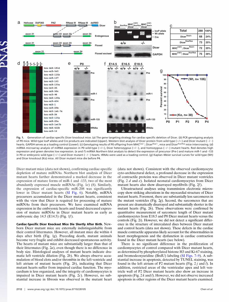
Dicer mutant mice (data not shown), confirming cardiac-specificdepletion of mature miRNAs. Northern blot analysis of Dicermutant hearts further demonstrated a marked decrease in theexpression of mature forms of miR-1 and -133, two of the mostabundantly expressed muscle miRNAs (Fig. 1e) (8). Similarly,the expression of cardiac-specific miR-208 was significantlylower in Dicer mutant hearts (SI Fig. 6). Notably, miRNAprecursors accumulated in the Dicer mutant hearts, consistentwith the view that Dicer is required for processing of maturemiRNAs from their precursors. We have examined miRNAexpression in the embryonic hearts and found decreased expres-sion of mature miRNAs in Dicer mutant hearts as early asembryonic day 14.5 (E14.5) (Fig. 1f ).
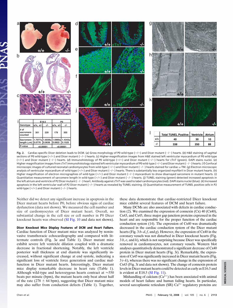
Cardiac-Specific Dicer Knockout Mice Die Shortly After Birth. New-born Dicer mutant mice are externally indistinguishable fromtheir control littermates. However, all mutant mice die within 4days after birth (Fig. 1g). Preceding death, mutant animalsbecome very fragile and exhibit decreased spontaneous activity.The hearts of mutant mice are substantially larger than that oftheir littermates (Fig. 2a), even though there is no difference inbody size. Histological analyses of mutant hearts indicate dra-matic left ventricle dilation (Fig. 2b). We always observe accu-mulation of blood clots and/or thrombin in the left ventricle andleft atrium of mutant hearts (Fig. 2b), indicating that Dicermutant hearts suffer from impaired cardiac function. The myo-cardium is less organized, and the integrity of cardiomyocytes isimpaired in Dicer mutant hearts (Fig. 2c). However, no sub-stantial increase in fibrosis was observed in the mutant heart
(data not shown). Consistent with the observed cardiomyocytecyto-architectural defect, a profound decrease in the expressionof contractile proteins was observed in Dicer mutant ventricles(Fig. 2 d and e). Isolated neonatal cardiomyocytes from Dicermutant hearts also show disarrayed myofibrils (Fig. 2f ).
Ultrastructural analyses using transmission electronic micros-copy show striking alterations in the myocardial structure of Dicermutant hearts. Foremost, there are substantially less sarcomeres inthe mutant ventricles (Fig. 2g). Second, the sarcomeres that arepresent are dramatically disarrayed and substantially shorter in themutant hearts (Fig. 2h). These observations were confirmed byquantitative measurement of sarcomere length of Dicer mutantcardiomyocytes from E18.5 and P0 Dicer mutant hearts versus thecontrols (Fig. 2i). However, we did not detect a significant differ-ence in the structure of intercalated discs between Dicer mutantand control hearts (data not shown). These defects in the cardiacmuscle contractile apparatus likely account for the abnormalities inheart morphogenesis and the dysfunction in cardiac contractionfound in the Dicer mutant hearts (see below).
There is no significant difference in the proliferation ofcardiomyocytes of control compared with Dicer mutant hearts,as determined by phosphorylated histone H3 and Ki-67 staining,and bromodeoxyuridine (BrdU) labeling (SI Figs. 7–9). A sub-stantial increase in apoptosis, detected by TUNEL staining, wasfound in the left atrium of P2 mutant hearts (Fig. 2 j and l). Inaddition, restricted areas of the ventricular apex and left ven-tricle wall of P2 Dicer mutant hearts also show an increase inapoptosis (Fig. 2 k and l). However, we did not observe increasedapoptosis in other regions of the Dicer mutant hearts examined.
a Helicase DUF283 PAZ RNase III RNase III dsRBD
exon 21 exon 22 exon 23 exon 24
exon 21 exon 24
Dicer
Endogenous
Floxed
Floxed excised
exon 21 exon 22 exon 23 exon 24
bcMHC
cre/+
Dicerflox/flox
Dicerflox/flox MHC
cre/+
Dicerflox/+
loxP alleleWt allele
Cre
Dicer
GAPDH
+/+ -/-
e P0+/+ -/-
d -/-
+/++/+ -/-
-/+-/+ f E14.5
+/+ -/-E16.5
+/+ -/-E18.5
+/+ -/-
Total 288 100%
Dicerflox/+ 68 24%
Dicerflox/flox 73 25%
MHCcre/+
; Dicerflox/+ 72 25%
MHCcre/+
; Dicerflox/flox 75 26%
g
0
10
20
30
40
50
60
70
80
90
100
(Wt, n= 106)
(Ko, n= 38)
P0 P1 P2 P3 P4
Per
cen
t su
rviv
al
Pre
miR-1
Pre
miR-1
Pre
miR-133
Pre
miR-133
tRNAs tRNAs
Fig. 1. Generation of cardiac-specific Dicer knockout mice. (a) The gene targeting strategy for cardiac-specific deletion of Dicer. (b) PCR genotyping analysisof P0 mice. Wild-type loxP alleles and Cre products are indicated (Upper). Western blot analysis of Dicer protein from wild-type (�/�) and Dicer mutant (�/�)hearts. GAPDH serves as a loading control (Lower). (c) Genotyping results of P0 offspring from MHCCre/�; Dicerflox/�, mice and Dicerflox/flox mice intercrossing. (d)miRNA microarray analysis of miRNA expression in P0 wild-type (�/�), Dicer heterozygous (�/�), and homozygous (�/�) mutant hearts. Red denotes highexpression and green denotes low expression. (e and f ) miRNA Northern blot analysis to detect the expression of precursor (Pre-) and mature miR-1 and -133in P0 or embryonic wild-type (�/�) and Dicer mutant (�/�) hearts. tRNAs were used as a loading control. (g) Kaplan–Meier survival curves for wild-type (Wt)and Dicer knockout (Ko) mice. All Dicer mutant mice die before P4.
2112 � www.pnas.org�cgi�doi�10.1073�pnas.0710228105 Chen et al.
Neither did we detect any significant increase in apoptosis in theDicer mutant hearts before P0, before obvious signs of cardiacdysfunction (data not shown). We measured the cell number andsize of cardiomyocytes of Dicer mutant heart. Overall, nosubstantial change in the cell size or cell number in P0 Dicerknockout hearts was observed (SI Fig. 10 and data not shown).
Dicer Knockout Mice Display Features of DCM and Heart Failure.Cardiac function of Dicer mutant mice was analyzed by nonin-vasive transthoracic echocardiography and compared with lit-termate controls (Fig. 3a and Table 1). Dicer mutant miceexhibit severe left ventricle dilation coupled with a dramaticdecrease in fractional shortening. Notably, the left ventricleposterior wall thickness at end diastole was significantly in-creased, without significant change at end systole, indicating asignificant loss of ventricle force generation and cardiac mal-function in Dicer mutant hearts. Interestingly, Dicer mutantmice display remarkable decrease in heart rate (Table 1).Although wild-type and heterozygous hearts contract at �550beats per minute (bpm), the mutant hearts only beat about halfof the rate (278 � 64 bpm), suggesting that Dicer mutant micemay also suffer from conduction defects (Table 1). Together,
these data demonstrate that cardiac-restricted Dicer knockoutmice exhibit several features of DCM and heart failure.
Many DCMs are also associated with defects in cardiac conduc-tion (2). We examined the expression of connexin (Cx) 40 (Cx40),Cx43, and Cx45, three major gap junction proteins expressed in theheart and are responsible for the proper function of the cardiacconduction system (14). The expression of Cx40 was dramaticallydecreased in the cardiac conduction system of the Dicer mutanthearts (Fig. 3 b–d, f, and g). However, the expression of Cx40 in thecoronary vessels was not disturbed in Dicer knockout hearts (Fig.3 b, e, and h), which is not surprising because Dicer was specificallyremoved in cardiomyocytes, not coronary vessels. Western blotanalyses quantitatively demonstrated a significant decrease of Cx40protein in Dicer mutant hearts (Fig. 3i). Remarkably, the expres-sion of Cx45 was significantly increased in Dicer mutant hearts (Fig.3 i–k), whereas there was no significant change in the expression ofCx43 (Fig. 3i and data not shown). The change of connexin proteinlevels in Dicer mutant hearts could be detected as early as E16.5 andis evident at E18.5 (SI Fig. 11).
Mishandling of calcium (Ca2�) has been associated with animalmodels of heart failure and human failing hearts. In particular,several sarcoplasmic reticulum (SR) Ca2� regulatory proteins are
a
b
g
c
d
e
f
h
i
j
Genotype +/+, +/- -/- +/+, +/- -/-
# of sacromere counted
301 229 179 355
Sacromere length (µm)
1.7077± 0.0776
1.6039± 0.0636
1.6969±0.0822
1.4343± 0.1525
P-value
E18.5 P0
<0.00001 <0.00001
k
l
+/+ -/-
+/+ -/-
+/+ -/-
+/+ -/-
+/+ -/-
+/+ -/-
+/+ -/-
+/+ -/-
+/+ -/-
cTnT
α-TM
+/+ -/-
Total TUNEL Positive Ventricle Atrium
+/+ 40 30 10
-/- 338 250 88
Fig. 2. Cardiac-specific Dicer deletion leads to DCM. (a) Gross morphology of P0 wild-type (�/�) and Dicer mutant (�/�) hearts. (b) H&E staining of sagittalsections of P0 wild-type (�/�) and Dicer mutant (�/�) hearts. (c) Higher-magnification images from H&E stained left ventricular myocardium of P0 wild-type(�/�) and Dicer mutant (�/�) hearts. (d) Immunohistology of P0 wild-type (�/�) and Dicer mutant (�/�) hearts for cTnT (green). DAPI stains nuclei. (e)Higher-magnification images from cTnT immunohistology-stained left ventricular myocardium of P0 wild-type (�/�) and Dicer mutant (�/�) hearts. ( f) Confocalmicroscopic images of cultured neonatal cardiomyocytes from wild-type (�/�) and Dicer mutant (�/�) hearts stained for cardiac �-TM. (g) Electron microscopicanalysis of ventricular myocardium of wild-type (�/�) and Dicer mutant (�/�) hearts. There is substantially less organized myofibril in Dicer mutant hearts. (h)Higher magnification of electron micrographies of wild-type (�/�) and Dicer mutant (�/�) myocardium to show disarrayed sarcomere in mutant hearts. (i)Quantitative measurement of sarcomere length in wild-type (�/�) and Dicer mutant (�/�) hearts. (j) TUNEL staining (green) detected increased apoptosis inthe left atrium and ventricle of P2 Dicer mutant (�/�) heart. Antibody against cTnT was used to label cardiomyocytes (red). DAPI stains nuclei (blue). (k) Increasedapoptosis in the left ventricular wall of P2 Dicer mutant (�/�) hearts as revealed by TUNEL staining. (l) Quantitative measurement of TUNEL positive cells in P2wild-type (�/�) and Dicer mutant (�/�) hearts.
Chen et al. PNAS � February 12, 2008 � vol. 105 � no. 6 � 2113
MED
ICA
LSC
IEN
CES
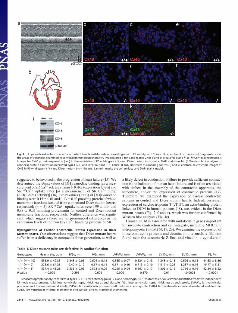
suggested to be involved in the progression of heart failure (15). Wedetermined the Bmax values of [3H]ryanodine binding [as a mea-surement of SR Ca2� release channel (RyR2) expression levels] andSR 45Ca2� uptake rates [as a measurement of SR Ca2� pump(SERCA2a) activity)] (16). Bmax values (�SE) of [3H]ryanodinebinding were 0.15 � 0.01 and 0.13 � 0.02 pmol/mg protein of wholemembrane fractions isolated from control and Dicer mutant hearts,respectively (n � 5). SR 45Ca2� uptake rates were 0.99 � 0.14 and0.88 � 0.05 nmol/mg protein/min for control and Dicer mutantmembrane fractions, respectively. Neither difference was signifi-cant, which suggests there are no pronounced differences in theexpression levels of the two key Ca2� handling proteins of SR.
Dysregulation of Cardiac Contractile Protein Expression in DicerMutant Hearts. Our observations suggest that Dicer mutant heartssuffer from a deficiency in contractile force generation, as well as
a likely defect in conduction. Failure to provide sufficient contrac-tion is the hallmark of human heart failure and is often associatedwith defects in the assembly of the contractile apparatus, thesarcomere, and/or the expression of contractile proteins (17).Therefore, we examined the expression of cardiac contractileproteins in control and Dicer mutant hearts. Indeed, decreasedexpression of cardiac troponin T (cTnT), an actin-binding proteinlinked to DCM in human patients (18), was evident in the Dicermutant hearts (Fig. 2 d and e), which was further confirmed byWestern blot analyses (Fig. 4g).
Human DCM is associated with mutations in genes importantfor myocyte contraction and cell integrity, including MHC and�-tropomyosin (�-TM) (4, 19, 20). We examine the expression ofthose contractile proteins and desmin, an intermediate filamentfound near the sarcomeric Z line, and vinculin, a cytoskeletal
a
b
+/+
-/-
1
2
3
mm
1
250 mSec
+/+ +/+ +/+
-/--/--/-
c d
f g
e
h
+/+ -/-
j k
Cx40 Cx40 Cx40
Cx40 Cx40 Cx40
Cx45Laminin Laminin
Cx45
i +/+ -/-
CX40
CX43
CX45
β-Tubulin
Fig. 3. Impaired cardiac function in Dicer mutant hearts. (a) M-mode echocardiograms of P0 wild-type (�/�) and Dicer mutant (�/�) mice. (b) Diagram to showthe areas of ventricles examined in confocal immunohistochemistry images: area 1 for c and f; area 2 for d and g; area 3 for e and h. (c–h) Confocal microscopicimages for Cx40 protein expression (red) in the ventricles of P0 wild-type (�/�) and Dicer mutant (�/�) mice. DAPI stains nuclei. (i) Western blot analyses ofconnexin protein expression in P0 wild-type (�/�) and Dicer mutant (�/�) mice. �-Tubulin serves as a loading control. (j and k) Confocal microscopic images ofCx45 in P0 wild-type (�/�) and Dicer mutant (�/�) hearts. Laminin marks the cell surface and DAPI stains nuclei.
Table 1. Dicer mutant mice are defective in cardiac function
Genotypes Heart rate, bpm IVSd, mm IVSs, mm LVPWd, mm LVPWs, mm LVIDd, mm LVIDs, mm FS, %
�/� (n � 10) 555.9 � 42.33 0.348 � 0.06 0.604 � 0.12 0.355 � 0.07 0.624 � 0.13 1.248 � 0.13 0.696 � 0.15 44.63 � 8.86�/� (n � 7) 278.6 � 64.38 0.40 � 0.12 0.57 � 0.15 0.571 � 0.10 0.713 � 0.10 1.517 � 0.25 1.287 � 0.16 19.77 � 5.31�/� (n � 8) 537.4 � 48.28 0.335 � 0.05 0.573 � 0.09 0.295 � 0.04 0.567 � 0.17 1.306 � 0.16 0.756 � 0.16 42.34 � 8.52P value �0.0001 0.296 0.629 0.0001 0.170 0.01 �0.0001 �0.0001
Echocardiographic analyses of P0 wild-type (�/�), Dicer heterozygous (�/-), and homozygous (-/-) mutant mice. Values were quantified from five independentM-mode measurements. IVSd, interventricular septal thickness at end diastole; IVSs, interventricular septal thickness at end systole; LVPWd, left ventricularposterior wall thickness at end diastole; LVPWs, left ventricular posterior wall thickness at end systole; LVIDd, left ventricular internal diameter at end diastole;LVIDs, left ventricular internal diameter at end systole; and FS, fractional shortening.
2114 � www.pnas.org�cgi�doi�10.1073�pnas.0710228105 Chen et al.
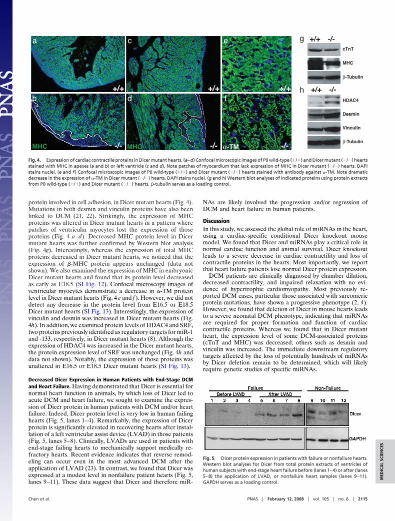
protein involved in cell adhesion, in Dicer mutant hearts (Fig. 4).Mutations in both desmin and vinculin proteins have also beenlinked to DCM (21, 22). Strikingly, the expression of MHCproteins was altered in Dicer mutant hearts in a pattern wherepatches of ventricular myocytes lost the expression of thoseproteins (Fig. 4 a–d). Decreased MHC protein level in Dicermutant hearts was further confirmed by Western blot analysis(Fig. 4g). Interestingly, whereas the expression of total MHCproteins decreased in Dicer mutant hearts, we noticed that theexpression of �-MHC protein appears unchanged (data notshown). We also examined the expression of MHC in embryonicDicer mutant hearts and found that its protein level decreasedas early as E18.5 (SI Fig. 12). Confocal microscopy images ofventricular myocytes demonstrate a decrease in �-TM proteinlevel in Dicer mutant hearts (Fig. 4 e and f ). However, we did notdetect any decrease in the protein level from E16.5 or E18.5Dicer mutant hearts (SI Fig. 13). Interestingly, the expression ofvinculin and desmin was increased in Dicer mutant hearts (Fig.4h). In addition, we examined protein levels of HDAC4 and SRF,two proteins previously identified as regulatory targets for miR-1and -133, respectively, in Dicer mutant hearts (8). Although theexpression of HDAC4 was increased in the Dicer mutant hearts,the protein expression level of SRF was unchanged (Fig. 4h anddata not shown). Notably, the expression of those proteins wasunaltered in E16.5 or E18.5 Dicer mutant hearts (SI Fig. 13).
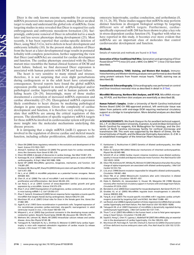
Decreased Dicer Expression in Human Patients with End-Stage DCMand Heart Failure. Having demonstrated that Dicer is essential fornormal heart function in animals, by which loss of Dicer led toacute DCM and heart failure, we sought to examine the expres-sion of Dicer protein in human patients with DCM and/or heartfailure. Indeed, Dicer protein level is very low in human failinghearts (Fig. 5, lanes 1–4). Remarkably, the expression of Dicerprotein is significantly elevated in recovering hearts after instal-lation of a left ventricular assist device (LVAD) in those patients(Fig. 5, lanes 5–8). Clinically, LVADs are used in patients withend-stage failing hearts to mechanically support medically re-fractory hearts. Recent evidence indicates that reverse remod-eling can occur even in the most advanced DCM after theapplication of LVAD (23). In contrast, we found that Dicer wasexpressed at a modest level in nonfailure patient hearts (Fig. 5,lanes 9–11). These data suggest that Dicer and therefore miR-
NAs are likely involved the progression and/or regression ofDCM and heart failure in human patients.
DiscussionIn this study, we assessed the global role of miRNAs in the heart,using a cardiac-specific conditional Dicer knockout mousemodel. We found that Dicer and miRNAs play a critical role innormal cardiac function and animal survival. Dicer knockoutleads to a severe decrease in cardiac contractility and loss ofcontractile proteins in the hearts. Most importantly, we reportthat heart failure patients lose normal Dicer protein expression.
DCM patients are clinically diagnosed by chamber dilation,decreased contractility, and impaired relaxation with no evi-dence of hypertrophic cardiomyopathy. Most previously re-ported DCM cases, particular those associated with sarcomericprotein mutations, have shown a progressive phenotype (2, 4).However, we found that deletion of Dicer in mouse hearts leadsto a severe neonatal DCM phenotype, indicating that miRNAsare required for proper formation and function of cardiaccontractile proteins. Whereas we found that in Dicer mutantheart, the expression level of some DCM-associated proteins(cTnT and MHC) was decreased, others such as desmin andvinculin was increased. The immediate downstream regulatorytargets affected by the loss of potentially hundreds of miRNAsby Dicer deletion remain to be determined, which will likelyrequire genetic studies of specific miRNAs.
HDAC4
Desmin
Vinculin
β-Tubulin
-/-+/+
cTnT
MHC
β-Tubulin
-/-+/+g
h+/+
a
b
c
d
e
f+/+ +/+
-/- -/--/-MHC MHC α-TM
Fig. 4. Expression of cardiac contractile proteins in Dicer mutant hearts. (a–d) Confocal microscopic images of P0 wild-type (�/�) and Dicer mutant (�/�) heartsstained with MHC in apexes (a and b) or left ventricle (c and d). Note patches of myocardium that lack expression of MHC in Dicer mutant (�/�) hearts. DAPIstains nuclei. (e and f ) Confocal microscopic images of P0 wild-type (�/�) and Dicer mutant (�/�) hearts stained with antibody against �-TM. Note dramaticdecrease in the expression of �-TM in Dicer mutant (�/�) hearts. DAPI stains nuclei. (g and h) Western blot analyses of indicated proteins using protein extractsfrom P0 wild-type (�/�) and Dicer mutant (�/�) hearts. �-tubulin serves as a loading control.
Fig. 5. Dicer protein expression in patients with failure or nonfailure hearts.Western blot analyses for Dicer from total protein extracts of ventricles ofhuman subjects with end-stage heart failure before (lanes 1–4) or after (lanes5–8) the application of LVAD, or nonfailure heart samples (lanes 9–11).GAPDH serves as a loading control.
Chen et al. PNAS � February 12, 2008 � vol. 105 � no. 6 � 2115
MED
ICA
LSC
IEN
CES
Dicer is the only known enzyme responsible for processingmiRNA precursors into mature products, making Dicer an idealtarget to study and understand the global role of miRNAs. Genetargeting studies in mice revealed that Dicer is required for earlyembryogenesis and embryonic mesoderm formation (24). Sur-prisingly, embryonic removal of Dicer in zebrafish led to a muchlater and less severe phenotype during development (25). Mostrecently, it has been reported that cardiac-specific deletion ofDicer, using Nkx2.5-Cre, led to defects in heart development andembryonic lethality (10). In the present study, deletion of Dicerfrom the heart at a later developmental stage results in postnatallethality with complete penetration, further supporting the viewthat Dicer (and miRNAs) are essential for cardiac developmentand function. The cardiac phenotype associated with the Dicermutant mice resembles the human clinical features of DCM andheart failure. Indeed, we find decreased Dicer expression isassociated with human patients with DCM and heart failure.
The heart is very sensitive to many stimuli and stresses;therefore, it is not surprising that even slight perturbationsduring cardiogenesis or in the adult heart have catastrophicconsequences. Several reports have found the global miRNAexpression profile regulated in models of physiological and/orpathological cardiac hypertrophy and in human patients withfailing hearts (26–29). Interestingly, many of those studiesdocumented a correlation of increased miRNA expression andcardiac abnormality (27–29). Dysregulated miRNA expressionlikely contributes to heart disease by mediating pathologicalchanges in gene expression. Given the complexity of cardiacdevelopment and function, it is therefore very exciting to spec-ulate that miRNAs are among the critical regulators of thisprocess. The identification of specific regulatory mRNA targetsfor those miRNAs involved in cardiovascular system will providemore insight into the molecular mechanisms underlying thisdisease process.
It is intriguing that a single miRNA (miR-1) appears to beinvolved in the regulation of diverse cardiac and skeletal musclefunction, including cellular proliferation, differentiation, cardi-
omyocyte hypertrophy, cardiac conduction, and arrhythmias (8,10, 11, 26, 30). Those studies suggest that miRNAs may performdistinct functions in divergent biological settings by targetingdifferent sets of mRNA targets. Furthermore, cardiac-specifically expressed miR-208 has been shown to play a key rolein stress-dependent cardiac function (9). Together with what wehave reported in this study, it becomes ever more evident thatmiRNAs are an important class of molecules that controlcardiovascular development and function.
MethodsDetails of materials and methods are found in SI Text.
Generation of Dicer Conditional Null Mice. Generation and genotyping of Dicerfloxed (Dicerflox/flox) mice (31) and �-MHC-Cre (MHCCre/�) mice (13) have beendescribed.
Immunohistochemistry, TUNEL Staining, and Western Blot Analysis. Immuno-histochemistry and Western blotting analyses were performed as described (8)using protein extracts from frozen mouse hearts. TUNEL staining was asdescribed (8).
Transthoracic Echocardiography. Echocardiography was performed on controland Dicer knockout neonatal mice as described in detail in SI Text.
MicroRNA Microarray, Northern Blot Analyses, and RT-PCR. MicroRNA microar-ray, RT-PCR, and Northern blot analyses are essentially as described (8).
Human Patient’s Samples. Under a University of North Carolina InstitutionalReview Board (UNC-CH IRB)-approved protocol, left ventricular tissue wastaken from patients with end-stage DCM requiring mechanical circulatorysupport as a bridge to transplantation. More detailed analyses are describedin SI Text.
ACKNOWLEDGMENTS. We thank Xiaoyun Hu for excellent technical support,Drs. J. P. Jin (Northwestern University, Evanston, IL) and Jim Bear (University ofNorth Carolina) for reagents, Kirk McNaughton for histology, and the Uni-versity of North Carolina microscopy facility for confocal microscopy andtransmission EM. This work was supported by the March of Dimes, the Na-tional Institutes of Health, and the Muscular Dystrophy Association. D.-Z.W. isan established investigator of the American Heart Association.
1. Olson EN (2006) Gene regulatory networks in the evolution and development of theheart. Science 313:1922–1927.
2. Ahmad F, Seidman JG, Seidman CE (2005) The genetic basis for cardiac remodeling.Annu Rev Genomics Hum Genet 6:185–216.
3. Olson EN (2004) A decade of discoveries in cardiac biology. Nat Med 10:467–474.4. Kamisago M, et al. (2000) Mutations in sarcomere protein genes as a cause of dilated
cardiomyopathy. N Engl J Med 343:1688–1696.5. Bartel DP (2004) MicroRNAs: genomics, biogenesis, mechanism, and function. Cell
116:281–297.6. Houbaviy HB, Murray MF, Sharp PA (2003) Embryonic stem cell-specific MicroRNAs. Dev
Cell 5:351–358.7. He L, et al. (2005) A microRNA polycistron as a potential human oncogene. Nature
435:828–833.8. Chen JF, et al. (2006) The role of microRNA-1 and microRNA-133 in skeletal muscle
proliferation and differentiation. Nat Genet 38:228–233.9. van Rooij E, et al. (2007) Control of stress-dependent cardiac growth and gene
expression by a microRNA. Science 316:575–579.10. Zhao Y, et al. (2007) Dysregulation of cardiogenesis, cardiac conduction, and cell cycle
in mice lacking miRNA-1–2. Cell 129:303–317.11. Zhao Y, Samal E, Srivastava D (2005) Serum response factor regulates a muscle-specific
microRNA that targets Hand2 during cardiogenesis. Nature 436:214–220.12. Murchison EP, et al. (2007) Critical roles for Dicer in the female germ line. Genes Dev
21:682–693.13. Agah R, et al. (1997) Gene recombination in postmitotic cells. Targeted expression of
Cre recombinase provokes cardiac-restricted, site-specific rearrangement in adultventricular muscle in vivo. J Clin Invest 100:169–179.
14. Miquerol L, et al. (2003) Gap junctional connexins in the developing mouse cardiacconduction system. Novartis Found Symp 250:80–98; discussion 98–109:276–279.
15. Wehrens XH, Lehnart SE, Marks AR (2005) Intracellular calcium release and cardiacdisease. Annu Rev Physiol 67:69–98.
16. Yamaguchi N, Takahashi N, Xu L, Smithies O, Meissner G (2007) Early cardiac hyper-trophy in mice with impaired calmodulin regulation of cardiac muscle Ca releasechannel. J Clin Invest 117:1344–1353.
17. Karkkainen S, Peuhkurinen K (2007) Genetics of dilated cardiomyopathy. Ann Med39:91–107.
18. Fatkin D, Graham RM (2002) Molecular mechanisms of inherited cardiomyopathies.Physiol Rev 82:945–980.
19. Schmitt JP, et al. (2006) Cardiac myosin missense mutations cause dilated cardiomy-opathy in mouse models and depress molecular motor function. Proc Natl Acad Sci USA103:14525–14530.
20. Olson TM, Kishimoto NY, Whitby FG, Michels VV (2001) Mutations that alter the surfacecharge of alpha-tropomyosin are associated with dilated cardiomyopathy. J Mol CellCardiol 33:723–732.
21. Li D, et al. (1999) Desmin mutation responsible for idiopathic dilated cardiomyopathy.Circulation 100:461–464.
22. Olson TM, et al. (2002) Metavinculin mutations alter actin interaction in dilatedcardiomyopathy. Circulation 105:431–437.
23. Dipla K, Mattiello JA, Jeevanandam V, Houser SR, Margulies KB (1998) Myocyterecovery after mechanical circulatory support in humans with end-stage heart failure.Circulation 97:2316–2322.
24. Bernstein E, et al. (2003) Dicer is essential for mouse development. Nat Genet 35:215–217.25. Giraldez AJ, et al. (2005) MicroRNAs regulate brain morphogenesis in zebrafish.
Science 308:833–838.26. Yang B, et al. (2007) The muscle-specific microRNA miR-1 regulates cardiac arrhyth-
mogenic potential by targeting GJA1 and KCNJ2. Nat Med 13:486–491.27. vanRooijE,etal. (2006)Asignaturepatternof stress-responsivemicroRNAsthat canevoke
cardiac hypertrophy and heart failure. Proc Natl Acad Sci USA 103:18255–18260.28. Tatsuguchi M, et al. (2007) Expression of microRNAs is dynamically regulated during
cardiomyocyte hypertrophy. J Mol Cell Cardiol 42:1137–1141.29. Thum T, et al. (2007) MicroRNAs in the human heart: a clue to fetal gene reprogram-
ming in heart failure. Circulation 116:258–267.30. Sayed D, Hong C, Chen IY, Lypowy J, Abdellatif M (2007) MicroRNAs play an essential
role in the development of cardiac hypertrophy. Circ Res 100:416–424.31. Murchison EP, Partridge JF, Tam OH, Cheloufi S, Hannon GJ (2005) Characterization
of Dicer-deficient murine embryonic stem cells. Proc Natl Acad Sci USA 102:12135–12140.
2116 � www.pnas.org�cgi�doi�10.1073�pnas.0710228105 Chen et al.


![Spinal cord-specific deletion of the glutamate transporter ... · Consistent with previous report [19], the strong tdTomato ... Hoxb8-Cre/GLT1 flox/flox mice had slightly lower body](https://img.pdfslide.us/doc/110x75/5b0a0b357f8b9ae61b8b7d58/spinal-cord-specific-deletion-of-the-glutamate-transporter-with-previous-report.jpg)