Embed Size (px)
Citation preview

Table S1. Bacterial strains and plasmids used in the study
Strain/plasmids Description Source/reference
R20291 NAP1/027 ribotype C. difficile strain, isolated in 2006 following an outbreak in Stoke Mandeville Hospital, UK [27]
CD646 078 ribotype C. difficile strain [28]
R20291::tcdE mutant C. difficile R20291 with intron insertion within the tcdE gene This study
CD646::tcdE mutant C. difficile CD646 with intron insertion within the tcdE gene This study
DH5α E coli strain with endA1, recA1, deoR, and hsdR17 (rK- mK
+) New England Bio-Labs
S17-1 E. coli strain that favors conjugation [30]
RGL100 R20291 with pRGL209 This study
RGL101 R20291::tcdE mutant with pRGL209 This study
pMTL007C-E5 Clostron plasmid [31, 32]
pMTL007E5:Cdi-tcdE-234a pMTL007C-E5 carrying the tcdE-specific intron This study
pRPF185 Shuttle vector for C. difficile [39]
pRGL60 pRPF185 carrying the tcdE ORF (TcdEWT) [15]
pRGL126 pRPF185 derivative with tcdE166 carrying Met25 and Met27 mutated to Leu25 and Leu27, respectively, to express TcdE166 This study (Figure 2A)
pRGL128 pRPF185 derivative with tcdE142 carrying Met1 and Met27 mutated to a stop codon and Leu27, respectively, to express TcdE142 This study (Figure 2A)
pRGL127 pRPF185 derivative with tcdE140 carrying Met1 and Met25 mutated to a stop codon and Leu25, respectively, to express TcdE140 This study (Figure 2A)
pRGL-TcdE142N26A pRGL128 derivative with mutated tcdE142, in which Asn26 was mutated to Ala26 This study
pRGL-TcdE142N26Q pRGL128 derivative with mutated tcdE142, in which Asn26 was mutated to Gln26 This study
pRGL-TcdE142N26V pRGL128 derivative with mutated tcdE142, in which Asn26 was mutated to Val26 This study
pRGL132 pRGL126 derivative with a deletion of the adenine at the -1 position from the Met1 ATG start codon This study (Figure 3A)
pRGL133 pRGL126 derivative with a deletion of the adenine at -1 and a mutation in the ATG at -3 from the Met1 ATG start codon This study (Figure 3A)
pRGL134 pRGL126 derivative with a deletion of the adenine at -1 and mutations in the ATGs at -3 and -6 from the Met1 ATG start codon This study (Figure 3A)
pRGL267 pRPF185 carrying tcdE166 with gus RBS This study
pRGL209 pRPF185 carrying tcdR ORF under the tet-inducible promoter This study

Table S2: Oligonucleotides used in this study
Name Description Sequence (5’ to 3’)
ORG45 tcdE forward CTA GGA GGC GTT ATG AAT ATG ACA ATATCT
ORG46 tcdE reverse CTT TTC ATC CTT AGC ATT CAT TTC ATC T
EBS-U Intron specific CGAAATTAGAAACTTGCGTTCAGTAAAC
ERM Intron specific ACG CGTGCGACTCATAGAATTATTTCCTCCCG
ORG102 tcdE forward with SacI GAG CTC CAATAA AAA GGT GGA CTATGA TGA
ORG103 tcdE reverse with BamHI GGATCC TTA ATG ATG ATG ATG ATG ATG CTT TTC ATC CTT AGC
ORG218 tcdE 166 forward-mutagenesis ATATTT CTA ATG GTA ACA AAATAT TTT TTT ATATAA ACCTAG GAG GCGTTT TAA ATT TAA CAATAT CTT TTT
ORG219 tcdE 166 reverse-mutagenesis ATT ACT AACTTT ATA AAT ATATGC TCT GAT AAA AAA GAT ATT GTT AAATTT AAA ACG CCT CCT AGGTTT
ORG216 tcdE 142 forward-mutagenesis AAA ATATTT TTT TAT ATA AAC CTA GGA GGC GTT ATG AAT TTA ACA ATATCT TTT TTATCA GAG CAT ATA
ORG217 tcdE 142 reverse-mutagenesis TAT AAATAT ATG CTC TGA TAA AAA AGATAT TGT TAA ATT CAT AAC GCCTCC TAG GTT TAT ATA AAA AAA
ORG214 tcdE 140 forward-mutagenesis CAA AAT ATT TTT TTATAT AAA CCT AGG AGG CGT TTT AAATAT GAC AAT ATC TTT TTT ATC AGA GCATAT
ORG215 tcdE 140 reverse-mutagenesis AAT ATATGC TCT GAT AAA AAA GAT ATT GTC ATATTT AAA ACG CCT CCT AGGTTT ATATAA AAA AAT ATT
ORG322 tcdEN26A forward-mutagenesis ATT TTT TTA TAT AAA CCT AGG AGG CGT TAT GGC TAT GAC AAT ATC TTT TTT ATC AGA GCA TAT A
ORG323 tcdEN26A reverse-mutagenesis TAT ATG CTC TGA TAA AAA AGA TAT TGT CAT AGC CAT AAC GCC TCC TAG GTT TAT ATA AAA AAAT
ORG320 tcdEN26Q forward-mutagenesis ATA TAT GCT CTG ATA AAA AAG ATAT TGT CAT CTG CAT AAC GCC TCC TAG GTT TAT ATA AAA AAA
ORG321 tcdEN26Q reverse-mutagenesis TTT TTT TAT ATA AAC CTA GGA GGC GTT ATG CAG ATG ACA ATA TCT TTT TTA TCA GAG CAT ATAT
ORG318 tcdEN26V forward-mutagenesis ATT TTT TTA TAT AAA CCT AGG AGG CGT TAT GGT TAT GAC AAT ATC TTT TTT ATC AGA GCA TATA
ORG319 tcdEN26V reverse-mutagenesis TAT ATG CTC TGA TAA AAA AGA TAT TGT CAT AAC CAT AAC GCC TCC TAG GTT TAT ATA AAA AAAT
ORG264 tcdE 132 forward-mutagenesis CCT TGC GAG CTC CAATAA AAA GGT GGA CTATGA TGA TG
ORG265 tcdE 133 forward-mutagenesis GAGCTCCAATAAAAAGGTGGACTTTAATGTTACACAGT
ORG266 tcdE 134 forward-mutagenesis GAGCTCCAATAAAAAGGTGGACTATGTTATTACACAGT
ORG107 tcdE forward with gus RBS GAGCTCC TGC AGT AAA GGA GAA AAT TTT ATG CAC AGT AGT TCA CCT TTT TAT ATT -3
ORG341 tcdR forward with SacI GAGCTCATATAAGAGAGGGTGATTTTATGCAAAAGTCTTTT
ORG359 tcdR reverse with BamHI GGATCCTTACTTATCGTCGTCATCCTTGTAATCCAAGTTAAAATAATTTTCATAGTC
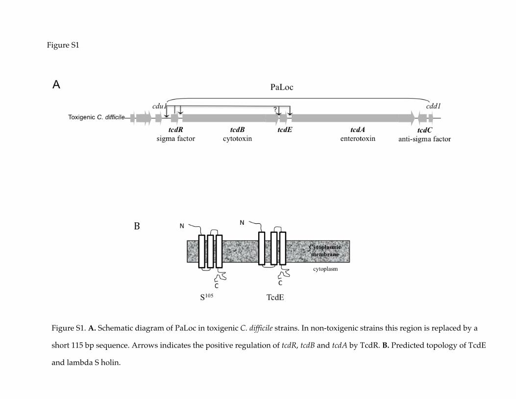
Figure S1
Figure S1. A. Schematic diagram of PaLoc in toxigenic C. difficile strains. In non-toxigenic strains this region is replaced by a
short 115 bp sequence. Arrows indicates the positive regulation of tcdR, tcdB and tcdA by TcdR. B. Predicted topology of TcdE
and lambda S holin.
B

Figure S2
Figure S2. Construction and characterization of tcdE mutants in R20291 and CD646 C. difficile
strains. A. Southern blot analysis of genomic DNA from parent and tcdE mutant strains using tcdE-
specific probes. Chromosomal DNA was digested with EcoRV. The tcdE-specific probe hybridized
with a larger DNA fragment in mutant strains, confirming the single insertion of the intron within
the tcdE gene. B. Concentrated culture supernatants used for the toxin ELISA test were spotted on a
PVDF membrane and probed with monoclonal antibodies against the L7/L12 ribosomal subunits.
B
A

Supplementary methods
Construction of tcdE insertion mutants
C. difficile R20291 and CD646 tcdE mutants were created by inserting the bacterial group II intron
using the ClosTron gene knockout system as described previously [31, 32]. Briefly, the plasmid
pMTL007C-E5 carrying the tcdE-specific retargeted intron (pMTL007C-E5:Cdi-tcdE-234a) was
transferred to C. difficile strains by conjugation as described previously [15, 33]. Transconjugants
resistant to thiamphenicol were resuspended in 200 µL of TY broth and plated on TY agar plates
containing lincomycin (10 µg/mL) to select for intron insertion. Putative tcdE mutants were screened
by PCR using the tcdE-specific primers ORG45 and ORG46 in combination with the EBS-U universal
and ERM primers (Table S2). The selected mutant was confirmed by Southern blot, as described
below.
Southern blot analysis
Ten micrograms of genomic DNA from the parent and the tcdE mutant strains was digested with the
EcoRV enzyme and separated on a 0.8% agarose gel by electrophoresis. DNA was transferred onto
an Immobilon-NY+ nylon membrane (Millipore, Bedford, MA, US) by the capillary transfer method.
Prehybridization of the filter was conducted for 2 h at 60°C in a solution containing 5 × SCC (1 × SSC
contains 0.15 M NaCl plus 0.015 M sodium citrate), 5 × Denhardt's solution, and 100 mg/mL salmon
sperm DNA. Probes specific for the tcdE gene were radiolabeled ([32P]dATP) using a High Prime kit
(Roche, NJ, USA) and hybridized overnight in 10 mL of fresh prehybridization buffer at 60°C. The
hybridized membrane was washed twice for 30 minutes in 2 × SCC and 0.5% SDS and once for 20
minutes 1 × SSC and 0.5% SDS, followed by analysis using a phosphorimager screen and a Typhoon
9410 scanner (GE Healthcare).

Dot blots to detect ribosomal subunits
Culture supernatants collected at different time points were concentrated by passage through
Amicon Microcon (YM-100) columns. To detect ribosomal subunits L7/L12, 200 ng of total protein
was spotted on nitrocellulose filters (Amersham Pharmacia) and probed with monoclonal antibodies
raised against streptococcal L7/L12 ribosomal subunits [39]. Following incubation with anti-mouse
horseradish peroxidase (HRP)-conjugated antibody, the antigen-antibody complexes were detected
using ECL Western blotting detection reagents (Pierce).

Figure S3
Figure S3. Total toxin production (secreted and in the cytosol) in C. difficile strains at different time points. Total toxin
contents in A. CD646 strain and its tcdE mutant CD646:: tcdE; and B. R20291 and its tcdE mutant R20291:: tcdE. Bacterial
cultures (1mL) were harvested at different time points and were sonicated for 30 seconds. After a brief centrifugation, 100
µl of the supernatant was used in ELISA to measure the total toxin content and the signal from the test was recorded as
the absorbance at 405 nm. Data are expressed as the mean ± standard error of three replicates.
A B

Figure S4
Expression analysis of TcdE166. A. The mutations introduced in various constructs are indicated in
bold, and the deleted adenine is marked with a box. Start codons for targeted mutagenesis are
underlined in red. B. Cytoplasmic membrane proteins prepared from C. difficile expressing various
TcdE166 constructs were separated by SDS-PAGE and were probed with anti-TcdE or anti-ATPase
beta subunit antibodies.

Figure S5
A
B
Figure S5. Effect of TcdE isoforms on cell death. A. C. difficile cultures were induced with 10 or 50
ng/mL ATc for 3 h. After induction, 10-fold serial dilutions were prepared, and 5 µL of the dilutions
was spotted for overnight growth at 37°C. B. Bar chart summarizing the FACs data (n = 4). C. difficile
cultures induced with 50 ng/mL Atc for 3 h were stained with PI and SYTO9 to detect dead and
injured cells in the population

Figure S6
Figure S6. FACS analysis of C. difficile cells with pRPF185 vector for membrane damage through
propidium iodide and SYTO9 staining. C. difficile R29291 strain and R20291:: tcdE mutant carrying
pRPPF185 plasmid were harvested at 4 h and subjected to FACS analysis following PI and SYTO
staining. Representative result of four independent experiments is shown here.

Figure S7
Figure S7. Model for the translation initiation events at the two start codons in the tcdE ORF. (A) The
30S ribosome–fMet-tRNA complex in a proposed initial binding configuration. In this model, the
initial binding is mediated by base-pairing between the end of the 16S rRNA and the sequence
upstream of the tcdE gene. (A) Transient denaturation of the stem-loop structure allows the
formation of the canonical Shine-Dalgarno interaction, resulting in initiation at Met25 and the
synthesis of TcdE142. (B). The formation of the stem and loop structures could prevent the translation
initiation at M25 and favors initiation at M27 resulting in the synthesis of TcdE140.
A
B