Embed Size (px)
Citation preview

Synthesis and Biological Evaluation of
Fucoidan-Mimetic Glycopolymers through
Cyanoxyl-Mediated Free-Radical
Polymerization
Mattias Tengdelius, Chyan-Jang Lee, Magnus Grenegård, May Griffith, Peter Påhlsson and
Peter Konradsson
Linköping University Post Print
N.B.: When citing this work, cite the original article.
Original Publication:
Mattias Tengdelius, Chyan-Jang Lee, Magnus Grenegård, May Griffith, Peter Påhlsson and
Peter Konradsson, Synthesis and Biological Evaluation of Fucoidan-Mimetic Glycopolymers
through Cyanoxyl-Mediated Free-Radical Polymerization, 2014, Biomacromolecules, (15), 7,
2359-2368.
http://dx.doi.org/10.1021/bm5002312
Copyright: American Chemical Society
http://pubs.acs.org/
Postprint available at: Linköping University Electronic Press
http://urn.kb.se/resolve?urn=urn:nbn:se:liu:diva-109382

1
Synthesis and Biological Evaluation of Fucoidan-
Mimetic Glycopolymers through Cyanoxyl-
Mediated Free-Radical Polymerization
Mattias Tengdelius†,‡
, Chyan-Jang Lee§,║,‡
, Magnus Grenegård╫,‡
, May Griffith§,║,‡
, Peter
Påhlsson║,‡
, Peter Konradsson†,
*
† Division of Organic Chemistry, § Division of Molecular Physics, Department of Physics,
Biology and Chemistry (IFM), ‡ Integrative Regenerative Medicine Center (IGEN), ║ Division
of Cell Biology, Department of Clinical and Experimental Medicine (IKE), Linköping
University, SE-581 83, Linköping, Sweden
╫ Department of Clinical Medicine, School of Health and Medical Sciences, Örebro University,
SE-701 82 Örebro, Sweden
2
ABSTRACT
The sulfated marine polysaccharide fucoidan has been reported to have health benefits ranging
from antivirus and anticancer properties to modulation of high blood pressure. Hence, they could
enhance the biological function of materials for biomedical applications. However, the
incorporation of fucoidan into biomaterials has been difficult, possibly due to its complex
structure and lack of suitable functional groups for covalent anchoring to biomaterials. We have
developed an approach for a rapid synthesis of fucoidan-mimetic glycopolymer chains through
cyanoxyl-mediated free-radical polymerization, a method suitable for chain-end functionalizing
and subsequent linkage to biomaterials. The resulting sulfated and nonsulfated methacrylamido
α-L-fucoside glycopolymers’ fucoidan-mimetic properties were studied in HSV-1 infection and
platelet activation assays. The sulfated glycopolymer showed similar properties to natural
fucoidan in inducing platelet activation and inhibiting HSV-1 binding and entry to cells, thus
indicating successful syntheses of fucoidan-mimetic glycopolymers.
INTRODUCTION
Fucoidans are one of the most extensively researched classes of marine polysaccharides, as
they have been reported to possess pleiotropic functions that include being anti-inflammatory,
antiviral, antioxidant, antitumor, anticoagulant, antithrombotic,1 and an inducer of platelet
activation.2 This should make fucoidans desirable additives for functionalization of biomaterials
for a wide range of biomedical applications. Yet only few reports on fucoidan-infused
biomaterials for bone tissue regeneration,3-5
burn healing,6 and antimicrobial studies
7 exist. The

3
lack of widespread application in functionalization of biomaterials may be due to the
heterogeneous and complex structural features of fucoidans isolated from natural sources.
Fucoidans are isolated from seaweed and marine invertebrates and primarily contain O-
sulfated α-L-fucosides but also display other structural features such as acetyl groups, proteins
and other types of saccharides.8 Several recurring motifs have been found in naturally occurring
fucoidans (Figure 1). However, these vary from species to species8 and also with the season of
harvest.9 Structure-activity relationships have also been studied regarding sulfation and fucosidic
linkage patterns10-12
as well as molecular weight.8 However, firm conclusions have been difficult
to draw. For example, Cumashi et al. examined various bioactivities in nine different fucoidans,
but were unable to make definite correlations between the activity displayed and the sulfation
and fucosidic linkage patterns.10
Extraction of fucoidan from its natural source often requires
harsh conditions thus lowering the accuracy of structural reproducibility.13
Also, few chemical
groups are found in fucoidans that could serve as anchors for covalent linkage to biomaterials.
All these factors raise the need for well-defined and reproducible synthetic fucoidan-mimetics
tailored for use in biomaterials.
Figure 1. Fucoidan fragments from Laminaria saccharina (a) and Fucus vesiculosus (b)10
.

4
The first synthesis of fucoidan fragments was presented by Jain et al., who synthesized (1→2)-
linked difucosides monosulfated in various positions.14, 15
Hua et al prepared fully and non-
sulfated (1→3)-linked tetrafucosides16
and a partly sulfated (1→3) and (1→4) alternately linked
pentafucoside.17
Anticancer studies showed the fully sulfated tetrafucoside to exhibit a higher
tumor growth inhibition rate than its nonsulfated counterpart.16
The pentafucoside displayed both
good antitumor activity in vivo and promising anticoagulant activity in vitro.17
The Nifantiev
group has presented extensive work synthesizing di-, tri-, tetra-, hexa-, octa-, dodeca- and
hexadecafucosides with various differences in branching-, sulfation- and fucosidic linkage
patterns.18-25
These fucoidan fragments were subjected to extensive conformational studies
through NMR,18-23, 25
yet no biological activity studies have been published. We also noted that
all the synthetic fucoidan fragments referred to were produced through saccharide formation,
which is often time-consuming and results in poor yields with growing saccharide chain length.
As an alternative to saccharide syntheses, glycopolymers are more rapidly synthesized than
oligosaccharides and give a higher yield. Glycopolymers are polymers with pendant saccharides
formed either through coupling of natural polysaccharides with synthetic polymers,26
polymers
formed through initiation from modified polysaccharides,26-28
coupling of mono- or
oligosaccharides to a polymer chain,29-35
or polymers formed from saccharide-pendant
polymerizable monomers.30-37
Although structurally different from natural polysaccharides,
glycopolymers incorporating key structural motifs have been shown to mimic the biological
activity of their naturally occurring counterpart.34, 35
However, to the best of our knowledge, no
reports have been published for the synthesis of fucoidan-mimetics by this approach.
In this paper we present an approach for the formation of long fucoidan-mimetic chains
through polymerization of monofucoside-pendant monomers. The fucoidan-mimetic nature of

5
the resulting polymers was tested through assays for platelet activation and inhibition against
Herpes Simplex Virus (HSV-1) infection.
EXPERIMENTAL SECTION
Materials. All chemicals used were purchased from Sigma-Aldrich unless stated otherwise.
Merck Silica gel 60 (0.040-0.063 mm) was used for flash column chromatography (FC).
Fucoidan from Fucus Vesiculosus (crude, Mw 20 000-200 000, product no. F5631) was bought
from Sigma-Aldrich and used without further purification. Polyacrylamide (typical Mw 1500, 50
wt % aq.) was bought from Sigma-Aldrich, lyophilized, and used without further purification.
Dextran sulfate sodium salt (Mw 500 000) was bought from Pharmacia Fine Chemicals. Heparin
sodium salt was bought from Sigma-Aldrich, product no. H3149.
General Methods. Thin-layer chromatography (TLC) was performed on Merck TLC Silica
gel 60 F254 glass plates and visualized by 254 nm UV light and/or charring after staining with
PAA-dip [EtOH (95%, 744 mL), H2SO4 (conc., 27.6 mL), AcOH (100%, 8.4 mL), and p-
anisaldehyde (20.4 mL)]. 1H and
13C NMR spectra (300 and 75.4 MHz, respectively) were
recorded on a Varian 300 spectrometer in CDCl3 or D2O at 25oC. The resonances of the
deuterated solvent were used as internal standard for all compounds in CDCl3 (1H NMR, δ =
7.26; 13
C NMR, δ = 77.16) and polymers in D2O (1H NMR, δ = 4.79); for monomers in D2O,
CH3OH (1H NMR, δ = 3.34;
13C NMR, δ = 49.50) was used as a reference.
38 NMR spectra were
processed in MestReNova v8.1.2-11880. Dialysis was performed using Spectra/Por Dialysis
Membrane 3 or 6 (MWCO = 3500 and 1000, respectively). Filtering prior to dialysis was done
using Acrodisc LC PVDF 0.45 µm filters. Gel permeation chromatography (GPC) was done at
Polymer Standards Service GmbH, Mainz, Germany using a PG16 instrument equipped with a
PSS SECcurity 1260 HPLC pump; a PSS PolarSil, 5 µm, ID 8.0 mm x 50 mm precolumn; three

6
PSS PolarSil, 5 µm, 100 Å, ID 8.0 mm x 300 mm columns; and a PSS SECcurity refractive
index detector with DMSO / 0.1 M LiBr as eluent at a flow rate of 1.0 mL/min at 70oC.
Calibrations were done with dextran/pullulan molar mass standards. Chromatograms were
processed using the PSS – WinGPC UniChrom Version 8.1 software. Elemental analysis was
performed at Eurofins Mikro Kemi AB, Uppsala, Sweden, for monomers and at Polymer
Standards Service GmbH, Mainz, Germany for polymers.
Synthesis of 2-(N-tert-Butyloxycarbonyl)-aminoethyl 2,3,4-tri-O-Acetyl-α-L-
Fucopyranoside (2). 2-Azidoethyl 2,3,4-Tri-O-acetyl-α-L-fucopyranoside39
(1) (6.58 g, 18.3
mmol), 10% Pd/C (0.301 g) and di-tert-butyl dicarbonate (8.02 g, 36.8 mmol) were dissolved in
EtOAc (200 mL), stirred at room temperature under H2(g) atmosphere (1 atm) for 23 h and
filtered through Celite. The solid was washed with EtOAc (200 mL) and the combined filtrates
evaporated. The crude product was purified through FC (CH2Cl2/EtOAc 9:1) which gave N-Boc-
protected fucoside 2 as a white solid (6.89 g, 15.9 mmol, 87%). Rf = 0.46 (CH2Cl2/EtOAc 4:1);
1H NMR (300 MHz, CDCl3): δ 5.33 (dd, 1H, J = 3.5, 10.6 Hz), 5.28 (dd, 1H, J = 1.2, 3.5 Hz),
5.12 (dd, 1H, J = 3.5, 10.6 Hz), 5.02 (d, 1H, J = 3.5 Hz), 4.86 (bs, 1H), 4.14 (q, 1H, J = 6.5 Hz),
3.79-3.72 (m, 1H), 3.51-3.44 (m, 1H), 3.41-3.23 (m, 2H), 2.15 (s, 3H), 2.07 (s, 3H), 1.98 (s, 3H),
1.43 (s, 9H), 1.13 (d, 3H, J = 6.5 Hz); 13
C NMR (75.4 MHz, CDCl3): δ 170.5, 170.2, 169.9,
155.7, 96.4, 79.3, 71.0, 68.1, 67.9, 67.8, 64.5, 40.2, 28.3, 20.6 (two carbons), 20.5, 15.8. Anal.
Calcd for C19H31NO10: C, 52.65; H, 7.21; N, 3.23. Found: C, 52.59; H, 7.24; N, 3.14.
Synthesis of 2-Acrylamidoethyl 2,3,4-tri-O-Acetyl-α-L-Fucopyranoside (3). To compound
2 (0.157 g, 0.362 mmol) in CH2Cl2 (5.0 mL) trifluoroacetic acid (TFA) (1.0 mL) was added and
the solution stirred at room temperature for 1.5 h. The solvent was evaporated and residual TFA
removed by coevaporation with toluene/MeOH. The resulting syrup was dissolved in CH2Cl2

7
(5.0 mL), and cooled to 0oC whereupon Et3N (0.202 mL, 1.46 mmol) and acryloyl chloride
(0.059 mL, 0.762 mmol) were added. The reaction mixture was allowed to reach room
temperature and stirred for 3.5 h. Water was added and the product extracted with CH2Cl2. The
organic phase was dried over MgSO4 and the solvent evaporated. FC (CH2Cl2/MeOH 49:1)
yielded acrylamido fucoside 3 as a slightly yellow syrup (0.097 g, 0.250 mmol, 69%). Rf = 0.51
(CH2Cl2/MeOH 9:1); 1H NMR (300 MHz, CDCl3): δ 6.29 (dd, 1H, J = 1.8, 17.0 Hz), 6.10 (dd,
1H, J = 10.0, 17.0 Hz), 5.98 (bs, 1H) 5.66 (dd, 1H, J = 1.8, 10.0 Hz), 5.35 (dd, 1H, J = 3.5, 10.6
Hz), 5.28 (dd, 1H, J = 1.2, 3.5 Hz), 5.14 (dd, 1H, J = 3.5, 10.6 Hz), 5.05 (d, 1H, J = 3.5 Hz), 4.13
(dq, 1H, J = 1.2, 6.5 Hz), 3.84-3.75 (m, 1H), 3.63-3.49 (m, 3H), 2.16 (s, 3H), 2.05 (s, 3H), 1.99
(s, 3H), 1.13 (d, 3H, J = 6.5 Hz); 13
C NMR (75.4 MHz, CDCl3): δ 170.6, 170.3, 170.2, 165.6,
130.7, 126.7, 96.5, 71.1, 68.2, 68.0, 67.3, 64.7, 39.2, 20.8, 20.7, 20.6, 15.9. Anal. Calcd for
C17H25NO9: C, 52.71; H, 6.50; N, 3.62. Found: C, 52.70; H, 6.54; N, 3.51.
Synthesis of 2-Methacrylamidoethyl 2,3,4-tri-O-Acetyl-α-L-Fucopyranoside (4).
Compound 2 (0.319 g, 0.736 mmol) was dissolved in CH2Cl2 (10.0 mL), TFA (2.0 mL) was
added and the mixture stirred at room temperature for 1 h. The solvent was evaporated, and
residual TFA was removed by coevaporation with toluene/MeOH. The resulting syrup was
dissolved in CH2Cl2 (20.0 mL) and Et3N (0.358 mL, 2.58 mmol) and N-hydroxysuccinimido
(NHS) methacrylate (0.273 g, 1.49 mmol) were added. The reaction mixture was protected from
light, stirred at room temperature for 2 h, and evaporated. The product was purified by FC
(CH2Cl2 → CH2Cl2/MeOH 9:1) to give methacrylamido fucoside 4 (0.240 g, 0.598 mmol, 81%)
as a colorless syrup. Rf = 0.73 (CH2Cl2/MeOH 9:1); 1H NMR (300 MHz, CDCl3): δ 6.22 (bs,
1H), 5.66 (d, 1H, J = 1.2 Hz), 5.33 (dd, 1H, J = 3.5, 10.6 Hz), 5.32 (d, 1H, J = 1.2 Hz), 5.26 (dd,
1H, J = 1.2, 3.5 Hz), 5.12 (dd, 1H, J = 3.5, 10.6 Hz), 5.03 (d, 1H, J = 3.5 Hz), 4.11 (dq, 1H, J =

8
1.2, 6.5 Hz), 3.81-3.72 (m, 1H), 3.60-3.42 (m, 3H), 2.14 (s, 3H), 2.02 (s, 3H), 1.96 (s, 3H), 1.95
(s, 3H), 1.11 (d, 3H, J = 6.5 Hz); 13
C NMR (75.4 MHz, CDCl3): δ 170.7, 170.3, 170.2, 168.4,
140.2, 119.6, 96.6, 71.2, 68.3, 68.0, 67.5, 64.9, 39.3, 20.8 (two carbons), 20.2, 18.7, 16.0. Anal.
Calcd for 4C18H27NO9•CH2Cl2: C, 51.86; H, 6.56; N, 3.31. Found: C, 51.77; H, 6.57; N, 3.28.
General Procedure for Deprotection of Triacetylated (Meth)acrylamido Fucosides (A).
To a tri-O-acetylated (meth)acrylamido fucoside (1 equiv) in MeOH (0.03 M), NaOMe (1 equiv)
was added. The reaction mixture was protected from light and stirred at room temperature for
0.5-3 h until complete deprotection of the acetyl groups (TLC). The mixture was neutralized with
Dowex Marathon C (H+) and filtered. The beads were washed with MeOH and the combined
filtrates evaporated. The resulting syrup was dissolved in H2O and lyophilized.
Synthesis of 2-Acrylamidoethyl α-L-Fucopyranoside (5). Tri-O-acetylated fucoside 3 was
deprotected according to general procedure A, which gave fucoside 5 (0.042 g, 0.161 mmol,
99%) as a white solid. Rf = 0.12 (CH2Cl2/MeOH 9:1); 1H NMR (300 MHz, D2O): δ 6.28 (dd, 1H,
J = 10.0, 17.0 Hz), 6.18 (dd, 1H, J = 1.8, 17.0 Hz), 5.76 (dd, 1H, J = 1.8, 10.0 Hz), 4.87 (d, 1H, J
= 3.5 Hz), 3.99 (q, 1H, J = 6.5 Hz), 3.85-3.73 (m, 4H), 3.64-3.57 (m, 1H), 3.55-3.42 (m, 2H),
1.17 (d, 3H, J = 6.5 Hz); 13
C NMR (75.4 MHz, D2O): δ 169.2, 130.5, 128.0, 98.9, 72.4, 70.2,
68.6, 67.3, 66.9, 39.8, 15.9. Anal. Calcd for 3C11H19NO6•2H2O: C, 48.34; H, 7.50; N, 5.13.
Found: C, 48.25; H, 7.46; N, 4.82.
Synthesis of 2-Methacrylamidoethyl α-L-Fucopyranoside (6). Tri-O-acetylated fucoside 4
was deprotected according to general procedure A, to give fucoside 6 (2.01 g, 7.28 mmol, 94%)
as a white solid. Rf = 0.17 (CH2Cl2/MeOH 9:1); 1H NMR (300 MHz, D2O): δ 5.69 (d, 1H, J = 1.2
Hz), 5.46 (d, 1H, J = 1.2 Hz), 4.87 (d, 1H, J = 3.5 Hz), 3.98 (q, 1H, J = 6.5 Hz), 3.84-3.73 (m,
4H), 3.63-3.51 (m, 2H), 3.49-3.41 (m, 1H), 1.92 (s, 3H), 1.18 (d, 3H, J = 6.5 Hz); 13
C NMR

9
(75.4 MHz, D2O): δ 172.4, 139.6, 121.5, 98.9, 72.4, 70.2, 68.6, 67.3, 66.9, 39.9, 18.3, 15.9. Anal.
Calcd for 3C12H21NO6•2H2O: C, 50.17; H, 7.84; N, 4.88. Found: C, 50.29; H, 7.67; N, 4.82.
General Procedure for Polymerization of (Meth)acrylamido Fucosides (B). p-
Chloroaniline (1 equiv) was suspended in water (0.04 M), HBF4 (50 wt % aq., 1.5 equiv) was
added, and the solution was stirred at 0oC under Ar(g) atmosphere until complete dissolution of
the solid. NaNO2 (1.5 equiv) was added, and the solution was stirred at 0oC under Ar(g)
atmosphere for 30 min. A solution of (meth)acrylamido fucoside monomer (25 equiv) and
NaOCN (5 equiv) in degassed H2O was added, and the solution was stirred at 55oC under Ar(g)
atmosphere for 18 h.
Polymerization of 2-Acrylamidoethyl α-L-Fucopyranoside 5 (7). Acrylamido fucoside
monomer 5 in degassed H2O (0.6 M) was polymerized according to general procedure B. The
solution was diluted with water, dialyzed against water (MWCO = 1000) for 2 days and
lyophilized to give glycopolymer 7 (0.235 g, 58%) as a slightly yellow fluffy solid.
Polymerization of 2-Methacrylamidoethyl α-L-Fucopyranoside 6 (8). Methacrylamido
fucoside 6 dissolved in degassed H2O (0.3 M) was polymerized according to general procedure
B, diluted with water and dialyzed against water (MWCO = 3500) for 4 days. Lyophilization
afforded glycopolymer 8 (0.374 g, 47%) as a slightly yellow fluffy solid.
O-Sulfation of Glycopolymer 8 (9). To nonsulfated glycopolymer 8 (0.185 g) dissolved in
DMF (25 mL), sulfur trioxide-pyridine (SO3•pyr) complex (1.63 g, 5 equiv per hydroxyl group)
was added. Stirring at room temperature led to instantaneous formation of a precipitate, which
was broken up with a spatula. The reaction mixture was sonicated and subsequently stirred at
room temperature for 2 days. Cooling in a freezer for 3 h allowed for further precipitation
whereupon the solvent was decanted off. The precipitate was dissolved in a solution of NaHCO3

10
(2.27 g, 40 equiv per hydroxyl group) in H2O (20 mL) and stirred at room temperature for 24 h.
Excess NaHCO3 was eliminated by careful additions of Dowex Marathon C (H+), the solution
filtered, and the beads rinsed with H2O. The combined filtrates were washed with CH2Cl2,
concentrated, and filtered. Purification through dialysis (MWCO = 3500) for 4 days, followed by
lyophilization afforded partially O-sulfated glycopolymer 9 (0.304 g) as a white fluffy solid.
WST-1 Assay. To ensure that the fucoidan-mimetics were noncytotoxic, we examined their
effects on cell viability using a colorimetric WST-1 assay (Roche, 11-644-807-001) according to
the manufacturer’s instructions. The cells were incubated with the cell proliferation reagent
WST-1 at 37oC for about 30 min to allow for color development, which was then quantified
using a microplate reader at 450 nm (Molecular Devices) (N=3 samples/group). A general linear
model was used to make multiple comparisons. Two groups were analyzed: proliferation at 24h
versus treatment and concentration of test compound; and proliferation at 48h versus treatment
and concentration of test compound. Statistical significance was set at P≤ 0.05.
Virus Infection and Titration. HCECs, a human corneal epithelial cell line,40
were cultured
in keratinocyte-serum free medium (KSFM) containing L-glutamine, human epidermal growth
factor (EGF), and bovine pituitary extract (BPE) (Life Technologies, 17005-075). HSV-1 strain
F41
kindly provided from Dr. Earl Brown (University of Ottawa, Canada) was used in this study.
Virus propagation was carried out in Vero cells (ATCC, CCL-81) in Dulbecco’s modified
Eagles’s medium (DMEM) medium containing 10% FBS.
To infect HCEC with HSV-1, monolayers of cells in 24-well plates were adsorbed with virus
(multiplicity of infection [MOI] = 1 or 0.1) for 1 h at 37oC. After adsorption, unbound virus was
removed by gentle washing with phosphate buffered saline (PBS, 0.01 M, pH=7.4), followed by
the addition of fresh medium and further incubation at 37oC.

11
Immunofluorescence Assay (IFA). Cells were fixed with 4% paraformaldehyde for 30 min at
4oC and then stained with primary antibodies at room temperature for 1 h. The anti-HSV-1/2
(Thermo Scientific, PA1-7488) antibody was used in these assays. After being washed with PBS
(0.1% Tween-20), the cells were reacted with Alexa Fluor 488-conjugated goat antirabbit
secondary antibody. Cells were incubated with 4',6-diamidino-2-phenylindole (DAPI) to stain
for nuclei and examined with a Zeiss fluorescent microscope (Zeiss Axio Observer A1).
Measurement of Platelet Aggregation. Heparinized human blood was obtained from
Linköping University Hospital’s blood bank. The blood was mixed (1:5 volumetric) with an
acid-citrate-dextrose solution composed of 71 mM citric acid, 85 mM sodium citrate, 111 mM
glucose (pH 4.5) and centrifuged at 150 g for 20 min. The upper platelet rich plasma phase was
collected and treated with 100 µM aspirin for 30 min followed by centrifugation at 520 g for 20
min. The pellet of platelets was carefully washed three times with Krebs-Ringer glucose (KRG)
buffer (120 mM NaCl, 4.9 mM KCl, 1.2 mM MgSO4, 1.7 mM KH2PO4, 8.3 mM Na2HPO4, 10
mM glucose (pH 7.3)) and finally resuspended in KRG supplemented with 0.05 U/mL apyrase.
Platelet density was adjusted to 2.5 x 10⁸/mL and extracellular [Ca
2+] was set to 1 mM
immediately before each experiment. All isolation steps were carried out in room temperature.
Measurements of aggregation were performed in aliquots of platelet suspensions (300 µl) at
37oC under stirring condition using a Chronolog Dual Channel lumi-aggregometer (Model 560,
Chrono-log, Haverston, PA, USA). The aggregation responses are expressed as percentage
increase in light transmission compared to platelet-free KRG (= 100%).
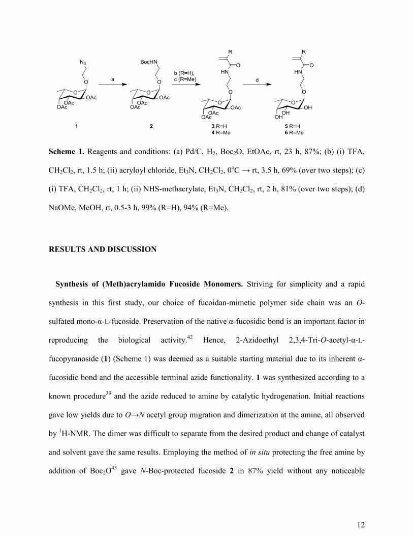
12
Scheme 1. Reagents and conditions: (a) Pd/C, H2, Boc2O, EtOAc, rt, 23 h, 87%; (b) (i) TFA,
CH2Cl2, rt, 1.5 h; (ii) acryloyl chloride, Et3N, CH2Cl2, 0oC → rt, 3.5 h, 69% (over two steps); (c)
(i) TFA, CH2Cl2, rt, 1 h; (ii) NHS-methacrylate, Et3N, CH2Cl2, rt, 2 h, 81% (over two steps); (d)
NaOMe, MeOH, rt, 0.5-3 h, 99% (R=H), 94% (R=Me).
RESULTS AND DISCUSSION
Synthesis of (Meth)acrylamido Fucoside Monomers. Striving for simplicity and a rapid
synthesis in this first study, our choice of fucoidan-mimetic polymer side chain was an O-
sulfated mono-α-L-fucoside. Preservation of the native α-fucosidic bond is an important factor in
reproducing the biological activity.42
Hence, 2-Azidoethyl 2,3,4-Tri-O-acetyl-α-L-
fucopyranoside (1) (Scheme 1) was deemed as a suitable starting material due to its inherent α-
fucosidic bond and the accessible terminal azide functionality. 1 was synthesized according to a
known procedure39
and the azide reduced to amine by catalytic hydrogenation. Initial reactions
gave low yields due to O→N acetyl group migration and dimerization at the amine, all observed
by 1H-NMR. The dimer was difficult to separate from the desired product and change of catalyst
and solvent gave the same results. Employing the method of in situ protecting the free amine by
addition of Boc2O43
gave N-Boc-protected fucoside 2 in 87% yield without any noticeable

13
byproducts formed. As functional groups to be polymerized acrylamide derivatives were chosen.
Deprotection of the N-Boc-group in TFA directly followed by coupling of the free amine with
acryloyl chloride produced acrylamido fucoside 3 in 69% yield. This reaction produced several
byproducts lowering the yield and complicating the cleanup. As an alternative the free amine
was, upon deprotection of the N-Boc-group in TFA, coupled with NHS-methacrylate to form
methacrylamido fucoside 4. This procedure gave a more product-specific reaction and increased
the yield to 81%. Since both acrylamido and methacrylamido functionalities are susceptible to
polymerization both set of fucosides were used in the subsequent steps. Tri-O-acetylated
fucosides 3 and 4 were then subjected to basic ester hydrolysis in NaOMe/MeOH which gave
deprotected fucoside monomers 5 and 6, respectively. These compounds were observed to self-
polymerize when evaporating the MeOH under heating. When protected from heat and light
during work-up and storage, no such observations were made. Attempts were made to O-sulfate
the free hydroxyl groups of the fucoside monomers, but the relatively small size of the tri-O-
sulfated products prohibited purification by either dialysis or size exclusion chromatography. O-
sulfation was hence carried out postpolymerization.
Polymerization of (Meth)acrylamido Fucoside Monomers. Several methods for
synthesizing chain-end functionalized glycopolymers for covalent binding to biomaterials have
been reported.44
We selected cyanoxyl-mediated free-radical polymerization due to its mild
reaction conditions and tolerance to hydroxyl groups.45
This method has demonstrated
successful incorporation of various functional groups at the initiating end, and yields an
inherent cyanate group at the terminating end, of the glycopolymer chain.46
Fucoside
monomers 5 and 6 were polymerized with p-chloroaniline chosen as the initiating phenylamine
due to its demonstrated high yield46
and its unreactive chloride group (Scheme 2). The aryl-

14
type active radical was produced in situ in aqueous solution via reaction between cyanate and
the aryl-diazonium salt formed by reacting p-chloroaniline with fluoroboric acid and nitrite
according to the mechanism described by Grande et al.47
Subsequent addition of fucoside
monomers 5 and 6 yielded glycopolymers 7 (58%) and 8 (47%), respectively, upon
purification by dialysis against deionized water. The slightly lower yield of glycopolymer 8
compared to glycopolymer 7 could be attributed to the well-known slower reactivity of
methacrylamides than of acrylamides in polymerizations. 1H-NMR confirmed the
polymerization of fucoside monomer 6 by the move of the methacrylate alkene proton signals
from 5.69 and 5.46 ppm upfield to form a singlet peak at 1.69 ppm (containing 2 protons) and
the move of the methacrylate methyl proton signal from 1.92 ppm upfield to 0.76 ppm (Figure
2). Signals corresponding to the chain-end aryl protons can be found at 7.26 and 7.01 ppm
confirming a successful cyanoxyl-mediated free-radical polymerization. In a similar fashion,
the move of the acrylate alkene proton signals of fucoside monomer 5 from 5.76, 6.18, and
6.28 ppm upfield to form two singlets at 1.51 (containing 2 protons) and 1.98 ppm (containing
1 proton) confirmed the successful formation of glycopolymer 7. The GPC elugram for
glycopolymer 8 showed one peak (Figure 3), and molecular weights were found to be
Mn=30 200 Da and Mw=65 100 Da giving a dispersity of Ɖ=2.15. The elugram for
glycopolymer 7 however showed two peaks. Given these results, glycopolymer 8 alone was
furnished to a fucoidan-mimetic glycopolymer by O-sulfation.

15
Scheme 2. Reagents and conditions: (a) p-chloroaniline, HBF4, NaNO2, NaOCN, H2O, 0oC →
55oC, 18 h; (b) (i) SO3•Pyr, DMF, rt, 2 days; (ii) NaHCO3, H2O, rt, 24 h.
Figure 2. 1H-NMR spectrum of glycopolymer 8 in D2O, 300 MHz.

16
Figure 3. GPC elugram of glycopolymer 8.
O-Sulfation of Glycopolymer 8. To complete the fucoidan-mimetics, glycopolymer 8 was O-
sulfated through treatment with sulfur trioxide pyridine complex in DMF (Scheme 2). The
pyridinium salt of glycopolymer 9 formed almost instantly, precipitated out of solution and was
subsequently crushed, sonicated, and further stirred to maximize the O-sulfation. The sodium salt
product was then yielded through ion-exchange by dissolving the precipitates in a sodium
bicarbonate water solution and residual compounds removed by extraction with DCM and
dialysis against deionized water. The 1H-NMR spectrum of compound 9 confirmed the O-
sulfation by the move of the signals for fucoside protons H-2, H-3 and H-4 (h, i, and j in Figure
2) from 3.75 ppm downfield to 4.50, 4.64 (merged with H2O), and 4.89 ppm. A downfield move
in signal for a proton neighboring a hydroxyl group turned to ester, e.g., a sulfate ester, is well-
known, and hence expected and in line with previous reports on O-sulfation of α-L-
fucopyranosides.16, 24
The integral ratio between fucoside, ethylene linker or polymer backbone
peaks on 1H-NMR did not change compared to glycopolymer 8, indicating that the O-sulfation
conditions did not hydrolyze the glycosidic nor the amide bonds. Elemental analysis for
glycopolymer 9 showed carbon and sulfur contents of 23.01 and 10.18 %, respectively,
corresponding to a sulfation degree of 66 %. It should be noted that high water content due to the
17
hygroscopic sulfate esters might give some discrepancies to these results. To determine their
fucoidan-mimetic properties, glycopolymers 8 and 9 were tested in assays for biocompatibility,
HSV-1 infection and platelet aggregation.
Biocompatibility of Fucoidan-Mimetic Glycopolymers. Prior to examining the
glycopolymers for their biological activities, we first determined the biocompatibility of these
compounds with HCECs, as compared to the natural fucoidan, dextran sulfate, heparin and the
control polyacrylamide backbone. An important function of corneal epithelial cells is to be able
to proliferate and differentiate to replenish the epithelium. We showed that cell proliferation as
determined by a WST-1 assay was significant. As shown in Figure 4, cell proliferation was only
inhibited by the commercially available natural fucoidan. Effects of a dose of 100 g/mL were
significant (P≤ 0.05 by at 24 h after treatment and 48 h treatment.

18
Figure 4. Effect of glycopolymers 8, and 9 compared to natural fucoidan, heparin, dextran
sulfate and polyacrylamide on cell proliferation as an indication of biocompatibility of these
compounds. HCECs were cultured in the presence of 0, 1, 10, and 100 g/mL of each compound
for 24 and 48 h. Cell proliferation as determined by the WST-1 assay showed that the various
compounds had no effect on proliferation except for natural fucoidan at 10 and 100 g/mL
levels.
19
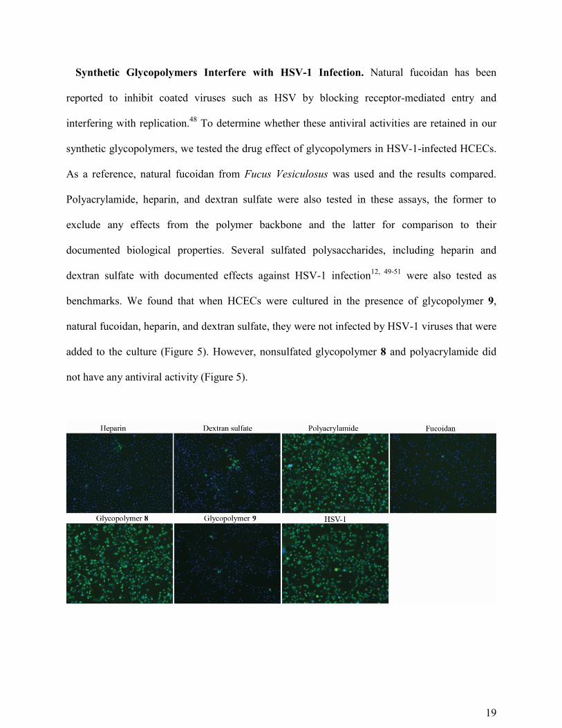
Synthetic Glycopolymers Interfere with HSV-1 Infection. Natural fucoidan has been
reported to inhibit coated viruses such as HSV by blocking receptor-mediated entry and
interfering with replication.48
To determine whether these antiviral activities are retained in our
synthetic glycopolymers, we tested the drug effect of glycopolymers in HSV-1-infected HCECs.
As a reference, natural fucoidan from Fucus Vesiculosus was used and the results compared.
Polyacrylamide, heparin, and dextran sulfate were also tested in these assays, the former to
exclude any effects from the polymer backbone and the latter for comparison to their
documented biological properties. Several sulfated polysaccharides, including heparin and
dextran sulfate with documented effects against HSV-1 infection12, 49-51
were also tested as
benchmarks. We found that when HCECs were cultured in the presence of glycopolymer 9,
natural fucoidan, heparin, and dextran sulfate, they were not infected by HSV-1 viruses that were
added to the culture (Figure 5). However, nonsulfated glycopolymer 8 and polyacrylamide did
not have any antiviral activity (Figure 5).

20
Figure 5. Effect of treatment of HCECs with glycopolymer 8, and 9 (100 g/mL) compared to
natural fucoidan, heparin, dextran sulfate and polyacrylamide. At 24 h post infection, HCECs
pretreated with sulfated glycopolymer 9 and other sulfated compounds (heparin, dextran sulfate,
and natural fucoidan) showed a marked decrease in the expression of active virus (green)
compared to nonsulfated glycopolymer 8, polyacrylamide, and untreated samples. HCEC cell
nuclei were stained with DAPI (blue).
Synthetic Glycopolymers Block HSV-1 Activity during the Virus-Cell Adsorption Step.
We then determined the potential mechanism by which glycopolymer 9 was blocking the herpes
infection. HCECs infected with HSV-1 were treated with 100 g/mL of sulfated glycopolymer 9
at different time points of infection. Pretreatment of cells with sulfated glycopolymers, after
which they were washed off had no noticeable antiviral effect (Figure 6A), indicating that the
sulfated glycopolymers did not cause any changes in the cell membrane to block viral entry.
However, the HSV-1 viral protein expression was greatly reduced by glycopolymer 9 as well
as heparin, dextran sulfate and natural fucoidan when it was added during viral adsorption
(Figure 6B). This strongly suggests that both glycopolymer 9, fucoidan, and the other sulfated
compounds reacted with the viral particles and blocked their entry into the cells.
However, post-treatment of already infected cells with glycopolymer 9 or other sulfated
compounds had no noticeable antiviral effect (Figure 6C) despite previous claims that natural
fucoidan interfered with viral replication.52
Furthermore, when we infected HCECs with a low
MOI of 0.1 to examine the effect of the gycopolymer 9 on viral spreading, we showed that the
sulfated glycopolymer (as well as heparin, dextran sulfate, and natural fucoidan) was able to
slow down but not stop the viral spreading (Figure 7). A low MOI results in infection of an

21
initially small number of cells allowing for observation of viral spreading. Without treatment,
most of the HCECs were infected 48 h post infection (Figure 7). With glycopolymer 9 treatment,
however, a large number of cells remained unstained by the anti-HSV-1/2 antibody. Collectively,
our results suggest that the sulfated glycopolymers most likely influenced the binding and entry
of HSV-1 in cells, and therefore slowed down the viral spreading in HCECs.

22
Figure 6. Effect of sulfated compounds (100 g/mL) - heparin (a), dextran sulfate (b), natural
fucoidan (c), or glycopolymer 9 (d) on HSV-1 infection of cornea epithelial cells (HCECs) under
different conditions, at 24 h postinfection. (A) HCECs were pretreated with compounds for 30
min, washed, then infected with HSV-1 (MOI=1). (B) Compounds were added in the culture

23
medium during viral adsorption. (C) Compounds were added after viral adsorption. Cells
infected with HVS-1 are stained green by an anti-HSV-1/2 antibody that binds viral particles.
HCEC nuclei were stained with DAPI (blue). Nontreated HCECs (e) are displayed as references.

24
Figure 7. Glycopolymer 9 inhibited HSV-1 spreading in already infected HCECs. HCECs were
infected with HSV-1 at a MOI = 0.1. After 1 h adsorption, the inoculated virus was washed
away, and the cells were cultured in fresh medium containing 100 g/mL of synthetic
glycopolymers. At 24, 48, and 72 h post infection, the cells were stained with an anti-HSV-1/2
antibody for viral protein expression (green). Cell nuclei were stained with DAPI (blue). (a)
Heparin, (b) dextran sulfate, (c) natural fucoidan, (d) glycopolymer 9, and (e) nontreated HCEC.
Sulfated Glycopolymers Induce Platelet Activation. The influence of nonsulfated and
sulfated glycopolymers was further assessed on isolated human blood platelets using the same
reference substances as in the HSV-1 infection studies. Addition of 100 µg/mL of glycopolymer
9 induced an immediate aggregation response of platelets (Figure 8A). The effect of the sulfated
glycopolymer was detected down to a dose of 0.3 µg/mL (not shown), and maximal aggregation
response was equivalent to that induced by natural fucoidan (Figure 8B). Nonsulfated
glycopolymer 8 did not stimulate platelet aggregation (Figures 8A and B), and it is notable that
platelets responded normally to subsequent activation by the thrombin receptor-activating
hexapeptide SFLLRN (10 µg/mL). Furthermore, heparin and dextran sulfate as well as backbone
molecule polyacrylamide had no detectable impact on human platelets (Figure 8B). Taken
together, these results show that sulfated but not nonsulfated glycopolymers activated human
blood platelets and consequently enhanced primary hemostasis.

25
Figure 8. Effect of glycopolymers on isolated human platelets. Aliquots of platelet suspensions
were exposed to nonsulfated glycopolymer 8 (100 µg/mL; green traces) and thereafter activated
by the thrombin receptor hexapeptide agonist SFLLRN (10 µg/mL). SFLLRN, but not the
nonsulfated glycoploymer, induced a prompt increase in light transmission which corresponds to
aggregation of platelets. Addition of sulfated glycopolymer 9 (100 µg/mL; red traces) caused an
immediate and pronounced aggregation response. The maximal increase in light transmission
through platelet suspensions following the addition of 100 µg/mL of either glycopolymers 8, 9,
fucoidan, dextran sulfate, heparin or polyacrylamide are summarized in (B). Platelet aggregation

26
by SFLLRN (10 µg/mL) is shown as a positive control. Data are presented as mean ± SEM (n=3-
4).
Fucoidan-Mimetic Activity Depends on Both Fucoside Moiety and Sulfation. These
studies have shown that sulfated glycopolymer 9 has the ability to induce platelet activation and
to prevent the binding and entry of HSV-1 to cells, in analogy with naturally derived fucoidan
from Fucus vesiculosus. Polyacrylamide, which was used as a reference to the polymeric
backbone, and nonsulfated glycopolymer 8 showed neither of these biological properties.
Although other sulfated oligosaccharides such as dextran sulfate and heparin showed similar
abilities to prevent HSV-1 infection, only fucoidan and sulfated glycopolymer 9 possessed the
ability to induce platelet aggregation. This confirms that sulfated glycopolymer 9 behaves as a
functional mimic of natural fucoidan.
CONCLUSIONS
Methacrylamido α-L-fucoside monomers were successfully synthesized, polymerized, and
subsequently partially O-sulfated to yield fucoidan-mimetic sulfated and nonsulfated
glycopolymers. The fucoidan-mimetic nature of the glycopolymers was established by HSV-1
infection and platelet activation studies.
We conclude that a sulfated fucoside polymer side chain is the key structural element for
yielding fucoidan-mimetic properties. We believe this to be a first step toward incorporating the
biological properties of fucoidan into biomaterials.
AUTHOR INFORMATION

27
Corresponding Author
* E-mail: [email protected]; Telephone: +46-(0)13-281728.
Author Contributions
The manuscript was written through contributions of all authors. All authors have given approval
to the final version of the manuscript.
Notes
The authors declare no competing financial interest.
ACKNOWLEDGMENTS
Funding for this project was from an EU Nanomedicine ERAnet grant, I-CARE, and a Swedish
Research Council Treatments of the Future Grant to May Griffith.
REFERENCES
1. Wijesinghe, W. A. J. P.; Jeon, Y. Carbohydr. Polym. 2012, 88, 13-20.
2. Berteau, O.; Mulloy, B. Glycobiology 2003, 13, 29R-40R.
3. Igondjo Tchen Changotade, S.; Korb, G.; Bassil, J.; Barroukh, B.; Willig, C.; Colliec-Jouault,
S.; Durand, P.; Godeau, G.; Senni, K. J. Biomed. Mater. Res., Part A 2008, 87, 666-675.
4. Jeong, H. ; Venkatesan, J.; Kim, S. Int. J. Biol. Macromol. 2013, 57, 138-141.
5. Jin, G.; Kim, G. H. J. Mater. Chem. 2011, 21, 17710-17718.

28
6. Sezer, A. D.; Cevher, E.; Hatipoǧlu, F.; Oǧurtan, Z.; Baş, A. L.; Akbuǧa, J. Biol. Pharm. Bull.
2008, 31, 2326-2333.
7. Lee, H. M.; Kim, J.; Cho, T. J. Ind. Eng. Chem. 2012, 18, 1197-1201.
8. Morya, V. K.; Kim, J.; Kim, E. Appl. Microbiol. Biotechnol. 2012, 93, 71-82.
9. Rioux, L.; Turgeon, S. L.; Beaulieu, M. Phytochemistry 2009, 70, 1069-1075.
10. Cumashi, A.; Ushakova, N. A.; Preobrazhenskaya, M. E.; D'Incecco, A.; Piccoli, A.; Totani,
L.; Tinari, N.; Morozevich, G. E.; Berman, A. E.; Bilan, M. I.; Usov, A. I.; Ustyuzhanina, N. E.;
Grachev, A. A.; Sanderson, C. J.; Kelly, M.; Rabinovich, G. A.; Iacobelli, S.; Nifantiev, N. E.
Glycobiology 2007, 17, 541-552.
11. Pomin, V. H. Biochim. Biophys. Acta, Gen. Subj. 2012, 1820, 1971-1979.
12. Ghosh, T.; Chattopadhyay, K.; Marschall, M.; Karmakar, P.; Mandal, P.; Ray, B.
Glycobiology 2009, 19, 2-15.
13. Ale, M. T.; Mikkelsen, J. D.; Meyer, A. S. Marine Drugs 2011, 9, 2106-2130.
14. Jain, R. K.; Matta, K. L. Carbohydr. Res. 1990, 208, 280-286.
15. Jain, R. K.; Matta, K. L. Carbohydr. Res. 1990, 208, 51-58.
16. Hua, Y.; Gu, G.; Du, Y. Carbohydr. Res. 2004, 339, 867-872.
17. Hua, Y.; Du, Y.; Yu, G.; Chu, S. Carbohydr. Res. 2004, 339, 2083-2090.

29
18. Gerbst, A. G.; Ustuzhanina, N. E.; Grachev, A. A.; Khatuntseva, E. A.; Tsvetkov, D. E.;
Whitfield, D. M.; Berces, A.; Nifantiev, N. E. J. Carbohydr. Chem. 2001, 20, 821-831.
19. Gerbst, A. G.; Ustuzhanina, N. E.; Grachev, A. A.; Zlotina, N. S.; Khatuntseva, E. A.;
Tsvetkov, D. E.; Shashkov, A. S.; Usov, A. I.; Nifantiev, N. E. J. Carbohydr. Chem. 2002, 21,
313-324.
20. Gerbst, A. G.; Ustuzhanina, N. E.; Grachev, A. A.; Khatuntseva, E. A.; Tsvetkov, D. E.;
Shashkov, A. S.; Usov, A. I.; Preobrazhenskaya, M. E.; Ushakova, N. A.; Nifantiev, N. E. J.
Carbohydr. Chem. 2003, 22, 109-122.
21. Grachev, A. A.; Gerbst, A. G.; Ustuzhanina, N. E.; Khatuntseva, E. A.; Shashkov, A. S.;
Usov, A. I.; Nifantiev, N. E. J. Carbohydr. Chem. 2005, 24, 85-100.
22. Gerbst, A. G.; Grachev, A. A.; Ustyuzhanina, N. E.; Khatuntseva, E. A.; Tsvetkov, D. E.;
Usov, A. I.; Shashkov, A. S.; Preobrazhenskaya, M. E.; Ushakova, N. A.; Nifantiev, N. E. Russ.
J. Bioorg. Chem. 2004, 30, 137-147.
23. Khatuntseva, E. A.; Ustuzhanina, N. E.; Zatonskii, G. V.; Shashkov, A. S.; Usov, A. I.;
Nifantiev, N. E. J. Carbohydr. Chem. 2000, 19, 1151-1173.
24. Krylov, V. B.; Kaskova, Z. M.; Vinnitskiy, D. Z.; Ustyuzhanina, N. E.; Grachev, A. A.;
Chizhov, A. O.; Nifantiev, N. E. Carbohydr. Res. 2011, 346, 540-550.
25. Ustyuzhanina, N.; Krylov, V.; Grachev, A.; Gerbst, A.; Nifantiev, N. Synthesis 2006, 4017-
4031.

30
26. Schatz, C.; Lecommandoux, S. Macromol. Rapid Commun. 2010, 31, 1664-1684.
27. Dupayage, L.; Nouvel, C.; Six, J. J. Polym. Sci. Part A 2011, 49, 35-46.
28. Ferji, K.; Nouvel, C.; Babin, J.; Albouy, P.; Li, M.; Six, J. J. Polym. Sci. Part A 2013, 51,
3829-3839.
29. Becer, C. R.; Gibson, M. I.; Geng, J.; Ilyas, R.; Wallis, R.; Mitchell, D. A.; Haddleton, D. M.
J. Am. Chem. Soc. 2010, 132, 15130-15132.
30. Becer, C. R. Macromol. Rapid Commun. 2012, 33, 742-752.
31. Yilmaz, G.; Becer, C. R. Eur. Polym. J. 2013, 49, 3046-3051.
32. Ahmed, M.; Wattanaarsakit, P.; Narain, R. Eur. Polym. J. 2013, 49, 3010-3033.
33. Ting, S. R. S.; Chen, G.; Stenzel, M. H. Polym. Chem. 2010, 1, 1392-1412.
34. Vázquez-Dorbatt, V.; Lee, J.; Lin, E.; Maynard, H. D. ChemBioChem 2012, 13, 2478-2487.
35. Voit, B.; Appelhans, D. Macromol. Chem. Phys. 2010, 211, 727-735.
36. Zhang, Q.; Collins, J.; Anastasaki, A.; Wallis, R.; Mitchell, D. A.; Becer, C. R.; Haddleton,
D. M. Angew. Chem., Int. Ed, 2013, 52, 4435-4439.
37. Gustafson, T. P.; Lonnecker, A. T.; Heo, G. S.; Zhang, S.; Dove, A. P.; Wooley, K. L.
Biomacromolecules 2013, 14, 3346-3353.

31
38. Fulmer, G. R.; Miller, A. J. M.; Sherden, N. H.; Gottlieb, H. E.; Nudelman, A.; Stoltz, B. M.;
Bercaw, J. E.; Goldberg, K. I. Organometallics 2010, 29, 2176-2179.
39. Ni, J.; Singh, S.; Wang, L. Bioconjugate Chem. 2003, 14, 232-238.
40. Araki-Sasaki, K.; Ohashi, Y.; Sasabe, T.; Hayashi, K.; Watanabe, H.; Tano, Y.; Handa, H.
Invest. Ophthalmol. Vis. Sci. 1995, 36, 614-621.
41. Ejercito, P. M.; Kieff, E. D.; Roizman, B. J. Gen. Virol. 1968, 2, 357-364.
42. Lee, Y. C.; Lee, R. T. Acc. Chem. Res. 1995, 28, 321-326.
43. Patel, A.; Lindhorst, T. K. J. Org. Chem. 2001, 66, 2674-2680.
44. Narla, S. N.; Nie, H.; Li, Y.; Sun, X. J. Carbohydr. Chem. 2012, 31, 67-92.
45. Sun, X.; Grande, D.; Baskaran, S.; Hanson, S. R.; Chaikof, E. L. Biomacromolecules 2002, 3,
1065-1070.
46. Hou, S.; Sun, X.; Dong, C.; Chaikof, E. L. Bioconjugate Chem. 2004, 15, 954-959.
47. Grande, D.; Baskaran, S.; Baskaran, C.; Gnanou, Y.; Chaikof, E. L. Macromolecules 2000,
33, 1123-1125.
48. Fitton, J. H. Mar. Drugs 2011, 9, 1731-1760.
49. Rabenstein, D. L. Nat. Prod. Rep. 2002, 19, 312-331.

32
50. Andrei, G.; Snoeck, R.; Goubau, P.; Desmyter, J.; De Clercq, E. Eur. J. Clin. Microbiol.
Infect. Dis. 1992, 11, 143-151.
51. Ramos-Kuri, M.; Barron Romero, B. L.; Aguilar-Setien, A. Arch. Med. Res. 1996, 27, 43-48.
52. Hayashi, K.; Nakano, T.; Hashimoto, M.; Kanekiyo, K.; Hayashi, T. Int. Immunopharmacol.
2008, 8, 109-116.