Embed Size (px)
Citation preview

nutrients
Article
A Novel Combination of Wheat Peptides andFucoidan Attenuates Ethanol-Induced GastricMucosal Damage through Anti-Oxidant,Anti-Inflammatory, and Pro-Survival Mechanisms
Juntao Kan 1,†, Molly Hood 2,†, Charlie Burns 2, Jeff Scholten 2, Jennifer Chuang 3, Feng Tian 1,Xingchang Pan 4, Jun Du 1,* and Min Gui 1,*
1 Nutrilite Health Institute, 720 Cailun Road, Shanghai 201203, China; [email protected] (J.K.);[email protected] (F.T.)
2 Nutrilite Health Institute, 7575 East Fulton Avenue, Ada, MI 49355, USA; [email protected] (M.H.);[email protected] (C.B.); [email protected] (J.S.)
3 Nutrilite Health Institute, 5600 Beach Boulevard, Buena Park, CA 90621, USA; [email protected] China National Research Institute of Food and Fermentation Industries, 24 Jiuxianqiao Middle Road,
Beijing 100015, China; [email protected]* Correspondence: [email protected] (J.D.); [email protected] (M.G.); Tel.: +86-21-23056956 (J.D.)† These authors contributed equally to this work.
Received: 13 July 2017; Accepted: 29 August 2017; Published: 6 September 2017
Abstract: Gastritis or peptic ulcer is believed to affect about half of people worldwide. Traditionalmedications can lead to adverse effects, therefore, alternative nutritional strategies are needed toprevent the development of gastric mucosal damage. A novel combination of two food-gradeingredients, wheat peptides and fucoidan (WPF), was prepared to treat male Sprague Dawley rats for30 days before gastric mucosal damage was induced by oral administration of ethanol. The serumlevels of biomarkers were determined by enzyme-linked immunosorbent assay. Biomarkers instomach tissue were analyzed using immunohistochemistry. In addition, human gastric epithelialcell line (GES-1) was used to investigate protein expression by Western blot. WPF could attenuateethanol-induced gastric mucosal damage in an inverse dose-dependent manner, with both ulcerindex and pathological index improved. WPF increased superoxide dismutase level and decreasedmalondialdehyde level. WPF also decreased the levels of interleukin-8, platelet-activating factor,and Caspase 3, while increasing the levels of prostaglandin E-2, epidermal growth factor (EGF),and EGF receptor (EGFR). Furthermore, phosphorylation of EGFR and extracellular signal–regulatedkinases was induced by WPF in GES-1 cells. In conclusion, the novel combination of wheat peptidesand fucoidan attenuated ethanol-induced gastric mucosal damage in rats through anti-oxidant,anti-inflammatory, and pro-survival mechanisms.
Keywords: gastric mucosal damage; wheat peptides; fucoidan; anti-oxidant; anti-inflammation; pro-survival
1. Introduction
Gastric mucosal damage occurs when risk factors, such as alcohol, stress, Helicobacter pylori,and nonsteroidal anti-inflammatory drugs (NSAIDs), overwhelm protective factors/mechanisms,and this can lead to the development of gastritis, peptic ulcer, or even gastric cancer [1,2]. Particularly,immoderate alcohol consumption can cause mucosal edema, congestion, hemorrhage, epithelialexfoliation, and inflammatory cell infiltration, resulting in ulcers in the stomach [3,4]. Therefore, a ratmodel of ethanol-induced gastric mucosal damage is widely used to study preventive or protectiveeffects of drugs or health foods [5,6].
Nutrients 2017, 9, 978; doi:10.3390/nu9090978 www.mdpi.com/journal/nutrients

Nutrients 2017, 9, 978 2 of 12
Wheat peptides are biologically active peptides obtained by enzymatic hydrolysis from wheatprotein [7,8], and they have anti-oxidant activity [9]. Wheat peptides could resist indomethacin-inducedoxidative stress in intestinal epithelial cells-6 (IEC-6) and hydrogen peroxide-induced oxidative stressin pheochromocytoma cells-12 [10,11]. Furthermore, treatment with wheat peptides could protectagainst NSAID-induced small intestinal damage by decreasing oxidative stress in rats [12].
Fucoidan is a sulfated polysaccharide obtained mainly in various species of brown algae andbrown seaweed such as Undaria pinnatifida, Laminaria angustata, Fucus vesiculosus, and Fucus evanescens,and it has anti-inflammatory activity [13,14]. Fucoidan can downregulate the production ofinterleukin-6 in colonic epithelial cells [15], and protect against aspirin-induced ulceration in ratsthrough its immunomodulatory properties [16].
The protective mechanisms underlying ethanol-induced gastric mucosal damage mainly includeanti-oxidation, anti-inflammation, and pro-survival [5,16,17]. Based on the anti-oxidant activity ofwheat peptides and anti-inflammatory activity of fucoidan, we proposed a novel combination of wheatpeptides and fucoidan (WPF), and tested its efficacy in a rat model of ethanol-induced gastric mucosaldamage, as well as investigated possible involved mechanisms.
2. Materials and Methods
2.1. Materials and WPF Preparation
Wheat peptides were obtained from China National Research Institute of Food & FermentationIndustries (Beijing, China), and prepared by protease hydrolysis method [10]. The peptides included3–6 amino acid residues, ranging from 140 to 1000 Da. Fucoidan (Beijing Gingko Group, Beijing, China)was extracted from Laminaria japonica (kelp) by water and filtered by membrane. An optimal ratio of10:3 (wheat peptides:fucoidan) was determined after previous bioassays and in vivo pilot study aswell as considering commercial cost and regulatory limits of China Food and Drug Administration(CFDA) on doses. WPF was prepared by Nutrilite Health Institute through mixture of 1 g of wheatpeptides and 300 mg of fucoidan (daily intake for human).
2.2. Cell Culture
Human gastric epithelial cell line (GES-1) was obtained from the American Type Culture Collection(ATCC, Rockville, MD, USA) and was cultured at 37 ◦C under 5% CO2 in Dulbecco’s Modified EagleMedium (Gibco, Grand Island, NY, USA) with 10% fetal bovine serum (Hyclone, Logan, UT, USA) and1% penicillin streptomycin (Gibco, Grand Island, NY, USA).
2.3. Animals
Male Sprague Dawley rats (180–220 g) were purchased from Charles River (Beijing, China).Animals were housed in a temperature and humidity-controlled room (22–23 ◦C and 46–63%,respectively) and had free access to food and water. The experimental protocols (ethic code: SYXK(SU) 2013-0037) were approved by the Institutional Animal Care and Use Committee of SoutheastUniversity (Nanjing, China).
2.4. Ethanol-Induced Gastric Mucosal Damage Model
This model was established by following the protocol from CFDA for health food registration withthe claim of “Assisting the Protection of Gastric Mucosa Function” [18]. After one-week acclimation,the animals were divided into eight groups (10 rats for each group): (1) naïve control; (2) modelcontrol; (3) low dose of WPF (108 mg/kg); (4) medium dose of WPF (217 mg/kg); (5) high dose ofWPF (325 mg/kg); (6) fucoidan (50 mg/kg); (7) wheat peptide (167 mg/kg); (8) cimetidine (65 mg/kg).The rats of model control and treatment groups were given either vehicle (saline) or test articles bydaily oral gavage for 30 days.

Nutrients 2017, 9, 978 3 of 12
Acute model of gastric mucosal damage was induced by oral administration of ethanol (1 mL) 24 hafter the 30-day-treatment period. Rats were sacrificed 1 h after ethanol treatment. The naïve controlgroup received vehicle only. Prior to termination, blood was collected and centrifugated to get serumfor further analysis. Then the animals were euthanized by CO2 inhalation, and stomach was excisedfor macroscopic and histopathologic evaluation.
2.5. Macroscopic Analysis
The excised stomach was opened along the greater curvature, and mucosa was washed withcold phosphate-buffered saline (PBS). Gastric mucosal damage was scored by an experiencedgastroenterologist, who was blinded to the samples, and expressed as ulcer index (Table 1, [18]).
Table 1. Scoring criteria for ulcer index.
Grade 1 2 3 4
Spot Erosion Each spotLinear Erosion (Length) 1–5 mm 5–10 mm 10–15 mm >15 mmLinear Erosion (Width) 1–2 mm >2 mm
Ulcer Index = Spot Erosion + Linear Erosion Length + (Linear Erosion Width) × 2
2.6. Histopathologic Evaluation
The stomach tissue was then fixed in 10% formaldehyde, embedded into paraffin, and cut into4 µm sections. A commercially available hematoxylin-eosin (H&E) kit (Beyotime, Shanghai, China)was used to stain sections. An expert pathologist evaluated the slides in a blinded fashion andcalculated the total pathological index as follows (Table 2, [18]).
Table 2. Scoring criteria for pathological index.
Grade 1 2 3 4 5
Congestion <20% 20–40% 40–60% 60–80% ≥80%Hemorrhage <20% 20–40% 40–60% 60–80% ≥80%
Necrosis <20% 20–40% 40–60% 60–80% ≥80%Pathological Index = Congestion + Hemorrhage × 2 + Necrosis × 3
2.7. Immunohistochemistry
Caspase 3 and epidermal growth factor receptor (EGFR) in paraffin sections were visualizedwith rabbit polyclonal Caspase 3 antibody (Abcam, Cambridge, MA, USA) and EGFRantibody (Zen BioScience, Chengdu, China) followed by staining with a horseradish peroxidase(HRP)-conjugated secondary antibody, and diaminobenzidine substrate (Maixin, Fuzhou, China).Five random fields from each section were photographed with a Zeiss digital camera, and analyzedusing a semi-quantitative scoring system as follows (Table 3, [19,20]).
Table 3. Semi-quantitative scoring system.
Grade 0 1 2 3 4
Percentage of Positive Cells ≤5% 6–25% 26–50% 51–75% >75%Intensity of Immunostaining No Low Moderate Strong
Total Score = Percentage of Positive Cells × Intensity of Immunostaining
2.8. Biochemical Analysis
Serum levels of malondialdehyde (MDA), superoxide dismutase (SOD), platelet-activating factor(PAF), prostaglandin E-2 (PGE2), epidermal growth factor (EGF), and interleukin-8 (IL-8) were
Nutrients 2017, 9, 978 4 of 12
determined by using commercially available kits (JRdun Biotechnology, Shanghai, China) according tothe manufacturer’s instructions.
In brief, the MDA assay kit uses thiobarbituric acid (TBA) to react with MDA to generate aMDA-TBA adduct which can be easily quantified colorimetrically at 532 nm. The SOD assay kit usesan enzyme mix that oxidizes WST-1 (2-(4-iodophenyl)-3-(4-nitrophenyl)-5-(2,4-disulfophenyl)-2H-tetrazolium, monosodium salt) and produces a water-soluble formazan dye to generate color that isread in a plate reader at 440 nm.
The levels of PAF, PGE2, EGF, and IL-8 were determined by sandwich enzyme-linkedimmunosorbent assay (ELISA). The principle of the assay is to capture each molecule from samplesin the wells of a microtiter plate coated by a pre-titered amount of primary antibody and then thebinding of biotinylated polyclonal antibody to the captured molecule. Next, horseradish peroxidase isadded to wells to bind to the immobilized biotinylated antibody, and quantification of immobilizedantibody-enzyme conjugate is determined by horseradish peroxidase activities in the presence of thesubstrate 3,3′,5,5′-tetramethylbenzidine. The enzyme activity is measured spectrophotometrically bythe increased absorbency at 450 nm, corrected from the absorbency at 590 nm, after acidification offormed product.
2.9. Western Blot
GES-1 cells were treated with control (PBS) or WPF (1.3 mg/mL) for 12 h before proteinextraction with cell lysis buffer (Cell Signaling, Danvers, MA, USA). Lysates were boiled with 5×loading buffer and then loaded onto sodium dodecyl sulfate-polyacrylamide gels. The separatedproteins were transferred to polyvinylidene fluoride (PVDF) membranes (Millipore, Billerica, MA,USA), then blocked with 5% nonfat dry milk in Tris-buffered saline/Tween (TBST). Primary andsecondary antibodies were used to successively incubate the PVDF membrane. The HRP-conjugatedprotein was detected by chemiluminescent horseradish peroxidase substrate solution (Millipore,Billerica, MA, USA). The primary antibodies against EGFR, p-EGFR, extracellular signal-regulatedkinases (ERK), and p-ERK were purchased from Cell Signaling, and GAPDH antibody was purchasedfrom Abcam.
2.10. Statistical Analysis
Statistical analysis was performed using SPSS 13.0 software (SPSS Inc., Chicago, IL, USA).Quantitative data are presented as mean ± standard deviation (SD). Differences between groupswere analyzed using Student’s t-test when only two groups were compared or assessed by one-wayanalysis of variance (ANOVA) with Dunnett’s post-hoc test when more than two groups were compared.A significant effect was defined as p < 0.05.
3. Results
3.1. WPF Attenuated Ethanol-Induced Gastric Mucosal Damage
Oral administration of ethanol induced gastric mucosal damage, resulting in an ulcer index from(0.1 ± 0.3) to (24.8 ± 6.5) (p < 0.05, Figure 1A). WPF attenuated ethanol-induced mucosal damage,with ulcer indexes of 2.5 ± 3.3, 6.3 ± 6.7, 9.6 ± 7.7, respectively (p < 0.05 vs. model control). The lowdose of WPF showed better protective effects when compared with the treatment of fucoidan alone(p < 0.05 vs. fucoidan).
Nutrients 2017, 9, 978 5 of 12Nutrients 2017, 9, 978 5 of 11
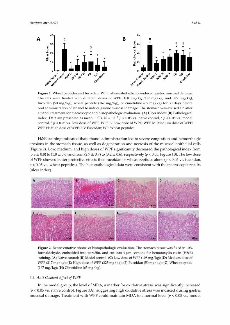
Figure 1. Wheat peptides and fucoidan (WPF) attenuated ethanol-induced gastric mucosal damage. The rats were treated with different doses of WPF (108 mg/kg, 217 mg/kg, and 325 mg/kg), fucoidan (50 mg/kg), wheat peptide (167 mg/kg), or cimetidine (65 mg/kg) for 30 days before oral administration of ethanol to induce gastric mucosal damage. The stomach was excised 1 h after ethanol treatment for macroscopic and histopathologic evaluation. (A) Ulcer index; (B) Pathological index. Data are presented as mean ± SD. N = 10. # p < 0.05 vs. naïve control, * p < 0.05 vs. model control, $ p < 0.05 vs. low dose of WPF. WPF L: Low dose of WPF; WPF M: Medium dose of WPF; WPF H: High dose of WPF; FD: Fucoidan; WP: Wheat peptides.
H&E staining indicated that ethanol administration led to severe congestion and hemorrhagic erosions in the stomach tissue, as well as degeneration and necrosis of the mucosal epithelial cells (Figure 2). Low, medium, and high doses of WPF significantly decreased the pathological index from (5.8 ± 0.8) to (1.8 ± 0.6) and from (2.7 ± 0.7) to (3.2 ± 0.6), respectively (p < 0.05, Figure 1B). The low dose of WPF showed better protective effects then fucoidan or wheat peptides alone (p < 0.05 vs. fucoidan, p < 0.05 vs. wheat peptides). The histopathological data were consistent with the macroscopic results (ulcer index).
Figure 2. Representative photos of histopathologic evaluation. The stomach tissue was fixed in 10% formaldehyde, embedded into paraffin, and cut into 4 μm sections for hematoxylin-eosin (H&E) staining. (A) Naïve control; (B) Model control; (C) Low dose of WPF (108 mg/kg); (D) Medium dose of WPF (217 mg/kg); (E) High dose of WPF (325 mg/kg); (F) Fucoidan (50 mg/kg); (G) Wheat peptide (167 mg/kg); (H) Cimetidine (65 mg/kg).
3.2. Anti-Oxidant Effect of WPF
In the model group, the level of MDA, a marker for oxidative stress, was significantly increased (p < 0.05 vs. naïve control, Figure 3A), suggesting high oxidative stress was induced during gastric mucosal
Figure 1. Wheat peptides and fucoidan (WPF) attenuated ethanol-induced gastric mucosal damage.The rats were treated with different doses of WPF (108 mg/kg, 217 mg/kg, and 325 mg/kg),fucoidan (50 mg/kg), wheat peptide (167 mg/kg), or cimetidine (65 mg/kg) for 30 days beforeoral administration of ethanol to induce gastric mucosal damage. The stomach was excised 1 h afterethanol treatment for macroscopic and histopathologic evaluation. (A) Ulcer index; (B) Pathologicalindex. Data are presented as mean ± SD. N = 10. # p < 0.05 vs. naïve control, * p < 0.05 vs. modelcontrol, $ p < 0.05 vs. low dose of WPF. WPF L: Low dose of WPF; WPF M: Medium dose of WPF;WPF H: High dose of WPF; FD: Fucoidan; WP: Wheat peptides.
H&E staining indicated that ethanol administration led to severe congestion and hemorrhagicerosions in the stomach tissue, as well as degeneration and necrosis of the mucosal epithelial cells(Figure 2). Low, medium, and high doses of WPF significantly decreased the pathological index from(5.8± 0.8) to (1.8± 0.6) and from (2.7± 0.7) to (3.2± 0.6), respectively (p < 0.05, Figure 1B). The low doseof WPF showed better protective effects then fucoidan or wheat peptides alone (p < 0.05 vs. fucoidan,p < 0.05 vs. wheat peptides). The histopathological data were consistent with the macroscopic results(ulcer index).
Nutrients 2017, 9, 978 5 of 11
Figure 1. Wheat peptides and fucoidan (WPF) attenuated ethanol-induced gastric mucosal damage. The rats were treated with different doses of WPF (108 mg/kg, 217 mg/kg, and 325 mg/kg), fucoidan (50 mg/kg), wheat peptide (167 mg/kg), or cimetidine (65 mg/kg) for 30 days before oral administration of ethanol to induce gastric mucosal damage. The stomach was excised 1 h after ethanol treatment for macroscopic and histopathologic evaluation. (A) Ulcer index; (B) Pathological index. Data are presented as mean ± SD. N = 10. # p < 0.05 vs. naïve control, * p < 0.05 vs. model control, $ p < 0.05 vs. low dose of WPF. WPF L: Low dose of WPF; WPF M: Medium dose of WPF; WPF H: High dose of WPF; FD: Fucoidan; WP: Wheat peptides.
H&E staining indicated that ethanol administration led to severe congestion and hemorrhagic erosions in the stomach tissue, as well as degeneration and necrosis of the mucosal epithelial cells (Figure 2). Low, medium, and high doses of WPF significantly decreased the pathological index from (5.8 ± 0.8) to (1.8 ± 0.6) and from (2.7 ± 0.7) to (3.2 ± 0.6), respectively (p < 0.05, Figure 1B). The low dose of WPF showed better protective effects then fucoidan or wheat peptides alone (p < 0.05 vs. fucoidan, p < 0.05 vs. wheat peptides). The histopathological data were consistent with the macroscopic results (ulcer index).
Figure 2. Representative photos of histopathologic evaluation. The stomach tissue was fixed in 10% formaldehyde, embedded into paraffin, and cut into 4 μm sections for hematoxylin-eosin (H&E) staining. (A) Naïve control; (B) Model control; (C) Low dose of WPF (108 mg/kg); (D) Medium dose of WPF (217 mg/kg); (E) High dose of WPF (325 mg/kg); (F) Fucoidan (50 mg/kg); (G) Wheat peptide (167 mg/kg); (H) Cimetidine (65 mg/kg).
3.2. Anti-Oxidant Effect of WPF
In the model group, the level of MDA, a marker for oxidative stress, was significantly increased (p < 0.05 vs. naïve control, Figure 3A), suggesting high oxidative stress was induced during gastric mucosal
Figure 2. Representative photos of histopathologic evaluation. The stomach tissue was fixed in 10%formaldehyde, embedded into paraffin, and cut into 4 µm sections for hematoxylin-eosin (H&E)staining. (A) Naïve control; (B) Model control; (C) Low dose of WPF (108 mg/kg); (D) Medium dose ofWPF (217 mg/kg); (E) High dose of WPF (325 mg/kg); (F) Fucoidan (50 mg/kg); (G) Wheat peptide(167 mg/kg); (H) Cimetidine (65 mg/kg).
3.2. Anti-Oxidant Effect of WPF
In the model group, the level of MDA, a marker for oxidative stress, was significantly increased(p < 0.05 vs. naïve control, Figure 3A), suggesting high oxidative stress was induced during gastricmucosal damage. Treatment with WPF could maintain MDA to a normal level (p < 0.05 vs. model

Nutrients 2017, 9, 978 6 of 12
control). Likewise, WPF increased the level of SOD, an antioxidant enzyme (p < 0.05 vs. model control,Figure 3B).
Nutrients 2017, 9, 978 6 of 11
damage. Treatment with WPF could maintain MDA to a normal level (p < 0.05 vs. model control). Likewise, WPF increased the level of SOD, an antioxidant enzyme (p < 0.05 vs. model control, Figure 3B).
Figure 3. Anti-oxidant effect of WPF. Serum levels of malondialdehyde (MDA) (A) and superoxide dismutase (SOD) (B) were determined by using commercially available colorimetric kits. Data are presented as mean ± SD. N = 10. # p < 0.05 vs. naïve control, * p < 0.05 vs. model control. WPF L: Low dose of WPF; WPF M: Medium dose of WPF; WPF H: High dose of WPF; FD: Fucoidan; WP: Wheat peptides.
3.3. Anti-Inflammatory Effect of WPF
Ethanol administration significantly induced the secretion of IL-8, which could be reversed by treatment with WPF (p < 0.05, Figure 4A). As a mediator of allergic and inflammatory processes, PAF was also inhibited by treatment with WPF (p < 0.05, Figure 4B).
Figure 4. Anti-inflammation and pro-survival effects of WPF. Serum levels of interleukin (IL)-8 (A), platelet-activating factor (PAF) (B), prostaglandin E-2 (PGE2) (C), and epidermal growth factor (EGF) (D) were determined by using commercially available enzyme-linked immunosorbent assay (ELISA) kits. Data are presented as mean ± SD. N = 10. # p < 0.05 vs. naïve control; * p < 0.05 vs. model control. WPF L: Low dose of WPF; WPF M: Medium dose of WPF; WPF H: High dose of WPF; FD: Fucoidan; WP: Wheat peptides.
Figure 3. Anti-oxidant effect of WPF. Serum levels of malondialdehyde (MDA) (A) and superoxidedismutase (SOD) (B) were determined by using commercially available colorimetric kits. Data arepresented as mean ± SD. N = 10. # p < 0.05 vs. naïve control, * p < 0.05 vs. model control.WPF L: Low dose of WPF; WPF M: Medium dose of WPF; WPF H: High dose of WPF; FD: Fucoidan;WP: Wheat peptides.
3.3. Anti-Inflammatory Effect of WPF
Ethanol administration significantly induced the secretion of IL-8, which could be reversed bytreatment with WPF (p < 0.05, Figure 4A). As a mediator of allergic and inflammatory processes,PAF was also inhibited by treatment with WPF (p < 0.05, Figure 4B).
Nutrients 2017, 9, 978 6 of 11
damage. Treatment with WPF could maintain MDA to a normal level (p < 0.05 vs. model control). Likewise, WPF increased the level of SOD, an antioxidant enzyme (p < 0.05 vs. model control, Figure 3B).
Figure 3. Anti-oxidant effect of WPF. Serum levels of malondialdehyde (MDA) (A) and superoxide dismutase (SOD) (B) were determined by using commercially available colorimetric kits. Data are presented as mean ± SD. N = 10. # p < 0.05 vs. naïve control, * p < 0.05 vs. model control. WPF L: Low dose of WPF; WPF M: Medium dose of WPF; WPF H: High dose of WPF; FD: Fucoidan; WP: Wheat peptides.
3.3. Anti-Inflammatory Effect of WPF
Ethanol administration significantly induced the secretion of IL-8, which could be reversed by treatment with WPF (p < 0.05, Figure 4A). As a mediator of allergic and inflammatory processes, PAF was also inhibited by treatment with WPF (p < 0.05, Figure 4B).
Figure 4. Anti-inflammation and pro-survival effects of WPF. Serum levels of interleukin (IL)-8 (A), platelet-activating factor (PAF) (B), prostaglandin E-2 (PGE2) (C), and epidermal growth factor (EGF) (D) were determined by using commercially available enzyme-linked immunosorbent assay (ELISA) kits. Data are presented as mean ± SD. N = 10. # p < 0.05 vs. naïve control; * p < 0.05 vs. model control. WPF L: Low dose of WPF; WPF M: Medium dose of WPF; WPF H: High dose of WPF; FD: Fucoidan; WP: Wheat peptides.
Figure 4. Anti-inflammation and pro-survival effects of WPF. Serum levels of interleukin (IL)-8 (A),platelet-activating factor (PAF) (B), prostaglandin E-2 (PGE2) (C), and epidermal growth factor (EGF)(D) were determined by using commercially available enzyme-linked immunosorbent assay (ELISA)kits. Data are presented as mean ± SD. N = 10. # p < 0.05 vs. naïve control; * p < 0.05 vs. model control.WPF L: Low dose of WPF; WPF M: Medium dose of WPF; WPF H: High dose of WPF; FD: Fucoidan;WP: Wheat peptides.
Nutrients 2017, 9, 978 7 of 12
3.4. Pro-Survival Effect of WPF
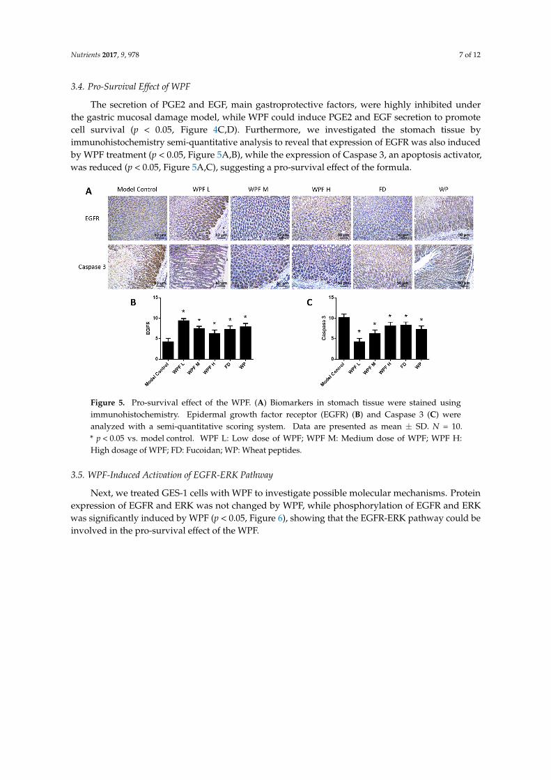
The secretion of PGE2 and EGF, main gastroprotective factors, were highly inhibited underthe gastric mucosal damage model, while WPF could induce PGE2 and EGF secretion to promotecell survival (p < 0.05, Figure 4C,D). Furthermore, we investigated the stomach tissue byimmunohistochemistry semi-quantitative analysis to reveal that expression of EGFR was also inducedby WPF treatment (p < 0.05, Figure 5A,B), while the expression of Caspase 3, an apoptosis activator,was reduced (p < 0.05, Figure 5A,C), suggesting a pro-survival effect of the formula.
Nutrients 2017, 9, 978 7 of 11
3.4. Pro-Survival Effect of WPF
The secretion of PGE2 and EGF, main gastroprotective factors, were highly inhibited under the gastric mucosal damage model, while WPF could induce PGE2 and EGF secretion to promote cell survival (p < 0.05, Figure 4C,D). Furthermore, we investigated the stomach tissue by immunohistochemistry semi-quantitative analysis to reveal that expression of EGFR was also induced by WPF treatment (p < 0.05, Figure 5A,B), while the expression of Caspase 3, an apoptosis activator, was reduced (p < 0.05, Figure 5A,C), suggesting a pro-survival effect of the formula.
Figure 5. Pro-survival effect of the WPF. (A) Biomarkers in stomach tissue were stained using immunohistochemistry. Epidermal growth factor receptor (EGFR) (B) and Caspase 3 (C) were analyzed with a semi-quantitative scoring system. Data are presented as mean ± SD. N = 10. * p < 0.05 vs. model control. WPF L: Low dose of WPF; WPF M: Medium dose of WPF; WPF H: High dosage of WPF; FD: Fucoidan; WP: Wheat peptides.
3.5. WPF-Induced Activation of EGFR-ERK Pathway
Next, we treated GES-1 cells with WPF to investigate possible molecular mechanisms. Protein expression of EGFR and ERK was not changed by WPF, while phosphorylation of EGFR and ERK was significantly induced by WPF (p < 0.05, Figure 6), showing that the EGFR-ERK pathway could be involved in the pro-survival effect of the WPF.
Figure 5. Pro-survival effect of the WPF. (A) Biomarkers in stomach tissue were stained usingimmunohistochemistry. Epidermal growth factor receptor (EGFR) (B) and Caspase 3 (C) wereanalyzed with a semi-quantitative scoring system. Data are presented as mean ± SD. N = 10.* p < 0.05 vs. model control. WPF L: Low dose of WPF; WPF M: Medium dose of WPF; WPF H:High dosage of WPF; FD: Fucoidan; WP: Wheat peptides.
3.5. WPF-Induced Activation of EGFR-ERK Pathway
Next, we treated GES-1 cells with WPF to investigate possible molecular mechanisms. Proteinexpression of EGFR and ERK was not changed by WPF, while phosphorylation of EGFR and ERKwas significantly induced by WPF (p < 0.05, Figure 6), showing that the EGFR-ERK pathway could beinvolved in the pro-survival effect of the WPF.

Nutrients 2017, 9, 978 8 of 12
Nutrients 2017, 9, 978 7 of 11
3.4. Pro-Survival Effect of WPF
The secretion of PGE2 and EGF, main gastroprotective factors, were highly inhibited under the gastric mucosal damage model, while WPF could induce PGE2 and EGF secretion to promote cell survival (p < 0.05, Figure 4C,D). Furthermore, we investigated the stomach tissue by immunohistochemistry semi-quantitative analysis to reveal that expression of EGFR was also induced by WPF treatment (p < 0.05, Figure 5A,B), while the expression of Caspase 3, an apoptosis activator, was reduced (p < 0.05, Figure 5A,C), suggesting a pro-survival effect of the formula.
Figure 5. Pro-survival effect of the WPF. (A) Biomarkers in stomach tissue were stained using immunohistochemistry. Epidermal growth factor receptor (EGFR) (B) and Caspase 3 (C) were analyzed with a semi-quantitative scoring system. Data are presented as mean ± SD. N = 10. * p < 0.05 vs. model control. WPF L: Low dose of WPF; WPF M: Medium dose of WPF; WPF H: High dosage of WPF; FD: Fucoidan; WP: Wheat peptides.
3.5. WPF-Induced Activation of EGFR-ERK Pathway
Next, we treated GES-1 cells with WPF to investigate possible molecular mechanisms. Protein expression of EGFR and ERK was not changed by WPF, while phosphorylation of EGFR and ERK was significantly induced by WPF (p < 0.05, Figure 6), showing that the EGFR-ERK pathway could be involved in the pro-survival effect of the WPF.
Figure 6. WPF-induced activation of EGFR-ERK pathway. Human gastric epithelial cell line (GES-1)cells were treated with control (PBS) or WPF (1.3 mg/mL) for 12 h before protein was extracted by celllysis buffer and analyzed using Western blot. GAPDH was used as a loading control. (A) Representativeblots of p-ERK, ERK, p-EGFR and EGFR; (B) Changes of p-ERK/ERK; (C) Changes of p-EGFR/EGFR.Data are presented as mean ± SD. * p < 0.05 vs. control. Experiments repeated at least three times.
4. Discussion
Gastritis or peptic ulcer is believed to affect about half of people worldwide, and traditionallythey are treated by medications such as antacids, histamine H2 receptor antagonists, or proton pumpinhibitors [21]. However, the adverse effects of these therapies are known, including hypersensitivity,arrhythmia, impotence, gynecomastia, and hematopoietic changes [22]. Bioactive ingredients extractedfrom food resources can provide alternative nutritional strategies without intolerable side effects toprevent the development of gastric mucosal damage into gastritis or peptic ulcer [23–25]. In this study,we showed that a novel combination of two food-grade ingredients, wheat peptides and fucoidan,could attenuate ethanol-induced gastric mucosal damage, with both macroscopic and histopathologicevaluation confirmed.
Three doses of WPF were used in the ethanol-induced gastric mucosa damage model to assess anoptimal dose. In our current study, the lowest treatment dose provided the most benefit by maintainingvalues for ulcer index, pathological index, MDA, SOD, IL-8, PAF, PGE2, and EGF near naïve control.The higher doses of WPF tested also provided statistically significant protection from mucosal damage,but to a lesser extent. This inverse dose response has been reported previously [12,26,27], however,more work needs to be done to determine if the lowest dose tested is the optimal dose, and whatwould be the minimally effective dose. This study did establish that the combination of wheat peptidesand fucoidan is effective in protecting the gastric mucosa from ethanol-induced damage.
The pathogenesis of ethanol-induced gastric mucosal damage is usually complex [3,4], in whichoxidative stress and depletion of antioxidants have been considered as a critical step in ethanol-inducedmucosal damage [5,17]. MDA is the principal and most studied product of polyunsaturated fatty acidperoxidation, and is a biomarker for oxidative stress [28,29]. SOD has been identified as an importantantioxidant defense in nearly all living cells exposed to oxygen [30]. In the ethanol-induced gastricmucosal damage model, the level of MDA was increased while the level of SOD was decreased [31,32].We also found the similar trend in our model and WPF could maintain the antioxidant defense.
Inflammation is another critical mechanism in ethanol-induced gastric mucosaldamage [16,33], in which pro-inflammatory cytokines, such as IL-1, IL-6, and IL-12, are elevated,while anti-inflammatory cytokines, such as IL-4 and IL-10, are decreased [16,34,35]. We first reportedIL-8 secretion was increased under gastric mucosal damage, which could be attenuated by WPF.PAF is an endogenous phospholipid which has been implicated as a mediator of allergic andinflammatory processes and most potent gastric ulcerogen [36,37]. It is synthesized and released by

Nutrients 2017, 9, 978 9 of 12
inflammatory cells including neutrophils [36], which was found to infiltrate in the injured gastricmucosa [38,39]. The inhibitory effect on PAF indicated that WPF can mediate inflammatory processes.
PGE2 and EGF exhibit gastroprotective and ulcer healing properties, mainly due to their mitogenic,pro-survival, and anti-apoptotic actions [40–42]. In addition, EGF could increase gastric blood flowto protect mucosa [43,44]. In the present model, the level of PGE2 and EGF was reduced by ethanol,which was consistent with previous studies [45,46]. Furthermore, EGF and PGE2 could upregulateBcl-2 and prevent Cytochrome C release from mitochondria to activate Caspase 3 [42]. In our study,we also found decreased expression of Caspase 3 in stomach tissue accompanied with an elevatedlevel of PGE2 and EGF in serum after treatment with WPF.
EGFR, a transmembrane receptor tyrosine kinase, is very critical in wound healing [47].High expression of EGF/EGFR in damaged gastric mucosa may be important for the repair of gastricmucosa [48]. In our study, the expression of EGFR in stomach tissue was inversely related to theextent of gastric mucosal damage. In a previous report, inhibition of tyrosine kinase activity ofEGFR attenuated gastric mucosal regeneration [49]. Here, we showed that WPF could induce thephosphorylation of EGFR to protect gastric epithelial cells, in addition to the phosphorylation ofERK, an intracellular signaling molecule downstream of EGFR [50,51], suggesting that the EGFR-ERKpathway was involved in a pro-survival effect of WPF.
5. Conclusions
Taken together, we have shown the protective effects of a novel combination of wheat peptidesand fucoidan in a rat model of ethanol-induced gastric mucosal damage, and disclosed anti-oxidant,anti-inflammatory, and pro-survival mechanisms of WPF. More importantly, these findings shed newlight on alternative nutritional strategies without intolerable side effects to prevent the development ofgastric mucosal damage.
Acknowledgments: The Nutrilite Health Institute fully funded this study.
Author Contributions: M.G., J.C. and J.S. designed the study. J.K., M.H., C.B. and X.P. performed the experiments.J.K., M.H., F.T. and J.D. prepared the manuscript. All authors have read and approved the final manuscript.
Conflicts of Interest: All the authors except X.P. are employees of the Nutrilite Health Institute. X.P. declares noconflict of interest.
References
1. Allen, A.; Flemström, G.; Garner, A.; Kivilaakso, E. Gastroduodenal mucosal protection. Physiol. Rev. 1993,73, 823–857. [PubMed]
2. Flemstrom, G.; Garner, A. Gastroduodenal hco3(-) transport: Characteristics and proposed role in acidityregulation and mucosal protection. Am. J. Physiol. 1982, 242, 183–193.
3. Guslandi, M. Effects of ethanol on the gastric mucosa. Dig. Dis. 1987, 5, 21–32. [CrossRef] [PubMed]4. Szabo, S.; Trier, J.S.; Brown, A.; Schnoor, J. Early vascular injury and increased vascular permeability in
gastric mucosal injury caused by ethanol in the rat. Gastroenterology 1985, 88, 228–236. [CrossRef]5. Bilici, D.; Süleyman, H.; Banoglu, Z.N. Melatonin prevents ethanol-induced gastric mucosal damage possibly
due to its antioxidant effect. Dig. Dis. Sci. 2002, 47, 856–861. [CrossRef] [PubMed]6. Capasso, R.; Pinto, L.; Vuotto, M.L.; Di, C.G. Preventive effect of eugenol on paf and ethanol-induced gastric
mucosal damage. Fitoterapia 2000, 71, S131–S137. [CrossRef]7. Finlayson, A.J. The compositions of some peptides produced by an enzymic hydrolysis of wheat gliadin.
Can. J. Biochem. 1964, 42, 1133–1140. [CrossRef] [PubMed]8. Prentice, N.; Burger, W.C.; Moeller, M. Partial purification and characterization of peptide hydrolases from
germinated wheat. Phytochemistry 1968, 7, 1899–1905. [CrossRef]9. Cavazos, A.; Mejia, E.G.D. Identification of bioactive peptides from cereal storage proteins and their potential
role in prevention of chronic diseases. Compr. Rev. Food Sci. Food Saf. 2013, 12, 364–380. [CrossRef]

Nutrients 2017, 9, 978 10 of 12
10. Hong, Y.; Pan, X.; Song, Z.; Wang, S.; Yang, L.; Sun, G. Protective effect of wheat peptides againstindomethacin-induced oxidative stress in iec-6 cells. Nutrients 2014, 6, 564–574.
11. Zhang, Q.X.; Ling, Y.F.; Sun, Z.; Zhang, L.; Yu, H.X.; Kamau, S.M.; Lu, R.R. Protective effect of whey proteinhydrolysates against hydrogen peroxide-induced oxidative stress on pc12 cells. Biotechnol. Lett. 2012, 34,2001–2006. [CrossRef] [PubMed]
12. Hong, Y.; Pan, X.C.; Wang, S.K.; Yang, L.G.; Sun, G.J. Protective effect of wheat peptides against smallintestinal damage induced by non-steroidal anti-inlfammatory drugs in rats. J. Integr. Agric. 2014, 13,2019–2027.
13. Li, B.; Lu, F.; Wei, X.; Zhao, R. Fucoidan: Structure and bioactivity. Molecules 2008, 13, 1671. [CrossRef][PubMed]
14. Chizhov, A.O.; Dell, A.; Morris, H.R.; Haslam, S.M.; Mcdowell, R.A.; Shashkov, A.S.; Nifant’Ev, N.E.;Khatuntseva, E.A.; Usov, A.I. A study of fucoidan from the brown seaweed chorda filum. Carbohydr. Res.1999, 320, 108–119. [CrossRef]
15. Matsumoto, S.; Nagaoka, M.; Hara, T.; Kimuratakagi, I.; Mistuyama, K.; Ueyama, S. Fucoidan derivedfrom cladosiphon okamuranus tokida ameliorates murine chronic colitis through the down-regulation ofinterleukin-6 production on colonic epithelial cells. Clin. Exp. Immunol. 2004, 136, 432–439. [CrossRef][PubMed]
16. Raghavendran, H.R.; Srinivasan, P.; Rekha, S. Immunomodulatory activity of fucoidan againstaspirin-induced gastric mucosal damage in rats. Int. Immunopharmacol. 2011, 11, 157–163. [CrossRef][PubMed]
17. La Casa, C.; Villegas, I.; de la Lastra, C.A.; Motilva, V.; Martín Calero, M.J. Evidence for protective andantioxidant properties of rutin, a natural flavone, against ethanol induced gastric lesions. J. Ethnopharmacol.2000, 71, 45–53. [CrossRef] [PubMed]
18. CFDA. Technical Standards for Testing and Assessment of Health Food; People’s Medical Publishing House:Beijing, China, 2003.
19. Klein, M.; Vignaud, J.M.; Hennequin, V.; Toussaint, B.; Bresler, L.; Plénat, F.; Leclère, J.; Duprez, A.; Weryha, G.Increased expression of the vascular endothelial growth factor is a pejorative prognosis marker in papillarythyroid carcinoma. J. Clin. Endocrinol. Metab. 2001, 86, 656–658. [CrossRef] [PubMed]
20. Chen, J.X.; Deng, N.; Chen, X.; Chen, L.W.; Qiu, S.P.; Li, X.F.; Li, J.P. A novel molecular grading model:Combination of ki67 and vegf in predicting tumor recurrence and progression in non-invasive urothelialbladder cancer. Asian Pac. J. Cancer Prev. 2012, 13, 2229–2234. [CrossRef] [PubMed]
21. Ferri, F.F. Ferri’s Clinical Advisor 2015; Elsevier: New York, NY, USA, 2015.22. Chan, F.K.; Leung, W.K. Peptic-ulcer disease. Lancet 2002, 360, 933–941. [CrossRef]23. Mcclain, C.J.; Dryden, G.W.; Krueger, K. Complementary and Alternative Medicine in Gastroenterology; Blackwell
Publishing Ltd.: Oxford, UK, 2009; pp. 2844–2859.24. Cao, X.Y.; Liu, J.L.; Liu, C.J.; Wang, X.; Guo, W.W. Toxicity study and safety assessment of wheat bran peptide.
Food Sci. 2008, 8, 138.25. Li, N.; Zhang, Q.; Song, J. Toxicological evaluation of fucoidan extracted from laminaria japonica in wistar
rats. Food Chem. Toxicol. 2005, 43, 421–426. [CrossRef] [PubMed]26. Pan, X.; Yin, H.; Gu, R.; Xu, Y.; Cai, M.; Sun, G. Effect of wheat peptide on the nitrogen metabolism and
gastrointestinal mucosal structure of rats. Food Sci. 2013, 34, 264–269.27. Yang, X.; Wang, Y.; Wang, F.; Xia, H.; Pan, X.; Zhu, H.; Gu, R.; Ma, Y.; Tang, H.; Wang, S.; et al. Effect of
hydrolyzed wheat protein peptide on ethanol-induced acute gastric mucosal damage in rats. Food Sci. 2016,37, 178–182.
28. Del, R.D.; Stewart, A.J.; Pellegrini, N. A review of recent studies on malondialdehyde as toxic molecule andbiological marker of oxidative stress. Nutr. Metab. Cardiovas. Dis. 2005, 15, 316–328. [CrossRef] [PubMed]
29. Nielsen, F.; Bo, B.M.; Nielsen, J.B.; Andersen, H.R.; Grandjean, P. Plasma malondialdehyde as biomarkerfor oxidative stress: Reference interval and effects of life-style factors. Clin. Chem. 1997, 43, 1209–1214.[PubMed]
30. Bowler, C.; And, M.V.M.; Inze, D. Superoxide dismutase and stress tolerance. Annu. Rev. Plant. Biol. 1992, 43,83–116. [CrossRef]

Nutrients 2017, 9, 978 11 of 12
31. Rouhollahi, E.; Moghadamtousi, S.Z.; Hamdi, O.A.; Fadaeinasab, M.; Hajrezaie, M.; Awang, K.; Looi, C.Y.;Abdulla, M.A.; Mohamed, Z. Evaluation of acute toxicity and gastroprotective activity of curcumapurpurascens bi. Rhizome against ethanol-induced gastric mucosal injury in rats. BMC Complement.Altern. Med. 2014, 14, 1–10. [CrossRef] [PubMed]
32. Alvarez-Suarez, J.M.; Dragana, D.; Slavica, R.; Radonjic, N.V.; Petronijevic, N.D.; Francesca, G.; Paola, A.;González-Paramás, A.M.; Celestino, S.B.; Sara, T. Strawberry polyphenols attenuate ethanol-induced gastriclesions in rats by activation of antioxidant enzymes and attenuation of mda increase. PLoS ONE 2011, 6,e25878. [CrossRef] [PubMed]
33. Park, S.; Hahm, K.B.; Oh, T.Y.; Jin, J.H.; Choue, R. Preventive effect of the flavonoid, wogonin, againstethanol-induced gastric mucosal damage in rats. Dig. Dis. Sci. 2004, 49, 384–394. [CrossRef] [PubMed]
34. Sang, W.P.; Oh, T.Y.; Yong, S.K.; Sim, H.; Sang, J.P.; Jang, E.J.; Park, J.S.; Baik, H.W.; Hahm, K.B. Artemisiaasiatica extracts protect against ethanol-induced injury in gastric mucosa of rats. J. Gastroenterol. Hepatol.2008, 23, 976–984.
35. Chang, X.; Luo, F.; Jiang, W.; Zhu, L.; Jin, G.; He, H.; Wei, T.; Gong, S.; Yan, T. Protective activity of salidrosideagainst ethanol-induced gastric ulcer via the mapk/nf-κb pathway in vivo in vivo and in vitro in vitro.Int. Immunopharmacol. 2015, 28, 604–615. [CrossRef] [PubMed]
36. Rosam, A.C.; Wallace, J.L.; Whittle, B.J.R. Potent ulcerogenic actions of platelet-activating factor on thestomach. Nature 1986, 319, 54–56. [CrossRef] [PubMed]
37. Jeanneton, O.; Delvaux, M.; Langlois, Y.; Bars, P.L.; Bars, J.L.; Delisle, M.B.; Frexinos, J.; Bueno, L. Correlationof desensitisation of platelet activating factor (paf) receptors with intensity of inflammation and intestinalpaf content during experimental ileitis in guinea pig. Gut 1998, 43, 356–364. [CrossRef] [PubMed]
38. Kvietys, P.R.; Twohig, B.; Danzell, J.; Specian, R.D. Ethanol-induced injury to the rat gastric mucosa. Role ofneutrophils and xanthine oxidase-derived radicals. Gastroenterology 1990, 98, 909–920. [CrossRef]
39. Erkasap, S.; Erkasap, N.; Aral, E.; Koken, T.; Kahraman, A.; Aydin, Y.; Yilmaz, S.; Ates, E. Mast cell stabilizatorand antioxidant effects of epidermal growth factor (egf) on gastric mucosal injury induced by ethanol in rats.Chin. J. Physiol. 2005, 48, 1–6. [PubMed]
40. Konturek, P.K.; Brzozowski, T.; Konturek, S.J.; Dembinski, A. Role of epidermal growth factor, prostaglandin,and sulfhydryls in stress-induced gastric lesions. Gastroenterology 1990, 99, 1607–1615. [CrossRef]
41. Konturek, S.J.; Brzozowski, T.; Piastucki, I.; Dembinski, A.; Radecki, T.; Dembinska-Kiec, A.; Zmuda, A.;Gregory, H. Role of mucosal prostaglandins and DNA synthesis in gastric cytoprotection by luminalepidermal growth factor. Gut 1981, 22, 927–932. [CrossRef] [PubMed]
42. Konturek, P.C.; Brzozowski, T.; Duda, A.; Kwiecien, S.; Löber, S.; Dembinski, A.; Hahn, E.G.; Konturek, S.J.Epidermal growth factor and prostaglandin e 2 accelerate mucosal recovery from stress-induced gastriclesions via inhibition of apoptosis. J. Physiol. Paris 2001, 95, 361–367. [CrossRef]
43. Hui, W.M.; Chen, B.W.; Kung, A.W.; Cho, C.H.; Luk, C.T.; Lam, S.K. Effect of epidermal growth factoron gastric blood flow in rats: Possible role in mucosal protection. Gastroenterology 1993, 104, 1605–1610.[CrossRef]
44. Konturek, S.J.; Brzozowski, T.; Majka, J.; Dembinski, A.; Slomiany, A.; Slomiany, B.L. Transforminggrowth factor alpha and epidermal growth factor in protection and healing of gastric mucosal injury.Scand. J. Gastroenterol. 1992, 27, 649–655. [CrossRef] [PubMed]
45. Golbabapour, S.; Hajrezaie, M.; Hassandarvish, P.; Abdul, M.N.; Hadi, A.H.; Nordin, N.; Abdulla, M.A.Acute toxicity and gastroprotective role of M. pruriens in ethanol-induced gastric mucosal injuries in rats.Biomed. Res. 2013, 2013, 974185.
46. Suo, H.; Feng, X.; Zhu, K.; Wang, C.; Zhao, X.; Kan, J. Shuidouchi (fermented soybean) fermented in differentvessels attenuates hcl/ethanol-induced gastric mucosal injury. Molecules 2015, 20, 19748–19763. [CrossRef][PubMed]
47. Werner, S.; Grose, R. Regulation of wound healing by growth factors and cytokines. Physiol. Rev. 2003, 83,835–870. [PubMed]
48. Hao, Z.K.; Liu, X.S.; Endoscopy, D.O. Expression of epidermal growth factor and epidermal growth factorreceptor in gastric mucosal erosion. World Chin. J. Digestol. 2013. [CrossRef]
49. Majumdar, A.P.; Fligiel, S.E.; Jaszewski, R.; Tureaud, J.; Dutta, S.; Chelluderai, B. Inhibition of gastric mucosalregeneration by tyrphostin: Evaluation of the role of epidermal growth factor receptor tyrosine kinase. J. Lab.Clin. Med. 1996, 128, 173–180. [CrossRef]
Nutrients 2017, 9, 978 12 of 12
50. Zwick, E.; Hackel, P.O.; Prenzel, N.; Ullrich, A. The egf receptor as central transducer of heterologoussignalling systems. Trends Pharmacol. Sci. 1999, 20, 408–412. [CrossRef]
51. Jorissen, R.N.; Walker, F.; Pouliot, N.; Garrett, T.P.; Ward, C.W.; Burgess, A.W. Epidermal growth factorreceptor: Mechanisms of activation and signalling. Exp. Cell Res. 2003, 284, 31–53. [CrossRef]
© 2017 by the authors. Licensee MDPI, Basel, Switzerland. This article is an open accessarticle distributed under the terms and conditions of the Creative Commons Attribution(CC BY) license (http://creativecommons.org/licenses/by/4.0/).