Embed Size (px)
Citation preview

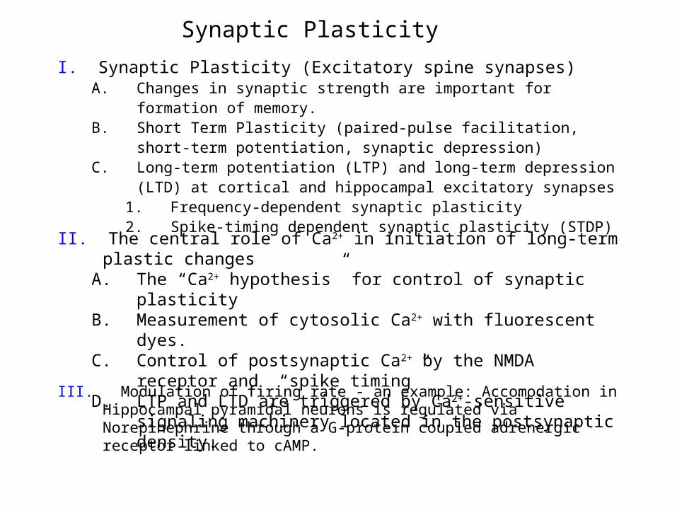
Synaptic Plasticity
Synaptic Plasticity
II. The central role of Ca2+ in initiation of long-term plastic changesA. The “Ca2+ hypothesis” for control of synaptic plasticityB. Measurement of cytosolic Ca2+ with fluorescent dyes.C. Control of postsynaptic Ca2+ by the NMDA receptor and “spike
timing”D. LTP and LTD are triggered by Ca2+-sensitive signaling machinery
located in the postsynaptic density.
I. Synaptic Plasticity (Excitatory spine synapses)A. Changes in synaptic strength are important for formation of memory.B. Short Term Plasticity (paired-pulse facilitation, short-term potentiation, synaptic
depression)C. Long-term potentiation (LTP) and long-term depression (LTD) at cortical and
hippocampal excitatory synapses1. Frequency-dependent synaptic plasticity2. Spike-timing dependent synaptic plasticity (STDP)
III. Modulation of firing rate - an example: Accomodation in Hippocampal pyramidal neurons is regulated via Norepinephrine through a G-protein coupled adrenergic receptor linked to cAMP.
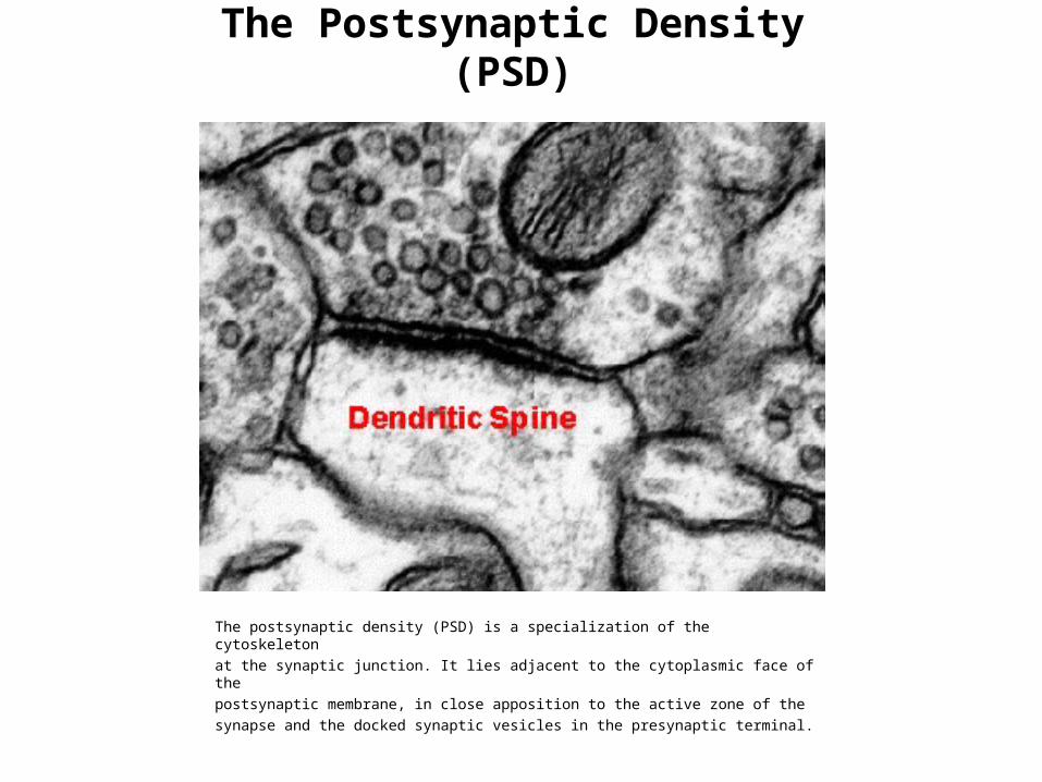
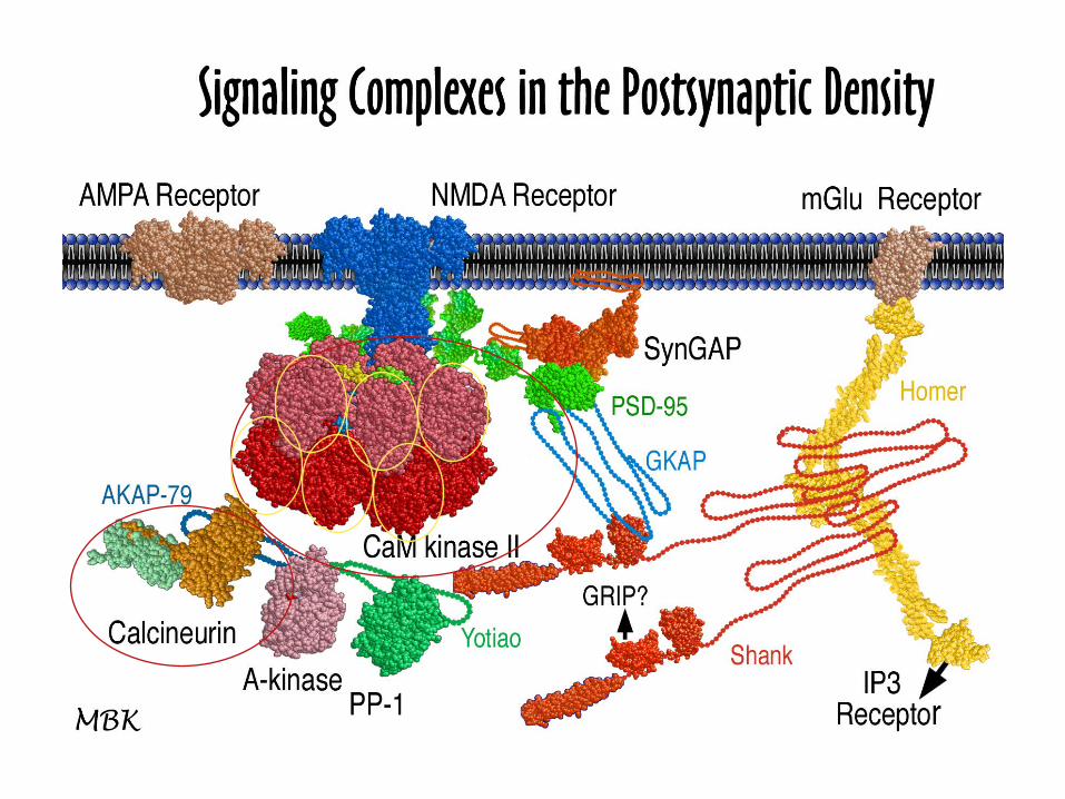
The Postsynaptic Density (PSD)
The postsynaptic density (PSD) is a specialization of the cytoskeletonat the synaptic junction. It lies adjacent to the cytoplasmic face of thepostsynaptic membrane, in close apposition to the active zone of thesynapse and the docked synaptic vesicles in the presynaptic terminal.
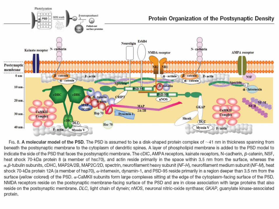
Liu et al., 2006. Molecular & Cellular Proteomics 5:1019–1032.
Synaptic Plasticity in the Hippocampus and Cortex
Synapses in the cortex and hippocampus are tightly regulated.
1. Regulation is used to maintain homeostatic balance2. It is also used to process and store information in neural circuits.3. Homeostasis and information storage must be coordinated to
maintain proper function.
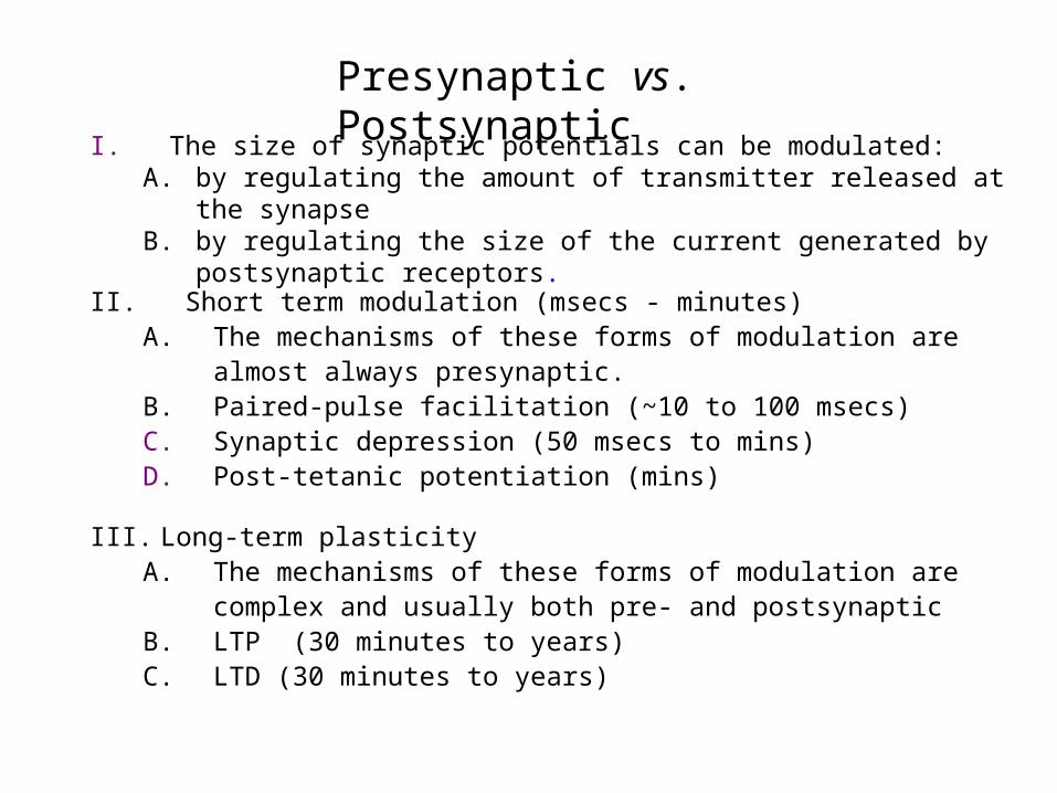
I. The size of synaptic potentials can be modulated: A. by regulating the amount of transmitter released at the synapse B. by regulating the size of the current generated by postsynaptic receptors.
II. Short term modulation (msecs - minutes)A. The mechanisms of these forms of modulation are almost always
presynaptic.B. Paired-pulse facilitation (~10 to 100 msecs)C. Synaptic depression (50 msecs to mins)D. Post-tetanic potentiation (mins)
III. Long-term plasticityA. The mechanisms of these forms of modulation are complex and usually
both pre- and postsynapticB. LTP (30 minutes to years)C. LTD (30 minutes to years)
Presynaptic vs. Postsynaptic
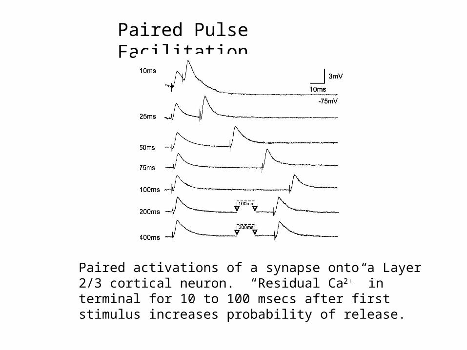
Paired Pulse Facilitation
Paired activations of a synapse onto a Layer 2/3 cortical neuron. “Residual Ca2+” in terminal for 10 to 100 msecs after first stimulus increases probability of release.
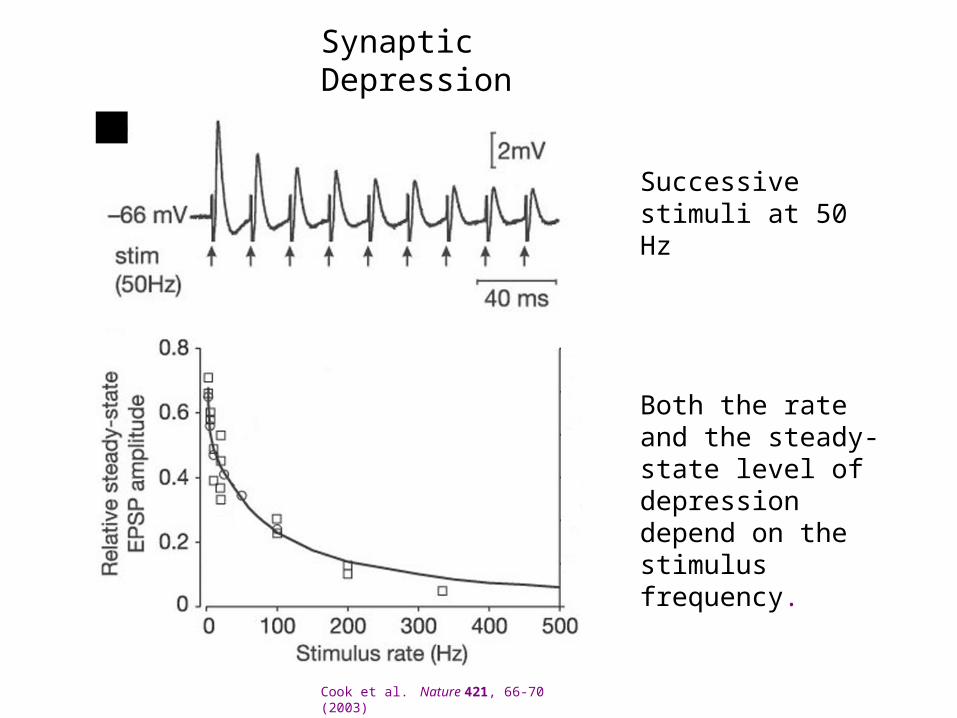
Synaptic Depression
Successive stimuli at 50 Hz
Cook et al. Nature 421, 66-70 (2003)
Both the rate and the steady-state level of depression depend on the stimulus frequency.
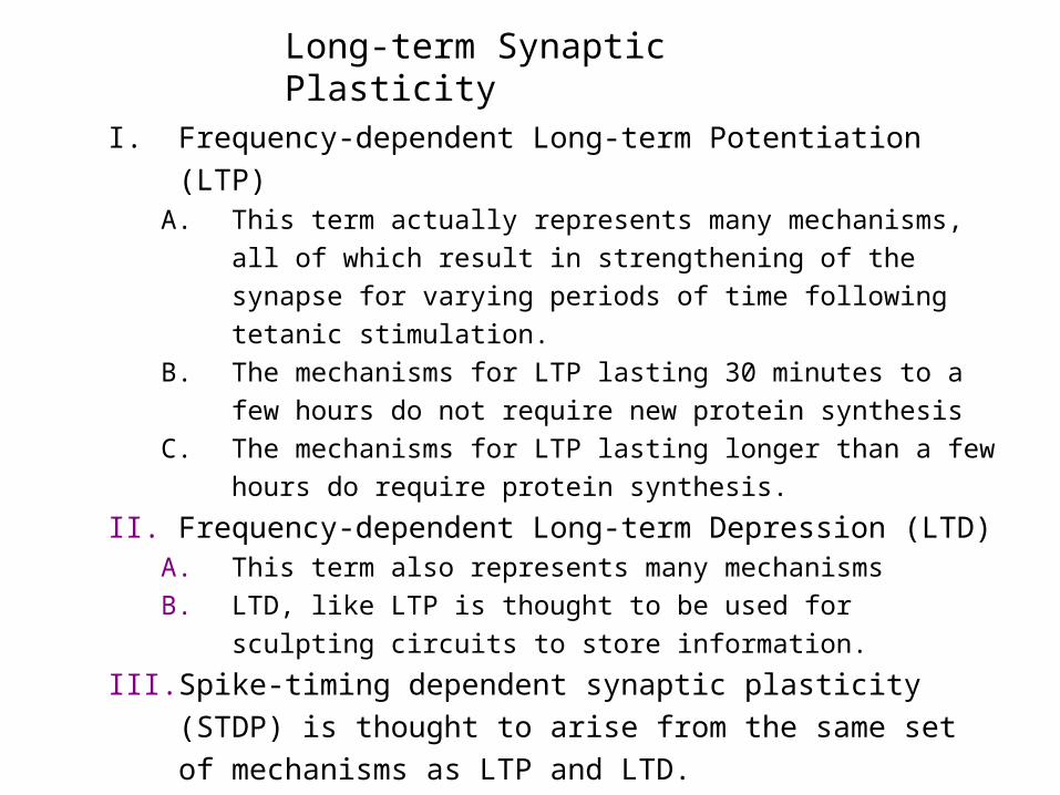
I. Frequency-dependent Long-term Potentiation (LTP)A. This term actually represents many mechanisms, all of which result in
strengthening of the synapse for varying periods of time following
tetanic stimulation.
B. The mechanisms for LTP lasting 30 minutes to a few hours do not
require new protein synthesis
C. The mechanisms for LTP lasting longer than a few hours do require
protein synthesis.
II. Frequency-dependent Long-term Depression (LTD)A. This term also represents many mechanisms
B. LTD, like LTP is thought to be used for sculpting circuits to store
information.
III. Spike-timing dependent synaptic plasticity (STDP) is thought to arise
from the same set of mechanisms as LTP and LTD.
Long-term Synaptic Plasticity
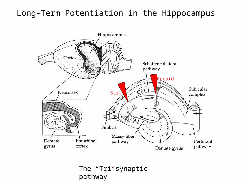
Long-Term Potentiation in the Hippocampus
Stim.
Record
The “Tri-synaptic pathway”
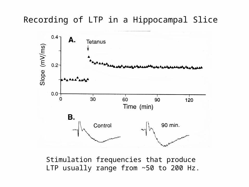
Recording of LTP in a Hippocampal Slice
Stimulation frequencies that produce LTP usually range from ~50 to 200 Hz.
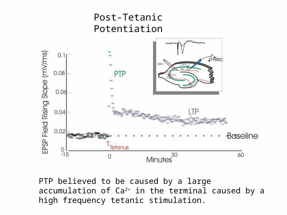
Post-Tetanic Potentiation
PTP believed to be caused by a large accumulation of Ca2+ in the terminal caused by a high frequency tetanic stimulation.
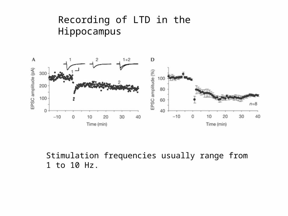
Recording of LTD in the Hippocampus
Stimulation frequencies usually range from 1 to 10 Hz.
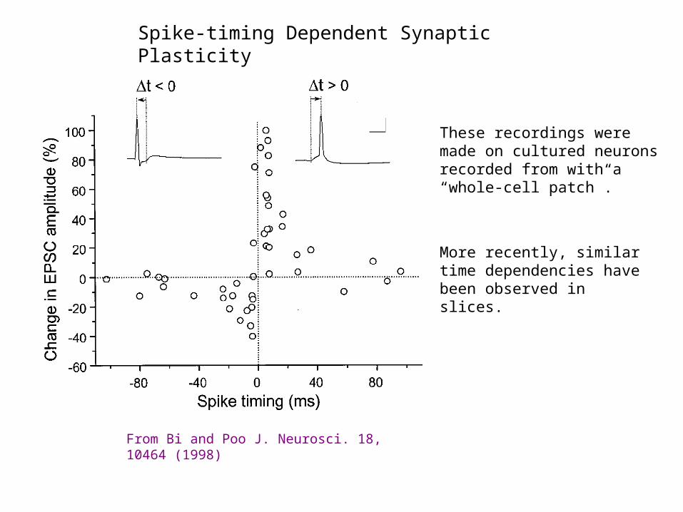
Spike-timing Dependent Synaptic Plasticity
From Bi and Poo J. Neurosci. 18, 10464 (1998)
These recordings were made on cultured neurons recorded from with a “whole-cell patch”.
More recently, similar time dependencies have been observed in slices.
Pre- fires 5-30 msecs before post - LTP
Pre- fires 5-30 msecs after post - LTD
Spike-timing Dependent Synaptic Plasticity
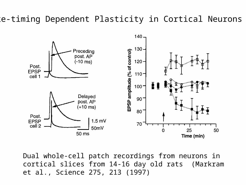
Spike-timing Dependent Plasticity in Cortical Neurons
Dual whole-cell patch recordings from neurons in cortical slices from 14-16 day old rats (Markram et al., Science 275, 213 (1997)
From The Organization of Behavior by Donald Hebb, 1949:
“When an axon of cell A is near enough to excite cell B and repeatedly or persistently takes part in firing it, some growth process or metabolic change takes place in one or both cells such that A's efficiency, as one of the cells firing B, is increased.”
Hebb postulated that this behavior of synapses in neuronal networks would permit the networks to store memories.
NMDA receptors, back-propagating action potentials, and summation of epsp’s appear to be the components that confer “Hebbian” behavior on the synapse.
The Hebbian Synapse
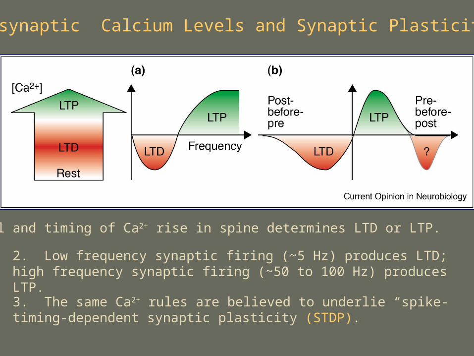
Postsynaptic Calcium Levels and Synaptic Plasticity
1. Level and timing of Ca2+ rise in spine determines LTD or LTP.
2. Low frequency synaptic firing (~5 Hz) produces LTD; high frequency synaptic firing (~50 to 100 Hz) produces LTP.
3. The same Ca2+ rules are believed to underlie “spike-timing-dependent synaptic plasticity (STDP).
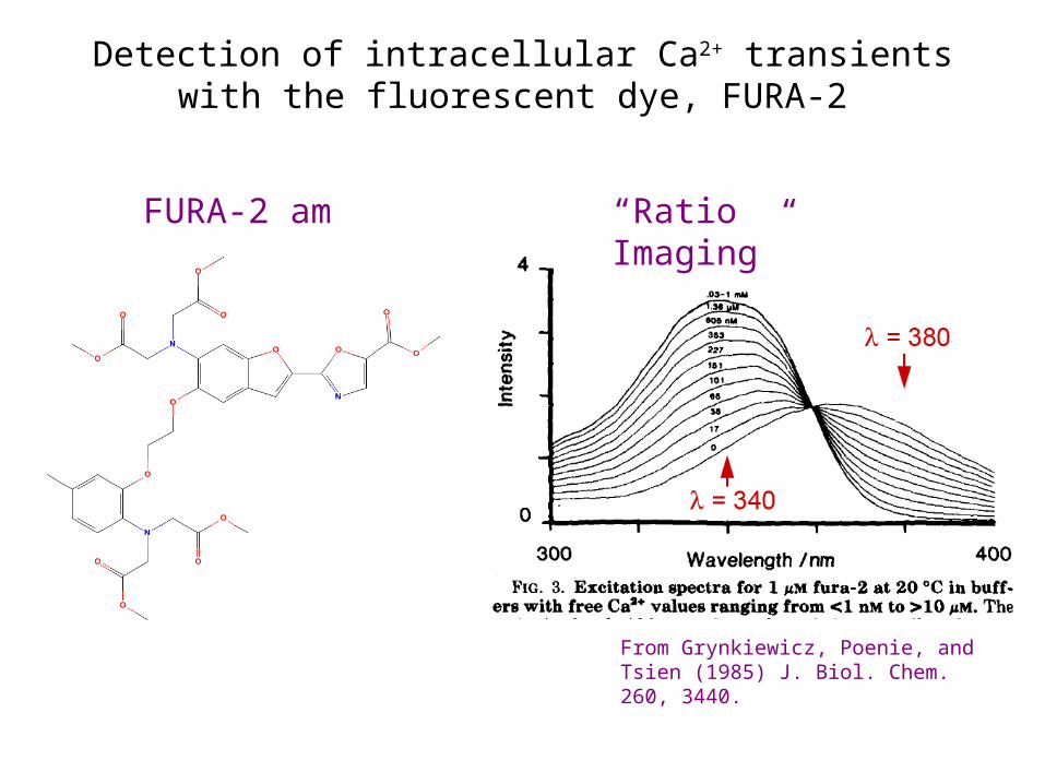
From Grynkiewicz, Poenie, and Tsien (1985) J. Biol. Chem. 260, 3440.
FURA-2 am
Detection of intracellular Ca2+ transients with the fluorescent dye, FURA-2
“Ratio Imaging”
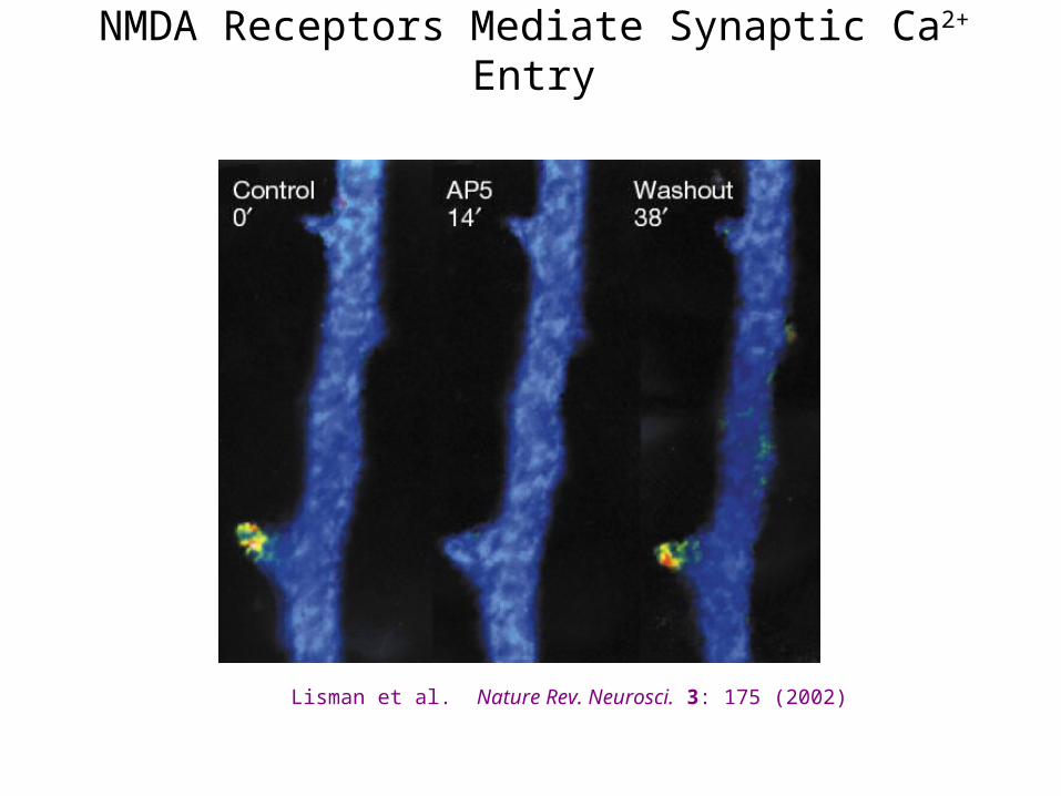
NMDA Receptors Mediate Synaptic Ca2+ Entry
Lisman et al. Nature Rev. Neurosci. 3: 175 (2002)
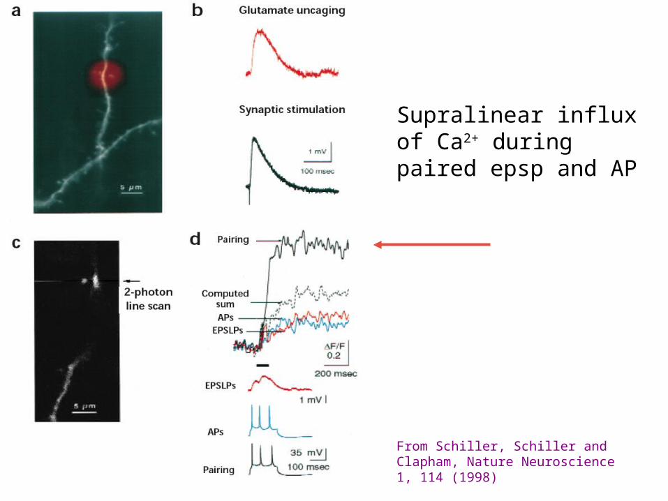
Supralinear influx of Ca2+ during paired epsp and AP
From Schiller, Schiller and Clapham, Nature Neuroscience 1, 114 (1998)
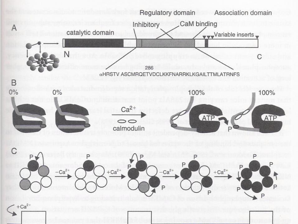
Recall the CaMKII Molecular Mechanism of Memory?
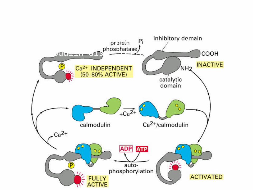
Ca2+/calmodulin dependent protein kinase (CaM-kinase)Memory function: 1. calmodulin dissociate after 10 sec of low calcium level; 2. remain active after calmodulin dissociation
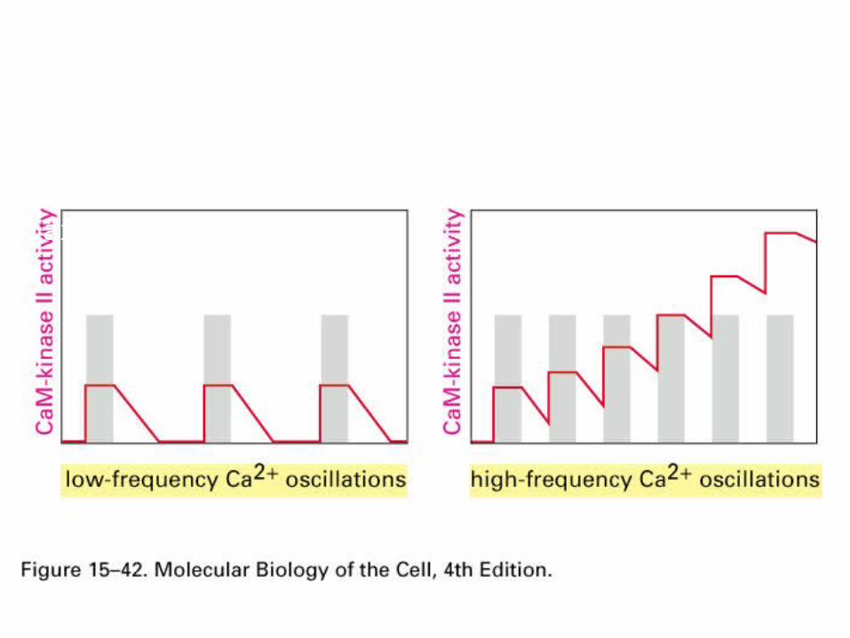
Ca2+/calmodulin dependent protein kinase (CaM-kinase)Frequency decoder of Calcium oscillation
High frequence, CaM-kinase does not return to basal level before the second wave of activation starts
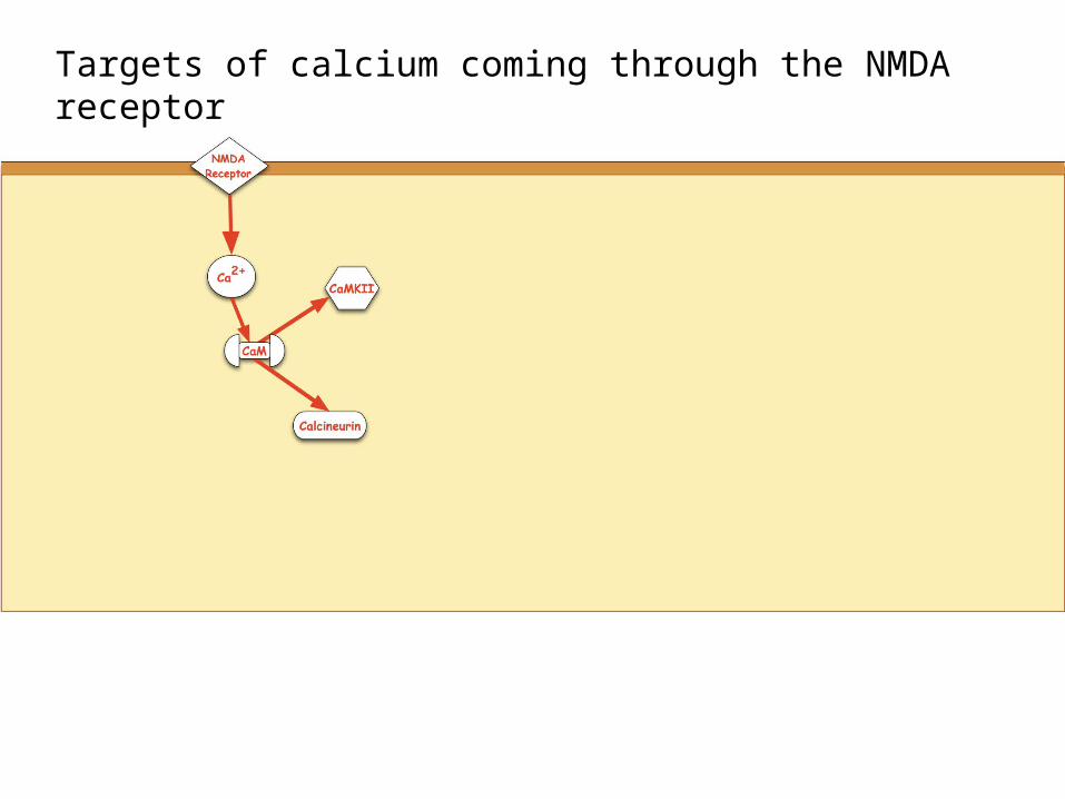
Targets of calcium coming through the NMDA receptor
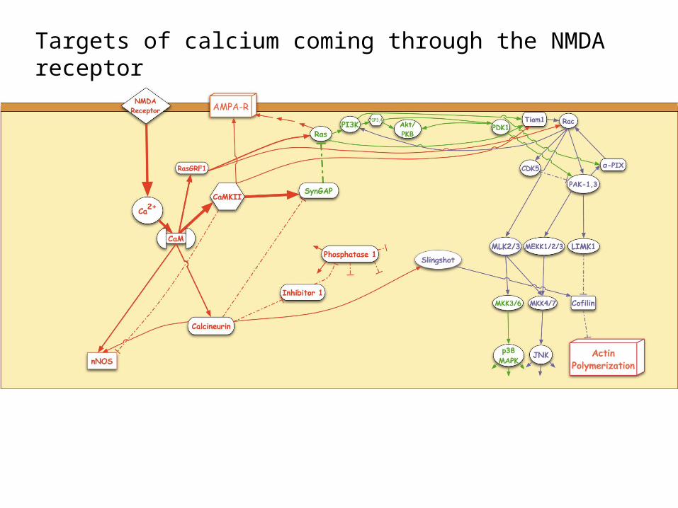
Targets of calcium coming through the NMDA receptor
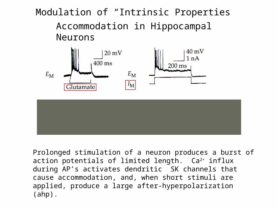
Accommodation in Hippocampal Neurons
Prolonged stimulation of a neuron produces a burst of action potentials of limited length. Ca2+ influx during AP’s activates dendritic SK channels that cause accommodation, and, when short stimuli are applied, produce a large after-hyperpolarization (ahp).
Modulation of “Intrinsic Properties”
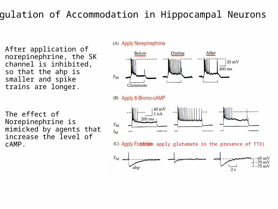
Regulation of Accommodation in Hippocampal Neurons
After application of norepinephrine, the SK channel is inhibited, so that the ahp is smaller and spike trains are longer.
The effect of Norepinephrine is mimicked by agents that increase the level of cAMP.
(then apply glutamate in the presence of TTX)
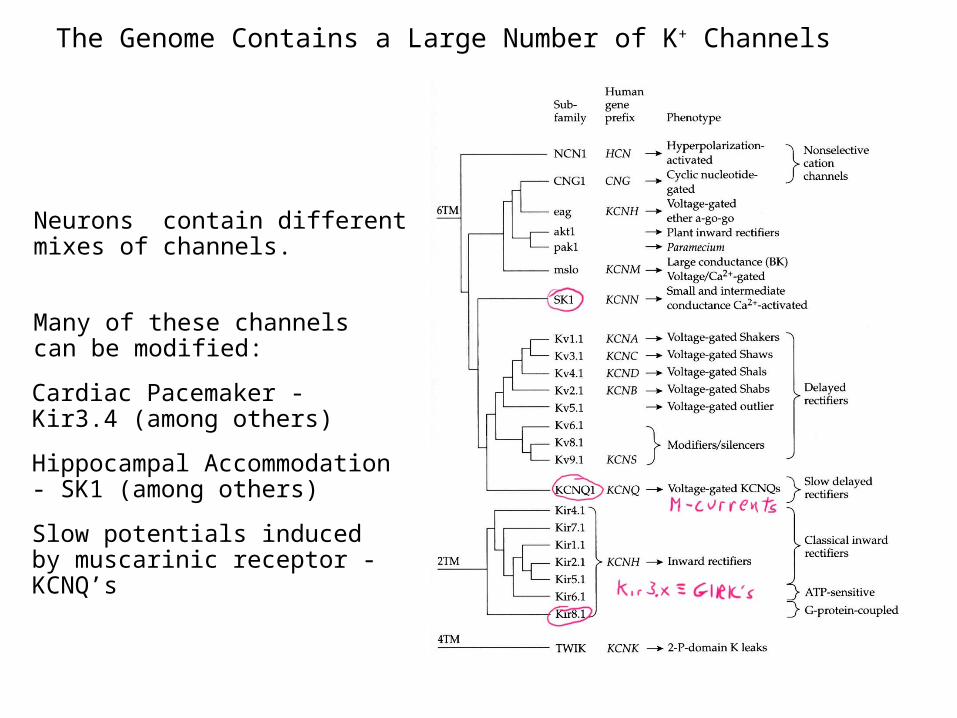
The Genome Contains a Large Number of K+ Channels
Simplified diagram of K+ channel families from Hille, “Ion Channels of Excitable Membranes”
Neurons contain different mixes of channels.
Many of these channels can be modified:
Cardiac Pacemaker - Kir3.4 (among others)
Hippocampal Accommodation - SK1 (among others)
Slow potentials induced by muscarinic receptor - KCNQ’s