Embed Size (px)
Citation preview
C H A P T E R T W O
C
IS
PU
urrent
SN 0
rogramnivers
Synapse Formation in Developing
Neural Circuits
Daniel A. Colon-Ramos
Contents
1. In
Top
070
inity
troduction
ics in Developmental Biology, Volume 87 # 2009
-2153, DOI: 10.1016/S0070-2153(09)01202-2 All rig
Cellular Neuroscience, Neurodegeneration and Repair, Department of Cell BiologySchool of Medicine, New Haven, Connecticut, USA
Else
hts
, Y
54
1
.1. A historical perspective 541
.2. S ynaptic structure and function 562. S
ynaptogenesis During Development 582
.1. N euronal cell fate and synaptogenesis 592
.2. A xon guidance and synaptogenesis 623. B
uilding a Synapse 683
.1. C ell adhesion in synaptic assembly 683
.2. A ssembling the synaptic components 693
.3. G uidepost cells, morphogens, and connectivity 714. P
erspective 75Ackn
owledgments 76Refe
rences 76Abstract
The nervous system consists of hundreds of billions of neurons interconnected
into the functional neural networks that underlie behaviors. The capacity of a
neuron to innervate and function within a network is mediated via specialized
cell junctions known as synapses. Synapses are macromolecular structures that
regulate intercellular communication in the nervous system, and are the main
gatekeepers of information flow within neural networks. Where and when
synapses form determines the connectivity and functionality of neural networks.
Therefore, our knowledge of how synapse formation is regulated is critical to
our understanding of the nervous system and how it goes awry in neurological
disorders.
Synapse formation involves pairing of the pre- and postsynaptic partners at a
specific neurospatial coordinate. The specificity of synapse formation requires
the precise execution of multiple developmental events, including cell fate
specification, cell migration, axon guidance, dendritic growth, synaptic target
vier Inc.
reserved.
ale
53

54 Daniel A. Colon-Ramos
selection, and synaptogenesis ( Juttner andRathjen in Cell. Mol. Life Sci.62:2811,
2005; Salie et al., in Neuron 45:189, 2005; Waites et al., in Annu. Rev. Neurosci.
28:251, 2005). Remarkably, during the development of the vertebrate nervous
system, these developmental processes occur almost simultaneously in billions
of neurons, resulting in the formation of trillions of synapses. How this remark-
able specificity is orchestrated during development is one of the outstanding
questions in the field of neurobiology, and the focus of discussion of this chapter.
We center the discussion of this chapter on the early developmental events
that orchestrate the process of synaptogenesis prior to activity-dependent
mechanisms. We have therefore limited the discussion of important activity-
dependent synaptogenic events, which are discussed in other chapters of this
book. Moreover, our discussion is biased toward lessons we have learned from
invertebrate systems, in particular from C. elegans and Drosophila. We did so to
complement the discussions from other chapters in this book, which focus on
the important findings that have recently emerged from the vertebrate
literature.
The chapter begins with a brief history of the field of synaptic biology.
This serves as a backdrop to introduce some of the historically outstanding
questions of synaptic development that have eluded us during the past century,
and which are the focus of this review. We then discuss some general features
of synaptic structure as it relates to its function. In particular, we will highlight
evolutionarily conserved traits shared by all synaptic structures, and how these
features have helped optimize these ancient cellular junctions for interneural
communication.
We then discuss the regulatory signals that orchestrate the precise assembly
of these conserved macromolecular structures. This discussion will be framed
in the context of the neurodevelopmental process. Specifically, much of our
discussion will focus on how the seemingly disparate developmental processes
are intimately linked at a molecular level, and how this relationship might be
crucial in the developmental orchestration of circuit assembly. We hope that the
discussion of the multifunctional cues that direct circuit development provides a
conceptual framework into understanding how, with a limited set of signaling
molecules, precise neural wiring can be coordinated between synaptic partners.
1. Introduction
1.1. A historical perspective
The history of synapse biology starts at the end of the eighteenth century,with the studies of Luigi Galvani and his descriptions of ‘‘animal electricity.’’In these classical studies, Galvani observed that he could induce the contrac-tion of limb muscles when he inserted a metal hook into the medulla of thefrog and attached the other end to an iron railing. These observationsmarked the first experimental demonstrations of synaptic transmission(Cowan and Kandel, 2001).

Synapse Formation in Developing Neural Circuits 55
Most of the subsequent synaptic studies in the nineteenth century andearlier half of the twentieth century also focused around the functionality ofsynapses, or synaptic transmission. It is therefore befitting that the actualterm ‘‘synapse’’ was not coined by a neuroanatomist, but by a physiologistnamed Charles Sherrington. Sherrington coined the term ‘‘synapse’’ to referto the special connections from one nerve cell to another that facilitated thetransmission of nervous impulses (Cowan and Kandel, 2001).
While physiologists and neuropharmacologists were functionally defin-ing the concept of synapses, neuroanatomists tangled in a bitter debate ontheir existence. The main reason for this debate was that during the nine-teenth century and earlier part of the twentieth century, nobody couldvisualize cell membranes and establish conclusively the existence of synapses.However, in spite of these technological limitations, some insightful neuro-biologists garnered enough experimental evidence to propose the anatomicalexistence of synapses.
Most of these early observations came from a specialized synapse: theneuromuscular junction (NMJ). Because of its size, morphology and func-tional readouts, NMJs informed then, as they do now, most of our knowl-edge on synaptic biology. Taking advantage of this system, physiologistWilly Kuhne and anatomist Wilhelm Krause independently hypothesizedthe existence of synapses at the site of contact between nerve cells andmuscles (Cowan and Kandel, 2001).
The question of the existence of interneuronal synapses was muchharder to settle. Synapses in the central nervous system are much smallerthan NMJs, in closed apposition to one another and packed at very highdensities. This made their visualization with the methods used duringnineteenth century downright impossible and triggered the postulation ofthe ‘‘reticular theory’’: the idea that the nervous system lacked functionalseparation of nerve cells and was syncytial, rather than synaptic, in nature(Cowan and Kandel, 2001; Westfall, 1996).
The theory turned out, of course, to be wrong. Although this was notconclusively shown until the advent of electron microscopy in the 1950s,the first evidence that neurons were discrete units came from developmen-tal, pathological, and anatomical observations in the nineteenth century.Most notable among these early studies are Santiago Ramon y Cajal’s.By using a method derived by Golgi, which stains only 1% of the cells,Cajal was able to visualize the morphology of individual cells in the contextof the nervous system. His detailed characterization of neurons not onlyprovided critical evidence for the neuron doctrine, but also stated the‘‘Principios de la Especificidad de la Conexion’’: the idea that nerve cellsconnect to each other in a specific fashion to form precise networks (Cowanand Kandel, 2001).
Although it would take another half a century for cell biologists tovisualize synapses, Cajal’s observations and insights at the turn of thenineteenth century provided the conceptual basis that has driven most of
56 Daniel A. Colon-Ramos
the neurodevelopmental questions since then. Over a century after Cajal’sinitial descriptions, we are still untangling the complex morass that is thecentral nervous system and tackling the questions staged by his landmarkobservations: How are the numerous cell types in the nervous systemspecified? What directs neurites to connect to each other? What are thecellular and molecular factors that underlie the ‘‘Principles of connectionspecificity’’?
1.2. Synaptic structure and function
During the last century however, and thanks in great part to technicaladvances in cell biology, the field has made great progress in its understand-ing of the synaptic structure as it relates to synaptic function. Most notably,electron microscopy allowed the visualization of synapses for the first timein the 1950s. This work, spearheaded by George Palade and Keith Porter,provided unequivocal evidence for the neuron doctrine and the existence ofsynapses, and identified the different types of synapses and their structuralcomponents (Cowan and Kandel, 2001; De Camilli et al., 2001).
There are two general categories of synapses: electrical synapses andchemical synapses. Physiologists and neuropharmacologists functionallydefined these two categories of synapses well before they were visualizedby cell biologists (Cowan and Kandel, 2001). But the cell biological workthat proceeded from the physiological studies demonstrated that these twofunctional categories corresponded to completely different structures.Electrical synapses are gap junctions that allow bidirectional propagationof signals, including electrical stimuli. They allow the fastest mode ofelectrical propagation across cells, and are now known to be important insynchronizing neural activity across networks (De Camilli et al., 2001).These gap junctions will not be further discussed in this chapter.
Chemical synapses allow communication between discontinuous neu-rons via the highly regulated secretion of chemical intermediate signals.Unlike electrical synapses, chemical synapses are polarized junctions thatallow the flow of information in just a single direction. Because of theirhighly regulated and directional transfer of information, chemical synapseshave been the focus of most of the synaptic biology studies, and as such willremain the focus of our chapter.
Although there is great morphological and molecular variability amongchemical synapses, all chemical synapses share common structural and func-tional features (De Camilli et al., 2001). They consist of two asymmetricallyjuxtaposed components linking two separate cells: a presynaptic specializa-tion and a postsynaptic region. The presynaptic specializations are specializedregions in the presynaptic cell with an abundance of neurotransmitter-filled
Synapse Formation in Developing Neural Circuits 57
synaptic vesicles. Presynaptic specializations also contain the active zonestructures that facilitate vesicle fusion and the release of neurotransmittercontent to the intersynaptic space, called the synaptic cleft. The postsynapticregion is an area of the postsynaptic cell with a high concentration ofneurotransmitter receptors, channels, and downstream signaling molecules.The neurotransmitters released by the presynaptic specializations are sensedby the receptors at the postsynaptic site, activating downstream signalingmolecules, opening channels, and propagating the nervous impulse tothe postsynaptic partner. These general features of the presynaptic andpostsynaptic specializations are shared by all classes of synaptic structures.
The synaptic structure as described above is also very well conservedacross evolution. Sea anemones and hydra (Phylum Cnidaria) have the mostprimitive nervous system, which consists of a diffuse network of neurons.These nerve nets, however, are connected via chemical and electricalsynapses that are fully capable of transmitting and regulating informationflow (Anderson and Spencer, 1989; Peteya, 1973; Westfall, 1996). Closeinspection of these synaptic structures reveal that Cnidarian synapses havesimilar structural components as those of higher organisms, with definedpresynaptic and postsynaptic specializations in close juxtaposition (Andersonand Spencer, 1989; Peteya, 1973; Westfall, 1996).
The presence of a conserved synaptic structure in these primitive nervoussystems reveals that synapses are as ancient as the nervous system itself.This evolutionary conservation of the synaptic structure also underscoresthe importance of these specialized cell junctions in interneuronal commu-nication and the functioning of the neural networks (Anderson andSpencer, 1989; Peteya, 1973; Westfall, 1996).
Interestingly, a recent study suggests that the evolution of the synapticmolecular machine might even precede the evolution of the nervous system(Sakarya et al., 2007). Although sponges (Phylum Porifera) are the onlymetazoans without a nervous system, it was found that sponges express anearly complete set of postsynaptic protein homologues that are hypothe-sized to assemble into synaptic-like scaffolds. Although sponges do not haveneurons, these postsynaptic-like structures are hypothesized to act as che-mosensory structures capable of responding to environmental cues (Sakaryaet al., 2007).
Other molecular components of the presynaptic machine, such as thesynaptic vesicle cycle regulators, also predate the existence of the nervoussystem and are very well conserved across evolution (Sudhof, 2004). It isprovocative that these macromolecular machines, presumable ‘‘buildingblocks’’ of the synapse, might be found even in the absence of a nervoussystem itself, an observation that underscores the importance and conserva-tion of these signaling complexes throughout evolution (Sakarya et al.,2007).
58 Daniel A. Colon-Ramos
Molecular and genetic studies in model invertebrate and vertebrateanimals have also supported the notion that the ultrastructural conservationof synapses corresponds to a conservation at the molecular level. Forinstance, in the simple nervous system of the nematode Caenorhabditis elegans,which consists of only 302 neurons, the number of neurotransmitters andreceptors required for the proper functioning of its �5000 synapsesapproaches in complexity those used by the hundreds of trillions of synapsesin the vertebrate nervous system (Rand and Nonet, 1997).
This suggests that throughout evolution, the increased capacity of infor-mation processing and storage observed in higher organisms is not the resultof marked changes in the complexity of the synaptic structure. We speculatethat this complexity results from an increasingly sophisticated neural frame-work in way of the abundance and organization of neural networks. Where,when, and how synapses form during development play critical roles on thewiring and function of neural networks. Although the neural networkorganization varies vastly across animals, the biological basis of synapses isshared from the simplest networks of Cnidaria to the complex neuropils ofthe human brain.
2. Synaptogenesis During Development
The organization of where, when, and how synapses are formed playsan instrumental role in directing the connectivity of circuits and organizingthe neuroarchitecture that enables information processing, storage, andultimately behaviors. As such, the developmental questions postulated byCajal in his ‘‘Principles of connection specificity’’ are of great importanceto our understanding of the assembly and function of the nervous system.What are themolecular and cellular factors that direct the precise innervationof hundreds of trillions of synapses during development?
Neural circuit formation requires the intricate orchestration of multipledevelopmental events including cell fate specification, cell migration, axonguidance, dendritic growth, synaptic target selection, and synaptogenesis( Juttner and Rathjen, 2005; Salie et al., 2005; Waites et al., 2005). The cor-rect innervation of a given circuit requires the successful completion of all ofthese developmental steps in both synaptic partners. As such, synaptogenesismarks the final step of a complicated developmental dance where, aftersuccessful completion of the aforementioned steps, both synaptic partnersconverge at a specific location to form a specialized junction.
Although the field has now identified a number of molecules requiredfor each of these developmental steps, we know much less about howthese different developmental steps act in concert to direct the development
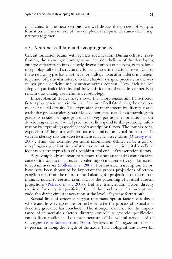
Synapse Formation in Developing Neural Circuits 59
of circuits. In the next sections, we will discuss the process of synapticformation in the context of the complex developmental dance that bringsneurons together.
2.1. Neuronal cell fate and synaptogenesis
Circuit formation begins with cell fate specification. During cell fate speci-fication, the seemingly homogeneous neuroepithelium of the developingembryo differentiates into a hugely diverse number of neurons, each tailoredmorphologically and structurally for its particular functional role. Each ofthese neuron types has a distinct morphology, axonal and dendritic trajec-tory, and, of particular interest to this chapter, synaptic property in the wayof synaptic specificity and neurotransmitter content. How each neuronadopts a particular identity and how this identity directs its connectivityremain outstanding problems in neurobiology.
Embryological studies have shown that morphogens and transcriptionfactors play crucial roles in the specification of cell fate during the develop-ment of neural circuits. The expression of morphogens by discrete tissuesestablishes gradients alongmultiple developmental axes. Thesemorphogenicgradients create a unique grid that conveys positional information in thedeveloping embryo. Neural precursor cells respond to this positional infor-mation by expressing a specific set of transcription factors. The combinatorialexpression of these transcription factors confers the neural precursor cellswith an identity that can then be inherited by its descendants (O’Leary et al.,2007). Thus, the extrinsic positional information delineated by a grid ofmorphogenic gradients is translated into an intrinsic and inheritable cellularidentity via the expression of a combinatorial code of transcription factors.
A growing body of literature supports the notion that this combinatorialcode of transcription factors can confer important connectivity informationto certain neurons (Polleux et al., 2007). For instance, transcription factorshave now been shown to be important for proper projections of retino-ganglion cells from the retina to the thalamus, for projections of axons fromthalamic nuclei to cortical areas and for the patterning of cortical efferentprojections (Polleux et al., 2007). But are transcription factors directlyrequired for synaptic specificity? Could the combinatorial transcriptionalcode also direct circuit innervation at the level of synapse formation?
Several lines of evidence suggest that transcription factors can directwhere and how synapses are formed even after the process of axonal anddendritic guidance has concluded. The strongest evidence for the impor-tance of transcription factors directly controlling synaptic specificationcomes from studies in the motor neurons of the ventral nerve cord ofC. elegans (Von Stetina et al., 2006). Synapses in C. elegans are formeden passant, or along the length of the axon. This biological trait allows for

60 Daniel A. Colon-Ramos
an easier developmental dissection of the axon guidance versus the synapto-genesis steps as, unlike end-button synapses, en passant synapses are formedon the side of axons, which contact many potential synaptic targets (Whiteet al., 1986). Early genetic studies in the motor neurons of the ventral nervecord showed that mutant animals lacking the gene unc-4, which encodes aPrd-like homeodomain transcription factor, display a strong motor move-ment defect in backward locomotion. Ultrastructural studies on the inner-vation and morphology of nerve cord neurons showed that absence of thetranscription factor did not alter the organization of the nerve cord, and allneurons looked normal in terms of morphology, position in the nerve cordfascicle and guidance. Interestingly, unc-4 mutant animals displayed abnor-mal synaptic specificity, with motor neurons innervating their incorrectpartners. This suggested that the Prd-like homeodomain transcription factorUNC-4 directly controls synaptic choice without affecting other neuraltraits such as outgrowth and fasciculation (Miller et al., 1992; White et al.,1992).
Further studies in other neurons determined that the UNC-4 transcriptionfactor also controls the expression of molecules involved in synaptic strength(Lickteig et al., 2001; Von Stetina et al., 2007). This capacity to regulate theexpression of molecules involved in synaptic strength is independent from itscapacity to regulate synaptic specificity (Lickteig et al., 2001). Although theidentity of the targets of UNC-4 remains unknown, these studies show thattranscription factors can directly regulate different aspects of synaptic biology,from formation of the synapse during development to the strength of synapticconnections (Von Stetina et al., 2006).
Another example of how transcription factors can regulate synapseformation is found in the cockroach cercal system. The cerci are an append-ages in the rear of the cockroach where filiform hairs are innervated by asingle sensory neuron. In newly hatched (first-instar) roaches there are justtwo sensory neurons that respond to stimuli: the lateral sensory neuron,which responds to stimuli form the front of the animal, and the medialsensory neuron, which responds to stimuli from the rear of the animal(Fig. 2.1). Although the arborization of these two sensory neurons overlaps,the two sensory neurons connect specifically and exclusively to differentsubsets of giant interneurons. This synaptic specificity directs directionalsensitivity to stimuli, allowing the animal to discern if the stimuli come fromthe front or the rear, and eliciting the corresponding escape response(Blagburn and Bacon, 2004).
Since these two filiform hair afferents have overlapping arborizations,the axonal projections cannot be the primary determinant of synapticspecificity. Instead, synaptic specificity must be directed by additional cuesthat allow these overlapping arbors to innervate specific targets. Loss-of-function studies in this system showed that the homeodomain transcription
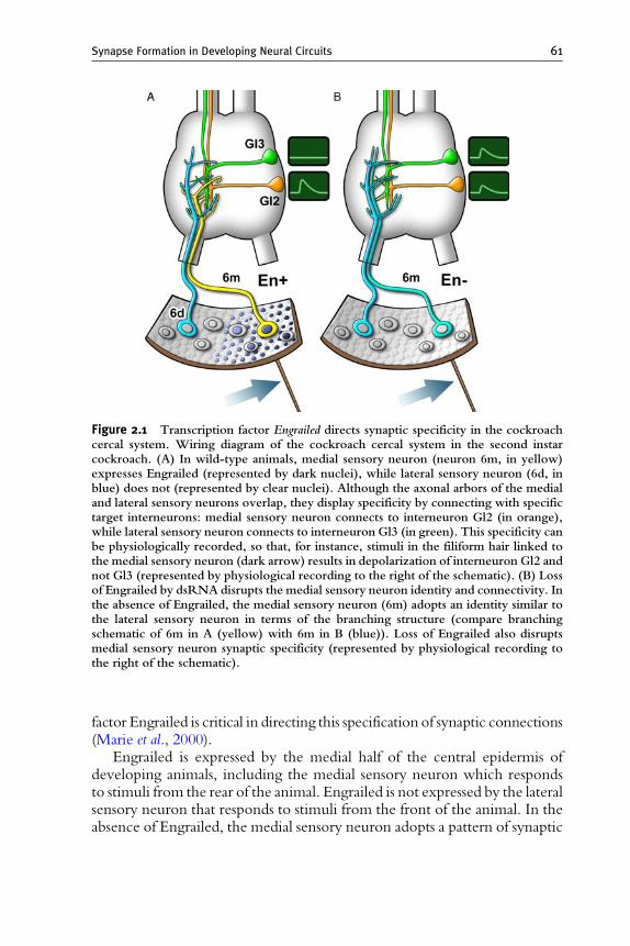
Figure 2.1 Transcription factor Engrailed directs synaptic specificity in the cockroachcercal system. Wiring diagram of the cockroach cercal system in the second instarcockroach. (A) In wild-type animals, medial sensory neuron (neuron 6m, in yellow)expresses Engrailed (represented by dark nuclei), while lateral sensory neuron (6d, inblue) does not (represented by clear nuclei). Although the axonal arbors of the medialand lateral sensory neurons overlap, they display specificity by connecting with specifictarget interneurons: medial sensory neuron connects to interneuron Gl2 (in orange),while lateral sensory neuron connects to interneuron Gl3 (in green). This specificity canbe physiologically recorded, so that, for instance, stimuli in the filiform hair linked tothe medial sensory neuron (dark arrow) results in depolarization of interneuron Gl2 andnot Gl3 (represented by physiological recording to the right of the schematic). (B) Lossof Engrailed by dsRNA disrupts the medial sensory neuron identity and connectivity. Inthe absence of Engrailed, the medial sensory neuron (6m) adopts an identity similar tothe lateral sensory neuron in terms of the branching structure (compare branchingschematic of 6m in A (yellow) with 6m in B (blue)). Loss of Engrailed also disruptsmedial sensory neuron synaptic specificity (represented by physiological recording tothe right of the schematic).
Synapse Formation in Developing Neural Circuits 61
factor Engrailed is critical in directing this specification of synaptic connections(Marie et al., 2000).
Engrailed is expressed by the medial half of the central epidermis ofdeveloping animals, including the medial sensory neuron which respondsto stimuli from the rear of the animal. Engrailed is not expressed by the lateralsensory neuron that responds to stimuli from the front of the animal. In theabsence of Engrailed, the medial sensory neuron adopts a pattern of synaptic
62 Daniel A. Colon-Ramos
connections similar to that of the Engrailed-negative, lateral sensory neuron(Fig. 2.1). These results indicate that transcription factor Engrailed isrequired for correct specification of synaptic connections (Marie et al., 2000).
Interestingly, persistent expression of Engrailed was also shown to berequired for the specification of other developmental traits of the medialsensory neuron. By manipulating Engrailed levels at different developmen-tal stages, the authors went on to show that Engrailed is required inpostmitotic neurons to control axon arborization and synaptic specification.They showed that these two events are developmentally separable, but areboth dependent on the same transcription factor. These findings demon-strate that Engrailed can direct discrete connectivity decisions at differentdevelopmental stages (Marie and Blagburn, 2003; Marie et al., 2002).Furthermore, it highlights the role of transcription factors in coordinatingand integrating the different developmental decisions that need to be madeto direct neural connectivity.
Therefore, transcription factors can act as conveying points, receivinginputs, and directing different developmental steps that range from cell iden-tity and neurite guidance to synapse assembly. When integrated (as in thecase of Engrailed), these different activities could orchestrate the interde-pendent development and innervation of circuits. Although we understandthe importance of transcription factors in directing discrete developmentalsteps, our knowledge on how their combinatorial and interdependent activ-ity leads to correct innervation is still limited. For instance, in most of theexperimental systems in which transcriptional regulation has been shown toaffect circuit formation we do not yet know the identity of the guidance orsynaptic specificity cues, how these cues are controlled and how their activitydirects connectivity.
2.2. Axon guidance and synaptogenesis
Once neural cell fate is specified and neuron precursors have migrated to theappropriate regions, they extend polarized projections that become theiraxons and dendrites. The axonal processes can extend long distances, navi-gating complex cellular environments before reaching their postsynapticpartner. This guidance is mediated through the growth cone, a specializedsensing device at the tip of the outgrowing axon. Growth cones express aseries of guidance receptors that are capable of sensing a variety of long-range (diffusible) and short-range (surface-bound) guidance cues. Theseguidance cues, which can be attractive or repulsive, are secreted by guide-post cells and intermediate targets. The spatial and temporal presence of theguidance cues, combined with the expression of the receptors in the growthcone, enables the axon to navigate through the labyrinth that is the devel-oping nervous system to reach its target (Plachez and Richards, 2005;Tessier-Lavigne and Goodman, 1996). Upon reaching and contacting its

Synapse Formation in Developing Neural Circuits 63
target, the axon transforms into a presynaptic specialization capable oftransducing synaptic signals to the postsynaptic target.
One of the outstanding questions in the field of synaptogenesis is how thistransformation is mediated. How does the axon identify its correct postsyn-aptic target? During guidance, how does the growth cone differentiatebetween intermediate guidepost targets and its final target? Upon reachingthe target region, how does it discriminate between potential partners toinnervate its correct postsynaptic partner? And what are the cell biologicalchanges that occur in the axon to transform it into a specialized presynapticjunction?
Clues on how this transformation occurs have come from studies in oneof the better-understood systems of growth cone guidance and synaptictargeting: the RP3 motoneuron in the Drosophila embryos. The RP3motoneuron can be visualized during development of theDrosophila embry-onic CNS with single-cell resolution and in the context of the intactnervous system. This is done by using immunocytochemistry techniquesthat allow detailed characterization of the developmental decisions made bythis motoneuron. These studies demonstrated that the RP3 axon undergoesa stereotypical sequence of guidance events before reaching its final targets,two muscles known as muscles 6 and 7 (Fig. 2.2). Remarkably, uponreaching the target region RP3 comes within filopodial reach of over adozen different muscles, yet specifically innervates only its correct targets.In wild-type embryos this stereotyped sequence of developmental events,and the innervation of the correct targets, happens with 100% accuracy(Chiba and Rose, 1998).
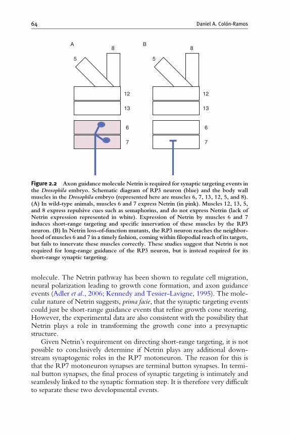
Genetic studies in the RP3 system showed that the long-range guidancedecisions and the short-range synaptic targeting choices are directed bydifferent molecular cues. The RP3 targets, muscles 6 and 7, express the axonguidance cue Netrin. However, deletion of the Netrin gene does not affectlong-range guidance decisions of the RP3 axon: in the absence of Netrin orits receptor (Frazzled), the RP3 growth cone makes its normal guidancedecisions, exiting the CNS, leaving the nerve bundles and entering theappropriate ventral muscle domain in a timely fashion to reach the neighbor-hoods of its targets, muscles 6 and 7 (Fig. 2.2). However, although the RP3growth cone comes within filopodial reach of its targets, in these mutants itfails to innervate them robustly (Mitchell et al., 1996). These studies suggestthat Netrin is not required for the long-range guidance decisions of the RP3motoneuron, but instead is required for its short-range synaptic targeting.Furthermore, these studies indicate that the long-range guidance decisionsand the short-range, synaptic targeting decisions of theRP3motoneuron aretwo different, genetically separable events that are not dependent on thesame molecular factors.
Although the RP3 guidance and synaptic targeting events are geneticallyseparable, short-range targeting in this system is still mediated by a guidance
8
5
A B
12
13
6
7
8
5
12
13
6
7
Figure 2.2 Axon guidance molecule Netrin is required for synaptic targeting events inthe Drosophila embryo. Schematic diagram of RP3 neuron (blue) and the body wallmuscles in the Drosophila embryo (represented here are muscles 6, 7, 13, 12, 5, and 8).(A) In wild-type animals, muscles 6 and 7 express Netrin (in pink). Muscles 12, 13, 5,and 8 express repulsive cues such as semaphorins, and do not express Netrin (lack ofNetrin expression represented in white). Expression of Netrin by muscles 6 and 7induces short-range targeting and specific innervation of these muscles by the RP3neuron. (B) In Netrin loss-of-function mutants, the RP3 neuron reaches the neighbor-hood of muscles 6 and 7 in a timely fashion, coming within filopodial reach of its targets,but fails to innervate these muscles correctly. These studies suggest that Netrin is notrequired for long-range guidance of the RP3 neuron, but is instead required for itsshort-range synaptic targeting.
64 Daniel A. Colon-Ramos
molecule. The Netrin pathway has been shown to regulate cell migration,neural polarization leading to growth cone formation, and axon guidanceevents (Adler et al., 2006; Kennedy and Tessier-Lavigne, 1995). The mole-cular nature of Netrin suggests, prima facie, that the synaptic targeting eventscould just be short-range guidance events that refine growth cone steering.However, the experimental data are also consistent with the possibility thatNetrin plays a role in transforming the growth cone into a presynapticstructure.
Given Netrin’s requirement on directing short-range targeting, it is notpossible to conclusively determine if Netrin plays any additional down-stream synaptogenic roles in the RP7 motoneuron. The reason for this isthat the RP7 motoneuron synapses are terminal button synapses. In termi-nal button synapses, the final process of synaptic targeting is intimately andseamlessly linked to the synaptic formation step. It is therefore very difficultto separate these two developmental events.
Synapse Formation in Developing Neural Circuits 65
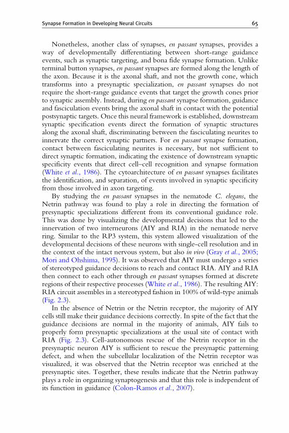
Nonetheless, another class of synapses, en passant synapses, provides away of developmentally differentiating between short-range guidanceevents, such as synaptic targeting, and bona fide synapse formation. Unliketerminal button synapses, en passant synapses are formed along the length ofthe axon. Because it is the axonal shaft, and not the growth cone, whichtransforms into a presynaptic specialization, en passant synapses do notrequire the short-range guidance events that target the growth cones priorto synaptic assembly. Instead, during en passant synapse formation, guidanceand fasciculation events bring the axonal shaft in contact with the potentialpostsynaptic targets. Once this neural framework is established, downstreamsynaptic specification events direct the formation of synaptic structuresalong the axonal shaft, discriminating between the fasciculating neurites toinnervate the correct synaptic partners. For en passant synapse formation,contact between fasciculating neurites is necessary, but not sufficient todirect synaptic formation, indicating the existence of downstream synapticspecificity events that direct cell–cell recognition and synapse formation(White et al., 1986). The cytoarchitecture of en passant synapses facilitatesthe identification, and separation, of events involved in synaptic specificityfrom those involved in axon targeting.
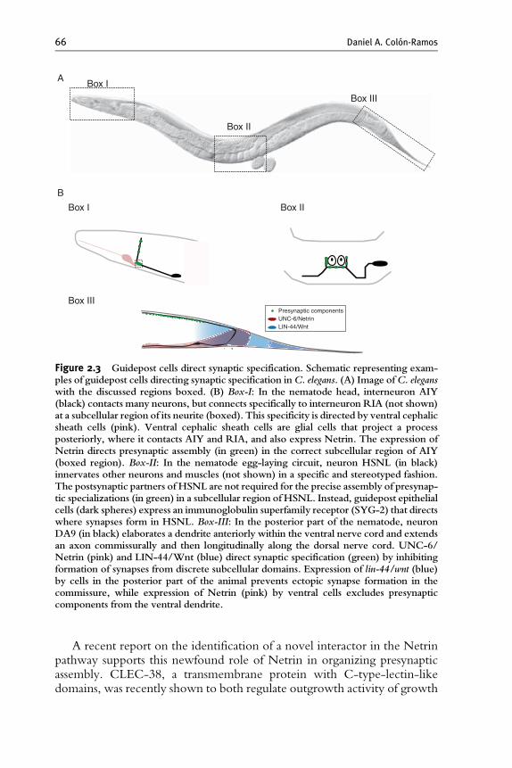
By studying the en passant synapses in the nematode C. elegans, theNetrin pathway was found to play a role in directing the formation ofpresynaptic specializations different from its conventional guidance role.This was done by visualizing the developmental decisions that led to theinnervation of two interneurons (AIY and RIA) in the nematode nervering. Similar to the RP3 system, this system allowed visualization of thedevelopmental decisions of these neurons with single-cell resolution and inthe context of the intact nervous system, but also in vivo (Gray et al., 2005;Mori and Ohshima, 1995). It was observed that AIY must undergo a seriesof stereotyped guidance decisions to reach and contact RIA. AIY and RIAthen connect to each other through en passant synapses formed at discreteregions of their respective processes (White et al., 1986). The resulting AIY:RIA circuit assembles in a stereotyped fashion in 100% of wild-type animals(Fig. 2.3).
In the absence of Netrin or the Netrin receptor, the majority of AIYcells still make their guidance decisions correctly. In spite of the fact that theguidance decisions are normal in the majority of animals, AIY fails toproperly form presynaptic specializations at the usual site of contact withRIA (Fig. 2.3). Cell-autonomous rescue of the Netrin receptor in thepresynaptic neuron AIY is sufficient to rescue the presynaptic patterningdefect, and when the subcellular localization of the Netrin receptor wasvisualized, it was observed that the Netrin receptor was enriched at thepresynaptic sites. Together, these results indicate that the Netrin pathwayplays a role in organizing synaptogenesis and that this role is independent ofits function in guidance (Colon-Ramos et al., 2007).
Presynaptic components UNC-6/Netrin LIN-44/Wnt
Box I
Box II
Box III
Box I
A
B
Box III
Box II
Figure 2.3 Guidepost cells direct synaptic specification. Schematic representing exam-ples of guidepost cells directing synaptic specification inC. elegans. (A) Image ofC. eleganswith the discussed regions boxed. (B) Box-I: In the nematode head, interneuron AIY(black) contacts many neurons, but connects specifically to interneuron RIA (not shown)at a subcellular region of its neurite (boxed). This specificity is directed by ventral cephalicsheath cells (pink). Ventral cephalic sheath cells are glial cells that project a processposteriorly, where it contacts AIY and RIA, and also express Netrin. The expression ofNetrin directs presynaptic assembly (in green) in the correct subcellular region of AIY(boxed region). Box-II: In the nematode egg-laying circuit, neuron HSNL (in black)innervates other neurons and muscles (not shown) in a specific and stereotyped fashion.The postsynaptic partners of HSNL are not required for the precise assembly of presynap-tic specializations (in green) in a subcellular region of HSNL. Instead, guidepost epithelialcells (dark spheres) express an immunoglobulin superfamily receptor (SYG-2) that directswhere synapses form in HSNL. Box-III: In the posterior part of the nematode, neuronDA9 (in black) elaborates a dendrite anteriorly within the ventral nerve cord and extendsan axon commissurally and then longitudinally along the dorsal nerve cord. UNC-6/Netrin (pink) and LIN-44/Wnt (blue) direct synaptic specification (green) by inhibitingformation of synapses from discrete subcellular domains. Expression of lin-44/wnt (blue)by cells in the posterior part of the animal prevents ectopic synapse formation in thecommissure, while expression of Netrin (pink) by ventral cells excludes presynapticcomponents from the ventral dendrite.
66 Daniel A. Colon-Ramos
A recent report on the identification of a novel interactor in the Netrinpathway supports this newfound role of Netrin in organizing presynapticassembly. CLEC-38, a transmembrane protein with C-type-lectin-likedomains, was recently shown to both regulate outgrowth activity of growth
Synapse Formation in Developing Neural Circuits 67
cones, and the organization of presynaptic terminals. In some developmentalcontexts CLEC-38 acts by inhibiting the Netrin receptor, thereby regulat-ing neural outgrowth during guidance. In other developmental contexts,however, CLEC-38 does not regulate outgrowth, but is instead required forthe organization of presynaptic terminals. Although it is not yet known ifthis presynaptic role of CLEC-38 is also mediated via the Netrin receptor,these data showed that CLEC-38, a regulator of Netrin activity and axonoutgrowth, plays additional roles in organizing presynaptic specializations(Kulkarni et al., 2008).
Other families of guidance molecules have also been observed to playsynaptogenic roles depending on the developmental context. For instance,in vertebrates, guidance molecules such as the Eph family of receptors andtheir ephrin ligands have been shown to play roles in growth cone guidanceas well as the development of mature excitatory synapses (Dalva et al., 2000).The distinct cellular responses of these different developmental events arealso likely generated by the developmental context and by diverse down-stream targets (Murai and Pasquale, 2004). Another family of receptors, theLAR-like phosphatase receptors, has also been shown to function at thelevel of axon guidance and presynaptic organization. Cell biological andgenetic characterizations of this receptor showed that in nematodes, thesetwo different activities are regulated by differentially spliced isoforms of thesame receptor (Ackley et al., 2005).
The existence of these shared pathways underscores the molecular linkbetween guidance and synaptogenesis. It also provides a conceptual frame-work on how growth cones, upon reaching their synaptic targets, couldtransition from being an outgrowth structure to a presynaptic terminal. Buthow can the same protein ‘‘molecularly multitask’’ and direct differentdevelopmental functions? How can the same receptor and ligand elicitdiverse cellular responses?
In the case of the Netrin receptor, for instance, the same molecule canregulate cell migration, neuronal polarization, axon guidance, and synapseformation in different developmental contexts. Although these events havevery different outcomes, there is an underlying similarity at the cell biolo-gical level: all of these events impinge on a polarization process thatrestructures the cytoskeleton. For example, during the maturation of theC. elegans neuron HSN, Netrin restricts the Netrin receptor localization tothe neuronal side facing the source of Netrin. This leads to the polarizedformation of the HSN growth cone (Adler et al., 2006). During guidance,Netrin polarizes the growth cone cytoskeleton to generate directed growth(Kennedy and Tessier-Lavigne, 1995). Similarly, in the AIY interneuronNetrin induces a localized polarization event to transform a region of theaxon shaft into a specialized presynaptic area (Colon-Ramos et al., 2007).
It is not well understood how downstream factors and regulators parcelout these signals to result in different developmental outcomes. However,
68 Daniel A. Colon-Ramos
with our increasing molecular understanding of these developmentalevents, it has become clear that guidance and synaptogenesis are intimatelylinked at a molecular level. Future work on these signal transductionpathways will allow us to understand how the same ligand/receptor paircan elicit distinct developmental outcomes, and how these events can beorchestrated in different cells to enable circuit assembly.
3. Building a Synapse
The process of guidance and target recognition is followed by synapseformation. How synapses are assembled is a formidable developmental ques-tion in its own right. As discussed previously, synapses need to form ontothe right partner, at the right density, and at a specific subcellular locationwith respect to the dendrites. Moreover, the assembly of presynaptic sitesalso needs to match the postsynaptic densities in terms of localization andidentity of the neurotransmitter and postsynaptic receptor ( Juttner andRathjen, 2005).
3.1. Cell adhesion in synaptic assembly
Synaptogenesis can be subdivided into two developmentally distinct steps(1) synaptic specificity and (2) synaptic assembly. Synaptic specificitydescribes the process that directs where synapses form: from the selectionof the right partner to the formation of synapses at the right subcellularcompartment. Synaptic assembly describes how synapses are formed: fromthe assembly of the macromolecular presynaptic structure to the formationof the signaling-rich postsynaptic specializations.
How are these two processes integrated to result in correct synapticdevelopment? The genesis of the synapse officially starts with the contactand communication between the pre- and the postsynaptic partners. There-fore, synaptic specificity is traditionally thought to be determined by cellsurface molecules that mediate this synaptic partner interaction. The classi-cal model states that contact between correct partners, mediated via celladhesion molecules, would then trigger inductive events that lead to theassembly and/or differentiation of pre- and postsynaptic specializations(Waites et al., 2005).
Spurred by this classic model, a number of studies have focused on theidentification of synaptogenic cell surface molecules. These studies led to theidentification of cell adhesion molecules that direct a variety of events insynaptic biology. The nature and importance of these cell–cell signalingmole-cules in synapse biology have been discussed elsewhere (Akins and Biederer,2006; Benson et al., 2001; Dalva et al., 2007; Juttner and Rathjen, 2005;
Synapse Formation in Developing Neural Circuits 69
Scheiffele, 2003; Yamagata et al., 2003), so in this section wewill only providea very brief summary of the conceptual findings stemming from these studies.
The molecules identified in these studies fall into four functional cate-gories: they either (1) promote stability by linking synaptic partners,(2) direct target recognition, (3) regulate differentiation of pre- and post-synaptic specializations, or (4) modulate synaptic structure and function(Yamagata et al., 2003). For instance, cadherins have been shown to localizeto puncta adherentia and direct synaptic morphology (Scheiffele, 2003).Immunoglobulin superfamily (IgSF) adhesion molecules Dscam, DscamL,Sidekick-1, and Sidekick-2 direct lamina-specific connectivity betweenspecific interneurons and retinal ganglion cells in the vertebrate retina(Yamagata et al., 2002). In Drosophila, LRR transmembrane protein capri-cious directs target specificity between muscle 12 and the motoneurons thatinnervate it (Shishido et al., 1998). Ephrins, on the other hand, can actthrough the EphB receptor to induce the clustering of NMDA receptorsand postsynaptic development (Dalva et al., 2000).
Interestingly, despite the focused efforts of identifying cell adhesionmolecules directly involved in synaptogenesis, only two adhesion moleculeshave been shown to induce formation of presynaptic specializations: neu-roligins and SynCAM1 (Akins and Biederer, 2006). The limited number ofidentified cell adhesion molecules capable of directly regulating synapseformation suggests that additional cues remain to be discovered. Thesefindings also beg the question of how the connectivity of hundreds oftrillions of synaptic connections are specified with a limited number ofmolecular cues.
3.2. Assembling the synaptic components
The classical view of synaptogenesis suggests that upon synaptic contactbetween partners, cell adhesion molecules induce the assembly of pre- andpostsynaptic specifications. This places assembly downstream of theadhesion-mediated specification events. Nonetheless, in a number of devel-opmental contexts in vivo, synaptic assembly occurs prior to synaptic contact.For instance, during muscle development in vertebrates, AChR clustersconcentrate into high density ‘‘hotspots’’ well before the growing axon hasarrived. This postsynaptic clustering of AChR receptors is also observed inaneural myotube cultures and in muscles of animals that have been geneti-cally rendered aneural (Kummer et al., 2006). These experiments indicatethat postsynaptic AChR clusters can occur prior to synaptic contact and inthe absence of presynaptic neural factors.
Presynaptic specializations can also form prior to cell–cell contactbetween synaptic partners. For instance, detailed ultrastructural studies inXenopus laevis tadpoles revealed that presynaptic specializations developprior to the association of the axon with the dendrites (Vaughn, 1989).
70 Daniel A. Colon-Ramos
These observations are also supported by ultrastructural studies in thedeveloping cortex of vertebrates, which revealed the existence of pre-and postsynaptic specializations that formed prior to the contact betweensynaptic partners (Craig and Lichtman, 2001).
Tissue culture studies have also supported the notion that synapticcontact is not necessary for the establishment of pre- and postsynapticspecializations. For instance, studies in dissociated hippocampal neuronshave demonstrated that, prior to cell–cell contact, functional NMDA andnon-NMDA-type glutamate receptors are present on the cell surface (Craigand Lichtman, 2001). Functional studies indicate that these ‘‘free’’ pre- andpostsynaptic structures contain the core molecular machinery necessary fortheir function (Krueger et al., 2003).
Together, these studies indicate some important aspects of the process ofsynapse formation. First, they demonstrate that synaptic partners are notnecessary for the assembly of synaptic components: both the release machin-ery in presynaptic structures and the neurotransmitter receptor clusters inpostsynaptic structures can be established independent of one another.Second, these experiments highlight the developmental and the geneticseparation between synaptic assembly and synaptic specificity.
This functional separation is further underscored in tissue culture systemsthat can reconstitute assembly, but not specificity events. In tissue culturesystems, neurons are dissociated, plated, and allowed to form synapticconnections onto neighboring neurons. The dissociation of neurons disruptsthe architecture of the nervous system, thereby destroying much of thepositional information which mediates synaptic specificity. Nonetheless,dissociated neurons still retain the ability to form synapses to neighboringneurons, to themselves, or even onto polylysine-coated beads (Vaughn,1989). These data suggest that assembly and specificity events are likelymediated through distinct signal transduction pathways.
Given the genetic separation between these events, how are they linkedto enable the assembly of a precisely wired nervous system? For instance, isthe observed assembly of ‘‘half-synapses’’ a transient and nonspecific featureof neuronal development? Can they actually influence where synapses willbe ultimately formed?
A number of studies have now shown that these ‘‘half-synapses’’ canparticipate in directing where synapses form. Neurodevelopmental studiesin zebrafish embryos showed that postsynaptic AChR aggregates formed inadvance of growing axons. Although some aggregates dispersed beforeinnervation, surprisingly, filopodia were stabilized when they contactedthe AChR aggregates (Kummer et al., 2006). Furthermore, in dissociatedhippocampal cultures, preformed postsynaptic scaffold protein complexes,containing PSD-95, GKAP, Shank, and neuroligin 1, localized to prede-fined postsynaptic hotspots. Upon contact with axons, these scaffoldingcomplexes induced the recruitment of synaptophysin-containing transport
Synapse Formation in Developing Neural Circuits 71
vesicles and the formation of presynaptic specializations (Gerrow et al.,2006). These results suggest that assembly prior to synaptic contact canlater influence where synapses will form.
3.3. Guidepost cells, morphogens, and connectivity
The studies discussed in the previous section indicate that the assembly ofsynapses does not require contact between synaptic partners. Moreover,given the role of these preformed specializations in directing where synapsesform, these data suggest that in some developmental contexts, the specifica-tion of where synapses form is also not dependent on the contact betweensynaptic partners. Which molecular mechanisms direct synaptic specificityin these contexts?
Accumulating evidence suggests that both intrinsic and extrinsicmechan-isms can influence where synapses are formed. For instance, studies indissociated cortical neuronal cultures revealed that initial formation of pre-synaptic terminals preferentially occurs at predefined sites within the axonalshaft. In these studies, time-lapse imaging was conducted to track themovement of synaptic vesicle protein transport vesicles (STVs), an organellecontaining synaptic vesicle-associated proteins which gets recruited tonascent synapses. It was observed that, even in the absence of postsynapticpartners or glia contact, STVs paused at predefined sites. Upon contact withpresumptive postsynaptic partners, presynaptic terminals developed specifi-cally at these predefined sites. Moreover, these sites promoted the formationof stable contacts with dendritic filopodia (Sabo et al., 2006). These studiesindicate that intrinsic, predefined pause sites in axon shafts can influencethe development of nascent synapses at particular sites along the axon indissociated neurons.
Extrinsic signals generated by guidepost cells can also provide cues thatdirect where synapses are assembled. For instance, in C. elegans theegg-laying motor neuron (HSNL) specifically innervates muscles and VCneurons in the vulva region of the animal (Fig. 2.3). Surprisingly, thepostsynaptic partners are not required for correct formation of presynapticspecializations in HSNL. Instead, guidepost epithelial cells provide a posi-tional cue that directs HSNL presynaptic assembly. This is molecularlymediated through a pair of IgSF proteins, SYG-1 and SYG-2. In syg-1 orsyg-2 mutants, presynaptic neuron HSNL contacts its normal synapticpartners but fails to form synaptic connections with them. Instead, ectopicsynapses are formed onto abnormal postsynaptic targets. SYG-1 and SYG-2both localize to synapses and bind to each other, acting as a receptor and aligand. SYG-1 functions cell autonomously in the presynaptic neuron,while SYG-2 functions in the guidepost epithelial cells that are essentialfor the correct formation of HSNL synapses (Shen and Bargmann, 2003;Shen et al., 2004).
72 Daniel A. Colon-Ramos
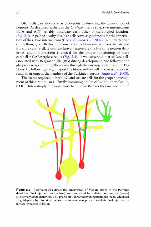
Glial cells can also serve as guideposts in directing the innervation ofneurons. As discussed earlier, in the C. elegans nerve ring, two interneurons(RIA and AIY) reliably innervate each other at stereotyped locations(Fig. 2.3). A pair of nearby glia-like cells serve as guideposts for the innerva-tion of these two interneurons (Colon-Ramos et al., 2007). In the vertebratecerebellum, glia cells direct the innervation of two interneurons: stellate andPurkinje cells. Stellate cells exclusively innervate the Purkinje neuron den-drites, and this precision is critical for the proper functioning of thesecerebellar GABAergic circuits (Fig. 2.4). It was observed that stellate cellsassociated with Bergmann glia (BG) during development, and followed theglia process by extending their axon through the curving contours of the BGfibers. By following the guidepost BG fibers, stellate cell processes are able toreach their targets: the dendrite of the Purkinje neurons (Ango et al., 2008).
The factor required in both BG and stellate cells for the proper develop-ment of this circuit is an L1 family immunoglobulin cell adhesion molecule,CHL1. Interestingly, previous work had shown that another member of the
Figure 2.4 Bergmann glia direct the innervation of Stellate axons to the Purkinjedendrites. Purkinje neurons (yellow) are innervated by stellate interneurons (green)exclusively at the dendrites. This precision is directed by Bergmann glia (red), which actas guideposts by directing the stellate interneuron process to their Purkinje neurontargets (synapses in blue).
Synapse Formation in Developing Neural Circuits 73
L1 family, neurofascin186, is required for the specification of another part ofthis GABAergic circuit: the innervation of the Basket cells and the Purkinjecell axon initial segment (Ango et al., 2004). This molecular characterizationof the cerebellar GABAergic circuit suggests that different members ofthe L1CAM protein family contribute to circuit formation through theircell-specific expression in subsets of neurons and guidepost glial cells.
Some neurons in the vertebrate hippocampus and cortex can also act asguideposts and direct synaptogenesis. For instance, the transient populationof Cajal-Retzius cells in the hippocampus serves as a placeholder to facilitatethe meeting of the appropriate pre- and postsynaptic cells (Del Rio et al.,1997). Also, during the development of the mammalian cortex, the subplateneurons display a similar guidepost function to arrange the connectivitybetween the thalamic axons and the layer 4 cortical neurons (McConnellet al., 1989). The significance of these guidepost cells was demonstrated byablating the guidepost cells and showing a synaptic connectivity defect inablated animals (Del Rio et al., 1997; Ghosh et al., 1990).
Tissues can also provide inhibitory signals to direct the formation ofsynapses with subcellular precision. In the DA9 motoneuron of C. elegans,Wnt lin-44 is secreted by four hypodermal cells in the tail. This expressionlocalizes receptor lin-17/Frizzled (Fz) to a subdomain of DA9, near theposterior part of the neurite adjacent to the hypodermal cells. This part ofthe neurite where lin-17/Fz localizes is normally devoid of presynapticspecializations (Fig. 2.3). When the Wnt pathway is compromised, how-ever, synapses develop ectopically in this subdomain. Moreover, overex-pression of WNT lin-44 in the posterior part of the animal expands LIN-17localization and inhibits presynaptic assembly in these new sites of ectopicLIN-17 localization (Klassen and Shen, 2007). The Wnt pathway was alsoshown to act as a local repressive cue to direct target specificity inDrosophila.Studies in embryonic motor neurons showed that Wnt4 acts via Frizzled 2,Derailed-2, and Disheveled to generate target specificity by preventingsynapse formation onto nontarget muscles (Inaki et al., 2007).
Wnts can also stimulate the formation of synapses in both vertebrates andinvertebrates (Salinas and Zou, 2008). InDrosophila, Wnt/wingless is requiredfor the correct development of presynaptic boutons, in terms of both theirnumbers and their structure, and this activity occurs in a transcription-independent manner (Miech et al., 2008). These observations, together withstudies showing a role of Wnts in postsynaptic activation of Frizzled recep-tors, indicate that Wnt signaling can alter synaptic development by simulta-neously modulating the development of presynaptic and postsynapticstructures (Miech et al., 2008).
But how can Wnts both promote and inhibit the formation of synapses?Recent studies in dissociated hippocampal cultures have demonstrated thatthe opposing effects of Wnts on synapse formation depend on differentWnt ligands. Interestingly, the different ligands differentially activate either
74 Daniel A. Colon-Ramos
the canonical or the noncanonical pathways: activation of the canonicalpathway promotes synapse formation, while activation of the noncanonicalone inhibits synapse formation (Davis et al., 2008).
Together, these studies demonstrate that in both vertebrates and inverte-brates, morphogenic signals such as Wnts can spatially regulate the patterningof synaptic connections. This is achieved by subdividing the neurite intodiscrete domains and either preventing or promoting synapse formation atspecific subcellular compartments.
Other extrinsic cues can also direct neuronal polarity that ultimatelyimpinges on the site of synaptic assembly in vivo. In a study that also used theDA9 motoneuron system, it was shown that the axon guidance cue UNC-6/netrin and its receptor UNC-5 act to exclude synaptic vesicles and active zoneproteins from the dendrite of DA9 (Fig. 2.3). In unc-6/netrin and unc-5 loss-of-function mutants, presynaptic components mislocalize to the DA9 dendrite,where the level of endogenous UNC-6/netrin is high. In addition, ectopicallyexpressed UNC-6/netrin, acting through UNC-5, was sufficient to excludeendogenous synapses from adjacent subcellular domains within the DA9 axon.Interestingly, this antisynaptogenic activity was interchangeable with that ofLIN-44/Wnt (Poon et al., 2008). This suggests that extracellular cues such asnetrin andWnts not only guide axon navigation but also regulate the polarizedaccumulation of presynaptic components through local exclusion.
Together, these studies indicate that extrinsic signals generated fromguidepost cells or neighboring tissues can direct the site of synaptic assembly,thereby modulating synaptic specificity. Although these examples indicatethat synaptic contact is not required for the initial specification of synapto-genesis, it should be noted that contact with a postsynaptic-like substrate isrequired for the eventual stabilization and perseverance of many of these‘‘free’’ pre- and postsynaptic sites (Vaughn, 1989).
Contact and communication between the synaptic partners is also requiredfor the regulation of the size and shape of the synapse, a process known assynaptic homeostasis (Keshishian, 2002).During synaptic homeostasis, a cross-talk between pre- and postsynaptic specializations takes place across thesynaptic cleft. Depending on the developmental context, this crosstalk canbemediated by a number of differentmolecular cues,which includes fibroblastgrowth factors (FGFs), bone morphogenetic protein (BMP), signal transduc-tion pathways, and MEF-2 transcriptional responses (Aberle et al., 2002;Goold and Davis, 2007; Salinas, 2005; Simon et al., 2008). The trans-synapticcommunication between partners elicits a coordinated regulation of synapticsize, shape, and functionality.
Contact between synaptic partners is also required for the proper mod-ulation of synaptic activity. Although in this chapter we focused ourdiscussion on the early synaptogenic decisions preceding synaptic activity,it should be noted that it is this synaptic activity that, in most developmentalcontexts, ultimately regulates the stabilization or elimination of many
Synapse Formation in Developing Neural Circuits 75
synapses (Flavell and Greenberg, 2008). The early developmental decisionsdiscussed in this chapter generate a neural framework over which activity-dependent changes occur. The activity-dependent regulation of synapticbiology is required, in both the developing and the mature nervous systems,for the maturation, refinement, and plasticity of synaptic connections.
4. Perspective
Correct circuit formation requires an intricate orchestration of multipledevelopmental events including cell migration, axon guidance, dendriticgrowth, target selection, and synaptogenesis ( Juttner and Rathjen, 2005;Salie et al., 2005; Waites et al., 2005). These events are integrated to enablecorrect synapse formation between neuronal partners. The developmentalinnervation of synaptic partners results in hundreds of trillions of preciselywired synaptic connections. Since the human genome has an estimated 25,000genes, and not all genes are involved in synaptogenesis, these events need to besimultaneously orchestrated in billions of neurons using a limited set ofmolecular cues.
Genetic, biochemical, and cell biological studies have revealed some ofthe molecular cues that regulate these developmental processes. Studies ofthese signaling molecules have revealed classical roles for different proteinfamilies. For instance, morphogens, which are known to create gradientswith important positional information, are critical for the specification ofcell fate decisions. Transmembrane proteins that recognize diffusible factorshave been shown to direct processes such as dendritic and axonal outgrowthand guidance. On the other hand, cell–cell adhesion proteins can controlsynaptogenesis. This has led to the conceptual understanding that genefamilies with evolutionarily conserved functions could play modular rolesin patterning the nervous system (Salie et al., 2005).
Although different protein families can play distinct roles at discretedevelopmental steps, this modular model of nervous system developmentdoes not reflect the complexity of this process in vivo. A growing body ofliterature shows that molecular cues, far from playing exclusive roles atdiscrete steps, are instead capable of ‘‘molecular multitasking’’ (Salie et al.,2005). For example, morphogenic proteins such as Sonic hedgehog, Wnts,FGF, and BMP have long been known to direct neuronal cell fate byeliciting transcriptional programs. More recent studies have demonstratedthat, depending on the cellular context, these canonical morphogens canalso provide instructive, transcription-independent signals to control pro-cesses such as axon outgrowth, neuronal cell polarity, and synapse formation(Salie et al., 2005). Additionally, signal transduction cascades that have beentraditionally thought of as long-range guidance cues have now also beenobserved to regulate synaptic formation events.
76 Daniel A. Colon-Ramos
It is provocative to think that ‘‘molecular multitasking’’ could haveprofound implications for the development of neural circuits. For instance,through the same receptor:ligand pair, multiple signal transduction path-ways could be simultaneously activated in different cells with differentdevelopmental outcomes. One could speculate that this would allow asingle molecular cue to simultaneously direct several independent develop-mental outcomes in different cells, thereby coordinating circuit assembly byorchestrating the innervation of multiple partners.
The identification of molecular factors and signal transduction cascadesinvolved in synapse formation, combined with an increased understandingof how these molecular factors are integrated to direct circuit formationin vivo, will provide us with an increasingly clear picture on how precisesynaptogenesis is orchestrated during nervous system development.
ACKNOWLEDGMENTS
I thank M. Hammarlund, S. Margolis, M. Margeta, and G. Maro for thoughtful commentsconcerning this chapter. I particularly thank K. Shen for helpful discussions, generous advice,and thoughtful comments on the chapter. I also thank J. Blagburn, V. Poon, and F. Ango forcontributing images. I apologize to those whose work I did not cite here due to oversight orspace constrains. During the preparation of this chapter, I was supported by NIH grant4R00NS057931-03.
REFERENCES
Aberle, H., Haghighi, A. P., Fetter, R. D., McCabe, B. D., Magalhaes, T. R., andGoodman, C. S. (2002). Wishful thinking encodes a BMP type II receptor that regulatessynaptic growth in Drosophila. Neuron 33, 545–558.
Ackley, B. D., Harrington, R. J., Hudson, M. L., Williams, L., Kenyon, C. J.,Chisholm, A. D., and Jin, Y. (2005). The two isoforms of the Caenorhabditis elegansleukocyte-common antigen related receptor tyrosine phosphatase PTP-3 function inde-pendently in axon guidance and synapse formation. J. Neurosci. 25, 7517–7528.
Adler, C. E., Fetter, R. D., and Bargmann, C. I. (2006). UNC-6/Netrin induces neuronalasymmetry and defines the site of axon formation. Nat. Neurosci. 9, 511–518.
Akins, M. R., and Biederer, T. (2006). Cell-cell interactions in synaptogenesis. Curr. Opin.Neurobiol. 16, 83–89.
Anderson, P. A., and Spencer, A. N. (1989). The importance of cnidarian synapses forneurobiology. J. Neurobiol. 20, 435–457.
Ango, F., di Cristo, G., Higashiyama, H., Bennett, V., Wu, P., and Huang, Z. J. (2004).Ankyrin-based subcellular gradient of neurofascin, an immunoglobulin family protein,directs GABAergic innervation at purkinje axon initial segment. Cell 119, 257–272.
Ango, F., Wu, C., Van der Want, J. J., Wu, P., Schachner, M., and Huang, Z. J. (2008).Bergmann glia and the recognition molecule CHL1 organize GABAergic axons anddirect innervation of Purkinje cell dendrites. PLoS Biol. 6, e103.
Benson, D. L., Colman, D. R., and Huntley, G. W. (2001). Molecules, maps and synapsespecificity. Nat. Rev. Neurosci. 2, 899–909.
Synapse Formation in Developing Neural Circuits 77
Blagburn, J. M., and Bacon, J. P. (2004). Control of central synaptic specificity in insectsensory neurons. Annu. Rev. Neurosci. 27, 29–51.
Chiba, A., and Rose, D. (1998). ‘‘Painting’’ the target: How local molecular cues definesynaptic relationships. Bioessays 20, 941–948.
Colon-Ramos, D. A., Margeta, M. A., and Shen, K. (2007). Glia promote local synaptogen-esis through UNC-6 (netrin) signaling in C. elegans. Science 318, 103–106.
Cowan, W. M., and Kandel, E. R. (2001). A brief history of synapses and synaptictransmittion. In ‘‘Synapses’’ (T. C. Sudhof, W. M. Cowan, and C. F. Stevens, Eds.),pp. 1–88. The Johns Hopkins University Press, Baltimore.
Craig, A. M., and Lichtman, J. W. (2001). Synapse formation and maturation. In ‘‘Synapses’’(T. C. Sudhof, W. M. Cowan, and C. F. Stevens, Eds.), pp. 1–88. The Johns HopkinsUniversity Press, Baltimore.
Dalva, M. B., McClelland, A. C., and Kayser, M. S. (2007). Cell adhesion molecules:Signalling functions at the synapse. Nat. Rev. Neurosci. 8, 206–220.
Dalva, M. B., Takasu, M. A., Lin, M. Z., Shamah, S. M., Hu, L., Gale, N. W., andGreenberg, M. E. (2000). EphB receptors interact with NMDA receptors and regulateexcitatory synapse formation. Cell 103, 945–956.
Davis, E. K., Zou, Y., and Ghosh, A. (2008). Wnts acting through canonical and nonca-nonical signaling pathways exert opposite effects on hippocampal synapse formation.Neural Develop. 3, 32.
De Camilli, P., Haucke, V., Takei, K., and Mugnaini, E. (2001). The structure of synapses.In ‘‘Synapses’’ (T. C. Sudhof, W. M. Cowan, and C. F. Stevens, Eds.), pp. 1–88. TheJohns Hopkins University Press, Baltimore.
Del Rio, J. A., Heimrich, B., Borrell, V., Forster, E., Drakew, A., Alcantara, S.,Nakajima, K., Miyata, T., Ogawa, M., Mikoshiba, K., Derer, P., Frotsher, M., andSoriano, E. (1997). A role for Cajal-Retzius cells and reelin in the development ofhippocampal connections. Nature 385, 70–74.
Flavell, S. W., and Greenberg, M. E. (2008). Signaling mechanisms linking neuronal activityto gene expression and plasticity of the nervous system. Annu. Rev. Neurosci. 31,563–590.
Gerrow, K., Romorini, S., Nabi, S. M., Colicos, M. A., Sala, C., and El-Husseini, A.(2006). A preformed complex of postsynaptic proteins is involved in excitatory synapsedevelopment. Neuron 49, 547–562.
Ghosh, A., Antonini, A., McConnell, S. K., and Shatz, C. J. (1990). Requirement forsubplate neurons in the formation of thalamocortical connections. Nature 347, 179–181.
Goold, C. P., and Davis, G. W. (2007). The BMP ligand Gbb gates the expression ofsynaptic homeostasis independent of synaptic growth control. Neuron 56, 109–123.
Gray, J. M., Hill, J. J., and Bargmann, C. I. (2005). Inaugural article: A circuit for navigationin Caenorhabditis elegans. Proc. Natl. Acad. Sci. USA 102, 3184–3191.
Inaki, M., Yoshikawa, S., Thomas, J. B., Aburatani, H., and Nose, A. (2007). Wnt4 is a localrepulsive cue that determines synaptic target specificity. Curr. Biol. 17, 1574–1579.
Juttner, R., and Rathjen, F. G. (2005). Molecular analysis of axonal target specificity andsynapse formation. Cell. Mol. Life Sci. 62, 2811–2827.
Kennedy, T. E., and Tessier-Lavigne, M. (1995). Guidance and induction of branchformation in developing axons by target-derived diffusible factors. Curr. Opin. Neurobiol.5, 83–90.
Keshishian, H. (2002). Is synaptic homeostasis just wishful thinking? Neuron 33, 491–492.Klassen, M. P., and Shen, K. (2007). Wnt signaling positions neuromuscular connectivity by
inhibiting synapse formation in C. elegans. Cell 130, 704–716.Krueger, S. R., Kolar, A., and Fitzsimonds, R. M. (2003). The presynaptic release apparatus
is functional in the absence of dendritic contact and highly mobile within isolated axons.Neuron 40, 945–957.
78 Daniel A. Colon-Ramos
Kulkarni, G., Li, H., and Wadsworth, W. G. (2008). CLEC-38, a transmembrane proteinwith C-type lectin-like domains, negatively regulates UNC-40-mediated axon out-growth and promotes presynaptic development in Caenorhabditis elegans. J. Neurosci. 28,4541–4550.
Kummer, T. T., Misgeld, T., and Sanes, J. R. (2006). Assembly of the postsynapticmembrane at the neuromuscular junction: Paradigm lost. Curr. Opin. Neurobiol. 16,74–82.
Lickteig, K. M., Duerr, J. S., Frisby, D. L., Hall, D. H., Rand, J. B., and Miller, D. M. 3rd.(2001). Regulation of neurotransmitter vesicles by the homeodomain protein UNC-4and its transcriptional corepressor UNC-37/groucho in Caenorhabditis elegans cholinergicmotor neurons. J. Neurosci. 21, 2001–2014.
Marie, B., Bacon, J. P., and Blagburn, J. M. (2000). Double-stranded RNA interferenceshows that Engrailed controls the synaptic specificity of identified sensory neurons. Curr.Biol. 10, 289–292.
Marie, B., and Blagburn, J. M. (2003). Differential roles of engrailed paralogs in determiningsensory axon guidance and synaptic target recognition. J. Neurosci. 23, 7854–7862.
Marie, B., Cruz-Orengo, L., and Blagburn, J. M. (2002). Persistent engrailed expression isrequired to determine sensory axon trajectory, branching, and target choice. J. Neurosci.22, 832–841.
McConnell, S. K., Ghosh, A., and Shatz, C. J. (1989). Subplate neurons pioneer the firstaxon pathway from the cerebral cortex. Science 245, 978–982.
Miech, C., Pauer, H. U., He, X., and Schwarz, T. L. (2008). Presynaptic local signaling by acanonical wingless pathway regulates development of the Drosophila neuromuscularjunction. J. Neurosci. 28, 10875–10884.
Miller, D. M., Shen, M. M., Shamu, C. E., Burglin, T. R., Ruvkun, G., Dubois, M. L.,Ghee, M., andWilson, L. (1992).C. elegans unc-4 gene encodes a homeodomain proteinthat determines the pattern of synaptic input to specific motor neurons. Nature 355,841–845.
Mitchell, K. J., Doyle, J. L., Serafini, T., Kennedy, T. E., Tessier-Lavigne, M.,Goodman, C. S., and Dickson, B. J. (1996). Genetic analysis of Netrin genes inDrosophila: Netrins guide CNS commissural axons and peripheral motor axons. Neuron17, 203–215.
Mori, I., and Ohshima, Y. (1995). Neural regulation of thermotaxis in Caenorhabditis elegans.Nature 376, 344–348.
Murai, K. K., and Pasquale, E. B. (2004). Eph receptors, ephrins, and synaptic function.Neuroscientist 10, 304–314.
O’Leary, D. D., Chou, S. J., and Sahara, S. (2007). Area patterning of the mammalian cortex.Neuron 56, 252–269.
Peteya, D. J. (1973). A light and electron microscope study of the nervous system ofCeriantheopsis americanus (Cnidaria, Ceriantharia). Z. Zellforsch Mikrosk Anat. 141,301–317.
Plachez, C., and Richards, L. J. (2005). Mechanisms of axon guidance in the developingnervous system. Curr. Top. Dev. Biol. 69, 267–346.
Polleux, F., Ince-Dunn, G., and Ghosh, A. (2007). Transcriptional regulation of vertebrateaxon guidance and synapse formation. Nat. Rev. Neurosci. 8, 331–340.
Poon, V. Y., Klassen, M. P., and Shen, K. (2008). UNC-6/netrin and its receptor UNC-5locally exclude presynaptic components from dendrites. Nature 455, 669–673.
Rand, J. B., andNonet,M. L. (1997).C. elegans II. In ‘‘Synaptic Transmission’’ (D. L. Riddle,T. Blumenthal, B. J. Meyer, and J. R. Priess, Eds.), vol. II, pp. 611–643. Cold SpringHarbor Laboratory Press, Cold Spring Harbor.
Sabo, S. L., Gomes, R. A., and McAllister, A. K. (2006). Formation of presynaptic terminalsat predefined sites along axons. J. Neurosci. 26, 10813–10825.

Synapse Formation in Developing Neural Circuits 79
Sakarya, O., Armstrong, K. A., Adamska, M., Adamski, M., Wang, I. F., Tidor, B.,Degnan, B. M., Oakley, T. H., and Kosik, K. S. (2007). A post-synaptic scaffold at theorigin of the animal kingdom. PLoS ONE 2, e506.
Salie, R., Niederkofler, V., and Arber, S. (2005). Patterning molecules; multitasking in thenervous system. Neuron 45, 189–192.
Salinas, P. C. (2005). Signaling at the vertebrate synapse: New roles for embryonic morpho-gens? J. Neurobiol. 64, 435–445.
Salinas, P. C., and Zou, Y. (2008). Wnt signaling in neural circuit assembly. Annu. Rev.Neurosci. 31, 339–358.
Scheiffele, P. (2003). Cell-cell signaling during synapse formation in the CNS. Annu. Rev.Neurosci. 26, 485–508.
Shen, K., and Bargmann, C. I. (2003). The immunoglobulin superfamily protein SYG-1determines the location of specific synapses in C. elegans. Cell 112, 619–630.
Shen, K., Fetter, R. D., and Bargmann, C. I. (2004). Synaptic specificity is generated by thesynaptic guidepost protein SYG-2 and its receptor, SYG-1. Cell 116, 869–881.
Shishido, E., Takeichi, M., and Nose, A. (1998). Drosophila synapse formation: Regulationby transmembrane protein with Leu-rich repeats, CAPRICIOUS. Science 280,2118–2121.
Simon, D. J., Madison, J. M., Conery, A. L., Thompson-Peer, K. L., Soskis, M.,Ruvkun, G. B., Kaplan, J. M., and Kim, J. K. (2008). The microRNA miR-1 regulatesa MEF-2-dependent retrograde signal at neuromuscular junctions. Cell 133, 903–915.
Sudhof, T. C. (2004). The synaptic vesicle cycle. Annu. Rev. Neurosci. 27, 509–547.Tessier-Lavigne, M., and Goodman, C. S. (1996). The molecular biology of axon guidance.
Science 274, 1123–1133.Vaughn, J. E. (1989). Fine structure of synaptogenesis in the vertebrate central nervous
system. Synapse 3, 255–285.Von Stetina, S. E., Fox,R.M.,Watkins, K. L., Starich, T.A., Shaw, J. E., andMiller,D.M. 3rd.
(2007). UNC-4 represses CEH-12/HB9 to specify synaptic inputs to VAmotor neurons inC. elegans. Genes Dev. 21 332–346.
Von Stetina, S. E., Treinin, M., and Miller, D. M. 3rd. (2006). The motor circuit. Int. Rev.Neurobiol. 69 125–167.
Waites, C. L., Craig, A. M., and Garner, C. C. (2005). Mechanisms of vertebrate synapto-genesis. Annu. Rev. Neurosci. 28, 251–274.
Westfall, I. A. (1996). Ultrastructure of synapses in the first-evolved nervous systems.J. Neurocytol. 25, 735–746.
White, J. G., Southgate, E., and Thomson, J. N. (1992). Mutations in the Caenorhabditiselegans unc-4 gene alter the synaptic input to ventral cord motor neurons. Nature 355,838–841.
White, J. G., Southgate, E., Thomson, J. N., and Brenner, S. (1986). The structure of thenervous system of the nematode Caenorhabditis elegans. Philosophical Transactions of theRoyal Society of London 314, 1–340.
Yamagata, M., Sanes, J. R., and Weiner, J. A. (2003). Synaptic adhesion molecules. Curr.Opin. Cell. Biol. 15, 621–632.
Yamagata, M., Weiner, J. A., and Sanes, J. R. (2002). Sidekicks: Synaptic adhesion mole-cules that promote lamina-specific connectivity in the retina. Cell 110, 649–660.