Embed Size (px)
Citation preview

J Vasc Br 2002, Vol. 1, Nº3 207
Surgical treatment of thoracic and thoracoabdominal aorticaneurysms: technical notes and the use of left heart bypass
Tratamento cirúrgico de aneurismas da aorta torácica e toracoabdominal:considerações técnicas e uso de derivação cardíaca esquerda
Roberto Chiesa1, Germano Melissano1, Marcelo Ruettimann Liberato de Moura1, Efrem Civilini1,Yamume Tshomba1, Matheus Ruettimann Liberato de Moura1, Silvio Magrin2, Andrea Carozzo2
1. Department of Vascular Surgery, Università Vita-Salute, IRCCS San Raffaele, Milan, Italy.2. Department of Anesthesiology, Università Vita-Salute, IRCCS San Raffaele, Milan, Italy.
J Vasc Br 2002;1(3):207-18.Copyright © 2002 by Sociedade Brasileira de Angiologia e Cirurgia Vascular.
207
ResumoObjetivos: O objetivo deste estudo foi o de analisar morbidade e
mortalidade pós-cirúrgicas em pacientes submetidos à cirurgia deaneurisma da aorta torácica e aneurisma toracoabdominal no Departa-mento de Cirurgia Vascular do IRCCS San Raffaele, Milão.
Métodos: O estudo incluiu 332 pacientes (256 homens e 76 mu-lheres) com idade média de 65 anos (variação de 34 a 82 anos) queforam submetidos a 333 operações para aneurismectomia de aneurismada aorta torácica e aneurisma toracoabdominal entre janeiro de 1988 eoutubro de 2002. A drenagem do líquido cefalorraquidiano foi utiliza-da em 212 casos (75% dos aneurismas toracoabdominais, 53% dosaneurismas da aorta torácica); 215 pacientes (110 aneurismastoracoabdominais e 105 aneurismas da aorta torácica) foram submeti-dos à cirurgia de bypass coronário esquerdo através do uso de bombaBiomedicus.
Resultados: A taxa total de mortalidade aos 30 dias foi de 40/332(12%); um total de 32 mortes (10,5%) foram registradas durante ascirurgias eletivas e oito (29,6%) em pacientes submetidos a reparosemergenciais. As seguintes complicações pós-cirúrgicas foram relata-das: paraplegia/paraparesia em 21 casos (6,3%), insuficiência respira-tória com necessidade de entubação prolongada em 79 casos (24%),complicações cardíacas (arritmia grave, enfarte do miocárdio) em 29casos (9%), insuficiência renal em 23 casos (7%), hemorragia pós-ci-rúrgica com necessidade de revisão cirúrgica em 17 casos (5%), infec-ção do enxerto em seis casos (1,8%).
Conclusões: As taxas de morbidade e mortalidade ocorridas após acirurgia de aneurisma toracoabdominal e aneurisma da aorta torácicaainda são altas. Contudo, de acordo com nossa experiência, o uso desuporte circulatório distal ativo, clampeamento seqüencial e drenagemde líquido cerebrorraquidiano faz com que resultados aceitáveis sejamalcançados e reduz complicações secundárias à isquemia visceral e is-quemia da medula espinhal, sem a necessidade de tempo declampeamento rápido.
Palavras-chave: aneurisma aórtico, aneurisma torácico, aneurismada aorta abdominal, procedimentos cirúrgicos cardíacos, coração auxi-liar.
AbstractObjective: The aim of this study was to analyze perioperative
morbidity and mortality in patients undergoing thoracic andthoracoabdominal aortic aneurysm surgery at the Department ofVascular Surgery of IRCCS San Raffaele, Milan.
Methods: The study included 332 patients (256 males and 76females) with a mean age of 65 (range from 34 to 82 years) undergoing333 operations for aneurysmectomy of thoracic and thoracoabdominalaortic aneurysm between January 1988 and October 2002.Cerebrospinal fluid drainage was used in 212 cases (75% ofthoracoabdominal aortic aneurysms, 53% of thoracic aortic aneurysms);215 patients (110 thoracoabdominal aortic aneurysms and 105 thoracicaortic aneurysms) were operated under left heart bypass using aBiomedicus pump.
Results: The overall mortality rate at 30 days was 40/332 (12%); atotal of 32 deaths (10.5%) were recorded during elective surgery andeight (29.6%) in patients undergoing emergency repairs. The followingperioperative complications were reported: paraplegia/paraparesis in 21cases (6.3%), respiratory failure requiring prolonged intubation in 79cases (24%), cardiac complications (major arrhythmia, myocardialinfarction) in 29 cases (9%), renal failure in 23 cases (7%), postoperativebleeding requiring redo surgery in 17 cases (5%), graft infection in sixcases (1.8%).
Conclusions: Morbidity and mortality consequent tothoracoabdominal aortic aneurysm and thoracic aortic aneurysmssurgery are still high. However, based on our experience, the use of anactive distal circulatory support, sequential cross-clamping andcerebrospinal fluid drainage enables acceptable results to be achievedand reduces complications secondary to spinal cord and visceral ischemia,without the need for expeditious clamping times.
Key words: aortic aneurysm, thoracic aneurysm, abdominalaneurysm, cardiac surgical procedures, heart-assist devices.
ARTIGO ORIGINAL
208 J Vasc Br 2002, Vol. 1, Nº3
Numerous problems are related to the surgicaltreatment of thoracic and thoracoabdominal aneurysmsand their anatomical localization. These problems aresecondary to massive blood losses, extensive surgicalexposure, radical hemodynamic alterations and mainlyto the limited tolerance of organs subject to a temporaryischemia during aortic clamping.
During clamping of the descending aorta, the arterialpressure proximal to the clamp increases rapidly, andthis is accompanied by a sudden fall in the pressuredistal to the clamped aorta. The high elevation of thecardiac postload results in increased ventricular tensionand hence also in preload. This results in an increasedright heart pressure and central venous pressure. Suchcascade of events leads to a rise in venous pressure andcerebrospinal fluid pressure. As a direct consequence ofinterrupted aortic flow, the organ below the clampingpoint is deprived from the normal oxygen intake andenergy substrates, and has a reduced removal of cellularcatabolites.
The effects of the hypoperfusion affecting the spinalcord, kidneys, intestine and liver are of particularclinical interest (Table 1).1
– Reduction of metabolic activity: using moderate orprofound hypothermia;
– Pharmacological aids: such as the use of mannitol,steroids, scavengers and new agents to preventspinal cord damage caused by ischemia orreperfusion.
Materials and methods
Patients
A total of 333 operations for descending thoracicaortic or thoracoabdominal aneurysms were carried outfrom January 1988 and October 2002 at the VascularSurgery Department of IRCCS S. Raffaele in Milan on332 patients (256 males and 76 females) with a meanage of 65 years (range 34-82). These included a patientsuffering from chronic aortic dissection (Stanford typeB) who initially underwent isthmus repair and, threeyears later, complete replacement of the descendingthoracic aorta due to the subsequent dilatation of thedistal tract.
Table 2 shows the anatomical distribution of thetreated aneurysms.
Aortic repair was performed on thoracic orthoracoabdominal aneurysms with restricted indicationsuntil 1993 (symptomatic aneurysms, emergency surgeryfollowing rupture, voluminous aneurysms) andsubsequently on all patients, extending the indicationsfor surgery to the guidelines set out in the recentliterature (Table 3).2
Data on the preoperative, intraoperative, and follow-up periods have been collected for all patients referredto our department with this pathology since January1993.
Complications Range Mean
Paraparesis/plegia 4-32% 14%Respiratory failure 16-43% 32%AMI 2-23% 11%Renal failure 4-37% 18%Multiple organ failure 2-13% 5%Postoperative bleeding 3-29% 7%
Table 1 - Complications of thoracoabdominal aortasurgery reported in the literature
AMI: acute myocardial infarction
There are currently several strategies to reduce theduration and extent of tissue ischemia:
– Mechanical devices: including extracorporealcirculation, shunt, sequential cross-clamping,retrograde perfusion and cerebrospinal fluiddrainage (CSFD);
– Surgical strategies: including the selectivereattachment of intercostal arteries critical for spinalcord perfusion, selected through somatosensoryevoked potentials (SEP) or motor evoked potentials(MEP);
Extent of aneurysm n. of cases %
Thoracic (TAA) 168 51Thoracoabdominal (TAAA) 164 49Type I* 32 20§
Type II* 50 30§
Type III* 48 29§
Type IV* 34 21§
Table 2 - Distribution of cases in this series according toaneurysm size
Thoracic and thoracoabdominal aortic aneurysms – Chiesa R et alii
* Classification according to Crawford§ Percentage referred to TAAA
J Vasc Br 2002, Vol. 1, Nº3 209
Aneurysm was of atherosclerotic origin in 278patients (84%). Type B aortic dissection was observedin 46 cases (14%); two cases were treated for para-anastomotic pseudoaneurysm in a previous suprarenalabdominal aortic replacement, one case of mycoticaneurysm in a patient undergoing dialysis and fivepseudoaneurysms of the isthmus and descending aortain patients injured in previous car accidents.
On presentation, 73 patients (22%) weresymptomatic (Table 4). Surgery was elective in 92% ofcases (305 patients) and urgent in 8% (27 patients).
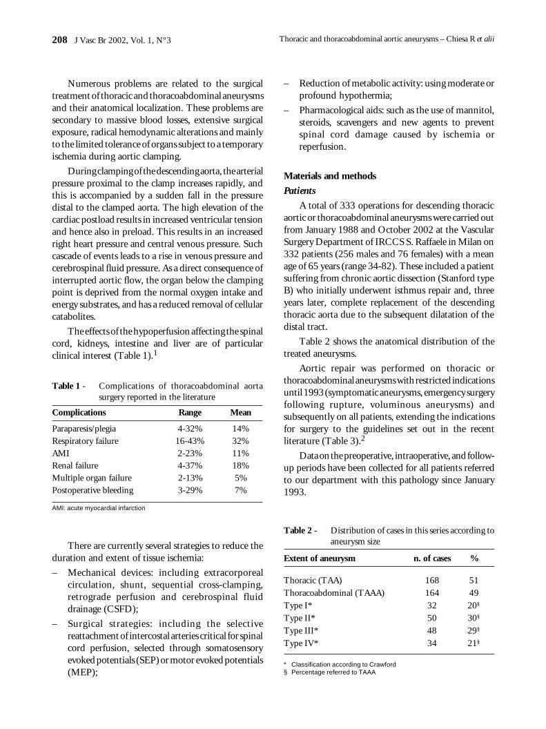
All patients undergoing elective surgery weresubmitted to multiplanar scans of the thoracic andabdominal aorta using magnetic resonance (MR) withweighted T1 and T2 SE sequences (Toshiba MRT 50,0.5 Tesla) and, until December 1994, we used alsosubtraction digital angiography. From January 1995onwards, the aorta was only studied using angio-MR+ Gd DTPA bolus (GE Med. Syst. Horizon IST)(Figure 1).
Table 3 - Surgical indication
Medical treatment Clinical status
Control of risk factorsClose instrumental follow-up Maximum diameter < 5 cmMagnetic resonance, annual CT
Relative indication Maximum diameter between 5-8 cmGiven the scant information available concerningthe expansion rate and risk of rupture of aneurysms,the data reported in the literature delegate indica-tions for surgery to the individual Centers dependingon their results and surgical series. A clinical andinstrumental evaluation of the risk-benefit ratio foreach individual case must be made in relation tosurgical risk factors and possibilities of rupture.
Absolute indication Symptomatic aneurysmDiameter > 8 cmFissured/rupturedExpansion rate > 1 cm/year
Surgical risk factors Risk of rupturePatient’s age and clinical status SymptomatologyDiabetes mellitus Presence of COBPExtent of pathology Dissecting aneurysmHeart, respiratory and renal function Max diameter and expansion rate
According to the Società Italiana di Chirurgia Vascolare ed Endovascolare (SICVE) Guidelines
Symptoms n. of cases
Chest pain 51Microembolization 9Dysphonia 5Acute renal failure 4Dyspnea 3Hemoptysis 1Total symptoms 73
Table 4 - Symptoms on presentation (excluding emer-gency cases)
Patients selected for surgery were also submitted tocardiological screening using ECG and to examinationby a specialist, which was extended, if necessary, usingthallium-dipyridamole scintigraphy or echo-stress testin cases of suspected coronary pathology. Those patientswith positive results underwent also coronarography,
Thoracic and thoracoabdominal aortic aneurysms – Chiesa R et alii
210 J Vasc Br 2002, Vol. 1, Nº3
and possible myocardial revascularization prior to aorticrepair.
The systematic preoperative use of TSA colorDoppler ultrasonography enabled those patients withsurgically relevant carotid stenosis to be identified.They were then submitted to carotid endarterectomyprior to major vascular repair, thus reducing the risk ofneurological ischemic events correlated with the radicalhemodynamic alterations during aortic surgery.
Routine preoperative screening was completed byassessment of peripheral arterial system using colorDoppler ultrasonography of the lower limbs. Lungfunction was studied using chest X-ray, blood gasanalysis and respiratory function tests (FVC, FEV1 andFEV 25-75%).
Technique
The preparation of patients for surgery starts in theward approximately one hour before admission to theoperating room, with the administration of antibioticprophylaxis (IV cefazolin 2 g.) and premedication (IMscopolamine 0.25-0.5 mg.; oral diazepam 0.1 mg/Kg,
IM morphine 0.1 mg/kg). Ten units of concentratederythrocytes, 10 units of frozen fresh plasma and 10units of platelet concentrate were available.
Anesthesia was induced using peripheral venousaccess with a 14G needle cannula in the right arm. In theoperating room, an 18G peridural catheter was thenpositioned at level T6-T7 for perioperative analgesia.
General anesthesia was then induced and the doublelumen endotracheal tube (Robertshaw) formonopulmonary ventilation was inserted and monitoredusing fibroscopy. If compression of the trachealbifurcation by a voluminous aortic dilatation is present,there is a consequent risk of left main bronchial oraneurysm rupture. In these situations it would bepreferable to use a single-lumen tube with a bronchialexcluder (Univent).
Mean arterial pressure was monitored bycannulation of the radial and right femoral arteries tocontrol proximal and distal pressure during aorticclamping. The venous accesses were then completedusing 4-lumen central catheter in the right subclavianvein and a high capacity 3-lumen catheter in the rightinternal jugular vein.
The use of a vesical catheter with a thermometricsensor allows body temperature to be monitored in thephase of moderate hypothermia during aortic clamping.
After inserting the subarachnoid catheter for CSFD(14G Tuohy needle) in the intervertebral space betweenL2 and L3 or L3 and L4, the patient was positioned inthe right lateral decubitus (shoulders 60°, pelvis 30°)propped up by a beanbag.
The patient was then sterilized to create an operatingfield extending from the left axillary cavity to mid thighand from the spine to the right anterior axillary line.
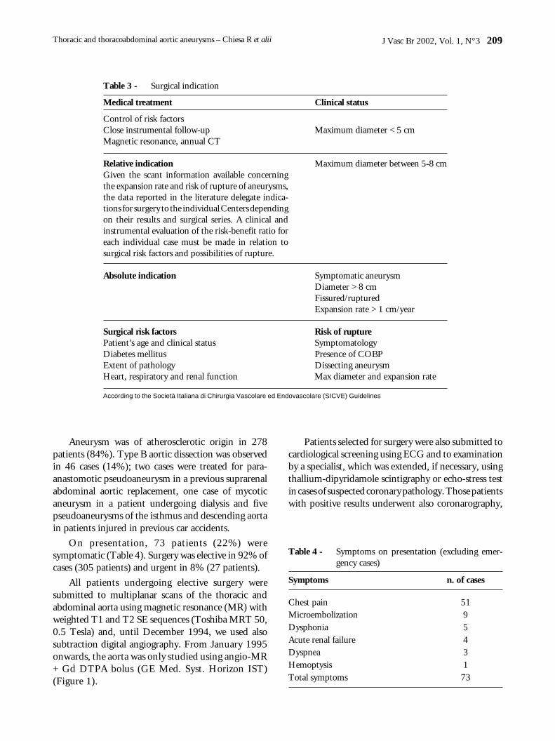
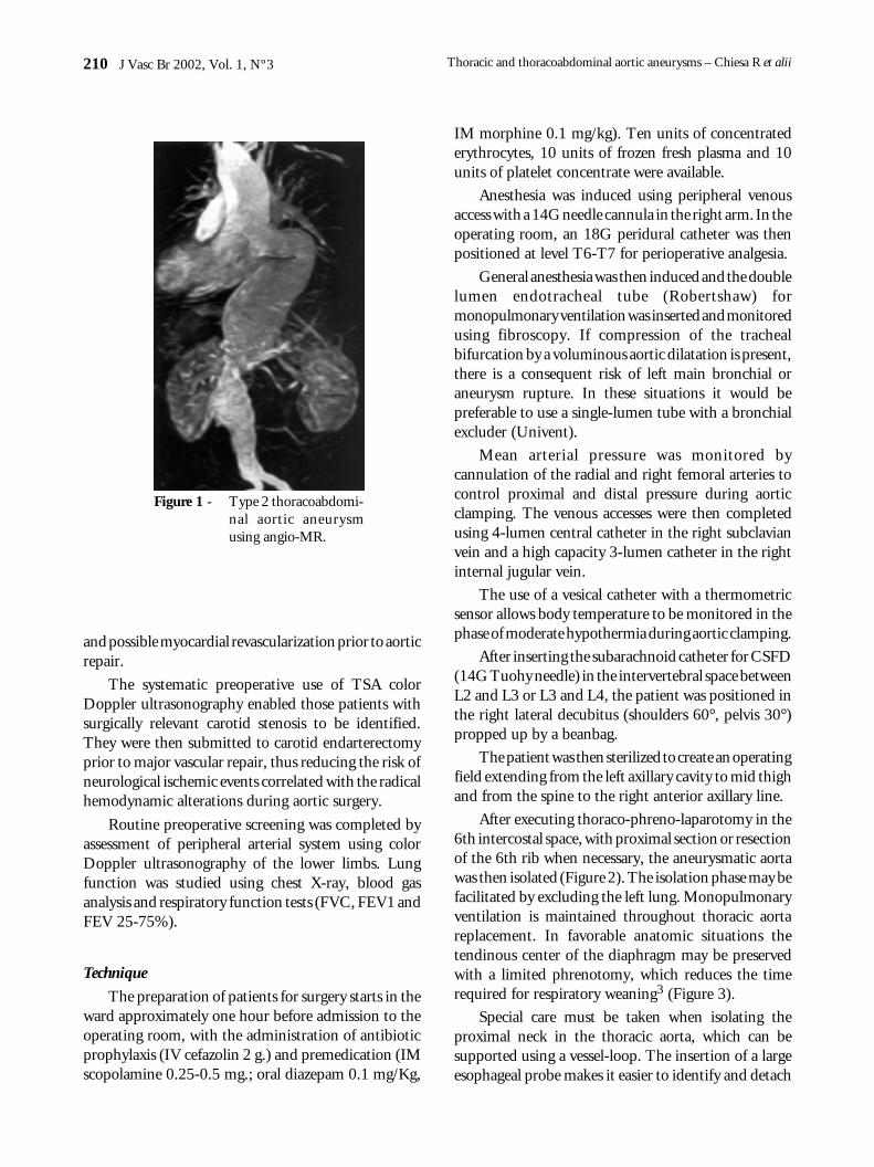
After executing thoraco-phreno-laparotomy in the6th intercostal space, with proximal section or resectionof the 6th rib when necessary, the aneurysmatic aortawas then isolated (Figure 2). The isolation phase may befacilitated by excluding the left lung. Monopulmonaryventilation is maintained throughout thoracic aortareplacement. In favorable anatomic situations thetendinous center of the diaphragm may be preservedwith a limited phrenotomy, which reduces the timerequired for respiratory weaning3 (Figure 3).
Special care must be taken when isolating theproximal neck in the thoracic aorta, which can besupported using a vessel-loop. The insertion of a largeesophageal probe makes it easier to identify and detach
Figure 1 - Type 2 thoracoabdomi-nal aortic aneurysmusing angio-MR.
Thoracic and thoracoabdominal aortic aneurysms – Chiesa R et alii
J Vasc Br 2002, Vol. 1, Nº3 211
the esophagus from the proximal aortic neck. The vagusand the source of the recurrent nerve must also beidentified at this stage since they can be damaged duringisolation and clamping maneuvers. Identification andclipping of some “high” intercostal arteries cansometimes facilitate the preparation for the proximalanastomosis, thus reducing aortic bleeding.
The abdominal aorta as far as the bifurcation andthe splanchnic vessels at source are identified andisolated using transperitoneal access, with medial visceralrotation after left parietocolic groove resection. Thisaccess allows preoperative exploration of the viscera andenables the correct revascularization following aorticreconstruction to be evaluated.
Systemic low-dose heparinization (70 UI/Kg) wasused in all patients to prevent embolization and preservethe microcirculation during clamping.
When executing left circulatory assistance, the leftatrium was cannulated (32 Fr angled cannula) aftermaking a pericardial incision posterior to the course ofthe phrenic nerve. In the event of difficult cannulationowing to previous heart surgery or the presence of atrialthrombosis, the pulmonary veins or descending aortacan be cannulated. More recently the descending aortais the site of choice to proximal cannulation.
Our preferred method for distal perfusion, given itsrelative simplicity, is the direct cannulation of thesubdiaphragmatic aorta (20-24 Fr flexible cannula). Acareful evaluation of preoperative CT or angio-MRmakes it easier to recognize the ideal site for aorticcannulation, avoiding areas with intraluminal thrombus,which might cause peripheral embolization. Thismethod avoids an inguinal incision and the need toreconstruct the femoral artery (Figure 4).
The circuit is then completed external to theoperating field with the interposition of a centrifugalpump and reservoir. In addition to the basal circulation,a heat exchanger can be also associated, together with asupplementary arterial line onto which four 9 Frocclusion/perfusion catheters (Pruitt-Inhara) are fixedto allow the selective perfusion of visceral arteries(octopus). The body temperature is maintained around32 °C (moderate hypothermia).
Flow detectors on the circuit allow constantmonitoring of bypass flow, enabling better control ofdistal aortic and visceral perfusion. It is initially keptlow (500 ml/min) to avoid retrograde embolization andthen increased after aortic clamping. Distal aorticpressure should be about 70 mmHg, a value that isachieved using a mean flow between 1,500 and 2,500ml/min. These flow rates should correspond to 2/3 ofthe basal cardiac output measured at the start of surgery.
Sequential cross-clamping is used from the aneurysmproximal tract, and when necessary, has been donebetween the carotid and left subclavian arteries ensuring
Figure 3 - Limited phrenotomy can reduce respiratoryweaning times.
Figure 2 - Preparation of aneurysminvolving the entire thoraco-abdominal aorta (Type 2).
Thoracic and thoracoabdominal aortic aneurysms – Chiesa R et alii
212 J Vasc Br 2002, Vol. 1, Nº3
control of aneurysms extending proximally as far as theisthmus. The aorta is resected transversely and theproximal anastomosis is prepared using graft (Dacronwoven double velour soaked in collagen) and 3-0polypropylene thread for all aortic sutures, withreinforcement pledgets (teflon) at points of maximumtension. The complete section of proximal aortic neckallows the anastomosis to be made avoiding esophageallesions when passing the suture thread (Figure 5).
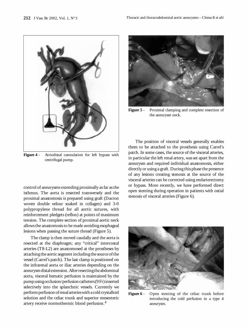
The clamp is then moved caudally and the aorta isresected at the diaphragm; any “critical” intercostalarteries (T8-L2) are anastomosed at the prostheses byattaching the aortic segment including the source of thevessel (Carrel’s patch). The last clamp is positioned onthe infrarenal aorta or iliac arteries depending on theaneurysm distal extension. After resecting the abdominalaorta, visceral hematic perfusion is maintained by thepump using occlusion/perfusion catheters (9 Fr) insertedselectively into the splanchnic vessels. Currently weperform perfusion of renal arteries with a cold crystalloidsolution and the celiac trunk and superior mesentericartery receive normothermic blood perfusion.4
The position of visceral vessels generally enablesthem to be attached to the prosthesis using Carrel’spatch. In some cases, the source of the visceral arteries,in particular the left renal artery, was set apart from theaneurysm and required individual anastomosis, eitherdirectly or using a graft. During this phase the presenceof any lesions creating stenosis at the source of thevisceral arteries can be corrected using endarterectomyor bypass. More recently, we have performed directopen stenting during operation in patients with ostialstenosis of visceral arteries (Figure 6).
Figure 4 - Atriodistal cannulation for left bypass withcentrifugal pump.
Figure 6 - Open stenting of the celiac trunk beforeintroducing the cold perfusion in a type 4aneurysm.
Figure 5 - Proximal clamping and complete resection ofthe aneurysm neck.
Thoracic and thoracoabdominal aortic aneurysms – Chiesa R et alii
J Vasc Br 2002, Vol. 1, Nº3 213
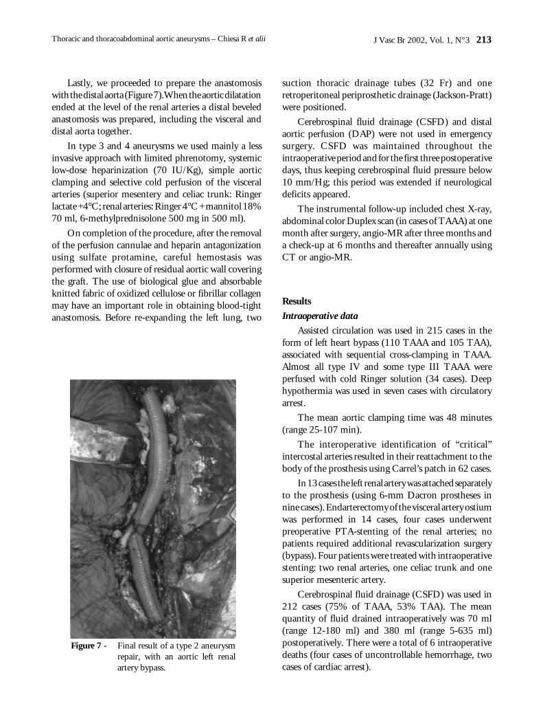
Lastly, we proceeded to prepare the anastomosiswith the distal aorta (Figure 7).When the aortic dilatationended at the level of the renal arteries a distal beveledanastomosis was prepared, including the visceral anddistal aorta together.
In type 3 and 4 aneurysms we used mainly a lessinvasive approach with limited phrenotomy, systemiclow-dose heparinization (70 IU/Kg), simple aorticclamping and selective cold perfusion of the visceralarteries (superior mesentery and celiac trunk: Ringerlactate +4°C; renal arteries: Ringer 4°C + mannitol 18%70 ml, 6-methylprednisolone 500 mg in 500 ml).
On completion of the procedure, after the removalof the perfusion cannulae and heparin antagonizationusing sulfate protamine, careful hemostasis wasperformed with closure of residual aortic wall coveringthe graft. The use of biological glue and absorbableknitted fabric of oxidized cellulose or fibrillar collagenmay have an important role in obtaining blood-tightanastomosis. Before re-expanding the left lung, two
suction thoracic drainage tubes (32 Fr) and oneretroperitoneal periprosthetic drainage (Jackson-Pratt)were positioned.
Cerebrospinal fluid drainage (CSFD) and distalaortic perfusion (DAP) were not used in emergencysurgery. CSFD was maintained throughout theintraoperative period and for the first three postoperativedays, thus keeping cerebrospinal fluid pressure below10 mm/Hg; this period was extended if neurologicaldeficits appeared.
The instrumental follow-up included chest X-ray,abdominal color Duplex scan (in cases of TAAA) at onemonth after surgery, angio-MR after three months anda check-up at 6 months and thereafter annually usingCT or angio-MR.
Results
Intraoperative data
Assisted circulation was used in 215 cases in theform of left heart bypass (110 TAAA and 105 TAA),associated with sequential cross-clamping in TAAA.Almost all type IV and some type III TAAA wereperfused with cold Ringer solution (34 cases). Deephypothermia was used in seven cases with circulatoryarrest.
The mean aortic clamping time was 48 minutes(range 25-107 min).
The interoperative identification of “critical”intercostal arteries resulted in their reattachment to thebody of the prosthesis using Carrel’s patch in 62 cases.
In 13 cases the left renal artery was attached separatelyto the prosthesis (using 6-mm Dacron prostheses innine cases). Endarterectomy of the visceral artery ostiumwas performed in 14 cases, four cases underwentpreoperative PTA-stenting of the renal arteries; nopatients required additional revascularization surgery(bypass). Four patients were treated with intraoperativestenting: two renal arteries, one celiac trunk and onesuperior mesenteric artery.
Cerebrospinal fluid drainage (CSFD) was used in212 cases (75% of TAAA, 53% TAA). The meanquantity of fluid drained intraoperatively was 70 ml(range 12-180 ml) and 380 ml (range 5-635 ml)postoperatively. There were a total of 6 intraoperativedeaths (four cases of uncontrollable hemorrhage, twocases of cardiac arrest).
Figure 7 - Final result of a type 2 aneurysmrepair, with an aortic left renalartery bypass.
Thoracic and thoracoabdominal aortic aneurysms – Chiesa R et alii
214 J Vasc Br 2002, Vol. 1, Nº3
Postoperative data
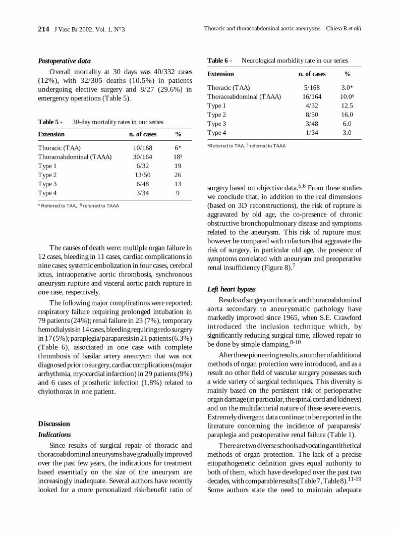
Overall mortality at 30 days was 40/332 cases(12%), with 32/305 deaths (10.5%) in patientsundergoing elective surgery and 8/27 (29.6%) inemergency operations (Table 5).
Table 5 - 30-day mortality rates in our series
* Referred to TAA, § referred to TAAA
Extension n. of cases %
Thoracic (TAA) 10/168 6*Thoracoabdominal (TAAA) 30/164 18§
Type 1 6/32 19Type 2 13/50 26Type 3 6/48 13Type 4 3/34 9
The causes of death were: multiple organ failure in12 cases, bleeding in 11 cases, cardiac complications innine cases; systemic embolization in four cases, cerebralictus, intraoperative aortic thrombosis, synchronousaneurysm rupture and visceral aortic patch rupture inone case, respectively.
The following major complications were reported:respiratory failure requiring prolonged intubation in79 patients (24%); renal failure in 23 (7%), temporaryhemodialysis in 14 cases, bleeding requiring redo surgeryin 17 (5%); paraplegia/paraparesis in 21 patients (6.3%)(Table 6), associated in one case with completethrombosis of basilar artery aneurysm that was notdiagnosed prior to surgery, cardiac complications (majorarrhythmia, myocardial infarction) in 29 patients (9%)and 6 cases of prosthetic infection (1.8%) related tochylothorax in one patient.
Discussion
Indications
Since results of surgical repair of thoracic andthoracoabdominal aneurysms have gradually improvedover the past few years, the indications for treatmentbased essentially on the size of the aneurysm areincreasingly inadequate. Several authors have recentlylooked for a more personalized risk/benefit ratio of
Table 6 - Neurological morbidity rate in our series
*Referred to TAA; § referred to TAAA
Extension n. of cases %
Thoracic (TAA) 5/168 3.0*Thoracoabdominal (TAAA) 16/164 10.0§
Type 1 4/32 12.5Type 2 8/50 16.0Type 3 3/48 6.0Type 4 1/34 3.0
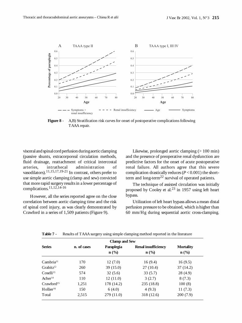
surgery based on objective data.5,6 From these studieswe conclude that, in addition to the real dimensions(based on 3D reconstructions), the risk of rupture isaggravated by old age, the co-presence of chronicobstructive bronchopulmonary disease and symptomsrelated to the aneurysm. This risk of rupture musthowever be compared with cofactors that aggravate therisk of surgery, in particular old age, the presence ofsymptoms correlated with aneurysm and preoperativerenal insufficiency (Figure 8).7
Left heart bypass
Results of surgery on thoracic and thoracoabdominalaorta secondary to aneurysmatic pathology havemarkedly improved since 1965, when S.E. Crawfordintroduced the inclusion technique which, bysignificantly reducing surgical time, allowed repair tobe done by simple clamping.8-10
After these pioneering results, a number of additionalmethods of organ protection were introduced, and as aresult no other field of vascular surgery possesses sucha wide variety of surgical techniques. This diversity ismainly based on the persistent risk of perioperativeorgan damage (in particular, the spinal cord and kidneys)and on the multifactorial nature of these severe events.Extremely divergent data continue to be reported in theliterature concerning the incidence of paraparesis/paraplegia and postoperative renal failure (Table 1).
There are two diverse schools advocating antitheticalmethods of organ protection. The lack of a preciseetiopathogenetic definition gives equal authority toboth of them, which have developed over the past twodecades, with comparable results (Table 7, Table 8).11-19
Some authors state the need to maintain adequate
Thoracic and thoracoabdominal aortic aneurysms – Chiesa R et alii
J Vasc Br 2002, Vol. 1, Nº3 215
visceral and spinal cord perfusion during aortic clamping(passive shunts, extracorporeal circulation methods,fluid drainage, reattachment of critical intercostalarteries, intrathecal administration ofvasodilators).11,15,17,19-21 In contrast, others prefer touse simple aortic clamping (clamp and sew) convictedthat more rapid surgery results in a lower percentage ofcomplications.11,12,14-16
However, all the series reported agree on the closecorrelation between aortic clamping time and the riskof spinal cord injury, as was clearly demonstrated byCrawford in a series of 1,509 patients (Figure 9).
Likewise, prolonged aortic clamping (> 100 min)and the presence of preoperative renal dysfunction arepredictive factors for the onset of acute postoperativerenal failure. All authors agree that this severecomplication drastically reduces (P < 0.001) the short-term and long-term22 survival of operated patients.
The technique of assisted circulation was initiallyproposed by Cooley et al.23 in 1957 using left heartbypass.
Utilization of left heart bypass allows a mean distalperfusion pressure to be obtained, which is higher than60 mm/Hg during sequential aortic cross-clamping.
Figure 8 - A,B) Stratification risk curves for onset of postoperative complications followingTAAA repair.
Table 7 - Results of TAAA surgery using simple clamping method reported in the literature
Clamp and SewSeries n. of cases Paraplegia Renal insufficiency Mortality
n (%) n (%) n (%)
Cambria11 170 12 (7.0) 16 (9.4) 16 (9.5)Grabitz12 260 39 (15.0) 27 (10.4) 37 (14.2)Coselli13 574 32 (5.6) 33 (5.7) 28 (4.9)Acher14 110 12 (11.0) 3 (2.7) 8 (7.3)Crawford15 1,251 178 (14.2) 235 (18.8) 100 (8)Hollier16 150 6 (4.0) 4 (9.3) 11 (7.3)Total 2,515 279 (11.0) 318 (12.6) 200 (7.9)
Thoracic and thoracoabdominal aortic aneurysms – Chiesa R et alii

216 J Vasc Br 2002, Vol. 1, Nº3
The employment of selective additional perfusion ofvisceral arteries, using occlusion/perfusion catheterslinked to the pump (Octopus), enables the entirereplacement of thoracoabdominal aorta to be madewith continuous visceral perfusion. By using theseadditional methods, the duration of “hot” visceralischemia (in particular, renal ischemia) is reduced tofew minutes (range 2-7 min in our series) required toexplore and cannulate the arterial openings withperfusion catheters. By augmenting the duration ofclamping tolerance, visceral perfusion contributes to ahigher level of surgical accuracy and precision.
A reduction in left ventricular tension is also achievedwith this method, which decreases preload and preventshypertension of the proximal aorta. This hemodynamiccontrol cannot be achieved with the same efficacy by
extraluminal shunt systems,24,25 unless vasodilators areused (sodium nitroprusside and/or anesthetic drugs).26
Moreover, blood flow is not constant along the shuntbut depends on various concomitant factors, such as thetube diameter, the presence of kinking and the “drivingforce” in the proximal aorta.27
An additional advantage of left heart bypass is thepossibility of connecting a heat exchanger to the circuit,thus allowing homeothermia/moderate hypothermiato be achieved during surgery. On completion of aorticreconstruction, prior to decannulation, this exchangeralso enables the patient’s physiological temperature tobe restored, thus diminishing the risk of coagulationdisorders and cardiac arrhythmia.
Left circulatory assistance is generally supported bya centrifugal pump, unless the patient has undergonefull heparinization. First described by Rafferty &Kletschka28 in 1968 and introduced into clinical practicein 1975, this kinetic pump offers several advantagescompared to traditional methods (Roller pump). Thekinetic energy transmitted to the blood is generated bythe high-speed rotation of a series of coaxial cones,producing a hematic vortex identical to a cyclone. TheBio Console motor interacts with a magnet fitted to thebase of the cones which regulates the speed of revolution:increased velocity means an increased centrifugal forceof blood and hence an increased output. The cones aredesigned to minimize the traumatic lyses of red bloodcells. Owing to the dynamics generating the hematicvortex, any potentially thrombogenic element is trappedat the tip of the cones (the eye of the cyclone), thuspreventing its emission into the circulation. Lastly, the
Figure 9 - Risk of paraplegia correlated with aorticclamping time.
Table 8 - Results of TAAA surgery using distal aortic perfusion method reported in the literature
Clamp and SewSeries n. of cases Paraplegia Renal insufficiency Mortality
n (%) n (%) n (%)
Safi17 186 12 (7.0) 22 (15.1) 18 (9.6)Coselli18 312 15 (4.8) 33 (10.6) 16 (5.1)Schepens19 50 5 (10.0) 5 (10.0) 4 (8.0)Svensson15 258 56 (21.7) 34 (13.2) 23 (8.9)Total 806 88 (10.9) 94 (11.6) 61 (7.5)
Thoracic and thoracoabdominal aortic aneurysms – Chiesa R et alii
J Vasc Br 2002, Vol. 1, Nº3 217
circuit is fitted with a filter thus guaranteeing thecomplete safety of the system.
Although the preliminary results were oftendivergent in small series of patients,29-33 it is now clearthat, if executed using left heart bypass and sequentialaortic cross-clamping, distal aortic perfusion (DAP)exerts a protective effect on the kidneys and spinal cordin the event of prolonged aortic clamping.34-40
Conclusions
Our personal experience confirms that the use ofDAP in the form of left heart bypass with Biomedicuspump and sequential cross-clamping, associated withcerebrospinal fluid drainage, allowed us to achievecomplication rates comparable to internationalexperience.
In spite of the technical evolution in the past twentyyears, aneurysmatic pathologies of thoracic andthoracoabdominal aorta continue to represent achallenge for vascular surgeons, owing to the complexityof their surgical repair and perioperative management.
References1. Panneton JM, Hollier LH. Nondissecting thoracoabdominal
aortic aneurysms: Part I. Ann Vasc Surg 1995;9:503-14.2. Linee guida SICVE. Aneurismi toracici e toracoaddominali.
G Ital Chir Vasc 2001;8 Suppl 3:1-14.3. Safi HJ. How I do it: thoracoabdominal aortic aneurysm graft
replacement. Cardiovasc Surg. 1999;7(6):607-13.4. Koksoy C, LeMaire SA, Curling PE, et al. Renal perfusion
during thoracoabdominal aortic operations: cold crystalloid issuperior to normothermic blood. Ann Thorac Surg2002;73(3):730-8.
5. Juvonen T, Ergin MA, Galla JD, et al. Prospective study of thenatural history of thoracic aortic aneurysms. Ann ThoracSurg. 1997;63(6):1533-45.
6. LeMaire SA, Miller III CC, Conklin LD, Schmittling ZC,Koskoy C, Coselli JS. A new predictive model for adverseoutcomes after elective thoracoabdominal aortic aneurysmrepair. Ann Thorac Surg 2001;71:1233-8.
7. LeMaire SA, Miller CC 3rd, Conklin LD, Schmittling ZC,Koksoy C, Coselli JS. A new predictive model for adverseoutcomes after elective thoracoabdominal aortic aneurysmrepair. Ann Thorac Surg. 2001;71(4):1233-8.
8. Crawford ES, Crawford JL. Disease of the aorta including anatlas of angiographic pathology and surgical technique.Baltimore: William & Wilkins; 1984.
9. Crawford ES, Coselli JS. Thoracoabdominal aneurysm surgery.Semin Thorac Cardiovasc Surg 1991;3:300-22.
10. Coselli JS. Suprarenal aortic reconstruction-perioperativemanagement: Patient selection, patient workup, operativemanagement, and postoperative management. In: RutherfordRB, editor. Semin Vasc Surg 1992;5(3):146-56.
11. Cambria RP, Davison JK, Carter C, et al. Epidural cooling forspinal cord protection during thoracoabdominal aneurysmrepair: a five year experience. J Vasc Surg 2000;31(6):1093-102.
12. Grabitz K, Sandmann W, Stuhmeier K, et al. The risk ofischemic spinal cord injury in patients undergoing graftreplacement for thoracoabdominal aortic aneurysms. J VascSurg 1996;23:230-40.
13. Coselli JS. Recent advances in surgical treatment ofthoracoabdominal aortic aneurysms. In: Chiesa R, MelissanoG, editors. Gli aneurismi dell’aorta addominale. Milano:Europa Scienze Umane Editrice; 1996. p.269-84.
14. Acher CW, Wynn MM, Hoch JR, Popic P, Archibald J,Turnipseed WD. Combined use of cerebral spinal fluiddrainage and naloxone reduces the risk of paraplegia inthoracoabdominal aneurysm repair. J Vasc Surg 1994;19:236-48.
15. Svensson LG, Crawford ES, Hess KR, Coselli JS, Safi HJ.Experience with 1509 patients undergoing thoracoabdominalaortic operations. J Vasc Surg 1993;17:357-70.
16. Hollier LM, Money S, Naslund T, et al. Risk of spinal corddysfunction in patients undergoing aortic replacements. Am JSurg 1992;164:210-4.
17. Safi HJ, Campbell MP, Miller III CC, et al. Cerebral Spinalfluid drainage and distal aortic perfusion decrease the incidenceof neurological deficit: the results of 343 descending andthoracoabdominal aortic aneurysm repairs. Eur J Vasc EndovascSurg 1997;14:118-24.
18. Coselli JS, LeMaire SA. Left heart bypass reduces paraplegiarates after thoracoabdominal aortic aneurysm repair. AnnThorac Surg 1999;67:1931-4.
19. Schepens M, Defauw J, Hamerlijnck R, Vermeulen F. Use ofleft heart bypass in the surgical repair of thoracoabdominalaortic aneurysms. Ann Vasc Surg 1995;9:327-38.
20. Svensson LG, Stewart RW, Cosgrove DM 3rd, et al. Intrathecalpapaverine for the prevention of paraplegia after operation onthe thoracic or thoracoabdominal aorta. J Thorac CardiovascSurg 1988;96:823-9.
21. Safi HJ, Miller III CC, Carr C, Iliopoulos DC, Dorsay DA,Baldwin JC. Importance of intercostal artery reattachmentduring thoracoabdominal aortic aneurysm repair. J Vasc Surg1998;27:58-68.
22. Svensson LG, Crawford ES, Hess KR, Coselli JS, Safi HJ.Thoracoabdominal aortic aneurysms associated with celiac,superior mesenteric, and renal artery occlusive disease: methodsand analysis of results in 271 patients. J Vasc Surg 1992:16;378-90.
23. Cooley DA, Belmonte BA, DeBakey ME. Temporaryextracorporeal circulation in the surgical treatment of cardiacand aortic disease. Report of 98 cases Ann Surg 1957;145:898-914.
24. Lawrence GH, Hessel EA, Sauvage LR, Krause AH. Results ofthe use of the TDMAC-heparin shunt in the surgery ofaneurysms of the descending thoracic aorta. J Thorac CardiovascSurg 1977;73:393-8.
Thoracic and thoracoabdominal aortic aneurysms – Chiesa R et alii

218 J Vasc Br 2002, Vol. 1, Nº3
25. Verdant A, Page A, Cossette R, Dontigny L, Page P, Baillot R.Surgery of the descending thoracic aorta: spinal cord protectionwith the Gott shunt. Ann Thorac Surg 1988;46:147-54.
26. Gelman S, Reves JG, Fowler K, Samuelson PN, Lell WA,Smith LR. Regional blood flow during cross-clamping of thethoracic aorta and infusion of sodium nitroprusside. J ThoracCardiovasc Surg 1983;85:287-91.
27. Cunningham JN, Laschinger JC, Spencer FC. Monitoring ofsomatosensory evoked potentials during surgical procedureson the thoracoabdominal aorta. J Thorac Cardiovasc Surg1987;94:275-85.
28. Rafferty EH, Kletschka HD; Artificial Heart: application ofnon pulsatile force-vortex principle. Minn Med 1968;51(1):11-6.
29. Cooley DA, Debakey ME, Morris GC. Controlledextracorporeal circulation in surgical treatment of aorticaneurysm. Ann Surg 1957;146:473-85.
30. Debakey ME, Cooley DA, Crawford ES. Aneurysms of thethoracic aorta. J Thorac Surg 1958;36:393-420.
31. Connolly JE, Kountz SL, Boyd RJ. Left heart by-pass:experimental and clinical observations on its regulation withparticular reference to maintenance of maximal renal bloodflow. J Thorac Cardiovasc Surg 1962;44:577-88.
32. Connolly JE, Wakabayashi A, German JC, Stemmer EA,Serres EJ. Clinical experience with pulsatile left heart bypasswithout anticoagulation for thoracic aneurysms. J ThoracCardiovasc Surg 1971;62:568-76.
33. Crawford ES, Mizrahi EM, Hess KR, Coselli JS, Safi HJ, PatelVM. The impact of distal aortic perfusion and somatosensoryevoked potential monitoring on prevention of paraplegia afteraortic aneurysm operation. J Thorac Cardiovasc Surg1988;95:357-67. (Published erratum appears in J ThoracCardiovasc Surg 1989;97:665.)
34. Svensson LG, Crawford ES, Hess KR, Coselli JS, Safi HJ.Variables predictive of outcome in 832 patients undergoingrepairs of the descending thoracic aorta. Chest 1993;104:1248-53.
35. Borst HG, Jurmann M, Buhner B. Laas J. Risk of replacementof descending aorta with a standardized left heart bypasstechnique. J Thorac Cardiovas Surg 1994:107:126-33.
36. Lawrie GM, Earle N, DeBakey ME. Evolution of surgicaltechniques for aneurysms of the descending thoracic aorta:twenty-nine years experience with 659 patients. J CardiacSurg 1994;9:648-61.
37. Verdant A, Cossette R, Page A, Baillot R, Dontigny L, Page P.Aneurysms of the descending thoracic aorta: three hundredsixty-six consecutive cases resected without paraplegia. J VascSurg 1995;21:385-91.
38. Kashyap VS, Cambria RP, Davison JK, L’Italien GJ. Renalfailure after thoracoabdominal aortic surgery. J Vasc Surg.1997;26(6):949-55; discussion 955-7.
39. Jacobs MJHM, Meylaerts SA, de Haan P, de Mol BA,Kalkman CJ. Strategies to prevent neurologic deficit based onmotor-evoked potentials in type I and II thoracoabdominalaortic aneurysm repair. J Vasc Surg 1999;29(1):48-57;discussion 57-9.
40. Cooley DA, Golino A, Frazier OH. Single-clamp techniquefor aneurysms of the descending thoracic aorta: report of 132consecutive cases. Eur J Cardiothorac Surg 2000;18(2):162-7.
Correspondence:Dr. Roberto ChiesaDepartment of Vascular SurgeryIRCCS H. San RaffaeleVia Olgettina, 6020132 - Milano - ItaliaE-mail: [email protected]
Thoracic and thoracoabdominal aortic aneurysms – Chiesa R et alii


![INDEX [d3d6mf6ofxeyve.cloudfront.net]d3d6mf6ofxeyve.cloudfront.net/wieckautodeadline60/files... · 2017-09-11 · INDEX 02 introduction 09 206 cc vs 207 cc coMPAriSon 0 207 cc ModEL](https://img.pdfslide.us/doc/110x75/5f5d928d51821d074c240fa7/index-2017-09-11-index-02-introduction-09-206-cc-vs-207-cc-comparison-0-207.jpg)