Embed Size (px)
Citation preview
Surgical options includeplacement of an activeor passive drain orfenestration on theconcave skin of thepinna.
Peer ReviewedProcedures Pro Surgery
Surgical Treatment for Aural Hematoma
Rachel Seibert, DVM, & Karen M. Tobias, DVM, DACVSUniversity of Tennessee
ranches of the caudal auricular artery passingfrom the convex to the concave surface of thepinna through tiny channels within the carti-
lage provide blood to the auricular cartilage. Nor-mally, the concave surface of pinna cartilage isfirmly attached to the skin, but trauma may createdead space between the cartilage and skin that fillswith blood. Hemorrhage from the penetrating ves-sels continues, ultimately forming an out-pouching(ie, aural hematoma). If untreated, granulation tis-sue replaces aural hematomas; subsequent contrac-tion and fibrosis of this tissue can result in pinnaldeformity and, in cats, obstruction of the externalacoustic opening.
Aural hematomas can be caused by direct damage(eg, bite wounds, vehicular trauma) but are morecommon from head shaking and ear scratchingassociated with otitis externa or atopy. Affectedanimals should undergo thorough dermatologicand otoscopic examinations, and any underlyingdisease should be treated to facilitate resolutionand prevent recurrence.
TreatmentAural hematomas can be treated surgically or nonsurgically. Therapeutic goals include removal ofcontents, maintenance of apposition between skin and cartilage, and appropriate treatment for thesource of head shaking or scratching. Drainage should be performed expeditiously to prevent con-tracture, fibrosis, and subsequent deformity.
Surgical options include placement of an active or passive drain or fenestration on the concaveskin of the pinna, using either a single long incision or multiple small incisions, to empty thepocket and prevent recurrence of fluid accumulation. Apposition of skin to cartilage is maintainedby placement of sutures between skin and cartilage, application of a compressive bandage, or both.Drains are removed within 1 week, but bandaging may be required for 2 additional weeks.
Nonsurgical treatment involves needle drainage and flushing of the hematoma cavity. Recurrenceis common with needle drainage alone, even if bandages are applied, but treatment with systemic
MORE
March 2013 • clinician’s brief 29
B

or intralesional steroids reduces recurrence. For needle drainage, the concave surface of the pinnashould be clipped and aseptically prepared. A 14- or 16-gauge needle or 19- or 21-gauge butterflycatheter is inserted into the dependent portion of the hematoma and directed into the pocket(Figure 1). For floppy-eared dogs, the needle is inserted into the end of the hematoma near thepinna apex so fluid draining from the needle hole does not enter the ear canal.
Fluid is drained, and the pocket is flushed with sterile saline to removeclots. Steroids can be injected into the hematoma cavity before band-age placement. Because of the antiinflammatory effects of glucocorti-coids, local steroid injection can predispose abscess formation; use ofsterile technique is extremely important with intralesional infusion.Successful resolution has been reported in 90% of animals with drain-age followed by injection of intralesional triamcinolone acetonide (10mg/mL; 0.1–1 mL q7d for 1–3 treatments) or dexamethasone (0.2–0.4mg diluted to 0.5–1.8 mL, infused q24h for 1–5 days).1-3 Bandaging isstill recommended to prevent damage during continued head shakingor scratching. Resolution of hematomas with drainage followed by IVadministration of immunosuppresive dose of glucocorticoids has also
been described1; however, this treatment is not recommended because of potential adverse events.
Closed-Suction DrainsClosed-suction drainage using a butterfly catheter and vacutainer tube may inexpensively preventor resolve SC fluid accumulation. In active patients, the tube and catheter must be carefullysecured to prevent accidental needle dislodgement. The owner can be taught to change the vacu-tainer tube when it has filled or lost negative suction and to monitor fluid production. Drains areusually removed within 5 to 7 days of placement; the ear can be bandaged for another week toprevent disruption from head shaking or scratching. Closed-suction drainage works for acute orchronic hematomas that have fluid accumulation, as long as the underlying cause is treated. Mostpatients show resolution in 7 to 10 days with minimal pinna distortion. Recurrence rates are 22%,with recurrence reported in animals with uncontrolled allergic dermatitis.3
Procedures Pro
30 cliniciansbrief.com • March 2013
What You Will Need
� #40 clipper blade
� Chlorhexidine scrub and solution
� Butterfly catheter (19- or 21-gauge)
� Mayo scissors
� Sterile gloves
� needle drivers
� #11 or #15 surgical blade
� 2-0 or 3-0 monofilament suture
� Tongue depressors
� White adhesive tape
� Bandage material
� vacutainer blood tubes (bd.com)
A butterfly catheter used toremove fluid from the hematomacavity.
1
March 2013 • clinician’s brief 31
Step-by-Step � Continuous Suction Drainage
Step 1
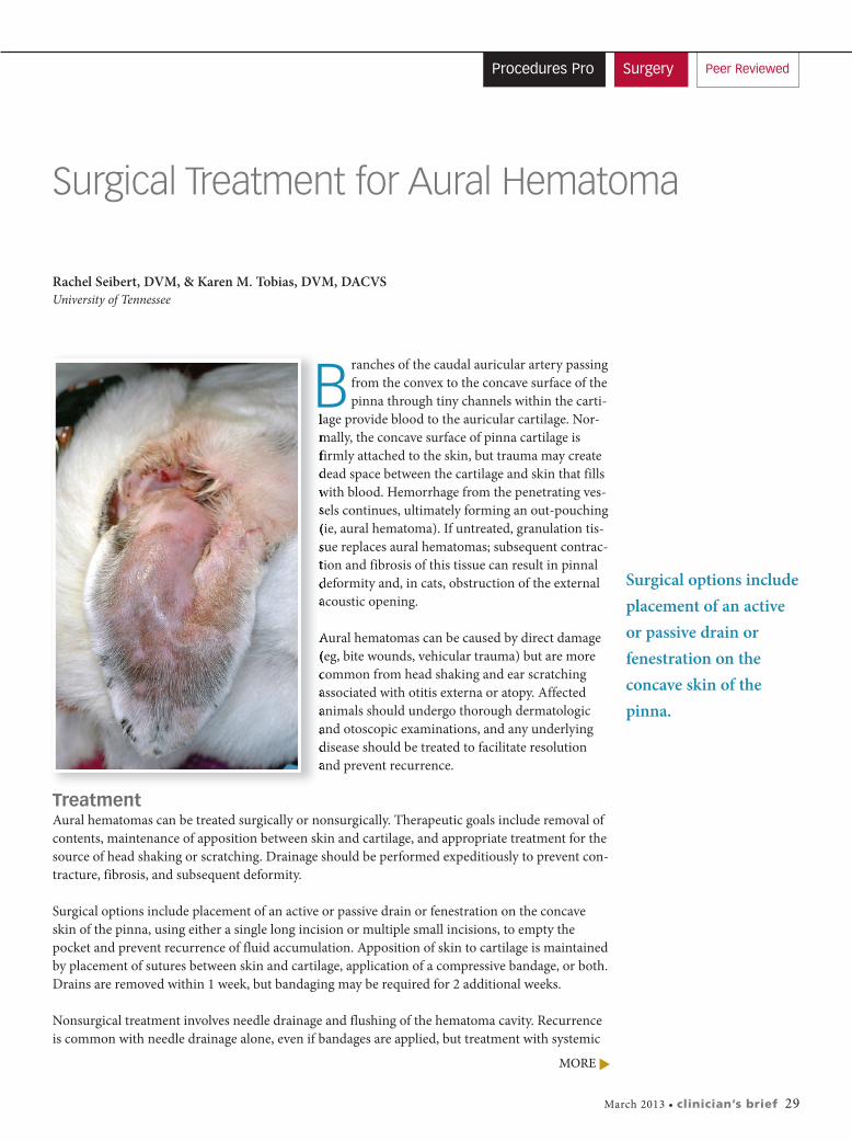
Cut the syringe adaptor from thebutterfly catheter (A). Fold over thetubing near the cut end, and snipoff the corner of the fold with Mayoscissors to create a fenestration (B).Add several fenestrations to the ter-minal 1.5–2 cm of the tubing, mak-ing sure that fenestrations are lessthan half the diameter of the tubingto prevent accidental breakage (C).
Step 2
Clip and vacuum the pinna, and place cotton or gauze in the ear canal to preventfluid accumulation. Aseptically prepare the surgical site with chlorhexidine scruband solution. If desired, drain some fluid from the hematoma with a needle andsyringe. With the tip of a #11 blade, make a small stab incision into the hematomacavity; alternatively, use a large-gauge–needle or small-skin–punch that is thesame diameter as or slightly larger than the drain tubing. The incision should onlybe large enough for tube placement. If desired, flush the hematoma through theincision to remove any clots or fibrin.
Author InsightLeave some fluid in thehematoma to permit stabincision into the fluidpocket.
A
B C
Step 3
Using a needle holder or hemostat, inserttubing into the hematoma cavity. Ensure allfenestrations are located within the cavity.Place a purse string suture in the skin aroundthe tubing exit site to maintain negative pressure.
MORE
Procedures Pro
32 cliniciansbrief.com • March 2013
Step 4
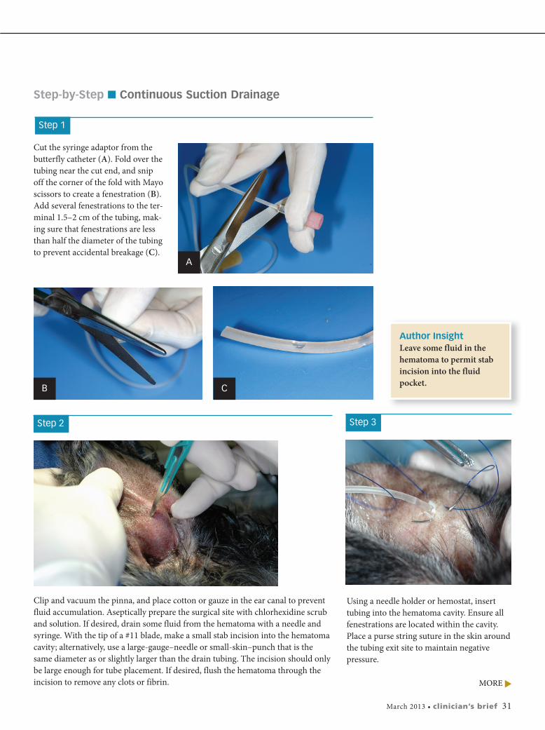
Secure the tube to the ear with a finger trap suture pattern. Use surgeon’s throws where the suture crosses the tubing. Tiethese carefully so they grip the tubing without kinking or compressing it. After tying off the finger trap pattern, take a final bite through the skin to finish securing the tube.
Step 5
Insert the needle end of the butterfly tubing into the vacutainertube to create a closed seal and active drainage. Once the tubehas partially filled, the vacuum on the drain will be lost. Kinkoff the drain, remove the needle from the vacutainer tube, andplace it in a new tube to reestablish active suction. The vacu-tainer tube may need to be changed 2 or 3 times immediatelyafter drain placement. After, it can be changed q24h or when the tubes are half full.
Step 6
Position the pinna apex dorsally to expose the external ear canaland concave surface of the pinna. Place a long strip of whiteadhesive tape along the haired surface of each pinna margin(adhesive side facing outward); carefully wrap each strip aroundthe neck or chin and back to the ear margin and stick it to theexposed adhesive. Use tongue depressors to prevent the tapefrom sticking together during placement. Tape should be looseenough to prevent neck constriction.
Step 7
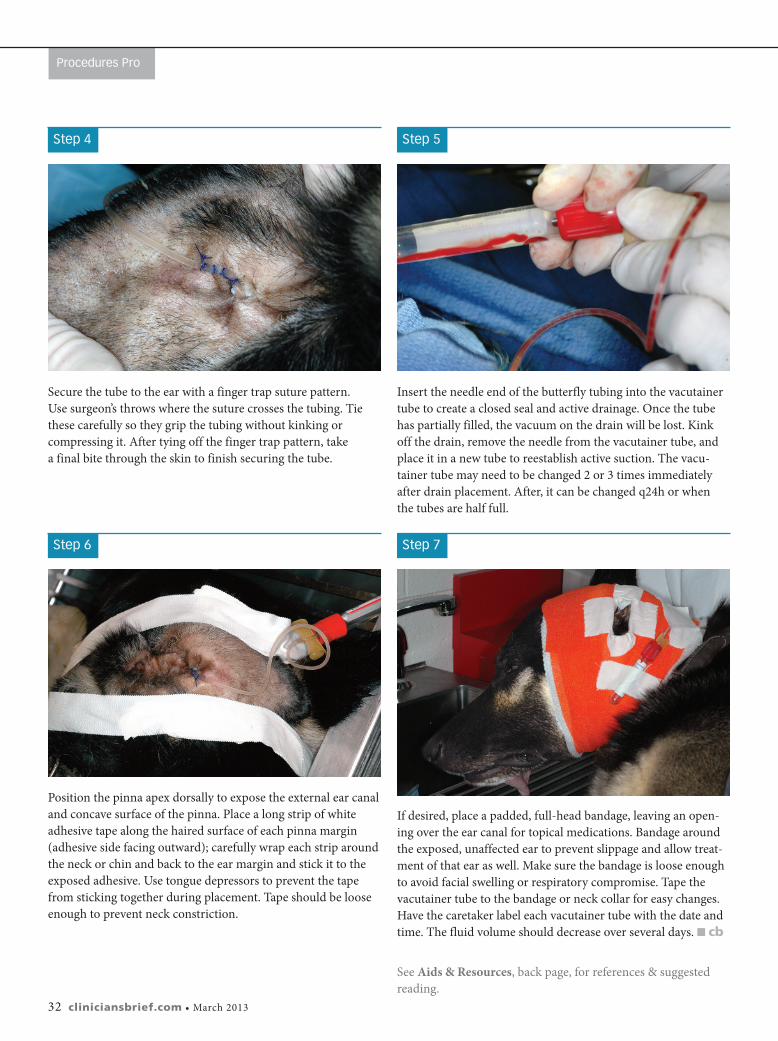
If desired, place a padded, full-head bandage, leaving an open-ing over the ear canal for topical medications. Bandage aroundthe exposed, unaffected ear to prevent slippage and allow treat-ment of that ear as well. Make sure the bandage is loose enoughto avoid facial swelling or respiratory compromise. Tape thevacutainer tube to the bandage or neck collar for easy changes.Have the caretaker label each vacutainer tube with the date andtime. The fluid volume should decrease over several days. � cb
See Aids & Resources, back page, for references & suggestedreading.