Embed Size (px)
Citation preview
Sulfurtransferases 1 and 2 Play Essential Roles in Embryo andSeed Development in Arabidopsis thaliana*□S
Received for publication, September 8, 2010, and in revised form, December 16, 2010 Published, JBC Papers in Press, December 28, 2010, DOI 10.1074/jbc.M110.182865
Guohong Mao‡1, Ruigang Wang§, Yuefeng Guan‡, Yidong Liu‡, and Shuqun Zhang‡2
From the ‡Division of Biochemistry, Interdisciplinary Plant Group, and Christopher S. Bond Life Sciences Center, University ofMissouri, Columbia, Missouri 65211 and the §College of Life Sciences, Inner Mongolia Agricultural University,Hohhot, Inner Mongolia 010018, China
Sulfurtransferases (STRs) catalyze the transfer of a sulfuratom from a donor to a suitable acceptor molecule. The Arabi-dopsis thaliana genome encodes 20 putative STR proteins.The biological functions of most are unclear. We found thatSTR1 and STR2 play important roles in embryo/seed develop-ment. Mutation of STR1 alone resulted in a shrunken seedphenotype, although growth and development of vegetativeand reproductive organs were not affected. The shrunken seedphenotype was associated with the delayed/arrested embryodevelopment, in most cases, at the heart stage. The embryodefect of str1mutant is not fully penetrant. Approximately12.5% of embryos developed further and formed normal look-ing seeds. In severely shrunken seeds, no embryo could beidentified after seed collection. Partially shrunken seeds thatcontained viable embryos could still germinate. However, cot-yledons of the seedlings from such seeds were abnormal. AnSTR1-GUS fusion reporter revealed that the STR1 gene wasuniversally expressed, with high levels of expression in specifictissues/organs including embryos. The incomplete penetranceof str1 embryo/seed phenotype is a result of functional STR2.Single str2mutant had no phenotype. However, no str1�/�/str2�/� double mutant embryos were able to develop past theheart stage. Furthermore, STR2 is haplo-insufficient in str1mutant background, and str1�/�/str2�/� embryos were 100%lethal. These data provide new insights into the biologicalfunctions of the ubiquitous sulfurtransferase in Arabidopsisembryogenesis and seed development.
Sulfurtransferases (STRs,3 or STs), also known as rhodane-ses, are ubiquitous enzymes that catalyze the transfer of a sul-fur atom from suitable sulfur donors to nucleophilic sulfuracceptors. In animals, sulfurtransferases function in cyanidedetoxification (1–3), hydrogen sulfide detoxification (4), sul-
fur metabolism (5, 6), and synthesis or repair of iron-sulfurproteins (7, 8). However, information about their biologicalfunctions in plants is limited. In Arabidopsis, there are 20 pu-tative sulfurtransferase proteins containing either one or tworhodanese domains. Based on amino acid sequence homol-ogy, these proteins are classified into six groups (GroupsI–VI) (9). STR gene expression has been examined duringplant defense response and senescence and under differentgrowth conditions including various sulfate concentrations,phosphate deficiency, and different diurnal light/dark cycles(9–12). AtSTR15 gene expression is enhanced by differenthormone treatments (13, 14), and AtSTR13/SIR1 is a key reg-ulator of many auxin-inducible genes (15). These findingssuggest that sulfurtransferases may play divergent roles inplant growth, development, stress response, and metabolism.STR1 and STR2 share high sequence identity. They are the
only two members in the Group I sulfurtransferase, which ischaracterized by having two rhodanese domains. Recombi-nant STR1 and STR2 have similar Km values, and both prefer3-mercaptopyruvate to thiosulfate as a substrate; as a result,they are also called mercaptopyruvate sulfurtransferase 1(MST1) and MST2 (16–18). However, they have differentlocalizations in cells. STR1 is localized in mitochondria,whereas STR2 is localized in the cytosol (19, 20). Their poten-tial functions in plant senescence and cyanide detoxificationhave been investigated (11). It was reported that ethylene aswell as its precursor, 1-aminocyclopropane-1-carboxylic acid,could induce the expression of STR1. STR1 expression is alsoelevated in plants grown under low sulfate conditions and lowphosphate conditions and in the presence of thiosulfate. How-ever, total sulfurtransferase activity is significantly changedonly under low phosphate conditions (9). In the Arabidopsislife cycle, STR1 and STR2 gene expression and total sul-furtransferase activity increase continuously as the plant ages,suggesting that STR1 and STR2 may be involved in senes-cence (9, 17, 18).In this report, we demonstrate that Arabidopsis STR1 and
STR2 play an important role in embryo and seed develop-ment. Null str1mutant alleles show a shrunken seed pheno-type, a result of defective embryo development. Expression ofa wild-type STR1 or STR1-GUS fusion can fully complementthe mutant phenotype. An STR1-GUS reporter revealed thatthe STR1 gene is ubiquitously expressed, with high levels ofexpression in certain tissues/organs including embryos, whichis consistent with a role in embryo development. In contrast,no change in the progress of senescence was observed in str1
* This work was supported by National Science Foundation Grant IOS-0743957 (to S. Z.).
□S The on-line version of this article (available at http://www.jbc.org) con-tains a supplemental method, Table I, and Figs. S1–S5.
1 Present address: Donald Danforth Plant Science Center, 975 North WarsonRd., St. Louis, MO 63132.
2 To whom correspondence should be addressed: Division of Biochemistry,371G Bond Life Sciences Center, University of Missouri, Columbia, MO65211. Tel.: 573-882-5837; Fax: 573-884-9676; E-mail: [email protected].
3 The abbreviations used are: STR, sulfurtransferase; GUS, �-glucuronidase;MST, mercaptopyruvate sulfurtransferase; TST, thiosulfate sulfurtrans-ferase; DAP, day(s) after pollination; DR, direct repeat; Rubisco, ribulose-bisphosphate carboxylase/oxygenase; T-DNA, transfer DNA.
THE JOURNAL OF BIOLOGICAL CHEMISTRY VOL. 286, NO. 9, pp. 7548 –7557, March 4, 2011© 2011 by The American Society for Biochemistry and Molecular Biology, Inc. Printed in the U.S.A.
7548 JOURNAL OF BIOLOGICAL CHEMISTRY VOLUME 286 • NUMBER 9 • MARCH 4, 2011
by guest on January 12, 2019http://w
ww
.jbc.org/D
ownloaded from
mutant despite a reduction of �80% of the total cellular MSTactivity. The embryo/seed developmental defect in str1 singlemutant is not fully penetrant, and about 12.5% of homozygousstr1 embryos are able to develop to full term and form normallooking seeds because of functional STR2. Single str2mutanthas no embryo/seed defect. However, mutation of STR2 instr1 background results in complete embryo lethality. Neitherstr1�/�/str2�/� nor str1�/�/str2�/� embryo can develop pastthe heart stage. The Arabidopsis genome contains 20 STRgenes, and the biological functions of most are unclear. Thisresearch provides new insight into the biological functions ofSTR1 and STR2 in plant embryo and seed development.
EXPERIMENTAL PROCEDURES
Plant Material and Growth Conditions—Mutant and wild-type plants in Arabidopsis thaliana ecotype Columbia (Col-0)were used in all experiments. Plants were grown under longday conditions (16-h light/8-h dark cycle) with about 100 mi-croeinsteins m�2 s�1 light at 22 °C. For observation of seed-ling phenotypes, sterilized seeds were plated on a half-strength Murashige and Skoog medium with 0.6% agar. Plateswere incubated in a growth chamber at 22 °C under continu-ous light (70 microeinsteins m�2 s�1).
T-DNA insertion alleles of STR1 (At1g79230), str1-1(SALK-015593), str1-2 (SAIL_69_D10), STR2 (At1g16460),and str2 (SALK_067994) were obtained from the ArabidopsisBiological Resource Center (21, 22). Homozygous null mu-tants were screened by genomic PCR using gene-specificprimers, 5�-gcaaatagttttggcgtctttc-3� and 5�-taataaaggcagacac-cgaaca-3� for str1, and 5�-tgaagaagcagtttgaacagga-3� and 5�-aaatctgtttatacgcggagga-3� for str2. Data presented in this re-port were collected mostly from str1-1 alleles, and those fromstr1-2 alleles were supplied in the supplemental figures.Total RNA Extraction and RT-PCR—Total RNAwas ex-
tracted using TRIzol reagent (Invitrogen). After reverse tran-scription, STR1 cDNAwas amplified using gene-specific prim-ers, 5�-atggcctcgacccttttct-3� and 5�-taataaaggcagacaccgaaca-3�.Protein Extraction and Sulfurtransferase Activity Assay—
Protein was extracted from seedlings and various organs ofmature plants as described (23). The concentration of proteinextracts was determined by using a Bio-Rad protein assay kit.Sulfurtransferase activity was determined as reported (18).The MST enzymatic activity was assayed with 50 �g of totalproteins in a reaction buffer consisting of 0.1 M Tris-HCl, pH9.0, 10 mM KCN, 5 mM �-mercaptoethanol. The reaction wasinitiated by the addition of 3-mercaptopyruvate to a final con-centration of 5 mM. The total volume of each assay reactionwas 1 ml. After being incubated at 37 °C for 40 min, the reac-tion was stopped by the addition of 200 �l of acidic iron rea-gent (50 g liter�1 FeCl3, 200 ml liter�1 65% HNO3). The ab-sorption was read at 460 nm. The thiocyanate formationrepresents the activity. Thiosulfate sulfurtransferase (TST)enzymatic activity was assayed as described above and wasinitiated by the addition of 5 mM Na2S2O3.Constructs and Plant Transformation—To make the STR1
complementation construct, we PCR-amplified the full-lengthSTR1 genomic DNA, including the 1109-bp region upstreamof the ATG start codon and 260-bp region downstream of the
stop codon, using STR1-F1 (5�cccaagcttagaggtgttgcgcagagt-cat3�) and STR1-B1 (5�gctctagaaatcgttgagaaatttcctgg-3�)primers. The fragment was cloned into SmaI-digestedpCAMBIA2300 to generate the final construct of ProSTR1:STR1. For the GUS fusion construct, a SmaI site was intro-duced before the TGA stop codon by PCR using STR1-S1(5�gggtgaaagtctccaccgtaaaggctagcgac3�) and STR1-G1(5�gggtgaagaagattcaacactctctatgggc3�) primers. The cDNA ofGUS was amplified by using primers (5�atgttacgtcctgtagaaac-cccaacccg3� and 5�tcattgtttgcctccctgctgcg3�) from the pBI121vector. The fragment was then inserted into the SmaI-di-gested ProSTR1:STR1 to generate the final ProSTR1:STR1-GUSconstruct. Both constructs were introduced into Agrobacte-rium tumefaciens strain GV3101 and then transformed intowild-type or str1-1mutant plants by the floral dippingmethod (24).DR5:GUS reporter construct, which contains an auxin-re-
sponsive promoter with direct repeat (DR) elements, is com-monly used to monitor endogenous auxin levels/activities(25–27). In this report, a DR5:GUS construct (25) in pBI101backbone was transformed into Col-0 and str1-1 plants. Em-bryos from DR5:GUS transgenic Col-0 and str1-1 plants weredissected out of siliques and stained for GUS activity.Histological Analysis—For GUS expression assays, various
tissues from transgenic plants were incubated in GUS stainingsolution (10 mM EDTA, 0.1% Triton X-100, 2 mM potassiumferricyanide, 2 mM potassium ferrocyanide, and 1 mg ml�1
X-glucuronide in a 50 mM sodium phosphate buffer, pH 7.0)at 37 °C. After staining, the solution was replaced with 75%ethanol to remove chlorophyll. GUS expression was examinedby using an Olympus microscope.For morphological studies of embryos, the siliques were
fixed in an ethanol:acetic acid (3:1, v/v) solution and thencleared in Hoyer’s solution(28). The seeds were dissected outand observed on a microscope (Leica CTR 5000) equippedwith Nomarski optics.For vein patterning, cotyledons of 7-day-old seedlings were
fixed in an ethanol:acetic acid (3:1, v/v) solution for 3 h andthen treated with 75% ethanol to remove chlorophyll. Thesamples were cleared using Hoyer’s solution, and vein pat-terns were observed with a microscope.
RESULTS
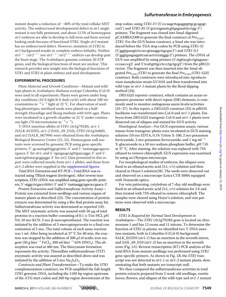
STR1 Is Required for Normal Seed Development inArabidopsis—The STR1 (At1g79230) gene is located on chro-mosome 1 and has 12 exons and 11 introns. To determine thefunction of STR1 in planta, we identified two T-DNA inser-tion mutants, both in Columbia-0 (Col-0) background.SALK_015593 (str1-1) has an insertion in the seventh intron,and SAIL_69_D10 (str1-2) has an insertion in the seventhexon (Fig. 1A). Reverse transcription (RT)-PCR analysis of thetotal RNA from mutant seedlings was performed using STR1gene-specific primers. As shown in Fig. 1B, the STR1 tran-script was not detected in str1-1 or str1-2mutant plants, dem-onstrating that both mutants are transcript-null.We then compared the sulfurtransferase activities in total
protein extracts prepared from 2-week-old seedlings, rosetteleaves, flowers, and siliques of the wild-type and str1-1mutant
Sulfurtransferase in Embryogenesis
MARCH 4, 2011 • VOLUME 286 • NUMBER 9 JOURNAL OF BIOLOGICAL CHEMISTRY 7549
by guest on January 12, 2019http://w
ww
.jbc.org/D
ownloaded from
plants. Two sulfur donors, 3-mercaptopyruvate and thiosul-fate, were used to determine the MST and TST activities, re-spectively. We found that total Arabidopsis protein extracts
had very low TST activity (Fig. 2A), and in contrast, MST ac-tivity was much higher (Fig. 2B). In the str1-1mutant seed-lings, TST and MST activities were both lower than those inthe wild-type seedlings, especially the MST activity (Fig. 2, Aand B). In extracts prepared from various organs of str1-1plants, MST activity was only about 20% of that in the wildtype (Fig. 2, B and C). In addition, we found that MST activitywas higher in flowers and siliques than in leaves and 2-week-old seedlings. The major reduction of MST activity in the str1mutant provided genetic evidence that the STR1 gene en-codes the major MST activity in Arabidopsis and that othermembers in the STR gene family are minor contributors. Thehigh reduction of MST activity in the str1mutant is also con-sistent with the previous finding that recombinant STR1 en-zyme prefers 3-mercaptopyruvate as its substrate rather thanthiosulfate (18).The mutant str1 plants displayed no visible phenotypic al-
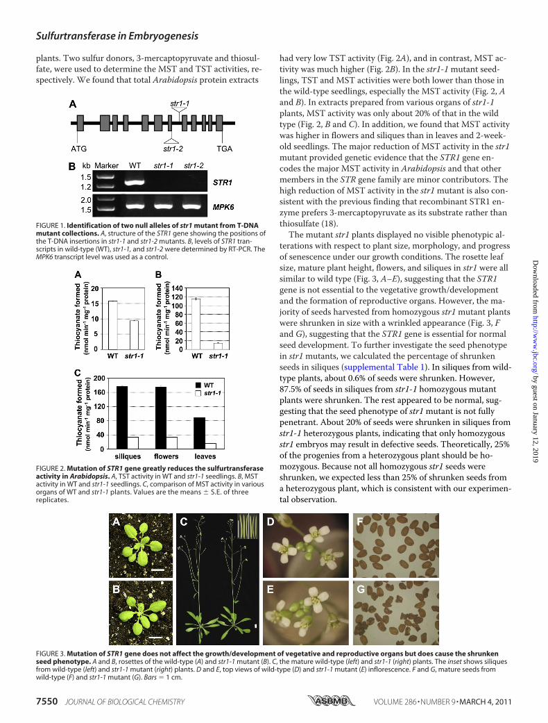
terations with respect to plant size, morphology, and progressof senescence under our growth conditions. The rosette leafsize, mature plant height, flowers, and siliques in str1 were allsimilar to wild type (Fig. 3, A–E), suggesting that the STR1gene is not essential to the vegetative growth/developmentand the formation of reproductive organs. However, the ma-jority of seeds harvested from homozygous str1mutant plantswere shrunken in size with a wrinkled appearance (Fig. 3, Fand G), suggesting that the STR1 gene is essential for normalseed development. To further investigate the seed phenotypein str1mutants, we calculated the percentage of shrunkenseeds in siliques (supplemental Table 1). In siliques from wild-type plants, about 0.6% of seeds were shrunken. However,87.5% of seeds in siliques from str1-1 homozygous mutantplants were shrunken. The rest appeared to be normal, sug-gesting that the seed phenotype of str1mutant is not fullypenetrant. About 20% of seeds were shrunken in siliques fromstr1-1 heterozygous plants, indicating that only homozygousstr1 embryos may result in defective seeds. Theoretically, 25%of the progenies from a heterozygous plant should be ho-mozygous. Because not all homozygous str1 seeds wereshrunken, we expected less than 25% of shrunken seeds froma heterozygous plant, which is consistent with our experimen-tal observation.
FIGURE 1. Identification of two null alleles of str1 mutant from T-DNAmutant collections. A, structure of the STR1 gene showing the positions ofthe T-DNA insertions in str1-1 and str1-2 mutants. B, levels of STR1 tran-scripts in wild-type (WT), str1-1, and str1-2 were determined by RT-PCR. TheMPK6 transcript level was used as a control.
FIGURE 2. Mutation of STR1 gene greatly reduces the sulfurtransferaseactivity in Arabidopsis. A, TST activity in WT and str1-1 seedlings. B, MSTactivity in WT and str1-1 seedlings. C, comparison of MST activity in variousorgans of WT and str1-1 plants. Values are the means � S.E. of threereplicates.
FIGURE 3. Mutation of STR1 gene does not affect the growth/development of vegetative and reproductive organs but does cause the shrunkenseed phenotype. A and B, rosettes of the wild-type (A) and str1-1 mutant (B). C, the mature wild-type (left) and str1-1 (right) plants. The inset shows siliquesfrom wild-type (left) and str1-1 mutant (right) plants. D and E, top views of wild-type (D) and str1-1 mutant (E) inflorescence. F and G, mature seeds fromwild-type (F) and str1-1 mutant (G). Bars � 1 cm.
Sulfurtransferase in Embryogenesis
7550 JOURNAL OF BIOLOGICAL CHEMISTRY VOLUME 286 • NUMBER 9 • MARCH 4, 2011
by guest on January 12, 2019http://w
ww
.jbc.org/D
ownloaded from
To genetically characterize the shrunken seed phenotype ofthe str1mutant, we performed reciprocal crosses. Whenstr1-1 plants were used as the pollen donors and wild-typeplants were used as the female parents, 0.87% of the F1 seedswere shrunken, which was consistent with the wild-type level(p � 0.05) (supplemental Table 1). This result suggests thatthe defective seed development was not a paternal effect dueto defective pollen. When str1-1 plants were pollinated withpollen grains from wild-type plants, the percentage ofshrunken seeds was at the wild-type level (1.21%) as well (p �0.05), ruling out the possibility of a maternal effect. Data fromreciprocal crosses also demonstrated that str1 is recessive andthat the heterozygous str1�/� zygotes can develop normallyand only the homozygous str1�/� zygotes have the potentialto develop to shrunken seeds.Homozygous str1 Embryos Show Delayed Development, and
Most Are Arrested at the Heart Stage—Seed development canbe divided into two major phases: embryo morphogenesis andseed maturation (29). Embryo morphogenesis is initiated by
the double fertilization of the embryo sac that gives rise to thezygote (2n) and endosperm (3n). The time of pollination (0days after pollination, DAP) is frequently used as the refer-ence point to define the stages of seed development. Arabi-dopsis embryos have five different developmental stages,namely octant, globular, heart, torpedo, and bent cotyledon(30), and the suspensor degenerates at the late heart stage orearly torpedo stage (31, 32).In wild-type plants, the embryos developed through the
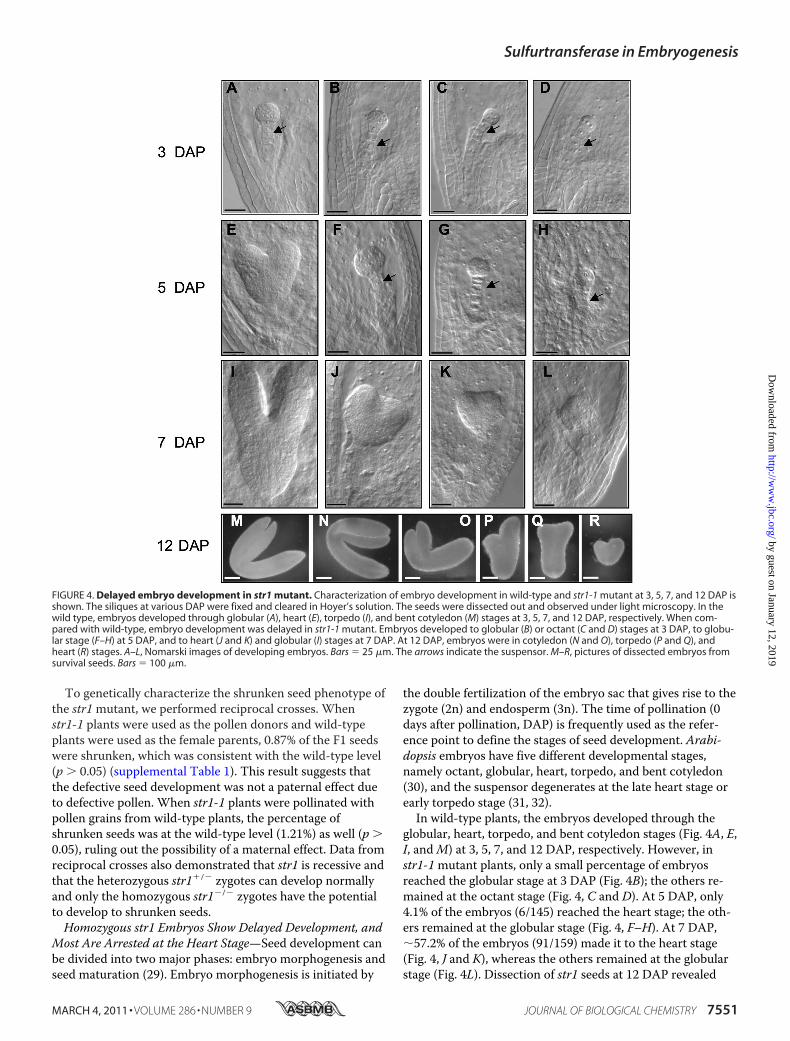
globular, heart, torpedo, and bent cotyledon stages (Fig. 4A, E,I, andM) at 3, 5, 7, and 12 DAP, respectively. However, instr1-1mutant plants, only a small percentage of embryosreached the globular stage at 3 DAP (Fig. 4B); the others re-mained at the octant stage (Fig. 4, C and D). At 5 DAP, only4.1% of the embryos (6/145) reached the heart stage; the oth-ers remained at the globular stage (Fig. 4, F–H). At 7 DAP,�57.2% of the embryos (91/159) made it to the heart stage(Fig. 4, J and K), whereas the others remained at the globularstage (Fig. 4L). Dissection of str1 seeds at 12 DAP revealed
FIGURE 4. Delayed embryo development in str1 mutant. Characterization of embryo development in wild-type and str1-1 mutant at 3, 5, 7, and 12 DAP isshown. The siliques at various DAP were fixed and cleared in Hoyer’s solution. The seeds were dissected out and observed under light microscopy. In thewild type, embryos developed through globular (A), heart (E), torpedo (I), and bent cotyledon (M) stages at 3, 5, 7, and 12 DAP, respectively. When com-pared with wild-type, embryo development was delayed in str1-1 mutant. Embryos developed to globular (B) or octant (C and D) stages at 3 DAP, to globu-lar stage (F–H) at 5 DAP, and to heart (J and K) and globular (I) stages at 7 DAP. At 12 DAP, embryos were in cotyledon (N and O), torpedo (P and Q), andheart (R) stages. A–L, Nomarski images of developing embryos. Bars � 25 �m. The arrows indicate the suspensor. M–R, pictures of dissected embryos fromsurvival seeds. Bars � 100 �m.
Sulfurtransferase in Embryogenesis
MARCH 4, 2011 • VOLUME 286 • NUMBER 9 JOURNAL OF BIOLOGICAL CHEMISTRY 7551
by guest on January 12, 2019http://w
ww
.jbc.org/D
ownloaded from
embryos at different developmental stages, including heart,torpedo, and bent cotyledon stages (Fig. 4, N–R), and the ma-jority were at the heart stage. At this time, the arrested str1embryos were bigger with more cells in comparison with thenormal wild-type embryos at morphologically similar stage.For instance, the heart-shaped str1 embryos at 12 DAP were�200 �m in width (Fig. 4R), which is much bigger than thenormal wild-type heart-stage embryos with �50 �m in width(Fig. 4E). This observation suggests that the cell division con-tinued to a certain extent in the affected str1mutant embryos.Eventually, �12.5% str1 embryos reached maturity andformed normal looking seeds (supplemental Table 1), and therest formed seeds with different degrees of shrinkage. Themost severely shrunken seeds (about 75% of the total) con-tained no embryo. The rest (12.5%) were partially shrunkenseeds that were able to germinate (discussed later). The str1-2mutant had the same embryo phenotype as str1-1 (supple-mental Fig. S1). The suspensors of the delayed embryos ap-peared to be normal in both str1mutant alleles (Fig. 4 andsupplemental Fig. S1).At the beginning of the seed maturation stage (7 DAP), em-
bryos are at the torpedo stage. They have already establishedthe body structure of plants, exhibiting a polarity along anapical-basal axis (33, 34). Normal embryos at this stage alsoshow bilateral symmetry along the apical-basal axis. Some str1embryos showed asymmetry in body structure, with two coty-ledons of unequal sizes (Fig. 4, P and Q), which is likely thecause of an observed cotyledon defect (discussed later).During the early seed maturation stage (7–10 DAP), Arabi-
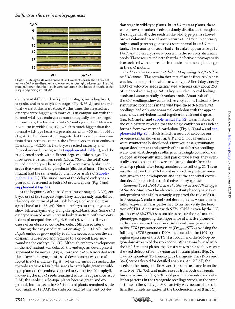
dopsis embryos grow rapidly to fill the seeds, whereas the en-dosperm is absorbed and reduced to a one-cell layer sur-rounding the embryo (35, 36). Although embryo developmentin the str1mutant was delayed, the endosperm developmentappeared to be normal (Fig. 4, B–D and F–H). Associated withthe delayed embryogenesis, seed development was also af-fected in str1mutants (Fig. 5). When the embryos reached thetorpedo stage at 6 DAP, the seeds became light green in wild-type plants as the embryos started to synthesize chlorophyll.However, the str1-1 seeds remained white in appearance. At 8DAP, the seeds in wild-type plants became green and ex-panded, but the seeds in str1-1mutant plants remained whiteand small. At 12 DAP, the embryos reached the bent cotyle-
don stage in wild-type plants. In str1-1mutant plants, therewere brown shrunken seeds randomly distributed throughoutthe silique. Finally, the seeds in the wild-type plants showedbrown color and were almost mature at 17 DAP. In contrast,only a small percentage of seeds were normal in str1-1mu-tants. The majority of seeds had a shrunken appearance at 17DAP, and no embryos were present in the severely shrunkenseeds. These results indicate that the defective embryogenesisis associated with and results in the shrunken seed phenotypein the str1mutant.Seed Germination and Cotyledon Morphology Is Affected in
str1 Mutants—The germination rate of seeds from str1 plantswas low in comparison with the wild type. After 9 days, nearly100% of wild-type seeds germinated, whereas only about 25%of str1 seeds did so (Fig. 6A). They included normal lookingseeds and some partially shrunken seeds. About half ofthe str1 seedlings showed defective cotyledons. Instead of twosymmetric cotyledons in the wild type, these defective str1seedlings had only one abnormal cotyledon with the appear-ance of two cotyledons fused together in different degrees(Fig. 6, D and E, and supplemental Fig. S2). Examination ofthe vein pattern revealed that the single cotyledon was indeedformed from two merged cotyledons (Fig. 6, H and I, and sup-plemental Fig. S2), which is likely a result of defective em-bryogenesis. As shown in Fig. 4, P and Q, not all embryoswere symmetrically developed. However, post-germinationorgan development and growth of these defective seedlingswere normal. Although seedlings with a single cotyledon de-veloped an unequally sized first pair of true leaves, they even-tually grew to plants that were indistinguishable from thewild-type plants after being transferred to soil (Fig. 3). Theseresults indicate that STR1 is not essential for post-germina-tion growth and development and that the abnormal cotyle-don development is due to defective embryogenesis.Genomic STR1 DNA Rescues the Shrunken Seed Phenotype
of the str1 Mutant—The identical mutant phenotype in twoindependent str1 alleles strongly supported the role of STR1in Arabidopsis embryo and seed development. A complemen-tation experiment was performed to further verify the func-tion of STR1. A construct with STR1 cDNA driven by the 35Spromoter (35S:STR1) was unable to rescue the str1mutantphenotype, suggesting the importance of a native promoterand/or elements in the introns. As a result, we generated anative STR1 promoter construct (ProSTR1:STR1) by using thefull-length STR1 genomic DNA that included the 1109-bpregion upstream of the ATG start codon and the 260-bp re-gion downstream of the stop codon. When transformed intothe str1-1mutant plants, the construct was able to fully rescuethe seed defects of homozygous str1mutant plants (Fig. 7).Two independent T3 homozygous transgenic lines (51-2 and36-3) were selected for detailed analyses. At 12 DAP, theseeds in the transgenic lines were the same as those from thewild type (Fig. 7A), and mature seeds from both transgeniclines were normal (Fig. 7B). Seed germination rates and coty-ledon patterns in the transgenic seedlings were also the sameas those in the wild type. MST activity was measured to con-firm the complementation at the biochemical level (Fig. 7C).
FIGURE 5. Delayed development of str1 mutant seeds. The siliques atvarious DAP were dissected and observed under light microscopy. In str1-1mutant, brown shrunken seeds were randomly distributed throughout thesilique beginning at 10 DAP.
Sulfurtransferase in Embryogenesis
7552 JOURNAL OF BIOLOGICAL CHEMISTRY VOLUME 286 • NUMBER 9 • MARCH 4, 2011
by guest on January 12, 2019http://w
ww
.jbc.org/D
ownloaded from
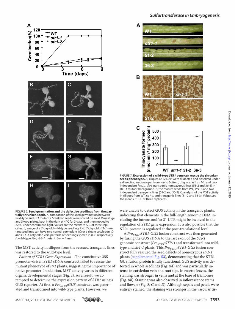
The MST activity in siliques from the rescued transgenic lineswas restored to the wild-type level.Pattern of STR1 Gene Expression—The constitutive 35S
promoter-driven STR1 cDNA construct failed to rescue themutant phenotype of str1 plants, suggesting the importance ofnative promoter. In addition, MST activity varies in differentorgans/developmental stages (Fig. 2). As a result, we at-tempted to determine the expression pattern of STR1 using aGUS reporter. At first, a ProSTR1:GUS construct was gener-ated and transformed into wild-type plants. However, we
were unable to detect GUS activity in the transgenic plants,indicating that elements in the full-length genomic DNA in-cluding the introns and/or 3�-UTR might be involved in theregulation of STR1 gene expression. It is also possible that theSTR1 protein is regulated at the post-translational level.A ProSTR1:STR1-GUS fusion construct was then generated
by fusing the GUS cDNA to the last exon of the STR1genomic construct (ProSTR1:STR1) and transformed into wild-type and str1-1 plants. This ProSTR1:STR1-GUS fusion con-struct fully rescued the seed defects of homozygous str1-1plants (supplemental Fig. S3), demonstrating that the STR1-GUS fusion protein is fully functional. GUS activity was de-tected in whole seedlings (Fig. 8A) and was particularly in-tense in cotyledon vein and root tips. In rosette leaves, thestaining was stronger in veins and at the base of trichomes(Fig. 8B). Staining was also observed in inflorescence stemsand flowers (Fig. 8, C and D). Although sepals and petals wereentirely stained, the staining was stronger in the vascular tis-
FIGURE 6. Seed germination and the defective seedlings from the par-tially shrunken seeds. A, comparison of the seed germination betweenwild-type and str1 mutants. Sterilized seeds were sowed on solid Murashigeand Skoog plates, kept in the dark at 4 °C for 3 days, and then moved to22 °C under continuous light. Values are the means � S.E. of three repli-cates. B, image of a 7-day-old wild-type seedling. C–E, 7-day-old str1-1 mu-tant seedlings can have two normal cotyledons (C) or a single cotyledon (Dand E). F–I, cotyledon vein patterns of seedlings shown in B–E, respectively.F, wild-type; G–I, str1-1 mutant. Bar � 1 mm.
FIGURE 7. Expression of a wild-type STR1 gene can rescue the shrunkenseeds phenotype. A, siliques at 12 DAP were dissected and observed undera dissecting microscope. From top to bottom, they are: WT, str1-1, and twoindependent ProSTR1:Str1 transgenic homozygous lines (51-2 and 36-3) instr1-1 mutant background. B, the mature seeds from WT, str1-1, and twoindependent transgenic lines (51-2 and 36-3). C, analysis of the MST activityin siliques from WT, str1-1, and transgenic lines (51-2 and 36-3). Values arethe means � S.E. of three replicates.
Sulfurtransferase in Embryogenesis
MARCH 4, 2011 • VOLUME 286 • NUMBER 9 JOURNAL OF BIOLOGICAL CHEMISTRY 7553
by guest on January 12, 2019http://w
ww
.jbc.org/D
ownloaded from
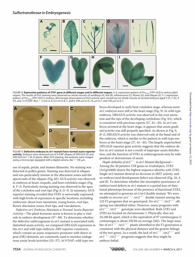
sue of sepals, petals, and stamen filaments. No staining wasdetected in pollen grains. Staining was detected in siliquesand was particularly intense in the abscission zones and theapical ends of the siliques (Fig. 8E). GUS activity was observedin embryos at heart, torpedo, and bent cotyledon stages (Fig.8, F–I). Particularly strong staining was observed in the apexof the cotyledon and root tips (Fig. 8, G–I). In summary, GUSreporter staining revealed that STR1 is universally expressed,with high levels of expression at specific locations, includingembryonic shoot/root meristems, young leaves, root tips,flower abscission zones, fruit tips, and vasculatures.Defective str1 Embryos Maintain a Normal Auxin Reporter
Activity—The plant hormone auxin is known to play a vitalrole in embryo development (37–40). To determine whetherthe defective embryogenesis in str1mutant is associated withdisturbed auxin activity, we compared DR5:GUS expression inthe str1 and wild-type embryos. DR5 reporter constructs,which contain an auxin-responsive promoter with direct re-peat (DR) elements, are commonly used to monitor endoge-nous auxin levels/activities (25–27). At 9 DAP, wild-type em-
bryos developed to early bent cotyledon stage, whereas moststr1 embryos were still at the heart stage (Fig. 9). In wild-typeembryos, DR5:GUS activity was observed in the root meris-tem and the tips of the developing cotyledons (Fig. 9A), whichis consistent with previous reports (27, 41–43). In str1 em-bryos arrested at the heart stage, it appears that auxin gradi-ent/activity was still properly specified. As shown in Fig. 9,B–E, DR5:GUS activity was observed only at the basal end ofthe embryos, which is similar to the pattern in wild-type em-bryos at the heart stage (27, 41–43). The largely unperturbedDR5:GUS reporter gene activity suggests that the embryo de-fect in str1mutant is not a result of improper auxin distribu-tion, and the function of STR1 in embryogenesis may be inde-pendent or downstream of auxin.Haplo-lethality of str2�/� in str1Mutant Background—
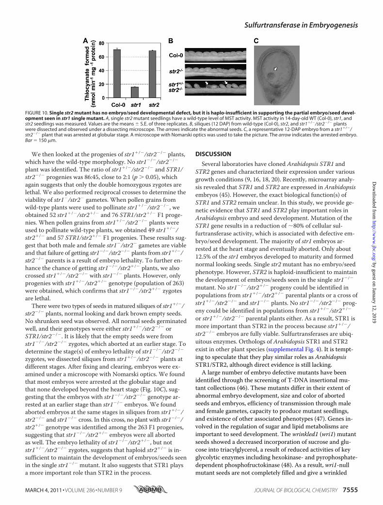
Among the 20 putative STR genes in Arabidopsis, STR2(At1g16460) shares the highest sequence identity with STR1.Single str2mutant showed no decrease in MST activity, andno embryo/seed development defect was observed (Fig. 10, Aand B). To determine whether the incomplete penetrance ofembryo/seed defects in str1mutant is a partial loss-of-func-tional phenotype because of the presence of functional STR2,we attempted to generate str1/str2 double mutant. We wereunable to recover str1�/�/str2�/� double mutant among the133 F2 progenies that we genotyped. No str1�/�/str2�/� off-spring was identified either. However, many progenies withstr1�/�/str2�/� genotype were identified. Both STR1 andSTR2 are located on chromosome 1. Physically, they are24,180 kb apart, which is the equivalent of 97 centimorgans (1centimorgan is about 250 kb on average (44)). The large num-ber of str1�/�/str2�/� plants identified in the F2 generation isconsistent with the physical distance and the genetic linkageof the two genes. As a result, the lack of str1�/�/str2�/� andstr1�/�/str2�/� progenies suggests that they might beembryo-lethal.
FIGURE 8. Expression patterns of STR1 gene at different stages and in different organs. A–E, expression pattern of ProSTR1:STR1-GUS in various plantorgans. The results of GUS staining were observed on whole mounts of seedlings (A), leaf (B), inflorescence (C), flower (D), and siliques (E). F–I, expressionpattern of ProSTR1:STR1-GUS in embryo. Microscope observations of GUS activity were carried out on whole mounts of excised embryos aged 5 (F), 7 (G), 9(H), and 12 (I) DAP. Bars � 2 mm in A, 0.5 cm in B, C, and E, 500 �m in D, 25 �m in F, and 100 �m in G–I.
FIGURE 9. Defective embryos in str1 mutant have normal auxin reporteractivity. Embryos were dissected out of 9-DAP siliques of DR5:GUS (A) andDR5:GUS/str1-1 (B–E) plants. After GUS staining, the embryos were imagedusing a microscope equipped with a digital camera. Bar � 100 �m.
Sulfurtransferase in Embryogenesis
7554 JOURNAL OF BIOLOGICAL CHEMISTRY VOLUME 286 • NUMBER 9 • MARCH 4, 2011
by guest on January 12, 2019http://w
ww
.jbc.org/D
ownloaded from
We then looked at the progenies of str1�/�/str2�/� plants,which have the wild-type morphology. No str1�/�/str2�/�
plant was identified. The ratio of str1�/�/str2�/� and STR1/str2�/� progenies was 86:45, close to 2:1 (p � 0.05), whichagain suggests that only the double homozygous zygotes arelethal. We also performed reciprocal crosses to determine theviability of str1�/str2� gametes. When pollen grains fromwild-type plants were used to pollinate str1�/�/str2�/�, weobtained 52 str1�/�/str2�/� and 76 STR1/str2�/� F1 proge-nies. When pollen grains from str1�/�/str2�/� plants wereused to pollinate wild-type plants, we obtained 49 str1�/�/str2�/� and 57 STR1/str2�/� F1 progenies. These results sug-gest that both male and female str1�/str2� gametes are viableand that failure of getting str1�/�/str2�/� plants from str1�/�/str2�/� parents is a result of embryo lethality. To further en-hance the chance of getting str1�/�/str2�/� plants, we alsocrossed str1�/�/str2�/� with str1�/� plants. However, onlyprogenies with str1�/�/str2�/� genotype (population of 263)were obtained, which confirms that str1�/�/str2�/� zygotesare lethal.There were two types of seeds in matured siliques of str1�/�/
str2�/� plants, normal looking and dark brown empty seeds.No shrunken seed was observed. All normal seeds germinatedwell, and their genotypes were either str1�/�/str2�/� orSTR1/str2�/�. It is likely that the empty seeds were fromstr1�/�/str2�/� zygotes, which aborted at an earlier stage. Todetermine the stage(s) of embryo lethality of str1�/�/str2�/�
zygotes, we dissected siliques from str1�/�/str2�/� plants atdifferent stages. After fixing and clearing, embryos were ex-amined under a microscope with Nomarski optics. We foundthat most embryos were arrested at the globular stage andthat none developed beyond the heart stage (Fig. 10C), sug-gesting that the embryos with str1�/�/str2�/� genotype ar-rested at an earlier stage than str1�/� embryos. We foundaborted embryos at the same stages in siliques from str1�/�/str2�/� and str1�/� cross. In this cross, no plant with str1�/�/str2�/� genotype was identified among the 263 F1 progenies,suggesting that str1�/�/str2�/� embryos were all abortedas well. The embryo lethality of str1�/�/str2�/�, but notstr1�/�/str2�/� zygotes, suggests that haploid str2�/� is in-sufficient to maintain the development of embryos/seeds seenin the single str1�/� mutant. It also suggests that STR1 playsa more important role than STR2 in the process.
DISCUSSION
Several laboratories have cloned Arabidopsis STR1 andSTR2 genes and characterized their expression under variousgrowth conditions (9, 16, 18, 20). Recently, microarray analy-sis revealed that STR1 and STR2 are expressed in Arabidopsisembryos (45). However, the exact biological function(s) ofSTR1 and STR2 remain unclear. In this study, we provide ge-netic evidence that STR1 and STR2 play important roles inArabidopsis embryo and seed development. Mutation of theSTR1 gene results in a reduction of �80% of cellular sul-furtransferase activity, which is associated with defective em-bryo/seed development. The majority of str1 embryos ar-rested at the heart stage and eventually aborted. Only about12.5% of the str1 embryos developed to maturity and formednormal looking seeds. Single str2mutant has no embryo/seedphenotype. However, STR2 is haploid-insufficient to maintainthe development of embryos/seeds seen in the single str1�/�
mutant. No str1�/�/str2�/� progeny could be identified inpopulations from str1�/�/str2�/� parental plants or a cross ofstr1�/�/str2�/� and str1�/� plants. No str1�/�/str2�/� prog-eny could be identified in populations from str1�/�/str2�/�
or str1�/�/str2�/� parental plants either. As a result, STR1 ismore important than STR2 in the process because str1�/�/str2�/� embryos are fully viable. Sulfurtransferases are ubiq-uitous enzymes. Orthologs of Arabidopsis STR1 and STR2exist in other plant species (supplemental Fig. 4). It is tempt-ing to speculate that they play similar roles as ArabidopsisSTR1/STR2, although direct evidence is still lacking.A large number of embryo defective mutants have been
identified through the screening of T-DNA insertional mu-tant collections (46). These mutants differ in their extent ofabnormal embryo development, size and color of abortedseeds and embryos, efficiency of transmission through maleand female gametes, capacity to produce mutant seedlings,and existence of other associated phenotypes (47). Genes in-volved in the regulation of sugar and lipid metabolisms areimportant to seed development. The wrinkled1 (wri1) mutantseeds showed a decreased incorporation of sucrose and glu-cose into triacylglycerol, a result of reduced activities of keyglycolytic enzymes including hexokinase- and pyrophosphate-dependent phosphofructokinase (48). As a result, wri1-nullmutant seeds are not completely filled and give a wrinkled
FIGURE 10. Single str2 mutant has no embryo/seed developmental defect, but it is haplo-insufficient in supporting the partial embryo/seed devel-opment seen in str1 single mutant. A, single str2 mutant seedlings have a wild-type level of MST activity. MST activity in 14-day-old WT (Col-0), str1, andstr2 seedlings was measured. Values are the means � S.E. of three replicates. B, siliques (12 DAP) from wild-type (Col-0), str2, and str1�/�/str2�/� plantswere dissected and observed under a dissecting microscope. The arrows indicate the abnormal seeds. C, a representative 12-DAP embryo from a str1�/�/str2�/� plant that was arrested at globular stage. A microscope with Nomarski optics was used to take the picture. The arrow indicates the arrested embryo.Bar � 150 �m.
Sulfurtransferase in Embryogenesis
MARCH 4, 2011 • VOLUME 286 • NUMBER 9 JOURNAL OF BIOLOGICAL CHEMISTRY 7555
by guest on January 12, 2019http://w
ww
.jbc.org/D
ownloaded from
appearance. WRI1 encodes an AP2/EREB domain transcrip-tion factor, and overexpression ofWRI1 resulted in an in-creased triacylglycerol level in both seeds and leaves (49). Amore recent study revealed that LEC2, a plant-specific B3transcription factor, directly regulated WRI1, which, in turn,controlled the expression of a subset of genes involved in fattyacid biosynthesis and seed maturation (50, 51).The shrunken seed phenotype of str1mutant is similar to
that of wri1 but more severe. The embryo defect appears ear-lier before the seed-filling stage, resulting in the abortion ofthe majority of the developing embryos and empty seeds. Mu-tation of STR2 in str1 background enhances the embryo/seedphenotype, indicating that both are involved in embryo/seeddevelopment. At this stage, the exact biochemical function ofSTR1/STR2 in embryo and seed development is unknown. Itis possible that they are either directly or indirectly involvedin a metabolic pathway that is essential to supply/convert theessential nutrients to support the normal embryogenesis andseed development, based on the putative function of sul-furtransferase in sulfur metabolism (5, 6). Alternatively, theycould be involved in the removal of a toxic substance pro-duced during the seed-setting stage. In the absence of STR1/STR2, embryos either are in a nutrient-deficient state or suf-fer from toxicity. Both can result in the delay of embryogrowth/development and/or aborted embryos.Plant sulfurtransferases have been speculated to function in
the detoxification of cyanide, a co-product of ethylene biosyn-thesis (11, 16, 52). In vitro, sulfurtransferases catalyze thetransfer of a sulfur atom from a donor such as thiosulfate and3-mercaptopyruvate to cyanide, leading to the formation ofthe less toxic thiocyanate (18). However, in vivo evidence sup-porting a role of STR1/STR2 in cyanide removal in plants isstill lacking. Plants produce high levels of ethylene duringflowering and seed setting (53, 54). In Arabidopsis, flowers/siliques produce �100 times more ethylene than leaves (sup-plemental Fig. 5, Supplemental Methods). In this study, theflowers were staged according to Smyth et al. (55), and flow-ers at stage 16 contain embryos at the torpedo stage. The av-erage rate of ethylene production in flowers from stage 16 andyounger was �600 pmol/h/g of fresh weight. Cyanide shouldbe produced at the same rate. Unlike ethylene, which can dif-fuse out of the cells, cyanide will accumulate in the cells if notbiochemically removed. A �600 pmol/h/g of fresh weightproduction rate is equivalent to an increase of 0.6 �M/h incellular cyanide concentration if 1 g of fresh weight is equal to1 ml. This clearly imposes a threat to normal cellular activityif cyanide is not removed. Cytochrome oxidase and Rubiscohave Kd or IC50 values of 1 and 6 �M, respectively. A numberof other redox-related enzymes are also sensitive to HCN atthe micromolar range (56).STR1 is localized in mitochondria, based on immunodetec-
tion of isolated mitochondria and transient expression ofSTR1-GFP fusion in Arabidopsis protoplasts (19). Further-more, 3-mercaptopyruvate exists in mitochondria (17), sug-gesting that the biochemical function of STR1 may indeedinvolve the utilization of 3-mercaptopyruvate as a substrate inthe detoxification of cyanide in mitochondria. As a co-prod-uct of ethylene biosynthesis, cyanide is produced outside of
mitochondria. However, at cellular pH, the majority of cya-nide is in the protonated form, which is membrane-perme-able. It has been reported that cyanide can move into mito-chondria by both facilitated transport and passive diffusion(57). Due to the high sensitivity of cytochrome oxidase to cya-nide (Kd of 1 �M) (56), protection of normal mitochondrialactivity in the presence of cyanide is imperative to the survivalof the cells and normal cellular activities. It is tempting tospeculate that STR1 may play a role in the protection of de-veloping embryos from cyanide toxicity, based on its mito-chondrial localization and in vitro biochemical activity in cya-nide detoxification. As a cytoplasmic protein, STR2 may beinvolved in a similar detoxification process. The lack of phe-notype in other tissues/organs (i.e. leaves) of the str1mutantplants could be a result of low level biosynthesis of ethylene(supplemental Fig. 5) and its co-product cyanide. In summary,we demonstrate in this report that STR1/STR2 sulfurtrans-ferases play an important role in plant embryo/seed develop-ment, which highlights the important biological function ofthis group of ubiquitous enzymes in plants.
Acknowledgments—We thank the Arabidopsis Biological ResourceCenter for seed stocks and Melody Kroll for proofreading themanuscript.
REFERENCES1. Vennesland, B., Castric, P. A., Conn, E. E., Solomonson, L. P., Volini, M.,
and Westley, J. (1982) Fed. Proc. 41, 2639–26482. Nagahara, N., Ito, T., and Minami, M. (1999) Histol. Histopathol. 14,
1277–12863. Billaut-Laden, I., Rat, E., Allorge, D., Crunelle-Thibaut, A., Cauffiez, C.,
Chevalier, D., Lo-Guidice, J. M., and Broly, F. (2006) Toxicol. Lett. 165,101–111
4. Ramasamy, S., Singh, S., Taniere, P., Langman, M. J., and Eggo, M. C.(2006) Am. J. Physiol. Gastrointest. Liver Physiol. 291, G288–G296
5. Donadio, S., Shafiee, A., and Hutchinson, C. R. (1990) J. Bacteriol. 172,350–360
6. Nagahara, N., and Sawada, N. (2006) Curr. Med. Chem. 13, 1219–12307. Bonomi, F., Pagani, S., Cerletti, P., and Cannella, C. (1977) Eur.
J. Biochem. 72, 17–248. Cerletti, P. (1986) Trends Biochem. Sci. 11, 369–3729. Bartels, A., Mock, H. P., and Papenbrock, J. (2007) Plant Physiol.
Biochem. 45, 178–18710. Schenk, P. M., Kazan, K., Rusu, A. G., Manners, J. M., and Maclean, D. J.
(2005) Plant Physiol. Biochem. 43, 997–100511. Meyer, T., Burow, M., Bauer, M., and Papenbrock, J. (2003) Planta 217,
1–1012. Fujiki, Y., Yoshikawa, Y., Sato, T., Inada, N., Ito, M., Nishida, I., and Wa-
tanabe, A. (2001) Physiol. Plant 111, 345–35213. Chung, B. C., Lee, S. Y., Oh, S. A., Rhew, T. H., Nam, H. G., and Lee,
C. H. (1997) J. Plant Physiol. 151, 339–34514. Weaver, L. M., Gan, S., Quirino, B., and Amasino, R. M. (1998) Plant
Mol. Biol. 37, 455–46915. Zhao, Y., Dai, X., Blackwell, H. E., Schreiber, S. L., and Chory, J. (2003)
Science 301, 1107–111016. Nakamura, T., Yamaguchi, Y., and Sano, H. (2000) Eur. J. Biochem. 267,
5621–563017. Papenbrock, J., and Schmidt, A. (2000) Eur. J. Biochem. 267, 5571–557918. Papenbrock, J., and Schmidt, A. (2000) Eur. J. Biochem. 267, 145–15419. Bauer, M., Dietrich, C., Nowak, K., Sierralta, W. D., and Papenbrock, J.
(2004) Plant Physiol. 135, 916–92620. Hatzfeld, Y., and Saito, K. (2000) FEBS Lett. 470, 147–15021. Alonso, J. M., Stepanova, A. N., Leisse, T. J., Kim, C. J., Chen, H., Shinn,
Sulfurtransferase in Embryogenesis
7556 JOURNAL OF BIOLOGICAL CHEMISTRY VOLUME 286 • NUMBER 9 • MARCH 4, 2011
by guest on January 12, 2019http://w
ww
.jbc.org/D
ownloaded from
P., Stevenson, D. K., Zimmerman, J., Barajas, P., Cheuk, R., Gadrinab, C.,Heller, C., Jeske, A., Koesema, E., Meyers, C. C., Parker, H., Prednis, L.,Ansari, Y., Choy, N., Deen, H., Geralt, M., Hazari, N., Hom, E., Karnes,M., Mulholland, C., Ndubaku, R., Schmidt, I., Guzman, P., Aguilar-Hen-onin, L., Schmid, M., Weigel, D., Carter, D. E., Marchand, T., Risseeuw,E., Brogden, D., Zeko, A., Crosby, W. L., Berry, C. C., and Ecker, J. R.(2003) Science 301, 653–657
22. Sessions, A., Burke, E., Presting, G., Aux, G., McElver, J., Patton, D., Di-etrich, B., Ho, P., Bacwaden, J., Ko, C., Clarke, J. D., Cotton, D., Bullis, D.,Snell, J., Miguel, T., Hutchison, D., Kimmerly, B., Mitzel, T., Katagiri, F.,Glazebrook, J., Law, M., and Goff, S. A. (2002) Plant Cell 14, 2985–2994
23. Liu, Y., and Zhang, S. (2004) Plant Cell 16, 3386–339924. Clough, S. J., and Bent, A. F. (1998) Plant J. 16, 735–74325. Ulmasov, T., Murfett, J., Hagen, G., and Guilfoyle, T. J. (1997) Plant Cell
9, 1963–197126. Sabatini, S., Beis, D., Wolkenfelt, H., Murfett, J., Guilfoyle, T., Malamy,
J., Benfey, P., Leyser, O., Bechtold, N., Weisbeek, P., and Scheres, B.(1999) Cell 99, 463–472
27. Friml, J., Vieten, A., Sauer, M., Weijers, D., Schwarz, H., Hamann, T.,Offringa, R., and Jurgens, G. (2003) Nature 426, 147–153
28. Liu, C. M., and Meinke, D. W. (1998) Plant J. 16, 21–3129. Baud, S., Dubreucq, B., Miquel, M., Rochat, C., and Lepiniec, L. (2008)
The Arabidopsis Book, pp. 1–24, American Society of Plant Biologists,Rockville, MD
30. Berleth, T. (1998) Plant Physiol. Biochem. 36, 69–8231. Umehara, M., and Kamada, H. (2005) Plant Biotech. 22, 253–26032. Schwartz, B., Vernon, D., and Meinke, D. (1997) Development of the Sus-
pensor: Differentiation, Communication, and Programmed Cell Deathduring Plant Embryogenesis, pp. 53–72, Kluwer Academic Publishers,Dordrecht, The Netherlands
33. Jenik, P. D., Gillmor, C. S., and Lukowitz, W. (2007) Annu. Rev. Cell Dev.Biol. 23, 207–236
34. Jurgens, G. (2001) EMBO J. 20, 3609–361635. Berger, F., Grini, P. E., and Schnittger, A. (2006) Curr. Opin. Plant Biol.
9, 664–67036. Olsen, O. A. (2001) Annu Rev Plant Physiol. Plant Mol. Biol. 52,
233–26737. Capron, A., Chatfield, S., Provart, N., and Berleth, T. (2008) The Arabi-
dopsis Book, 1–28, American Society of Plant Biologists, Rockville, MD38. Michniewicz, M., Brewer, P. B., and Friml, J. (2007) The Arabidopsis
Book, pp. 1–28, American Society of Plant Biologists, Rockville, MD39. Scheres, B., Benfey, P., and Dolan, L. (2002) The Arabidopsis Book, pp.
1–18, American Society of Plant Biologists, Rockville, MD40. De Smet, I., Lau, S., Mayer, U., and Jurgens, G. (2010) Plant J. 61,
959–97041. Friml, J., Benkova, E., Blilou, I., Wisniewska, J., Hamann, T., Ljung, K.,
Woody, S., Sandberg, G., Scheres, B., Jurgens, G., and Palme, K. (2002)Cell 108, 661–673
42. Xiao, W., Custard, K. D., Brown, R. C., Lemmon, B. E., Harada, J. J.,Goldberg, R. B., and Fischer, R. L. (2006) Plant Cell 18, 805–814
43. Song, S. K., Hofhuis, H., Lee, M. M., and Clark, S. E. (2008) Dev. Cell 15,98–109
44. Mezard, C. (2006) Biochem. Soc. Trans. 34, 531–53445. Spencer, M. W., Casson, S. A., and Lindsey, K. (2007) Plant Physiol. 143,
924–94046. Tzafrir, I., Dickerman, A., Brazhnik, O., Nguyen, Q., McElver, J., Frye,
C., Patton, D., and Meinke, D. (2003) Nucleic Acids Research 31, 90–9347. Tzafrir, I., Pena-Muralla, R., Dickerman, A., Berg, M., Rogers, R., Hutch-
ens, S., Sweeney, T. C., McElver, J., Aux, G., Patton, D., and Meinke, D.(2004) Plant Physiol. 135, 1206–1220
48. Focks, N., and Benning, C. (1998) Plant Physiol. 118, 91–10149. Cernac, A., and Benning, C. (2004) Plant J. 40, 575–58550. Baud, S., Mendoza, M. S., To, A., Harscoet, E., Lepiniec, L., and
Dubreucq, B. (2007) Plant J. 50, 825–83851. Braybrook, S. A., Stone, S. L., Park, S., Bui, A. Q., Le, B. H., Fischer, R. L.,
Goldberg, R. B., and Harada, J. J. (2006) Proc. Natl. Acad. Sci. U.S.A. 103,3468–3473
52. Grossmann, K. (1996) Physiol. Plant. 97, 772–77553. Roeder, S., Dreschler, K., Wirtz, M., Cristescu, S. M., van Harren, F. J.,
Hell, R., and Piechulla, B. (2009) Plant Mol. Biol. 70, 535–54654. Iannetta, P. P., Laarhoven, L. J., Medina-Escobar, N., James, E. K., Mc-
Manus, M. T., Davies, H. V., and Harren, F. J. (2006) Physiol. Plant. 127,247–259
55. Smyth, D. R., Bowman, J. L., and Meyerowitz, E. M. (1990) Plant Cell 2,755–767
56. Solomonson, L. (1981) Cyanide as a Metabolic Inhibitor, pp. 11–28,Academic Press, London, New York
57. Wisler, J. A., Dulaney, M. D., Pellicore, L. S., and Lenz, D. E. (1991) Toxi-col. Lett 56, 275–281
Sulfurtransferase in Embryogenesis
MARCH 4, 2011 • VOLUME 286 • NUMBER 9 JOURNAL OF BIOLOGICAL CHEMISTRY 7557
by guest on January 12, 2019http://w
ww
.jbc.org/D
ownloaded from
Guohong Mao, Ruigang Wang, Yuefeng Guan, Yidong Liu and Shuqun ZhangArabidopsis thalianain
Sulfurtransferases 1 and 2 Play Essential Roles in Embryo and Seed Development
doi: 10.1074/jbc.M110.182865 originally published online December 28, 20102011, 286:7548-7557.J. Biol. Chem.
10.1074/jbc.M110.182865Access the most updated version of this article at doi:
Alerts:
When a correction for this article is posted•
When this article is cited•
to choose from all of JBC's e-mail alertsClick here
Supplemental material:
http://www.jbc.org/content/suppl/2011/01/03/M110.182865.DC1
http://www.jbc.org/content/286/9/7548.full.html#ref-list-1
This article cites 51 references, 14 of which can be accessed free at
by guest on January 12, 2019http://w
ww
.jbc.org/D
ownloaded from