Embed Size (px)
DESCRIPTION
Embryo Development. Animal Science 434 John Parrish. Timing of Insemination. SpeciesTime of OvulationOptimal Insemination Time Cow29 hr after startEnd of estrus (12 hr after of estrusfirst seen in estrus) EweEnd of estrusEnd of 1st day or start of 2nd day of estrus - PowerPoint PPT Presentation
Citation preview
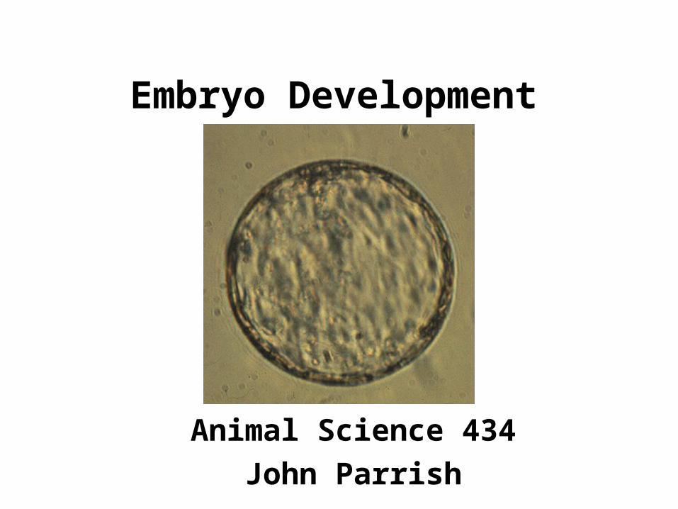
Embryo Development
Animal Science 434
John Parrish
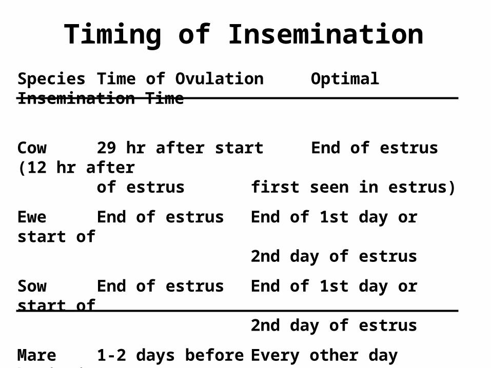
Timing of Insemination
Species Time of Ovulation Optimal Insemination Time
Cow 29 hr after start End of estrus (12 hr afterof estrus first seen in estrus)
Ewe End of estrus End of 1st day or start of2nd day of estrus
Sow End of estrus End of 1st day or start of2nd day of estrus
Mare 1-2 days before Every other day beginningend of estrus day 3 of estrus
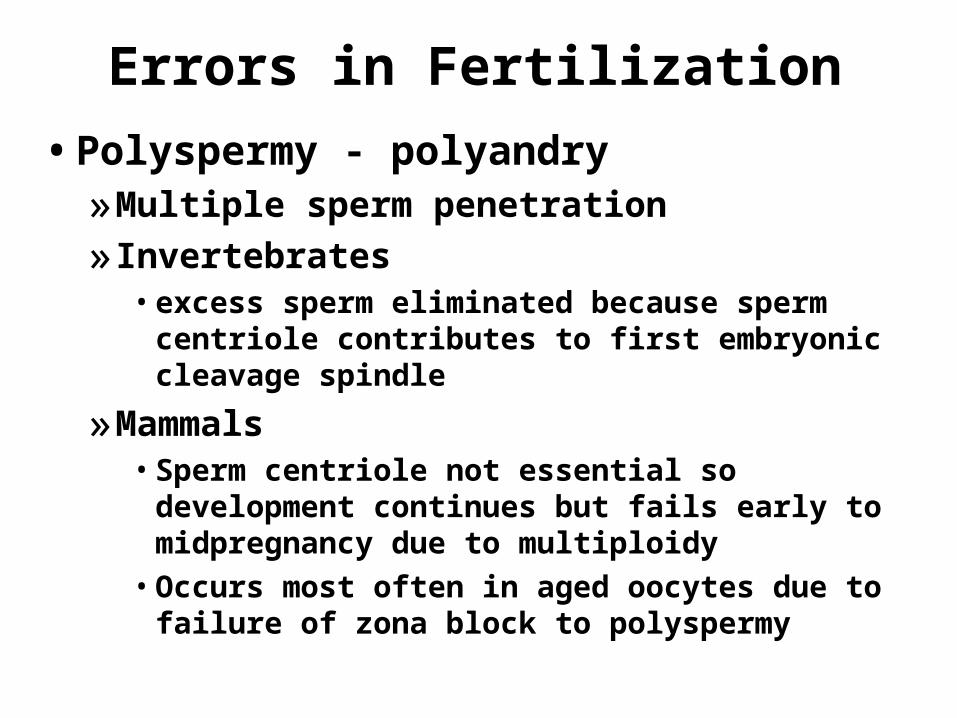
Errors in Fertilization
• Polyspermy - polyandry»Multiple sperm penetration
» Invertebrates• excess sperm eliminated because sperm centriole
contributes to first embryonic cleavage spindle
»Mammals• Sperm centriole not essential so development
continues but fails early to midpregnancy due to multiploidy• Occurs most often in aged oocytes due to failure
of zona block to polyspermy
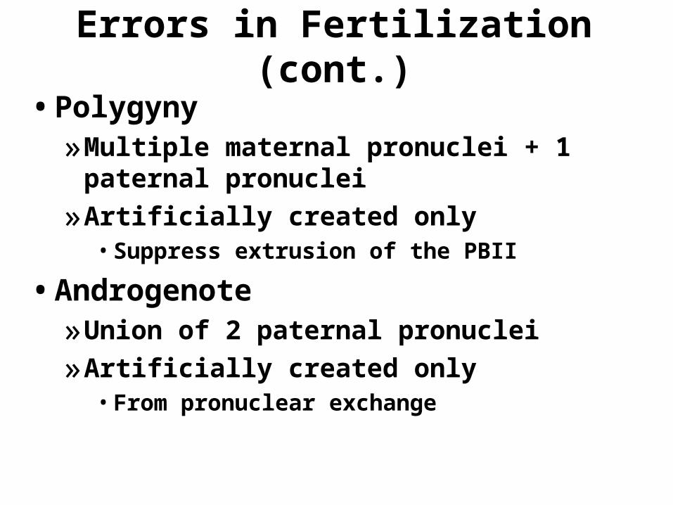
Errors in Fertilization (cont.)
• Polygyny»Multiple maternal pronuclei + 1 paternal
pronuclei
»Artificially created only• Suppress extrusion of the PBII
• Androgenote»Union of 2 paternal pronuclei
»Artificially created only• From pronuclear exchange
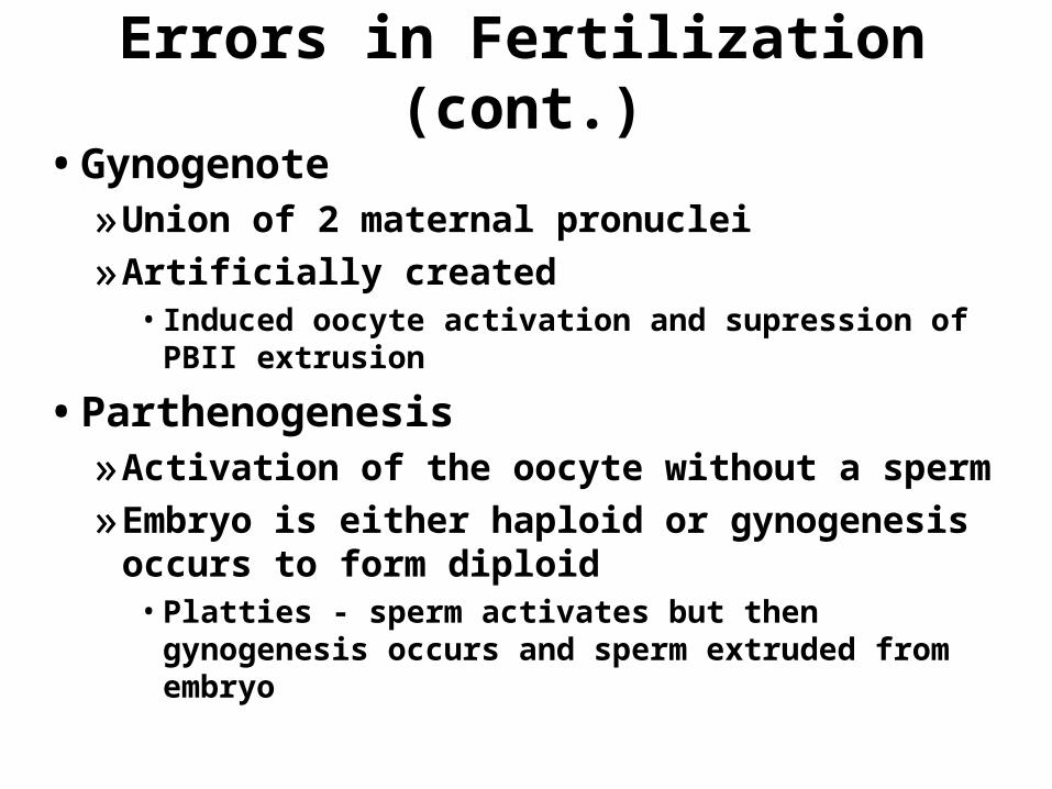
Errors in Fertilization (cont.)
• Gynogenote»Union of 2 maternal pronuclei
»Artificially created• Induced oocyte activation and supression of PBII
extrusion
• Parthenogenesis»Activation of the oocyte without a sperm
»Embryo is either haploid or gynogenesis occurs to form diploid• Platties - sperm activates but then gynogenesis
occurs and sperm extruded from embryo
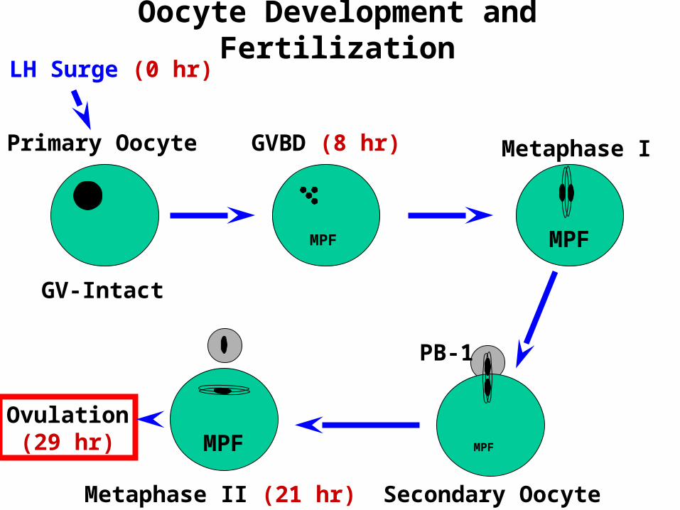
Oocyte Development and Fertilization
MPF MPF
MPFMPF
Primary Oocyte
GV-Intact
GVBD (8 hr) Metaphase I
PB-1
Secondary OocyteMetaphase II (21 hr)
Ovulation(29 hr)
LH Surge (0 hr)
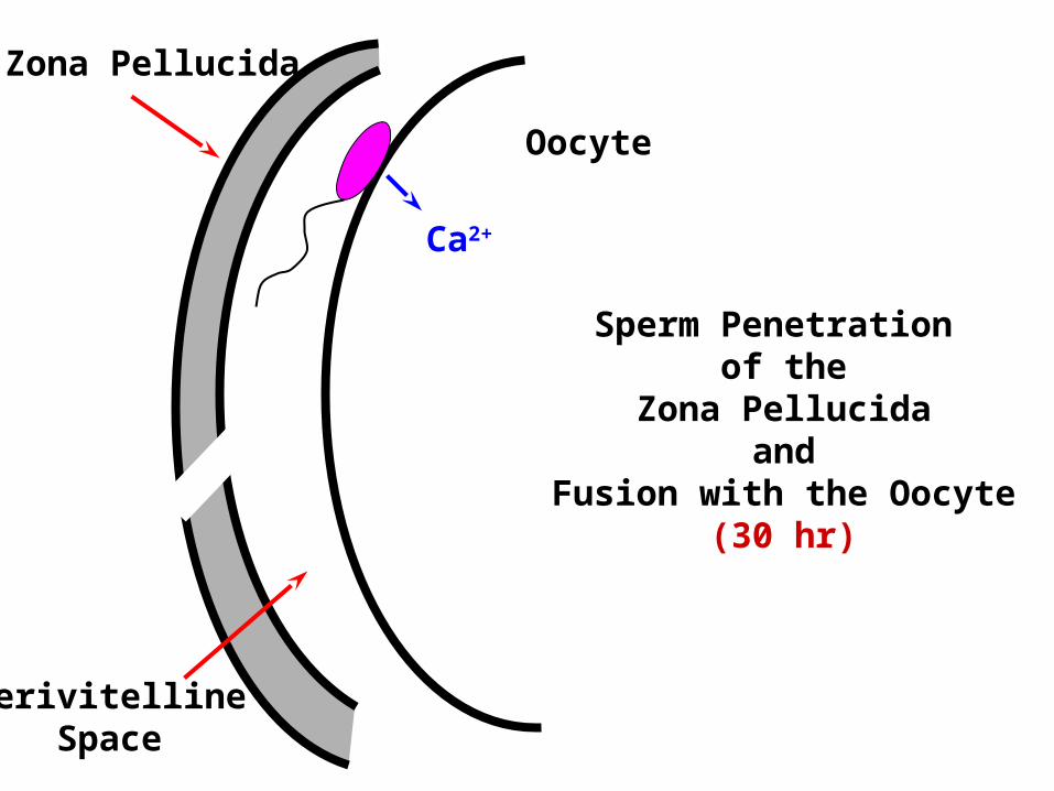
Zona Pellucida
PerivitellineSpace
Oocyte
Ca2+
Sperm Penetration of the
Zona Pellucidaand
Fusion with the Oocyte(30 hr)
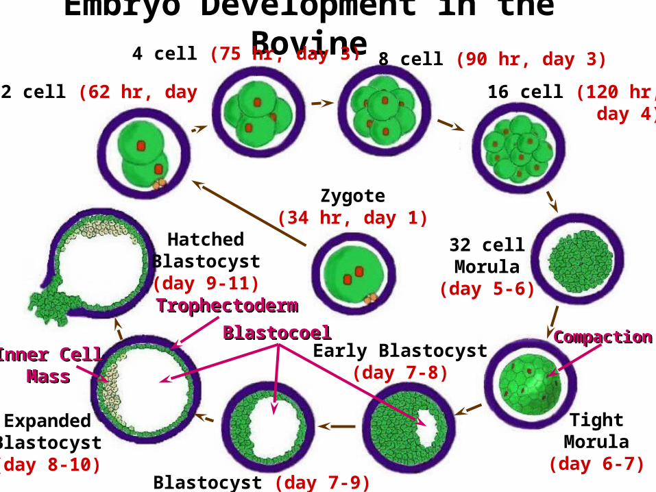
Embryo Development in the Bovine
2 cell (62 hr, day 2)
4 cell (75 hr, day 3) 8 cell (90 hr, day 3)
16 cell (120 hr, day 4)
32 cellMorula
(day 5-6)
Zygote(34 hr, day 1)
TightMorula
(day 6-7)
Early Blastocyst(day 7-8)
Blastocyst (day 7-9)
ExpandedBlastocyst(day 8-10)
HatchedBlastocyst(day 9-11)
CompactionCompactionBlastocoelBlastocoelInner Cell
MassInner Cell
Mass
TrophectodermTrophectoderm
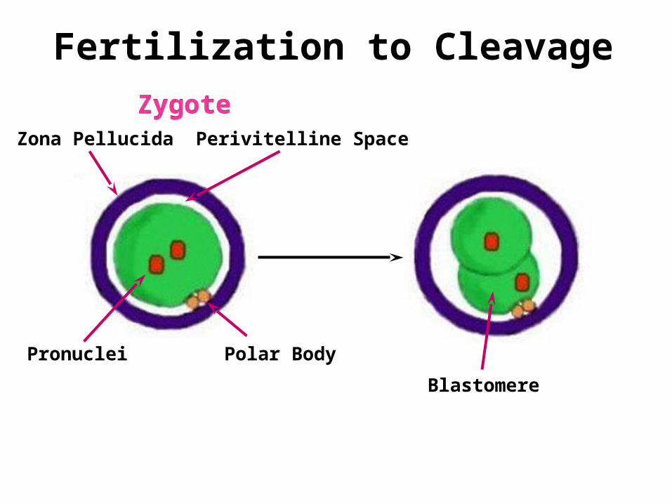
Fertilization to Cleavage
Polar Body
Zona Pellucida
Pronuclei
Perivitelline Space
ZygoteZygote
Blastomere
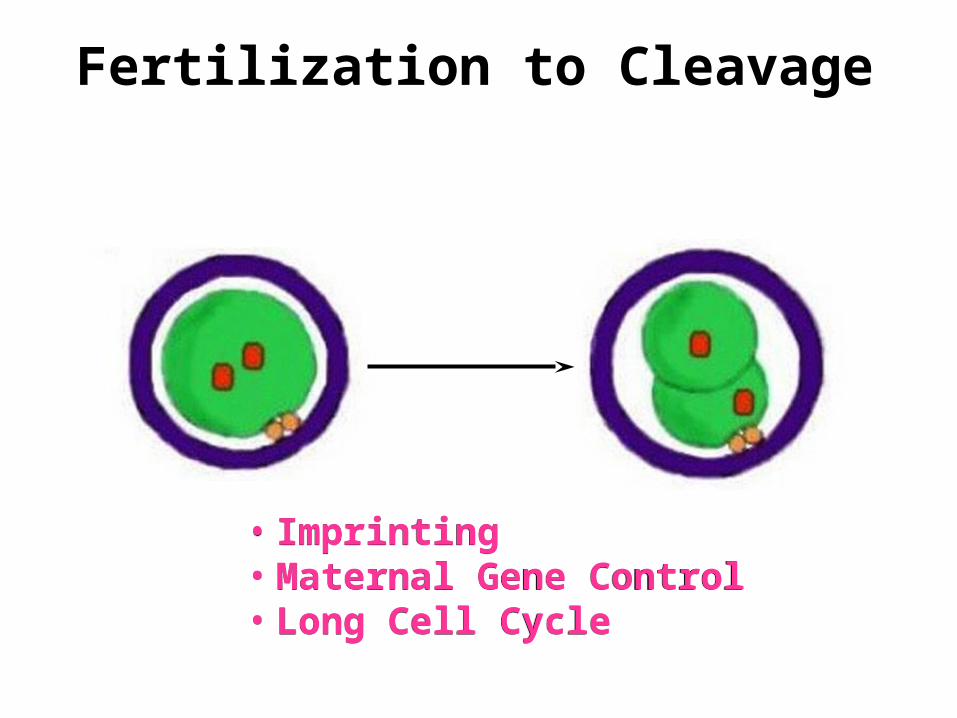
Fertilization to Cleavage
• Imprinting• Maternal Gene Control• Long Cell Cycle
• Imprinting• Maternal Gene Control• Long Cell Cycle
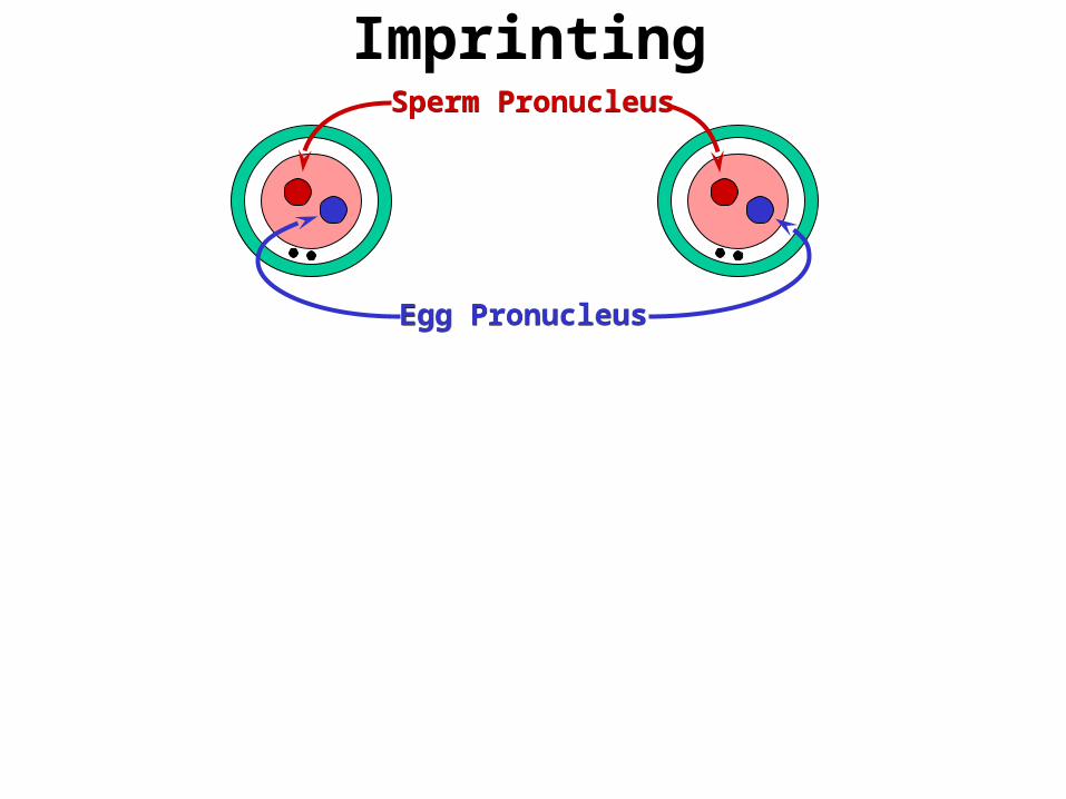
Imprinting
Egg PronucleusEgg Pronucleus
Sperm PronucleusSperm Pronucleus
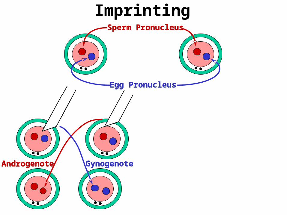
Imprinting
Egg PronucleusEgg Pronucleus
Sperm PronucleusSperm Pronucleus
AndrogenoteAndrogenote GynogenoteGynogenote
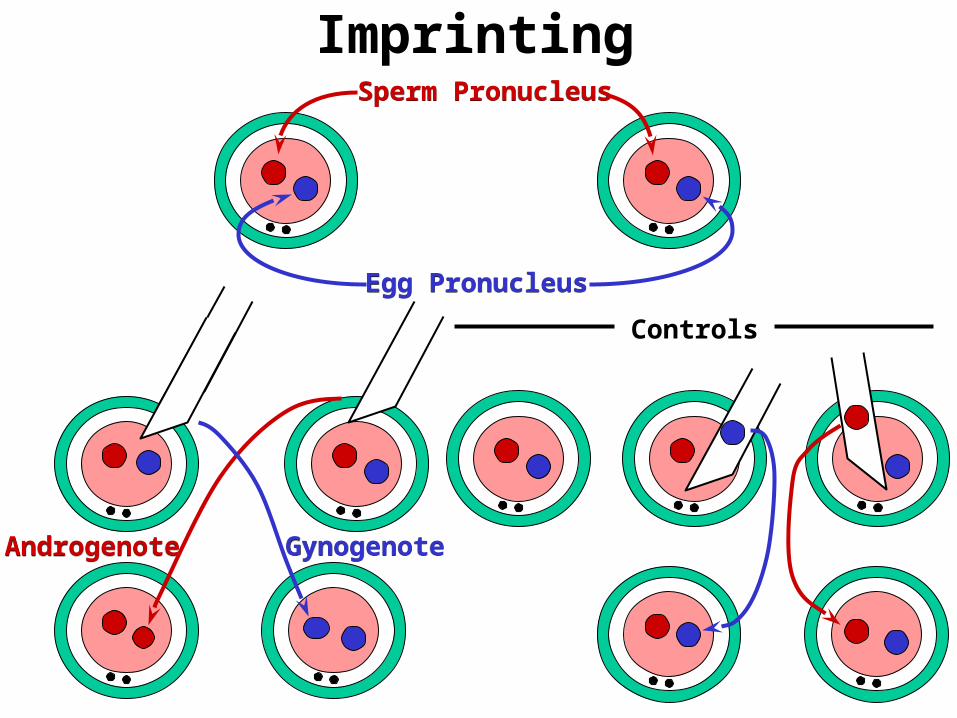
Imprinting
Controls
Egg PronucleusEgg Pronucleus
Sperm PronucleusSperm Pronucleus
AndrogenoteAndrogenote GynogenoteGynogenote
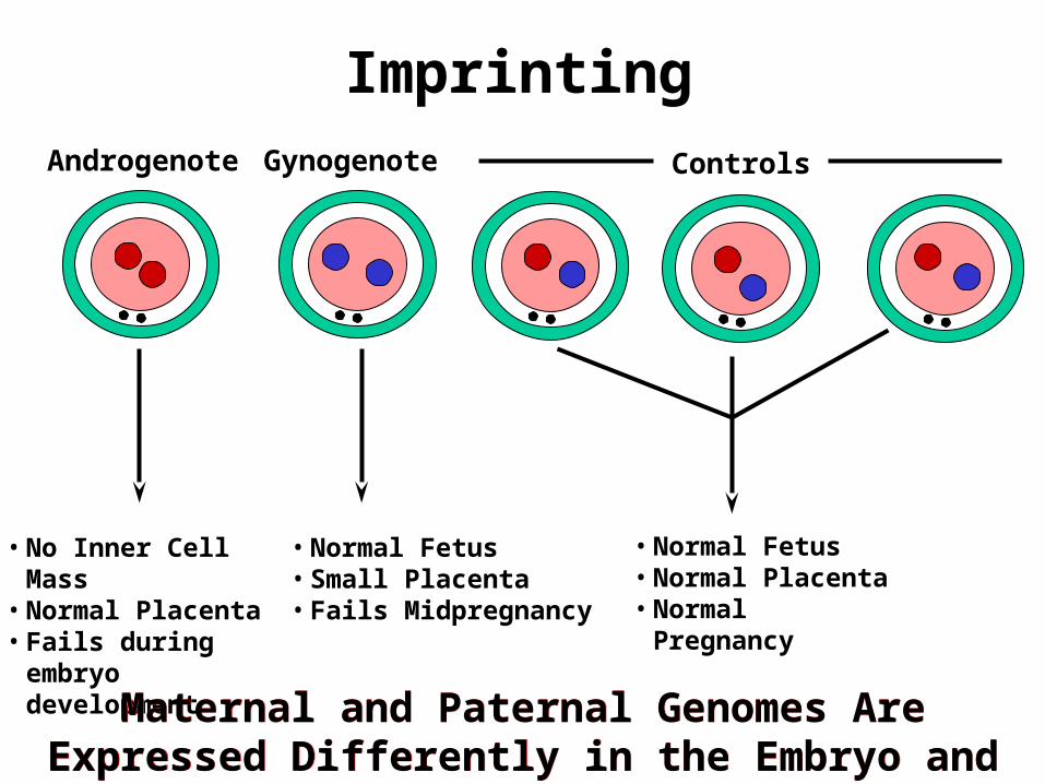
ImprintingAndrogenote Gynogenote Controls
• No Inner Cell Mass• Normal Placenta• Fails during embryo
development
• Normal Fetus• Small Placenta• Fails Midpregnancy
• Normal Fetus• Normal Placenta• Normal Pregnancy
Maternal and Paternal Genomes Are Expressed Differently in the Embryo and Fetus
Maternal and Paternal Genomes Are Expressed Differently in the Embryo and Fetus
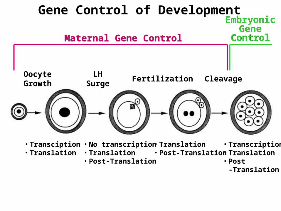
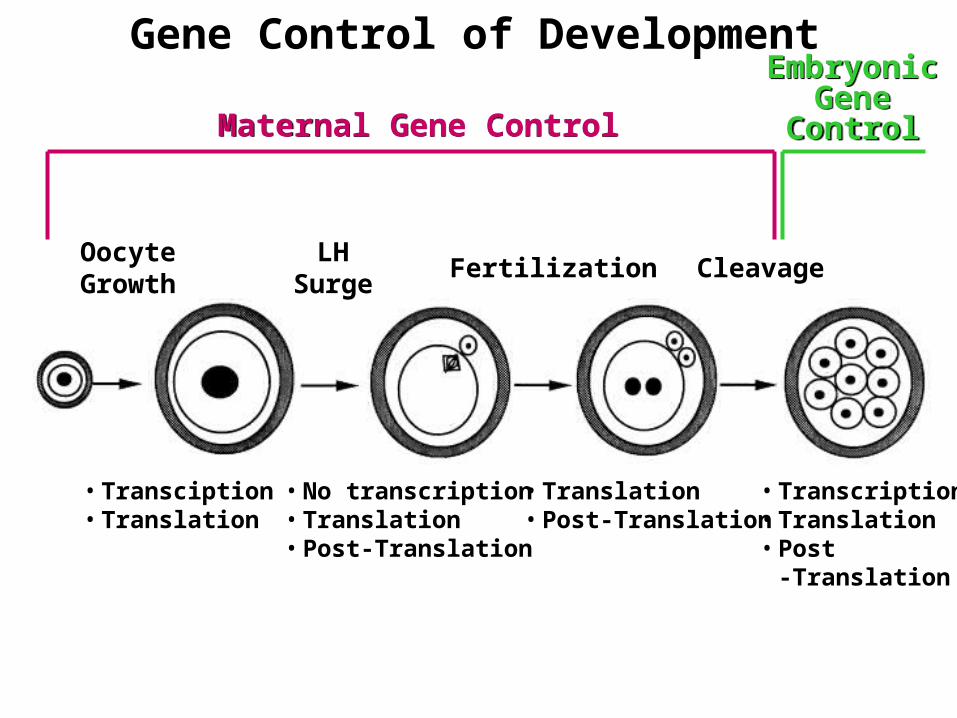
Gene Control of Development
OocyteGrowth
LHSurge
Fertilization Cleavage
• Transciption• Translation
• Transcription• Translation• Post
-Translation
• Translation• Post-Translation
• No transcription• Translation• Post-Translation
Maternal Gene ControlMaternal Gene Control
EmbryonicGene
Control
EmbryonicGene
Control
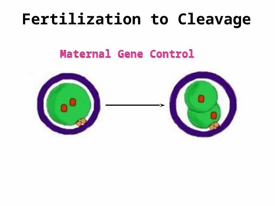
Fertilization to Cleavage
Maternal Gene ControlMaternal Gene Control
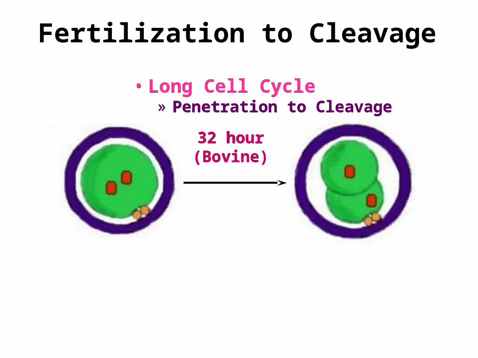
Fertilization to Cleavage
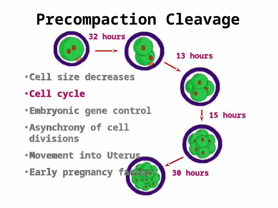
• Long Cell Cycle» Penetration to Cleavage
• Long Cell Cycle» Penetration to Cleavage
32 hour(Bovine)32 hour(Bovine)
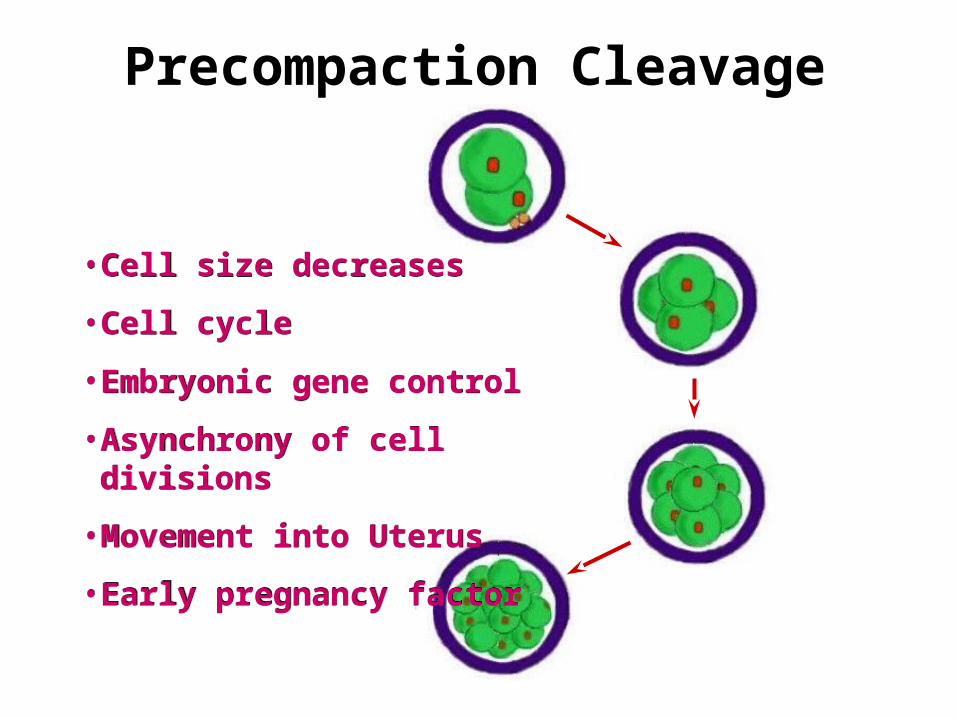
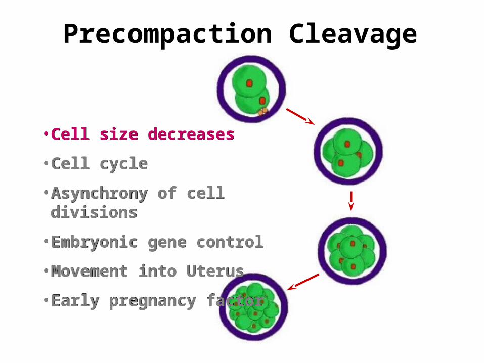
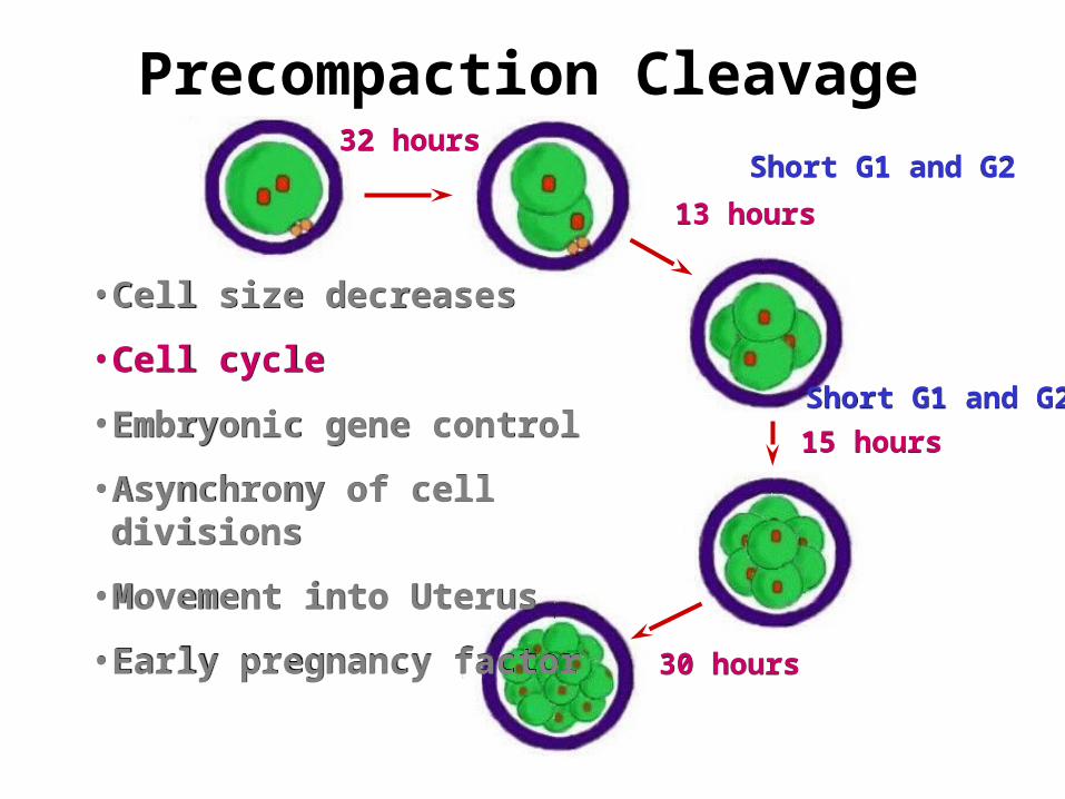
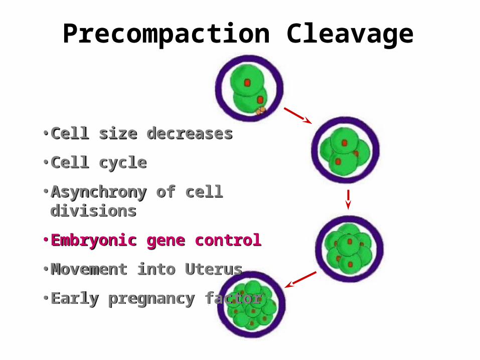
Precompaction Cleavage
• Cell size decreases
• Cell cycle
• Embryonic gene control
• Asynchrony of cell divisions
•Movement into Uterus
• Early pregnancy factor
• Cell size decreases
• Cell cycle
• Embryonic gene control
• Asynchrony of cell divisions
•Movement into Uterus
• Early pregnancy factor
Precompaction Cleavage
• Cell size decreases
• Cell cycle
• Asynchrony of cell divisions
• Embryonic gene control
•Movement into Uterus
• Early pregnancy factor
• Cell size decreases
• Cell cycle
• Asynchrony of cell divisions
• Embryonic gene control
•Movement into Uterus
• Early pregnancy factor
Precompaction Cleavage
• Cell size decreases
• Cell cycle
• Embryonic gene control
• Asynchrony of cell divisions
•Movement into Uterus
• Early pregnancy factor
• Cell size decreases
• Cell cycle
• Embryonic gene control
• Asynchrony of cell divisions
•Movement into Uterus
• Early pregnancy factor
13 hours13 hours
15 hours15 hours
30 hours30 hours
32 hours32 hours
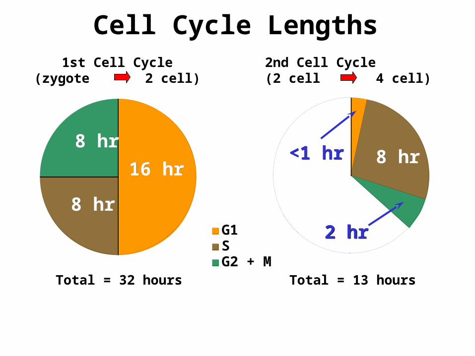
G1SG2 + M
Cell Cycle Lengths
16 hr16 hr
8 hr8 hr
8 hr8 hr8 hr8 hr
2 hr2 hr
<1 hr<1 hr
Total = 32 hours Total = 13 hours
1st Cell Cycle(zygote 2 cell)
2nd Cell Cycle(2 cell 4 cell)
Precompaction Cleavage
• Cell size decreases
• Cell cycle
• Embryonic gene control
• Asynchrony of cell divisions
•Movement into Uterus
• Early pregnancy factor
• Cell size decreases
• Cell cycle
• Embryonic gene control
• Asynchrony of cell divisions
•Movement into Uterus
• Early pregnancy factor
13 hours13 hours
15 hours15 hours
30 hours30 hours
32 hours32 hoursShort G1 and G2Short G1 and G2
Short G1 and G2Short G1 and G2
Precompaction Cleavage
• Cell size decreases
• Cell cycle
• Asynchrony of cell divisions
• Embryonic gene control
•Movement into Uterus
• Early pregnancy factor
• Cell size decreases
• Cell cycle
• Asynchrony of cell divisions
• Embryonic gene control
•Movement into Uterus
• Early pregnancy factor
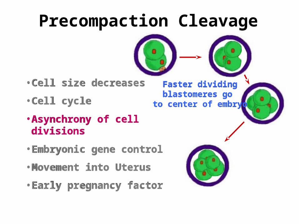
Faster dividingblastomeres go
to center of embryo
Faster dividingblastomeres go
to center of embryo
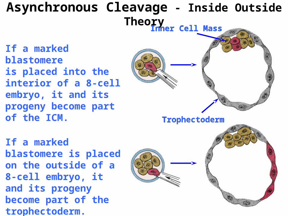
Asynchronous Cleavage - Inside Outside Theory
If a marked blastomereis placed into the interior of a 8-cell embryo, it and its progeny become part of the ICM.
If a marked blastomere is placed on the outside of a 8-cell embryo, it and its progeny become part of the trophectoderm.
TrophectodermTrophectoderm
Inner Cell MassInner Cell Mass
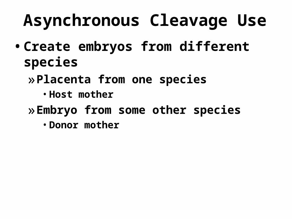
Asynchronous Cleavage Use
• Create embryos from different species»Placenta from one species• Host mother
»Embryo from some other species• Donor mother
Precompaction Cleavage
• Cell size decreases
• Cell cycle
• Asynchrony of cell divisions
• Embryonic gene control
•Movement into Uterus
• Early pregnancy factor
• Cell size decreases
• Cell cycle
• Asynchrony of cell divisions
• Embryonic gene control
•Movement into Uterus
• Early pregnancy factor
Gene Control of Development
OocyteGrowth
LHSurge
Fertilization Cleavage
• Transciption• Translation
• Transcription• Translation• Post
-Translation
• Translation• Post-Translation
• No transcription• Translation• Post-Translation
Maternal Gene ControlMaternal Gene Control
EmbryonicGene
Control
EmbryonicGene
Control
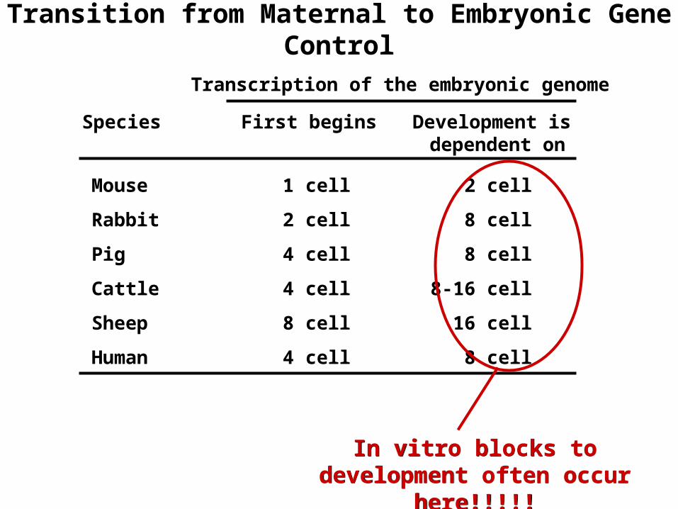
Transition from Maternal to Embryonic Gene Control
In vitro blocks to development often occur here!!!!!
In vitro blocks to development often occur here!!!!!
Transcription of the embryonic genome
First begins Development is dependent on
Species
Mouse 1 cell 2 cell
Rabbit 2 cell 8 cell
Pig 4 cell 8 cell
Cattle 4 cell 8-16 cell
Sheep 8 cell 16 cell
Human 4 cell 8 cell

Precompaction Cleavage
• Cell size decreases
• Cell cycle
• Asynchrony of cell divisions
• Embryonic gene control
•Movement into Uterus
• Early pregnancy factor
• Cell size decreases
• Cell cycle
• Asynchrony of cell divisions
• Embryonic gene control
•Movement into Uterus
• Early pregnancy factor
13 hours13 hours
15 hours15 hours
30 hours30 hours
32 hours32 hours
Cell Cycle Length Increases
Cell Cycle Length Increases
Embryo runs out of key factors coded for
by maternal mRNA
Embryo runs out of key factors coded for
by maternal mRNAPause in G1Pause in G1
Precompaction Cleavage
• Cell size decreases
• Cell cycle
• Asynchrony of cell divisions
• Embryonic gene control
•Movement into Uterus
• Early pregnancy factor
• Cell size decreases
• Cell cycle
• Asynchrony of cell divisions
• Embryonic gene control
•Movement into Uterus
• Early pregnancy factor
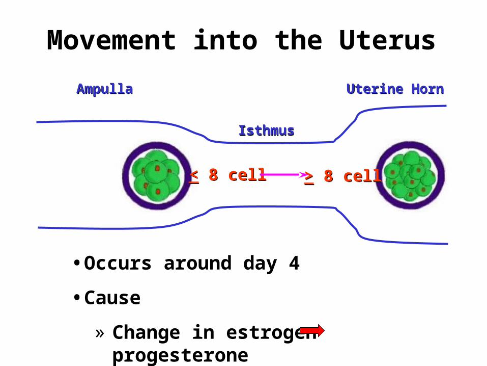
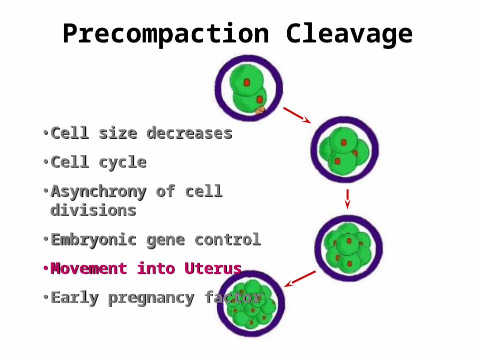
Movement into the Uterus
< 8 cell< 8 cell > 8 cell> 8 cell
AmpullaAmpulla
IsthmusIsthmus
Uterine HornUterine Horn
•Occurs around day 4
•Cause
» Change in estrogen progesterone

Precompaction Cleavage
• Cell size decreases
• Cell cycle
• Asynchrony of cell divisions
• Embryonic gene control
•Movement into Uterus
• Early pregnancy factor
• Cell size decreases
• Cell cycle
• Asynchrony of cell divisions
• Embryonic gene control
•Movement into Uterus
• Early pregnancy factor
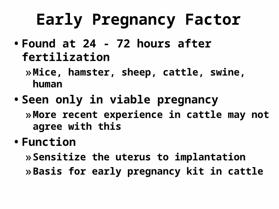
Early Pregnancy Factor
• Found at 24 - 72 hours after fertilization»Mice, hamster, sheep, cattle, swine, human
• Seen only in viable pregnancy»More recent experience in cattle may not
agree with this
• Function»Sensitize the uterus to implantation
»Basis for early pregnancy kit in cattle
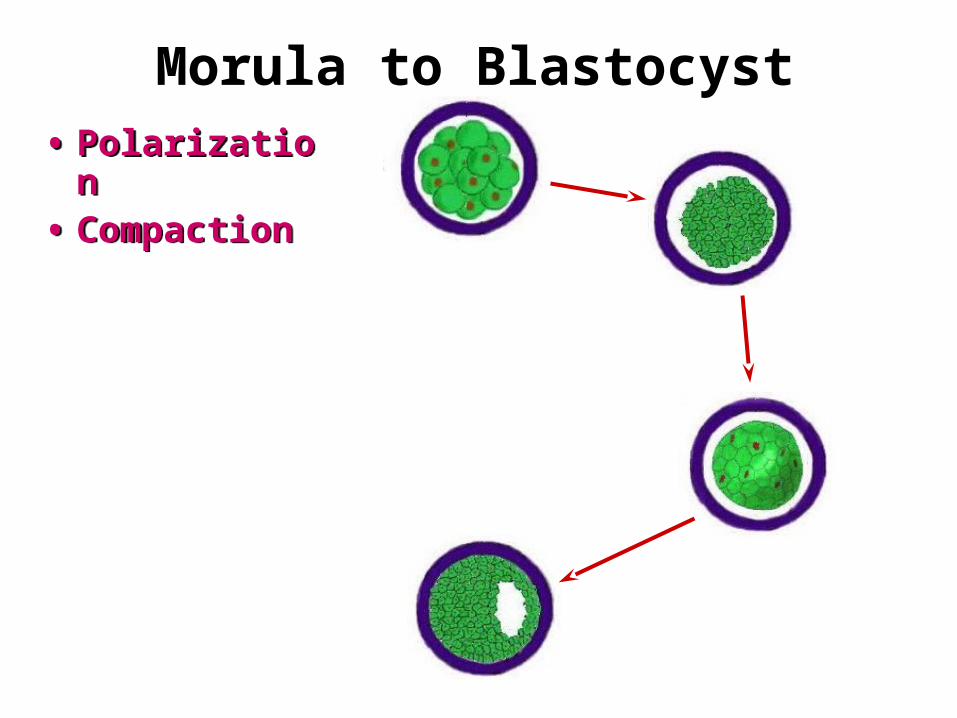
Morula to Blastocyst
• Polarization• Compaction• Polarization• Compaction
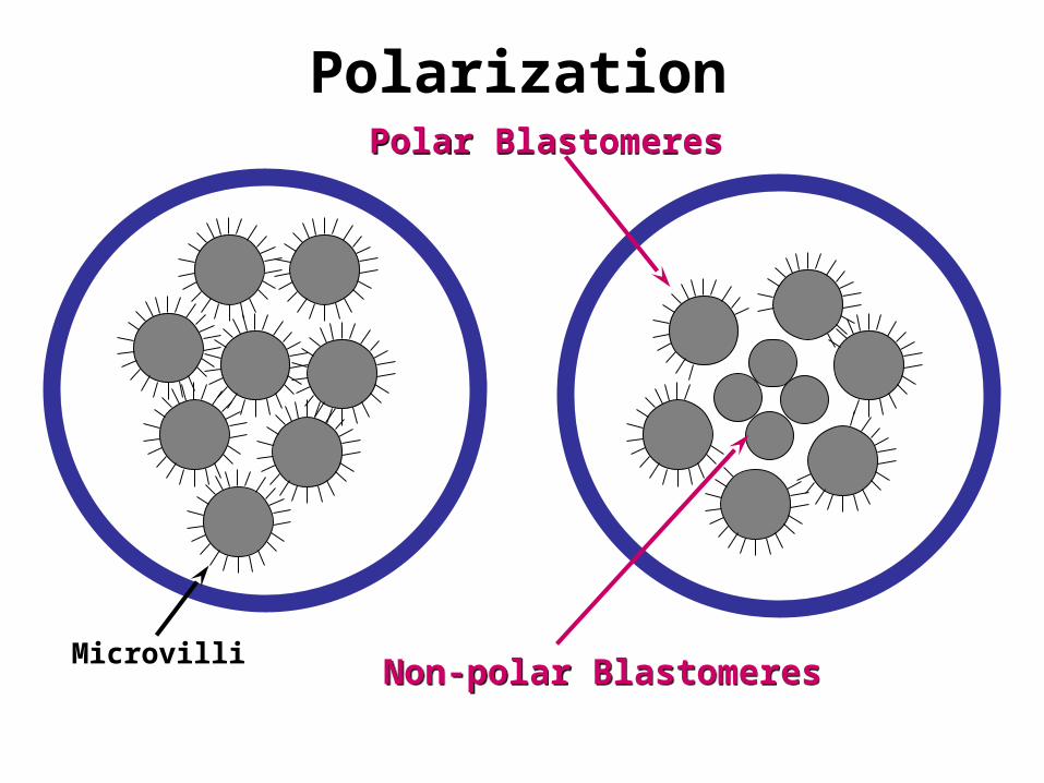
PolarizationPolar BlastomeresPolar Blastomeres
Non-polar BlastomeresNon-polar BlastomeresMicrovilli
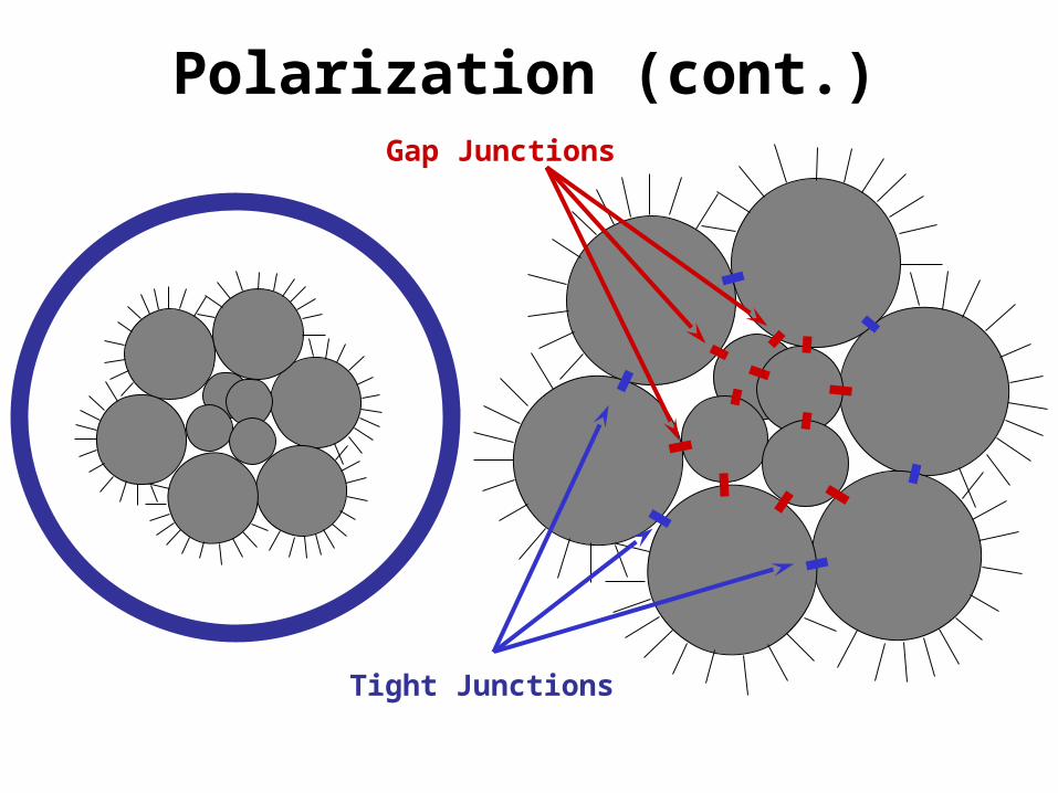
Polarization (cont.)
Tight Junctions
Gap Junctions
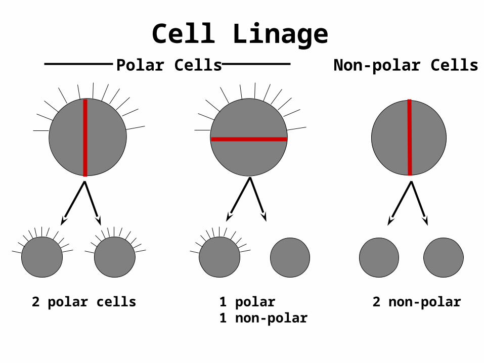
Cell LinagePolar Cells Non-polar Cells
2 polar cells 1 polar1 non-polar
2 non-polar
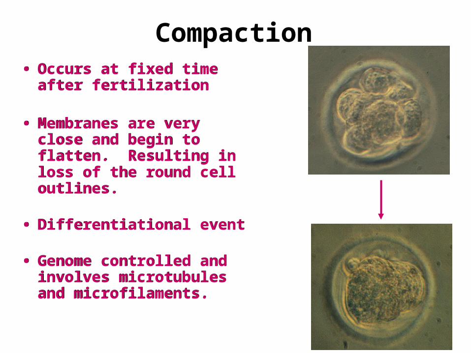
Compaction• Occurs at fixed time after
fertilization
• Membranes are very close and begin to flatten. Resulting in loss of the round cell outlines.
• Differentiational event
• Genome controlled and involves microtubules and microfilaments.
• Occurs at fixed time after fertilization
• Membranes are very close and begin to flatten. Resulting in loss of the round cell outlines.
• Differentiational event
• Genome controlled and involves microtubules and microfilaments.
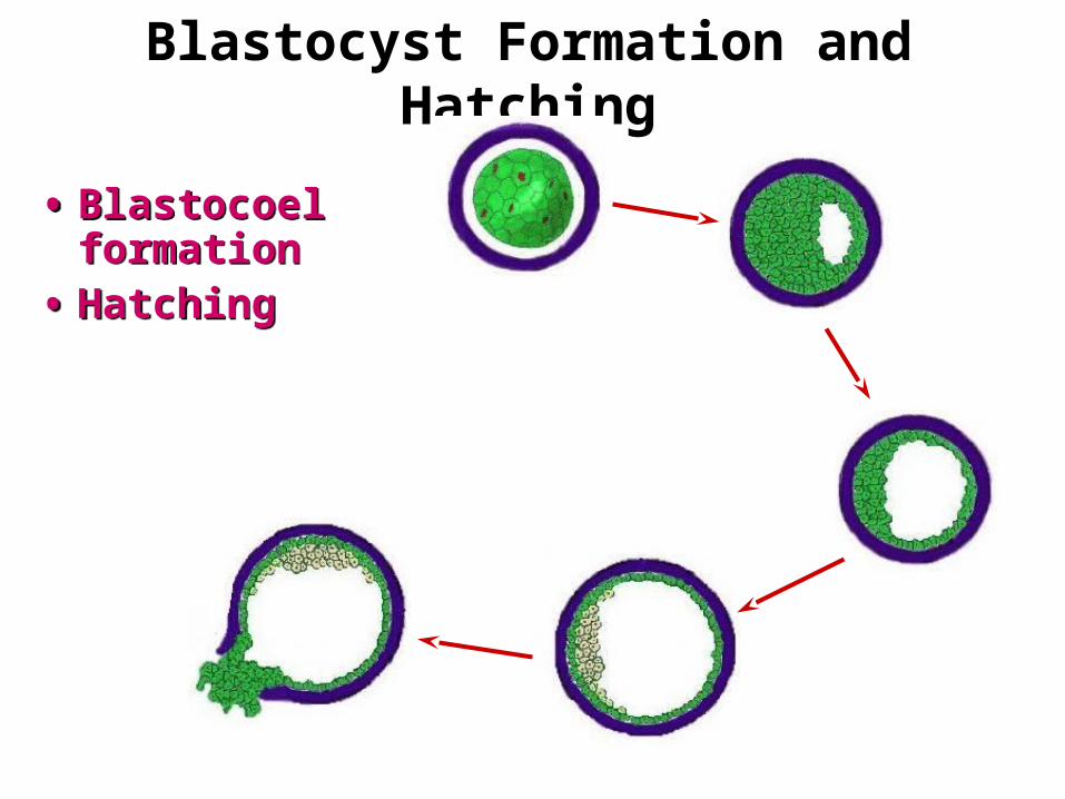
Blastocyst Formation and Hatching
• Blastocoel formation
• Hatching
• Blastocoel formation
• Hatching

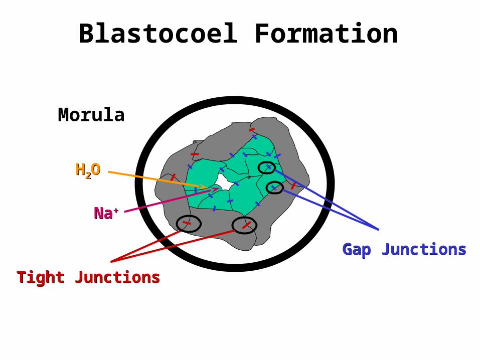
Blastocoel Formation
Tight JunctionsTight Junctions
Gap JunctionsGap Junctions
Na+Na+
H2OH2O
Morula
Blastocoel Formation
Tight JunctionsTight Junctions
Gap JunctionsGap Junctions
Na+Na+
H2OH2O
Morula
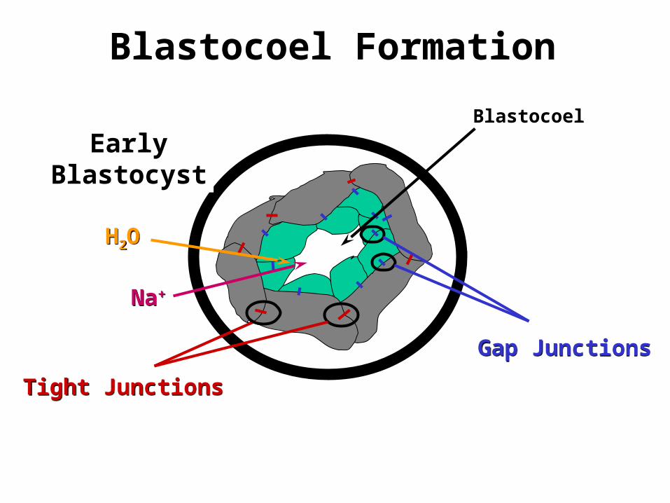
Morula
Blastocoel Formation
Tight JunctionsTight Junctions
Gap JunctionsGap Junctions
Na+Na+
H2OH2O
Blastocoel
EarlyBlastocyst
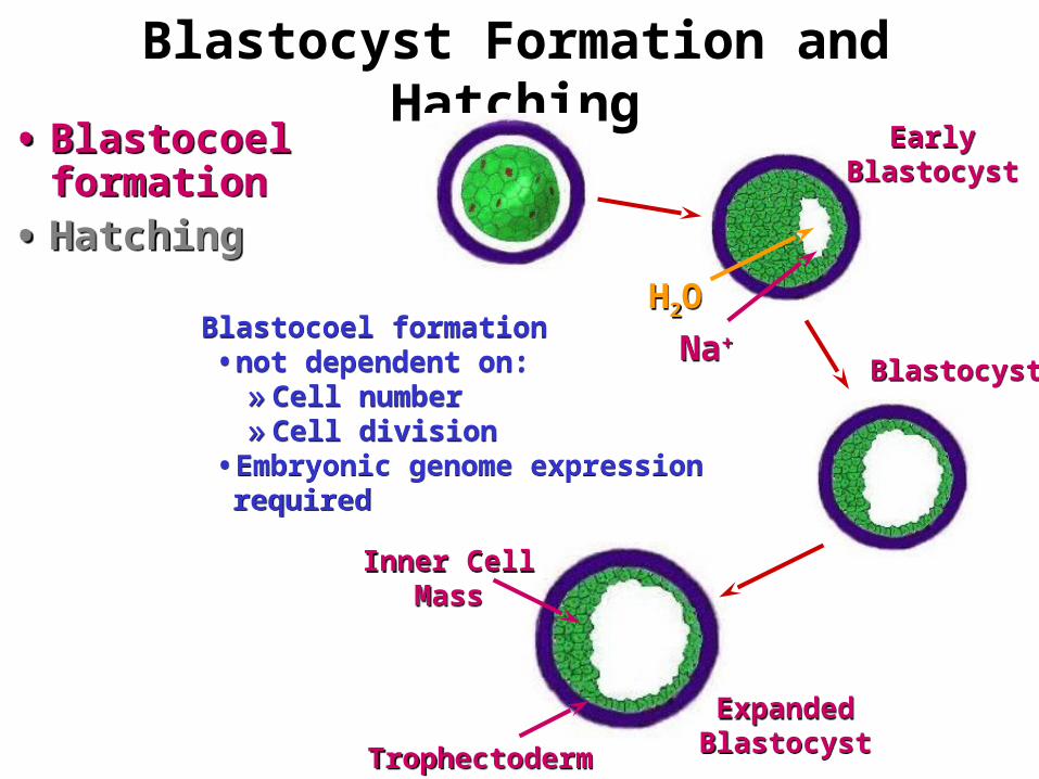
Blastocyst Formation and Hatching• Blastocoel
formation• Hatching
• Blastocoel formation
• Hatching
Na+Na+
H2OH2O
EarlyBlastocyst
EarlyBlastocyst
BlastocystBlastocyst
ExpandedBlastocystExpandedBlastocyst
Inner CellMass
Inner CellMass
TrophectodermTrophectoderm
Blastocoel formation • not dependent on:»Cell number»Cell division
• Embryonic genome expression required
Blastocoel formation • not dependent on:»Cell number»Cell division
• Embryonic genome expression required
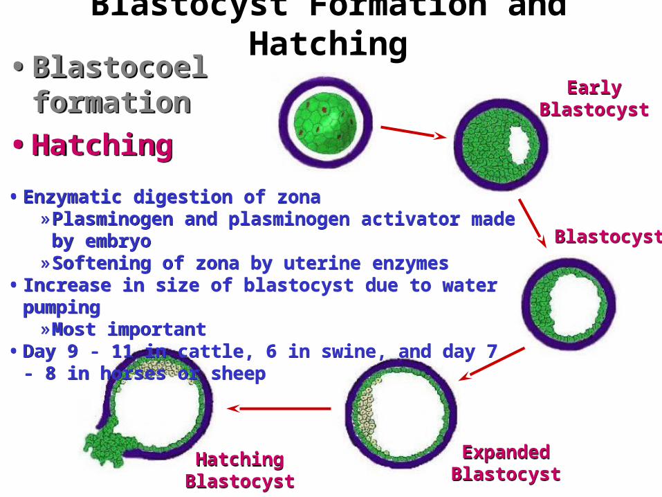
Blastocyst Formation and Hatching
BlastocystBlastocyst
ExpandedBlastocystExpandedBlastocyst
HatchingBlastocystHatching
Blastocyst
• Enzymatic digestion of zona»Plasminogen and plasminogen activator made by
embryo»Softening of zona by uterine enzymes
• Increase in size of blastocyst due to water pumping»Most important
• Day 9 - 11 in cattle, 6 in swine, and day 7 - 8 in horses or sheep
• Enzymatic digestion of zona»Plasminogen and plasminogen activator made by
embryo»Softening of zona by uterine enzymes
• Increase in size of blastocyst due to water pumping»Most important
• Day 9 - 11 in cattle, 6 in swine, and day 7 - 8 in horses or sheep
• Blastocoel formation
• Hatching
• Blastocoel formation
• HatchingEarly
BlastocystEarly
Blastocyst
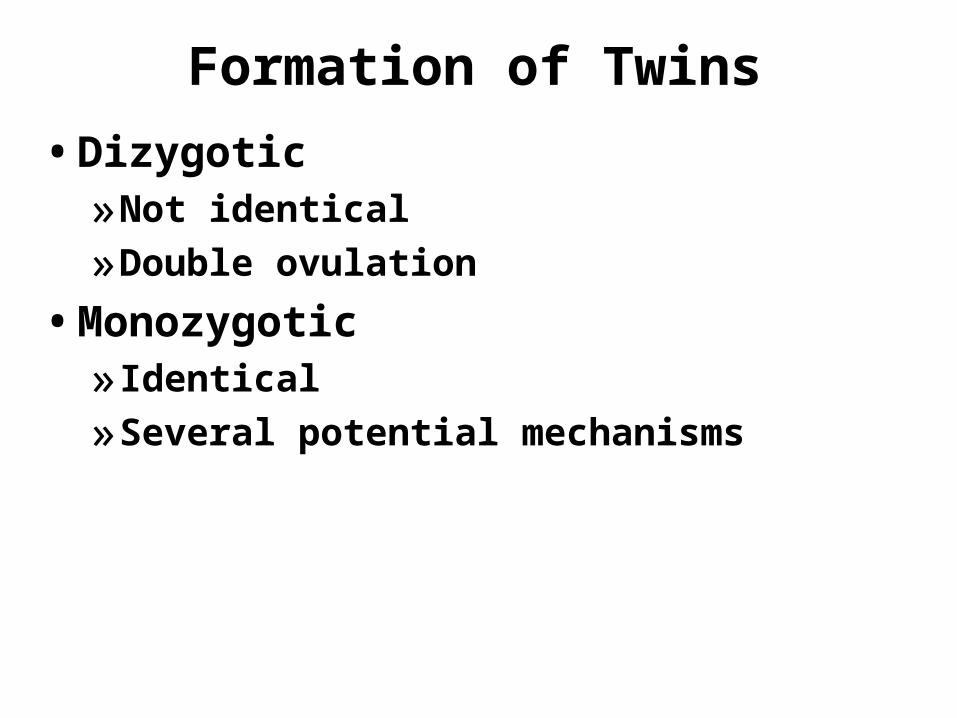
Formation of Twins
• Dizygotic»Not identical
»Double ovulation
• Monozygotic» Identical
»Several potential mechanisms
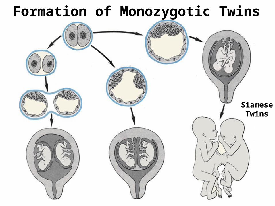
Formation of Monozygotic Twins
SiameseTwins