Embed Size (px)
Citation preview
Full Terms & Conditions of access and use can be found athttp://www.tandfonline.com/action/journalInformation?journalCode=idre20
Download by: [Northeastern University] Date: 12 October 2016, At: 14:36
Disability and Rehabilitation
ISSN: 0963-8288 (Print) 1464-5165 (Online) Journal homepage: http://www.tandfonline.com/loi/idre20
Motor skill changes and neurophysiologicadaptation to recovery-oriented virtualrehabilitation of hand function in a person withsubacute stroke: a case study
Gerard G. Fluet, Jigna Patel, Qinyin Qiu, Matthew Yarossi, Supriya Massood,Sergei V. Adamovich, Eugene Tunik & Alma S. Merians
To cite this article: Gerard G. Fluet, Jigna Patel, Qinyin Qiu, Matthew Yarossi, SupriyaMassood, Sergei V. Adamovich, Eugene Tunik & Alma S. Merians (2016): Motor skill changesand neurophysiologic adaptation to recovery-oriented virtual rehabilitation of handfunction in a person with subacute stroke: a case study, Disability and Rehabilitation, DOI:10.1080/09638288.2016.1226421
To link to this article: http://dx.doi.org/10.1080/09638288.2016.1226421
Published online: 27 Sep 2016.
Submit your article to this journal
Article views: 21
View related articles
View Crossmark data
CASE REPORT
Motor skill changes and neurophysiologic adaptation to recovery-oriented virtualrehabilitation of hand function in a person with subacute stroke: a case study
Gerard G. Flueta, Jigna Patela, Qinyin Qiua, Matthew Yarossia, Supriya Massoodb, Sergei V. Adamovicha,c,Eugene Tunikd and Alma S. Meriansa
aDepartment of Rehabilitation & Movement Sciences, Rutgers The State University of New Jersey, Newark, NJ, USA; bSt. Joseph’s WayneHospital, Acute Rehabilitation Unit, Wayne, NJ, USA; cDepartment of Biomedical Engineering, New Jersey Institute of Technology, UniversityHeights, Newark, NJ, USA; dDepartment of Physical Therapy, Movement, and Rehabilitation Science, Bouve College of Health Sciences,Northeastern University, Boston, MA, USA
ABSTRACTPurpose: The complexity of upper extremity (UE) behavior requires recovery of near normal neuromuscu-lar function to minimize residual disability following a stroke. This requirement places a premium on spon-taneous recovery and neuroplastic adaptation to rehabilitation by the lesioned hemisphere. Motor skilllearning is frequently cited as a requirement for neuroplasticity. Studies examining the links betweentraining, motor learning, neuroplasticity, and improvements in hand motor function are indicated.Methods: This case study describes a patient with slow recovering hand and finger movement (TotalUpper Extremity Fugl–Meyer examination score¼ 25/66, Wrist and Hand items¼ 2/24 on poststroke day37) following a stroke. The patient received an intensive eight-session intervention utilizing simulatedactivities that focused on the recovery of finger extension, finger individuation, and pinch-grasp forcemodulation.Results: Over the eight sessions, the patient demonstrated improvements on untrained transfer tasks,which suggest that motor learning had occurred, as well a dramatic increase in hand function and corre-sponding expansion of the cortical motor map area representing several key muscles of the paretic hand.Recovery of hand function and motor map expansion continued after discharge through the three-monthretention testing.Conclusion: This case study describes a neuroplasticity based intervention for UE hemiparesis and amodel for examining the relationship between training, motor skill acquisition, neuroplasticity, and motorfunction changes.
� IMPLICATIONS FOR REHABILITATION� Intensive hand and finger rehabilitation activities can be added to an in-patient rehabilitation pro-
gram for persons with subacute stroke.� Targeted training of the thumb may have an impact on activity level function in persons with upper
extremity hemiparesis.� Untrained transfer tasks can be utilized to confirm that training tasks have elicited motor learning.� Changes in cortical motor maps can be used to document changes in brain function which can be
used to evaluate changes in motor behavior persons with subacute stroke.
ARTICLE HISTORYReceived 13 November 2015Revised 29 June 2016Accepted 16 August 2016
KEYWORDSStroke; upper extremity;virtual reality; robotics;rehabilitation; hand
Introduction
Despite over a decade of investigation of upper limb motor thera-pies, 78% of people poststroke continue to have upper extremity(UE) deficits that decrease their independence.[1] Some authorssuggest this is due to the fact that the complexity of UE behaviorrequires recovery of neuromuscular function that approaches nor-mal. Arguably, this high level of function decreases the value ofcompensatory strategies and abnormal contralesional control ofthe hemiparetic UE,[2] placing a premium on spontaneous recov-ery and neuroplastic adaptation to rehabilitation by the lesionedhemisphere. In turn, this establishes an imperative to designrehabilitation activities that are congruent with establishedrequirements for neuroplasticity and studies that confirm the
behavioral changes elicited by rehabilitation interventions occur-ring as a result of adaptive patterns of neuroplasticity.[3]
A key factor to consider during the planning of treatmentfocused on neuroplasticity following a stroke is timing. A largemajority of the rehabilitation literature describing interventionstargeting hand function in persons with stroke are conducted inpersons during the chronic stage of recovery. Animal studies haveshown that earlier rehabilitation, leads to greater preservation ofthe cortical areas controlling the hand in both the lesioned andnon-lesioned hemispheres.[4,5] Similar studies in humans suggestthat rehabilitation interventions presented in the first few weeksfollowing a stroke may be more effective.[3] This case study willdescribe changes in lesioned motor cortex function occurring
CONTACT Gerard G Fluet DPT PhD [email protected] Department of Rehabilitation and Movement Sciences, Rutgers the State University ofNew Jersey, Room 714C, 65 Bergen Street, Newark, New Jersey, 07101 USA� 2016 Informa UK Limited, trading as Taylor & Francis Group
DISABILITY AND REHABILITATION, 2016http://dx.doi.org/10.1080/09638288.2016.1226421
during the course of a neuroplasticity-based intervention, per-formed by a person in the subacute stage of stroke recovery.
Another key factor in the design of rehabilitation programsduring this period is the amount of practice needed. Three hun-dred repetitions of UE activity has been identified as necessary toelicit neuro-plastic adaptations in persons with stroke.[6]Observational studies of typical rehabilitation sessions describesubstantially less activity.[7] Constraint induced movement proto-cols elicit more than three hundred repetitions per hour.[8]Studies of CIMT, delivered to persons who are less than onemonth following a stroke in inpatient rehabilitation facilities, haveestablished that this higher repetition arm and hand motor train-ing is safe, feasible, and effective.[9] The simulations making upthe intervention described in this paper are designed to deliver alarge number of repetitions of activity efficiently.
Another requirement for behavior-dependent neuroplasticity isthe continuous development of motor skill. In studies of animaland human response to behavioral interventions, changes inneural function are tied to changes in motor skill.[10] Thesechanges level off when subjects continue to practice skills afterthey have been mastered.[11] This case presents a set of simu-lated rehabilitation activities controlled by algorithms that makethe activity more challenging as soon as the current difficulty levelis mastered. Our lab has utilized this approach successfully in therehabilitation of persons with chronic [12] and it has beenemployed successfully in another lab’s study of persons with sub-acute stroke.[13] This case will utilize two approaches to themeasurement of motor skill learning. The first is the measurementof performance during training tasks. The second is measurementof improvement in the ability of the fingers to perform untrainedtasks.
This case study presents the responses of a subject with sub-acute stroke to an eight-session program of simulated activities invirtual reality (VR) that was designed to provide a strong stimulusfor neuroplastic adaptation. The activities target recovery of thehand, arm, and fingers and utilize high repetitions of activitiesscaled to the patient’s movement abilities that are constantly pro-gressed in difficulty. Training and transfer task kinematics aremeasured to document and confirm motor skill development.Transcranial magnetic stimulation (TMS) is utilized to describeplasticity of the brain structures controlling the fingers. Finally,clinical measures are presented to confirm the translation of thesechanges in motor skill and brain function into real-world move-ment ability.
Methods
Participant and protocol
AD was a 62 year-old right-handed female with a history of leftpontine stroke with right hemiplegia, non-fluent aphasia and dys-phagia, 37 days prior to her enrollment in the study. Her medicalhistory was positive for uncontrolled hypertension and hyperthy-roidism. AD was transferred to an Acute Rehabilitation Unit eightdays after her stroke and received a standard inpatient rehabilita-tion program of one hour each of Occupational, Physical, andSpeech Therapy, five days per week which started 9 days after herCVA. Prior to her enrollment in this study AD participated in 31(30min) sessions of Occupational Therapy. She began training inthis study 37 days poststroke and also continued with herongoing standard rehabilitation. AD returned to the communityfollowing her posttest. She completed 9 sessions of outpatientOccupational Therapy that focused on her UE prior to her reten-tion testing session which occurred three months later.
AD was part of a larger sample of individuals recruited for apilot study conducted at the Acute Rehabilitation Unit of St.Joseph’s Hospital in Wayne, NJ. The study protocol was approvedby the Institutional Review Board of St. Joseph’s Hospital. AD per-formed a one-hour VR-based intervention eight times in a two-week period. All VR simulations were designed to facilitate highlyrepetitive practice combined with accuracy demands that requiredconscious attention to task. Training time was split evenlybetween simulations that addressed arm and shoulder movementand simulations that addressed hand and finger movement. Thebalance of this case report will address the VR-based interventionsand outcomes related to the hand and fingers.
Hardware and software
The NJIT-Track Glove system consists of a CyberGloveTM (CyberGlove Systems San Jose, CA), which is an instrumented glove forfinger angle tracking, and a TrackStarTM three-dimensional mag-netic tracking system (Ascension Technology, Shelburne, VT) usedto track hand position and orientation. The CyberGlove was usedas an interface between AD and the virtual environments for threeof the simulations used in this case study, Piano Trainer, MirrorPong, and Space Pong. For the Piano Trainer and Mirror Pong sim-ulations, the CyberGrasp device, a force reflecting exoskeletonthat fits over a CyberGlove data glove was used to facilitate fingerextension.[14,15] Pinch force training and testing was performedusing an ATI Nano17TM force sensor (ATI Industrial Automation,Apex, NC). All the VR simulations were developed using Matlab(Mathworks, Nattick, MA), Cþþ, VirtoolsTM (Dassault Systemes,V�elizy-Villacoublay Cedex, France), or an Open GL library. AD per-formed four simulations that addressed hand function, PianoTrainer, Space Pong, Mirror Pong, and Monkey Business.
Mirror Pong is utilized to prime the motor cortex to maximizethe effectiveness of the three simulations that follow it. The par-ticipant utilizes a cyberglove worn on the unimpaired hand tocontrol the virtual image of their impaired hand and a Cybergraspworn on the paretic hand which passively extends the fingers.The participant views a virtual image of their paretic hand withtheir own hands covered by the viewing screen. The amount ofmovement of the paretic hand is yoked to the amount of move-ment of the unimpaired hand. In theory, priming of the motorcortex is accomplished via two mechanisms. Viewing a moving vir-tual image of the paretic hand controlled by the non-paretic handhas been linked to increased activity in the lesioned primary mor-tex cortex as measured by functional magnetic resonance imag-ing.[16] In addition, an extended period of repetitive passivemovement of a paretic hand has been linked to upregulation ofthe lesioned motor cortex and improvements in motor perform-ance, immediately following this period, in persons withstroke.[17] The simulation utilized in our intervention adds a gamein which the virtual paretic hand is used to strike a moving targetwith each movement in order to increase conscious attention tothe task.
Piano Trainer is designed to improve finger individuation. Thesimulation and its algorithm for shaping finger individuation aredescribed in detail elsewhere.[15] Our approach to initiating train-ing with facilitation from the Cybergrasp and weaning this facilita-tion over the course of the intervention is described in detail inour case report on a patient with chronic stroke.[18] Space Pongtargets the control of mass finger flexion and extension. The sub-jects control a pong paddle using flexion and extension of the fin-gers measured with a CyberGlove, playing a game against thecomputer. Gain can be magnified during the performance of thissimulation to allow a participant with as little as 3� of active finger
2 G. G. FLUET ET AL.
movement to perform a meaningful activity. Gain magnification ismodified on a daily basis to increase the challenge of this simula-tion as active finger movement abilities increase. Monkey Businesstargets control of a fore-finger and thumb, pinching movement ina fashion similar to Space Pong. The participant controls a mon-key avatar, as it jumps to grab tree branches. Larger pinch forcesresult in higher jumping height by the monkey. Threshold of theload cell utilized to measure pinch force is small enough to allowparticipants with trace finger flexion and thumb adductionstrength to participate in this activity. Pinch force is modified on adaily basis to increase the challenge of this simulation as fingerstrength increases.
TMS was used to assay changes in cortical neurophysiologyby mapping the topographic representation of the finger-handmuscles (first dorsal interossei [FDI], adductor policis brevis[APB], extensor digitorum communicus [EDC]) before and aftertraining and a third session three months after training. TMSmapping was performed using a Magstim Rapid2TM 70mmdouble coil. Surface EMG was recorded using a 2 kHz DelsysTrignoTM system.
Motor mapping
TMS mapping of the motor cortex was performed by deliveringTMS pulses over an expanse of the sensorimotor cortex andmeasuring the motor evoked potentials (MEPs) in the contralat-eral FDI, APB, and EDC muscles. Maps for the lesioned andnon-lesioned hemispheres were collected. For the mapping pro-cedure, AD was seated with her UE comfortably secured tolimit motion. To ensure spatial TMS precision, AD’s high-resolution anatomical MRI was used to render a 3D cortical sur-face that was co-registered with her head to allow for framelessneuronavigation (Advanced Neuro Technology). For all stimuli,the TMS coil was held tangential to the scalp with the handleposterior 45� off the sagittal plane. We first identified the hot-spot for the contralateral FDI, defined as the loci which pro-duced the maximal MEP in the FDI muscle. Following this, theresting motor threshold (RMT) was calculated as the minimumintensity required to elicit MEPs >50 lV in the FDI muscle on50% of six consecutive trials.[19] Stimulation intensity was setto 110% of the determined RMT [20] for mapping. A 10� 10cmarea surrounding the motor hotspot was marked using the neu-ronavigation software to provide consistent map boundaries.Real time visual feedback of the MEP time traces and neurona-vigated coil position provided to the experimenter during test-ing maximized the map information obtained by allowing forincreased density of points in excitable and border regions,with less attention given to far-away non-responsive areas.[21]For each stimulation point we computed the following meas-ures: (i) MEP as the peak-to-peak amplitude of the EMG signal20–50ms after the TMS pulse, and (ii) background EMG, calcu-lated as the EMG signal in the 50ms interval before the TMSpulse (second order Butterworth filter, 5–250Hz band-pass, full-wave rectified, 20Hz low-pass envelope). A threshold of 50lVwas used to identify MEPs from background EMG for the FDIand APB 36. MEPs were weaker for the EDC so a threshold of25lV was used to generate this map. To allow comparisonsacross maps and sessions, MEP amplitudes and stimulationpoints were interpolated to a 10� 10cm mesh of 5mm reso-lution centered on the M1 hotspot, using cubic surface interpol-ation.[22] Extent of the representation producing corticospinaloutput (MEPs), or map area, is calculated as the product of thenumber of interpolated scalp sites eliciting MEPs and the map
resolution (0.5mm).[22,23] Map area for each muscle is reportedas the primary outcome measure for this study.
Clinical measurements
The Upper Extremity Fugl–Meyer Exam (UEFMA) is a 33 item bat-tery, which evaluates reflexes, movement patterns, grasp, andcoordination, higher scores indicate better performance.[24] TheWolf Motor Function Test (WMFT) is a 15-item battery that meas-ures at the activity level.[25] A score of “unable” was recordedwhen the subject could not perform a task within 120 seconds.The ability to perform a test item within 120 seconds at a test sub-sequent to the pretest that a participant was previously unable toperform has been cited as a clinically meaningful change in per-sons with CVA.[26] The Box and Blocks Test (BBT) is a simplemeasure of unilateral gross manual dexterity. It measures thenumber of wooden blocks moved in one minute from one side ofa box to the other.[27]
Kinematic assessment
All measures were collected with the patient seated with theirtrunks supported and their hands resting on a table. Performancefor tracing tasks was measured following a single familiarizationtrial, which was not scored.
Finger range of motion was measured as the differencebetween all the joint angles with the fingers in a flexed position(fist) and the joint angles of all of the fingers extended (open) asmuch as possible. Finger angles were collected using aCybergloveTM (Cyberglove Systems, San Jose CA). Larger numbersindicated increased active finger extension range of motion.
Finger Trace is the ability to control active finger extensionand flexion between 0% and 80% of maximum finger extension,measured by having the subject flex and extend their fingers tocontrol a cursor tracking a sine wave (duration of 1 cycle � 6 s,period¼ 0.15 Hz). Smaller root mean square error (RMSE) valuesindicate improved performance.
Pinch force was measured as the maximum force a subject canexert on a force sensor held between their paretic thumb andindex finger, given two trials. Larger numbers indicate strongerpinch force. This task was designed as a transfer test of motorlearning accomplished during the Space Pong simualtion.
Pinch Trace measures the ability to control pinch forcebetween 0% and 80% of maximum pinch force, measured by hav-ing the subject vary pinch grip force to control a cursor tracking asine wave (duration of 1 cycle � 6 s, period¼ 0.15Hz). SmallerRMSE values indicate better performance. This task was designedas a transfer test of motor learning accomplished during theMonkey Business simualtion.
Results
Training kinematics
AD completed eight sessions of VR training with no adverse events,complaints of increased UE pain, or fatigue that limited her abilityto complete the rest of her in-patient acute rehabilitation program.During the first session AD was extremely excited that she could“do something with her hand”. Her first session using the VR sys-tem was the first time since her stroke that she actively controlledher fingers during an activity. She did not miss a session during thetrial and expressed a desire to use the equipment after the studyperiod was completed. AD performed more than 300 repetitions ineach of the eight sessions meeting the minimum repetition
VIRTUAL REHABILITATION OF HAND IN SUBACUTE STROKE 3
requirements for neuroplasticity. Two of the finger training simula-tions are relatively discrete activities that lend themselves to per-formance analysis during the training process.
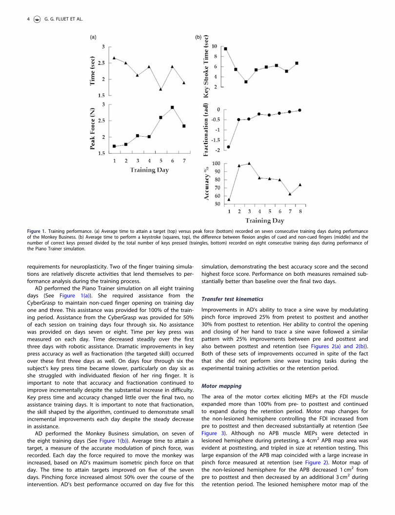
AD performed the Piano Trainer simulation on all eight trainingdays (See Figure 1(a)). She required assistance from theCyberGrasp to maintain non-cued finger opening on training dayone and three. This assistance was provided for 100% of the train-ing period. Assistance from the CyberGrasp was provided for 50%of each session on training days four through six. No assistancewas provided on days seven or eight. Time per key press wasmeasured on each day. Time decreased steadily over the firstthree days with robotic assistance. Dramatic improvements in keypress accuracy as well as fractionation (the targeted skill) occurredover these first three days as well. On days four through six thesubject’s key press time became slower, particularly on day six asshe struggled with individuated flexion of her ring finger. It isimportant to note that accuracy and fractionation continued toimprove incrementally despite the substantial increase in difficulty.Key press time and accuracy changed little over the final two, noassistance training days. It is important to note that fractionation,the skill shaped by the algorithm, continued to demonstrate smallincremental improvements each day despite the steady decreasein assistance.
AD performed the Monkey Business simulation, on seven ofthe eight training days (See Figure 1(b)). Average time to attain atarget, a measure of the accurate modulation of pinch force, wasrecorded. Each day the force required to move the monkey wasincreased, based on AD’s maximum isometric pinch force on thatday. The time to attain targets improved on five of the sevendays. Pinching force increased almost 50% over the course of theintervention. AD’s best performance occurred on day five for this
simulation, demonstrating the best accuracy score and the secondhighest force score. Performance on both measures remained sub-stantially better than baseline over the final two days.
Transfer test kinematics
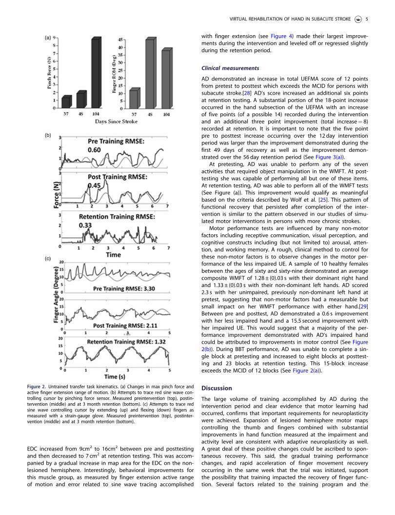
Improvements in AD’s ability to trace a sine wave by modulatingpinch force improved 25% from pretest to posttest and another30% from posttest to retention. Her ability to control the openingand closing of her hand to trace a sine wave followed a similarpattern with 25% improvements between pre and posttest andalso between posttest and retention (see Figures 2(a) and 2(b)).Both of these sets of improvements occurred in spite of the factthat she did not perform sine wave tracing tasks during theexperimental training activities or the retention period.
Motor mapping
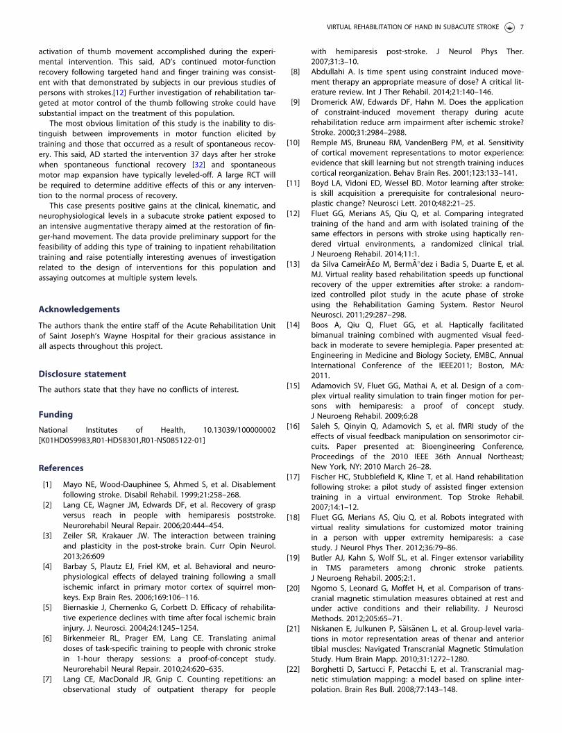
The area of the motor cortex eliciting MEPs at the FDI muscleexpanded more than 100% from pre- to posttest and continuedto expand during the retention period. Motor map changes forthe non-lesioned hemisphere controlling the FDI increased frompre to posttest and then decreased substantially at retention (SeeFigure 3). Although no APB muscle MEPs were detected inlesioned hemisphere during pretesting, a 4cm2 APB map area wasevident at posttesting, and tripled in size at retention testing. Thislarge expansion of the APB map coincided with a large increase inpinch force measured at retention (see Figure 2). Motor map ofthe non-lesioned hemisphere for the APB decreased 1 cm2 frompre to posttest and then decreased by an additional 3 cm2 duringthe retention period. The lesioned hemisphere motor map of the
Figure 1. Training performance. (a) Average time to attain a target (top) versus peak force (bottom) recorded on seven consecutive training days during performanceof the Monkey Business. (b) Average time to perform a keystroke (squares, top), the difference between flexion angles of cued and non-cued fingers (middle) and thenumber of correct keys pressed divided by the total number of keys pressed (traingles, bottom) recorded on eight consecutive training days during performance ofthe Piano Trainer simulation.
4 G. G. FLUET ET AL.
EDC increased from 9cm2 to 16cm2 between pre and posttestingand then decreased to 7 cm2 at retention testing. This was accom-panied by a gradual increase in map area for the EDC on the non-lesioned hemisphere. Interestingly, behavioral improvements forthis muscle group, as measured by finger extension active rangeof motion and error related to sine wave tracing accomplished
with finger extension (see Figure 4) made their largest improve-ments during the intervention and leveled off or regressed slightlyduring the retention period.
Clinical measurements
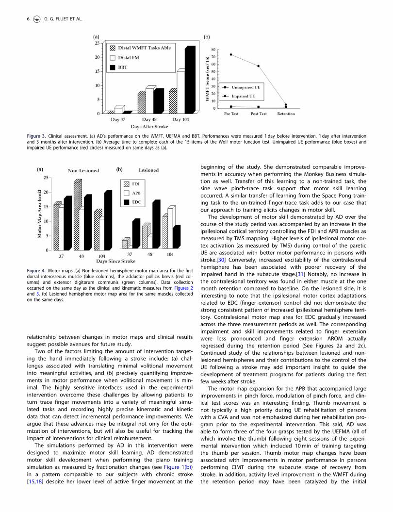
AD demonstrated an increase in total UEFMA score of 12 pointsfrom pretest to posttest which exceeds the MCID for persons withsubacute stroke.[28] AD’s score increased an additional six pointsat retention testing. A substantial portion of the 18-point increaseoccurred in the hand subsection of the UEFMA with an increaseof five points (of a possible 14) recorded during the interventionand an additional three point improvement (total increase¼ 8)recorded at retention. It is important to note that the five pointpre to posttest increase occurring over the 12 day interventionperiod was larger than the improvement demonstrated during thefirst 49 days of recovery as well as the improvement demon-strated over the 56 day retention period (See Figure 3(a)).
At pretesting, AD was unable to perform any of the sevenactivities that required object manipulation in the WMFT. At post-testing she was capable of performing all but one of these items.At retention testing, AD was able to perform all of the WMFT tests(See Figure (a)). This improvement would qualify as meaningfulbased on the criteria described by Wolf et al. [25]. This pattern offunctional recovery that persisted after completion of the inter-vention is similar to the pattern observed in our studies of simu-lated motor interventions in persons with more chronic strokes.
Motor performance tests are influenced by many non-motorfactors including receptive communication, visual perception, andcognitive constructs including (but not limited to) arousal, atten-tion, and working memory. A rough, clinical method to control forthese non-motor factors is to observe changes in the motor per-formance of the less impaired UE. A sample of 10 healthy femalesbetween the ages of sixty and sixty-nine demonstrated an averagecomposite WMFT of 1.28 ± (0).03 s with their dominant right handand 1.33 ± (0).03 s with their non-dominant left hands. AD scored2.3 s with her unimpaired, previously non-dominant left hand atpretest, suggesting that non-motor factors had a measurable butsmall impact on her WMFT performance with either hand.[29]Between pre and posttest, AD demonstrated a 0.6 s improvementwith her less impaired hand and a 15.5 second improvement withher impaired UE. This would suggest that a majority of the per-formance improvement demonstrated with AD’s impaired handcould be attributed to improvements in motor control (See Figure2(b)). During BBT performance, AD was unable to complete a sin-gle block at pretesting and increased to eight blocks at posttest-ing and 23 blocks at retention testing. This 15-block increaseexceeds the MCID of 12 blocks (See Figure 2(a)).
Discussion
The large volume of training accomplished by AD during theintervention period and clear evidence that motor learning hadoccurred, confirms that important requirements for neuroplasticitywere achieved. Expansion of lesioned hemisphere motor mapscontrolling the thumb and fingers combined with substantialimprovements in hand function measured at the impairment andactivity level are consistent with adaptive neuroplasticity as well.A great deal of these positive changes could be ascribed to spon-taneous recovery. This said, the gradual training performancechanges, and rapid acceleration of finger movement recoveryoccurring in the same week that the trial was initiated, supportthe possibility that training impacted the recovery of finger func-tion. Several factors related to the training program and the
Figure 2. Untrained transfer task kinematics. (a) Changes in max pinch force andactive finger extension range of motion. (b) Attempts to trace red sine wave con-trolling cursor by pinching force sensor. Measured preintervention (top), postin-tervention (middle) and at 3 month retention (bottom). (c) Attempts to trace redsine wave controlling cursor by extending (up) and flexing (down) fingers asmeasured with a strain-gauge glove. Measured preintervention (top), postinter-vention (middle) and at 3 month retention (bottom).
VIRTUAL REHABILITATION OF HAND IN SUBACUTE STROKE 5
relationship between changes in motor maps and clinical resultssuggest possible avenues for future study.
Two of the factors limiting the amount of intervention target-ing the hand immediately following a stroke include: (a) chal-lenges associated with translating minimal volitional movementinto meaningful activities, and (b) precisely quantifying improve-ments in motor performance when volitional movement is min-imal. The highly sensitive interfaces used in the experimentalintervention overcome these challenges by allowing patients toturn trace finger movements into a variety of meaningful simu-lated tasks and recording highly precise kinematic and kineticdata that can detect incremental performance improvements. Weargue that these advances may be integral not only for the opti-mization of interventions, but will also be useful for tracking theimpact of interventions for clinical reimbursement.
The simulations performed by AD in this intervention weredesigned to maximize motor skill learning. AD demonstratedmotor skill development when performing the piano trainingsimulation as measured by fractionation changes (see Figure 1(b))in a pattern comparable to our subjects with chronic stroke[15,18] despite her lower level of active finger movement at the
beginning of the study. She demonstrated comparable improve-ments in accuracy when performing the Monkey Business simula-tion as well. Transfer of this learning to a non-trained task, thesine wave pinch-trace task support that motor skill learningoccurred. A similar transfer of learning from the Space Pong train-ing task to the un-trained finger-trace task adds to our case thatour approach to training elicits changes in motor skill.
The development of motor skill demonstrated by AD over thecourse of the study period was accompanied by an increase in theipsilesional cortical territory controlling the FDI and APB muscles asmeasured by TMS mapping. Higher levels of ipsilesional motor cor-tex activation (as measured by TMS) during control of the pareticUE are associated with better motor performance in persons withstroke.[30] Conversely, increased excitability of the contralesionalhemisphere has been associated with poorer recovery of theimpaired hand in the subacute stage.[31] Notably, no increase inthe contralesional territory was found in either muscle at the onemonth retention compared to baseline. On the lesioned side, it isinteresting to note that the ipsilesional motor cortex adaptationsrelated to EDC (finger extensor) control did not demonstrate thestrong consistent pattern of increased ipsilesional hemisphere terri-tory. Contralesional motor map area for EDC gradually increasedacross the three measurement periods as well. The correspondingimpairment and skill improvements related to finger extensionwere less pronounced and finger extension AROM actuallyregressed during the retention period (See Figures 2a and 2c).Continued study of the relationships between lesioned and non-lesioned hemispheres and their contributions to the control of theUE following a stroke may add important insight to guide thedevelopment of treatment programs for patients during the firstfew weeks after stroke.
The motor map expansion for the APB that accompanied largeimprovements in pinch force, modulation of pinch force, and clin-ical test scores was an interesting finding. Thumb movement isnot typically a high priority during UE rehabilitation of personswith a CVA and was not emphasized during her rehabilitation pro-gram prior to the experimental intervention. This said, AD wasable to form three of the four grasps tested by the UEFMA (all ofwhich involve the thumb) following eight sessions of the experi-mental intervention which included 10min of training targetingthe thumb per session. Thumb motor map changes have beenassociated with improvements in motor performance in personsperforming CIMT during the subacute stage of recovery fromstroke. In addition, activity level improvement in the WMFT duringthe retention period may have been catalyzed by the initial
Figure 3. Clinical assessment. (a) AD’s performance on the WMFT, UEFMA and BBT. Performances were measured 1 day before intervention, 1 day after interventionand 3 months after intervention. (b) Average time to complete each of the 15 items of the Wolf motor function test. Unimpaired UE performance (blue boxes) andimpaired UE performance (red circles) measured on same days as (a).
Figure 4. Motor maps. (a) Non-lesioned hemisphere motor map area for the firstdorsal interosseous muscle (blue columns), the adductor pollicis brevis (red col-umns) and extensor digitorum communis (green columns). Data collectionoccurred on the same day as the clinical and kinematic measures from Figures 2and 3. (b) Lesioned hemisphere motor map area for the same muscles collectedon the same days.
6 G. G. FLUET ET AL.
activation of thumb movement accomplished during the experi-mental intervention. This said, AD’s continued motor-functionrecovery following targeted hand and finger training was consist-ent with that demonstrated by subjects in our previous studies ofpersons with strokes.[12] Further investigation of rehabilitation tar-geted at motor control of the thumb following stroke could havesubstantial impact on the treatment of this population.
The most obvious limitation of this study is the inability to dis-tinguish between improvements in motor function elicited bytraining and those that occurred as a result of spontaneous recov-ery. This said, AD started the intervention 37 days after her strokewhen spontaneous functional recovery [32] and spontaneousmotor map expansion have typically leveled-off. A large RCT willbe required to determine additive effects of this or any interven-tion to the normal process of recovery.
This case presents positive gains at the clinical, kinematic, andneurophysiological levels in a subacute stroke patient exposed toan intensive augmentative therapy aimed at the restoration of fin-ger-hand movement. The data provide preliminary support for thefeasibility of adding this type of training to inpatient rehabilitationtraining and raise potentially interesting avenues of investigationrelated to the design of interventions for this population andassaying outcomes at multiple system levels.
Acknowledgements
The authors thank the entire staff of the Acute Rehabilitation Unitof Saint Joseph’s Wayne Hospital for their gracious assistance inall aspects throughout this project.
Disclosure statement
The authors state that they have no conflicts of interest.
Funding
National Institutes of Health, 10.13039/100000002[K01HD059983,R01-HD58301,R01-NS085122-01]
References
[1] Mayo NE, Wood-Dauphinee S, Ahmed S, et al. Disablementfollowing stroke. Disabil Rehabil. 1999;21:258–268.
[2] Lang CE, Wagner JM, Edwards DF, et al. Recovery of graspversus reach in people with hemiparesis poststroke.Neurorehabil Neural Repair. 2006;20:444–454.
[3] Zeiler SR, Krakauer JW. The interaction between trainingand plasticity in the post-stroke brain. Curr Opin Neurol.2013;26:609
[4] Barbay S, Plautz EJ, Friel KM, et al. Behavioral and neuro-physiological effects of delayed training following a smallischemic infarct in primary motor cortex of squirrel mon-keys. Exp Brain Res. 2006;169:106–116.
[5] Biernaskie J, Chernenko G, Corbett D. Efficacy of rehabilita-tive experience declines with time after focal ischemic braininjury. J. Neurosci. 2004;24:1245–1254.
[6] Birkenmeier RL, Prager EM, Lang CE. Translating animaldoses of task-specific training to people with chronic strokein 1-hour therapy sessions: a proof-of-concept study.Neurorehabil Neural Repair. 2010;24:620–635.
[7] Lang CE, MacDonald JR, Gnip C. Counting repetitions: anobservational study of outpatient therapy for people
with hemiparesis post-stroke. J Neurol Phys Ther.2007;31:3–10.
[8] Abdullahi A. Is time spent using constraint induced move-ment therapy an appropriate measure of dose? A critical lit-erature review. Int J Ther Rehabil. 2014;21:140–146.
[9] Dromerick AW, Edwards DF, Hahn M. Does the applicationof constraint-induced movement therapy during acuterehabilitation reduce arm impairment after ischemic stroke?Stroke. 2000;31:2984–2988.
[10] Remple MS, Bruneau RM, VandenBerg PM, et al. Sensitivityof cortical movement representations to motor experience:evidence that skill learning but not strength training inducescortical reorganization. Behav Brain Res. 2001;123:133–141.
[11] Boyd LA, Vidoni ED, Wessel BD. Motor learning after stroke:is skill acquisition a prerequisite for contralesional neuro-plastic change? Neurosci Lett. 2010;482:21–25.
[12] Fluet GG, Merians AS, Qiu Q, et al. Comparing integratedtraining of the hand and arm with isolated training of thesame effectors in persons with stroke using haptically ren-dered virtual environments, a randomized clinical trial.J Neuroeng Rehabil. 2014;11:1.
[13] da Silva Cameir~A£o M, Berm~A�dez i Badia S, Duarte E, et al.MJ. Virtual reality based rehabilitation speeds up functionalrecovery of the upper extremities after stroke: a random-ized controlled pilot study in the acute phase of strokeusing the Rehabilitation Gaming System. Restor NeurolNeurosci. 2011;29:287–298.
[14] Boos A, Qiu Q, Fluet GG, et al. Haptically facilitatedbimanual training combined with augmented visual feed-back in moderate to severe hemiplegia. Paper presented at:Engineering in Medicine and Biology Society, EMBC, AnnualInternational Conference of the IEEE2011; Boston, MA:2011.
[15] Adamovich SV, Fluet GG, Mathai A, et al. Design of a com-plex virtual reality simulation to train finger motion for per-sons with hemiparesis: a proof of concept study.J Neuroeng Rehabil. 2009;6:28
[16] Saleh S, Qinyin Q, Adamovich S, et al. fMRI study of theeffects of visual feedback manipulation on sensorimotor cir-cuits. Paper presented at: Bioengineering Conference,Proceedings of the 2010 IEEE 36th Annual Northeast;New York, NY: 2010 March 26–28.
[17] Fischer HC, Stubblefield K, Kline T, et al. Hand rehabilitationfollowing stroke: a pilot study of assisted finger extensiontraining in a virtual environment. Top Stroke Rehabil.2007;14:1–12.
[18] Fluet GG, Merians AS, Qiu Q, et al. Robots integrated withvirtual reality simulations for customized motor trainingin a person with upper extremity hemiparesis: a casestudy. J Neurol Phys Ther. 2012;36:79–86.
[19] Butler AJ, Kahn S, Wolf SL, et al. Finger extensor variabilityin TMS parameters among chronic stroke patients.J Neuroeng Rehabil. 2005;2:1.
[20] Ngomo S, Leonard G, Moffet H, et al. Comparison of trans-cranial magnetic stimulation measures obtained at rest andunder active conditions and their reliability. J NeurosciMethods. 2012;205:65–71.
[21] Niskanen E, Julkunen P, S€ais€anen L, et al. Group-level varia-tions in motor representation areas of thenar and anteriortibial muscles: Navigated Transcranial Magnetic StimulationStudy. Hum Brain Mapp. 2010;31:1272–1280.
[22] Borghetti D, Sartucci F, Petacchi E, et al. Transcranial mag-netic stimulation mapping: a model based on spline inter-polation. Brain Res Bull. 2008;77:143–148.
VIRTUAL REHABILITATION OF HAND IN SUBACUTE STROKE 7
[23] Brott T, Adams H, Olinger CP, et al. Measurements of acutecerebral infarction: a clinical examination scale. Stroke.1989;20:864–870.
[24] Deakin A, Hill H, Pomeroy VM. Rough guide to the Fugl-Meyer assessment, Upper limb section. Physiotherapy.2003;89:751–767.
[25] Wolf SL, Thompson PA, Morris DM, et al. The EXCITE trial:attributes of the Wolf Motor Function Test in patients withsubacute stroke. Neurorehabil Neural Repair. 2005;19:194–205.
[26] Wolf S, Winstein C, Miller J, et al. Effect of constraint-induced movement therapy on upper extremity func-tion 3 to 9 months after stroke. JAMA. 2006;296:2095–2104.
[27] Mathiowetz V, Volland G, Kashman N, et al. Adult norms forthe Box and Block Test of manual dexterity. Am J OccupTher. 1985;39:386–391.
[28] Narayan Arya K, Verma R, Garg R. Estimating the minimalclinically important difference of an upper extremity recov-ery measure in subacute stroke patients. Top StrokeRehabil. 2011;18:599–610.
[29] Wolf SL, McJunkin JP, Swanson ML, et al. Pilot normativedatabase for the Wolf Motor Function Test. Arch Phys MedRehabil. 2006;87:443–445.
[30] Boake C, Noser EA, Ro T, et al. Constraint-InducedMovement Therapy During Early Stroke Rehabilitation.Neurorehabil Neural Repair. 2007;21:14–24.
[31] Mohapatra S, Harrington R, Chan E, et al. Role of contrale-sional hemisphere in paretic arm reaching in patients withsevere arm paresis due to stroke: a preliminary report.Neurosci. Lett.2016;617:52–58.
[32] Kwakkel G, Kollen B, Lindeman E. Understanding the pat-tern of functional recovery after stroke: facts and theories.Restor Neurol Neurosci. 2004;22:281–299.
8 G. G. FLUET ET AL.