Embed Size (px)
Citation preview
University of Tennessee, Knoxville University of Tennessee, Knoxville
TRACE: Tennessee Research and Creative TRACE: Tennessee Research and Creative
Exchange Exchange
Masters Theses Graduate School
12-2010
Study of the Structure and Function of CXC Chemokine Receptor Study of the Structure and Function of CXC Chemokine Receptor
2 2
Hae Ryong Kwon [email protected]
Follow this and additional works at: https://trace.tennessee.edu/utk_gradthes
Part of the Bioinformatics Commons, Cancer Biology Commons, Medical Pharmacology Commons,
and the Molecular Biology Commons
Recommended Citation Recommended Citation Kwon, Hae Ryong, "Study of the Structure and Function of CXC Chemokine Receptor 2. " Master's Thesis, University of Tennessee, 2010. https://trace.tennessee.edu/utk_gradthes/813
This Thesis is brought to you for free and open access by the Graduate School at TRACE: Tennessee Research and Creative Exchange. It has been accepted for inclusion in Masters Theses by an authorized administrator of TRACE: Tennessee Research and Creative Exchange. For more information, please contact [email protected].
To the Graduate Council:
I am submitting herewith a thesis written by Hae Ryong Kwon entitled "Study of the Structure
and Function of CXC Chemokine Receptor 2." I have examined the final electronic copy of this
thesis for form and content and recommend that it be accepted in partial fulfillment of the
requirements for the degree of Master of Science, with a major in Life Sciences.
Timothy E. Sparer, Major Professor
We have read this thesis and recommend its acceptance:
Jeffrey Becker, Daniel Roberts
Accepted for the Council:
Carolyn R. Hodges
Vice Provost and Dean of the Graduate School
(Original signatures are on file with official student records.)
To the Graduate Council: I am submitting herewith a thesis written by Hae Ryong Kwon entitled “Study of the Structure and Function of CXC Chemokine Receptor 2.” I have examined the final electronic copy of this thesis for form and content and recommend that it be accepted in partial fulfillment of the requirements for the degree of Master of Science, with a major in Life Sciences.
Tim Sparer
Major Professor We have read this thesis and recommend its acceptance: Jeffrey Becker
Daniel Roberts
Accepted for the Council: Carolyn R. Hodges
Vice Provost and Dean of the Graduate School
(Original signatures are on file with official student records.)
STUDY OF THE STRUCTURE AND FUNCTION OF
CXC CHEMOKINE RECEPTOR 2
A Thesis
Presented for the
Master of Science
Degree
The University of Tennessee, Knoxville
Hae Ryong Kwon
December 2010

ii
DEDICATION
I dedicate this thesis to my fiancé, Hyejune Park who equally shared all
the emotional burdens with me through this process. There is no doubt in my
mind that without your love and prayer I could not have done this. Most
importantly, I dedicate this thesis to my mother and sister, Kyung-Ja Choi and
Sung-Ah Kwon for their endless patient and prayer. I also dedicate this thesis to
my father, Hee-Dae Kwon, who has been resting in peace.
iii
ACKNOWLEDGEMENT
I would like to thank my advisor, Dr. Timothy Sparer. He inspired me with
his endless enthusiasm, knowledge and scientific insight. I am thankful that he
was so patient and supportive during the times I was struggling with my projects.
I am honored to have worked for you. I also would like to thank my committee
members: Dr. Jeffrey Becker and Dr. Daniel Roberts for their critical reviews and
guidance during my studies.
Furthermore, I also extend gratitude to Dr. Tom Masi for his advice and
scientific insight for my research. Additionally, I would like to thank my Korean
friends, Dr. Giljun Park, Dr. Hee-Jung Kim, and Dr. Jinho Heo for your player and
support for me. I also thank Courtney Copeland and Holly Saito for all the fun in
Dr. Sparer’s lab.
iv
ABSTRACT
It has been shown that the amino terminus and second extracellular loop
(EC2) of CXCR2 are crucial for ligand binding and receptor activation. The lack
of an ionic lock motif in the third intracellular loop of CXCR2 focuses an
investigation of the mechanism by which these two extracellular regions
contribute to receptor recognition and activation.
The first objective of this investigation was to predict the structure of
CXCR2 based on known structures of crystallized GPCRs. Rhodopsin, β2-
adrenergic receptor, CXCR4 were used for homology modeling of CXCR2
structure. Highly conserved motifs found in sequence alignments of the template
GPCRs were helpful to generate CXCR2 models. We also studied solvent
accessibility of residues in the EC2 of CXCR2 in the inactive state. Most of the
residues in the EC2 were found to be solvent accessible in the inactive state,
suggesting the residues might be involved in ligand recognition.
Second, we studied the role of charged residues in the EC2 of CXCR2 in
ligand binding and receptor activation using constitutively active mutants (CAM)
of CXCR2, D9K and D9R. Combinatorial mutations consisting of the CAM in the
amino terminus and single mutations of charged residues in the EC2 were
generated to study two concepts including “attraction” and “repulsion” models.
The mutant receptors were used to test their effects on cell surface expression,
ligand binding, receptor activation through PLC-β3, and cellular transformation.
v
All the mutations in the repulsion model result in CXCR2 receptors that are
unable to bind ligand, suggesting that each of the Arg residues in the EC2 are
important for ligand recognition. Interestingly, mutations in the attraction model
partially inhibited receptor activation by the CAM D9K, suggesting that Glu198
and Asp199 residues in the EC2 are associated with receptor activation.
Furthermore, a novel CAM, E198A/D199A, was identified in this study. These
negatively charged residues are very close to a conserved disulfide bond linking
the EC2 and the third transmembrane.
In this sense, these current discoveries concerning the structural basis of
CXCR2 and interdisciplinary approaches would provide new insights to
investigate unknown mechanisms of interaction with its cognate ligands and
receptor activation.
vi
Table of Contents
PART I GENERAL INTRODUCTION ..................................................................1
Chapter 1 G protein-coupled receptors....................................................................2
An overview.............................................................................................................2
Classification of GPCRs ..........................................................................................3
Tertiary structure of GPCRs.....................................................................................6
Ligand binding and receptor activation ..................................................................13
Signal transduction of GPCRs ...............................................................................14
Chapter 2 CXC Chemokine Receptor 2 (CXCR2) and Cognate Chemokines ......21
Chemokines and cognate chemokine receptors ....................................................21
Interaction of CXCR2 and its cognate chemokines ................................................23
Receptor activation and signal transduction of CXCR2..........................................25
Statement of research aims...................................................................................31
PART II PREDICTION OF THE TERTIARY STRUCTURE OF CXCR2 AND
STUDY OF THE CONFORMATION OF THE SECOND EXTRACELLULAR
LOOP OF THE RECEPTOR IN AN INACTIVE STATE......................................33
Chapter 1 Abstract ..................................................................................................34
Chapter 2 Introduction............................................................................................36
Chapter 3 Materials and Methods...........................................................................39
Cell line and medium .............................................................................................39
Cysteine-scanning mutagenesis ............................................................................39
Expression of the CXCR2 receptor in HEK293 cells ..............................................40
MTSEA-biotin labeling and flow cytometry analysis ...............................................40
vii
Homology modeling of human CXCR2 ..................................................................41
Prediction of secondary structure of GPCRs..........................................................42
Chapter 4 Results....................................................................................................44
Structural similarity and differences between homology modeled CXCR2 and
crystal structures of GPCRs...................................................................................44
Secondary structure of the second extracellular loop of CXCR2............................54
Solvent accessibility of residues on the second extracellular loop of CXCR2 in the
inactive state .........................................................................................................58
Chapter 5 Discussion..............................................................................................61
PART III INVOLVEMENT OF THE SECOND EXTRACELLULAR LOOP OF
CXCR2 IN LIGAND RECOGNITION AND RECEPTOR ACTIVATION..............65
Chapter 1 Abstract ..................................................................................................66
Chapter 2 Introduction............................................................................................68
Chapter 3 Materials and Methods...........................................................................74
Cell lines and media ..............................................................................................74
Site-directed mutagenesis and transfection ...........................................................74
Ligand binding assay.............................................................................................75
Immunostaining of phospho-PLC-β3......................................................................75
Loss of contact inhibition (foci formation) assay.....................................................76
Chapter 4 Results....................................................................................................77
Charged residues in the second extracellular loop of CXCR2 are required for ligand
recognition.............................................................................................................77
Involvement of the amino terminus and second extracellular loop of CXCR2 in
ligand recognition ..................................................................................................78
viii
Constitutively inactive mutants CXCR2 induced by charge-reversal complementary
mutations (derived from repulsion model) ..............................................................80
Negatively charged residues in EC2 of CXCR2 are highly associated with receptor
activation of CXCR2 (derived from attraction model) .............................................81
A novel constitutively active mutant with double alanine mutations in CXCR2 .......82
Chapter 5 Discussion..............................................................................................87
PART IV CONCLUSIONS ..................................................................................91
REFERENCES ...................................................................................................96
VITA..................................................................................................................107
ix
LIST OF TABLES
Table 1. GPCR as drug targets.............................................................................5
Table 2. Chemokine receptor and their cognate chemokine families in humans 22
Table 3. Primers for cysteine-scanning mutagenesis on different regions of
CXCR2 ........................................................................................................43
Table 4. CXCL8-binding and surface expression of wild-type CXCR2 and mutant
receptors......................................................................................................79
x
LIST OF FIGURES
Figure 1. Comparison of GPCR structures. ........................................................10
Figure 2. Crystal structure of the chemokine receptor CXCR4 and its structural
similarity with other crystal GPCRs..............................................................12
Figure 3. Molecular model of the M3 muscarinic acetylcholine receptor (M3R)-Gαq
complex. ......................................................................................................15
Figure 4. Diversity of G-protein-coupled receptor signaling. ...............................19
Figure 5. Characterized IL-8 signaling pathways. ...............................................27
Figure 6. Computational modeling of the EC2 of rhodopsin, CXCR2, CXCR1, and
the receptor mutants BD199VA, BD199N, and AV190DB. .....................................30
Figure 7. Sequence alignment of CXCR2 and bovine rhodopsin........................46
Figure 8. Sequence alignment of CXCR2 and β2AR...........................................47
Figure 9. Sequence alignment of CXCR2 and CXCR4. ......................................48
Figure 10. Homology modeled CXCR2 structures based on the crystal structures
of bovine rhodopsin, human β2AR, and human CXCR4. .............................49
Figure 11. Superposition and structural similarity of homology modeled CXCR2
structures with other GPCRs. ......................................................................50
Figure 12. Prediction of ligand binding sites in β2AR-based and CXCR4-based
CXCR2 structure..........................................................................................52
Figure 13. Prediction of secondary structure of the second extracellular loop of
CXCR2 ........................................................................................................56
xi
Figure 14. Solvent accessibility maps of the single cysteine mutants of CXCR2.
.....................................................................................................................60
Figure 15. Models of receptor activation of CXCR2............................................73
Figure 16. Phosphorylation of PLC-β3 in wild-type and mutant CXCR2 receptors.
.....................................................................................................................83
Figure 17. Foci formation assay with NIH3T3 transfectants stably expressing
charge-reversal complementary mutant receptors derived from the repulsion
model...........................................................................................................84
Figure 18. Foci formation assay with NIH3T3 transfectants stably expressing
combinatorial mutant receptors derived from the attraction model. .............85
Figure 19. Foci formation assay in the presence of inhibitors of intracellular
signaling. .....................................................................................................86
1
PART I
GENERAL INTRODUCTION
2
Chapter 1 G protein-coupled receptors
An overview
G protein-coupled receptors (GPCRs) are the largest family of membrane-
bound receptors found in eukaryotes including yeast and mammals. There are
1,000 genes in the human genome that encode GPCRs and comprise diverse
groups of receptors for ligands such as hormones, neurotransmitters,
chemokines, and calcium ions. GPCRs can be sensory receptors for various
odorants, bitter and sweet taste, and photons of light (1, 2). Furthermore, GPCRs
regulate a variety of biological pathways inside cells and are an ideal target for
the development of effective drugs to treat human conditons/disorders (Table 1)
(3).
The main structural feature of GPCRs is the conservation of three
intracellular and extracellular loops connecting seven transmembrane domains.
Localization of GPCRs on the cell surface allows for the transfer of extracellular
stimuli into intracellular biochemical responses. Upon ligand binding, GPCRs
undergo conformational changes to activate intracellular signaling cascades (4).
The change initiates the activation of heterotrimeric G proteins. This protein
complex includes G alpha (Gα), G beta (Gβ), and G gamma (Gγ) subunits and
upon activation leads to the release of guanosine diphosphate (GDP) and the
concomitant binding of guanosine triphosphate (GTP). Upon exchange, the Gα
protein disassembles from the Gβγ dimer. The released G protein can regulate
3
the stimulation or inhibition of secondary messenger molecules including
adenylate cyclase, guanylyl cyclase, phospholipases, or ion channels.
Phosphorylation of the cytoplasmic domains of GPCRs by GPCR kinases and
arrestins are involved in receptor desensitization and internalization (5, 6).
Resolution of GPCR structures has been solved in the past few years.
Although there are many limitations to structural resolution such as heterologous
expression systems, purification of GPCRs, and their large molecular size, five
crystal structures of GPCRs have been successfully elucidated including bovine
rhodopsin, human β2 adrenergic (β2AR), avian β1AR, human A2A adenosine
receptor, opsin, and human CXC chemokine receptor 4 (CXCR4) (7, 8).
Furthermore, structural information derived from mutagenesis, biochemical,
biophysical, and computational modeling approaches have added to our
understanding of the conformational changes in GPCRs (2, 9-11). These studies
have provided new insights into GPCR structure and activation.
Classification of GPCRs
All GPCRs share the same structural basis of seven transmembranes
connected via three extracellular and intracellular loops, an amino terminus, and
carboxy terminus. However, few sequences are conserved among the different
GPCRs, which are commonly classified into six families (http://gpcr.org/).
The families of class A, B, and C are grouped on the basis of sequence
similarity (2). Class A is the largest family of GPCRs and is considered
4
rhodopsin-like receptors. This family comprises receptors for various ligands
including peptides and small molecules (12). Distinct regions were identified as
highly conserved residues such as the (D/E)R(Y/W) motif at the bottom of
transmembrane (TM) 3 and the NPXXY motif in TM7 that play critical roles in
receptor structure and function (13-15). Class B, also called secretin-like
receptors, is composed of the receptors for gastrointestinal peptide hormone
family, corticotropin-releasing hormone, calcitonin and parathyroid hormone (2).
In this class of receptors, the amino terminus and extracellular loops are highly
associated with ligand binding, whereas there is no evidence for the involvement
of transmembrane helices. However this is not based on the crystal structure as
no class B receptor structures have been solved to date (16). This family of
receptors is characterized by forming tight network structures of disulfide bonds
with several cysteine residues localized on the amino terminus. Class C is known
as metabotropic glutamate receptors. Their very large amino terminus (300-600
amino acids) is involved in ligand recognition. Two distinct lobes of the amino
terminus of these receptors are used to bind ligand in a “Venus flytrap” manner
(1, 17). Class D and E are minor families containing STE2 and STE3 yeast
pheromone receptors, respectively. Class F is composed of cyclic adenosine
monophosphate (cAMP) and archaebacterial opsin receptors.
5
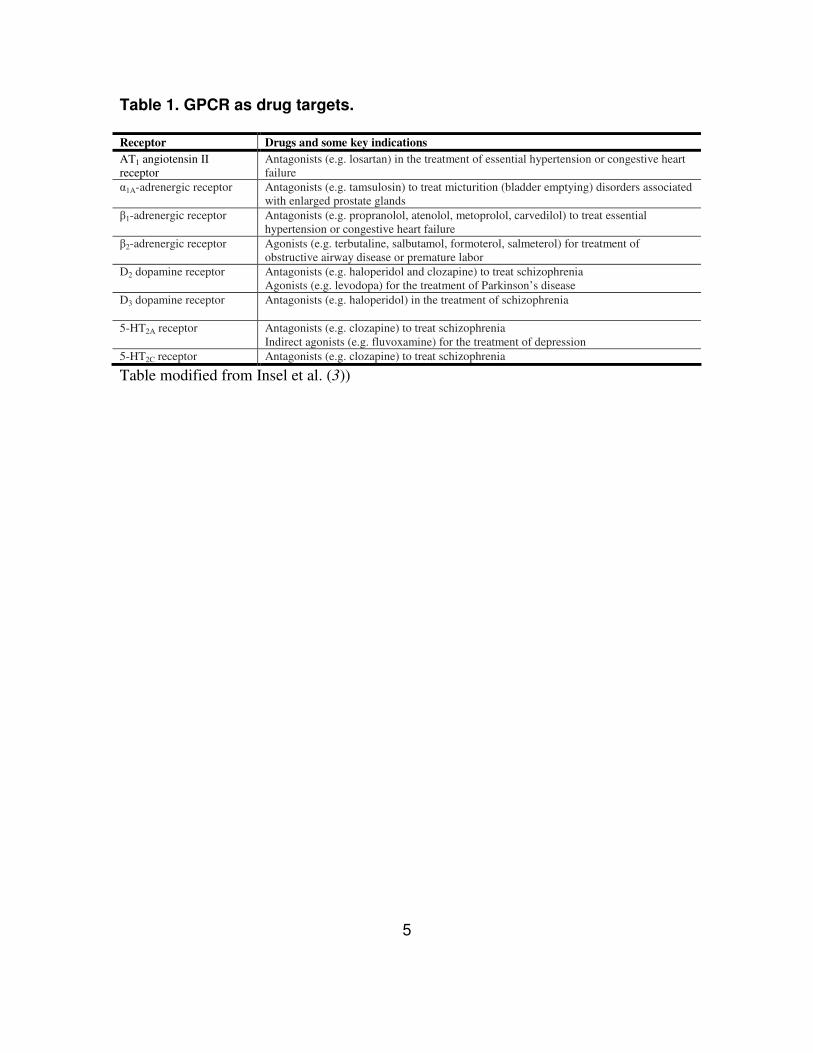
Table 1. GPCR as drug targets.
Receptor Drugs and some key indications
AT1 angiotensin II
receptor
Antagonists (e.g. losartan) in the treatment of essential hypertension or congestive heart
failure
α1A-adrenergic receptor
Antagonists (e.g. tamsulosin) to treat micturition (bladder emptying) disorders associated
with enlarged prostate glands
β1-adrenergic receptor
Antagonists (e.g. propranolol, atenolol, metoprolol, carvedilol) to treat essential
hypertension or congestive heart failure
β2-adrenergic receptor
Agonists (e.g. terbutaline, salbutamol, formoterol, salmeterol) for treatment of
obstructive airway disease or premature labor
D2 dopamine receptor
Antagonists (e.g. haloperidol and clozapine) to treat schizophrenia
Agonists (e.g. levodopa) for the treatment of Parkinson’s disease
D3 dopamine receptor
Antagonists (e.g. haloperidol) in the treatment of schizophrenia
5-HT2A receptor Antagonists (e.g. clozapine) to treat schizophrenia
Indirect agonists (e.g. fluvoxamine) for the treatment of depression
5-HT2C receptor Antagonists (e.g. clozapine) to treat schizophrenia
Table modified from Insel et al. (3))
6
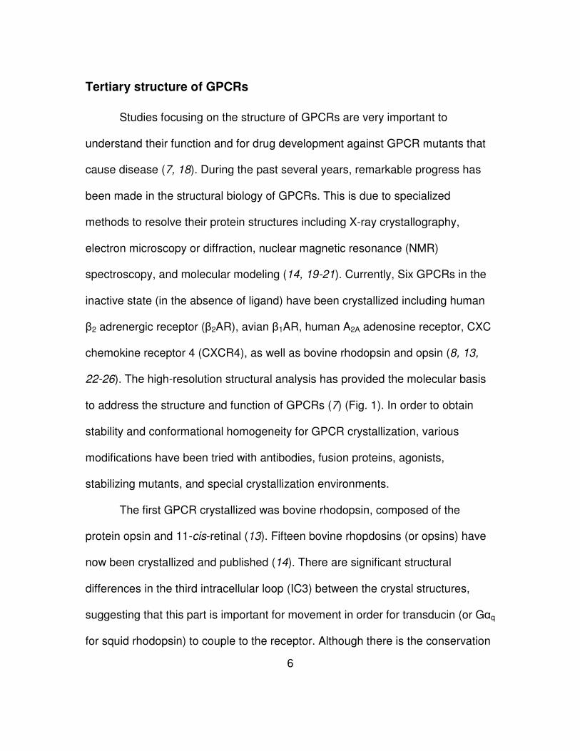
Tertiary structure of GPCRs
Studies focusing on the structure of GPCRs are very important to
understand their function and for drug development against GPCR mutants that
cause disease (7, 18). During the past several years, remarkable progress has
been made in the structural biology of GPCRs. This is due to specialized
methods to resolve their protein structures including X-ray crystallography,
electron microscopy or diffraction, nuclear magnetic resonance (NMR)
spectroscopy, and molecular modeling (14, 19-21). Currently, Six GPCRs in the
inactive state (in the absence of ligand) have been crystallized including human
β2 adrenergic receptor (β2AR), avian β1AR, human A2A adenosine receptor, CXC
chemokine receptor 4 (CXCR4), as well as bovine rhodopsin and opsin (8, 13,
22-26). The high-resolution structural analysis has provided the molecular basis
to address the structure and function of GPCRs (7) (Fig. 1). In order to obtain
stability and conformational homogeneity for GPCR crystallization, various
modifications have been tried with antibodies, fusion proteins, agonists,
stabilizing mutants, and special crystallization environments.
The first GPCR crystallized was bovine rhodopsin, composed of the
protein opsin and 11-cis-retinal (13). Fifteen bovine rhopdosins (or opsins) have
now been crystallized and published (14). There are significant structural
differences in the third intracellular loop (IC3) between the crystal structures,
suggesting that this part is important for movement in order for transducin (or Gαq
for squid rhodopsin) to couple to the receptor. Although there is the conservation
7
of common structural features of the GPCRs, bovine rhodopsin has irregular
transmembrane helices, especially with respect to bending at Pro residues and
kinking around Gly-Gly residues (13, 27). The presence of Pro267, a highly
conserved residue in TM6, causes a significant distortion suggesting that the
proline may function as a toggle switch to transfer motions from the extracellular
surface to the cytoplasmic surface (28). The proline in other GPCRs like β2AR
may also be a crucial residue for receptor function. Rhodopsin and β2AR have a
conserved Pro in TM6 that is important for receptor activation (29, 30). Two
adjacent Gly residues (Gly89 and Gly90 of rhodopsin) that are conserved in
many GPCRs induce a π-helix (i+5�i hydrogen bonding) in TM2 (31, 32). The
amino terminus and three extracellular loops are moved towards each other to
make a compact structure forming the chromophore-binding pocket (22, 33). In
the amino terminus of rhodopsin, post-translational modifications were found on
several distinct residues including glycosylations of Asn2 and Asn15 and
acetylation of Met1 (34, 35). Thus, pairs of β-sheets almost parallel to the
membrane are localized on the amino terminus.
The crystal structures of class A GPCRs show a highly conserved
disulfide bond linking the Cys residue on the second extracellular loop (EC2)
between TM4 and TM5 and a Cys on the front region of TM3. Rhodopsin has two
short β-sheets associated with the ligand-binding pocket on its EC2 (13). The
EC2 of rhodopsin, but not EC1 and EC3, is folded deeply into the helix bundles,
whereas the EC2 of β2AR and β1AR are exposed to the solvent and contain a
8
short α-helix structure tied by intra- and inter-disulfide bonds to create a cavity for
ligand binding (22, 24). The EC2 is crucial for the formation of the ligand binding
pocket has also been observed in other class B GPCRs including complement
factor 5a receptor (C5aR) as well as other class A GPCRs (36, 37).
One of highly conserved motifs, (D/E)R(Y/W), is positioned in the second
intracellular loop (IC2) of all the crystal structures. The tripeptide motif on the
boundary of TM3 forms an ‘ionic lock’ with Glu on TM6 and has been shown that
this linkage is crucial for maintaining the receptors in an inactive state (38). The
ionic lock was observed on the crystal structure of rhodopsin, but not β2AR and
β1AR. However, the fact that mutation of the ionic lock in β2AR increases
constitutive activity supports the formation of the linkage in the receptor (39).
Disruption of the ionic lock in β2AR is accomplished by the contact of the Arg (R)
residue of the (D/E)R(Y/W) motif and T4 lysozyme fused to IC3 (22, 40). On the
other hand, it is unclear why an inverse agonist bound to β1AR showed disruption
of the linkage (24). Interestingly, in rhodopsin, the tripeptide Val137-Val138-
Val139 is located next to the highly conserved (D/E)R(Y/W) motif and might keep
rhodopsin in the inactive state (33).
The crystal structures of rhodopsin and β2AR have been used to create
homology models for other GPCRs that have not been solved (32, 41, 42). These
model predictions are based on the fact that GPCRs have highly conserved
motifs and structural conservation, even though their primary sequences are
9
quite divergent (43). One study compared all the transmembrane proteins of the
different GPCRs to one another (14).
10
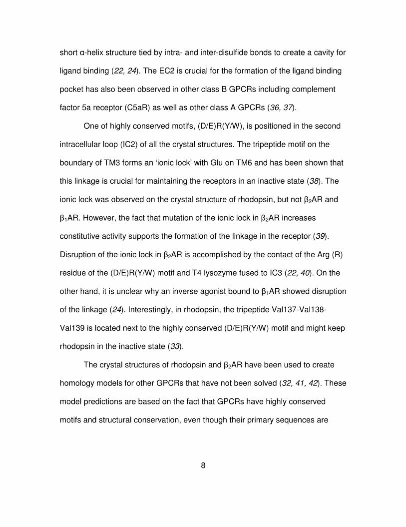
Figure 1. Comparison of GPCR structures.
Bovine rhodopsin (purple), avian β1AR (orange) and human A2A adenosine
receptor (green) are each superimposed on the human β2AR structure (blue).
The extracellular loop2 (EC2), intracellular loop (IC2), cytoplasmic helix 8 (H8)
and transmembrane (TM) segments are labeled on one of the structures. Figure
from Rosenbaum et al (7).
11
This study showed that all the rhodopsin structures superposed well, except for
squid rhodopsin. Interestingly, the β2AR and β1AR superpose poorly on each
other, whereas they fit quite well with rhodopsin. Additional studies using
homology modeling suggests which crystallized GPCR is a better template for
predicting the structures of the other uncrystallized GPCRs (44). This study
showed that current crystal structures of GPCRs are not sufficient for homology
modeling and drug design, because only 3 subfamilies including rhodopsin,
adenosine, and adrenergic receptor have crystal structures. However, it was
suggested that multiple-template homology modeling using all crystallized GPCR
structures could improve the current templates for dynamic simulations like
ligand docking.
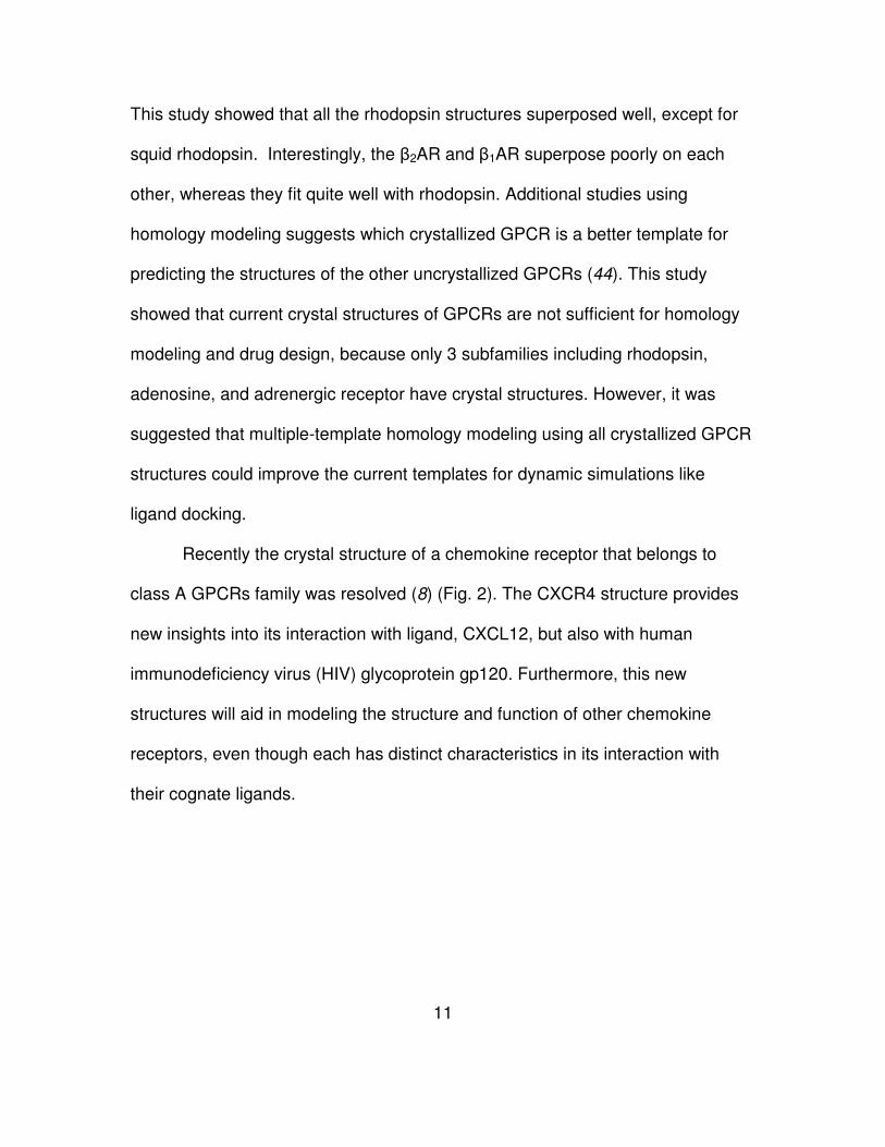
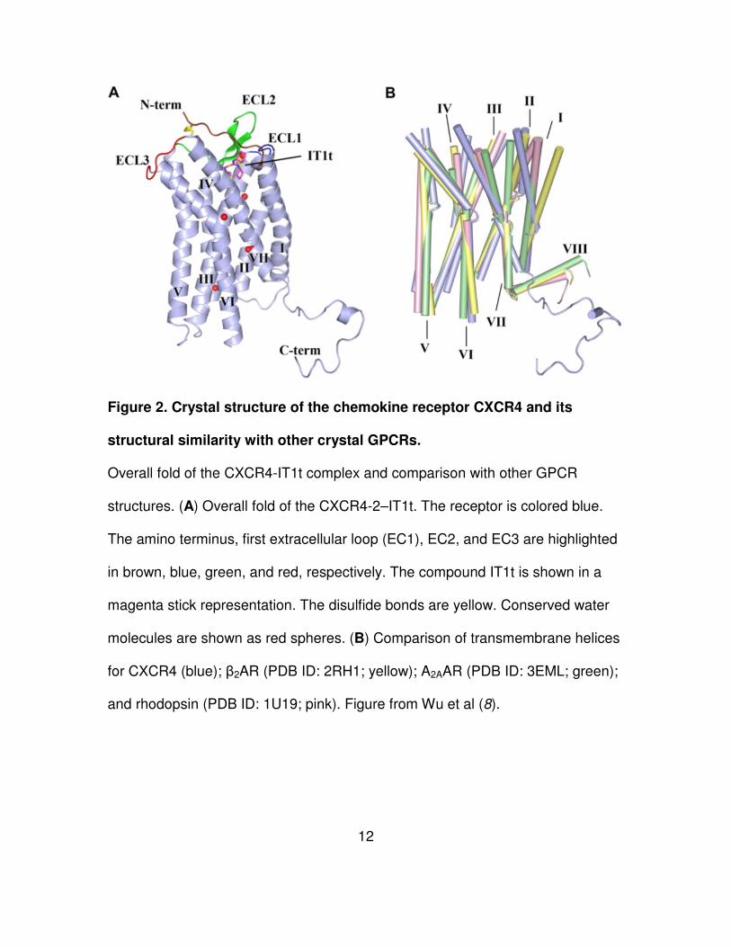
Recently the crystal structure of a chemokine receptor that belongs to
class A GPCRs family was resolved (8) (Fig. 2). The CXCR4 structure provides
new insights into its interaction with ligand, CXCL12, but also with human
immunodeficiency virus (HIV) glycoprotein gp120. Furthermore, this new
structures will aid in modeling the structure and function of other chemokine
receptors, even though each has distinct characteristics in its interaction with
their cognate ligands.
12
Figure 2. Crystal structure of the chemokine receptor CXCR4 and its
structural similarity with other crystal GPCRs.
Overall fold of the CXCR4-IT1t complex and comparison with other GPCR
structures. (A) Overall fold of the CXCR4-2–IT1t. The receptor is colored blue.
The amino terminus, first extracellular loop (EC1), EC2, and EC3 are highlighted
in brown, blue, green, and red, respectively. The compound IT1t is shown in a
magenta stick representation. The disulfide bonds are yellow. Conserved water
molecules are shown as red spheres. (B) Comparison of transmembrane helices
for CXCR4 (blue); β2AR (PDB ID: 2RH1; yellow); A2AAR (PDB ID: 3EML; green);
and rhodopsin (PDB ID: 1U19; pink). Figure from Wu et al (8).
13
Ligand binding and receptor activation
Numerous studies of GPCRs aim to understanding the mechanism of
ligand binding and receptor activation using biological, biochemical, and
biophysical approaches (45). Various methods were used to identify the binding
domains including site-directed and random mutagenesis, receptor chimeras,
competition with synthesized peptides, and addition of probes. For
bioinformatical approaches, this experimental data was applied to improve
ligand-docking model (46).
Despite the variation in the mechanism of ligand binding, there are some
similarities between the interaction of ligands and receptors. Small ligands
(photon, biogenic amines and nucleosides) bind within transmembrane regions,
whereas large molecules like peptides and proteins bind to extracellular loops
(45). Peptide ligands show direct interaction of both amino-terminus and
extracellular loops (4). However, some peptide ligands can interact with
transmembrane domains as well as extracellular loops (47).
Initially, a simple model was used to explain how agonist binding induces
a conformational change. The model was called the “two-state model” with an
equilibrium between the inactive conformation (R) and active conformation (R*)
(48). However, more recently crystal structures and biophysical studies led to the
creation of new model known as the “multi-state” model (7, 49). Upon ligand
binding, GPCRs become activated and undergo a conformational change,
resulting in G protein activation. The conformational changes leads to the
14
rearrangement of the interhelical interactions on transmembranes including TM3,
TM6 and TM7. The extracellular domains and the flexible third intracellular loop
are involved in this rearrangement. The recognition sites such as the second
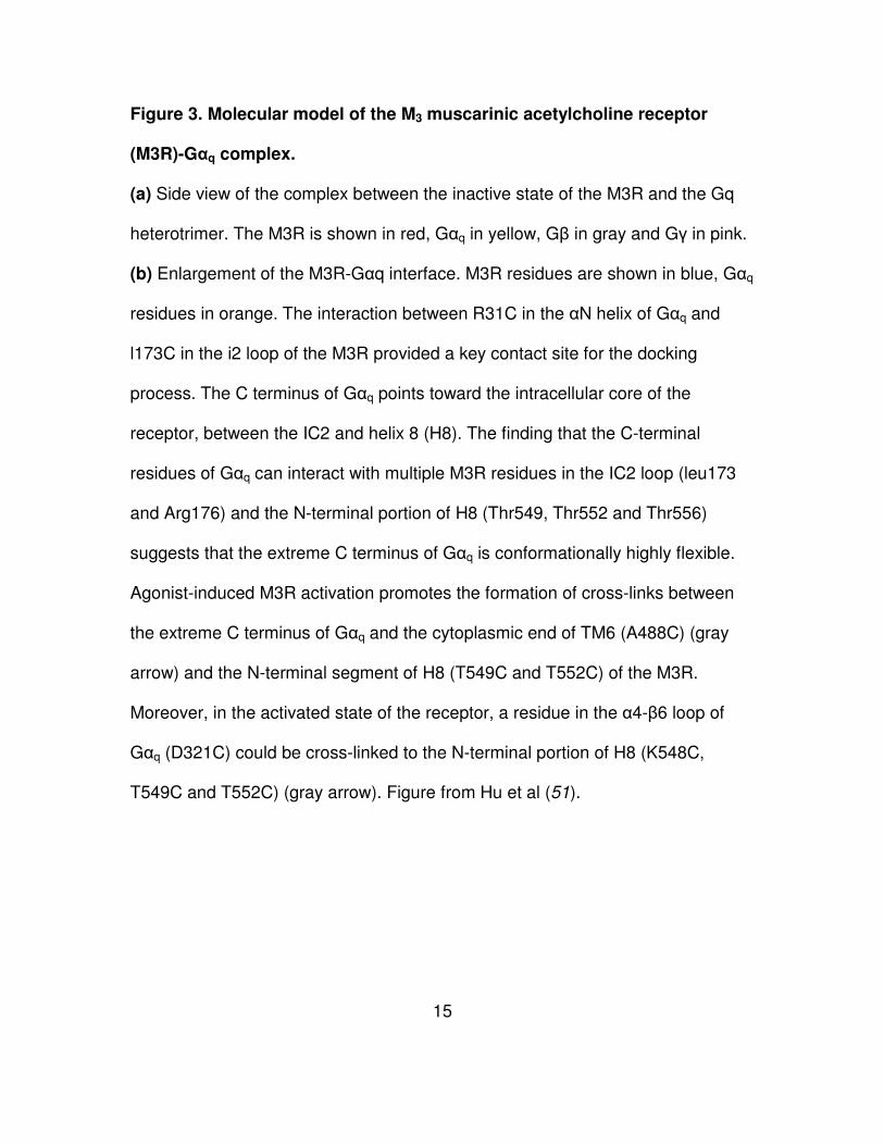
intracellular loop (IC2) including (D/E)R(Y/W) motif close to TM3 are exposed to
activate signaling partners including heterotrimeric G proteins and arrestins (50,
51) (Fig. 3). A highly conserved Pro residue on TM6 allows the rigid
transmembrane helices to move as part of the conformational change (29).
Post-translational modifications on cytoplasmic domains of GPCRs are
involved in receptor desensitization and internalization (5, 6). GPCR kinases and
arrestins are involved in these modifications. In the cases of rhodopsin and β2AR,
there are residues for different post-translational modifications on their
cytoplasmic loops, including palmitoylation at Cys, phosphorylation at Ser, and
ubiquitinylation at Lys (35, 52).
Signal transduction of GPCRs
The cytoplasmic domains of GPCRs interact with proteins that are
responsible for initiating intracellular signaling cascades (53) (Fig. 4).
Heterotrimeric G proteins are predominantly used as the first intracellular
modulators of GPCR signaling (54). To date, 23 Gα protein subtypes have been
identified and are classified into 4 different groups such as Gαi/o, Gαs, Gαq/11, and
Gα12/13. There are also 6 Gβ and 11 Gγ proteins (55).
15
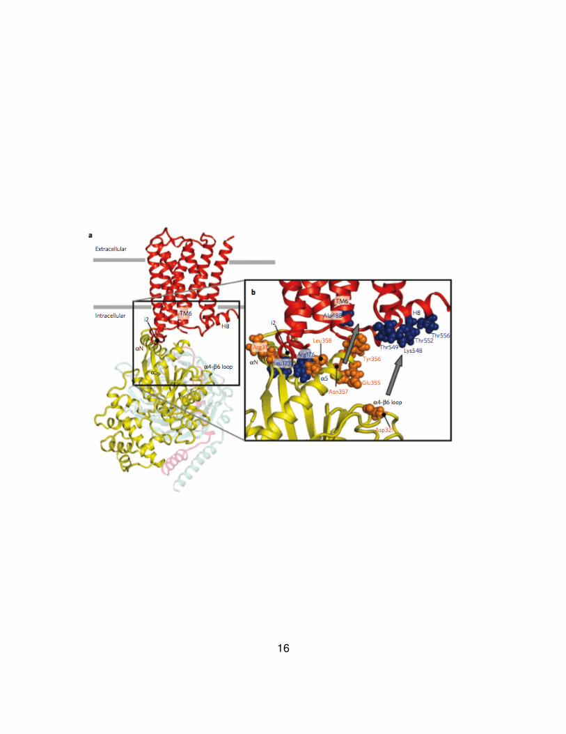
Figure 3. Molecular model of the M3 muscarinic acetylcholine receptor
(M3R)-Gαq complex.
(a) Side view of the complex between the inactive state of the M3R and the Gq
heterotrimer. The M3R is shown in red, Gαq in yellow, Gβ in gray and Gγ in pink.
(b) Enlargement of the M3R-Gαq interface. M3R residues are shown in blue, Gαq
residues in orange. The interaction between R31C in the αN helix of Gαq and
l173C in the i2 loop of the M3R provided a key contact site for the docking
process. The C terminus of Gαq points toward the intracellular core of the
receptor, between the IC2 and helix 8 (H8). The finding that the C-terminal
residues of Gαq can interact with multiple M3R residues in the IC2 loop (leu173
and Arg176) and the N-terminal portion of H8 (Thr549, Thr552 and Thr556)
suggests that the extreme C terminus of Gαq is conformationally highly flexible.
Agonist-induced M3R activation promotes the formation of cross-links between
the extreme C terminus of Gαq and the cytoplasmic end of TM6 (A488C) (gray
arrow) and the N-terminal segment of H8 (T549C and T552C) of the M3R.
Moreover, in the activated state of the receptor, a residue in the α4-β6 loop of
Gαq (D321C) could be cross-linked to the N-terminal portion of H8 (K548C,
T549C and T552C) (gray arrow). Figure from Hu et al (51).
16
17
The expression pattern and combination of the heterotrimeric G proteins is cell
type-dependent, indicating that GPCRs can interact with certain G proteins that
are available in a given cell or localize closely to the receptors. Although it has
been shown that second intracellular loop (IC2), IC3, and cytoplasmic terminus
function as key regions for interaction with G protein coupling, no consensus
motif has been identified (56).
Activation of the three G proteins leads to exchange of guanosine
diphosphate (GDP) and guanosine triphosphate (GTP), followed by dissociation
of Gα protein and Gβγ dimer. The released G proteins can regulate the
stimulation or inhibition of secondary messenger molecules including adenylate
cyclase, guanylyl cyclase, phospholipases, or ion channels.
Each family of G proteins has distinct characteristics (2). The Gαs family
functions as a stimulator of adenylyl cyclase to increase cyclic adenosine
monophosphate (cAMP) levels. Whereas, Gαi/o proteins play a role in the
inactivation of adenylyl cyclase, resulting in a decrease of cAMP levels.
Furthermore, almost all the proteins that belong to Gαi/o family are pertussis
toxin-sensitive (PTX). Gαq/11 including Gα14, Gα15, and Gα16 can bind
phospholipases C-β (PLC-β) (57). Proteins of the Gα12/13 family are ubituitously
expressed and localized (58). This family can activate small G proteins (Rho and
Ras family), Src family tyrosine kinase, and phospholipase D (PLD). Interestingly,
when Gα12/13 mutants are in a constitutively active form, cellular transformation is
induced in a Rho signaling-dependent manner (59). Gβ and Gγ proteins have
18
been shown to function as a heterodimer. This complex induces adenylyl cyclase,
PLC-β, and phosphoinositide-3 kinase (PI3K) (60, 61).
Agonist-occupied GPCRs induce not only signal transduction cascades
but also activation of various intracellular regulatory molecules. The G protein-
coupled receptor kinases (GRKs) are serine/threonine kinases. GRKs catalyze
phosphorylation of GPCRs and recruit arrestins to the phosphorylated GPCR for
receptor desensitization and internalization (62, 63). After activation, the
regulators of G protein signaling (RGS) proteins are used to return the GTP-
bound Gα proteins to their basal state (Gα) via GTP hydrolysis. This results in
the reassociation of the heterotrimeric G proteins and inactivation of the GPCR
(64). Interestingly, arrestins function not only in GPCR desensitization and
internalization but also as a G protein-independent signal transducer (65, 66).
19
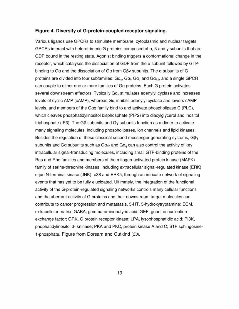
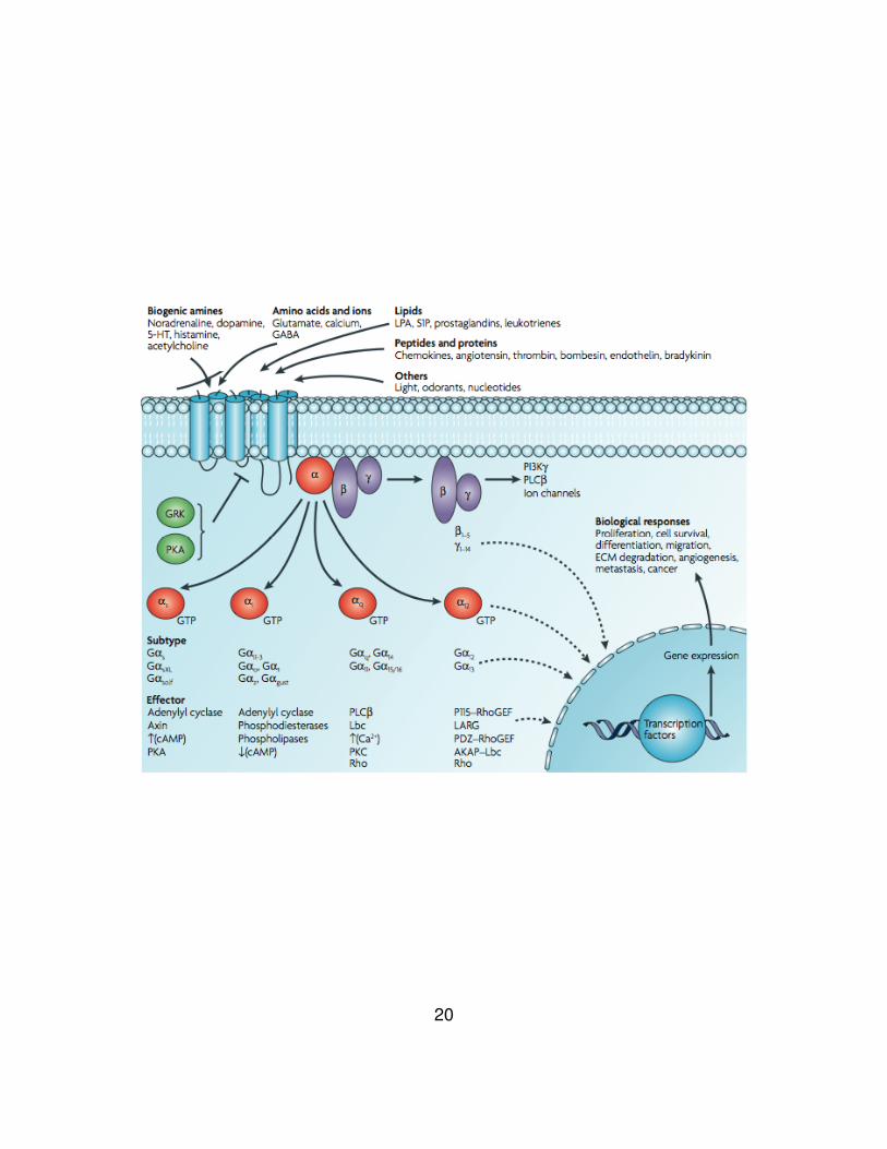
Figure 4. Diversity of G-protein-coupled receptor signaling.
Various ligands use GPCRs to stimulate membrane, cytoplasmic and nuclear targets.
GPCRs interact with heterotrimeric G proteins composed of α, β and γ subunits that are
GDP bound in the resting state. Agonist binding triggers a conformational change in the
receptor, which catalyses the dissociation of GDP from the α subunit followed by GTP-
binding to Gα and the dissociation of Gα from Gβγ subunits. The α subunits of G
proteins are divided into four subfamilies: Gαs, Gαi, Gαq and Gα12, and a single GPCR
can couple to either one or more families of Gα proteins. Each G protein activates
several downstream effectors. Typically Gαs stimulates adenylyl cyclase and increases
levels of cyclic AMP (cAMP), whereas Gαi inhibits adenylyl cyclase and lowers cAMP
levels, and members of the Gαq family bind to and activate phospholipase C (PLC),
which cleaves phosphatidylinositol bisphosphate (PIP2) into diacylglycerol and inositol
triphosphate (IP3). The Gβ subunits and Gγ subunits function as a dimer to activate
many signaling molecules, including phospholipases, ion channels and lipid kinases.
Besides the regulation of these classical second-messenger generating systems, Gβγ
subunits and Gα subunits such as Gα12 and Gαq can also control the activity of key
intracellular signal-transducing molecules, including small GTP-binding proteins of the
Ras and Rho families and members of the mitogen-activated protein kinase (MAPK)
family of serine-threonine kinases, including extracellular signal-regulated kinase (ERK),
c-jun N-terminal kinase (JNK), p38 and ERK5, through an intricate network of signaling
events that has yet to be fully elucidated. Ultimately, the integration of the functional
activity of the G-protein-regulated signaling networks controls many cellular functions
and the aberrant activity of G proteins and their downstream target molecules can
contribute to cancer progression and metastasis. 5-HT, 5-hydroxytryptamine; ECM,
extracellular matrix; GABA, gamma-aminobutyric acid; GEF, guanine nucleotide
exchange factor; GRK, G protein receptor kinase; LPA, lysophosphatidic acid; PI3K,
phophatidylinositol 3- kninase; PKA and PKC, protein kinase A and C; S1P sphingosine-
1-phosphate. Figure from Dorsam and Gutkind (53).
20
21
Chapter 2 CXC Chemokine Receptor 2 (CXCR2) and
Cognate Chemokines
Chemokines and cognate chemokine receptors
Chemokines are a family of small cytokines, or cell-signaling molecules,
that regulate inflammation through GPCRs expressed on leukocytes (67). In
higher vertebrates, over 50 different chemokines have been identified (68).
These proteins induce various biological and biochemical activities such as cell
migration, cell adhesion, and exocytosis (69). There are four families of
chemokines including C, CC, CXC, and CX3C. The chemokines of C or CC
subfamily have a single Cys residue or two contiguous Cys residues on the
amino terminus of the protein, respectively. CXC or CX3C subfamilies contain
one or three amino acids between the two cysteine residues. The CXC
chemokines are classified into two subfamilies: the glutamate-leucine-arginine
(ELR) and non-ELR families. Generally, the ELR CXC chemokines promote
angiogenesis while the CXC chemokines that lack the ELR motif inhibit
angiogenesis (70). However, the non-ELR chemokine CXCL12 (SDF-1) induces
angiogenesis, which may relate to its ability to induce cancer metastasis (71).
In the early 1990s, two CXC chemokine receptors for CXCL8 (IL-8) were
the first identified chemokine receptors. Now there are 19 chemokines receptors
including 7 members for CXC, 11 for CC, 1 for C and 1 for CX3C type (72-76).
22
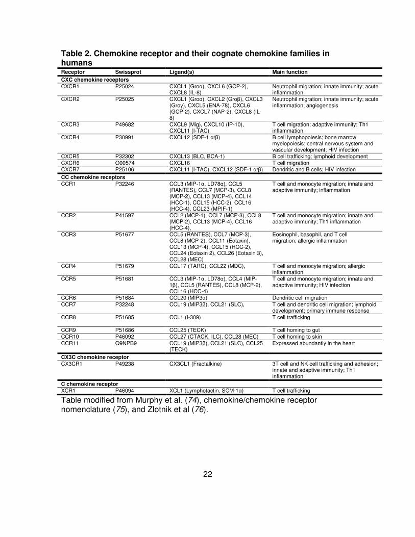
Table 2. Chemokine receptor and their cognate chemokine families in humans Receptor Swissprot Ligand(s) Main function
CXC chemokine receptors
CXCR1 P25024 CXCL1 (Groα), CXCL6 (GCP-2), CXCL8 (IL-8)
Neutrophil migration; innate immunity; acute inflammation
CXCR2 P25025 CXCL1 (Groα), CXCL2 (Groβ), CXCL3 (Groγ), CXCL5 (ENA-78), CXCL6 (GCP-2), CXCL7 (NAP-2), CXCL8 (IL-8)
Neutrophil migration; innate immunity; acute inflammation; angiogenesis
CXCR3 P49682 CXCL9 (Mig), CXCL10 (IP-10), CXCL11 (I-TAC)
T cell migration; adaptive immunity; Th1 inflammation
CXCR4 P30991 CXCL12 (SDF-1 α/β) B cell lymphopoiesis; bone marrow myelopoiesis; central nervous system and vascular development; HIV infection
CXCR5 P32302 CXCL13 (BLC, BCA-1) B cell trafficking; lymphoid development CXCR6 O00574 CXCL16 T cell migration CXCR7 P25106 CXCL11 (I-TAC), CXCL12 (SDF-1 α/β) Dendritic and B cells; HIV infection CC chemokine receptors
CCR1 P32246 CCL3 (MIP-1α, LD78α), CCL5 (RANTES), CCL7 (MCP-3), CCL8 (MCP-2), CCL13 (MCP-4), CCL14 (HCC-1), CCL15 (HCC-2), CCL16 (HCC-4), CCL23 (MPIF-1)
T cell and monocyte migration; innate and adaptive immunity; inflammation
CCR2 P41597 CCL2 (MCP-1), CCL7 (MCP-3), CCL8 (MCP-2), CCL13 (MCP-4), CCL16 (HCC-4),
T cell and monocyte migration; innate and adaptive immunity; Th1 inflammation
CCR3 P51677 CCL5 (RANTES), CCL7 (MCP-3), CCL8 (MCP-2), CCL11 (Eotaxin), CCL13 (MCP-4), CCL15 (HCC-2), CCL24 (Eotaxin 2), CCL26 (Eotaxin 3), CCL28 (MEC)
Eosinophil, basophil, and T cell migration; allergic inflammation
CCR4 P51679 CCL17 (TARC), CCL22 (MDC), T cell and monocyte migration; allergic inflammation
CCR5 P51681 CCL3 (MIP-1α, LD78α), CCL4 (MIP-1β), CCL5 (RANTES), CCL8 (MCP-2), CCL16 (HCC-4)
T cell and monocyte migration; innate and adaptive immunity; HIV infection
CCR6 P51684 CCL20 (MIP3α) Dendritic cell migration CCR7 P32248 CCL19 (MIP3β), CCL21 (SLC), T cell and dendritic cell migration; lymphoid
development; primary immune response CCR8 P51685 CCL1 (I-309) T cell trafficking
CCR9 P51686 CCL25 (TECK) T cell homing to gut CCR10 P46092 CCL27 (CTACK, ILC), CCL28 (MEC) T cell homing to skin CCR11 Q9NPB9 CCL19 (MIP3β), CCL21 (SLC), CCL25
(TECK) Expressed abundantly in the heart
CX3C chemokine receptor
CX3CR1 P49238 CX3CL1 (Fractalkine) 3T cell and NK cell trafficking and adhesion; innate and adaptive immunity; Th1 inflammation
C chemokine receptor
XCR1 P46094 XCL1 (Lymphotactin, SCM-1α) T cell trafficking
Table modified from Murphy et al. (74), chemokine/chemokine receptor nomenclature (75), and Zlotnik et al (76).
23
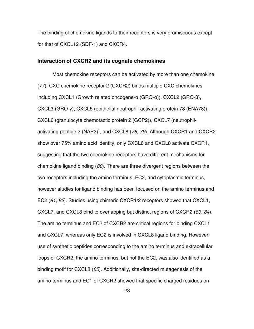
The binding of chemokine ligands to their receptors is very promiscuous except
for that of CXCL12 (SDF-1) and CXCR4.
Interaction of CXCR2 and its cognate chemokines
Most chemokine receptors can be activated by more than one chemokine
(77). CXC chemokine receptor 2 (CXCR2) binds multiple CXC chemokines
including CXCL1 (Growth related oncogene-α (GRO-α)), CXCL2 (GRO-β),
CXCL3 (GRO-γ), CXCL5 (epithelial neutrophil-activating protein 78 (ENA78)),
CXCL6 (granulocyte chemotactic protein 2 (GCP2)), CXCL7 (neutrophil-
activating peptide 2 (NAP2)), and CXCL8 (78, 79). Although CXCR1 and CXCR2
show over 75% amino acid identity, only CXCL6 and CXCL8 activate CXCR1,
suggesting that the two chemokine receptors have different mechanisms for
chemokine ligand binding (80). There are three divergent regions between the
two receptors including the amino terminus, EC2, and cytoplasmic terminus,
however studies for ligand binding has been focused on the amino terminus and
EC2 (81, 82). Studies using chimeric CXCR1/2 receptors showed that CXCL1,
CXCL7, and CXCL8 bind to overlapping but distinct regions of CXCR2 (83, 84).
The amino terminus and EC2 of CXCR2 are critical regions for binding CXCL1
and CXCL7, whereas only EC2 is involved in CXCL8 ligand binding. However,
use of synthetic peptides corresponding to the amino terminus and extracellular
loops of CXCR2, the amino terminus, but not the EC2, was also identified as a
binding motif for CXCL8 (85). Additionally, site-directed mutagenesis of the
amino terminus and EC1 of CXCR2 showed that specific charged residues on
24
the amino terminus of CXCR2 are critical for CXCL1 (Asp9, Glu12, Lys108 and
Lys120 of CXCR2) and CXCL8 (Glu7, Asp9 and Glu12 of CXCR2) binding (86).
Site-directed mutagenesis and chimeric receptors of CXCR1/2 including the
amino terminus, TM4, EC2, TM5, EC3, and TM7 showed that the amino terminus
of CXCR1 and EC2 of CXCR2 are important for CXCL8 binding, while the amino
terminus of CXCR2 is involved in CXCL1 binding (87). A recent study using a
random phage-epitope library showed certain synthetic epitopes inhibited binding
of CXCL8 (or antibody) to CXCR1/2 and blocked chemotaxis of human
neutrophils (88). The consensus sequences of the synthetic epitopes were
identical with residues on amino terminus of CXCR1/2 (Trp10-Asp11-Phe12 for
CXCR1 and Asp13-Phe14-Trp15 for CXCR2), indicating that amino terminus of
the receptors is important for ligand binding. Although some of the information
about ligand binding is not consistent, the amino terminus and/or the EC2 are
critical for binding to its cognate chemokines. Additionally, amino acid
substitutions showed that the Cys residues of CXCR2 are highly associated with
CXCL8 ligand binding, but not surface expression, including Cys119, Cys196,
and Cys286 in the extracellular loops and Cys308 in TM7 (89).
The amino terminus of CXCL8 is important for high affinity and binding
specificity (90). Site-directed mutagenesis of CXCL8, two binding sites for
CXCR1 and CXCR2 were predicted (91). In the study, two residues, Tyr13 and
Lys15, in the amino terminus of CXCL8 were identified as important sites for
CXCR1 binding. Both Glu4-Leu5-Arg6 (ELR) motif and Tyr13-Ser14-Lys15 are
25
exposed to the surface and localize on opposite ends on the same face of the
ligand. The fact that the distance between the two regions is 23 angstroms (2.3
nm) suggests that the two sites of CXCL8 can span the distance between the
amino terminus and third extracellular loop of CXCR1/2. Additionally,
substitutions of residues of bovine CXCL8 were used to identify analogues with
high affinity (K11R) or antagonist activity (K11R/G31P) (92, 93). The
human/bovine chimeric hbG31P (bovine CXCL8(3-44)K11R/G31P-hCXCL8(45-
72)) preserved antagonist activity, indicating that the analogue can be used as
potential antagonist for human CXCL8 and its receptors (94).
CXCR2 normally expressed on neutrophils contains two N-glycosylation
sites (95). There are three possible residues for N-glycosylation at Asn17,
Asn186, and Asn197. Because of the proximity of the Asn residues and
transmembranes, Asn17 and Asn197 are probably the most likely candidates for
glycosylation. Asn186 is closer to the transmembrane domain and probably
cannot handle the N-linked sugar moieties. N-glycosylation on the extracellular
domains of CXCR2 is not involved in ligand binding but contributes to the
maintenance of the receptors expressed on the cell surface (73, 84, 95).
Receptor activation and signal transduction of CXCR2
CXCR2 is expressed on a various tumor cells including breast cancer,
head and neck cancer, melanoma, pancreatic, and ovarian cancer (96-100).
CXCL8 can activate both CXCR1 and CXCR2 receptors and has been
26
characterized as a mediator of cancer progression. Activation of these receptors
promote the expression of genes highly associated with cell survival, proliferation,
angiogenesis, and invasion (78, 101) (Fig. 5).
Antagonists have been used to understand the mechanism of CXCR1 and
CXCR2 activation. Competition binding with CXCL1 and CXCL8 and a small
molecule antagonist, SB225002, to CXCR2 mutants was used to show how
these chemokines activate the receptor (87). The antagonist binds to the
transmembrane helices within CXCR2, but not to the ligand binding sites, and
causes an allosteric change that blocks to binding of CXCL1 and CXCL8.
Currently, two different antagonists were used to understand the difference
between CXCR1 and CXCR2 in antagonism (102). It has been shown that the
EC2 of the receptors does not play a crucial role in compound antagonism, and a
single amino acid (Lys320 for CXCR2 and Asn311 for CXCR1) of the receptor is
important to allosteric binding.
Viral oncogene ORF74 encoded by Kaposi’s sarcoma-associated
herpesvirus (human herpesvirus 8), also called KSHV-GPCR, is the closest
homologue to CXCR2 (103). KSHV-GPCR constitutively activates AP-1, CREB,
NFAT, and MAPK via Gαi or Gαq, and NF-κB through only Gαq. This is true in
different cell lines including COS-7, primary effusion lymphoma (PEL), and
endothelial cells (104-107). Currently, studies of M33 from murine
cytomegalovirus (MCMV) and US28 of human cytomegalovirus (HCMV), both
viral GPCR homologues,

27
Figure 5. Characterized IL-8 signaling pathways.
A schematic diagram illustrating the range of signaling pathways that are activated after stimulation of CXCR1 and/or CXCR2 receptors with IL-8. After activation of heterotrimeric small G proteins, IL-8 signaling promotes activation of the primary effectors phosphatidyl-inositol-3-kinase or phospholipase C, promoting the activation of Akt, PKC, calcium mobilization and/or MAPK signaling cascades. These signaling pathways have been shown to promote protein translation (left) and regulate the activity of a range of transcription factors (bottom). Solid gold lines, transcription factors whose activity has been shown to be positively regulated by IL-8 signaling using various reporter assays. In the case of signal transducers and activators of transcription 3 (STAT3) and h-catenin, IL-8 signaling has been shown to promote nuclear translocation of these factors; however, transcriptional activation of either factor remains to be shown. Dashed lines, the putative pathways through which IL-8 signaling regulates transcription factor activity. In addition, IL-8 signaling activates members of the RhoGTPase family and activates a number of nonreceptor tyrosine kinases (e.g., Src family kinases and FAK) that regulate the architecture of the cell cytoskeleton and its interaction with the surrounding extracellular environment. Figure from Waugh and Wilson (101).
28
have been shown to activate the Gαq protein and induce phopholipase C-β (PLC-
β) activity, followed by stimulation of CREB and NF-κB signals (108-111).
Additionally, M33 can induce CREB and NF-κB stimulation via a PLC-β
independent manner (112). On the other hand, another GPCR homolog, UL33, of
HCMV enhances CREB transcriptional activities by the Gs pathway not the Gq/11
pathway (113).
It has been shown that CXCR2 can couple to different G proteins in
different cell lines, including Gαi, Gα12, Gα13, Gα14, Gα15, Gα16, and Gβγ subunits
(114-116). CXCR2 can interact with non-G proteins like vasodilator-stimulated
phosphoprotein (VASP) to activate downstream signaling (117). It is suggested
that multiple signaling cascades can be activated in a G-protein-dependent and -
independent manner.
A point mutation of Asp143 (to Val, the same as KSHV-GPCR) of the
highly conserved (D/E)R(Y/W) motif in CXCR2 leads to constitutive activation of
the receptor and cellular transformation similar to KSHV-GPCR (118).
Transformation was totally inhibited using AG490, a specific kinase inhibitor for
JAK2-STAT3 signaling (119). Constitutive activation of KSHV-GPCR and CXCR2
mutant D143V is highly associated with STAT3 phosphorylation. Another motif
related to CXCR2 activation was identified in the cytoplasmic terminus (120).
Mutation of the LLKIL motif inhibited chemotaxis via disruption of Akt, Rac1, and
Cdc42 recruitment. Interestingly, EC2 as well as the carboxy terminus of CXCR2
is important for activation and regulation of the receptor (84). Mutation of Asp199
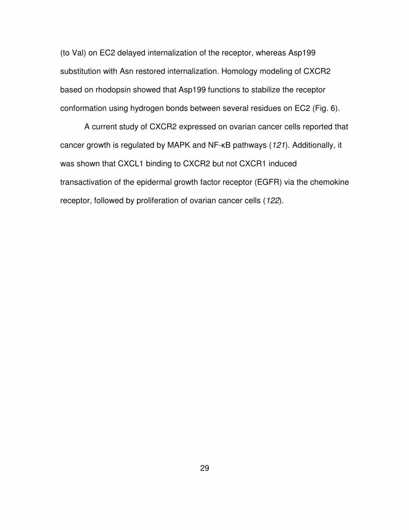
29
(to Val) on EC2 delayed internalization of the receptor, whereas Asp199
substitution with Asn restored internalization. Homology modeling of CXCR2
based on rhodopsin showed that Asp199 functions to stabilize the receptor
conformation using hydrogen bonds between several residues on EC2 (Fig. 6).
A current study of CXCR2 expressed on ovarian cancer cells reported that
cancer growth is regulated by MAPK and NF-κB pathways (121). Additionally, it
was shown that CXCL1 binding to CXCR2 but not CXCR1 induced
transactivation of the epidermal growth factor receptor (EGFR) via the chemokine
receptor, followed by proliferation of ovarian cancer cells (122).
30
Figure 6. Computational modeling of the EC2 of rhodopsin, CXCR2, CXCR1,
and the receptor mutants BD199VA, BD199N, and AV190DB.
Shown are receptor interactions involving Asp190 of rhodopsin (A) and its
corresponding residues Asp199 of the EC2 of CXCR2 (B), Val190 of CXCR1 (C),
Val199 of BD199VA (D), Asn199 of BD199N (E), and Asp190 of AV190DB (F). The
protein secondary structural elements are displayed as follows: α-helix, white; β-
sheet, yellow; turn, cyan; and random coil, purple. The color coding of receptor
residue atoms is as follows: green, carbon; red, orange; and blue, nitrogen. H-
bond-forming residues (in stick depiction) in association with Asp190 in
rhodopsin (Tyr192, Thr193, and Arg177) (A), Asp199 in CXCR2 (Thr186, Arg208,
and Arg185) (B), Asn199 in BD199N (Thr186 and Arg185) (E), and Asp190 in
AV190DB (Arg199) (F) are shown by white dotted lines. No H-bond was formed in
CXCR1 (C) and BD199VA (D). The residues within 3.0 Å of Val190 (CXCR1) or
Val199 (BD199VA) are represented. Figure from Nasser et al (84).
31
Statement of research aims
Most chemokine receptors can be activated by more than one chemokine
(77). CXC chemokine receptor 2 (CXCR2) binds multiple CXC chemokines
including CXCL1 (GRO-α), CXCL2 (GRO-β), CXCL3 (GRO-γ), CXCL5 (ENA78),
CXCL6 (GCP2), CXCL7 (NAP2), and CXCL8 (78, 79). Although CXCR1 and
CXCR2 show over 75% amino acid identity, only CXCL6 and CXCL8 activate
CXCR1, suggesting that the two chemokine receptors have different
mechanisms for ligand binding (80). In previous studies with multiple chimeric
receptors, it was shown that CXCR1 and CXCR2 have distinct mechanisms of
ligand binding and receptor activation (80, 81, 83-88). The studies suggested that
divergent regions including the amino terminus and second extracellular loop
between the receptors are crucial in construction of selectivity determinants for
different cellular responses. However, the mechanism of ligand binding and
receptor activation is still unknown.
First, in our study, CXCR2 structure is predicted on the basis of crystal
GPCR structures including rhodopsin, β2AR by homology modeling, and CXCR4.
The topology of the EC2 region of CXCR2 in the inactive state is determined by
using substituted cysteine accessibility method (SCAM).
Second, we studied the role of charged residues in EC2 of CXCR2 in
ligand binding and receptor activation. Two concepts, “attraction” and “repulsion”
models, were designed to investigate the mechanism of CXCR2 activation (Fig.
14). To end that, we constructed combinatorial mutation sets consisting of
32
constitutively active mutants (CAM) Asp9 in the amino terminus and substitutions
in EC2. These mutant receptors were generated by site-directed mutagenesis
and transfected into mouse fibroblast NIH3T3 cell lines. The NIH3T3 cells stably
expressing wild-type CXCR2 and mutant receptors were used to investigate cell
surface expression of the receptor, ligand-binding, and receptor activation
through phospholipase C-β3 (PLC-β3).
33
PART II
PREDICTION OF THE TERTIARY STRUCTURE OF
CXCR2 AND STUDY OF THE CONFORMATION OF THE
SECOND EXTRACELLULAR LOOP OF THE RECEPTOR
IN AN INACTIVE STATE
34
Chapter 1 Abstract
The first objective of this investigation was to predict the structure of
CXCR2 based on known structures of crystallized GPCRs. Bovine rhodopsin,
human β2-adrenergic receptor, and human CXCR4 were used for homology
modeling of CXCR2 structure. Highly conserved motifs ((D/E)R(Y/W), NPXXY,
toggle switch, and disulfide bonds) found in sequence alignments of the template
GPCRs were helpful to generate CXCR2 models. Using Cα RMSDs, structural
alignments of the seven transmembrane domains showed the individual models
tend to be similar to their templates. Based on the sequence alignments and
RMSD values, the CXCR4-based model shows a very similar structure to
CXCR2. The second extracellular loop of the homology modeled CXCR2
provided two β-strands like CXCR4 and the prediction is very similar with results
obtained from web-based prediction programs. Superimposition of the second
extracellular loop of CXCR4 and CXCR4-based CXCR2, showed the second
extracellular loop of CXCR2 is structurally very similar with that of CXCR4 with
below 2.0 Å RMSD.
Furthermore, we studied solvent accessibility of residues in the second
extracellular loop of CXCR2, a key region for ligand binding and receptor
activation, in the inactive state of the receptor. MTSEA-biotin accessibility of
substituted cysteine residues in second extracellular loop was measured by
immunostaining of FITC-streptavidin using flow cytometric analysis. The
35
normalization of the accessibility was determined by surface expression levels of
the substituted cysteine mutant receptors. Most of residues in the second
extracellular loop were found to be solvent accessible in the inactive state of
CXCR2, indicating that the residues might be involved in ligand binding. Most of
the charged residues in second extracellular loop also have solvent accessibility.
However, five amino acids 204TANWR208 showed no solvent accessibility,
pointing to possible interaction with the amino terminus or other extracellular
loops. Our finding of ligand binding sites for CXCR2 also indicated that most of
the residues in the second extracellular loop could form a ligand binding pocket
to contact CXCL8.
In this sense, these current discoveries about structural basis of CXCR2
and interdisciplinary approaches would provide new insights to investigate
unknown mechanisms of interaction with its cognate ligands and receptor
activation.
36
Chapter 2 Introduction
CXCR2, also called IL-8RB, was initially identified as a chemokine
receptor expressed on neutrophils (123). CXCR2 is a member of the rhodopsin-
like subfamily (class A) of G protein-coupled receptors, and has the closest
homology to CXCR1, with 77% amino acid identity (72, 73, 123). Multiple
chemokines can bind CXCR2 including CXCL1 (Growth related oncogene-α
(GRO-α)), CXCL2 (GRO-β), CXCL3 (GRO-γ), CXCL5 (epithelial neutrophil-
activating protein 78 (ENA78)), CXCL6 (granulocyte chemotactic protein 2
(GCP2)), CXCL7 (neutrophil-activating peptide 2 (NAP2)), and CXCL8 (78, 79).
The function of CXCR2 has been studied in a variety of immune cells such as
neutrophils and macrophages, indicating that the receptor plays a critical role in
immune response and inflammation (121). Furthermore, it has been shown that
CXCR2 receptors are also expressed on various cancer cells including breast
cancer, head and neck cancer, melanoma, pancreatic, and ovarian cancer (96-
100).
In previous studies utilizing chimeric receptors, it has been shown that
CXCR1 and CXCR2 have distinct mechanisms of ligand binding and receptor
activation (80, 81, 83-88). The studies suggested that divergent regions including
the amino terminus and second extracellular loop between the receptors are
crucial in construction of selectivity determinants for different cellular responses.
The CXCL8, a chemokine ligand that binds to both CXCR1 and CXCR2, has
37
been used to study which part of the ligand is important to bind the receptors (90,
91). Both the Glu4-Leu5-Arg6 (ELR) motif and Tyr13-Ser14-Lys15 on the amino
terminus of CXCL8 might initially interact with the amino terminus and third
extracellular loop of the receptors. However, a current study suggested another
model that the N-loop (His18 to Phe21) of CXCL8 binds the amino terminus of
the cognate receptors and then the ELR motif of the ligand interacts with the
second extracellular loop of the receptors (124). Although there are different
possible mechanisms of the interaction of CXCL8 and its cognate receptors, the
fact that extracellular parts of CXCR1 and CXCR2 are crucial for ligand binding
and receptor activation has been conserved.
Six GPCRs in the inactive state have been crystallized including human β2
adrenergic receptor (β2AR), avian β1AR, human A2A adenosine receptor, CXC
chemokine receptor 4 (CXCR4) as well as bovine rhodopsin and opsin (8, 13, 22-
26). The high-resolution structural analysis has provided a crucial molecular
basis to address structure and function of GPCRs (7). Previously, the crystal
structures of rhodopsin and β2AR have been used to predict structures of GPCRs
that are not structurally solved (32, 41, 42). The prediction is based on the fact
that GPCRs have highly conserved motifs and structural conservation, even
though their primary sequences are quite divergent (43). However, the limited
crystallized structures are not sufficient for homology modeling and drug design
of structurally unknown GPCRs (44). Recently, a novel crystal structure of
chemokine receptor CXCR4 has been solved belonging to class A GPCRs (8).
38
The CXCR4 structure is expected to be the best template to homology model
other chemokine receptors including CXCR1 and CXCR2.
In GPCRs, the second extracellular loops in particular show highly
variable length and non-conserved amino acid sequences (43). The function of
EC2 in different GPCRs has been shown to negatively regulate receptor
activation. In C5aR and rhodopsin receptors, the EC2 region plays a critical role
in stabilization of the inactive conformation of GPCRs (36, 37, 125). Another
example has been observed in the melanocortin receptor with the fact that a
short length of EC2 and the absence of a conserved disulfide bond allow the
receptor to induce a high basal activity (126). However, in the M3 muscarinic
acetylcholine receptor (M3R), it has been shown that multiple residues on EC2
are important in stabilizing the active state of the receptor (127).
In our study, a CXCR2 structure is predicted on the basis of crystal GPCR
structures including rhodopsin, β2AR, and CXCR4 by using homology modeling.
Furthermore, the accessibily of residues in the EC2 region of CXCR2 in the
inactive state is determined by using substituted cysteine accessibility method
(SCAM).
39
Chapter 3 Materials and Methods
Cell line and medium
Human embryonic kidney (HEK) 293 cells were cultured with Dulbecco’s
modified eagle medium (DMEM) containing 10% newborn calf serum (NCS), 1X
non-essential amino acids (NEAA), and 1X penicillin/streptomycin.
Cysteine-scanning mutagenesis
The CXCR2 gene cloned into pRc/CMV vector was used as a template for
site-directed mutagenesis. Forward and reverse primers were manually designed
for single cysteine substitution of all the residues (from Phe183 to Lue214) in the
EC2 of CXCR2 (Table 3). PCR mutagenesis was performed with a modification
of the QuickChange® method (Stratagene). 1X PCR mixture (50 µl) contains
300-400 ng of template plasmid including wild-type CXCR2 gene, 0.5 U Taq
polymerase (Takara, Japan), 1X Taq polymerase buffer, 2 µl deoxynucleotide
mixture, 10 pmoles each primer, and nuclease-free H2O. Cycling conditions were
a cycle of 98 °C for 3 min, 35 cycles of 98 °C for 30 sec, 55 °C for 30 sec, and 72
°C for 10 min, and a cycle of 72 °C for 20 min. Dpn I digestion treatment was
performed, followed by cleaning via a PCR purification kit. All mutations were
conformed by sequencing analysis (Molecular biology resource facility, University
of Tennessee, Knoxville).
40
Expression of the CXCR2 receptor in HEK293 cells
Plasmid DNAs of wild-type CXCR2 and mutant receptors were transiently
transfected into HEK293 cells. The transfections were performed with
Lipofectamine 2000 reagent according to the manufacturer’s instructions. 4.0 x
105 HEK293 cells were seeded in 60 mm dishes one day before transfection. 4
µg of plasmid DNA and 4 µl of Lipofectamine 2000 were individually diluted into
250 µl fresh DMEM, incubated for 5 min at room temperature, and mixed
together followed by incubation for 30 min at room temperature. The mixtures
and 1 ml fresh DMEM were added to each 60 mm dish and incubated for 4 hr at
37 °C. The mixtures was substituted with new medium including 10% newborn
calf serum (NCS), followed by cultivation for 48 hr at 37 °C.
MTSEA-biotin labeling and flow cytometry analysis
2-[(biotinoyl)amino]ethyl methanethiosulfonate (MTSEA-biotin) labeling
was performed as previously described (9) with the following modifications.
MTSEA-biotin was dissolved in dimethyl sulfoxide (DMSO) and prepared as a 20
mM stock. A final concentration of 0.1 mM MTSEA-biotin in 1X PBS was used for
MTSEA-labeling. Cells expressing the wild-type CXCR2 and mutant receptors
were harvested by using cell scrapers (BD FalconTM, NJ) with 2 ml 1X PBS. After
41
centrifugation, 200 µl MTSEA-biotin was added to the cells for 2 min at room
temperature, followed by washing with 4ml 1X PBS.
In order to determine cell surface expression of CXCR2, 25 µl of 1% goat
serum in 1X PBS was used for blocking non-specific antibody binding for 15 min
at room temperature. 10 µl of an anti-CXCR2-specific monoclonal antibody,
phycoerythrin (PE)-conjugated mouse monoclonal anti-human IL-8 receptor B
(R&D systems, MN), was added to the cells for 1 hr at RT in the dark. After
washing with 4 ml 1X PBS, 200 µl strepavidin-fluorescein isothiocyanate (FITC)
(1:200 dilution) (BD Biosciences, CA) was added to the cells for 1 hr at RT in the
dark, followed by an additional washing. Fixation of the cells was performed with
200 µl 4% paraformaldehyde (PFA). A BD FACSCaliburTM (BD Sciences) was
used to obtain flow cytometry data from the immunostained samples. The flow
cytometry data were analyzed by FlowJo 8.7 software (Tree Star Inc., OR).
Homology modeling of human CXCR2
To construct a homology modeled CXCR2, published crystal structures of
bovine rhodopsin (Protein Data Bank (PDB) code: 1U19), β2AR (PDB code:
2RH1), and CXCR4 (PDB code: 3ODU) were used as templates. Homology
modeling was performed in Molecular Operating Environment (MOE 2008.10) of
chemical computing group (Montreal, Canada). Sequence alignments of CXCR2
and the three templates were performed using BLOSUM30 matrix, a gap open
penalty of 10.0 and a gap extension penalty of 0.05. The results of alignments
42
was evaluated by using the highly conserved residues on class A GPCR family,
including the (D/E)R(Y/W) motif in TM3, the Cys residues on the boundary of
EC1 and TM3 and within the EC2 that form disulfide bridges and the NPXXY
motif in TM7. The formation of disulfide bridges was induced by automatic search
option. The backbone atoms on transmembrane regions were kept fixed and
remaining residues are refined by energy-minimization with assisted model
building with energy refinement (Amber)-99 force fields. Root mean square
deviation (RMSD) was calculated to evaluate structural similarity and differences
between homology modeled CXCR2 and templates. Furthermore, Site Finder in
MOE software was used to search possible binding sites for ligands.
Prediction of secondary structure of GPCRs
Structures of the second extracellular loops were predicted by two web-
based programs, the self-optimized prediction method (SOPMA) of the network
protein sequence analysis (NPS@) (128) and the web server for protein surface
accessibility and secondary structure predictions (NetSurfP ver. 1.1) of center for
biological sequence analysis (CBS) (129).
43
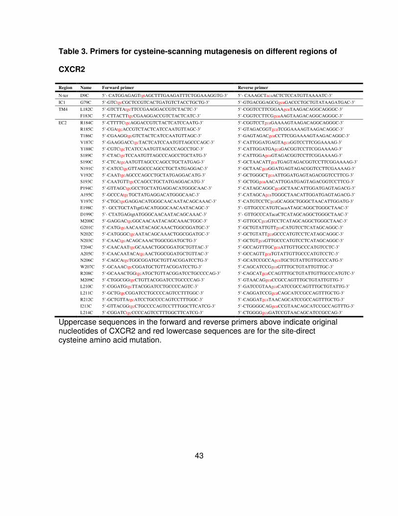
Table 3. Primers for cysteine-scanning mutagenesis on different regions of
CXCR2
Region Name Forward primer Reverse primer
N-ter D9C 5’- CATGGAGAGTtgtAGCTTTGAAGATTTCTGGAAAGGTG-3’ 5’- CAAAGCTacaACTCTCCATGTTAAAATC-3’
IC1 G79C 5’-GTCtgcCGCTCCGTCACTGATGTCTACCTGCTG-3’ 5’-GTGACGGAGCGgcaGACCCTGCTGTATAAGATGAC-3’
TM4 L182C 5’-GTCTTAtgcTTCCGAAGGACCGTCTACTC-3’ 5’-CGGTCCTTCGGAAgcaTAAGACAGGCAGGGC-3’
F183C 5’-CTTACTTtgcCGAAGGACCGTCTACTCATC-3’ 5’-CGGTCCTTCGgcaAAGTAAGACAGGCAGGGC-3’
EC2 R184C 5’-CTTTTCtgcAGGACCGTCTACTCATCCAATG-3’ 5’-CGGTCCTgcaGAAAAGTAAGACAGGCAGGGC-3’
R185C 5’-CGAtgcACCGTCTACTCATCCAATGTTAGC-3’ 5’-GTAGACGGTgcaTCGGAAAAGTAAGACAGGC-3’
T186C 5’-CGAAGGtgcGTCTACTCATCCAATGTTAGC-3’ 5’-GAGTAGACgcaCCTTCGGAAAAGTAAGACAGGC-3’
V187C 5’-GAAGGACCtgcTACTCATCCAATGTTAGCCCAGC-3’ 5’-CATTGGATGAGTAgcaGGTCCTTCGGAAAAG-3’
Y188C 5’-CGTCtgcTCATCCAATGTTAGCCCAGCCTGC-3’ 5’-CATTGGATGAgcaGACGGTCCTTCGGAAAAG-3’
S189C 5’-CTACtgcTCCAATGTTAGCCCAGCCTGCTATG-3’ 5’-CATTGGAgcaGTAGACGGTCCTTCGGAAAAG-3’
S190C 5’-CTCAtgcAATGTTAGCCCAGCCTGCTATGAG-3’ 5’-GCTAACATTgcaTGAGTAGACGGTCCTTCGGAAAAG-3’
N191C 5’-CATCCtgcGTTAGCCCAGCCTGCTATGAGGAC-3’ 5’-GCTAACgcaGGATGAGTAGACGGTCCTTCGAAAAG-3’
V192C 5’-CAATtgcAGCCCAGCCTGCTATGAGGACATG-3’ 5’-GCTGGGCTgcaATTGGATGAGTAGACGGTCCTTCG-3’
S193C 5’-CAATGTTtgcCCAGCCTGCTATGAGGACATG-3’ 5’-GCTGGgcaAACATTGGATGAGTAGACGGTCCTTCG-3’
P194C 5’-GTTAGCtgcGCCTGCTATGAGGACATGGGCAAC-3’ 5’-CATAGCAGGCgcaGCTAACATTGGATGAGTAGACG-3’
A195C 5’-GCCCAtgcTGCTATGAGGACATGGGCAAC-3’ 5’-CATAGCAgcaTGGGCTAACATTGGATGAGTAGACG-3’
Y197C 5’-CTGCtgcGAGGACATGGGCAACAATACAGCAAAC-3’ 5’-CATGTCCTCgcaGCAGGCTGGGCTAACATTGGATG-3’
E198C 5’- GCCTGCTATtgtGACATGGGCAACAATACAGC-3’ 5’- GTTGCCCATGTCacaATAGCAGGCTGGGCTAAC-3’
D199C 5’- CTATGAGtgtATGGGCAACAATACAGCAAAC-3’ 5’- GTTGCCCATacaCTCATAGCAGGCTGGGCTAAC-3’
M200C 5’-GAGGACtgcGGCAACAATACAGCAAACTGGC-3’ 5’-GTTGCCgcaGTCCTCATAGCAGGCTGGGCTAAC-3’
G201C 5’-CATGtgcAACAATACAGCAAACTGGCGGATGC-3’ 5’-GCTGTATTGTTgcaCATGTCCTCATAGCAGGC-3’
N202C 5’-CATGGGCtgcAATACAGCAAACTGGCGGATGC-3’ 5’-GCTGTATTgcaGCCCATGTCCTCATAGCAGGC-3’
N203C 5’-CAACtgcACAGCAAACTGGCGGATGCTG-3’ 5’-GCTGTgcaGTTGCCCATGTCCTCATAGCAGGC-3’
T204C 5’-CAACAATtgcGCAAACTGGCGGATGCTGTTAC-3’ 5’-GCCAGTTTGCgcaATTGTTGCCCATGTCCTC-3’
A205C 5’-CAACAATACAtgcAACTGGCGGATGCTGTTAC-3’ 5’-GCCAGTTgcaTGTATTGTTGCCCATGTCCTC-3’
N206C 5’-CAGCAtgcTGGCGGATGCTGTTACGGATCCTG-3’ 5’-GCATCCGCCAgcaTGCTGTATTGTTGCCCATG-3’
W207C 5’-GCAAACtgcCGGATGCTGTTACGGATCCTG-3’ 5’-CAGCATCCGgcaGTTTGCTGTATTGTTGC-3’
R208C 5’-GCAAACTGGtgcATGCTGTTACGGATCCTGCCCCAG-3’ 5’-CAGCATgcaCCAGTTTGCTGTATTGTTGCCCATGTC-3’
M209C 5’-CTGGCGGtgcCTGTTACGGATCCTGCCCCAG-3’ 5’-GTAACAGgcaCCGCCAGTTTGCTGTATTGTTG-3’
L210C 5’-CGGATGtgcTTACGGATCCTGCCCCAGTC-3’ 5’-GATCCGTAAgcaCATCCGCCAGTTTGCTGTATTG-3’
L211C 5’-GCTGtgcCGGATCCTGCCCCAGTCCTTTGGC-3’ 5’-CAGGATCCGgcaCAGCATCCGCCAGTTTGCTG-3’
R212C 5’-GCTGTTAtgcATCCTGCCCCAGTCCTTTGGC-3’ 5’-CAGGATgcaTAACAGCATCCGCCAGTTTGCTG-3’
I213C 5’-GTTACGGtgcCTGCCCCAGTCCTTTGGCTTCATCG-3’ 5’-CTGGGGCAGgcaCCGTAACAGCATCCGCCAGTTTG-3’
L214C 5’-CGGATCtgcCCCCAGTCCTTTGGCTTCATCG-3’ 5’-CTGGGGgcaGATCCGTAACAGCATCCGCCAG-3’
Uppercase sequences in the forward and reverse primers above indicate original nucleotides of CXCR2 and red lowercase sequences are for the site-direct cysteine amino acid mutation.
44
Chapter 4 Results
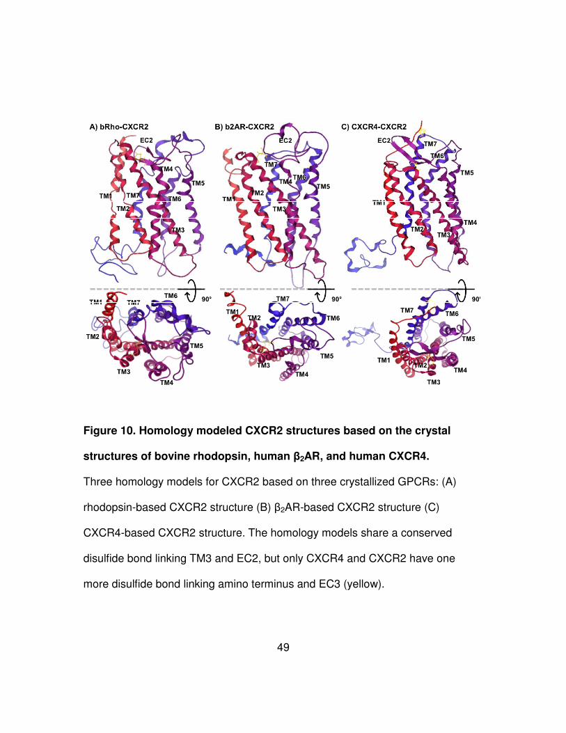
Structural similarity and differences between homology modeled
CXCR2 and crystal structures of GPCRs
Sequence alignment and homology modeling for CXCR2 were performed
with bovine rhodopsin, β2AR, and CXCR4 in MOE software. Sequence
alignments with rhodopsin and β2AR showed low identity in the alignment (18.1%
for rhodopsin, 24.2% for β2AR), whereas CXCR4 provided relatively high identity
(33.3%) to CXCR2 (Fig. 7, 8, 9). In all the cases, the (D/E)R(Y/W) and NPXXY
motifs are conserved in their primary sequences. The modeling produced a
rhodopsin-based CXCR2 model (bRho-CXCR2), a β2AR-based model (b2AR-
CXCR2), and a CXCR4-based model (CXCR4-CXCR2) (Fig. 10). All three
models contain the highly conserved disulfide bond of GPCRs however the
CXCR4-based model has one more disulfide bond linking amino terminus and
third extracellular loop. Like their templates, bRho-CXCR2 and CXCR4-CXCR2
produced two β-strands in its second extracellular loop, whereas b2AR-CXCR2
showed only one α-helix.
To evaluate structural similarity of homology modeled CXCR2 structures
and crystal structures of GPCRs, superposition of the structures were performed
and RMSD values of seven transmembrane helices were measured (Fig. 11).
Zero at RMSD means they are identical in conformation. The bRho-CXCR2
45
showed the lowest value (1.64 Å) in the comparison with rhodopsin, the b2AR-
CXCR2 was the most identical to β2AR (2.02 Å), and CXCR4-CXCR2 was very
similar to CXCR4 (0.78 Å). Interestingly, the RMSD from CXCR4-CXCR2 versus
CXCR4 was the lowest among comparison of the models with their own
templates. The highest RMSD value (2.93 Å) was derived from the comparison of
b2AR-CXCR2 and CXCR4. Furthermore, Site Finder was used to predict ligand
binding sites of b2AR-CXCR2 and CXCR4-CXCR2 (Fig. 12). The prediction
indicates that residues in all the extracellular loops are highly associated with
ligand binding. Residues in some of transmembranes domains showed the
involvement in ligand binding as well.
46
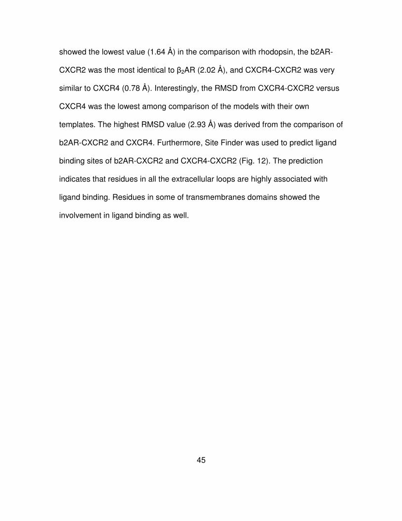
Figure 7. Sequence alignment of CXCR2 and bovine rhodopsin.
Identical residues are represented with color boxes. CXCR2 shows 18.1%
(65/360 amino acids) identity with rhodopsin. Both have the highly conserved
(D/E)R(Y/W), NPXXY motif, and rotamer toggle switch (#) (red texts and boxes),
whereas only bovine rhodopsin has a Glu residue in its IC3 forming an ionic lock
between TM3 and TM6. In the EC2 of rhodopsin, there are two β-strand
structures (Green). Asterisks indicate the Cys residues that form a conserved
disulfide bond in GPCRs. Each motif of rhodopsin and CXCR2 were based on
crystal structure of rhodopsin (PDB code: 1U19) and prediction of universal
protein resource (Uniprot, www.uniprot.org), respectively.
47
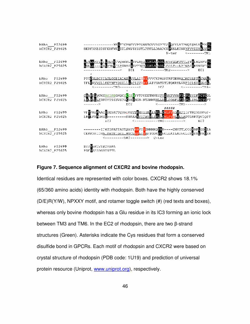
Figure 8. Sequence alignment of CXCR2 and β2AR.
Identical residues are represented with color boxes. CXCR2 shows 24.2%
(87/360 amino acids) identity with β2AR. Both share the highly conserved
(D/E)R(Y/W), NPXXY motif, and rotamer toggle switch (#) (red texts and boxes),
whereas a Glu residue in its IC3 forming an ionic lock between TM3 and TM6
only conserved in β2AR. In the EC2 of β2AR, there are two α-helices structures
(blue). Asterisks indicate the Cys residues that form a conserved disulfide bond
in GPCRs. Each motif of β2AR and CXCR2 were based on crystal structure of
β2AR (PDB code: 2RH1) and prediction of universal protein resource (Uniprot,
www.uniprot.org), respectively.
48
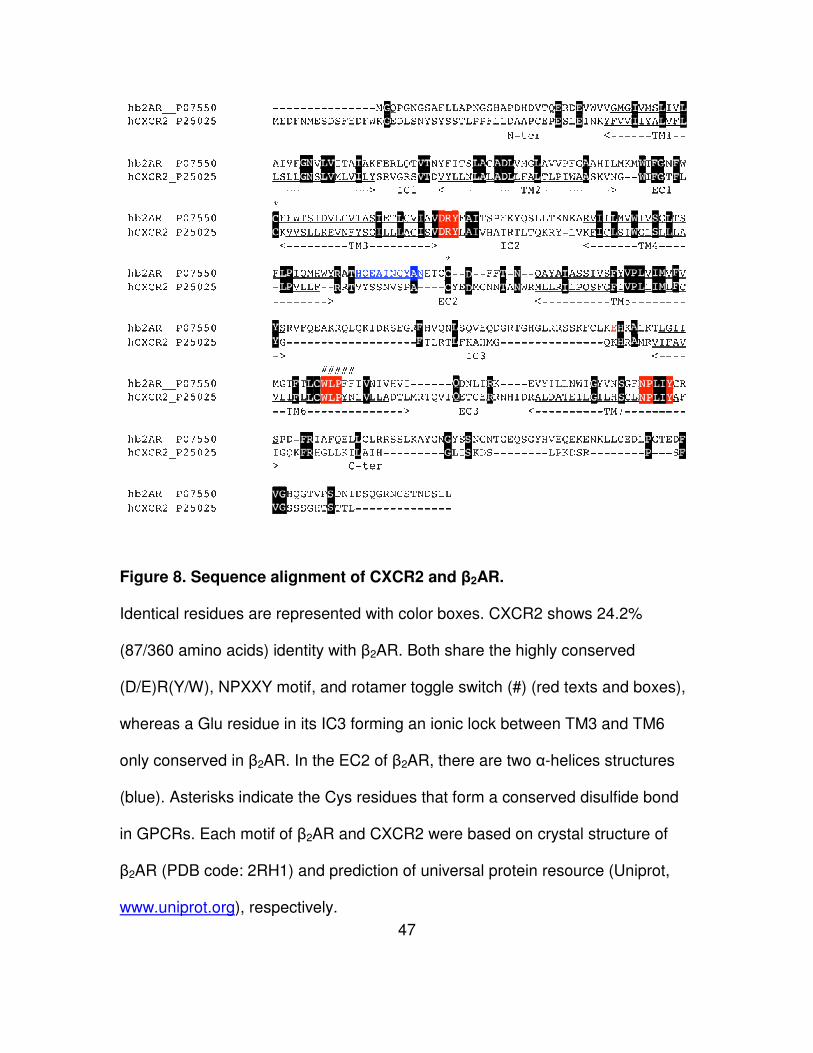
Figure 9. Sequence alignment of CXCR2 and CXCR4.
Identical residues are represented with color boxes. CXCR2 shows 33.3%
(120/360 amino acids) identity with CXCR4. Their EC2s shared few amino acids
(16% identity, 4/25 amino acids). Both share the highly conserved (D/E)R(Y/W),
NPXXY motif, and rotamer toggle switch (#) (red boxes). However, CXCR2 and
CXCR4 has no a Glu residue in its IC3 forming an ionic lock between TM3 and
TM6 conserved in rhodopsin and β2AR. In the EC2 of CXCR4, there are two β-
strand structures (green). There are two disulfide bonds (* and &) in the GPCRs.
Each motif of CXCR4 and CXCR2 were based on crystal structure of CXCR4
(PDB code: 3ODU) and prediction of universal protein resource (Uniprot,
www.uniprot.org), respectively.
49
Figure 10. Homology modeled CXCR2 structures based on the crystal
structures of bovine rhodopsin, human β2AR, and human CXCR4.
Three homology models for CXCR2 based on three crystallized GPCRs: (A)
rhodopsin-based CXCR2 structure (B) β2AR-based CXCR2 structure (C)
CXCR4-based CXCR2 structure. The homology models share a conserved
disulfide bond linking TM3 and EC2, but only CXCR4 and CXCR2 have one
more disulfide bond linking amino terminus and EC3 (yellow).
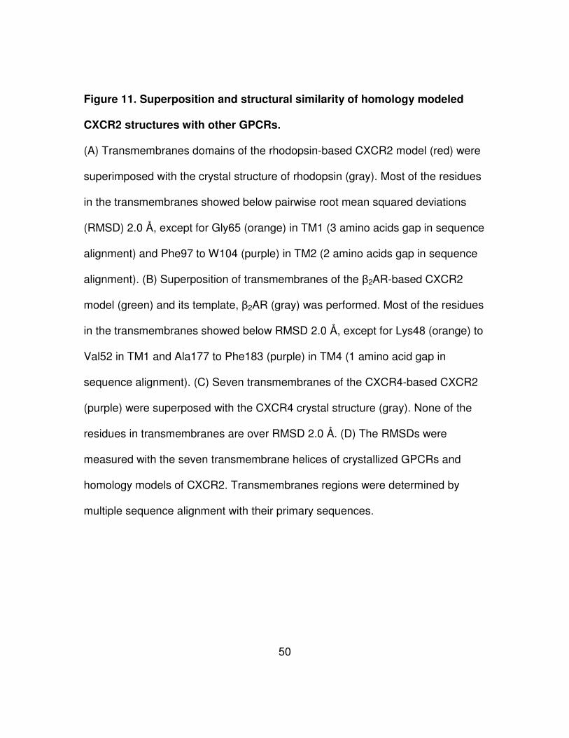
50
Figure 11. Superposition and structural similarity of homology modeled
CXCR2 structures with other GPCRs.
(A) Transmembranes domains of the rhodopsin-based CXCR2 model (red) were
superimposed with the crystal structure of rhodopsin (gray). Most of the residues
in the transmembranes showed below pairwise root mean squared deviations
(RMSD) 2.0 Å, except for Gly65 (orange) in TM1 (3 amino acids gap in sequence
alignment) and Phe97 to W104 (purple) in TM2 (2 amino acids gap in sequence
alignment). (B) Superposition of transmembranes of the β2AR-based CXCR2
model (green) and its template, β2AR (gray) was performed. Most of the residues
in the transmembranes showed below RMSD 2.0 Å, except for Lys48 (orange) to
Val52 in TM1 and Ala177 to Phe183 (purple) in TM4 (1 amino acid gap in
sequence alignment). (C) Seven transmembranes of the CXCR4-based CXCR2
(purple) were superposed with the CXCR4 crystal structure (gray). None of the
residues in transmembranes are over RMSD 2.0 Å. (D) The RMSDs were
measured with the seven transmembrane helices of crystallized GPCRs and
homology models of CXCR2. Transmembranes regions were determined by
multiple sequence alignment with their primary sequences.
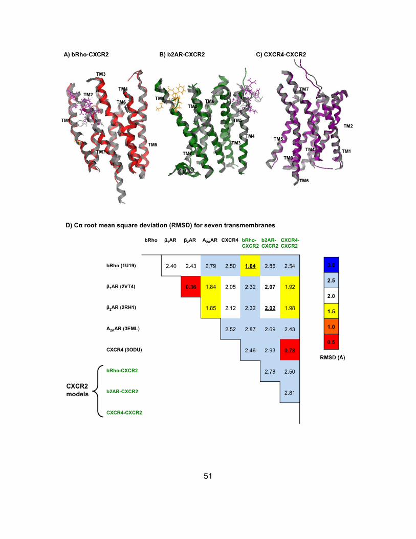
51
52
Figure 12. Prediction of ligand binding sites in β2AR-based and CXCR4-
based CXCR2 structure.
The Site Finder in Molecular Operation Environment (MOE) software was used to
search for potential ligand binding sites in β2AR-based CXCR2 (A) and CXCR4-
based CXCR2 models (B). Spheres indicate possible contact sites for ligand
binding (red = hydrophilic, white = hydrophoblic). (A) In the β2AR-based CXCR2,
most of the residues in the predicted binding site are localized in the extracellular
loops, including Lys108, Val109, Asn110, Gly111, and Trp112 in the first
extracellular loop, Arg184, Arg185, Cys196, Tyr197, Glu198, Asp199, Met200,
and Thr204 in the second extracellular loop (yellow), and Glu284, Thr285,
Cys286, Arg289, Asn290, Asp293, and Leu296 in the third extracellular loop.
Furthermore, some transmembranes residues were predicted as binding sites,
including Phe97, Thr100, Ile103, Trp104, and Ser107 in the second
transmembrane, Cys119, Val122, Ser123, Lys126, Glu127, and Phe130 in the
third transmembrane, and Phe218, Trp264, Tyr267, and Leu271 in the fifth
transmembrane. (B) CXCR4-based CXCR2 also has many residues in
extracellular domains as potential ligand binding sites; Glu40, Pro41, Glu42, and
Lys48 in the amino terminus, Trp104, Ser107, Lys108, Val109, Asn110, Gly111,
and Trp112 in the first extracellular loop, and Arg184, Arg185, Thr186, Tyr188,
Ser190, Asn191, Val192, Ser193, Pro194, Ala195, Cys196, Tyr197, Glu198,
Asp199, Arg208, Leu211, and Arg212 in the second extracellular loop.
Furthermore, several residues in the transmembrane domains were predicted as
ligand binding sites: Tyr55 in the first transmembrane, Phe97 and Leu101 in the
second transmembrane, Val122, Ser123, Lys126, Glu127, and Phe130 in the
third transmembrane, Val180 in the fourth transmembrane, Pro215 and Gln216
in the fifth transmembrane, Tyr267, Leu271, Asp274, and Arg278 in the sixth
transmembrane, and Arg289, Ile292, Asp293, Arg294, Leu296, Asp297, Glu300,
Ile301, and Ile304 in the seventh transmembrane.
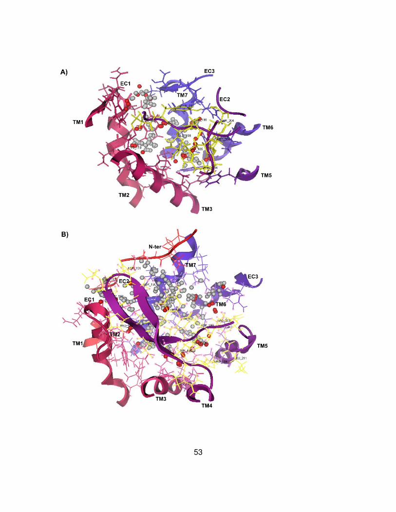
53
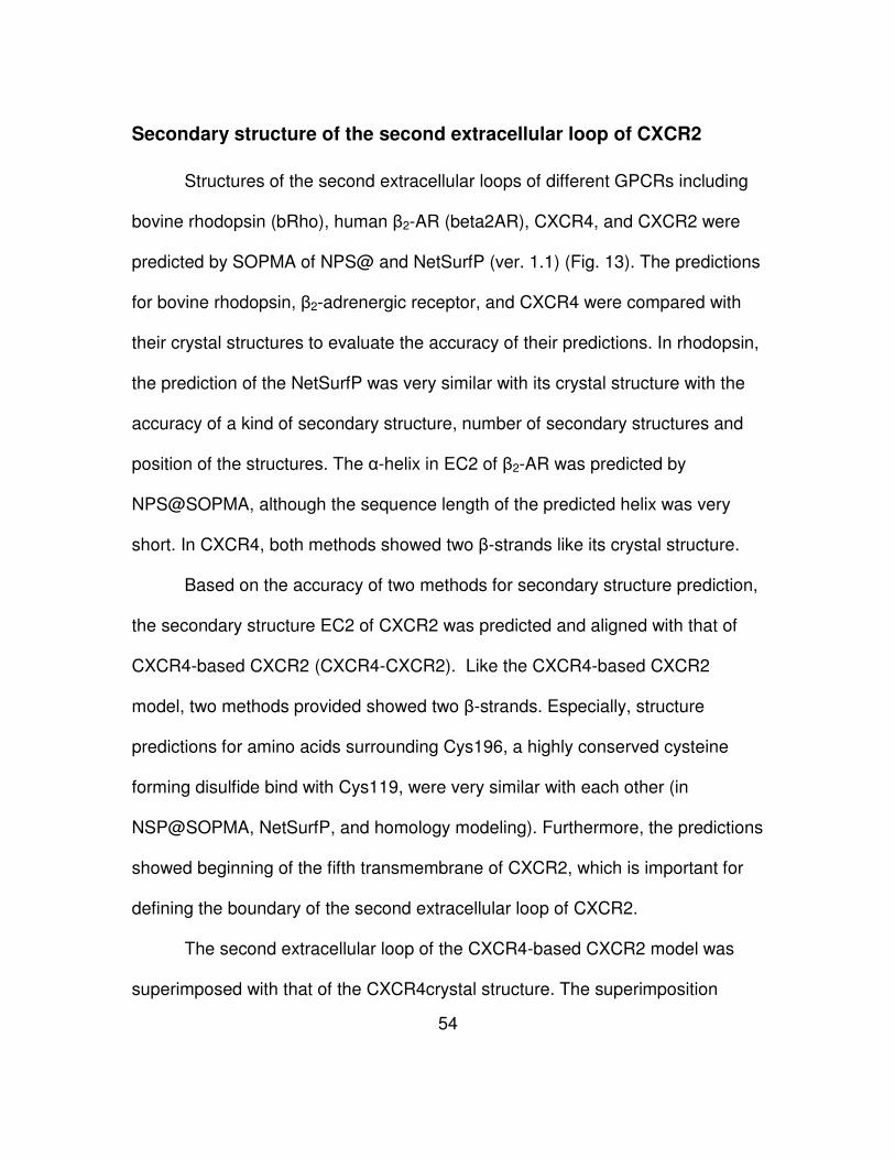
54
Secondary structure of the second extracellular loop of CXCR2
Structures of the second extracellular loops of different GPCRs including
bovine rhodopsin (bRho), human β2-AR (beta2AR), CXCR4, and CXCR2 were
predicted by SOPMA of NPS@ and NetSurfP (ver. 1.1) (Fig. 13). The predictions
for bovine rhodopsin, β2-adrenergic receptor, and CXCR4 were compared with
their crystal structures to evaluate the accuracy of their predictions. In rhodopsin,
the prediction of the NetSurfP was very similar with its crystal structure with the
accuracy of a kind of secondary structure, number of secondary structures and
position of the structures. The α-helix in EC2 of β2-AR was predicted by
NPS@SOPMA, although the sequence length of the predicted helix was very
short. In CXCR4, both methods showed two β-strands like its crystal structure.
Based on the accuracy of two methods for secondary structure prediction,
the secondary structure EC2 of CXCR2 was predicted and aligned with that of
CXCR4-based CXCR2 (CXCR4-CXCR2). Like the CXCR4-based CXCR2
model, two methods provided showed two β-strands. Especially, structure
predictions for amino acids surrounding Cys196, a highly conserved cysteine
forming disulfide bind with Cys119, were very similar with each other (in
NSP@SOPMA, NetSurfP, and homology modeling). Furthermore, the predictions
showed beginning of the fifth transmembrane of CXCR2, which is important for
defining the boundary of the second extracellular loop of CXCR2.
The second extracellular loop of the CXCR4-based CXCR2 model was
superimposed with that of the CXCR4crystal structure. The superimposition
55
showed the second extracellular loop of CXCR2 is structurally very similar with
that of CXCR4 (below 2.0 Å RMSD per residue, 0.6 Å RMSD in total), with an
exception of Asn203 of CXCR2 (Asp193 of CXCR4) (over 2.0 Å RMSD per
residue).
56
Figure 13. Prediction of secondary structure of the second extracellular
loop of CXCR2
A) Structures of the second extracellular loops of several GPCRs including
bovine rhodopsin (bRho), β2-adrenergic receptor (beta2AR), CXCR4, and
CXCR2 were predicted by using NPS@SOPMA and NetSurfP (ver. 1.1). The
predictions for bovine rhodopsin, β2-adrenergic receptor, and CXCR4 were also
compared with their crystal structures. The predicted secondary structure of the
EC2 of CXCR2 was aligned with that of CXCR4-based CXCR2 (CXCR4-CXCR2).
Color code was used to show secondary structures: h = α-helix, e = β-strand, t =
turn, and c = coil. Cys residues that form a conserved disulfide bond were
indicated with specific signs (* for rhodopsin, β2AR, CXCR4, and CXCR2, # for
intra disulfide bond in only β2AR). B) The structures of the second extracellular
loops of CXCR4 and CXCR4-based CXCR2 model were superimposed with
each other. All of the residues in the EC2 of CXCR2 except for Asp203 (over 2.0
Å RMSD, compared with Asp193 of CXCR4) were structurally very similar to
CXCR4. Two cysteine residues (Cys119 and Cys196) forming a conserved
disulfide bond were represented.
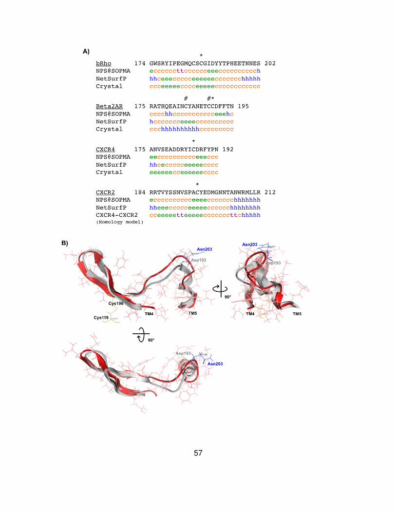
57
58
Solvent accessibility of residues on the second extracellular loop of
CXCR2 in the inactive state
Wild-type CXCR2 and single cysteine mutant receptors were transiently
transfected into HEK293 cells. After 48 h of cultivation, whole cells were
harvested and exposed to MTSEA-biotin for labeling of water-accessible cysteine
residues. Cell surface-expressed CXCR2 receptors and MTSEA-biotin labeled
receptors were immunostained with an anti-CXCR2 antibody and streptavidin-
FITC, respectively. Flow cytometry was used to show solvent accessibility of
individual CXCR2 receptors mutants. Accessibility values of wild-type CXCR2
and single cysteine mutant receptors were calculated from the mean
fluorescence intensity (MFI) of FITC (detects accessibility of an additional Cys
residue) divided by MFI of PE (detects total cell surface expression level of
CXCR2 receptors). The relative accessibility values were derived from
accessibility values of the mutant receptors fold over that of wild-type. A
representative graph was drawn for the relative accessibility values (Fig. 14).
The experiment reveals that residue D9C in the amino terminus of
CXCR2, is solvent exposed, whereas residue G79C in the first intracellular loop
is not water accessible. L182C and F183C which are predicted to be in the fourth
transmambrane (TM4) showed similar or less solvent accessibility compared with
that of wild-type CXCR2. Residues of the second extracellular loop are divided
into two groups by their location relative to residue Cys196 that is critical for a
highly conserved disulfide bond found in class A GPCRs. The front group is from
59
R184C to A195C and the rear group is from Y197C to L214C. Although there is
fluctuation between accessibility values, all of the residues in the front group of
EC2 are solvent exposed. Interestingly, in the rear group of EC2, Y197C next to
Cys196 and the penta-amino acids, T204 to R208, showed similar water
accessibility with wild-type CXCR2 and the rest of the rear group are solvent
exposed. The solvent accessibility of I213C close to the fifth transmembrane
(TM5) was the highest in our experiment.
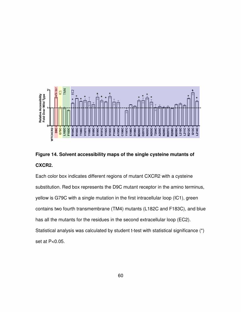
60
Figure 14. Solvent accessibility maps of the single cysteine mutants of
CXCR2.
Each color box indicates different regions of mutant CXCR2 with a cysteine
substitution. Red box represents the D9C mutant receptor in the amino terminus,
yellow is G79C with a single mutation in the first intracellular loop (IC1), green
contains two fourth transmembrane (TM4) mutants (L182C and F183C), and blue
has all the mutants for the residues in the second extracellular loop (EC2).
Statistical analysis was calculated by student t-test with statistical significance (*)
set at P<0.05.
61
Chapter 5 Discussion
Crystal structures of GPCRs have been used to predict structures of other
GPCRs that have not been solved (32, 41, 42). Homology modeling for CXCR2
using the crystallized bovine rhodopsin have been attempted to explain
experiments of receptor stabilization and antagonism (32, 41, 42, 84, 102).
However, their observations about the predicted CXCR2 models focused on
some limited regions of interested (e.g. second extracellular loop and seventh
transmembrane). Furthermore, their homology modeled CXCR2 structures were
not evaluated through the comparison with more recent crystal structures of other
GPCRs.
In this study, we performed homology modeling for CXCR2 by using three
crystallized GPCRs, rhodopsin, β2AR and CXCR4. Sequence alignments
between them showed conservation of highly conserved motifs (e.g.
(D/E)R(Y/W), NPXXY, and toggle switch), pointing to the possibility of using the
three crystal structures as templates for the homology modeling (Fig. 7, 8, 9).
Furthermore, the motifs and alignments were used to compare locations and
length of transmembrane helices for evaluation of homology modeled CXCR2.
The highly conserved disulfide bond linking TM3 and EC2 was automatically
generated during the homology modeling process in all the models, but only the
CXCR4-based CXCR2 model has one more disulfide bond linking the amino
terminus and EC3. The homology modeled structures are intended to show
62
structural similarity with their templates. Especially since the secondary
structures in the second extracellular loops were the same as their original
templates (e.g. β-strands of rhodopsin and α-helix of β2AR) (Fig. 10) and their
RMSD values also represented the intention (Fig. 11). However, we made use of
β2AR and CXCR4 as a template for homology modeling, even though they have
some structural modifications that interrupt modeling for other GPCRs (e.g.
fusion with T4 lysozyme in the third intracellular loop in both structures, and an
intra-disulfide bond in the second extracellular loop found only in β2AR). Even
though all three crystallized GPCR stuctures were used as templates and
showed significant structural conservation in transmembrane domains, CXCR4
might be the best template for CXCR2 based on high identity (33.3%) in
sequence alignment and the lowest RMSD values (0.78Å) in structure
superposition (Fig. 9,10). Furthermore, two secondary structure prediction
programs, NPS@SOPMA and NetSurfP were used to predict the secondary
structure of the second extracellular loop of CXCR2. In three GPCRs including
rhodopsin, β2AR, and CXCR4, the programs showed highly accurate results in
the comparison of crystal structures and their predictions. The prediction of the
programs suggested that the second extracellular of CXCR2 has two β-strands
like rhodopsin and CXCR4. Similarly, the CXCR4-based CXCR2 model
suggested two β-strands, even though CXCR2 and CXCR4 have very low
identity between the residues in their second extracellular loops. Interestingly, the
superimposition of CXCR4-based CXCR2 and CXCR4 suggested that the
63
structure of the second extracellular loop of the CXCR2 model might be very
similar with CXCR4 (Fig. 13).
We also studied the topology of residues in the EC2 of CXCR2 by using
substituted-cysteine accessibility method (SCAM), due to the importance of this
region in ligand binding and potential receptor activation. Unlike the common
SCAM process with immunoblotting (9, 130), flow cytometric analysis was
performed to amplify signals of FITC-streptavidin bound MTSEA-biotin.
Furthermore, cell surface expression levels of mutant receptors that are needed
to normalize MTSEA-biotin accessibility were measured at the same time
through immunostaining with a specific antibody against CXCR2. The refined
approach was evaluated by the accessibility of several mutant receptors
including D9C (amino terminus), G79C (IC1), L182C (TM4), and F183C (TM4).
Our SCAM results indicate that most of the residues in EC2 are water-
accessible in the inactive state of CXCR2 (in the absence of ligand) (Fig. 14).
Especially, all the charged residues (R184C, R185C, E198C, D199C, and
R212C) in EC2 except for R208C are exposed to solvent. Because charged
residues on the extracellular loops were considered as key residues to form a
binding pocket, ligands of CXCR2 could contact the exposed residues followed
by receptor activation. In the study to find CXCL8 binding sites to homology
modeled CXCR2 structures, most of the residues in EC2 including the charged
residues were predicted as potential key residues (Fig. 12). In EC2, five
sequential amino acids 204TANWR208 were predicted as non-accessible residues.
64
These residues might be involved in the interaction with the amino terminus or
other extracellular loops. Since the CXCR2 models lack an amino terminus, there
are limitations to studying the mechanisms for ligand binding and receptor
activation. Thus our interdisciplinary approaches and observations would be very
helpful to plan new approaches to discover unknown mechanisms about CXCR2.
65
PART III
INVOLVEMENT OF THE SECOND EXTRACELLULAR
LOOP OF CXCR2 IN LIGAND RECOGNITION AND
RECEPTOR ACTIVATION
66
Chapter 1 Abstract
It has been shown that the amino terminus and second extracellular loop
of CXCR2 are crucial for ligand binding and receptor activation. The lack of an
ionic lock motif (a Glu residue) in the third intracellular loop in CXCR2 focuses an
investigation of two extracellular regions in CXCR2 involved in receptor
recognition and activation. Using constitutively active mutants, D9K and D9R, in
CXCR2, the potential role of charged residues in the second extracellular loop of
CXCR2 was investigated.
Combinatorial mutations consisting of the constitutively active mutants in
the amino terminus and single mutations of charged residues in the second
extracellular loop were generated to study two concepts including “attraction” and
“repulsion” models. The mutant receptors were used to test their effects on cell
surface expression, ligand binding, receptor activation through PLC-β3, and
cellular transformation. All the mutations in the repulsion model result in CXCR2
receptors that are unable to bind ligand, indicating that all the Arg residues
(Arg184, Arg185, Arg208, and Arg212) in the second extracellular loop are
important for ligand recognition. Although charge-reversal complementary
mutations in the repulsion model were expected to retain structural constraints of
CXCR2, none of the mutant receptors function as wild-type CXCR2 in the
inactive and active states. Interestingly, mutations in the attraction model partially
inhibited receptor activation of constitutively active mutant D9K, suggesting that
Glu198 and Asp199 residues in the second extracellular loop are highly
67
associated with receptor activation. Furthermore, a novel constitutively active
mutant E198A/D199A was identified in this study. These negatively charged
residues are very close to a disulfide bond (Cys119-Cys196) linking the second
extracellular loop and the third transmembrane. The unique location suggests
that the neutralization (with Ala residues) of the residues might be needed to
control TM3 promote slight breakdown of structural constraint of the constitutively
activated CXCR2 with D9K mutation.
These observations are helpful to understand how the extracellular
regions of CXCR2 interact with its cognate ligands and transfer signals
intracellularly.
68
Chapter 2 Introduction
GPCRs respond to a variety of ligands that are classified into agonist and
antagonist. GPCRs can also induce constitutive activation in the absence of
external ligands. Single point mutations of GPCRs as well as some wild-type
GPCRs have been shown to lead to constitutively activity (131). Moreover, the
constitutively active mutants of GPCRs have been used as important
pharmacological targets for the development of potent drugs against mutant
GPCR-mediated diseases (132).
In class A GPCRs, it has been shown that the highly conserved
(D/E)R(Y/W) motif in TM3 is important for interaction with D/E residue at the
boundary of IC3 and TM6 (38). The interaction, also called the ‘ionic lock’, is
crucial to stabilize the inactive conformation of GPCR receptors. Mutations of
these residues result in an increase of constitutive activity (39, 133).
Furthermore, breakage of the ionic lock is linked to turning on the ‘toggle switch’
in TM6, with the change of configuration of a conserved Pro residue surrounding
aromatic residues in TM6 (38). These mechanisms for receptor activation are
conserved in both rhodopsin and β2AR have been well studied. However, in most
(~80%) of class A GPCRs, residues for the ionic lock or toggle switch are not
present (133).
Currently, novel mechanisms for receptor activation have been found. The
first example is a model with ionic locks on the extracellular surface (134). Free
fatty acid receptor 1 (FFAR1) does not contain residues for both an ionic lock and
69
toggle switch similar to rhodopsin and β2AR. FFAR1 contains a Lys instead of a
Glu in TM6 (for an ionic lock) and a Val and Asn rather than the aromatic
residues in TM6 (for a toggle switch). Interestingly, homology modeling of FFAR1
showed two ionic locks between two Glu residues on EC2 and two Arg residues
on distinct transmembranes including TM5 and TM7. Single alanine mutations for
the two Glu on EC2 induced constitutive activation of the receptor in the absence
of ligand, indicating that the EC2 of the receptor can participate in receptor
activation. Furthermore, a random mutagenesis study of residues on EC2 of the
complement factor 5a receptor (C5aR), a class B GPCR, showed that multiple
residues in EC2 are highly associated with receptor stabilization in the inactive
state (36).
A second example is the role of conserved Cys residues in the amino
terminus and third extracellular loop (EC3) of chemokine receptors (135). CXCR4
was used as a model to substitute a Ser pair, aromatic pair (Phe), or salt bridge
pair (Arg/Asp) for the conserved Cys residues. The mutation with a Ser pair in a
constitutively active mutant (CAM) CXCR4 (N119S) disrupted the receptor
activation, whereas the other substitutions with the Phe or Arg/Asp pair in the
CAM CXCR4 showed little effect on the constitutively activity of the receptor. The
result suggested that the disufide bond derived from the conserved Cys in the
amino terminus and EC3 of chemokine receptors is important for receptor
activation and signaling.
CXCR2 shares the highly conserved (D/E)R(Y/W) motif like other GPCRs
in class A. A point mutation of Asp143 (to Val, the same with KSHV-GPCR) of
70
the motif on CXCR2 leads to constitutive activation of the receptor and cellular
transformation similar to KSHV-GPCR (118). However, like FFAR1 (Lys219),
CXCR2 has Lys246 instead of D/E residue in TM6 to interact with Arg of the
(D/E)R(Y/W) motif for an ionic lock, whereas aromatic residues (Trp264 and
Tyr267 in TM6 of CXCR2) for a toggle switch are conserved in TM6. This
suggests that CXCR2 is different in the mechanism for receptor activation
compared to rhodopsin and β2AR. In a previous study of the role of EC2 in
activation and regulation of the receptor, it has been shown that mutation of
Asp199 (to Val) in EC2 delayed the internalization of the receptor, whereas
substitution with Asn restored the internalization speed (84). With homology
modeling of CXCR2 based on rhodopsin, the study suggested that Asp199
functions to stabilize the receptor conformation using hydrogen bonds between
several residues (Arg185, Thr186, and Arg208) in EC2. Currently, it has been
shown that single point mutations of Asp9 in the amino terminus of CXCR2 leads
to constitutive activation to promote cellular transformation activity in cells stably
expressing the mutant receptors (Park et al., unpublished). The study also
demonstrated that different constitutive active mutants show signal specificity.
Taken together, these observations suggest that both the amino terminus and
EC2 of CXCR2 are crucial in receptor activation.
Viral oncogene ORF74 encoded by Kaposi’s sarcoma-associated
herpesvirus (human herpesvirus 8), also called KSHV-GPCR, is the closest
homolog to CXCR2 (103). KSHV-GPCR constitutively activates AP-1, CREB,
NFAT, and MAPK via Gαi or Gαq, and NF-κB through only Gαq subtype in
71
different cell lines including COS-7, primary effusion lymphoma (PEL), and
endothelial cells (104-107). Currently, studies of M33 of murine cytomegalovirus
(MCMV) and US28 of human cytomegalovirus (HCMV), viral GPCR homologs,
have been shown that Gαq protein is activated to induce phopholipase C-β (PLC-
β) activity, followed by stimulation of CREB and NF-κB signals (108-111).
Additionally, M33 can induce CREB and NF-κB stimulation via a PLC-β
independent manner (112). On the other hand, another GPCR homolog UL33 of
HCMV enhances CREB transcriptional activities by Gs pathway not Gq/11 pathway
(113).
As stated previously, a point mutation of Asp143 to Val in the highly
conserved (D/E)R(Y/W) motif in CXCR2 leads to constitutive activation and
cellular transformation (118). The transforming capacities were totally inhibited by
using AG490, a specific kinase inhibitor of JAK2-STAT3 signaling (119). It has
been demonstrated that the constitutive activity of KSHV-GPCR and CXCR2
mutant D143V is highly associated with STAT3 phosphorylation. A current study
of CXCR2 expressed in ovarian cancer cells reported that cancer growth is
regulated by the MAPK and NF-κB pathways (121). Additionally, it was shown
that the chemokine CXCL1 binding to CXCR2 but not CXCR1 induces
transactivation of epidermal growth factor receptor (EGFR) via the chemokine
receptor, followed by proliferation of ovarian cancer cells (122).
In this study, we investigated a role of charged residues in EC2 of CXCR2
in ligand binding and receptor activation. Two concepts, “attraction” and
“repulsion” models were designed to investigate the mechanism of CXCR2
72
activation (Fig. 15). To end that, we constructed combinatorial mutation sets
consisting of constitutively active mutants (CAM) Asp9 in the amino terminus and
substitutions in EC2. These mutant receptors were generated by site-directed
mutagenesis and transfected into mouse fibroblast NIH3T3 cell lines. The
NIH3T3 cells stably expressing wild-type CXCR2 and mutant receptors were
used to investigate cell surface expression of the receptor, ligand-binding, and
receptor activation through phospholipase C-β3 (PLC-β3).
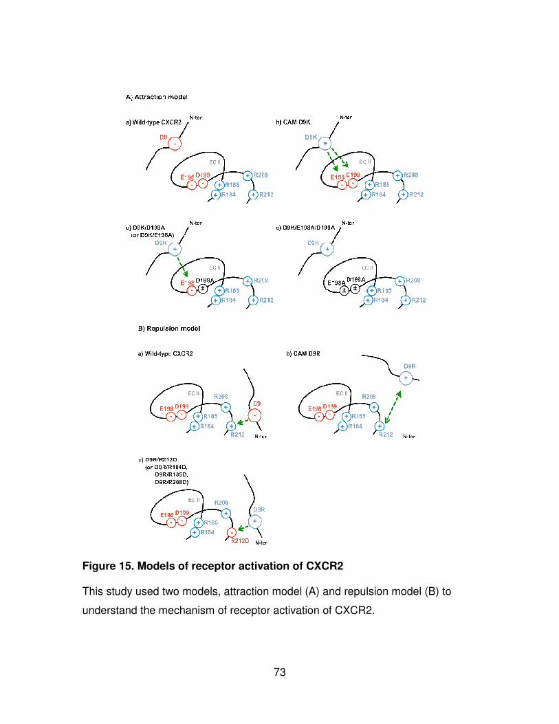
73
Figure 15. Models of receptor activation of CXCR2
This study used two models, attraction model (A) and repulsion model (B) to
understand the mechanism of receptor activation of CXCR2.
74
CHAPTER 3 MATERIALS AND METHODS
Cell lines and media
NIH3T3 mouse fibroblast cells were cultured with DMEM containing 10%
fetal clone 3 (FC3), 1X NEAA, and 1X penicillin/streptomycin. Transfectant
NIH3T3 cells stably expressing wild-type CXCR2 or mutant receptors were
maintained in the medium with 800 µg/ml G418, also known as Geneticin.
Site-directed mutagenesis and transfection
The CXCR2 gene cloned in pRc/CMV vector was used as a template for
site-directed mutagenesis. Forward and reverse primers were manually designed
for charged residues on EC2 of CXCR2 (Table 4). PCR mutagenesis was
performed with a modification of the QuickChange® method (Stratagene). Dpn I
digestion treatment was performed, followed by cleaning via a PCR purification
kit. All mutations were conformed by sequencing analysis (Molecular biology
resource facility, University of Tennessee, Knoxville).
Plasmid DNAs of wild-type CXCR2 and mutant receptors were used to
transfect NIH3T3 cells. The transfections were performed with Lipofectamine
2000 reagent according to the manufacturer’s instructions. 4.0 x 10⁵ NIH3T3
cells were seeded in 6-well plates one day before transfection. 2 µg of plasmid
DNA and 4 µl of Lipofectamine 2000 were individually diluted into 250 µl fresh
DMEM, incubated for 5 min at room temperature, and mixed together followed by
75
incubation for 30 min at room temperature. The mixtures and 500 µl fresh DMEM
were added to each well and incubated for 4 hr at 37 °C. The mixtures were
substituted to new medium including 10% FC3, followed by cultivation for 48 hr at
37 °C. Transfectant NIH3T3 cells stably expressing wild-type CXCR2 and mutant
receptors were selected using the medium with 800 µg/ml G418. Single clonal
cell lines were derived from the transfectants. An anti-CXCR2-specific
monoclonal antibody, phycoerythrin (PE)-conjugated mouse monoclonal anti-
human IL-8 receptor B (R&D systems, MN), and flow cytometry were used to
confirm the cell surface expression level of CXCR2 on the clonal cell lines.
Ligand binding assay
Competition ligand binding assays were performed as previously
described (136). Wild-type CXCR2 and mutant receptors were transiently
transfected into COS-7 cells. After 48 hr, binding was performed on whole cells
for 3 hr at 4 °C using radio-labeled 125I-CXCL8 ligand in binding buffer (50 mM
HEPES, pH 7.4, 1 mM CaCl2, 5 mM MgCl2, and 0.5% bovine serum albumin) and
increasing amounts of unlabeled CXCL8. Washing was performed four times with
binding buffer with 0.5 M NaCl at 4 °C.
Immunostaining of phospho-PLC-β3
The single clonal cell lines stably expressing wild-type CXCR2 or mutant
receptors were used to measure serine phosphorylation (for Ser537) of PLC-β3.
76
4.0 x 10⁵ cells were seeded in 60 mm dishes with the medium containing 10%
FC3 and after one day the medium is substituted to fresh DMEM without serum.
After 18-20 hr the cells were harvested with 2 ml 1X PBS using cell scraper,
centrifuged at 400 g, and fixed with 100 µl fixation buffer BD CytofixTM for 15 min
(BD Biosciences). The fixed cells were permeabilized with BDTM Phosflow Perm
Buffer III (BD Biosciences) for 30 min on ice. A rabbit polyclonal antibody, p-PLC-
β3 (Santa Cruz Biotechnology, Inc., CA) (1:50 dilution) was added to the
permeabilized cells for 1 hr at room temperature in dark. Alexa Fluor 488 goat
anti-rabbit IgG antibody (Invitrogen, CA) (1:50,000) was used as secondary
antibody to immunostain the cells for 1 hr at RT in dark. There was a washing
with 4ml 1X PBS between each step. BD FACSCaliburTM (BD Sciences) was
used to obtain flow cytometry data from the immunostained samples. The flow
cytometry data were analyzed by FlowJo 8.7 software (Tree Star Inc., OR).
Loss of contact inhibition (foci formation) assay
The foci formation assay was performed by previously described (118).
1.0 x 10⁵ untransfected NIH3T3 cells were cultured in each well of a 6-well plate.
After one day, 100 stable transfectants were seeded on top of the untransfected
cells and maintained with serum-reduced medium containing 2% FC3. The cells
were grown with or without inhibitors including PTX, U-73122, or AG490. Foci
were counted after 2 weeks.
77
Chapter 4 Results
Charged residues in the second extracellular loop of CXCR2 are
required for ligand recognition
To study the role of charged residues in second extracellular loop of
CXCR2, negatively charged residues including Glu198 and/or Asp199 and
positively charged residues such as Arg184, Arg185, Arg208, and Arg212 were
substituted to Ala residue to neutralize their charges. The positively charged
residues were also mutated to Asp residues in the charge-reversal manner. Non-
charged residues including Asn191, Asn202, and Asn203 were substituted to Ala
as controls. The mutant constructs were generated by using site-directed
mutagenesis and were conformed by sequence analysis.
The wild-type CXCR2 and mutant receptors were transfected into mouse
fibroblast NIH3T3 cell lines followed by single clonal selection. With NIH3T3 cells
stably expressing the wild-type CXCR2 and mutant receptors, cell surface
expression of the receptors was investigated by immunostaining with a specific
antibody for CXCR2 (Table 4). There was no detectable difference in cell surface
expression between wild-type CXCR2 and mutant receptors.
To measure CXCL8-binding affinity to the wild-type CXCR2 and mutant
receptors, the constructs were transiently transfected in COS-7 cell lines.
Competition binding assays with radio-labeled 125I-CXCL8 were performed to
determine ligand binding affinity (IC50) for each construct (Table 4). We found
78
that all the charged residues on EC2 of CXCR2, except for D199A, are involved
in ligand binding. On the other hand, the mutations of Asn, an uncharged
residue, in EC2 showed a similar level of binding affinity to the wild-type CXCR2
receptor.
Involvement of the amino terminus and second extracellular loop of
CXCR2 in ligand recognition
The combinatorial mutations containing the CAM D9 residue in the amino
terminus and mutations of charged residues in EC2 of CXCR2 were transiently
transfected into COS-7 cell lines to measure CXCL8 binding affinity. Competition
ligand binding with radio-labled 125I-CXCL8 was performed for each construct
(Table 4). Like the cases of the single mutations in EC2, all the combinatorial
mutant receptors showed no binding to CXCL8. Interestingly, D9K/D199A
showed no CXCL8 binding, whereas IC50 values of D9K and D199A individually
are similar to wild-type CXCR2.
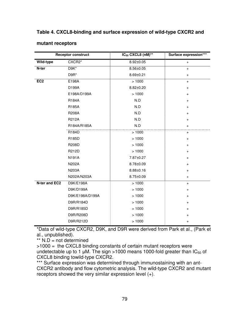
79
Table 4. CXCL8-binding and surface expression of wild-type CXCR2 and
mutant receptors
Receptor construct IC50 CXCL8 (nM)** Surface expression***
Wild-type CXCR2* 8.92±0.05 +
N-ter D9K* 8.56±0.05 +
D9R* 8.69±0.21 +
EC2 E198A > 1000 +
D199A 8.82±0.20 +
E198A/D199A > 1000 +
R184A N.D +
R185A N.D +
R208A N.D +
R212A N.D +
R184A/R185A N.D +
R184D > 1000 +
R185D > 1000 +
R208D > 1000 +
R212D > 1000 +
N191A 7.87±0.27 +
N202A 8.78±0.09 +
N203A 8.88±0.16 +
N202A/N203A 8.75±0.09 +
N-ter and EC2 D9K/E198A > 1000 +
D9K/D199A > 1000 +
D9K/E198A/D199A > 1000 +
D9R/R184D > 1000 +
D9R/R185D > 1000 +
D9R/R208D > 1000 +
D9R/R212D > 1000 +
*Data of wild-type CXCR2, D9K, and D9R were derived from Park et al., (Park et al., unpublished). ** N.D = not determined >1000 = the CXCL8 binding constants of certain mutant receptors were undetectable up to 1 µM. The sign >1000 means 1000-fold greater than IC50 of CXCL8 binding towild-type CXCR2. *** Surface expression was determined through immunostaining with an ant-CXCR2 antibody and flow cytometric analysis. The wild-type CXCR2 and mutant receptors showed the very similar expression level (+).
80
Constitutively inactive mutants CXCR2 induced by charge-reversal
complementary mutations (derived from repulsion model)
Charge-reversal combinatorial mutant receptors consisting of CAM D9R
and RxD mutations (R184D, R185D, R208D, and R212D) in EC2 were
constructed to study whether the mutant receptors lead to constitutive activation
like CAM D9R or change their structural constraints related to receptor function.
Charge-reversal single mutations alone (R184D, R185D, R208D, and R212D)
were used as controls. The NIH3T3 cells stably expressing the mutant receptors
were used to investigate receptor activation by PLC-β3 signaling. The
transfectants were immunostained with a specific PLC-β3 antibody to detect
serine phosphorylation (Ser537 of PLC-β3). Flow cytometric analysis showed
that CAM D9R induced significantly less phosphorylation than wild-type CXCR2
(Fig. 16). Both the charge-reversal single and combinatorial mutant receptors
showed more phosphorylated PLC-β3 than wild-type CXCR2, suggesting the
inactivation of PLC-β3.
To study whether the inactivation of PLC-β3 is involved in cellular
transformation by the mutant receptors, foci formation assays were performed.
None of the charge-reversal mutants formed foci, whereas CAM D9R
significantly produced foci through constitutive activation (Fig. 17). Therefore, the
charge-reversal single mutations as well as the combinatorial mutations lead to
disruption of downstream signal transduction of CXCR2 through inhibition of
PLC-β3 activity.
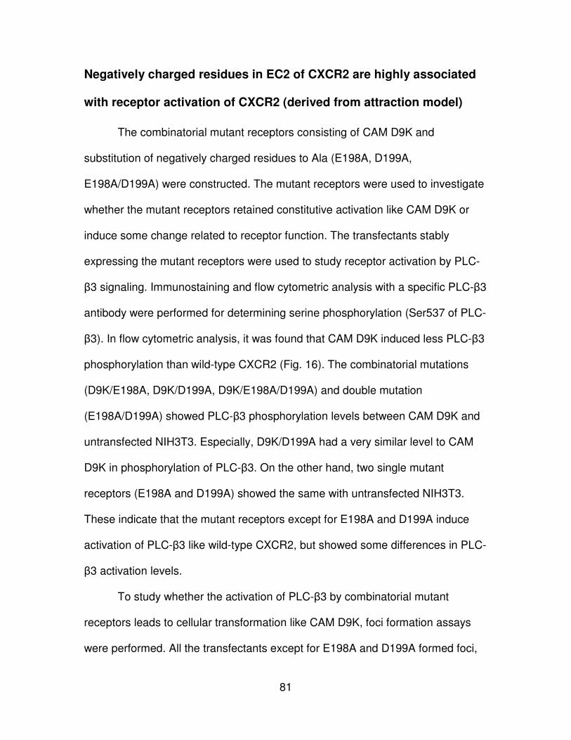
81
Negatively charged residues in EC2 of CXCR2 are highly associated
with receptor activation of CXCR2 (derived from attraction model)
The combinatorial mutant receptors consisting of CAM D9K and
substitution of negatively charged residues to Ala (E198A, D199A,
E198A/D199A) were constructed. The mutant receptors were used to investigate
whether the mutant receptors retained constitutive activation like CAM D9K or
induce some change related to receptor function. The transfectants stably
expressing the mutant receptors were used to study receptor activation by PLC-
β3 signaling. Immunostaining and flow cytometric analysis with a specific PLC-β3
antibody were performed for determining serine phosphorylation (Ser537 of PLC-
β3). In flow cytometric analysis, it was found that CAM D9K induced less PLC-β3
phosphorylation than wild-type CXCR2 (Fig. 16). The combinatorial mutations
(D9K/E198A, D9K/D199A, D9K/E198A/D199A) and double mutation
(E198A/D199A) showed PLC-β3 phosphorylation levels between CAM D9K and
untransfected NIH3T3. Especially, D9K/D199A had a very similar level to CAM
D9K in phosphorylation of PLC-β3. On the other hand, two single mutant
receptors (E198A and D199A) showed the same with untransfected NIH3T3.
These indicate that the mutant receptors except for E198A and D199A induce
activation of PLC-β3 like wild-type CXCR2, but showed some differences in PLC-
β3 activation levels.
To study whether the activation of PLC-β3 by combinatorial mutant
receptors leads to cellular transformation like CAM D9K, foci formation assays
were performed. All the transfectants except for E198A and D199A formed foci,
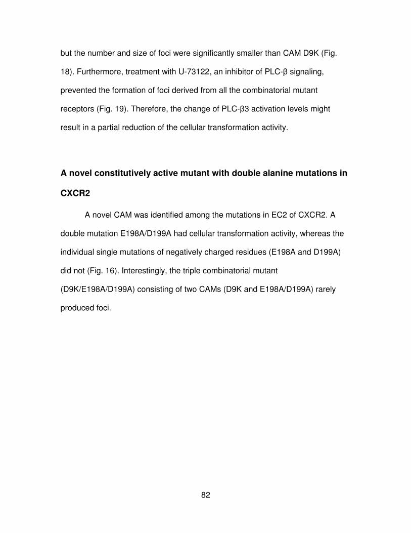
82
but the number and size of foci were significantly smaller than CAM D9K (Fig.
18). Furthermore, treatment with U-73122, an inhibitor of PLC-β signaling,
prevented the formation of foci derived from all the combinatorial mutant
receptors (Fig. 19). Therefore, the change of PLC-β3 activation levels might
result in a partial reduction of the cellular transformation activity.
A novel constitutively active mutant with double alanine mutations in
CXCR2
A novel CAM was identified among the mutations in EC2 of CXCR2. A
double mutation E198A/D199A had cellular transformation activity, whereas the
individual single mutations of negatively charged residues (E198A and D199A)
did not (Fig. 16). Interestingly, the triple combinatorial mutant
(D9K/E198A/D199A) consisting of two CAMs (D9K and E198A/D199A) rarely
produced foci.
83
Figure 16. Phosphorylation of PLC-β3 in wild-type and mutant CXCR2 receptors. These figures showed phosphorylation of PLC-β3 of CAM D9K and mutant receptors for the attraction model (A), CAM D9R and mutant receptors for the repulsion model (B, C, and D).
84
Figure 17. Foci formation assay with NIH3T3 transfectants stably
expressing charge-reversal complementary mutant receptors derived from
the repulsion model.
Constitutively active mutant D9R was used for positive control. None of all the
mutant transfectants showed foci formation.
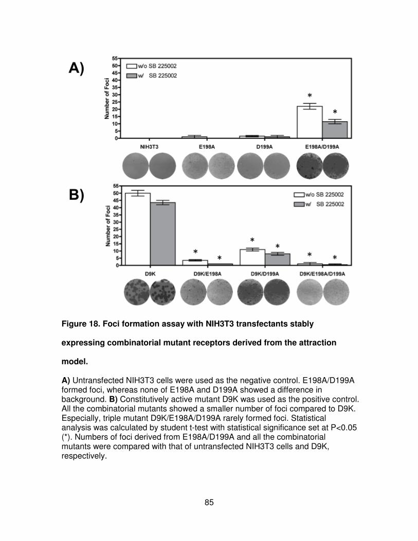
85
Figure 18. Foci formation assay with NIH3T3 transfectants stably
expressing combinatorial mutant receptors derived from the attraction
model.
A) Untransfected NIH3T3 cells were used as the negative control. E198A/D199A formed foci, whereas none of E198A and D199A showed a difference in background. B) Constitutively active mutant D9K was used as the positive control. All the combinatorial mutants showed a smaller number of foci compared to D9K. Especially, triple mutant D9K/E198A/D199A rarely formed foci. Statistical analysis was calculated by student t-test with statistical significance set at P<0.05 (*). Numbers of foci derived from E198A/D199A and all the combinatorial mutants were compared with that of untransfected NIH3T3 cells and D9K, respectively.
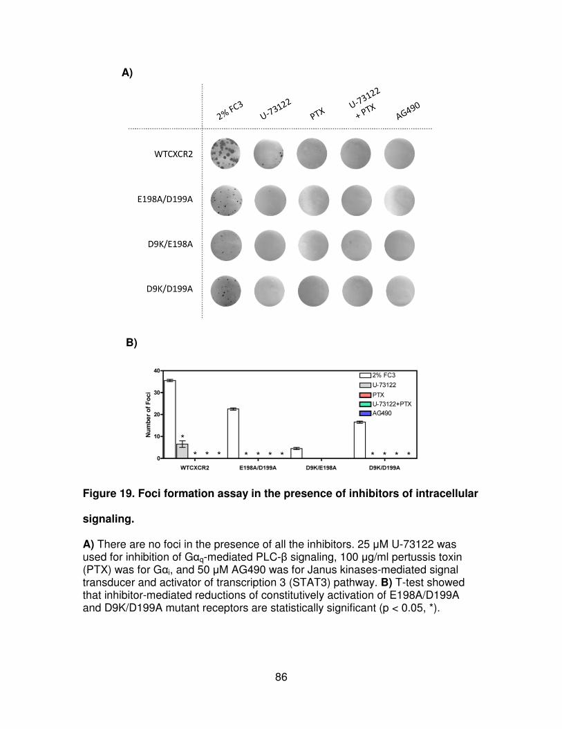
86
A)
B)
Figure 19. Foci formation assay in the presence of inhibitors of intracellular
signaling.
A) There are no foci in the presence of all the inhibitors. 25 µM U-73122 was used for inhibition of Gαq-mediated PLC-β signaling, 100 µg/ml pertussis toxin (PTX) was for Gαi, and 50 µM AG490 was for Janus kinases-mediated signal transducer and activator of transcription 3 (STAT3) pathway. B) T-test showed that inhibitor-mediated reductions of constitutively activation of E198A/D199A and D9K/D199A mutant receptors are statistically significant (p < 0.05, *).
87
Chapter 5 Discussion
An increasing number of studies have focused on the mechanism of
receptor activation of GPCRs due to its importance as pharmacological targets
(45, 132). Two major mechanisms for receptor activation including the ionic lock
and toggle switch were found in rhodopsin and β2AR, whereas most of class A
GPCRs do not contain these residues necessary for these mechanisms (38,
133). Some including rhodopsin and β2AR showed the conservation of a Glu
residue in the boundary of IC3 and TM6 to interact with the (D/E)R(Y/W) motif for
receptor stabilization in the inactive state. CXCR2 also does not contain a
conserved Glu residue for the ionic lock, suggesting CXCR2 undergoes an
unknown mechanism for receptor activation.
It has been shown that the amino terminus and second extracellular loop
of CXCR2 is crucial for ligand recognition and receptor activation (80, 81, 83-88,
124). Interestingly, a previous study of the EC2 of CXCR2 showed that a
negatively charged residue, Asp199 is crucial to manage internalization speed of
the receptor (84). Furthermore, another current investigation showed that single
mutations (e.g. D9K and D9R) of Asp9 in the amino terminus of CXCR2 lead to
constitutive activation (Park et al., unpublished). These observations suggest that
charged residues in the second extracellular loop of GPCRs may play a role in
receptor stabilization and activation.
To study a role for EC2 in ligand binding, we generated single mutations in
EC2, including the substitution of negatively charged residues including Glu198
88
and/or Asp199 and positively charged residues such as Arg184, Arg185, Arg208,
and Arg212 to Ala residue to neutralize their charges. The Arg residues are also
mutated to a charge-reversal residue, Asp residue. Like wild-type CXCR2, the
mutant receptors were well expressed at the cell surface. However, all the
mutant receptors except for D199A demonstrated no binding affinity for CXCL8.
This result suggests that charged residues in EC2 of CXCR2 are crucial for
ligand recognition, but do not affect cell surface expression.
Furthermore, we designed two concepts, the attraction and repulsion
models, to understand CXCR2 activation (Fig. 15). To end that, we constructed
two different combinatorial mutation sets consisting of CAMs (D9K and D9R) and
the substitutions in EC2. All the combinatorial mutant receptors were well
expressed at the cell surface but had no CXCL8 binding affinity similar to most of
the single mutations in EC2 of CXCR2. Interestingly, all the complementary
charge-reversal mutants to prove the repulsion model (D9R/R184D, D9R/R185D,
D9R/R208D, and D9R/R212D) showed no ligand binding affinity, inactivation of
PLC-β3 and inhibition of cellular transformation. The mutants were expected to
function like wild-type CXCR2 because some studies have generated charge-
reversal mutations to mimic the structural constraints of their wild-type receptors
(135). However, unexpectedly, their structural constraints would not be the same
with wild-type CXCR2, even though there is no problem in the protein folding of
the mutant receptors (Table 4). Therefore, the mutant receptors would be
constitutively inactive mutants (CIM) or dead receptors. On the other hand,
another set of the combinatorial mutants (D9K/E198A, D9K/E198A, and
89
D9K/E198A/D199A) retained their constitutive activity via PLC-β3 signaling even
though the mutants were unable to bind CXCL8.
Interestingly, similar results were observed with charged residues in the
extracellular loops of neurohyphophysical hormone receptors including
vasopressin and oxytocin receptors (137). Using Ala mutations and charge-
reversal substitutions, some of the charged residues showed a significant
reduction of ligand binding affinity, regardless of surface expression. The study
also generated two complementary mutants including R46D/D204R and
R125D/D204R to prove interaction between the mutated residues. However, the
cell surface expression level of the combinatorial mutants was very low (≤20%
fold over wild-type) and the binding affinities for their cognate ligands were
significantly reduced or undetectable.
Consistent with our result, it has been previously shown that Asp199 in
EC2 of CXCR2 is not related to ligand binding (84). D199N showed a similar
CXCL8 binding affinity to wild-type CXCR2 but D199V has a higher CXCL8
binding affinity than wild-type CXCR2. Furthermore, the mutant receptors
(D199N and D199V) had no effect on Ca2+ mobilization by CXCL1 or CXCL8
stimulation. Similarly, in this study, D199A showed no difference in PLC-β3
signaling, whereas E198A/D199A induced activation of PLC-β3. Previously, it
was shown that negatively charged residues in EC2 of FFAR1 are important for
receptor activation for constructing ionic locks to positively charged residues on
distinct transmembranes (134).
90
Interestingly, E198A/D199A was identified as a novel constitutively active
mutant via foci formation assays. Since many investigations have tried to
construct site-directed or random mutagenesis of the EC2 of GPCRs (36), it
would be the first CAM derived from double mutations of residues in EC2. The
double mutant receptor might have structural constraints for CXCR2 activation,
whereas the single mutations, E198A and D199A, allow themselves to have
inactive forms. The negatively charged residues are very close to a highly
conserved disulfide bond (Cys119-Cys196) linking EC2 and TM3. The special
location of the two residues in EC2 might explain how the double mutation leads
to constitutive activation. The neutralization (with Ala residue) of the residues
might induce a structural change in TM3 through the disulfide bond. Furthermore,
the substitution with Ala in combinatorial mutant receptors derived from the
attraction models might promote breakdown of the structural constraints of CAM
D9K.
Unlike rhodopsin and β2AR, most of class A GPCRs including CXCR2 lack
possible residues in IC3 for an ionic lock, pointing to the need to study possible
other mechanisms for activation of GPCRs. Our observations might provide new
insight to understand a role of extracellular regions (especially, amino terminus
and EC2) in ligand binding and receptor activation.

91
PART IV CONCLUSIONS
92
Most chemokine receptors can be activated by more than one chemokine
(77). CXC chemokine receptor 2 (CXCR2) binds multiple CXC chemokines
including CXCL1 (GRO-α), CXCL2 (GRO-β), CXCL3 (GRO-γ), CXCL5 (ENA78),
CXCL6 (GCP2), CXCL7 (NAP2), and CXCL8 (78, 79). Although CXCR1 and
CXCR2 show over 75% amino acid identity, only CXCL6 and CXCL8 activate
CXCR1, suggesting that the two chemokine receptors have different
mechanisms for chemokine ligand binding (80). It has been shown that the amino
terminus and second extracellular loop of CXCR2 is crucial for ligand recognition
and receptor activation (80, 81, 83-88, 124). Interestingly, a previous study of the
EC2 of CXCR2 showed that a negatively charged residue, Asp199, is crucial to
manage internalization speed of the receptor (84). Furthermore, another current
investigation showed that single mutations (e.g. D9K and D9R) of Asp9 in the
amino terminus of CXCR2 led to constitutive activation (Park et al., unpublished).
These observations suggest that charged residues in the second extracellular
loop of GPCRs may play a role in receptor stabilization and activation. However,
the mechanism of the ligand binding and receptor activation is still unknown.
First, in our study, a CXCR2 structure is predicted on the basis of crystal
GPCR structures including rhodopsin, β2AR, and CXCR4 by homology modeling
(Fig. 10). The highly conserved motifs ((D/E)R(Y/W), NPXXY, toggle switch, and
disulfide bonds found in sequence alignments of the template GPCRs were
helpful to generate CXCR2 models (Fig. 7,8,9). Using Cα RMSDs, structural
alignments of seven transmembrane domains showed individual models tend to
be similar to their templates (Fig, 11). Modeling results indicated that the crystal
93
structure of CXCR4 might be the best template for CXCR2 prediction due to the
relatively high identity (33.3% identity for CXCR4) in sequence alignments and
the lowest RMSD values (0.78Å). Site Finder was used to predict ligand binding
sites of b2AR-CXCR2 and CXCR4 (Fig. 12). The prediction suggests that
residues in all the extracellular loops are highly associated with ligand binding.
Residues in some of the transmembranes domains showed involvement in ligand
binding as well. The second extracellular loop of the CXCR4-based homology
modeled CXCR2 provided two β-strands like rhodopsin and CXCR4 and the
prediction is very similar with results of the predictions from two web-based
prediction programs, NPS@SOPMA and NetSurfP (Fig. 13). Superimposition of
the second extracellular loops of CXCR4 and CXCR4-based CXCR2 suggested
that the second extracellular loop of CXCR2 is structurally very similar with that
of CXCR4 with below 2.0 Å RMSD.
Furthermore, we studied solvent accessibility of residues in the second
extracellular loop of CXCR2, a key region for ligand binding and receptor
activation, in the inactive state of the receptor. MTSEA-biotin accessibility of
substituted cysteine residues in the second extracellular loop was measured by
immunostaining of FITC-streptavidin using flow cytometric analysis. The
normalization of the accessibility was determined based on the cell surface
expression level of the substituted cysteine mutant receptors (Fig. 14). Most of
the residues in the second extracellular loop were found to be solvent accessible
in the inactive state of CXCR2, indicating that the residues might be involved in
ligand recognition. The results showed that all the charged residues except for
94
Arg208 in second extracellular loop also have solvent accessibility, pointing to
possibility of ligand binding. However, 204TANWR208 residues showed no solvent
accessibility, suggesting that they might be involved in the interaction with the
amino terminus or other extracellular loops. Our finding of ligand binding sites for
CXCR2 models also suggests that the most of the residues in the region could
form a ligand binding pocket to contact its cognate ligands.
Second, we studied the role of charged residues in EC2 of CXCR2 in
ligand binding and receptor activation. Using constitutively active CXCR2
mutants, D9K and D9R, a potential role of charged residues in the second
extracellular loop of CXCR2 was investigated.
Combinatorial mutations consisting of the constitutively active mutants in
the amino terminus and single mutations of charged residues in the second
extracellular loop were generated to study two concepts including “attraction” and
“repulsion” model (Fig. 15). The mutant receptors were used to test their effects
on cell surface expression, ligand binding, receptor activation through PLC-β3,
and cellular transformation. All of the mutations in the repulsion model result in
CXCR2 receptors that are unable to bind ligand, suggesting that all the Arg
residues (Arg184, Arg185, Arg208, and Arg212) in the second extracellular loop
are important for ligand recognition. Although charge-reversal complementary
mutations in the repulsion model were expected to retain structural constraints of
CXCR2, none of the mutant receptors function as wild-type CXCR2 in the
inactive and active state (Fig. 16, 17). Interestingly, mutations in the attraction
model partially inhibited receptor activation of constitutively active mutant D9K,
95
suggesting that Glu198 and Asp199 residues in the second extracellular loop are
associated with receptor activation (Fig. 16, 18). Furthermore, a novel
constitutively active mutant, E198A/D199A, was identified in this study (Fig. 16,
18). These negatively charged residues are very close to a disulfide bond
(Cys119-Cys196) linking the second extracellular loop and the third
transmembrane.
In Part II, the discoveries about the structural basis of CXCR2 and
interdisciplinary approaches would provide new insights to investigate unknown
mechanisms of interaction with its cognate ligands and receptor activation. These
observations in Part III suggest the interaction between the amino terminus and
EC2 of CXCR2 for ligand binding and receptor activation. Although crystallization
studies of the amino terminus are limited for homology modeling and dynamic
simulation (e.g. ligand docking), several groups have resolved the structures of
the amino terminus in GPCRs using NMR, indicating that this approach with
NMR would be effective and accurate to understand unknown amino terminus
structures (138-140). Furthermore, some biochemical approaches including
disulfide cross-linking, disulfide trapping, and protein digestion, would be helpful
to discover unknown mechanism of ligand binding and receptor activation in
GPCRs including CXCR2 (11, 139, 141, 142).
96
REFERENCES
1. Fredriksson, R., Lagerstrom, M. C., Lundin, L. G., and Schioth, H. B. (2003) The G-protein-coupled receptors in the human genome form five main families. Phylogenetic analysis, paralogon groups, and fingerprints, Mol Pharmacol 63, 1256-1272.
2. Pierce, K. L., Premont, R. T., and Lefkowitz, R. J. (2002) Seven-transmembrane receptors, Nat Rev Mol Cell Biol 3, 639-650.
3. Insel, P. A., Tang, C. M., Hahntow, I., and Michel, M. C. (2007) Impact of GPCRs in clinical medicine: monogenic diseases, genetic variants and drug targets, Biochim Biophys Acta 1768, 994-1005.
4. Gether, U. (2000) Uncovering molecular mechanisms involved in activation of G protein-coupled receptors, Endocr Rev 21, 90-113.
5. Tsao, P., Cao, T., and von Zastrow, M. (2001) Role of endocytosis in mediating downregulation of G-protein-coupled receptors, Trends Pharmacol Sci 22, 91-96.
6. Kohout, T. A., and Lefkowitz, R. J. (2003) Regulation of G protein-coupled receptor kinases and arrestins during receptor desensitization, Mol Pharmacol 63, 9-18.
7. Rosenbaum, D. M., Rasmussen, S. G., and Kobilka, B. K. (2009) The structure and function of G-protein-coupled receptors, Nature 459, 356-363.
8. Wu, B., Chien, E. Y., Mol, C. D., Fenalti, G., Liu, W., Katritch, V., Abagyan, R., Brooun, A., Wells, P., Bi, F. C., Hamel, D. J., Kuhn, P., Handel, T. M., Cherezov, V., and Stevens, R. C. (2010) Structures of the CXCR4 Chemokine GPCR with Small-Molecule and Cyclic Peptide Antagonists, Science.
9. Hauser, M., Kauffman, S., Lee, B. K., Naider, F., and Becker, J. M. (2007) The first extracellular loop of the Saccharomyces cerevisiae G protein-coupled receptor Ste2p undergoes a conformational change upon ligand binding, J Biol Chem 282, 10387-10397.
10. Day, P. W., Rasmussen, S. G., Parnot, C., Fung, J. J., Masood, A., Kobilka, T. S., Yao, X. J., Choi, H. J., Weis, W. I., Rohrer, D. K., and Kobilka, B. K. (2007) A monoclonal antibody for G protein-coupled receptor crystallography, Nat Methods 4, 927-929.
11. Wess, J., Han, S. J., Kim, S. K., Jacobson, K. A., and Li, J. H. (2008) Conformational changes involved in G-protein-coupled-receptor activation, Trends Pharmacol Sci 29, 616-625.
12. George, S. R., O'Dowd, B. F., and Lee, S. P. (2002) G-protein-coupled receptor oligomerization and its potential for drug discovery, Nat Rev Drug Discov 1, 808-820.
13. Palczewski, K., Kumasaka, T., Hori, T., Behnke, C. A., Motoshima, H., Fox, B. A., Le Trong, I., Teller, D. C., Okada, T., Stenkamp, R. E., Yamamoto, M., and Miyano, M. (2000) Crystal structure of rhodopsin: A G protein-coupled receptor, Science 289, 739-745.
97
14. Lodowski, D. T., and Palczewski, K. (2009) Chemokine receptors and other G protein-coupled receptors, Curr Opin HIV AIDS 4, 88-95.
15. Ludeke, S., Mahalingam, M., and Vogel, R. (2009) Rhodopsin activation switches in a native membrane environment, Photochem Photobiol 85, 437-441.
16. Neumann, J. M., Couvineau, A., Murail, S., Lacapere, J. J., Jamin, N., and Laburthe, M. (2008) Class-B GPCR activation: is ligand helix-capping the key?, Trends Biochem Sci 33, 314-319.
17. Kunishima, N., Shimada, Y., Tsuji, Y., Sato, T., Yamamoto, M., Kumasaka, T., Nakanishi, S., Jingami, H., and Morikawa, K. (2000) Structural basis of glutamate recognition by a dimeric metabotropic glutamate receptor, Nature 407, 971-977.
18. Klabunde, T., and Hessler, G. (2002) Drug design strategies for targeting G-protein-coupled receptors, Chembiochem 3, 928-944.
19. Pebay-Peyroula, E., Rummel, G., Rosenbusch, J. P., and Landau, E. M. (1997) X-ray structure of bacteriorhodopsin at 2.5 angstroms from microcrystals grown in lipidic cubic phases, Science 277, 1676-1681.
20. Arshava, B., Taran, I., Xie, H., Becker, J. M., and Naider, F. (2002) High resolution NMR analysis of the seven transmembrane domains of a heptahelical receptor in organic-aqueous medium, Biopolymers 64, 161-176.
21. Unger, V. M., and Schertler, G. F. (1995) Low resolution structure of bovine rhodopsin determined by electron cryo-microscopy, Biophys J 68, 1776-1786.
22. Cherezov, V., Rosenbaum, D. M., Hanson, M. A., Rasmussen, S. G., Thian, F. S., Kobilka, T. S., Choi, H. J., Kuhn, P., Weis, W. I., Kobilka, B. K., and Stevens, R. C. (2007) High-resolution crystal structure of an engineered human beta2-adrenergic G protein-coupled receptor, Science 318, 1258-1265.
23. Rosenbaum, D. M., Cherezov, V., Hanson, M. A., Rasmussen, S. G., Thian, F. S., Kobilka, T. S., Choi, H. J., Yao, X. J., Weis, W. I., Stevens, R. C., and Kobilka, B. K. (2007) GPCR engineering yields high-resolution structural insights into beta2-adrenergic receptor function, Science 318, 1266-1273.
24. Warne, T., Serrano-Vega, M. J., Baker, J. G., Moukhametzianov, R., Edwards, P. C., Henderson, R., Leslie, A. G., Tate, C. G., and Schertler, G. F. (2008) Structure of a beta1-adrenergic G-protein-coupled receptor, Nature 454, 486-491.
25. Park, J. H., Scheerer, P., Hofmann, K. P., Choe, H. W., and Ernst, O. P. (2008) Crystal structure of the ligand-free G-protein-coupled receptor opsin, Nature 454, 183-187.
26. Jaakola, V. P., Griffith, M. T., Hanson, M. A., Cherezov, V., Chien, E. Y., Lane, J. R., Ijzerman, A. P., and Stevens, R. C. (2008) The 2.6 angstrom crystal structure of a human A2A adenosine receptor bound to an antagonist, Science 322, 1211-1217.
27. Teller, D. C., Okada, T., Behnke, C. A., Palczewski, K., and Stenkamp, R. E. (2001) Advances in determination of a high-resolution three-dimensional structure of rhodopsin, a model of G-protein-coupled receptors (GPCRs), Biochemistry 40, 7761-7772.
98
28. Sansom, M. S., and Weinstein, H. (2000) Hinges, swivels and switches: the role of prolines in signalling via transmembrane alpha-helices, Trends Pharmacol Sci 21, 445-451.
29. Shi, L., Liapakis, G., Xu, R., Guarnieri, F., Ballesteros, J. A., and Javitch, J. A. (2002) Beta2 adrenergic receptor activation. Modulation of the proline kink in transmembrane 6 by a rotamer toggle switch, J Biol Chem 277, 40989-40996.
30. Altenbach, C., Kusnetzow, A. K., Ernst, O. P., Hofmann, K. P., and Hubbell, W. L. (2008) High-resolution distance mapping in rhodopsin reveals the pattern of helix movement due to activation, Proc Natl Acad Sci U S A 105, 7439-7444.
31. Horn, F., Vriend, G., and Cohen, F. E. (2001) Collecting and harvesting biological data: the GPCRDB and NucleaRDB information systems, Nucleic Acids Res 29, 346-349.
32. Filipek, S., Teller, D. C., Palczewski, K., and Stenkamp, R. (2003) The crystallographic model of rhodopsin and its use in studies of other G protein-coupled receptors, Annu Rev Biophys Biomol Struct 32, 375-397.
33. Palczewski, K. (2006) G protein-coupled receptor rhodopsin, Annu Rev Biochem 75, 743-767.
34. Kaushal, S., Ridge, K. D., and Khorana, H. G. (1994) Structure and function in rhodopsin: the role of asparagine-linked glycosylation, Proc Natl Acad Sci U S A 91, 4024-4028.
35. Murray, A. R., Fliesler, S. J., and Al-Ubaidi, M. R. (2009) Rhodopsin: the functional significance of asn-linked glycosylation and other post-translational modifications, Ophthalmic Genet 30, 109-120.
36. Klco, J. M., Wiegand, C. B., Narzinski, K., and Baranski, T. J. (2005) Essential role for the second extracellular loop in C5a receptor activation, Nat Struct Mol Biol 12, 320-326.
37. Massotte, D., and Kieffer, B. L. (2005) The second extracellular loop: a damper for G protein-coupled receptors?, Nat Struct Mol Biol 12, 287-288.
38. Audet, M., and Bouvier, M. (2008) Insights into signaling from the beta2-adrenergic receptor structure, Nat Chem Biol 4, 397-403.
39. Ballesteros, J. A., Jensen, A. D., Liapakis, G., Rasmussen, S. G., Shi, L., Gether, U., and Javitch, J. A. (2001) Activation of the beta 2-adrenergic receptor involves disruption of an ionic lock between the cytoplasmic ends of transmembrane segments 3 and 6, J Biol Chem 276, 29171-29177.
40. Hanson, M. A., Cherezov, V., Griffith, M. T., Roth, C. B., Jaakola, V. P., Chien, E. Y., Velasquez, J., Kuhn, P., and Stevens, R. C. (2008) A specific cholesterol binding site is established by the 2.8 A structure of the human beta2-adrenergic receptor, Structure 16, 897-905.
41. Zhang, Y., Devries, M. E., and Skolnick, J. (2006) Structure modeling of all identified G protein-coupled receptors in the human genome, PLoS Comput Biol 2, e13.
42. Costanzi, S. (2008) On the applicability of GPCR homology models to computer-aided drug discovery: a comparison between in silico and crystal structures of the beta2-adrenergic receptor, J Med Chem 51, 2907-2914.
99
43. Mirzadegan, T., Benko, G., Filipek, S., and Palczewski, K. (2003) Sequence analyses of G-protein-coupled receptors: similarities to rhodopsin, Biochemistry 42, 2759-2767.
44. Mobarec, J. C., Sanchez, R., and Filizola, M. (2009) Modern homology modeling of G-protein coupled receptors: which structural template to use?, J Med Chem 52, 5207-5216.
45. Kristiansen, K. (2004) Molecular mechanisms of ligand binding, signaling, and regulation within the superfamily of G-protein-coupled receptors: molecular modeling and mutagenesis approaches to receptor structure and function, Pharmacol Ther 103, 21-80.
46. Vaidehi, N., Floriano, W. B., Trabanino, R., Hall, S. E., Freddolino, P., Choi, E. J., Zamanakos, G., and Goddard, W. A., 3rd. (2002) Prediction of structure and function of G protein-coupled receptors, Proc Natl Acad Sci U S A 99, 12622-12627.
47. Eglen, R. M., and Reisine, T. (2009) New insights into GPCR function: implications for HTS, Methods Mol Biol 552, 1-13.
48. Hall, D. A. (2000) Modeling the functional effects of allosteric modulators at pharmacological receptors: an extension of the two-state model of receptor activation, Mol Pharmacol 58, 1412-1423.
49. Kobilka, B. (2004) Agonist binding: a multistep process, Mol Pharmacol 65, 1060-1062.
50. Schwartz, T. W., Frimurer, T. M., Holst, B., Rosenkilde, M. M., and Elling, C. E. (2006) Molecular mechanism of 7TM receptor activation--a global toggle switch model, Annu Rev Pharmacol Toxicol 46, 481-519.
51. Hu, J., Wang, Y., Zhang, X., Lloyd, J. R., Li, J. H., Karpiak, J., Costanzi, S., and Wess, J. (2010) Structural basis of G protein-coupled receptor-G protein interactions, Nat Chem Biol 6, 541-548.
52. Liang, W., Hoang, Q., Clark, R. B., and Fishman, P. H. (2008) Accelerated dephosphorylation of the beta2-adrenergic receptor by mutation of the C-terminal lysines: effects on ubiquitination, intracellular trafficking, and degradation, Biochemistry 47, 11750-11762.
53. Dorsam, R. T., and Gutkind, J. S. (2007) G-protein-coupled receptors and cancer, Nat Rev Cancer 7, 79-94.
54. Oldham, W. M., and Hamm, H. E. (2008) Heterotrimeric G protein activation by G-protein-coupled receptors, Nat Rev Mol Cell Biol 9, 60-71.
55. Hermans, E. (2003) Biochemical and pharmacological control of the multiplicity of coupling at G-protein-coupled receptors, Pharmacol Ther 99, 25-44.
56. Wong, S. K. (2003) G protein selectivity is regulated by multiple intracellular regions of GPCRs, Neurosignals 12, 1-12.
57. Rebecchi, M. J., and Pentyala, S. N. (2000) Structure, function, and control of phosphoinositide-specific phospholipase C, Physiol Rev 80, 1291-1335.
58. Strathmann, M. P., and Simon, M. I. (1991) G alpha 12 and G alpha 13 subunits define a fourth class of G protein alpha subunits, Proc Natl Acad Sci U S A 88, 5582-5586.
100
59. Dhanasekaran, N., and Dermott, J. M. (1996) Signaling by the G12 class of G proteins, Cell Signal 8, 235-245.
60. Clapham, D. E., and Neer, E. J. (1997) G protein beta gamma subunits, Annu Rev Pharmacol Toxicol 37, 167-203.
61. Schwindinger, W. F., and Robishaw, J. D. (2001) Heterotrimeric G-protein betagamma-dimers in growth and differentiation, Oncogene 20, 1653-1660.
62. Pitcher, J. A., Hall, R. A., Daaka, Y., Zhang, J., Ferguson, S. S., Hester, S., Miller, S., Caron, M. G., Lefkowitz, R. J., and Barak, L. S. (1998) The G protein-coupled receptor kinase 2 is a microtubule-associated protein kinase that phosphorylates tubulin, J Biol Chem 273, 12316-12324.
63. Ferguson, S. S. (2001) Evolving concepts in G protein-coupled receptor endocytosis: the role in receptor desensitization and signaling, Pharmacol Rev 53, 1-24.
64. Ross, E. M., and Wilkie, T. M. (2000) GTPase-activating proteins for heterotrimeric G proteins: regulators of G protein signaling (RGS) and RGS-like proteins, Annu Rev Biochem 69, 795-827.
65. Miller, W. E., and Lefkowitz, R. J. (2001) Expanding roles for beta-arrestins as scaffolds and adapters in GPCR signaling and trafficking, Curr Opin Cell Biol 13, 139-145.
66. Perry, S. J., Baillie, G. S., Kohout, T. A., McPhee, I., Magiera, M. M., Ang, K. L., Miller, W. E., McLean, A. J., Conti, M., Houslay, M. D., and Lefkowitz, R. J. (2002) Targeting of cyclic AMP degradation to beta 2-adrenergic receptors by beta-arrestins, Science 298, 834-836.
67. Baggiolini, M. (1998) Chemokines and leukocyte traffic, Nature 392, 565-568.
68. Rossi, D., and Zlotnik, A. (2000) The biology of chemokines and their receptors, Annu Rev Immunol 18, 217-242.
69. Locati, M., and Murphy, P. M. (1999) Chemokines and chemokine receptors: biology and clinical relevance in inflammation and AIDS, Annu Rev Med 50, 425-440.
70. Strieter, R. M., Polverini, P. J., Kunkel, S. L., Arenberg, D. A., Burdick, M. D., Kasper, J., Dzuiba, J., Van Damme, J., Walz, A., Marriott, D., and et al. (1995) The functional role of the ELR motif in CXC chemokine-mediated angiogenesis, J Biol Chem 270, 27348-27357.
71. Phillips, R. J., Burdick, M. D., Lutz, M., Belperio, J. A., Keane, M. P., and Strieter, R. M. (2003) The stromal derived factor-1/CXCL12-CXC chemokine receptor 4 biological axis in non-small cell lung cancer metastases, Am J Respir Crit Care Med 167, 1676-1686.
72. Holmes, W. E., Lee, J., Kuang, W. J., Rice, G. C., and Wood, W. I. (1991) Structure and functional expression of a human interleukin-8 receptor, Science 253, 1278-1280.
73. Murphy, P. M., and Tiffany, H. L. (1991) Cloning of complementary DNA encoding a functional human interleukin-8 receptor, Science 253, 1280-1283.
74. Murphy, P. M. (2002) International Union of Pharmacology. XXX. Update on chemokine receptor nomenclature, Pharmacol Rev 54, 227-229.
101
75. (2001) Chemokine/chemokine receptor nomenclature, J Leukoc Biol 70, 465-466.
76. Zlotnik, A., Yoshie, O., and Nomiyama, H. (2006) The chemokine and chemokine receptor superfamilies and their molecular evolution, Genome Biol 7, 243.
77. Rot, A., and von Andrian, U. H. (2004) Chemokines in innate and adaptive host defense: basic chemokinese grammar for immune cells, Annu Rev Immunol 22, 891-928.
78. Addison, C. L., Daniel, T. O., Burdick, M. D., Liu, H., Ehlert, J. E., Xue, Y. Y., Buechi, L., Walz, A., Richmond, A., and Strieter, R. M. (2000) The CXC chemokine receptor 2, CXCR2, is the putative receptor for ELR+ CXC chemokine-induced angiogenic activity, J Immunol 165, 5269-5277.
79. Strieter, R. M., Gomperts, B. N., and Keane, M. P. (2007) The role of CXC chemokines in pulmonary fibrosis, J Clin Invest 117, 549-556.
80. Murphy, P. M. (1994) The molecular biology of leukocyte chemoattractant receptors, Annu Rev Immunol 12, 593-633.
81. Ben-Baruch, A., Bengali, K. M., Biragyn, A., Johnston, J. J., Wang, J. M., Kim, J., Chuntharapai, A., Michiel, D. F., Oppenheim, J. J., and Kelvin, D. J. (1995) Interleukin-8 receptor beta. The role of the carboxyl terminus in signal transduction, J Biol Chem 270, 9121-9128.
82. Catusse, J., Faye, P., Loillier, B., Cremers, B., Franck, R. M., Luccarini, J. M., Pruneau, D., and Paquet, J. L. (2003) Cloning and characterization of guinea pig interleukin-8 receptor, Biochem Pharmacol 66, 1171-1180.
83. Ahuja, S. K., Lee, J. C., and Murphy, P. M. (1996) CXC chemokines bind to unique sets of selectivity determinants that can function independently and are broadly distributed on multiple domains of human interleukin-8 receptor B. Determinants of high affinity binding and receptor activation are distinct, J Biol Chem 271, 225-232.
84. Nasser, M. W., Raghuwanshi, S. K., Malloy, K. M., Gangavarapu, P., Shim, J. Y., Rajarathnam, K., and Richardson, R. M. (2007) CXCR1 and CXCR2 activation and regulation. Role of aspartate 199 of the second extracellular loop of CXCR2 in CXCL8-mediated rapid receptor internalization, J Biol Chem 282, 6906-6915.
85. Katancik, J. A., Sharma, A., Radel, S. J., and De Nardin, E. (1997) Mapping of the extracellular binding regions of the human interleukin-8 type B receptor, Biochem Biophys Res Commun 232, 663-668.
86. Katancik, J. A., Sharma, A., and de Nardin, E. (2000) Interleukin 8, neutrophil-activating peptide-2 and GRO-alpha bind to and elicit cell activation via specific and different amino acid residues of CXCR2, Cytokine 12, 1480-1488.
87. Catusse, J., Liotard, A., Loillier, B., Pruneau, D., and Paquet, J. L. (2003) Characterization of the molecular interactions of interleukin-8 (CXCL8), growth related oncogen alpha (CXCL1) and a non-peptide antagonist (SB 225002) with the human CXCR2, Biochem Pharmacol 65, 813-821.
102
88. Houimel, M., and Mazzucchelli, L. (2009) Identification of biologically active peptides that inhibit binding of human CXCL8 to its receptors from a random phage-epitope library, J Leukoc Biol 85, 728-738.
89. Limatola, C., Di Bartolomeo, S., Catalano, M., Trettel, F., Fucile, S., Castellani, L., and Eusebi, F. (2005) Cysteine residues are critical for chemokine receptor CXCR2 functional properties, Exp Cell Res 307, 65-75.
90. Suzuki, H., Prado, G. N., Wilkinson, N., and Navarro, J. (1994) The N terminus of interleukin-8 (IL-8) receptor confers high affinity binding to human IL-8, J Biol Chem 269, 18263-18266.
91. Schraufstatter, I. U., Ma, M., Oades, Z. G., Barritt, D. S., and Cochrane, C. G. (1995) The role of Tyr13 and Lys15 of interleukin-8 in the high affinity interaction with the interleukin-8 receptor type A, J Biol Chem 270, 10428-10431.
92. Li, F., and Gordon, J. R. (2001) Il-8((3-73))K11R is a high affinity agonist of the neutrophil CXCR1 and CXCR2, Biochem Biophys Res Commun 286, 595-600.
93. Li, F., Zhang, X., and Gordon, J. R. (2002) CXCL8((3-73))K11R/G31P antagonizes ligand binding to the neutrophil CXCR1 and CXCR2 receptors and cellular responses to CXCL8/IL-8, Biochem Biophys Res Commun 293, 939-944.
94. Zhao, X., Li, F., Town, J. R., Zhang, X., Wang, W., and Gordon, J. R. (2007) Humanized forms of the CXCR1/CXCR2 antagonist, bovine CXCL8((3-74))K11R/G31P, effectively block ELR-CXC chemokine activity and airway endotoxemia pathology, Int Immunopharmacol 7, 1723-1731.
95. Ludwig, A., Ehlert, J. E., Flad, H. D., and Brandt, E. (2000) Identification of distinct surface-expressed and intracellular CXC-chemokine receptor 2 glycoforms in neutrophils: N-glycosylation is essential for maintenance of receptor surface expression, J Immunol 165, 1044-1052.
96. Miller, L. J., Kurtzman, S. H., Wang, Y., Anderson, K. H., Lindquist, R. R., and Kreutzer, D. L. (1998) Expression of interleukin-8 receptors on tumor cells and vascular endothelial cells in human breast cancer tissue, Anticancer Res 18, 77-81.
97. Richards, B. L., Eisma, R. J., Spiro, J. D., Lindquist, R. L., and Kreutzer, D. L. (1997) Coexpression of interleukin-8 receptors in head and neck squamous cell carcinoma, Am J Surg 174, 507-512.
98. Norgauer, J., Metzner, B., and Schraufstatter, I. (1996) Expression and growth-promoting function of the IL-8 receptor beta in human melanoma cells, J Immunol 156, 1132-1137.
99. Takamori, H., Oades, Z. G., Hoch, O. C., Burger, M., and Schraufstatter, I. U. (2000) Autocrine growth effect of IL-8 and GROalpha on a human pancreatic cancer cell line, Capan-1, Pancreas 21, 52-56.
100. Venkatakrishnan, G., Salgia, R., and Groopman, J. E. (2000) Chemokine receptors CXCR-1/2 activate mitogen-activated protein kinase via the epidermal growth factor receptor in ovarian cancer cells, J Biol Chem 275, 6868-6875.
103
101. Waugh, D. J., and Wilson, C. (2008) The interleukin-8 pathway in cancer, Clin Cancer Res 14, 6735-6741.
102. Nicholls, D. J., Tomkinson, N. P., Wiley, K. E., Brammall, A., Bowers, L., Grahames, C., Gaw, A., Meghani, P., Shelton, P., Wright, T. J., and Mallinder, P. R. (2008) Identification of a putative intracellular allosteric antagonist binding-site in the CXC chemokine receptors 1 and 2, Mol Pharmacol 74, 1193-1202.
103. Arvanitakis, L., Geras-Raaka, E., Varma, A., Gershengorn, M. C., and Cesarman, E. (1997) Human herpesvirus KSHV encodes a constitutively active G-protein-coupled receptor linked to cell proliferation, Nature 385, 347-350.
104. Couty, J. P., Geras-Raaka, E., Weksler, B. B., and Gershengorn, M. C. (2001) Kaposi's sarcoma-associated herpesvirus G protein-coupled receptor signals through multiple pathways in endothelial cells, J Biol Chem 276, 33805-33811.
105. Montaner, S., Sodhi, A., Pece, S., Mesri, E. A., and Gutkind, J. S. (2001) The Kaposi's sarcoma-associated herpesvirus G protein-coupled receptor promotes endothelial cell survival through the activation of Akt/protein kinase B, Cancer Res 61, 2641-2648.
106. Smit, M. J., Verzijl, D., Casarosa, P., Navis, M., Timmerman, H., and Leurs, R. (2002) Kaposi's sarcoma-associated herpesvirus-encoded G protein-coupled receptor ORF74 constitutively activates p44/p42 MAPK and Akt via G(i) and phospholipase C-dependent signaling pathways, J Virol 76, 1744-1752.
107. Cannon, M. L., and Cesarman, E. (2004) The KSHV G protein-coupled receptor signals via multiple pathways to induce transcription factor activation in primary effusion lymphoma cells, Oncogene 23, 514-523.
108. Casarosa, P., Bakker, R. A., Verzijl, D., Navis, M., Timmerman, H., Leurs, R., and Smit, M. J. (2001) Constitutive signaling of the human cytomegalovirus-encoded chemokine receptor US28, J Biol Chem 276, 1133-1137.
109. McLean, K. A., Holst, P. J., Martini, L., Schwartz, T. W., and Rosenkilde, M. M. (2004) Similar activation of signal transduction pathways by the herpesvirus-encoded chemokine receptors US28 and ORF74, Virology 325, 241-251.
110. Sherrill, J. D., and Miller, W. E. (2006) G protein-coupled receptor (GPCR) kinase 2 regulates agonist-independent Gq/11 signaling from the mouse cytomegalovirus GPCR M33, J Biol Chem 281, 39796-39805.
111. Stropes, M. P., and Miller, W. E. (2008) Functional analysis of human cytomegalovirus pUS28 mutants in infected cells, J Gen Virol 89, 97-105.
112. Sherrill, J. D., Stropes, M. P., Schneider, O. D., Koch, D. E., Bittencourt, F. M., Miller, J. L., and Miller, W. E. (2009) Activation of intracellular signaling pathways by the murine cytomegalovirus G protein-coupled receptor M33 occurs via PLC-{beta}/PKC-dependent and -independent mechanisms, J Virol 83, 8141-8152.
104
113. Casarosa, P., Gruijthuijsen, Y. K., Michel, D., Beisser, P. S., Holl, J., Fitzsimons, C. P., Verzijl, D., Bruggeman, C. A., Mertens, T., Leurs, R., Vink, C., and Smit, M. J. (2003) Constitutive signaling of the human cytomegalovirus-encoded receptor UL33 differs from that of its rat cytomegalovirus homolog R33 by promiscuous activation of G proteins of the Gq, Gi, and Gs classes, J Biol Chem 278, 50010-50023.
114. Wu, D., LaRosa, G. J., and Simon, M. I. (1993) G protein-coupled signal transduction pathways for interleukin-8, Science 261, 101-103.
115. Fromm, C., Coso, O. A., Montaner, S., Xu, N., and Gutkind, J. S. (1997) The small GTP-binding protein Rho links G protein-coupled receptors and Galpha12 to the serum response element and to cellular transformation, Proc Natl Acad Sci U S A 94, 10098-10103.
116. Schraufstatter, I. U., Chung, J., and Burger, M. (2001) IL-8 activates endothelial cell CXCR1 and CXCR2 through Rho and Rac signaling pathways, Am J Physiol Lung Cell Mol Physiol 280, L1094-1103.
117. Neel, N. F., Barzik, M., Raman, D., Sobolik-Delmaire, T., Sai, J., Ham, A. J., Mernaugh, R. L., Gertler, F. B., and Richmond, A. (2009) VASP is a CXCR2-interacting protein that regulates CXCR2-mediated polarization and chemotaxis, J Cell Sci 122, 1882-1894.
118. Burger, M., Burger, J. A., Hoch, R. C., Oades, Z., Takamori, H., and Schraufstatter, I. U. (1999) Point mutation causing constitutive signaling of CXCR2 leads to transforming activity similar to Kaposi's sarcoma herpesvirus-G protein-coupled receptor, J Immunol 163, 2017-2022.
119. Burger, M., Hartmann, T., Burger, J. A., and Schraufstatter, I. (2005) KSHV-GPCR and CXCR2 transforming capacity and angiogenic responses are mediated through a JAK2-STAT3-dependent pathway, Oncogene 24, 2067-2075.
120. Sai, J., Fan, G. H., Wang, D., and Richmond, A. (2004) The C-terminal domain LLKIL motif of CXCR2 is required for ligand-mediated polarization of early signals during chemotaxis, J Cell Sci 117, 5489-5496.
121. Yang, G., Rosen, D. G., Liu, G., Yang, F., Guo, X., Xiao, X., Xue, F., Mercado-Uribe, I., Huang, J., Lin, S. H., Mills, G. B., and Liu, J. (2010) CXCR2 promotes ovarian cancer growth through dysregulated cell cycle, diminished apoptosis, and enhanced angiogenesis, Clin Cancer Res 16, 3875-3886.
122. Bolitho, C., Hahn, M. A., Baxter, R. C., and Marsh, D. J. (2010) The chemokine CXCL1 induces proliferation in epithelial ovarian cancer cells by transactivation of the epidermal growth factor receptor, Endocr Relat Cancer 17, 929-940.
123. Murphy, P. M., Baggiolini, M., Charo, I. F., Hebert, C. A., Horuk, R., Matsushima, K., Miller, L. H., Oppenheim, J. J., and Power, C. A. (2000) International union of pharmacology. XXII. Nomenclature for chemokine receptors, Pharmacol Rev 52, 145-176.
124. Prado, G. N., Suetomi, K., Shumate, D., Maxwell, C., Ravindran, A., Rajarathnam, K., and Navarro, J. (2007) Chemokine signaling specificity: essential role for the N-terminal domain of chemokine receptors, Biochemistry 46, 8961-8968.
105
125. Ahuja, S., Crocker, E., Eilers, M., Hornak, V., Hirshfeld, A., Ziliox, M., Syrett, N., Reeves, P. J., Khorana, H. G., Sheves, M., and Smith, S. O. (2009) Location of the retinal chromophore in the activated state of rhodopsin*, J Biol Chem 284, 10190-10201.
126. Holst, B., and Schwartz, T. W. (2003) Molecular mechanism of agonism and inverse agonism in the melanocortin receptors: Zn(2+) as a structural and functional probe, Ann N Y Acad Sci 994, 1-11.
127. Scarselli, M., Li, B., Kim, S. K., and Wess, J. (2007) Multiple residues in the second extracellular loop are critical for M3 muscarinic acetylcholine receptor activation, J Biol Chem 282, 7385-7396.
128. Geourjon, C., and Deleage, G. (1995) SOPMA: significant improvements in protein secondary structure prediction by consensus prediction from multiple alignments, Comput Appl Biosci 11, 681-684.
129. Petersen, B., Petersen, T. N., Andersen, P., Nielsen, M., and Lundegaard, C. (2009) A generic method for assignment of reliability scores applied to solvent accessibility predictions, BMC Struct Biol 9, 51.
130. Unal, H., Jagannathan, R., Bhat, M. B., and Karnik, S. S. (2010) Ligand-specific conformation of extracellular loop-2 in the angiotensin II type 1 receptor, J Biol Chem 285, 16341-16350.
131. Seifert, R., and Wenzel-Seifert, K. (2002) Constitutive activity of G-protein-coupled receptors: cause of disease and common property of wild-type receptors, Naunyn Schmiedebergs Arch Pharmacol 366, 381-416.
132. Smit, M. J., Vischer, H. F., Bakker, R. A., Jongejan, A., Timmerman, H., Pardo, L., and Leurs, R. (2007) Pharmacogenomic and structural analysis of constitutive g protein-coupled receptor activity, Annu Rev Pharmacol Toxicol 47, 53-87.
133. Rovati, G. E., Capra, V., and Neubig, R. R. (2007) The highly conserved DRY motif of class A G protein-coupled receptors: beyond the ground state, Mol Pharmacol 71, 959-964.
134. Sum, C. S., Tikhonova, I. G., Costanzi, S., and Gershengorn, M. C. (2009) Two arginine-glutamate ionic locks near the extracellular surface of FFAR1 gate receptor activation, J Biol Chem 284, 3529-3536.
135. Rana, S., and Baranski, T. J. (2010) Third extracellular loop (EC3)-N terminus interaction is important for seven-transmembrane domain receptor function: implications for an activation microswitch region, J Biol Chem 285, 31472-31483.
136. Bakker, R. A., Casarosa, P., Timmerman, H., Smit, M. J., and Leurs, R. (2004) Constitutively active Gq/11-coupled receptors enable signaling by co-expressed G(i/o)-coupled receptors, J Biol Chem 279, 5152-5161.
137. Hawtin, S. R., Simms, J., Conner, M., Lawson, Z., Parslow, R. A., Trim, J., Sheppard, A., and Wheatley, M. (2006) Charged extracellular residues, conserved throughout a G-protein-coupled receptor family, are required for ligand binding, receptor activation, and cell-surface expression, J Biol Chem 281, 38478-38488.
106
138. Pellegrini, M., and Mierke, D. F. (1999) Molecular complex of cholecystokinin-8 and N-terminus of the cholecystokinin A receptor by NMR spectroscopy, Biochemistry 38, 14775-14783.
139. Veldkamp, C. T., Seibert, C., Peterson, F. C., De la Cruz, N. B., Haugner, J. C., 3rd, Basnet, H., Sakmar, T. P., and Volkman, B. F. (2008) Structural basis of CXCR4 sulfotyrosine recognition by the chemokine SDF-1/CXCL12, Sci Signal 1, ra4.
140. Tikhonova, I. G., and Costanzi, S. (2009) Unraveling the structure and function of G protein-coupled receptors through NMR spectroscopy, Curr Pharm Des 15, 4003-4016.
141. Buck, E., and Wells, J. A. (2005) Disulfide trapping to localize small-molecule agonists and antagonists for a G protein-coupled receptor, Proc Natl Acad Sci U S A 102, 2719-2724.
142. Umanah, G. K., Son, C., Ding, F., Naider, F., and Becker, J. M. (2009) Cross-linking of a DOPA-containing peptide ligand into its G protein-coupled receptor, Biochemistry 48, 2033-2044.
107
VITA
Hae Ryong Kwon was born in January 30, 1981 in South Korea. He
attended the Chungbuk National University in Cheongju, South Korea where he
received the degree of Bachelor of Science in Life Sciences (concentration:
Microbiology) in February 2003. Afterwards, he continued his master studies at
the same university where he received the degree of Master of Science in
Microbiology in February 2005. In August 2007, Hae Ryong started his Master’s
degree of Life Sciences (concentration: the Genome Science and Technology) at
the University of Tennessee, Knoxville. Upon completion of his studies, his
diploma (Master of Science in Life Sciences) was awarded in December 2010.