Embed Size (px)
Citation preview
Journal of Clinical InvestigationVol. 44, No. 12, 1965
Studies of Parietal Cell Antibody in Pernicious Anemia *GRAHAMH. JEFFRIES t ANDMARVINH. SLEISENGERWITH THETECHNICAL
ASSISTANCEOF SUEMARGOLIS(From the Department of Medicine, Division of Gastroenterology, Cornell University Medical
College, New York, N. Y.)
The etiology of atrophic gastritis, the primarypathologic lesion in Addisonian pernicious anemia,has not been established. It has been suggested,however, on the basis of indirect evidence, that themucosal glands in the body and fundus of thestomach may be destroyed by an autoimmuneprocess (2). Antibodies to gastric intrinsic fac-tor (3-6) and to gastric parietal cell cytoplasmicantigen (2, 7-9) may be present in sera from pa-tients with pernicious anemia. Furthermore,treatment of pernicious anemia patients with pred-nisolone may result in a regeneration of chief andparietal cells in the gastric mucosa, with secretionof acid and intrinsic factor and improvement invitamin B12 absorption (10, 11). This cortico-steroid effect may result from the suppression ofimmunologic processes that destroy parenchymalcells in the gastric mucosa.
The evidence that has been cited in support ofan autoimmune etiology in pernicious anemia doesnot exclude the alternative hypothesis that im-munologic mechanisms play no etiologic role inthis disease and that circulating antibodies reactivewith gastric mucosal antigens are merely the re-sult of previous gastric mucosal injury (12, 13).The recovery of the gastric mucosa during pred-nisolone therapy could be explained alternativelyby a stimulation of mucosal cell regeneration.
Previous studies have shown that parietal cell
* Submitted for publication June 23, 1965; acceptedSeptember 7, 1965.
Supported by research grant CA 09386 from the Na-tional Cancer Institute, U. S. Public Health Service, andin part by a grant from the John A. Hartford Founda-tion. Presented in part at the annual meeting of theAmerican Gastroenterological Association, Montreal, Can-ada, May 28, 1965 (1).
tRecipient of U. S. Public Health Service ResearchCareer Development award 5-K3-AM-14, 153 from theNational Institute of Arthritis and Metabolic Diseases.
Address requests for reprints to Dr. Graham H. Jef-fries, 525 East 68th St., New York, N. Y. 10021.
antibody, present almost exclusively in sera frompatients with atrophic gastritis or pernicious ane-mia, reacts with an antigen or antigens in the cy-toplasm of gastric parietal cells and is unreactivewith antigens in other tissues (2, 8). The distri-bution of this antibody in the immunoglobulinfractions of serum has not been determined, andthe titer of antibody in serum has been measuredonly by a complement fixation reaction using crudegastric mucosal extracts as antigen (2, 7, 14).
In the present experiments, sera and gastricjuices from patients with pernicious anemia weretested for the presence of parietal cell antibody byan immunofluorescent method. The titer of anti-body in each specimen was measured, and its dis-tribution in the specific immunoglobulin fractionswas determined. The distribution of immuno-globulins in frozen sections of gastric mucosal bi-opsies from patients with pernicious anemia wasalso studied by immunofluorescent staining.Parietal cell antibody, present in sera from 86%oof patients with pernicious anemia, was character-ized for the first time as a yG-globulin and wasalso identified in gastric juice.
MethodsSera and gastric juice samples and gastric mucosal bi-
opsies were obtained from patients with Addisonian per-nicious anemia. These patients had suffered from amegaloblastic anemia that responded to parenteral vitaminBu2 therapy, hazd subnormal vitamin Bn2 absorption thatwas corrected with intrinsic factor, and secreted no acidduring augmented histamine stimulation (15).
Sera were stored at -20° C without added preserva-tive for periods up to 4 years. Gastric juice was ob-tained by intermittent hand suction through a nasogastrictube. Both fasting and stimulated gastric secretions (his-tamine acid phosphate, 0.04 mg per kg subcutaneously, orbetazole hydrochloride, 100 mg subcutaneously) were col-lected and stored at -20° C. Gastric biopsy specimenswere obtained from the body and fundus of the stomachthrough a peroral hydraulic multiple biopsy tube (16)positioned fluoroscopically. Paraffin sections of formalin-fixed tissue were stained with hematoxylin and eosin for
2021
GRAHAMH. JEFFRIES AND MARVIN H. SLEISENGER
routine light microscopic examination. For immuno-fluorescent studies, the gastric biopsy specimens were im-mediately embedded in 7.5%o gelatin (17), frozen in anacetone-dry ice bath at - 700 C, and stored at - 200 C insealed containers to prevent dehydration.
Goat antisera to human immunoglobulins (-yG-, 'yA-,and yM-globulins, respectively) were obtained commer-cially.1 Each antiserum, tested by immunoelectrophore-sis and by double diffusion in agar, gave a single precipi-tin band against fresh normal human serum. Fluoresceinconjugates of each antiserum were prepared by a methodthat has been previously described (18).
Immunofluorescent demonstration of parietal cell anti-body in serum. A modification of the methods describedpreviously was used (2, 8, 19). Sections 6 /A in thick-ness were cut from fresh-frozen rat stomach in an Inter-national-Harris cryostat at -200 C, placed on glass mi-croscope slides, air dried, and fixed in acetone for 1minute at room temperature. After fixation, the slideswere air dried at room temperature and used within 24hours.
The parietal cell antibody test was carried out as fol-lows. The sections of rat gastric mucosa were washedfor 5 minutes in phosphate-buffered saline (PBS) atpH 7.4, incubated for 30 minutes at 370 C with per-nicious anemia serum diluted 1/4 with PBS, washed inthree changes of PBS for 15 minutes, incubated for 30minutes at room temperature with fluoresceinated anti-human 'yG-globulin antiserum, and finally washed inthree changes of PBS for 30 minutes. The sections werethen mounted in 0.025 M, pH, 7.4 phosphate-bufferedglycerol and covered with a glass cover slip.
The slides were studied within 6 hours of staining witha Zeiss fluorescence microscope, using an Osram-200 mer-
1 Hyland Laboratories, Los Angeles, Calif.
cury arc, one BG12 exciter filter, a dark field condenser,and a Zeiss no. 53 barrier filter. Representative coloredphotomicrographs were obtained with Anscochrome T/200tungsten film.
Sections of rat esophagus, small intestine, pancreas,liver, and kidney were also used in preliminary controlstudies to confirm the specificity of the immunofluorescentreaction between parietal cell antibody and gastricparietal cell cytoplasm.
Measurement of the titer of parietal cell antibody inserum. Serial dilutions of each serum (1/4, 1/8, . . . to1/512) were used in these parietal cell antibody tests. Asingle pool of fluoresceinated antihuman -yG-globulin anti-serum was used to measure parietal cell antibody titers;this ensured that there was no variation in parietal cellimmunofluorescence due to variations in the titer of thefluoresceinated antibody. The titer of parietal cell anti-body in each serum specimen was expressed as the lowestconcentration of diluted serum at which parietal cell im-munofluorescence could be detected.
Determination of the distribution of parietal cell anti-body in the immunoglobulin fractions of serum. Sec-tions of rat gastric mucosa, preincubated as above withpernicious anemia serum (1/4 dilution), were incubatedwith fluoresceinated antisera to yG-, 'yA-, and yM-globu-lin, respectively. The specificity of parietal cell stainingby each fluoresceinated antiserum was tested either byexposing sections to nonfluoresceinated antiserum beforethe incubation with fluoresceinated antiserum, or in thecase of fluoresceinated anti--yA-globulin antiserum, bydirect absorption of the antiserum with purified 'yG-globu-lin. This purified -yG-globulin was prepared from nor-mal serum by precipitation with 18% sodium sulfate andpurified by column chromatography on DEAE-cellulosewith 0.1 M, pH 7.0 phosphate buffer. It gave a single
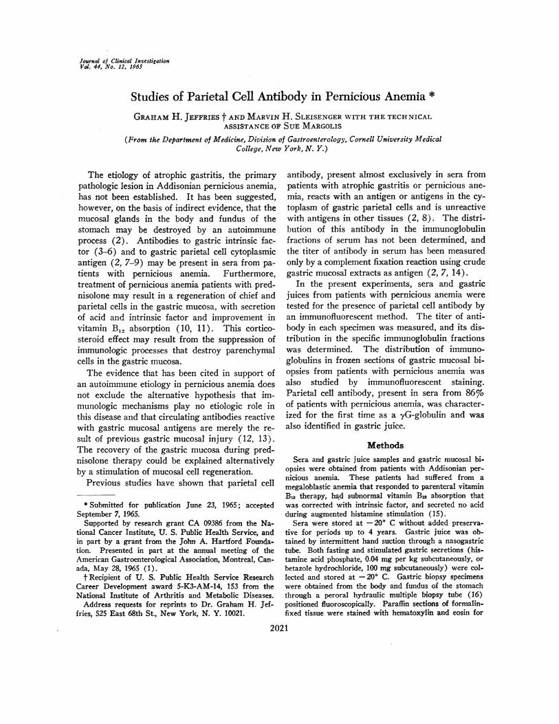
FIG. 1. PARIETAL CELL IMMUNOFLUORESCENCE.A frozen section of ratgastric mucosa was incubated with pernicious anemia serum (diluted 1/4with phosphate-buffered saline) followed by fluoresceinated antiserum to'yG-globulin. Magnification X 375.
2022
STUDIES OF PARIETAL CELL ANTIBODY IN PERNICIOUS ANEMIA
16
14
12
In .11'U
08
Cu
'4--
0 4
NEC 4 8 16 32 64 128 256 512 1024parietal cell antibody titer
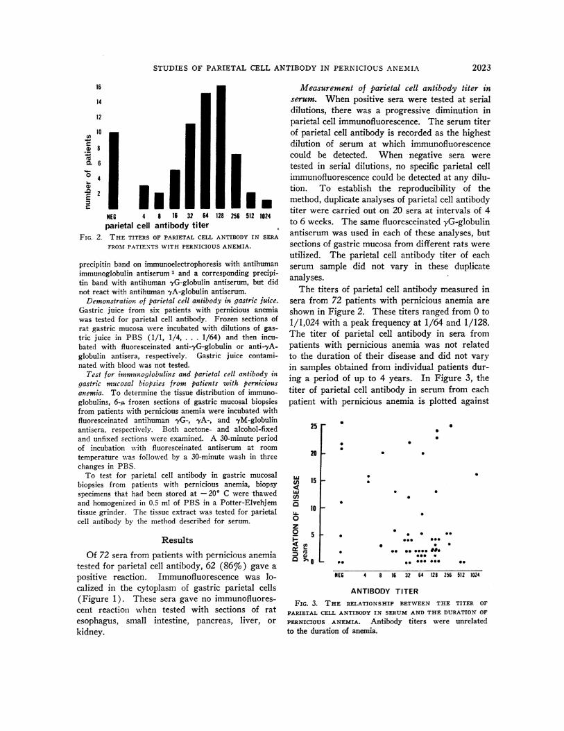
FIG. 2. THE TITERS OF PARIETAL CELL ANTIBODY IN SERA
FROMPATIENTS WITH PERNICIOUS ANEMIA.
precipitin band on immunoelectrophoresis with antihumanimmunoglobulin antiserum 1 and a corresponding precipi-tin band with antihuman yG-globulin antiserum, but didnot react with antihuman yA-globulin antiserum.
Demonstration of parietal cell antibody in gastric juice.Gastric juice from six patients with pernicious anemiawas tested for parietal cell antibody. Frozen sections ofrat gastric mucosa were incubated with dilutions of gas-
tric juice in PBS (1/1, 1/4, . . . 1/64) and then incu-bated with fluoresceinated anti-yG-globulin or anti-yA-globulin antisera, respectively. Gastric juice contami-nated with blood was not tested.
Test for immunoglobulins and parietal cell antibody in
gastric mucosal biopsies from patients with perniciousanemia. To determine the tissue distribution of immuno-globulins, 6-/z frozen sections of gastric mucosal biopsiesfrom patients with pernicious anemia were incubated withfluoresceinated antihuman yG-, yA-, and -yM-globulinantisera, respectively. Both acetone- and alcohol-fixedand unfixed sections were examined. A 30-minute periodof incubation with fluoresceinated antiserum at room
temperature was followed by a 30-minute wash in threechanges in PBS.
To test for parietal cell antibody in gastric mucosalbiopsies from patients with pernicious anemia, biopsyspecimens that had been stored at -200 C were thawedand homogenized in 0.5 ml of PBS in a Potter-Elvehjemtissue grinder. The tissue extract was tested for parietalcell antibody by the method described for serum.
Results
Of 72 sera from patients with pernicious anemiatested for parietal cell antibody, 62 (86%o) gave a
positive reaction. Immunofluorescence was lo-calized in the cytoplasm of gastric parietal cells(Figure 1). These sera gave no immunofluores-cent reaction when tested with sections of ratesophagus, small intestine, pancreas, liver, or
kidney.
Measurement of parietal cell antibody titer inserum. When positive sera were tested at serialdilutions, there was a progressive diminution inparietal cell immunofluorescence. The serum titerof parietal cell antibody is recorded as the highestdilution of serum at which immunofluorescencecould be detected. When negative sera weretested in serial dilutions, no specific parietal cellimmunofluorescence could be detected at any dilu-tion. To establish the reproducibility of themethod, duplicate analyses of parietal cell antibodytiter were carried out on 20 sera at intervals of 4to 6 weeks. The same fluoresceinated yG-globulinantiserum was used in each of these analyses, butsections of gastric mucosa from different rats wereutilized. The parietal cell antibody titer of eachserum sample did not vary in these duplicateanalyses.
The titers of parietal cell antibody measured insera from 72 patients with pernicious anemia areshown in Figure 2. These titers ranged from 0 to1/1,024 with a peak frequency at 1/64 and 1/128.The titer of parietal cell antibody in sera frompatients with pernicious anemia was not relatedto the duration of their disease and did not varyin samples obtained from individual patients dur-ing a period of up to 4 years. In Figure 3, thetiter of parietal cell antibody in serum from eachpatient with pernicious anemia is plotted against
25r 0
S
S
.
0
20 F
ujC,)
U1)LLI
0z0I-
'I
a
15 F S
0
10-
5Un0
_0
Os* S SS 0
0 o -000 0E 00
NEG 4 8 16 32 64 128 256 512 1024
ANTIBODY TITER
FIG. 3. THE RELATIONSHIP BETWEEN THE TITER OFPARIETAL CELL ANTIBODY IN SERUMAND THE DURATIONOFPERNICIOUS ANEMIA. Antibody titers were unrelatedto the duration of anemia.
2023
GRAHAMH. JEFFRIES ANDMARVIN H. SLEISENGER
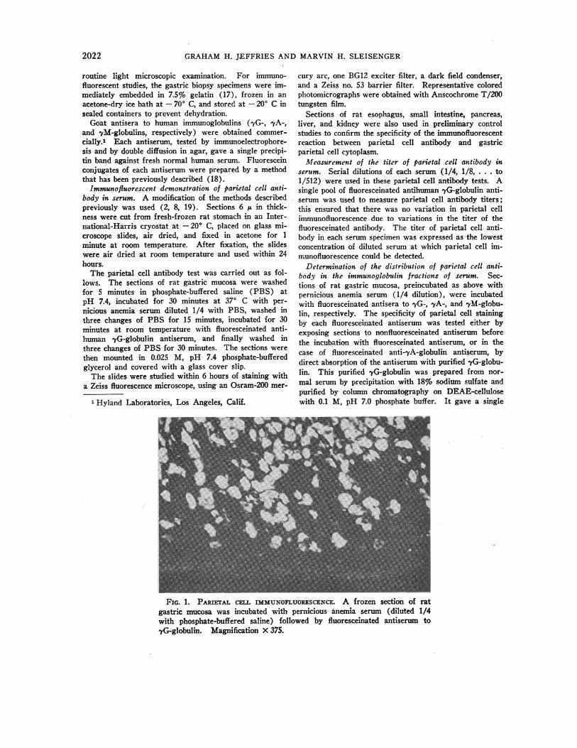
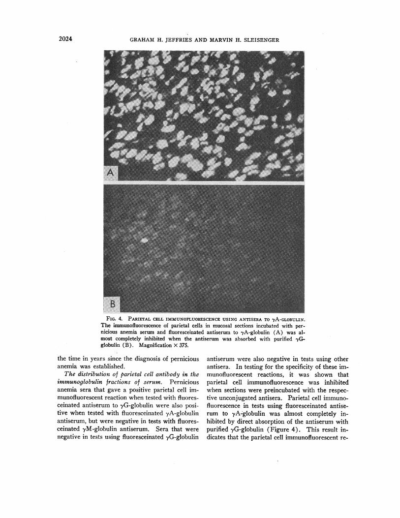
FIG. 4. PARIETAL CELL IMMUNOFLUORESCENCEUSING ANTISERA TO yA-GLOBULIN.The immunofluorescence of parietal cells in mucosal sections incubated with per-nicious anemia serum and fluoresceinated antiserum to -yA-globulin (A) was al-most completely inhibited when the antiserum was absorbed with purified 'yG-globulin (B). Magnification X 375.
the time in years since the diagnosis of perniciousanemia was established.
The distribution of parietal cell antibody in theimmunoglobulin fractions of serum. Perniciousanemia sera that gave a positive parietal cell im-munofluorescent reaction when tested with fluores-ceinated antiserum to -yG-globulin were also posi-tive when tested with fluoresceinated yA-globulinantiserum, but were negative in tests with fluores-ceinated yM-globulin antiserum. Sera that werenegative in tests using fluoresceinated yG-globulin
antiserum were also negative in tests using otherantisera. In testing for the specificity of these im-munofluorescent reactions, it was shown thatparietal cell immunofluorescence was inhibitedwhen sections were preincubated with the respec-tive unconjugated antisera. Parietal cell immuno-fluorescence in tests using fluoresceinated antise-rum to yA-globulin was almost completely in-hibited by direct absorption of the antiserum withpurified yG-globulin (Figure 4). This result in-dicates that the parietal cell immunofluorescent re-
2024
STUDIES OF PARIETAL CELL ANTIBODY IN PERNICIOUS ANEMIA
action with fluoresceinated antisera to yA-globulinmay result mainly from the cross-reaction of thisantiserum with yG-globulin. From these obser-vations we concluded that parietal cell antibodyresides predominantly in the yG-globulin fractionof pernicious anemia sera, although minor amountsmay be present in the yA-globulin fraction.
Demonstration of parietal cell antibody in gas-
tric juice from patients with pernicious anemia.Parietal cell antibody was detected in the achlorhy-dric gastric juice from six patients with perniciousanemia, both during fasting and when stimulated.The titer of antibody in each gastric juice was con-
siderably less than that present in the patient'sserum (Table I), and tests using fluoresceinatedantisera to yG- and yA-globulins indicated thatthe antibody was a yG-globulin as in the serum.
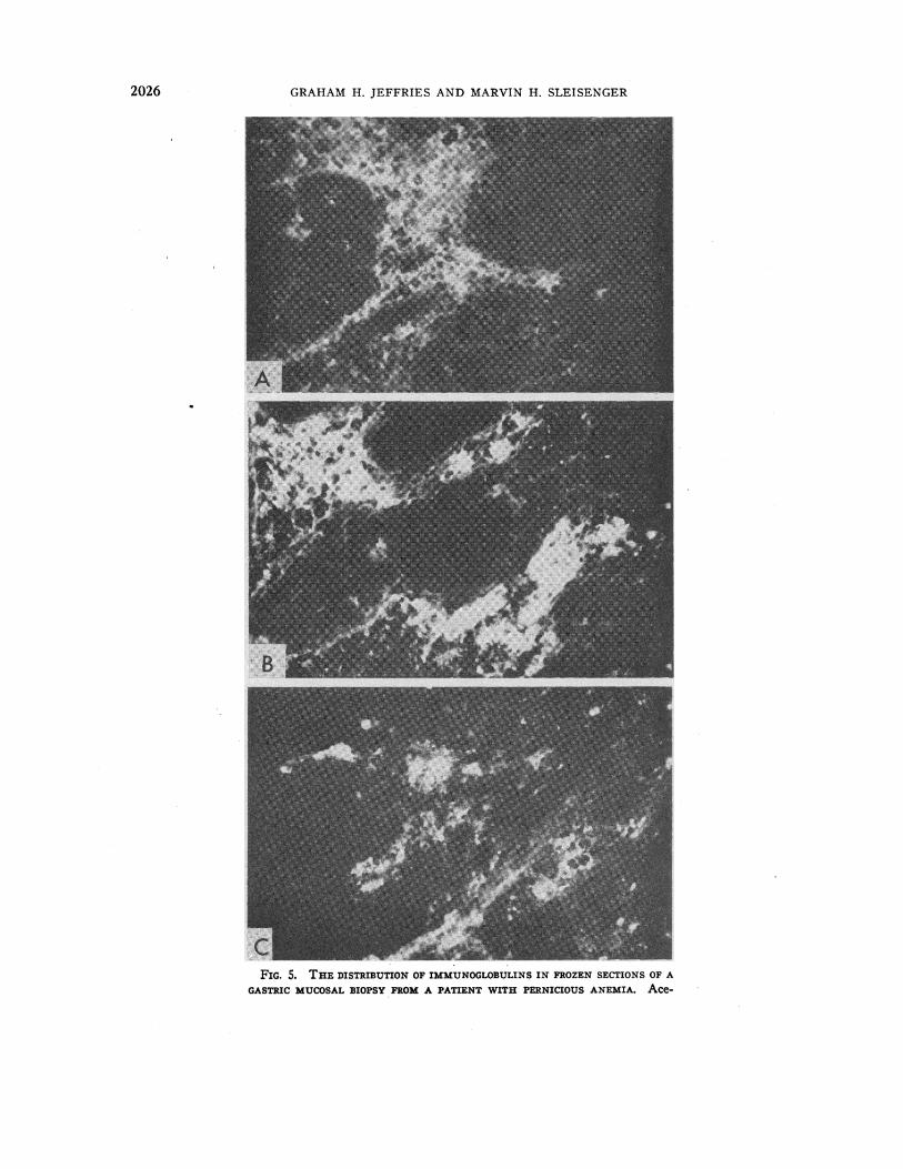
Identification of immunoglobulins and parietalcell antibody in gastric mucosal biopsies from pa-tients with pernicious anemia. The distributionof immunoglobulins was studied in frozen sectionsof gastric mucosal biopsies from three patients withpernicious anemia. Incubation of these sectionswith fluoresceinated antisera to -yG-, yA-, and yM-globulins produced tissue fluorescence that was lo-calized in the lamina propria of the mucosa andparticularly concentrated in mononuclear cells.This was best seen in acetone-fixed sections (Fig-ures 5). In hematoxylin and eosin stained sec-
tions, both lymphocytes and plasma cells were
identified in the lamina propria. The specificity ofthe immunofluorescent staining with antisera toyG- and yA-globulins was confirmed by preincu-bating mucosal sections with unconjugated anti-sera. Staining with fluoresceinated antiserum toyG-globulin was inhibited by prior incubation withunconjugated yG-globulin antiserum, but was notinhibited by unconjugated yA-globulin antiserum.Conversely, staining with fluoresceinated antiserumto yA-globulin was inhibited by homologous un-
conjugated antiserum, but was not inhibited byunconjugated yG-globulin antiserum. Both yG-and yA-globulins were detected in many of theinfiltrating cells (Figure 5 A, B), whereas yM-globulin was seen in only a few cells (Figure 5 C).
Gastric mucosal biopsies from five patients withpernicious anemia were homogenized in PBS, andthe tissue extract was tested for parietal cell anti-body. Sera from each of these patients contained
TABLE I
Titers of parietal cell antibody inserum and gastric juice
GastricSerum juice
Patient titer titer
1 32 42 64 43 128 164 256 165 256 86 512 8
antibody. No parietal cell antibody could be de-tected in these extracts of gastric mucosa.
Discussion
Antibodies reactive with parietal cell-cytoplasmin an immunofluorescent test using rat gastricmucosa were detected in sera for 86% of 72 pa-tients with adult pernicious anemia. This inci-dence of antibody corresponds to that reported inthe literature (2, 14, 19, 20).
The titer of parietal cell antibody has been mea-sured with serial dilutions of serum in the immuno-fluorescent test, and reproducible results have beenobtained. In these studies all measurements ofparietal cell antibody titer were carried out witha single pool of fluoresceinated antiserum to yG-globulin; thus the titers measured in different per-nicious anemia sera reflect variations in the con-centration of parietal cell antibody alone. Thesetiters are slightly higher than those reported byIrvine and his associates (8, 14), who used a com-plement fixation test with saline extracts of humangastric mucosa as antigen.
Taylor and co-workers (2) reported that theincidence of gastric antibodies in sera from pa-tients with pernicious anemia was not related tothe duration of disease. In the present study wehave shown that the titer of parietal cell antibodyis also unrelated to the duration of pernicious ane-mia; this confirms recent observations made byIrvine and associates. (14). In some patients therewere high titers of serum antibody 20 to 25 yearsafter the diagnosis of pernicious anemia had beenestablished, whereas in other patients no antibodywas detected in serum obtained at the time ofdiagnosis. Furthermore, serum samples obtainedfrom individual patients during a period of 4 yearsshowed no variation in antibody titer.
2025
GRAHAMH. JEFFRIES AND MARVIN H. SLEISENGER
CoFIG. 5. THEDISTRIBUTION OF IMMUNOGLOBULINSIN FROZENSECTIONS OF A
GASTRIC MUCOSALBIOPSY FROMA PATIENT WITH PERNICIOUS ANEMIA. Ace-
2026
STUDIES OF PARIETAL CELL ANTIBODY IN PERNICIOUS ANEMIA
In patients with adult pernicious anemia, gastricmucosal atrophy precedes the development of vita-min B,, deficiency. If gastric mucosal atrophyresults from a loss of the mucosal potential to re-generate parietal cells, there should be no furtherstimulation of parietal cell antibody production bymucosal antigen so that the titer of circulatingparietal cell antibody should diminish progres-sively with time. The observation that the titerof parietal cell antibody does not change with timesuggests that in many patients there is a continu-ous stimulation of antibody production. This mayresult from continuing regeneration and destruc-tion of parietal cell precursors in the atrophic gas-tric mucosa. That this mucosa may retain its po-tential to regenerate parietal cells is indicated bythe histologic and secretory changes that have beenobserved during prednisolone therapy (10, 11).The absence of parietal cell antibody in serumfrom some patients with pernicious anemia is notexplained by this hypothesis. It is possible, how-ever, that these patients have indeed lost the po-tential to regenerate gastric parietal cells; thismight occur with extensive replacement of themucosa in the body and fundus of the stomach byintestinal- or pyloric-type epithelium.
Parietal cell antibody, present in both serumand gastric juice, was identified as a yG-globulin.The presence of this antibody in gastric juice frompatients with pernicious anemia has been confirmedindependently by Fisher and Taylor (21). Thelow titer of antibody in gastric juice as comparedwith serum may result from a transudation ofplasma across the mucosa; plasma proteins, in-cluding the immunoglobulins, have been identifiedpreviously in gastric juice (22-24). The possi-bility that parietal cell antibody is selectively se-creted by the mucosa cannot be excluded, how-ever, as the absolute concentrations of yG-globulinin serum and gastric juice were not measured.
Sera from patients with pernicious anemia mayalso contain antibodies that react with gastric in-trinsic factor (3-6). This antibody has not beendetected in gastric juice (20) ; nevertheless, it maybe present in low concentrations sufficient to in-
hibit the biologic activity of small amounts of in-trinsic factor that may be secreted.
Mononuclear cells containing each of the im-munoglobulins were identified in the lamina pro-pria of gastric mucosal biopsies from patients withpernicious anemia. There is no evidence, how-ever, that these cells produce antibodies that reactwith mucosal cell antigens. No parietal cell anti-body could be detected in buffered saline extractsof gastric mucosal biopsies. This negative ob-servation does not exclude the presence of parietalcell antibody either in the interstitial lymph or inmononuclear cells in the lamina propria; the con-centration of antibody in the saline extracts mayhave been too low to be detected in the immuno-fluorescent test.
Summary1) Parietal cell antibody was detected by an im-
munofluorescent method in sera from 62 of 72 pa-tients (86%o) with adult pernicious anemia. Thetiter of parietal cell antibody in the serum was notrelated to the duration of the patient's perniciousanemia and did not vary in individual patientsover a period of up to 4 years.
2) Parietal cell antibody was present in the,yG-globulin fraction of serum; it was either absentor present in very low titers in the yA-globulinfraction, and it was not detected in the yM-globu-lin fraction.
3) Parietal cell antibody was demonstrated ingastric juice from patients with pernicious anemia.It was present in the yG-globulin fraction, and itstiter in the juice was considerably lower than thatin the patient's serum.
4) Immunoglobulins were present in the mono-nuclear cells in the lamina propria of the gastricmucosa from patients with pernicious anemia.Parietal cell antibody could not be detected in mu-cosal extracts.
References1. Jeffries, G. H., and M. H. Sleisenger. Immuno-
fluorescent studies in pernicious anemia (abstract).Gastroenterology 1965, 48, 823.
2. Taylor, K. B., I. M. Roitt, D. Doniach, K. G. Couch-man, and C. Shapland. Autoimmune phenomena in
tone-fixed sections of gastric mucosa from a patient with pernicious anemiawere incubated with fluoresceinated antisera to yG-globulin (A), yA-globu-lin (B), and 'yM-globulin (C). Fluorescence is concentrated in mononuclearcells in the lamina propria.
2027
GRAHAMH. JEFFRIES AND MARVIN H. SLEISENGER
pernicious anaemia. Gastric antibodies. Brit. med.J. 1962, 2, 1347.
3. Taylor, K. B. Inhibition of intrinsic factor by per-
nicious anaemia serum. Lancet 1959, 2, 106.4. Schwartz, M. Intrinsic factor antibody in serum
from patients with pernicious anaemia. Lancet1960, 2, 1263.
5. Jeffries, G. H., D. W. Hoskins, and M. H. Sleisenger.Antibody to intrinsic factor in serum from patientswith pernicious anemia. J. clin. Invest. 1962, 41,1106.
6. Abels, J., W. Bouma, A. Jansz, M. G. Woldring, A.Bakker, and H. 0. Nieweg. Experiments on in-trinsic factor antibody in serum from patientswith pernicious anemia. J. Lab. clin. Med. 1963,61, 893.
7. Irvine, W. J., S. H. Davies, I. W. Delamore, andA. W. Williams. Immunological relationship be-tween pernicious anaemia and thyroid disease.Brit. med. J. 1962, 2, 454.
8. Irvine, W. J. Gastric antibodies studied by fluores-cence microscopy. Quart. J. exp. Physiol. 1963, 48,427.
9. Markson, J. L., and J. M. Moore. Autoimmunity inpernicious anaemia and iron-deficiency anaemia: a
complement-fixation test using human gastric mu-
cosa. Lancet 1962, 2, 1240.10. Jeffries, G. H. Recovery of gastric mucosal struc-
ture and function in pernicious anemia duringprednisolone therapy. Gastroenterology 1965, 48,371.
11. Jeffries, G. H., J. E. Todd, and M. H. Sleisenger.Pernicious anemia. The effect of prednisolone on
gastric mucosal histology, gastric secretion, andvitamin B,2 absorption. Submitted for publication.
12. Thomas, L. Circulating autoantibodies and humandisease; with a note on primary atypical pneumonia.New Engl. J. Med. 1964, 270, 1157.
13. Herbert, V., R. R. Streiff, and L. W. Sullivan. Noteson vitamin B,2 absorption: autoimmunity and child-hood pernicious anemia; relation of intrinsic fac-
tor to blood group substance. Medicine (Balti-more) 1964, 43, 679.
14. Irvine, W. J., S. H. Davies, S. Teitelbaum, I. W.Delamore, and A. W. Williams. The clinical andpathological significance of gastric parietal cell anti-body. Ann. N. Y. Acad. Sci. 1965, 124, 657.
15. Kay, A. W. Effect of large doses of histamine ongastric secretion of HCl; an augmented histaminetest. Brit. med. J. 1953, 2, 77.
16. Flick, A. L., W. E. Quinton, and C. E. Rubin. Aperoral hydraulic biopsy tube for multiple samplingat any level of the gastrointestinal tract. Gastro-enterology 1961, 40, 120.
17. Burkholder, P. M., A. H. Littell, and P. G. Klein.Sectioning at room temperature of unfixed tissues,frozen in a gelatin matrix, for immunohistologicprocedures. Stain Technol. 1961, 36, 89.
18. Rubin, W., A. S. Fauci, M. H. Sleisenger, and G. H.Jeffries. Immunofluorescent studies in adult celiacdisease. J. clin. Invest. 1965, 44, 475.
19. Te Velde, K., J. Abels, G. J. P. A. Anders, A. Arends,Ph. J. Hoedemaeker, and H. 0. Nieweg. A fam-ily study of pernicious anemia by an immunologicmethod. J. Lab. clin. Med. 1964, 64, 177.
20. Fisher, J. M., and K. B. Taylor. A comparison ofautoimmune phenomena in pernicious anemia andchronic atrophic gastritis. New Engl. J. Med.1965, 272, 499.
21. Fisher, J. M., and K. B. Taylor. Personal communi-cation.
22. Heiskell, C. L., T. Wada, S. J. Stempien, M. Fukuda,S. Nakagawa, A. Yachi, A. Dagradi, and C. M.Carpenter. Normal serum proteins in gastric juice.A preliminary report. Gastroenterology 1961, 40,775.
23. Tenorovk, M., E. Stuchlikova, and J. Kohrnek.Agarophoresis and immuno-electrophoresis of theproteins of gastric juice. Nature (Lond.) 1961,192, 763.
24. Hurlimann, J. Les proteines du suc gastrique. Rtudeimmunoelectrophoretique. Helv. med. Acta 1963,30, 126.
2028