Embed Size (px)
Citation preview

Research Article Open Access
Ray et al., J Clin Cell Immunol 2012, S10http://dx.doi.org/10.4172/2155-9899.S10-003
Review Article Open Access
Clinical & Cellular
Immunology
J Clin Cell Immunol ISSN:2155-9899 JCCI, an open access journal Clinical, Cellular & Molecular Biology
of Autoimmune Disorders
Received October 02, 2012; Accepted October 22, 2012; Published October 29, 2012
Citation: Ray S, Sonthalia N, Kundu S, Ganguly S (2012) Autoimmune Disorders: An Overview of Molecular and Cellular Basis in Today’s Perspective. J Clin Cell Immunol S10:003. doi:10.4172/2155-9899.S10-003
Copyright: © 2012 Ray S, et al. This is an open-access article distributed under the terms of the Creative Commons Attribution License, which permits unrestricted use, distribution, and reproduction in any medium, provided the original author and source are credited.
AbstractAutoimmunity arises when immune responses mounted in the host are directed against self-components.
Autoimmune diseases are pathophysiological states that result from a loss of self-tolerance and the consequent immune destruction of host tissues. Autoimmunity is mediated by a variety of molecular and cellular events, and responses. The development of an autoimmune disease is a very complex process in which recognition of self-antigens by lymphocytes is centrally involved in pathologic organ damage. Autoimmune disease is inherited as a complex trait, with multiple loci controlling various aspects of disease susceptibility. More recently, some of these susceptibility genes have been identified. Certain environmental influences, such as cigarette smoke, ultraviolet light, or infectious agents, may interplay with this genetic predisposition to initiate the disease process. Silica exposure and its role in systemic lupus erythematosus (SLE) have been identified in studies of occupational exposure, and experimental studies have explored potential mechanisms related to immune dysregulation. Some autoimmune responses emerge following infection by a pathogen, whose protein(s) hold structural similarities to regions on proteins of the host. Thus, antibodies evoked against a pathogen might cross-react with a self-protein and act as autoantibodies, and the concerned autoantigen then provides a source for persistent stimulation. Evidence is emerging that activation of autoimmune B cells and T cells can be influenced by innate immune receptors, such as Toll-like receptors, which primarily recognize pathogen-derived molecular structures but may cross-react with host molecules. Proteins to which the immune system is generally self-tolerant might, if altered, elicit autoimmune responses. Potential involvement of chaperones in the induction of autoimmune disease pathogenesis has also been explored. The contributions of microRNA to pathogenesis of autoimmune diseases like SLE are beginning to be uncovered and may provide us a new arena for exploration of mechanisms responsible for initiation and pathogenesis of autoimmune diseases.
Autoimmune Disorders: An Overview of Molecular and Cellular Basis in Today’s PerspectiveSayantan Ray*, Nikhil Sonthalia, Supratip Kundu and Satyabrata GangulyDepartment of Medicine, Medical College and Hospital, Kolkata, West Bengal, India
Keywords: Autoimmunity; Autoimmune disease; Self-antigens; Lymphocytes; Autoantibodies; MicroRNA
IntroductionHuman autoimmune diseases (AD) occur frequently (affecting in
aggregate more than 5% of the population worldwide), and impose a significant burden of morbidity and mortality on the human population [1]. AD are defined as diseases in which immune responses to specific self-antigens contribute to the ongoing tissue damage that occurs in that disease. ADs may be either tissue-specific (e.g., thyroid, β-cells of the pancreas), where unique tissue-specific antigens are targeted, or may be more systemic, in which multiple tissues are affected, and a variety of apparently ubiquitously expressed autoantigens are targeted [2]. Although the definition appears relatively simple in concept, the complexity of this spectrum of disorders is enormous, and has greatly challenged elucidation of simple shared mechanisms. This complexity affects almost every domain, including genetics, phenotypic expression, and kinetics. In the latter case, there is frequently a prolonged period between initial onset of symptoms and development of the diagnostic phenotype, and disease may vary in expression in the same individual over time.
Autoimmunity is not set off by a single cause and is triggered by a variety of agents and molecular and cellular pathways and events. Several elements and mechanisms underlying autoimmune responses have been identified. However, even if a given AD were to be initiated primarily by a single trigger, other events and regulating mechanisms come into play, thereby adding complexity to the process. This review focuses on the current understanding of the mechanistic principles that underlie ADs. We provide an outlook on novel class of immune regulators that play an essential role in multiple pathophysiological processes of multiple ADs.
Overview of Development of Autoimmunity A major barrier to understanding mechanisms of autoimmunity
comes from difficulty in defining early events in these diseases. Since, diseases are only recognizable after development of the diagnostic phenotype, there has been the tendency to interpret findings made at diagnosis with findings present at initiation. Based on recent findings [3-5], the development of ADs can be divided in four phases- і. Susceptibility phase, іі. Initiation phase, ііі. Propagation phase and іν. Regulation phase.
The susceptibility to ADs can be either inherited or acquired (and in many diseases, both). ADs result from a complex interplay of pathways and events which initially allow autoreactivity to manifest, and then, after an initiating event, allow development of self-sustaining tissue damage. Factors that trigger the initiation include abnormalities in tolerance induction, regulatory T-cell (Treg) development, or immune signaling thresholds. The propagation phase is marked by a feed-forward cycle of autoimmunity and tissue damage, in which immune effector pathways cause damage and provide antigen to drive the
*Corresponding author: Sayantan Ray, MD, Department of Medicine, Medical College and Hospital, Kolkata 700073, West Bengal, India, E-mail: [email protected]

Citation: Ray S, Sonthalia N, Kundu S, Ganguly S (2012) Autoimmune Disorders: An Overview of Molecular and Cellular Basis in Today’s Perspective. J Clin Cell Immunol S10:003. doi:10.4172/2155-9899.S10-003
Page 2 of 12
J Clin Cell Immunol ISSN:2155-9899 JCCI, an open access journal Clinical, Cellular & Molecular Biology
of Autoimmune Disorders
ongoing immune response. Figure 1 presents a conceptual framework for the development of AD. It should also be noted that in many cases during disease propagation, immunoregulatory pathways are also activated, which may result in natural inhibition of clinical disease over time. Such immunoregulation is likely absent or fails in a susceptible host.
ADs traditionally have been categorized as organ specific or
systemic or both (Table 1). The organ-specific ADs may represent examples of normal immune responses that produce disease because they are “misdirected” against a self-antigen or organ. By contrast, in systemic ADs, multiple organs are targets for immune attack, and chronic activation of innate and adaptive immune cells is usually present. SLE is considered to be the prototypic systemic AD. However, it should be noted that the categorization of an AD as organ-specific or systemic is based primarily on clinical observations rather than the expression pattern of the self antigen that appears to be targeted in the attack.
Determinants of Autoimmune DiseaseAlthough ADs in humans are genetically complex, significant
advances in understanding have occurred over the past several years. For many ADs, the break in peripheral self-tolerance leading to an anti-self immune response is linked to an encounter with a particular pathogen, chemical, drug, toxin, or hormone. However, the single most important factor contributing to AD is the genetic make-up of the host. A complex constellation of AD susceptibility alleles and haplotypes exists that determines the ongoing deregulation of self-tolerance mechanisms.
Genetic predisposition
There have been important advances in the genetics of autoimmunity in several mouse models. These studies highlight a critical role for pathways of tolerance induction, immunoregulation, and setpoints/thresholds for immune signaling in avoiding emergence of autoimmunity [6-8]. It should be emphasized that regardless of the underlying cause for autoimmunity, predisposition to a given autoimmune response is associated with certain human leukocyte antigen (HLA) allele(s). If the host’s major histocompatibility complex (MHC) cannot present an antigen, that antigen cannot elicit a response
EnvironmentIndividual
Genetic factors
Innate immune
responsiveness
Self-limiting non- specific
inflammation
Smoking, UV light,
infectious agents
Adaptive immunesystem activation
Loss of self-tolerance
Gender, ethnicorigin
Host defensegenes (TLR7, FcR,
TNF)
Immuneresponse genes
(HLA-DR,PTPN22, CTLA4,
IL10, FcRIIB)Proinflammatory
cytokines
Immunoregulation(Treg,clonal
deletion, anergy)
AutoreactiveT cells
Autoantibodyproduction
Autoimmunity
Tissue damage Tissue responsegenes
Figure 1: Pathways influencing the development and perpetuation of autoimmune diseases.
Table 1: Examples of selected human autoimmune diseases.
Organ-Specific Autoimmune DiseasesOrgan Disease(s) Self-Antigen Major Autoimmune MechanismAdrenal cells Addison's disease Cytochrome P-450 antigens AutoantibodiesRed blood cells Autoimmune hemolytic anemia Red blood cell membrane proteins AutoantibodiesPlatelets Idiopathic thrombocytopenic purpura Platelet antigens (GP IIb/IIIa) AutoantibodiesStomach Pernicious anemia Gastric parietal cell antigens (H+/ATPase, intrinsic factor) Autoantibodies/T cellsSmall bowel Celiac sprue (gluten enteropathy) Transglutaminase Autoantibodies/T cells
ThyroidHashimoto's thyroiditis Thyroid cell antigens (e.g., thyroglobulin) T cells/autoantibodies
Graves' disease Thyroid-stimulating hormone receptor AutoantibodiesMuscle Myasthenia gravis Acetylcholine receptors AutoantibodiesPancreatic islets Type 1 diabetes Beta cell antigens (glutamic acid decarboxylase, insulin) T cells (autoantibodies present
Hepatocytes Autoimmune hepatitis Hepatocyte antigens (cytochrome P450 2D6) T cells/antibodiesBile duct cells Primary biliary cirrhosis Intrahepatic bile duct (pyruvate dehydrogenase complex
protein)Autoantibodies/ T cells
Heart Rheumatic heart disease Myocardial antigens AutoantibodiesKidney/lung Goodpasture's syndrome Basement membrane antigens (type IV collagen α3 chain) Autoantibodies
Systemic Autoimmune DiseasesDisease(s) Self-Antigen Major Autoimmune MechanismAnkylosing sponkylitis Vertebrae Immune complexesMultiple sclerosis Brain or white matter TH1 cells and TC cells, auto-antibodiesRheumatoid arthritis Connective tissue, IgG Auto-antibodies, immune complexesSystemic lupus erythematosus DNA, nuclear protein, RBC and platelet membranes Auto-antibodies, immune complexesScleroderma Nuclei, heart, lungs, gastrointestinal tract, kidney Auto-antibodiesSjogren’s syndrome Salivary gland, liver, kidney, thyroid Auto-antibodies

Citation: Ray S, Sonthalia N, Kundu S, Ganguly S (2012) Autoimmune Disorders: An Overview of Molecular and Cellular Basis in Today’s Perspective. J Clin Cell Immunol S10:003. doi:10.4172/2155-9899.S10-003
Page 3 of 12
J Clin Cell Immunol ISSN:2155-9899 JCCI, an open access journal Clinical, Cellular & Molecular Biology
of Autoimmune Disorders
and would not be an autoantigen in that host. The presence or absence of the appropriate MHC would determine whether the potential autoantigen is presented and the occurrence or otherwise of a response to the antigen.
Due to their direct involvement in T cell responses, the most important genes that predispose both humans and animals to AD are the MHC genes (Table 2). Perhaps the best illustration of AD-HLA association in humans can be found in ankylosing spondyliitis (AS). Over 90% of Caucasians with AS express an allele belonging to the HLA-B27 family. Other AD also show strong associations with specific HLA allele families. For example, expression of HLA-DR2 and HLA-DR3 predisposes an individual to developing SLE, while T1 diabetes mellitus (DM) has particularly strong links to HLA-DR3, -DR4, -DQ2, and -DQ8. Individuals expressing certain alleles of HLA-DR4 are especially prone to rheumatoid arthritis (RA) or Juvenile RA, while primary Sjogren’s syndrome (SS) and polymyositis (PM) are associated with HLA-DR3 in some populations.
The requisite HLA alleles work at the level of antigen presenting cell, whose presence or absence determine the presentation and the resultant response to an autoantigen. However, no genetic pattern is specific to any disease and some patients with specific genetic pattern manifests different diseases. Predisposition of disease can also be seen in families however, phenotypic manifestation can be different.
Environmental triggers
The role of environmental factors in the etiology of ADs is clearly apparent when considering the disease concordance rate between monozygotic twins. More than 50 and sometimes 70 or 80% of monozygotic twins are discordant for major ADs. Despite the existing evidence, however, definitive proof which suggests that that an encounter with an environmental stimulus actually triggers the initial onset of human AD is still lacking.
Environmental stimuli, including chemical agents and pathogens, show significant links to AD onset or flare-ups in both humans and animal models [9]. Certain chemical and pharmaceutical agents have been linked to the onset of particular systemic AD symptoms. For example, toxins such as the heavy metal mercuric-chloride or polyvinyl-chloride can precipitate immune complex nephritis, systemic sclerosis, or the development of autoantibodies. Smoking, use of hair dyes (which contain aromatic amines), glue-sniffing, or exposure to silica dust (as occurs in many types of manufacturing and mining jobs) or
other toxins can bring on an episode of RA, SLE, Graves’ disease (GD), or scleroderma. Workers in industries such as furniture re-finishing, spray-painting, perfume or cosmetic manufacturing also have a slightly increased risk of developing AD.
Exposure to UV radiation, particularly UV-B rays, has been linked to a physical insult that results in flare-ups of SLE. In vitro studies suggest that exposure of DNA and small nuclear ribonucleoproteins (snRNPs) to UV-B results in changes to the conformation and location of these molecules that increase their chances of activating an autoreactive lymphocyte. The mechanism by which these environmental factors induce autoimmunity includes epigenetic changes (DNA methylation and histone modification), reaction with the self component to generate novel antigens, aberrant cell death releasing cellular material that can lead to inflammasome activation and production of pro-inflammatory cytokines and molecular mimicry [10].
Some AD may initiate in response to drug treatment. For example, thiol-containing drugs and sulfonamide derivatives, as well as certain antibiotics and non-steroidal anti-inflammatory drugs, appear to trigger the onset of pemphigus. Drugs such as hydralazine and procainamide or similar aromatic amine drugs prescribed can induce SLE-like symptoms such as arthritis, pleuropericarditis, and myocarditis.
Infections with certain viruses, bacteria, and mycoplasma appear to provoke the initiation of systemic AD in genetically predisposed individuals. Moreover, a severe bacterial or viral infection may trigger an increase in autoreactive antibodies or conventional T cells that leads to a flare-up of quiescent AD or an exacerbation of existing symptoms [14,15]. With respect to viruses, the onset of various AD has been variably associated with infection by HSV-1, Coxsackie virus, Epstein-Barr virus (EBV), human immunodeficiency virus (HIV), human papilloma virus (HPV), or influenza virus. In particular, viral infections have been closely associated with flare-ups of SLE. Similarly, the development of Guillain Barre syndrome (GBS) may follow infection with herpes simplex virus (HSV), EBV, or cytomegalovirus (CMV), and the onset of acute idiopathic thrombocytopenic purpura (ITP) may be preceded by varicella infection. Infections with various bacterial species have also been associated with AD. The most striking example is the development of rheumatic fever (RF) following recovery from infection with a virulent member of the Group A streptococci. Another close link is that between the onset of GBS and C. jejuni infection. Antibodies directed against the lipopolysaccharide (LPS) of C. jejuni that cross-react with human nerve gangliosides have been isolated from GBS patients.
Infections are major players in the environmental factors which
Autoimmune Diseases HLA Molecule Strength of AssociationAnkylosing spondylitis HLA-B27 (Caucasians) ++++Rheumatoid arthritis HLA-DR4
HLA-DRB1*04+++
Systemic lupus erythematosus HLA-DR2, DR3 ++Sjögren's syndrome HLA-DR3 ++Psoriatic spondylitis HLA-B27 +++Dermatitis herpetiformis HLA-DR3 +++Gluten-sensitive enteropathy (celiac disease)
HLA-DQ2 +++
Type 1 diabetes mellitus HLA-DR3, DR4, DQ2, DQ8 +++Hyperthyroidism (Graves') HLA-DR3, B8 +Hashimoto’s Thyroiditis HLA-DR3, DR5 ++Adrenal insufficiency HLA-DR3 ++Myasthenia gravis HLA-B8, HLA-DR3 +Multiple sclerosis HLA-DR2 ++
Table 2: HLA alleles associated with selected human AD. Relationship between silica exposure and AD was demonstrated way back in 1914 by Bramwell [11] who showed an increase in the occurrence of scleroderma in stone masons. A study by Sanchez-Roman et al., demonstrated the high probability of workers occupationally exposed to silica developing a multiple spectrum of clinical and serological autoimmune manifestations like SS, scleroderma, SLE, overlap syndrome [12]. Epidemiologic studies have demonstrated moderate to strong associations between occupational silica exposure and SLE. A reduction of Treg cell function and size has been linked to excessive loss of these cells as they become increasingly susceptible to CD95 mediated apoptosis in persons with silica exposure. There is also activation of responder T cells. Taken together, the reduction of Treg cell function and size caused by excessive loss of Treg cells and substitution by chronically activated responder T cells facilitate the immune dysregulation in persons with silica exposure [13].

Citation: Ray S, Sonthalia N, Kundu S, Ganguly S (2012) Autoimmune Disorders: An Overview of Molecular and Cellular Basis in Today’s Perspective. J Clin Cell Immunol S10:003. doi:10.4172/2155-9899.S10-003
Page 4 of 12
J Clin Cell Immunol ISSN:2155-9899 JCCI, an open access journal Clinical, Cellular & Molecular Biology
of Autoimmune Disorders
modulate the development of ADs. Underlying mechanisms are multiple and complex, probably different according to pathogens. It will be extremely interesting to correlate these mechanisms and more generally the infections in question with the polymorphism of genes predisposing to or protecting against the various ADs.
Hormonal influences
The two types of autoimmune thyroiditis, Hashimoto’s thyroiditis (HT) and GD, also occur predominantly in women. Significant numbers of HT and GD patients first develop their disease in the postpartum period, suggesting that major hormonal changes can precipitate onset. In animal models of hypothyroidism, estrogen exacerbates HT symptoms while testosterone reverses it. Another hormonal influence that may be relevant to AD etiology is the hypothalamic–pituitary–adrenal (HPA) axis. Animals with defects in their HPA axis show increased susceptibility to AD, implying that stress-induced increases in glucocorticoids such as corticosterone and cortisol are required to restrain autoreactive lymphocytes.
Regional/ethnic differences
Many ADs appear to vary in incidence by region or by ethnic group, although such data have been relatively hard to come by and are not consistent for all AD or countries. In some cases, this variation may be due to the uneven prevalence of an HLA allele linked to a particular AD (due to ethnic differences) or of a triggering pathogen or chemical agent (due to geographic or environmental differences). In other cases, the reasons for variation in AD incidence among countries or ethnic groups are not obvious. Tied to the regional/ethnic issue is the observation that the decreasing incidence of infections in western countries and more recently in developing countries is at the origin of the increasing incidence of both autoimmune and allergic diseases including Crohn’s disease (CD), T1DM, and multiple sclerosis (MS) [26,27].
Mechanisms Underlying Autoimmune DisordersThe vast majority of AD stem from abnormalities in the mechanisms
of peripheral tolerance that fine-tune the repertoires of mature T and B peripheral lymphocytes. The mere presence of autoreactive lymphocytes in an individual’s repertoire is not enough to trigger AD: it only predisposes that individual to developing AD. For AD to develop, a stimulus that activates the autoreactive cells must be present, and mechanisms designed to regulate autoreactive lymphocyte responses must fail.
We will now discuss several mechanisms, some of which remain controversial, that are believed to contribute to the development of AD in susceptible individuals.
Pathogen-related mechanisms
The onset or flare-ups of many AD appear to be triggered by particular pathogens. However, one should keep in mind that, apart from infection, there must be other factors involved in AD development because infection is common but autoimmunity is not. Millions of people experience pathogen infections, many of them very serious, but only a small fraction of infected individuals develop AD.
Molecular mimicry: The first pathogen-related hypothesis, called molecular mimicry (or antigenic mimicry), holds that autoreactive lymphocytes in the periphery are sometimes activated by cross-reacting pathogen antigens [28-31]. The process of antigen mimicry has frequently been proposed as a potential initiator of ADs (Table 3). This mechanism, particularly when isolated, is only likely relevant to those autoimmune processes clearly associated with antecedent infections, and particularly those that resolve spontaneously. The mechanism may, however, also play a role in initiation of the autoimmune response in self-sustaining autoimmune processes, but in this case, requires that T-cell responses to the cross-reacting self-antigen are initiated.
Foreign antigens, which often differ from their homologous self antigens in some areas, may nevertheless bear significant structural similarity to self-antigens in other regions. Initiation of an immune
Pathogen Antigen Cross-reacting Mammalian Self Antigen ADStreptococcus cell wall M protein Myosin, other heart valve proteins RFPeptides of EBV, influenza virus, HPV, measles virus, HHV-6 Myelin basic protein MSLPS of Campylobacter jejuni Peripheral nerve gangliosides GBSProteins of Salmonella typhimurium or Yersinia enterocolitica HLA-B27 Reactive arthritisBorrelia burgdorferi, OspA protein Lymphocyte function-associated antigen 1 (LFA-1) Lyme arthritisP2-C protein of Coxsackie virus Glutamic acid decarboxylase T1DMProtein of Yersinia enterocolitica Thyrotropin receptor GDB13 protein of Trypanosoma cruzi Cardiac myosin Chagas heart disease
Table 3: Examples of human autoimmune disease potentially linked to molecular mimicry.
A striking common feature of many ADs in both humans and experimental animal models is that females are more susceptible to autoimmune conditions than males [16-18]. More than 85 percent of patients with thyroiditis, scleroderma, lupus, and SS are females [19]. In addition to genetic factors such as X-chromosome abnormalities, sex hormones such as estrogens and androgens are believed to play a significant role in the sex-based susceptibility to many ADs. Researchers hypothesize that the expression of hormones or factors associated with the development of sex-specific organs can activate previously tolerant or ignorant lymphocytes. Indeed, in a mouse model of SLE, the administration of estrogen unregulated Bcl-2 in B cells and blocked B cell tolerization [20-22]. Disease symptoms were exacerbated in the estrogen-treated animals. Estrogen metabolism is often abnormal in SLE patients, and flare-ups of SLE may on occasion be associated with changes in hormonal status, such as during pregnancy or the initiation of hormone replacement therapy. It is clear that sex hormones have profound influence on immune system development and function. Recent studies revealed that estrogen receptor ERα, rather than ERβ plays a critical role in the regulation estrogen-mediated promotion of autoimmunity in NZB/W mice, [23] and also microRNA (miRNA) induction. Of relevance, the decreased level of miR-146a and miR-125a and increased level of miR-148a have been identified in human patients with lupus and reported to contribute to lupus pathogenesis by regulating type I interferon(IFN) pathway [24,25]. Together, these data suggest that sex hormones such as estrogen may contribute to the pathogenesis of lupus and other gender biased AD via the regulation of miRNA expression.

Citation: Ray S, Sonthalia N, Kundu S, Ganguly S (2012) Autoimmune Disorders: An Overview of Molecular and Cellular Basis in Today’s Perspective. J Clin Cell Immunol S10:003. doi:10.4172/2155-9899.S10-003
Page 5 of 12
J Clin Cell Immunol ISSN:2155-9899 JCCI, an open access journal Clinical, Cellular & Molecular Biology
of Autoimmune Disorders
response to the foreign antigen may generate a cross-reactive antibody response that also recognizes the self-protein (antigen mimicry). When the antigen is a cell surface molecule, antibody-mediated effector pathways can lead to host tissue damage. It is important to realize that antigen mimicry alone cannot explain self-sustaining ADs, which are driven by self-antigens and autoreactive T cells. In these cases, there is a requirement for overcoming T-cell tolerance to the self protein. The simultaneous liberation of self-antigen in the presence of the cross-reactive antibody response may allow effective presentation of cryptic epitopes in the self-antigen to autoreactive T cells by activated cross-reactive B cells [32,33]. If continued release of self-antigen occurs, a specific, adaptive immune response to self will be sustained. Antigen release from tissues likely plays a critical role in driving this autoimmune process.
Given the vast number of microbial protein sequences that mimic sequences in human proteins, it is likely that exposure to most microbes does not necessarily trigger an immune response that cross-reacts with human proteins. However, such an initial cross-reactive immune response could lead to subsequent exposure of other regions on the same self-antigen that will then stimulate the emergence of further antibodies, some of them pathogenic, through a process of “epitope spreading”.
Induction of inflammation and DC maturation: Infection by a pathogen induces inflammation, supplying “danger signals” and a cytokine milieu that favors dendritic cell (DC) maturation and activation. Many investigators [34-36] have now provided evidence that this inflammation-induced maturation of DCs that may be the key link between pathogen infection and autoimmunity, the so called “adjuvant effect.” The hypothesis is that bacterial DNA, bacterial components, and endogenous nucleic acids released upon pathogen-induced cell death are particularly potent adjuvants because they engage the Toll-like receptors (TLRs) of immature DCs. Following TLR engagement, DCs are induced to mature and upregulate their expression of costimulatory molecules. When such mature DCs encounter autoreactive T cells in the lymph node, activation leading to an autoimmune response may result if the pMHC derived from a pathogen or self antigen is recognized by the T cell. Thus, autoreactive T cells that might have been held quiescent due to a lack of costimulation and/or the effector actions of Treg cells regain their capacity for activation.
In humans, increased numbers of DCs can be found in the cellular infiltrates affecting the target tissues in several AD, including GD, HT, RA, T1DM, SLE, and SS. These DCs appear to be mature in phenotype, although it is not clear whether they arrive in the lesions as mature cells or are induced to mature once they arrive. Pathogen-induced inflammation and the release of pathogen-associated molecular patterns (PAMPs) from infected host cells may not be the only way to drive DC maturation leading to AD. Cells that have become necrotic due to mechanical injury, transformation, or other forms of stress may release host stress molecules such as HSP70, HSP60, and gp96 with effects on DCs [37]. While the precise mechanism by which DC function is enhanced by stress molecules remains to be clarified, the results of in vitro as well as in vivo studies suggest a means by which AD can be induced by endogenous host stress molecules in the absence of pathogen infection.
Microbial superantigens: Another theory to account for at least some episodes of pathogen-linked AD involves microbial superantigens. These molecules can non-specifically activate a large number of different T cell clones by binding directly to particular T-cell receptor (TCR) Vβ sequences [38]. Superantigens are believed to play
role in relapses of AD or the exacerbation of existing AD, but they do not appear to be able to initiate AD. In humans, there is evidence that a bacterial superantigen from an unknown species may be a factor in CD. Researchers have also noted that certain TCR Vβ T cell subsets are elevated in cases of Kawasaki disease (KD) and Psoriasis (PS). Indeed, T cells whose TCR Vβ regions are recognized by Group A streptococcal superantigens have been isolated from PS skin lesions.
Disruption in the level or activity of regulatory proteins
The immune system is regulated by complex and intricate cellular and molecular interactions that organize direct and control its functions. Molecular and/or cellular changes that compromise the correct performance of this network have been found to be associated with ADs. Non-HLA genes, including cytotoxic T lymphocyte-associated antigen-4 (CTLA4) gene, protein tyrosine phosphate nonreceptor type 22 (PTPN22), together with other autoimmune susceptibility loci (PDCD1, FCRL3, SUMO4, CD25, PADI4 and SLC22A4), tumor TNF-α and FOXP3 have been associated with susceptibility to ADs [39-42].
CTLA4: CTLA4 is essential for T lymphocyte-mediated immunoregulation. Certain alleles of the CTLA4 gene, encoding a regulatory molecule in the immune system, have been proposed to act as nonspecific costimulatory elements in autoimmunity. Polymorphisms of the T cell regulatory molecule CTLA-4 have been implicated in certain ADs [43], particularly type 1 diabetes [44], autoimmune thyroid disease and lupus [45,46]. A CTLA4 allele has been strongly associated with a type 1 diabetes subgroup with a female bias characterized by failure in tolerance to thyroid peroxidase at an early age [47]. However, the CTLA4 gene is seemingly not a major risk factor or a major determinant of disease progression in primary biliary cirrhosis [48] or ulcerative colitis [49].
PTPN22: The human lymphoid PTPN22 gene encodes an 807-amino acid residue protein referred to as lymphoid tyrosine phosphatase. A single-nucleotide polymorphism (SNP) in PTPN22 has been identified as a major risk factor for several human ADs, including type 1 diabetes, RA, SLE, GD, generalized vitiligo [47,50-53].
FOXP3: FoxP3 is a member of the forkhead family of transcription factors, and is essential for the development of Tregs, which regulate the activation and differentiation of effector T cells at many different levels. Mutations in the FoxP3 gene is associated with emergence of autoimmunity when regulatory T-cell (Treg) differentiation is abnormal in humans with immune dysregulation, polyendocrinopathy, enteropathy, X-linked (IPEX) syndrome [54].
TNF-α: TNF-α is involved in chronic inflammation and autoimmunity [55,56]. For example, TNF-α strongly affects the differentiation of DCs and intraorbital inflammatory macrophages from monocytic precursors. Dysregulation of the TNF/TNFR superfamilies may provide a systemic pathogenic link in GD [57]. T cell clones derived from patients with ADs were found to produce TNF-α, and interaction of TNF-α with type I IFN may contribute to AD development [58].
Alterations in the expression levels of regulatory proteins also cause disturbance of normal functions and produce autoimmune responses. Changes in the level or activity of the regulatory molecular chaperones results in the generation of disordered or misfolded proteins that can become targets of autoimmune responses. Cell-mediated functions of the immune system diminish with age, leading to increased susceptibility to infection and autoimmunity. Disruptions of TCR signal transduction pathways occur in ageing and are believed to be

Citation: Ray S, Sonthalia N, Kundu S, Ganguly S (2012) Autoimmune Disorders: An Overview of Molecular and Cellular Basis in Today’s Perspective. J Clin Cell Immunol S10:003. doi:10.4172/2155-9899.S10-003
Page 6 of 12
J Clin Cell Immunol ISSN:2155-9899 JCCI, an open access journal Clinical, Cellular & Molecular Biology
of Autoimmune Disorders
major causes in the disruption of immune tolerance and expression of autoimmunity.
Altered proteins
Altered proteins can be effective triggers of autoimmunity. Proteins to which the immune system is self-tolerant might, if altered, elicit autoimmune responses. Self-proteins can be altered in a number of ways.
Although, there are not yet good examples where natural autoimmunity arises due to the progressive accumulation of somatic mutations over time, with the expression of mutant, truncated forms of autoantigens, the study by Engelhorn et al. [61] has provide an important mechanistic underpinning for the proposal that accumulated mutations have a role in the initiation of autoimmunity.
Posttranslational modification: Posttranslational modification of self-proteins has an effect on intracellular signalling and protein recognition by the immune system and creates auto-antigens that are not subjected to immune tolerance [62,63]. There is a range of possible post-translational modifications (PTMs) of autoantigens that can allow immune recognition of neo-self epitopes, including phosphorylation, proteolytic cleavage, ubiquitination, transglutamination, citrullination, and isoaspartyl modification [64,65]. An example of a posttranslational modification is the non-enzymatic modification of the carboxyl side groups of aspartate residues to isoaspartyl side chains that causes altered T cell function and autoimmune responses [66,67]. Citrullination of the guanidinium side chains of arginine in proteins by peptidyl arginine deaminase has been implicated in rheumatoid arthritis pathogenesis [68]. There is also evidence that citrullination may play a role in T cell autoreactivity in RA. Immune recognition of citrullinated proteins has also been implicated in MS. In normal CNS tissue a proportion of myelin basic protein (MBP) has citrulline conversions. This proportion has been shown to increase by up to threefold in chronic MS [69]. In several cases, autoantibodies recognize exclusively the modified form of the antigen.
The mechanisms by which PTMs can influence the generation of neo-epitopes for autoimmune attack are complex. These mechanisms can result in a straightforward increase in affinity of binding for MHC or TCR. There can be more subtle effects through altering the susceptibility of a protein to proteolytic cleavage during antigen processing [70]. The extent to which proteins are modified (either spontaneously or enzymatically) can alter as a result of cell stress, inflammation, or infection. The continued dissection of these complex interactions, particularly the effects of infection and inflammatory mediators, is therefore an important area for future research.
Enzyme-processing of proteins: Enzyme-processing of proteins
Sequestered proteins: Sequestered proteins are normally sheltered from immune recognition. However, they can become immunogenic once exposed to recognition by immune cells and induce efficient immune responses.
Several autoimmune disorders have been linked to apoptosis [78,79]. Apoptosis, a process of programmed cell death and removal of damaged cells, results in the release of cell components that are then made accessible to immune recognition.
Apoptosis exposes cytoplasm as well as nuclear components ordinarily sheltered from the immune system. In the presence of defective clearance of cellular debris or subcellular particles, apoptosis can be a significant trigger of autoimmune responses against nuclear components. Proteasome defect resulting from downregulation of expression of a proteasome subunit that prevents the proteolytic processing required for the production and activation of the transcription factor nuclear factor-κB (NF-κB), is reported to play a role in type 1 diabetes [80]. Nucleosomes, which are ordinarily sequestered from the immune system, can, if exposed to immune recognition, become immunogenic and induce autoimmunity, particularly in SLE [81].
Epitope spreading
The determinants that are most efficiently presented are termed ‘dominant’; those that are not loaded on to MHC class II to a significant degree are termed ‘cryptic.’ Epitope spreading is the phenomenon by which immune system appears to expand its response beyond the dominant epitopes recognized by B or T cell to induce new cryptic, non cross-rective epitopes that are recognized much later [82,83]. The appearance of responses to these later epitopes underlies the progression of AD and characterizes its chronic phase. For self-antigens, it is likely that a constant set of dominant determinants are generated during antigen processing under most circumstances, with similar outcomes in the thymus and periphery. Antigens processed by the “standard” pathway are therefore fully tolerized, with the T-cell repertoire purged of reactivity to the dominant self. However, since the balance of dominant and cryptic epitopes is significantly influenced by protein structure, post-translational modification, and folding, changes
can trigger or amplify specific autoimmune responses. For instance, celiac disease involves immune targeting of glutamine-rich gliadin components of wheat gluten. Deamidation of gliadin by the enzyme tissue transglutaminase (tTG) produces a more potent antigen for stimulation of DQ2-restricted gliadin-specific T cell clones derived from the gut of celiac disease patients [71]. A recent study has shown that while acid deamidated glutens are less allergenic and autoantigenic, enzymatically deamidated gliadins increase these responses [72]. Thus, the manner in which food is processed may be a contributing trigger of ADs.
Disordered proteins: Denatured proteins [73], natively disordered or misfolded proteins can trigger immune responses against self-proteins. Misfolding produces molecular species that have incorrectly-formed three-dimensional structures. Heat-shock proteins and other molecular chaperones assist the correct folding, stabilization, and translocation of proteins. Defects in the function or expression of heat-shock proteins and other molecular chaperones might play a causative role in the stimulation of autoimmunity [74]. Antibodies against heat-shock proteins have been found to recur in ADs [75-77]. Such autoantibodies can interfere with the ability of heat-shock proteins to effect their function in protein refolding.
Protein mutation and altered expression: Mutations and altered expression of proteins provide important sources of self-antigens that trigger autoimmune responses. Novel forms of autoantigens generated by mutation, truncation, or splicing. Since the final epitopes generated and loaded on to MHC class II can be profoundly influenced by single early cleavage events during antigen processing, relatively minor but critically placed changes in the primary structure of autoantigens may have the capacity to influence peptide selection. Mutations in the autoimmune regulator (AIRE) gene are responsible for the development of autoimmune polyendocrinopathy candidiasis ectodermal dystrophy (APECED) [59]. Mutations in the coding part of human mannose-binding lectin increase the risk of infection and autoimmunity [60].

Citation: Ray S, Sonthalia N, Kundu S, Ganguly S (2012) Autoimmune Disorders: An Overview of Molecular and Cellular Basis in Today’s Perspective. J Clin Cell Immunol S10:003. doi:10.4172/2155-9899.S10-003
Page 7 of 12
J Clin Cell Immunol ISSN:2155-9899 JCCI, an open access journal Clinical, Cellular & Molecular Biology
of Autoimmune Disorders
of protein structure may alter the balance of dominant and cryptic determinants that are presented during natural antigen processing. The effector actions unleashed during the first response to the original epitope may disrupt cell structure or protein conformation to expose either entirely new self antigens (intermolecular epitope spreading) or new epitopes on existing self antigens (intramolecular epitope spreading) [84]. It is likely that this paradigm is broadly applicable to autoimmunity, since numerous changes in autoantigen structure occur during various relevant physiological states, and can influence subsequent processing and selection of epitopes presented.
Proven examples of epitope spreading in humans are few. In Pemphigus, blistering of the mouth almost always precedes blistering of the skin, and mouth blisters are associated with the presence of autoantibodies directed against the desmoglein-3 protein. It is not until the attacks on desmoglein-3 expose epitopes on the related protein desmoglein-1 that autoantibodies directed against this latter protein are produced and skin blistering commences. Epitope spreading has also been invoked to account for the polyclonal lymphocyte activation evident in SLE. For example, multiple T and B cell clones reactive to different epitopes of snRNP can be found at different stages of AD progression in SLE patients.
Cellular Mechanisms of Autoimmune Disease
Role of T cells in initiating and regulating autoimmunity
There is abundant evidence that potentially autoreactive T cells can mature and reach the periphery in most individuals. Numerous “loopholes” in self-tolerance may allow this. Some organ-sequestered antigens are never presented adequately in the thymus. In addition, some self-peptides may not be processed and presented efficiently in the thymus. T cells that escape negative selection against these peptides may be activated in the periphery when these peptides are created by altered proteolysis during inflammation and by post-translational modifications of peptides, such as glycosylation or citrullination [85,86]. Several well-studied animal models are generated by immunization of animals with peripheral organ antigens, such as type II collagen in collagen-induced arthritis and myelin-associated proteins in experimental allergic encephalomyelitis (EAE), which can engender robust T cell responses in the presence of appropriate adjuvants. In addition, immunization of normal mice with nuclear antigens or peptides derived from these antigens can result in lupus-like autoantibody production [87-90]. These experiments indicate that peripheral autoreactive T cells exist and are needed to be kept under control to prevent autoimmunity. Many cellular mechanisms prevent peripheral self-reactive T cells from mediating autoimmune responses (Table 4).
Considerable evidence has emerged in recent years that certain subsets of classic αβ TCR-expressing T cells have the ability to suppress
Role of B cells
Peripheral T cell Tolerance Modes of Tolerance Breakdown
Immune ignorance
Release of sequestered antigensAberrant expression of MHC class IIIncreased expression of autoantigen/MHC class IIMolecular mimicryEpitope spreading
Anergy
Release of inflammatory mediatorsIncreased expression or function of costimulatory moleculesSuppression of IDO
Regulatory T Cells Release of inflammatory mediatorsApoptosis Defects in apoptosis signaling
Viral apoptosis inhibitors
Table 4: Mechanisms of peripheral T cell tolerance.
responder T cell proliferation and in vivo T cell responses to self-antigens and foreign antigens [90,91]. Natural Treg cells are a subset of 5% to 10% of peripheral CD4+ cells with a repertoire enriched in self-reactive specificities. These T cells can be identified by their constitutive expression of high levels of the T cell activation marker CD25 and CTLA4. Natural regulatory T cells express a unique transcription factor, FoxP3, which confers many of their properties, such as poor proliferation when activated and the ability to suppress proliferation and cytokine secretion by other T cells activated in co-culture with natural regulatory T cells, in a cell contact-dependent, but non-antigen-specific manner [92]. Genetic deficiency in regulatory T cells secondary to mutations in FoxP3 in the scurfy mouse and in humans with immune dysregulation, polyendocrinopathy, enteropathy, X-linked (IPEX) syndrome results in systemic AD, [93] and more recent experiments in animal models show that experimental elimination of regulatory T cells in adult animals can lead quickly to AD [94]. A recent review has linked regulatory T-cell abnormalities with the increased incidence of immuno-inflammatory disease globally [95].
A large body of evidence suggests that T cells are required for the full expression of most rheumatic and other ADs [96,97]. In particular, HLA associations, the presence of infiltrating CD4+ T cells at the sites of pathology in various organ-specific ADs, and evidence for T cell help in the repertoire of autoantibodies all point to a role for T cell help in AD. Pathogenic autoantibodies in SLE exhibit isotype switching and somatic mutation that are hallmarks of T cell help. Most animal models of lupus, type 1 DM, and antigen-induced AD can be prevented and in most cases ameliorated by T cell depletion. However, in humans, this has been difficult to accomplish.
Multiple checkpoints are involved in the prevention of activation of autoreactive B cells in the peripheral lymphoid tissues [98]. Autoreactive B cells are part of the normal peripheral B cell repertoire, and defects in central B cell tolerance do not seem to be necessary to allow for pathogenic autoantibody production. The escape of autoreactive T cells secondary to intrathymic deficiency of AIRE is sufficient for the subsequent development of autoantibodies to multiple organs [99]. The transfer of alloreactive CD4+ T cells and generation of chronic graft-versus-host disease cause pathogenic lupus-like autoantibody production in normal recipient animals [100]. The ability of normal animals to generate diverse antinuclear antibody responses after immunization with one nuclear antigen is further support of this conclusion [88,97]. Similar to regulation of autoreactive T cells, studies suggest that regulation of B cells in the peripheral lymphoid tissues may be important for the prevention of B cell autoimmunity. The therapeutic benefit of depleting B cells in mice and humans has refocused attention on B cells and their role in autoimmunity beyond autoantibody
We are all sitting on a minefield of self-reactive cells, with potential access to their respective autoantigens, but since, AD is more the exception than the rule, the body has homeostatic mechanisms to prevent them being triggered under normal circumstances. Initiation of an adaptive immune response requires presentation to T cells of suprathreshold concentrations of molecules with structure not previously tolerized by the host. Such tolerance requires generation of self-determinants in sufficient amounts to be recognized by T cells undergoing deletion in the thymus or anergy in the periphery. It is assumed that the key to the system is control of the autoreactive T-helper cell since the evidence heavily favors the T-dependence of virtually all autoimmune responses.
Citation: Ray S, Sonthalia N, Kundu S, Ganguly S (2012) Autoimmune Disorders: An Overview of Molecular and Cellular Basis in Today’s Perspective. J Clin Cell Immunol S10:003. doi:10.4172/2155-9899.S10-003
Page 8 of 12
J Clin Cell Immunol ISSN:2155-9899 JCCI, an open access journal Clinical, Cellular & Molecular Biology
of Autoimmune Disorders
Influence of antigen presenting and tissue environment on autoimmunity
It has been increasingly evident that the manner and environment in which T cells and B cells are activated can have profound effects on their subsequent differentiation and susceptibility to peripheral tolerance mechanisms [106,107]. Stimuli derived from different pathogens can instruct DCs to differentiate into different subtypes that prime T cells to become different effector subtypes. DCs presenting self-antigen without activation or through alternative activation pathways can induce T cell anergy or promote T cell differentiation into IL-10-producing or FoxP3-positive regulatory T cells. The cytokine TGF-β seems to be crucial for this alternative activation pathway, at least in animal models.
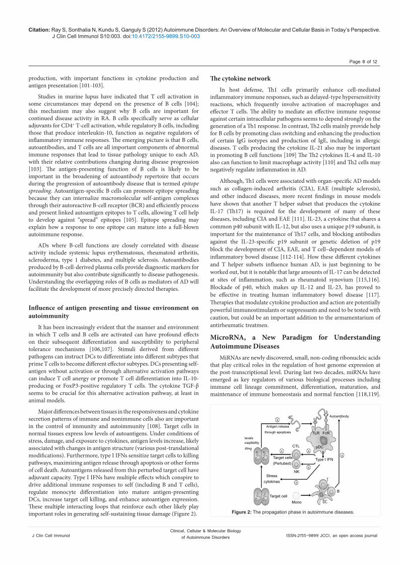
Major differences between tissues in the responsiveness and cytokine secretion patterns of immune and nonimmune cells also are important in the control of immunity and autoimmunity [108]. Target cells in normal tissues express low levels of autoantigens. Under conditions of stress, damage, and exposure to cytokines, antigen levels increase, likely associated with changes in antigen structure (various post-translational modifications). Furthermore, type I IFNs sensitize target cells to killing pathways, maximizing antigen release through apoptosis or other forms of cell death. Autoantigens released from this perturbed target cell have adjuvant capacity. Type I IFNs have multiple effects which conspire to drive additional immune responses to self (including B and T cells), regulate monocyte differentiation into mature antigen-presenting DCs, increase target cell killing, and enhance autoantigen expression. These multiple interacting loops that reinforce each other likely play important roles in generating self-sustaining tissue damage (Figure 2).
The cytokine network
Antigen release
through apoptosis
CTL
TLR FcR
pDC
NK
Autoantibody
Type I IFN
B
Ag levelsSusceptibility
to killing
Target cells(Pertubed)
Stresscytokines
Target cellMono
Figure 2: The propagation phase in autoimmune diseases.
Studies in murine lupus have indicated that T cell activation in some circumstances may depend on the presence of B cells [104]; this mechanism may also suggest why B cells are important for continued disease activity in RA. B cells specifically serve as cellular adjuvants for CD4+ T-cell activation, while regulatory B cells, including those that produce interleukin-10, function as negative regulators of inflammatory immune responses. The emerging picture is that B cells, autoantibodies, and T cells are all important components of abnormal immune responses that lead to tissue pathology unique to each AD, with their relative contributions changing during disease progression [103]. The antigen-presenting function of B cells is likely to be important in the broadening of autoantibody repertoire that occurs during the progression of autoantibody disease that is termed epitope spreading. Autoantigen-specific B cells can promote epitope spreading because they can internalize macromolecular self-antigen complexes through their autoreactive B-cell receptor (BCR) and efficiently process and present linked autoantigen epitopes to T cells, allowing T cell help to develop against “spread” epitopes [105]. Epitope spreading may explain how a response to one epitope can mature into a full-blown autoimmune response.
ADs where B-cell functions are closely correlated with disease activity include systemic lupus erythematosus, rheumatoid arthritis, scleroderma, type 1 diabetes, and multiple sclerosis. Autoantibodies produced by B-cell-derived plasma cells provide diagnostic markers for autoimmunity but also contribute significantly to disease pathogenesis. Understanding the overlapping roles of B cells as mediators of AD will facilitate the development of more precisely directed therapies.
production, with important functions in cytokine production and antigen presentation [101-103].
In host defense, Th1 cells primarily enhance cell-mediated inflammatory immune responses, such as delayed-type hypersensitivity reactions, which frequently involve activation of macrophages and effector T cells. The ability to mediate an effective immune response against certain intracellular pathogens seems to depend strongly on the generation of a Th1 response. In contrast, Th2 cells mainly provide help for B cells by promoting class switching and enhancing the production of certain IgG isotypes and production of IgE, including in allergic diseases. T cells producing the cytokine IL-21 also may be important in promoting B cell functions [109] The Th2 cytokines IL-4 and IL-10 also can function to limit macrophage activity [110] and Th2 cells may negatively regulate inflammation in AD.
Although, Th1 cells were associated with organ-specific AD models such as collagen-induced arthritis (CIA), EAE (multiple sclerosis), and other induced diseases, more recent findings in mouse models have shown that another T helper subset that produces the cytokine IL-17 (Th17) is required for the development of many of these diseases, including CIA and EAE [111]. IL-23, a cytokine that shares a common p40 subunit with IL-12, but also uses a unique p19 subunit, is important for the maintenance of Th17 cells, and blocking antibodies against the IL-23-specific p19 subunit or genetic deletion of p19 block the development of CIA, EAE, and T cell–dependent models of inflammatory bowel disease [112-114]. How these different cytokines and T helper subsets influence human AD, is just beginning to be worked out, but it is notable that large amounts of IL-17 can be detected at sites of inflammation, such as rheumatoid synovium [115,116]. Blockade of p40, which makes up IL-12 and IL-23, has proved to be effective in treating human inflammatory bowel disease [117]. Therapies that modulate cytokine production and action are potentially powerful immunostimulants or suppressants and need to be tested with caution, but could be an important addition to the armamentarium of antirheumatic treatmen.
MicroRNA, a New Paradigm for Understanding Autoimmune Diseases
MiRNAs are newly discovered, small, non-coding ribonucleic acids that play critical roles in the regulation of host genome expression at the post-transcriptional level. During last two decades, miRNAs have emerged as key regulators of various biological processes including immune cell lineage commitment, differentiation, maturation, and maintenance of immune homeostasis and normal function [118,119].

Citation: Ray S, Sonthalia N, Kundu S, Ganguly S (2012) Autoimmune Disorders: An Overview of Molecular and Cellular Basis in Today’s Perspective. J Clin Cell Immunol S10:003. doi:10.4172/2155-9899.S10-003
Page 9 of 12
J Clin Cell Immunol ISSN:2155-9899 JCCI, an open access journal Clinical, Cellular & Molecular Biology
of Autoimmune Disorders
This rapidly emerging field has revolutionized our understanding of normal immunoregulation and breakdown of self-tolerance. The powerful gene regulatory role of miRNAs is now well recognized. The expression and function of miRNAs are essential for the development of diverse physiological systems and the maintenance of the cellular homeostasis and normal function [120,121]. The field of miRNA research gained widespread attention with the recognition of aberrant expression and/or function of miRNAs in a broad range of ADs [122-124].
new molecular and cellular strategies for control and manipulation of autoimmune responses and diseases. The remarkable increase in information regarding the immune system, and the genetic basis of complex traits, is likely to accelerate the pace of our understanding of human autoimmunity in the near future. Major progresses have been made in understanding of miRNA biology, as well as obtaining insights into its role in pathogenesis of ADs. We anticipate that, the advances made by the application of novel and high-throughput technologies to the analysis of diseased tissues, including miRNA and the autoantibody repertoire, and the development of novel effective miRNA-based gene
References
1. Davidson A, Diamond B (2006) General features of autoimmune disease. In: Rose NR, Mackay IR (eds) The Autoimmune Diseases. Elsevier, St Louis: 25-36.
2. von Mühlen CA, Tan EM (1995) Autoantibodies in the diagnosis of systemic rheumatic diseases. Semin Arthritis Rheum 24: 323-358.
3. Arbuckle MR, McClain MT, Rubertone MV, Scofield RH, Dennis GJ, et al. (2003) Development of autoantibodies before the clinical onset of systemic lupus erythematosus. N Engl J Med 349: 1526-1533.
4. Marshak-Rothstein A (2006) Toll-like receptors in systemic autoimmune disease. Nat Rev Immunol 6: 823-835.
5. Nielen MM, van Schaardenburg D, Reesink HW, van de Stadt RJ, van der Horst-Bruinsma IE, et al. (2004) Specific autoantibodies precede the symptoms of rheumatoid arthritis: a study of serial measurements in blood donors. Arthritis Rheum 50: 380-386.
6. Morahan G, Morel L (2002) Genetics of autoimmune diseases in humans and in animal models. Curr Opin Immunol 14: 803-811.
7. Adorini L, Gregori S, Harrison LC (2002) Understanding autoimmune diabetes: insights from mouse models. Trends Mol Med 8: 31-38.
8. Christadoss P, Lennon VA, Krco CJ, David CS (1982) Genetic control of experimental autoimmune myasthenia gravis in mice. III. Ia molecules mediate cellular immune responsiveness to acetylcholine receptors. J Immunol 128: 1141-1144.
9. Dooley MA, Hogan SL (2003) Environmental epidemiology and risk factors for autoimmune disease. Curr Opin Rheumatol 15: 99-103.
10. Reeves WH, Lee PY, Weinstein JS, Satoh M, Lu L (2009) Induction of autoimmunity by pristane and other naturally occurring hydrocarbons. Trends
Diseases miRNAs Pathogenic contribution Cells
Systemic lupus erythematosus [24,25,129]
miR-146a Targets STAT-1 and IRF-5, negative regulator of Type I IFN pathway PBMCsmiR-148a Target DNMT1 directly and indirectly, induces DNA hypomethylation and the
expression autoimmune-associated genesCD4+ T cells
miR-125a Targets KLF13, Negative regulator of inflammatorychemokine RANTES
PBMCs
miR-21 Target RAS, induces DNA hypomethylation CD4+ T cells
Rheumatoid Arthritis [130-134]
miR-146a Targets FAF1, negative regulator of T cell apoptosis PBMC, CD4+ T cells, Th-17 cells, synovial fibroblasts
miR-155 Targets Matrix metalloproteinase(MMP)-3/1 in RASFs, Regulation ofinflammation and potentially involved in RASFs mediated tissue damages
PBMC, Th-17 cell, synovial fibroblasts
miR-124a Targets cyclin-dependent kinase 2 (CDK-2) and chemokine MCP-1, negative regulator of cell proliferation and MCP-1 secretion
synoviocytes
Multiple sclerosis [135-138]miR-326 Targets Ets-1, Promotes Th-17 cell
differentiation CD4+ T cells
miR-17-5P, miR-20a Potentially involved in the regulation of T cell activation CD4+ T cellsmiR-34a, miR-155 and miR-326
Targets CD47, promotes phagocytosis of myelin by releasing macrophage from inhibitory signaling
MS lesion
Abbreviation: PBMC: Peripheral Blood Mononuclear Cell; STAT: Signal Transducer and Activator of Transcription; IFN: Interferon; IRF-5: Interferon Regulatory Factor 5; KLF13: Kruppel-Like Factor 13; DNMT1: DNA methyltransferase 1; RANTES: Regulated upon Activation Normal T-cell Expressed and Secreted; FAF1: FAS-Associated Factor 1; RASFs: Rheumatoid Arthritis Synovial Fibroblasts; MCP 1: Monocyte Chemoattractant Protein 1
Table 5: MiRNAs in human inflammatory autoimmune diseases.
With the increased recognition that miRNAs are capable of controlling the immune cell development and function [125-128], it is conceivable that dysregulated miRNA expression will lead to the immune tolerance breakdown and the development of ADs. Moreover, the unique dysregulated miRNA expression patterns have been identified in human patients with SLE, RA, and MS. Table 5 illustrates selected AD-related miRNAs that have been shown to play critical pathogenic roles in the development of these diseases [24,25,129-138].
The importance of miRNAs to immune system maintenance and autoimmunity is now becoming increasingly clear, owing to concerted efforts invested on mechanistic insight into miRNA roles in AD pathogenesis during the past several years. These findings led to the identification and characterization of numerous novel miRNAs and thereby open up a new perspective on functional mechanism of autoimmune pathogenesis and highlight the possibility of miRNA-based disease interventions.
Conclusion and Future DirectionThe development of autoimmune disorder is a complex process.
The main molecular and cellular mechanisms of autoimmune responses and their origins are numerous and diverse. Although knowledge regarding different aspects of the immunopathogenesis of these disorders, especially related to animal studies, has advanced dramatically in recent years, major gaps in knowledge of human AD pathogenesis persist. The cellular immunologic abnormalities involved in the initiation and perpetuation of disease also need much greater definition. Better understanding of these processes would also provide
therapies will make the future of this field very bright.

Citation: Ray S, Sonthalia N, Kundu S, Ganguly S (2012) Autoimmune Disorders: An Overview of Molecular and Cellular Basis in Today’s Perspective. J Clin Cell Immunol S10:003. doi:10.4172/2155-9899.S10-003
Page 10 of 12
J Clin Cell Immunol ISSN:2155-9899 JCCI, an open access journal Clinical, Cellular & Molecular Biology
of Autoimmune Disorders
Immunol 30: 455-464.
11. Bramwell B (1914) Diffuse sclerodermia: its frequency; its occurrence in stone masons: its treatment by fibrolysis in elevations of temperature due to fibrolysis injections. Edinburgh Medical Journal 12: 387.
12. Sanchez-Roman J, Wichmann I, Salaberri J, Varela JM, Nuñez-Roldan A (1993) Multiple clinical and biological autoimmune manifestations in 50 workers after occupational exposure to silica. Ann Rheum Dis 52: 534-538.
13. Hayashi H (2010) Peripheral regulatory T cells from silicosis patients are susceptible to CD95-mediated apoptosis. Kawasaki Medical Journal 36:13-21.
14. Bach JF (2002) The effect of infections on susceptibility to autoimmune and allergic diseases. N Engl J Med 347: 911-920.
15. Wucherpfennig KW (2001) Mechanisms for the induction of autoimmunity by infectious agents. J Clin Invest 108: 1097-1104.
16. Whitacre CC (2001) Sex differences in autoimmune disease. Nat Immunol 2: 777-780.
17. Ansar Ahmed S, Penhale WJ, Talal N (1985) Sex hormones, immune responses, and autoimmune diseases. Mechanisms of sex hormone action. Am J Pathol 121: 531-551.
18. Ahmed SA, Hissong BD, Verthelyi D, Donner K, Becker K, et al. (1999) Gender and risk of autoimmune diseases: possible role of estrogenic compounds. Environ Health Perspect 107: 681-686.
19. Progress in Autoimmune Disease Research. Report to Congress (2005) The Autoimmune Diseases Coordinating Committee, National Institute of Allergy and Infectious Diseases, National Institutes of Health.
20. Roubinian JR, Talal N, Greenspan JS, Goodman JR, Siiteri PK (1978) Effect of castration and sex hormone treatment on survival, anti-nucleic acid antibodies, and glomerulonephritis in NZB/NZW F1 mice. J Exp Med 147: 1568-1583.
21. Roubinian J, Talal N, Siiteri PK, Sadakian JA (1979) Sex hormone modulation of autoimmunity in NZB/NZW mice. Arthritis Rheum 22: 1162-1169.
22. Grimaldi CM, Cleary J, Dagtas AS, Moussai D, Diamond B (2002) Estrogen alters thresholds for B cell apoptosis and activation. J Clin Invest 109: 1625-1633.
23. Bynoté KK, Hackenberg JM, Korach KS, Lubahn DB, Lane PH, et al. (2008) Estrogen receptor-alpha deficiency attenuates autoimmune disease in (NZB x NZW)F1 mice. Genes Immun 9: 137-152.
24. Tang Y, Luo X, Cui H, Ni X, Yuan M, et al. (2009) MicroRNA-146A contributes to abnormal activation of the type I interferon pathway in human lupus by targeting the key signaling proteins. Arthritis Rheum 60: 1065-1075.
25. Pan W, Zhu S, Yuan M, Cui H, Wang L, et al. (2010) MicroRNA-21 and microRNA-148a contribute to DNA hypomethylation in lupus CD4+ T cells by directly and indirectly targeting DNA methyltransferase 1. J Immunol 184: 6773-6781.
26. Yazdanbakhsh M, Kremsner PG, van Ree R (2002) Allergy, parasites, and the hygiene hypothesis. Science 296: 490-494.
27. Okada H, Kuhn C, Feillet H, Bach JF (2010) The ‘hygiene hypothesis’ for autoimmune and allergic diseases: an update. Clin Exp Immunol 160: 1-9.
28. Albani S (1994) Infection and molecular mimicry in autoimmune diseases of childhood. Clin Exp Rheumatol 12 Suppl 10: S35-S41.
29. Fourneau JM, Bach JM, van Endert PM, Bach JF (2004) The elusive case for a role of mimicry in autoimmune diseases. Mol Immunol 40: 1095-1102.
30. James JA, Harley JB, Scofield RH (2006) Epstein–Barr virus and systemic lupus erythematosus. Curr Opin Rheumatol 18: 462-467.
31. Blank M, Barzilai O, Shoenfeld Y (2007) Molecular mimicry and auto-immunity. Clin Rev Allergy Immunol 32: 111-118.
32. Powell AM, Black MM (2001) Epitope spreading: protection from pathogens, but propagation of autoimmunity? Clin Exp Dermatol 26: 427-433.
33. Bonsor DA, Grishkovskaya I, Dodson EJ, Kleanthous C (2007) Molecular mimicry enables competitive recruitment by a natively disordered protein. J Am Chem Soc 129: 4800-4807.
34. Turley SJ (2002) Dendritic cells: inciting and inhibiting autoimmunity. Curr Opin Immunol 14: 765-770.
35. Pasare C, Medzhitov R (2003) Toll-like receptors: balancing host resistance with immune tolerance. Curr Opin Immunol 15: 677-682.
36. Christensen SR, Shupe J, Nickerson K, Kashgarian M, Flavell RA, et al. (2006) Toll-like receptor 7 and TLR9 dictate autoantibody specificity and have opposing inflammatory and regulatory roles in a murine model of lupus. Immunity 25: 417-428.
37. Millar DG, Garza KM, Odermatt B, Elford AR, Ono N, et al. (2003) Hsp70 promotes antigen-presenting cell function and converts T-cell tolerance to autoimmunity in vivo. Nat Med 9: 1469-1476.
38. Torres BA, Johnson HM (1998) Modulation of disease by superantigens. Curr Opin Immunol 10: 465-470.
39. Merriman TR, Pearce SH (2006) Genetic progress towards the molecular basis of common autoimmunity. Discov Med 6: 40-45.
40. Serrano NC, Millan P, Páez MC (2006) Non-HLA associations with autoimmune diseases. Autoimmun Rev 5: 209-214.
41. Fruman DA, Walsh CM (2007) Signal transduction and autoimmunity: introduction. Autoimmunity 40: 403-404.
42. Michou L, Lasbleiz S, Rat AC, Migliorini P, Balsa A, et al. (2007) Linkage proof for PTPN22, a rheumatoid arthritis susceptibility gene and a human autoimmunity gene. Proc Natl Acad Sci U S A 104: 1649-1654.
43. Viganó P, Lattuada D, Somigliana E, Abbiati A, Candiani M, et al. (2005) Variants of the CTLA4 gene that segregate with autoimmune diseases are not associated with endometriosis. Mol Hum Reprod 11: 745-749.
44. Chu EB, Hobbs MV, Wilson CB, Romball CG, Linsley PS, et al. (1996) Intervention of CD4+ cell subset shifts and autoimmunity in the BXSB mouse by murine CTLA4Ig. J Immunol 156: 1262-1268.
45. Sigal LH (2006) Basic science for the clinician 37: Protecting against autoimmunity-tolerance: mechanisms of negative selection in the thymus. J Clin Rheumatol 12: 99-101.
46. Park Y (2007) Functional evaluation of the type 1 diabetes (T1D) susceptibility candidate genes. Diabetes Res Clin Pract 77 Suppl 1: S110-115.
47. Howson JM, Dunger DB, Nutland S, Stevens H, Wicker LS, et al. (2007) A type 1 diabetes subgroup with a female bias is characterised by failure in tolerance to thyroid peroxidase at an early age and a strong association with the cytotoxic T-lymphocyte-associated antigen-4 gene. Diabetologia 50: 741-746.
48. Donaldson P, Veeramani S, Baragiotta A, Floreani A, Venturi C, et al. (2007) Cytotoxic T-lymphocyte-associated antigen-4 single nucleotide polymorphisms and haplotypes in primary biliary cirrhosis. Clin Gastroenterol Hepatol 5: 755-760.
49. Magyari L, Faragó B, Bene J, Horvatovich K, Lakner L, et al. (2007) No association of the cytotoxic T-lymphocyte associated gene CTLA4 +49A/G polymorphisms with Crohn’s disease and ulcerative colitis in Hungarian population samples. World J Gastroenterol 13: 2205-2208.
50. Vang T, Miletic AV, Bottini N, Mustelin T (2007) Protein tyrosine phosphatase PTPN22 in human autoimmunity. Autoimmunity 40: 453-461.
51. Burkhardt H, Hüffmeier U, Spriewald B, Böhm B, Rau R, et al. (2006) Association between protein tyrosine phosphatase 22 variant R620W in conjunction with the HLA-DRB1 shared epitope and humoral autoimmunity to an immunodominant epitope of cartilage-specific type II collagen in early rheumatoid arthritis. Arthritis Rheum 54: 82-89.
52. Chelala C, Duchatelet S, Joffret ML, Bergholdt R, Dubois-Laforgue D, et al. (2007) PTPN22 R620W functional variant in type 1 diabetes and autoimmunity related traits. Diabetes 56: 522-526.
53. Chung SA, Criswell LA (2007) PTPN22: its role in SLE and autoimmunity. Autoimmunity 40: 582-590.
54. Gambineri E, Torgerson TR, Ochs HD (2003) Immune dysregulation, polyendocrinopathy, enteropathy, and X-linked inheritance (IPEX), a syndrome of systemic autoimmunity caused by mutations of FOXP3, a critical regulator of T-cell homeostasis. Curr Opin Rheumatol 15: 430-435.
55. Matsuzaki T, Takagi A, Ikemura H, Matsuguchi T, Yokokura T (2007) Intestinal microflora: probiotics and autoimmunity. J Nutr 137: 798S-802S.
56. Kastelan S, Zjacić-Rotkvić V, Kastelan Z (2007) Could diabetic retinopathy be an autoimmune disease? Med Hypotheses 68: 1016-1018.

Citation: Ray S, Sonthalia N, Kundu S, Ganguly S (2012) Autoimmune Disorders: An Overview of Molecular and Cellular Basis in Today’s Perspective. J Clin Cell Immunol S10:003. doi:10.4172/2155-9899.S10-003
Page 11 of 12
J Clin Cell Immunol ISSN:2155-9899 JCCI, an open access journal Clinical, Cellular & Molecular Biology
of Autoimmune Disorders
57. Quadbeck B, Stucke M, Eckstein AK, Heise DJ, Mann K, et al. (2006) Dysregulation of TNF/TNFR superfamily members: a systemic link between intra- and extrathyroidal manifestations in Graves’ disease. Scand J Immunol 64: 523-530.
58. Martin DA, Elkon KB (2006) Intracellular mammalian DNA stimulates myeloid dendritic cells to produce type I interferons predominantly through a toll-like receptor 9-independent pathway. Arthritis Rheum 54: 951-962.
59. Pitkänen J, Peterson P (2003) Autoimmune regulator: from loss of function to autoimmunity. Genes Immun 4: 12-21.
60. Larsen F, Madsen HO, Sim RB, Koch C, Garred P (2004) Disease-associated mutations in human mannose-binding lectin compromise oligomerization and activity of the final protein. J Biol Chem 279: 21302-21311.
61. Engelhorn ME, Guevara-Patiño JA, Noffz G, Hooper AT, Lou O, et al. (2006) Autoimmunity and tumor immunity induced by immune responses to mutations in self. Nat Med 12: 198-206.
62. Doyle HA, Mamula MJ (2002) Posttranslational protein modifications: new flavors in the menu of autoantigens. Curr Opin Rheumatol 14: 244-249.
63. Doyle HA, Mamula MJ (2005) Posttranslational modifications of self-antigens. Ann N Y Acad Sci 1050: 1-9.
64. Utz PJ, Anderson P (1998) Posttranslational protein modifications, apoptosis, and the bypass of tolerance to autoantigens. Arthritis Rheum 41: 1152-1160.
65. Rosen A, Casciola-Rosen L (1999) Autoantigens as substrates for apoptotic proteases: implications for the pathogenesis of systemic autoimmune disease. Cell Death Differ 6: 6-12.
66. Mamula MJ, Gee RJ, Elliott JI, Sette A, Southwood S, et al. (1999) Isoaspartyl post-translational modification triggers autoimmune responses to self-proteins. J Biol Chem 274: 22321-22327.
67. Yang ML, Doyle HA, Gee RJ, Lowenson JD, Clarke S, et al. (2006) Intracellular protein modification associated with altered T cell functions in autoimmunity. J Immunol 177: 4541-4549.
68. Yamada R, Suzuki A, Chang X, Yamamoto K (2005) Citrullinated proteins in rheumatoid arthritis. Front Biosci 10: 54-64.
69. Moscarello MA, Wood DD, Ackerley C, Boulias C (1994) Myelin in multiple sclerosis is developmentally immature. J Clin Invest 94: 146-154.
70. Anderton SM (2004) Post-translational modifications of self antigens: implications for autoimmunity. Curr Opin Immunol 16: 753-758.
71. Molberg O, Mcadam SN, Körner R, Quarsten H, Kristiansen C, et al. (1998) Tissue transglutaminase selectively modifies gliadin peptides that are recognized by gut-derived T cells in celiac disease. Nat Med 4: 713-717.
72. Berti C, Roncoroni L, Falini ML, Caramanico R, Dolfini E, et al. (2007) Celiac-related properties of chemically and enzymatically modified gluten proteins. J Agric Food Chem 55: 2482-2488.
73. Bouvet JP, Zouali M (2005) Silent antibodies. Arch Inst Pasteur Tunis 82: 3-8.
74. Macario AJ (1995) Heat-shock proteins and molecular chaperones: implications for pathogenesis, diagnostics, and therapeutics. Int J Clin Lab Res 25: 59-70.
75. Karopoulos C, Rowley MJ, Handley CJ, Strugnell RA (1995) Antibody reactivity to mycobacterial 65 kDa heat shock protein: relevance to autoimmunity. J Autoimmun 8: 235-248.
76. Prohászka Z (2007) Chaperones as part of immune networks. Adv Exp Med Biol 594: 159-166.
77. Routsias JG, Tzioufas AG (2006) The role of chaperone proteins in autoimmunity. Ann N Y Acad Sci 1088: 52-64.
78. Franz S, Gaipl US, Munoz LE, Sheriff A, Beer A, et al. (2006) Apoptosis and autoimmunity: when apoptotic cells break their silence. Curr Rheumatol Rep 8: 245-247.
79. Navratil JS, Liu CC, Ahearn JM (2006) Apoptosis and autoimmunity. Immunol Res 36: 3-12.
80. Hayashi T, Faustman D (2000) The role of the proteasome in autoimmunity. Diabetes Metab Res Rev 16: 325-337.
81. Decker P, Singh-Jasuja H, Haager S, Kötter I, Rammensee HG (2005) Nucleosome, the main autoantigen in systemic lupus erythematosus,
induces direct dendritic cell activation via a MyD88-independent pathway: consequences on inflammation. J Immunol 174: 3326-3334.
82. Gershwin ME (2008) The mosaic of autoimmunity. Autoimmun Rev 7: 161-163.
83. Sercarz EE, Lehmann PV, Ametani A, Benichou G, Miller A, et al. (1993) Dominance and crypticity of T cell antigenic determinants. Annu Rev Immunol 11: 729-766.
84. Lanzavecchia A (1995) How can cryptic epitopes trigger autoimmunity? J Exp Med 181: 1945-1948.
85. Casciola-Rosen L, Andrade F, Ulanet D, Wong WB, Rosen A (1999) Cleavage by granzyme B is strongly predictive of autoantigen status: implications for initiation of autoimmunity. J Exp Med 190: 815-826.
86. Bäcklund J, Carlsen S, Höger T, Holm B, Fugger L, et al. (2002) Predominant selection of T cells specific for the glycosylated collagen type II epitope (263-270) in humanized transgenic mice and in rheumatoid arthritis. Proc Natl Acad Sci USA 99: 9960-9965.
87. Topfer F, Gordon T, McCluskey J (1995) Intra- and intermolecular spreading of autoimmunity involving the nuclear self-antigens La (SS-B) and Ro (SS-A). Proc Natl Acad Sci U S A 92: 875-879.
88. Craft J, Fatenejad S (1997) Self antigens and epitope spreading in systemic autoimmunity. Arthritis Rheum 40: 1374-1382.
89. Ohashi PS, Oehen S, Buerki K, Pircher H, Ohashi CT, et al. (1991) Ablation of “tolerance” and induction of diabetes by virus infection in viral antigen transgenic mice. Cell 65: 305-317.
90. Piccirillo CA, Shevach EM (2004) Naturally-occurring CD4+CD25+ immunoregulatory T cells: central players in the arena of peripheral tolerance. Semin Immunol 16: 81-88.
91. Shevach EM (2004) Regulatory/suppressor T cells in health and disease. Arthritis Rheum 50: 2721-2724.
92. Zheng Y, Rudensky AY (2007) Foxp3 in control of the regulatory T cell lineage. Nat Immunol 8: 457-462.
93. Kim JM, Rasmussen JP, Rudensky AY (2007) Regulatory T cells prevent catastrophic autoimmunity throughout the lifespan of mice. Nat Immunol 8: 191-197.
94. Bennett CL, Christie J, Ramsdell F, Brunkow ME, Ferguson PJ, et al. (2001) The immune dysregulation, polyendocrinopathy, enteropathy, X-linked syndrome (IPEX) is caused by mutations of FOXP3. Nat Genet 27: 20-21.
95. de St Groth BF (2012) Regulatory T-cell abnormalities and the global epidemic of immuno-inflammatory disease. Immunol Cell Biol 90: 256-259.
96. Skapenko A, Lipsky PE, Schulze-Koops H (2006) T cell activation as starter and motor of rheumatic inflammation. Curr Top Microbiol Immunol 305: 195-211.
97. Bockenstedt LK, Gee RJ, Mamula MJ (1995) Self-peptides in the initiation of lupus autoimmunity. J Immunol 154: 3516-3524.
98. Goodnow CC, Cyster JG, Hartley SB, Bell SE, Cooke MP, et al. (1995) Self-tolerance checkpoints in B lymphocyte development. Adv Immunol 59: 279-368.
99. Anderson MS, Venanzi ES, Klein L, Chen Z, Berzins SP, et al. (2002) Projection of an immunological self shadow within the thymus by the aire protein. Science 298: 1395-1401.
100. Via CS, Shearer GM (1988) T-cell interactions in autoimmunity: insights from a murine model of graft-versus-host disease. Immunol Today 9: 207-213.
101. Chan OT, Madaio MP, Shlomchik MJ (1999) The central and multiple roles of B cells in lupus pathogenesis. Immunol Rev 169: 107-121.
102. Chan OT, Hannum LG, Haberman AM, Madaio MP, Shlomchik MJ (1999) A novel mouse with B cells but lacking serum antibody reveals an antibody-independent role for B cells in murine lupus. J Exp Med 189: 1639-1648.
103. Yanaba K, Bouaziz JD, Matsushita T, Magro CM, St Clair EW, et al. (2008) B-lymphocyte contributions to human autoimmune disease. Immunol Rev 223: 284-299.
104. Chan OT, Shlomchik MJ (2000) Cutting edge: B cells promote CD8+ T cell activation in MRL-Fas(lpr) mice independently of MHC class I antigen presentation. J Immunol 164: 1658-1662.

Citation: Ray S, Sonthalia N, Kundu S, Ganguly S (2012) Autoimmune Disorders: An Overview of Molecular and Cellular Basis in Today’s Perspective. J Clin Cell Immunol S10:003. doi:10.4172/2155-9899.S10-003
Page 12 of 12
J Clin Cell Immunol ISSN:2155-9899 JCCI, an open access journal Clinical, Cellular & Molecular Biology
of Autoimmune Disorders
105. Mamula MJ, Fatenejad S, Craft J (1994) B cells process and present lupus autoantigens that initiate autoimmune T cell responses. J Immunol 152: 1453-1461.
106. Steinman RM (2003) The control of immunity and tolerance by dendritic cell. Pathol Biol (Paris) 51: 59-60.
107. Banchereau J, Steinman RM (1998) Dendritic cells and the control of immunity. Nature 392: 245-252.
108. Raz E (2007) Organ-specific regulation of innate immunity. Nat Immunol 8: 3-4.
109. Leonard WJ, Spolski R (2005) Interleukin-21: a modulator of lymphoid proliferation, apoptosis and differentiation. Nat Rev Immunol 5: 688-698.
110. Oswald IP, Gazzinelli RT, Sher A, James SL (1992) IL-10 synergizes with IL-4 and transforming growth factor-beta to inhibit macrophage cytotoxic activity. J Immunol 148: 3578-3582.
111. Dong C (2006) Diversification of T-helper-cell lineages: finding the family root of IL-17-producing cells. Nat Rev Immunol 6: 329-333.
112. Kastelein RA, Hunter CA, Cua DJ (2007) Discovery and biology of IL-23 and IL-27: related but functionally distinct regulators of inflammation. Annu Rev Immunol 25: 221-242.
113. Oppmann B, Lesley R, Blom B, Timans JC, Xu Y, et al. (2000) Novel p19 protein engages IL-12p40 to form a cytokine, IL-23, with biological activities similar as well as distinct from IL-12. Immunity 13: 715-725.
114. Langrish CL, Chen Y, Blumenschein WM, Mattson J, Basham B, et al. (2005) IL-23 drives a pathogenic T cell population that induces autoimmune inflammation. J Exp Med 201: 233-240.
115. Koenders MI, Joosten LA, van den Berg WB (2006) Potential new targets in arthritis therapy: interleukin (IL)-17 and its relation to tumour necrosis factor and IL-1 in experimental arthritis. Ann Rheum Dis 65 Suppl 3: iii29-33.
116. Lubberts E, Joosten LA, Oppers B, van den Bersselaar L, Coenen-de Roo CJ, et al. (2001) IL-1-independent role of IL-17 in synovial inflammation and joint destruction during collagen-induced arthritis. J Immunol 167: 1004-1013.
117. Mannon PJ, Fuss IJ, Mayer L, Elson CO, Sandborn WJ, et al. (2004) Anti-interleukin-12 antibody for active Crohn’s disease. N Engl J Med 351: 2069-2079.
118. Lagos-Quintana M, Rauhut R, Lendeckel W, Tuschl T (2001) Identification of novel genes coding for small expressed RNAs. Science 294: 853-858.
119. Bartel DP (2004) MicroRNAs: genomics, biogenesis, mechanism, and function. Cell 116: 281-297.
120. Ambros V (2004) The functions of animal microRNAs. Nature 431: 350-355.
121. Neilson JR, Zheng GX, Burge CB, Sharp PA (2007) Dynamic regulation of miRNA expression in ordered stages of cellular development. Genes Dev 21: 578-589.
122. Baltimore D, Boldin MP, O’Connell RM, Rao DS, Taganov KD (2008) MicroRNAs: new regulators of immune cell development and function. Nat Immunol 9: 839-845.
123. Pauley KM, Cha S, Chan EK (2009) MicroRNA in autoimmunity and autoimmune diseases. J Autoimmun 32: 189-194.
124. Dai R, Ahmed SA (2011) MicroRNA, a new paradigm for understanding immunoregulation, inflammation, and autoimmune diseases. Transl Res 157: 163-179.
125. Xiao C, Rajewsky K (2009) MicroRNA control in the immune system: basic principles. Cell 136: 26-36.
126. Stittrich AB, Haftmann C, Sgouroudis E, Kühl AA, Hegazy AN, et al. (2010) The microRNA miR-182 is induced by IL-2 and promotes clonal expansion of activated helper T lymphocytes. Nat Immunol 11: 1057-1062.
127. Xiao C, Calado DP, Galler G, Thai TH, Patterson HC, et al. (2007) MiR-150 controls B cell differentiation by targeting the transcription factor c-Myb. Cell 131: 146-159.
128. Ooi AG, Sahoo D, Adorno M, Wang Y, Weissman IL, et al. (2010) MicroRNA-125b expands hematopoietic stem cells and enriches for the lymphoid-balanced and lymphoid-biased subsets. Proc Natl Acad Sci U S A 107: 21505-21510.
129. Zhao X, Tang Y, Qu B, Cui H, Wang S, et al. (2010) MicroRNA-125a contributes to elevated inflammatory chemokine RANTES levels via targeting KLF13 in systemic lupus erythematosus. Arthritis Rheum 62: 3425-3435.
130. Pauley KM, Satoh M, Chan AL, Bubb MR, Reeves WH, et al. (2008) Upregulated miR-146a expression in peripheral blood mononuclear cells from rheumatoid arthritis patients. Arthritis Res Ther 10: R101.
131. Stanczyk J, Pedrioli DM, Brentano F, Sanchez-Pernaute O, Kolling C, et al. (2008) Altered expression of MicroRNA in synovial fibroblasts and synovial tissue in rheumatoid arthritis. Arthritis Rheum 58: 1001-1009.
132. Murata K, Yoshitomi H, Tanida S, Ishikawa M, Nishitani K, et al. (2010) Plasma and synovial fluid microRNAs as potential biomarkers of rheumatoid arthritis and osteoarthritis. Arthritis Res Ther 12: R86.
133. Nakamachi Y, Kawano S, Takenokuchi M, Nishimura K, Sakai Y, et al. (2009) MicroRNA-124a is a key regulator of proliferation and monocyte chemoattractant protein 1 secretion in fibroblast-like synoviocytes from patients with rheumatoid arthritis. Arthritis Rheum 60: 1294-1304.
134. Niimoto T, Nakasa T, Ishikawa M, Okuhara A, Izumi B, et al. (2010) MicroRNA-146a expresses in interleukin-17 producing T cells in rheumatoid arthritis patients. BMC Musculoskelet Disord 11: 209.
135. Du C, Liu C, Kang J, Zhao G, Ye Z, et al. (2009) MicroRNA miR-326 regulates TH-17 differentiation and is associated with the pathogenesis of multiple sclerosis. Nat Immunol 10: 1252-1259.
136. Junker A, Krumbholz M, Eisele S, Mohan H, Augstein F, et al. (2009) MicroRNA profiling of multiple sclerosis lesions identifies modulators of the regulatory protein CD47. Brain 132: 3342-3352.
137. Lindberg RL, Hoffmann F, Mehling M, Kuhle J, Kappos L (2010) Altered expression of miR-17-5p in CD4+ lymphocytes of relapsing-remitting multiple sclerosis patients. Eur J Immunol 40: 888-898.
138. Cox MB, Cairns MJ, Gandhi KS, Carroll AP, Moscovis S, et al. (2010) MicroRNAs miR-17 and miR-20a inhibit T cell activation genes and are under-expressed in MS whole blood. PLoS One 5: e12132.
Submit your next manuscript and get advantages of OMICS Group submissions
Unique features:
• Userfriendly/feasiblewebsite-translationofyourpaperto50world’sleadinglanguages• AudioVersionofpublishedpaper• DigitalarticlestoshareandexploreSpecial features:
• 200OpenAccessJournals• 15,000editorialteam• 21daysrapidreviewprocess• Qualityandquickeditorial,reviewandpublicationprocessing• IndexingatPubMed(partial),Scopus,DOAJ,EBSCO,IndexCopernicusandGoogleScholaretc• SharingOption:SocialNetworkingEnabled• Authors,ReviewersandEditorsrewardedwithonlineScientificCredits• Betterdiscountforyoursubsequentarticles
Submityourmanuscriptat:www.editorialmanager.com/clinicalgroup
Thisarticlewasoriginallypublishedinaspecialissue,Clinical, Cellular & Molecular Biology of Autoimmune Disorders handledbyEditor(s).Dr.AbdulRahmanAsif,George-August-University,Germany