Embed Size (px)
Citation preview
We always have lots of interesting student projects available, for students in physics, mechanical and electrical engineering, image processing, optics, cell biology, cardiology etc.
Below, you can find a list of current projects - but if you feel inspired by our work and want to work at the cutting edge of biomedical engineering research, please let us know your educational background and field of interest and we will inform you on possible projects. Feel free to contact us!
For more information, you can contact the persons mentioned on each project description, or contact Hans Bosch, PhD, tel.: 010-7038088, e-mail: [email protected].
Ultrasound Contrast Agents
Probe development
3D ultrasound & image processing
Vascular biomechanics
Innovative applications
Measuring the radius of vibrating contrast bubbles in high-speed camera opticalimagesImplementation of repetition rate excitation
Design of a transducer driver circuit for a handheld ultrasound systemThe effect of an acoustic lens on the acoustic field transmitted by an unfocessedaxisymmetric single element transducerMeasurement of nonlinearity parameter of urine for a new bladder volume assessment methodEffect of backing geometry on transducer performanceUsing chirps for superharmonic imaging
Artifact Detection in three-dimensional ultrasound images of the heartRobust appearance modeling of the heart in three-dimensional ultrasound images4D interpolation of sparse, irregularly sampled ultrasound imagesAutomated analysis of 3D cardiac ultrasound images with contrast3D fast beam formation with good image quality
Model-based estimation of the outflow conditions of coronary arteries for patient specific shear stress computationsWall shear stress distribution in realistic human coronary Modelling of plaque ruptureMR imaging of vulnerable plaques: pressure induced deformation Relation between blood flow induced shear stress and plaque raptureRelationship between shear stress and plaque composition in human carotidbifurcationsChanges in wall shear stress distribution in human carotid bifurcations due to medical treatmentInfluence of CTA segmentation methods on the wall shear stress distribution in human carotid bifurcations
Tissue tracking on tendons in the hand using high-frequency ultrasound imagingModeling of Intravascular Photo-acoustic imaging
Student Projects
Erasmus MC : Student Projects
Measuring the radius of vibrating contrast bubbles in high-speed camera optical images MASTER'S PROJECT ASSIGNMENT
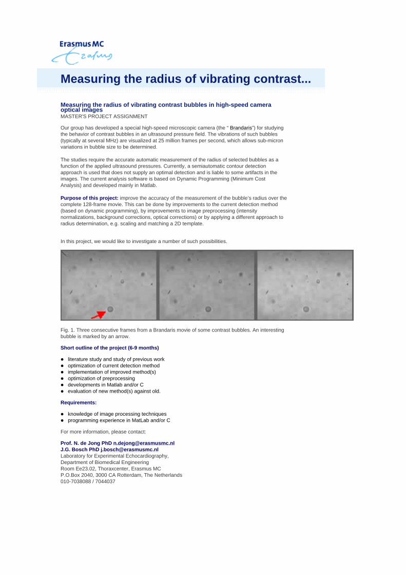
Our group has developed a special high-speed microscopic camera (the “ Brandaris”) for studying the behavior of contrast bubbles in an ultrasound pressure field. The vibrations of such bubbles (typically at several MHz) are visualized at 25 million frames per second, which allows sub-micron variations in bubble size to be determined. The studies require the accurate automatic measurement of the radius of selected bubbles as a function of the applied ultrasound pressures. Currently, a semiautomatic contour detection approach is used that does not supply an optimal detection and is liable to some artifacts in the images. The current analysis software is based on Dynamic Programming (Minimum Cost Analysis) and developed mainly in Matlab. Purpose of this project: improve the accuracy of the measurement of the bubble’s radius over the complete 128-frame movie. This can be done by improvements to the current detection method (based on dynamic programming), by improvements to image preprocessing (intensity normalizations, background corrections, optical corrections) or by applying a different approach to radius determination, e.g. scaling and matching a 2D template.
In this project, we would like to investigate a number of such possibilities.
Fig. 1. Three consecutive frames from a Brandaris movie of some contrast bubbles. An interesting bubble is marked by an arrow.
Short outline of the project (6-9 months)
Requirements:
For more information, please contact:
Prof. N. de Jong PhD [email protected] J.G. Bosch PhD [email protected] Laboratory for Experimental Echocardiography, Department of Biomedical Engineering Room Ee23.02, Thoraxcenter, Erasmus MC P.O.Box 2040, 3000 CA Rotterdam, The Netherlands 010-7038088 / 7044037
literature study and study of previous work optimization of current detection method implementation of improved method(s) optimization of preprocessing developments in Matlab and/or C evaluation of new method(s) against old.
knowledge of image processing techniques programming experience in MatLab and/or C
Measuring the radius of vibrating contrast...
Implementation of Pulse detection schemes in a research scanner Master’s project assignment
Introduction:
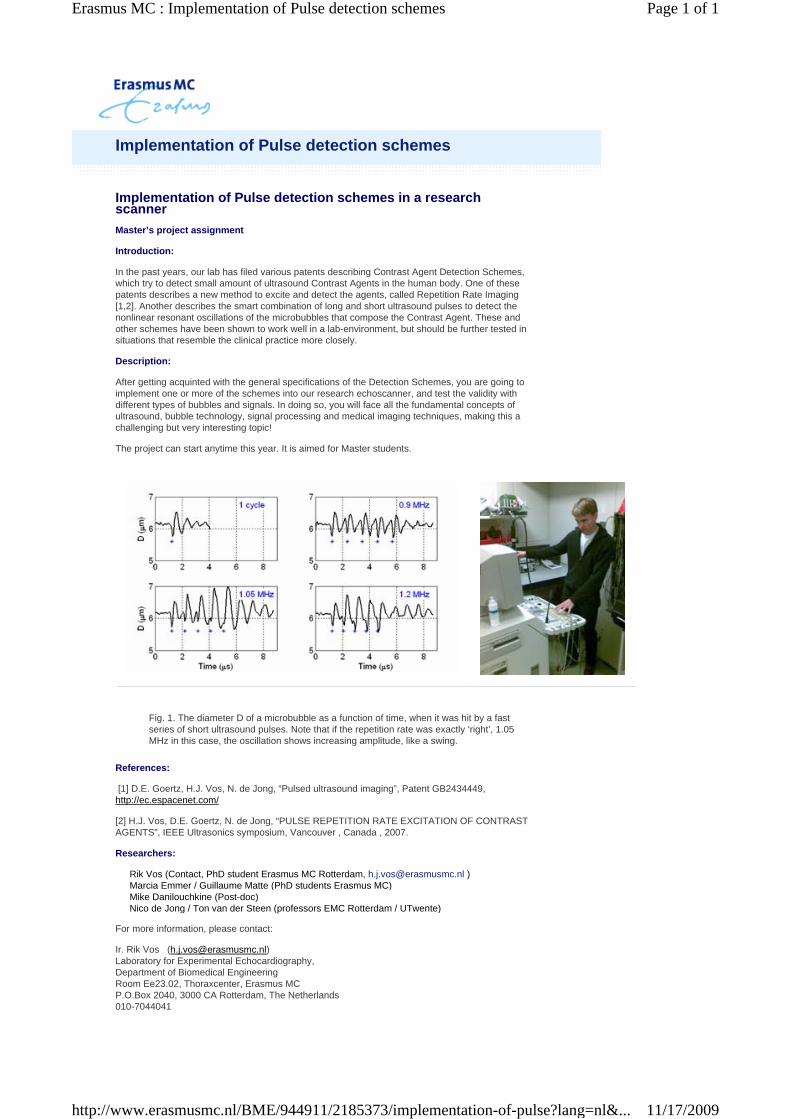
In the past years, our lab has filed various patents describing Contrast Agent Detection Schemes, which try to detect small amount of ultrasound Contrast Agents in the human body. One of these patents describes a new method to excite and detect the agents, called Repetition Rate Imaging [1,2]. Another describes the smart combination of long and short ultrasound pulses to detect the nonlinear resonant oscillations of the microbubbles that compose the Contrast Agent. These and other schemes have been shown to work well in a lab-environment, but should be further tested in situations that resemble the clinical practice more closely.
Description:
After getting acquinted with the general specifications of the Detection Schemes, you are going to implement one or more of the schemes into our research echoscanner, and test the validity with different types of bubbles and signals. In doing so, you will face all the fundamental concepts of ultrasound, bubble technology, signal processing and medical imaging techniques, making this a challenging but very interesting topic!
The project can start anytime this year. It is aimed for Master students.
Fig. 1. The diameter D of a microbubble as a function of time, when it was hit by a fast series of short ultrasound pulses. Note that if the repetition rate was exactly ‘right’, 1.05 MHz in this case, the oscillation shows increasing amplitude, like a swing.
References:
[1] D.E. Goertz, H.J. Vos, N. de Jong, “Pulsed ultrasound imaging”, Patent GB2434449, http://ec.espacenet.com/
[2] H.J. Vos, D.E. Goertz, N. de Jong, “PULSE REPETITION RATE EXCITATION OF CONTRAST AGENTS”, IEEE Ultrasonics symposium, Vancouver , Canada , 2007.
Researchers:
For more information, please contact:
Ir. Rik Vos ([email protected]) Laboratory for Experimental Echocardiography, Department of Biomedical Engineering Room Ee23.02, Thoraxcenter, Erasmus MC P.O.Box 2040, 3000 CA Rotterdam, The Netherlands 010-7044041
Rik Vos (Contact, PhD student Erasmus MC Rotterdam, [email protected] ) Marcia Emmer / Guillaume Matte (PhD students Erasmus MC) Mike Danilouchkine (Post-doc) Nico de Jong / Ton van der Steen (professors EMC Rotterdam / UTwente)
Implementation of Pulse detection schemes
Page 1 of 1Erasmus MC : Implementation of Pulse detection schemes
11/17/2009http://www.erasmusmc.nl/BME/944911/2185373/implementation-of-pulse?lang=nl&...
Design of a transducer driver circuit for a handheld ultrasound system. MASTER'S PROJECT ASSIGNMENT
Background: In several clinical situations, i.e. the intensive care, the recovery room, and the urology department, it is often necessary to know the bladder filling. Catheterisation is the golden standard for bladder volume assessment due to its accuracy and reliability. However, it has major disadvantages. Besides the fact that catheterisation is not comfortable for the patient, it is invasive and might produce infections and traumas. In an attempt to reduce the number of catheterisations, thus reducing the chance of infections, alternative measurement techniques are addressed. One possibility is the use of ultrasound.
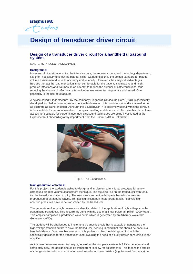
A device called “Bladderscan™” by the company Diagnostic Ultrasound Corp. (DxU) is specifically developed for bladder volume assessment with ultrasound. It is non-invasive and is claimed to be as accurate as catheterisation. Although the BladderScan™ is extremely useful within the clinic, it is less suitable for personal use due to complex handling and device cost. To make bladder volume assessment suitable for personal use, new ultrasound techniques are being investigated at the Experimental Echocardiography department from the ErasmusMC in Rotterdam.
Fig. 1. The Bladderscan.
Main graduation activities: For this project, the student is asked to design and implement a functional prototype for a new ultrasound bladder volume assessment technique. The focus will be on the transducer front-end, i.e. the transducer driver circuitry. The new measurement technique is based on non-linear propagation of ultrasound waves. To have significant non-linear propagation, relatively high acoustic pressures have to be transmitted by the transducer.
The generation of very high pressures is directly related to the application of high voltages on the transmitting transducer. This is currently done with the use of a linear power amplifier (1000 Watts). This amplifier amplifies a predefined waveburst, which is generated by an Arbitrary Waveform Generator (AWG).
The student will be challenged to implement a transmit circuit that is capable of generating the high-voltage transmit bursts to drive the transducer, bearing in mind that this should be done in a handheld device. One possible solution to this problem is that the driving circuit should be specifically designed for the transducer used, avoiding the need of a bulky power-consuming linear amplifier.
As the volume measurement technique, as well as the complete system, is fully experimental and completely new, the design should be transparent to allow for adjustments. This means the effects of changes in transducer specifications and waveform characteristics (e.g. transmit frequency) on
Design of transducer driver circuit
=nl&view=Print

the performance of the circuit should be easily interpreted and should allow for quick redesign.
Since a first functional prototype is almost operational and clinical experiments are pending, the driving circuit is of great importance to the project. Hence, implementation and testing of the design is an important part of this project.
During this project the student will gain knowledge of medical ultrasound applications and perform several acoustic measurements.
Requirements:
* discipline: Microelectronics
For more information, please contact:
E. Merks [email protected] Laboratory for Experimental Echocardiography, Department of Biomedical Engineering Room Ee23.02, Thoraxcenter, Erasmus MC P.O.Box 2040, 3000 CA Rotterdam, The Netherlands 010-7044633
Dr.ir. Wouter A. Serdijn [email protected] Microelectronics Department, Delft University of Technology , Delft Phone: +31 (0)15-2781715
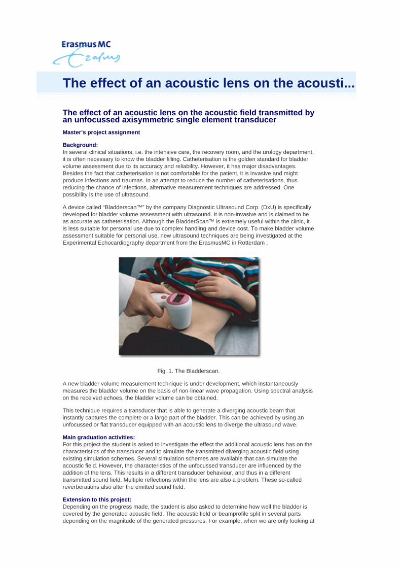
The effect of an acoustic lens on the acoustic field transmitted by an unfocussed axisymmetric single element transducer
Master’s project assignment
Background: In several clinical situations, i.e. the intensive care, the recovery room, and the urology department, it is often necessary to know the bladder filling. Catheterisation is the golden standard for bladder volume assessment due to its accuracy and reliability. However, it has major disadvantages. Besides the fact that catheterisation is not comfortable for the patient, it is invasive and might produce infections and traumas. In an attempt to reduce the number of catheterisations, thus reducing the chance of infections, alternative measurement techniques are addressed. One possibility is the use of ultrasound.
A device called “Bladderscan™” by the company Diagnostic Ultrasound Corp. (DxU) is specifically developed for bladder volume assessment with ultrasound. It is non-invasive and is claimed to be as accurate as catheterisation. Although the BladderScan™ is extremely useful within the clinic, it is less suitable for personal use due to complex handling and device cost. To make bladder volume assessment suitable for personal use, new ultrasound techniques are being investigated at the Experimental Echocardiography department from the ErasmusMC in Rotterdam .
Fig. 1. The Bladderscan.
A new bladder volume measurement technique is under development, which instantaneously measures the bladder volume on the basis of non-linear wave propagation. Using spectral analysis on the received echoes, the bladder volume can be obtained.
This technique requires a transducer that is able to generate a diverging acoustic beam that instantly captures the complete or a large part of the bladder. This can be achieved by using an unfocussed or flat transducer equipped with an acoustic lens to diverge the ultrasound wave.
Main graduation activities: For this project the student is asked to investigate the effect the additional acoustic lens has on the characteristics of the transducer and to simulate the transmitted diverging acoustic field using existing simulation schemes. Several simulation schemes are available that can simulate the acoustic field. However, the characteristics of the unfocussed transducer are influenced by the addition of the lens. This results in a different transducer behaviour, and thus in a different transmitted sound field. Multiple reflections within the lens are also a problem. These so-called reverberations also alter the emitted sound field.
Extension to this project: Depending on the progress made, the student is also asked to determine how well the bladder is covered by the generated acoustic field. The acoustic field or beamprofile split in several parts depending on the magnitude of the generated pressures. For example, when we are only looking at
The effect of an acoustic lens on the acousti...

beamprofile will be less diverging than when we are looking at pressures as low as one tenth (-20 dB) of the maximum. So, which part of the bladder is covered by pressures from 0 to –6 dB, which part is covered by pressures from –6dB to –12dB, and so on? This, of course depends on the size of the bladder and the location.
Requirements:
* discipline: Physics or electronics * programming experience in Matlab and /or C++
For more information, please contact:
E. Merks [email protected] Laboratory for Experimental Echocardiography, Department of Biomedical Engineering Room Ee23.02, Thoraxcenter, Erasmus MC P.O.Box 2040, 3000 CA Rotterdam, The Netherlands 010-7044633
Erasmus MC : The effect of an acoustic lens on the acoustic field

Measurement of the nonlinearity parameter of urine to determine its effect on a new bladder volume assessment method Bladder volume assessment on the basis of nonlinear wave propagation Internship project Background: A device called “BladderscanTM” by the company Verathon Medical is specifically developed for bladder volume assessment with ultrasound. It is non-invasive and is claimed to be as accurate as catheterisation, which is the golden standard for bladder volume measurement. Although the BladderScanTM is extremely useful within the clinic, it is less suitable for personal use due to relatively complex handling and device cost. To make bladder volume assessment suitable for personal use, a new ultrasound technique is being investigated at the Experimental Echocardiography department from the ErasmusMC in Rotterdam. This new measurement technique is based on non-linear propagation of ultrasonic waves. This non-linear effect causes the generation of higher harmonic components from the fundamental frequency as a function of travel distance and thus is a measure for the volume encompassed by the acoustic beam. The ratio of these higher harmonics with respect to the fundamental frequency depends on the medium specific non-linearity parameter and acoustic loss. Main activities For this project, the student is asked to use an in-house developed experimental setup to measure the non-linearity parameter, speed of sound, and acoustic loss of urine samples. This setup must first be calibrated with a ‘reference fluid’, of which the non-linearity parameter and loss are known. The calibration as well as the actual measurements will be performed at room and physiological temperatures. The student will also be asked to define and incorporate relevant parameters that might influence the loss and non-linearity measured, e.g. density, viscosity and ambient pressure. During this project the student will gain knowledge of nonlinear acoustics and perform several acoustic measurements.
Fig. 1. Experimental setup to measure the nonlinearity parameter, speed of sound and acoustic loss.
Required discipline: Physics or Electrical Engineering Payment: €250,-/month Location: Experimental Echocardiography, ErasmusMC, Rotterdam, Supervisor(s): Ir. E. Merks, (ErasmusMC, Rotterdam) Phone: 010-4089358 e-mail: [email protected]
Student projects Thoraxcenter Biomedical Engineering, Erasmus MC, Rotterdam, Oct 2007 Page 9 of 21
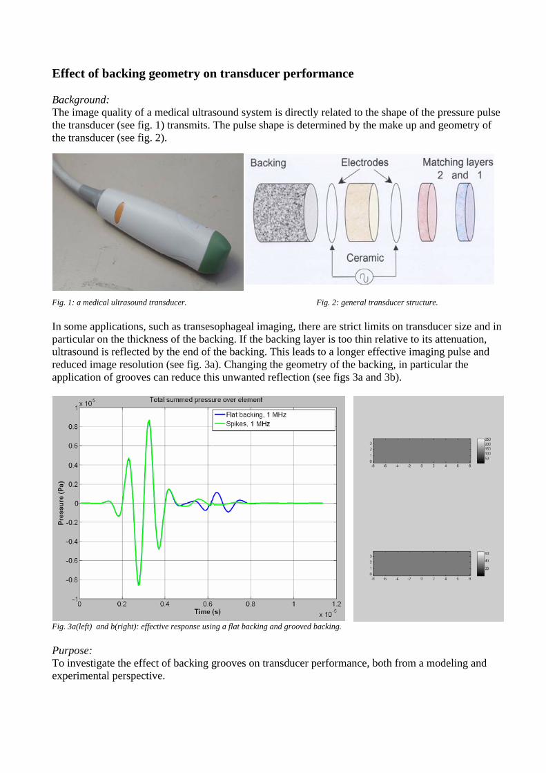
Effect of backing geometry on transducer performance Background: The image quality of a medical ultrasound system is directly related to the shape of the pressure pulse the transducer (see fig. 1) transmits. The pulse shape is determined by the make up and geometry of the transducer (see fig. 2).
Fig. 1: a medical ultrasound transducer. Fig. 2: general transducer structure. In some applications, such as transesophageal imaging, there are strict limits on transducer size and in particular on the thickness of the backing. If the backing layer is too thin relative to its attenuation, ultrasound is reflected by the end of the backing. This leads to a longer effective imaging pulse and reduced image resolution (see fig. 3a). Changing the geometry of the backing, in particular the application of grooves can reduce this unwanted reflection (see figs 3a and 3b).
Fig. 3a(left) and b(right): effective response using a flat backing and grooved backing. Purpose: To investigate the effect of backing grooves on transducer performance, both from a modeling and experimental perspective.

The project will be executed in close cooperation with Paul van Neer
Sho o- sducers and backing material, KLM model, pseudospectral modeling of
- Formula derivation to predict increase in backing attenuation by backing grooves.
Req ranical engineering/physics.
- An interest in both experimental work as in modeling.
or more information contact:
g, ErasmusMC Rotterdam, The Netherlands
4 Fax: +31 10 70 44720
rt utline of the project (6-9 months):
Literature study (tranwave propagation).
- Application of backing grooves to experimental arrays.
ui ements: - Background in biomedical engineering/mech- Programming experience (ie Matlab, C++).
F Paul van Neer MSc. ([email protected]) Room: Ee 23.02, Department of Biomedical EngineerinP.O. Box 2040, 3000 CA Phone: +31 10 70 4347
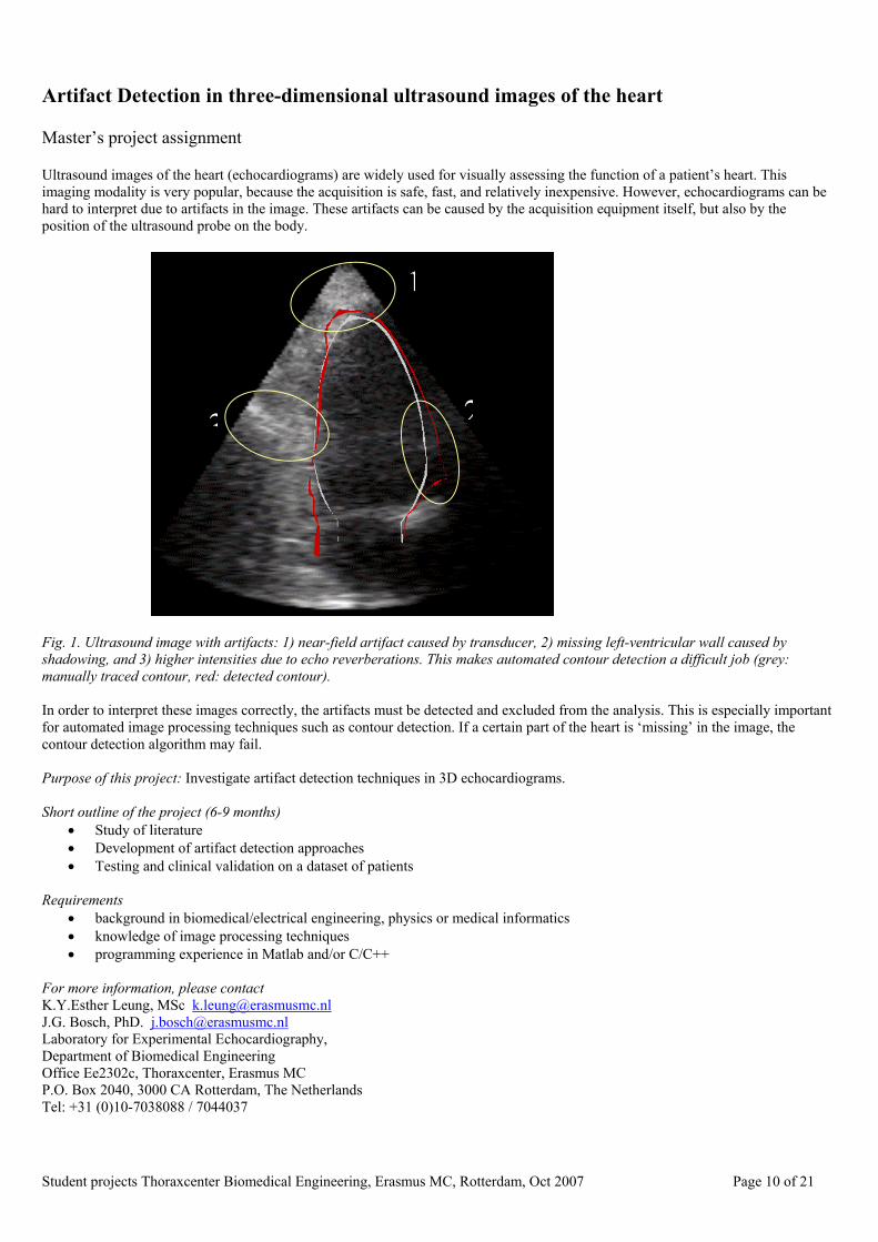
Artifact Detection in three-dimensional ultrasound images of the heart Master’s project assignment Ultrasound images of the heart (echocardiograms) are widely used for visually assessing the function of a patient’s heart. This imaging modality is very popular, because the acquisition is safe, fast, and relatively inexpensive. However, echocardiograms can be hard to interpret due to artifacts in the image. These artifacts can be caused by the acquisition equipment itself, but also by the position of the ultrasound probe on the body.
3 2
1
Fig. 1. Ultrasound image with artifacts: 1) near-field artifact caused by transducer, 2) missing left-ventricular wall caused by shadowing, and 3) higher intensities due to echo reverberations. This makes automated contour detection a difficult job (grey: manually traced contour, red: detected contour). In order to interpret these images correctly, the artifacts must be detected and excluded from the analysis. This is especially important for automated image processing techniques such as contour detection. If a certain part of the heart is ‘missing’ in the image, the contour detection algorithm may fail. Purpose of this project: Investigate artifact detection techniques in 3D echocardiograms. Short outline of the project (6-9 months)
• Study of literature • Development of artifact detection approaches • Testing and clinical validation on a dataset of patients
Requirements
• background in biomedical/electrical engineering, physics or medical informatics • knowledge of image processing techniques • programming experience in Matlab and/or C/C++
For more information, please contact K.Y.Esther Leung, MSc [email protected] J.G. Bosch, PhD. [email protected] Laboratory for Experimental Echocardiography, Department of Biomedical Engineering Office Ee2302c, Thoraxcenter, Erasmus MC P.O. Box 2040, 3000 CA Rotterdam, The Netherlands Tel: +31 (0)10-7038088 / 7044037
Student projects Thoraxcenter Biomedical Engineering, Erasmus MC, Rotterdam, Oct 2007 Page 10 of 21

Robust appearance modeling of the heart in three-dimensional ultrasound images Master’s project assignment
Background:
Our group is currently developing contour detection techniques for three-dimensional ultrasound images of the heart (3D echocardiograms). In these images, the beating heart is visible. Echocardiograms are primarily used for visual assessment of the functioning of a patient’s heart, but with an automated contour detection technique, they can also be used for measurement of the left ventricular volume, for detection of wall motion abnormalities etc. Manual segmentation of such 3D images is extremely time consuming and is not very reproducible.
Fig. 1. Rendering of 3D echocardiogram and endocardial surfaces.
The techniques which are being developed are based on active appearance models. These are statistical models of the contours and the greyscale ‘appearance’ of the heart, representing averages and variations across a patient population. Due to the complexity of the ultrasound images, nonstandard techniques are required to model the images adequately.
In this master’s project, we will be investigating the use of robust techniques for active appearance modeling. In particular, we will be focusing on the exclusion of typical ultrasound artifacts in the images, to obtain a more robust model for the heart. Several techniques have been proposed in the literature, e.g. principal component analysis for missing data.
Purpose of this project: Develop a robust technique for appearance modeling of 3D echocardiograms
Short outline of the project (6-9 months)
Requirements
K.Y.Esther Leung, MSc [email protected] J.G. Bosch PhD [email protected] Laboratory for Experimental Echocardiography, Department of Biomedical Engineering Room Ee23.02, Thoraxcenter, Erasmus MC P.O.Box 2040, 3000 CA Rotterdam, The Netherlands 010-7038088 / 7044037
Study of literature and comparison of methods Development of one or more robustness approaches Testing and clinical validation on a dataset of patients
background in biomedical/electrical engineering, physics or medical informatics knowledge of image processing techniques programming experience in Matlab and/or C/C++
Robust appearance modeling of the heart
4D interpolation of sparse, irregularly sampled ultrasound image data
MASTER'S PROJECT ASSIGNMENT
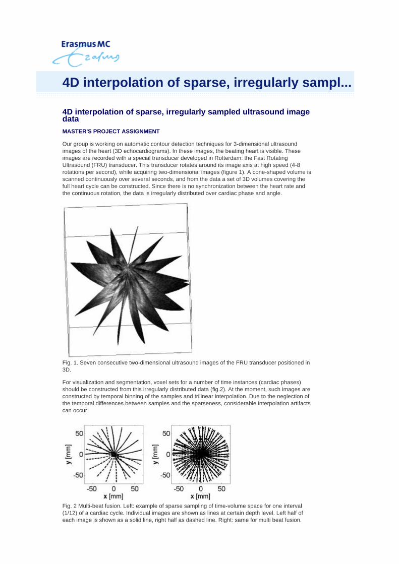
Our group is working on automatic contour detection techniques for 3-dimensional ultrasound images of the heart (3D echocardiograms). In these images, the beating heart is visible. These images are recorded with a special transducer developed in Rotterdam: the Fast Rotating Ultrasound (FRU) transducer. This transducer rotates around its image axis at high speed (4-8 rotations per second), while acquiring two-dimensional images (figure 1). A cone-shaped volume is scanned continuously over several seconds, and from the data a set of 3D volumes covering the full heart cycle can be constructed. Since there is no synchronization between the heart rate and the continuous rotation, the data is irregularly distributed over cardiac phase and angle.
Fig. 1. Seven consecutive two-dimensional ultrasound images of the FRU transducer positioned in 3D.
For visualization and segmentation, voxel sets for a number of time instances (cardiac phases) should be constructed from this irregularly distributed data (fig.2). At the moment, such images are constructed by temporal binning of the samples and trilinear interpolation. Due to the neglection of the temporal differences between samples and the sparseness, considerable interpolation artifacts can occur.
Fig. 2 Multi-beat fusion. Left: example of sparse sampling of time-volume space for one interval (1/12) of a cardiac cycle. Individual images are shown as lines at certain depth level. Left half of each image is shown as a solid line, right half as dashed line. Right: same for multi beat fusion.
4D interpolation of sparse, irregularly sampl...

We have developed a 4-dimensional Normalized Convolution interpolation approach that gives better results (fig.3), but improvements can still be achieved.
Issues involved:
Fig 3. Interpolation results. Left: temporal binning and trilinear interpolation. Notice the jagged edges. Right: interpolation with normalized convolution.
Purpose of this project: develop a suitable interpolation technique for this data
Short outline of the project (6-9 months)
Requirements:
For more information, please contact: K.Y. Esther Leung, MSc [email protected] J.G. Bosch PhD [email protected] Laboratory for Experimental Echocardiography, Department of Biomedical Engineering Room Ee23.02, Thoraxcenter, Erasmus MC P.O.Box 2040, 3000 CA Rotterdam, The Netherlands 010-7038088 / 7044037
theoretical comparison of our approach to other possibilities size and shape of the convolution kernels (currently Gaussian) in temporal and spatial dimensions multi-scale approach to better bridge gaps in the data better handling of motion artifacts
literature study of sparse data interpolation techniques, and study of previous work theoretical comparison of methods for this specific problem implementation of improvements and/or alternatives C++ developments, experimentation partly in Matlab evaluation of method on a number of patient images.
knowledge of image processing techniques programming experience in C++
Automated analysis of 3D cardiac ultrasound images with contrast Master’s project assignment
Background
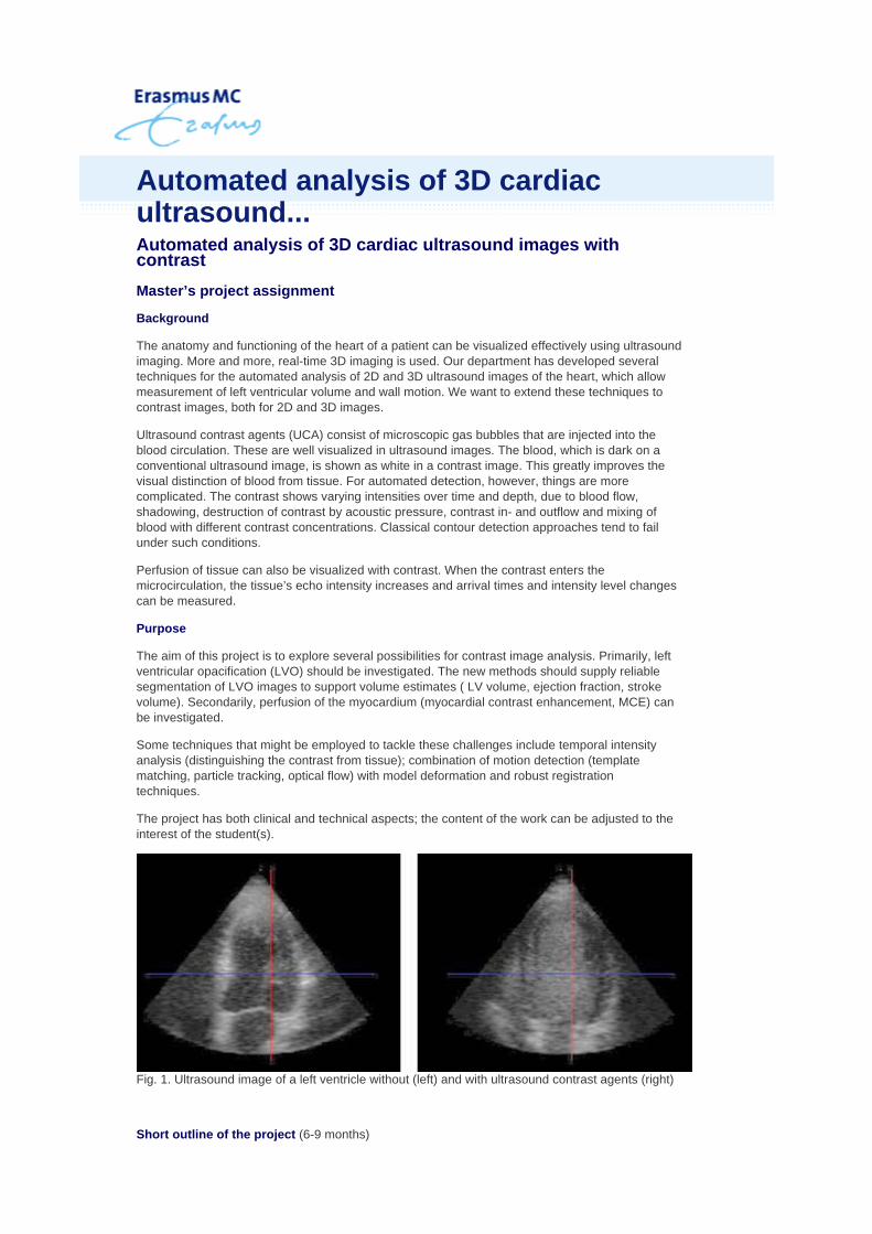
The anatomy and functioning of the heart of a patient can be visualized effectively using ultrasound imaging. More and more, real-time 3D imaging is used. Our department has developed several techniques for the automated analysis of 2D and 3D ultrasound images of the heart, which allow measurement of left ventricular volume and wall motion. We want to extend these techniques to contrast images, both for 2D and 3D images.
Ultrasound contrast agents (UCA) consist of microscopic gas bubbles that are injected into the blood circulation. These are well visualized in ultrasound images. The blood, which is dark on a conventional ultrasound image, is shown as white in a contrast image. This greatly improves the visual distinction of blood from tissue. For automated detection, however, things are more complicated. The contrast shows varying intensities over time and depth, due to blood flow, shadowing, destruction of contrast by acoustic pressure, contrast in- and outflow and mixing of blood with different contrast concentrations. Classical contour detection approaches tend to fail under such conditions.
Perfusion of tissue can also be visualized with contrast. When the contrast enters the microcirculation, the tissue’s echo intensity increases and arrival times and intensity level changes can be measured.
Purpose
The aim of this project is to explore several possibilities for contrast image analysis. Primarily, left ventricular opacification (LVO) should be investigated. The new methods should supply reliable segmentation of LVO images to support volume estimates ( LV volume, ejection fraction, stroke volume). Secondarily, perfusion of the myocardium (myocardial contrast enhancement, MCE) can be investigated.
Some techniques that might be employed to tackle these challenges include temporal intensity analysis (distinguishing the contrast from tissue); combination of motion detection (template matching, particle tracking, optical flow) with model deformation and robust registration techniques.
The project has both clinical and technical aspects; the content of the work can be adjusted to the interest of the student(s).
Fig. 1. Ultrasound image of a left ventricle without (left) and with ultrasound contrast agents (right)
Short outline of the project (6-9 months)
Automated analysis of 3D cardiacultrasound...

Requirements:
For more information, please contact:
K.Y.Esther Leung, MSc [email protected] J.G. Bosch PhD [email protected] Laboratory for Experimental Echocardiography, Department of Biomedical Engineering Room Ee23.02, Thoraxcenter, Erasmus MC P.O.Box 2040, 3000 CA Rotterdam, The Netherlands 010-7038088 / 7044037
Study of technical literature and previous work (pilot studies performed by our group) Acquire and analyze images of phantoms and patients Experiments with different analysis approaches Testing and clinical validation
background in biomedical/electrical/mechanical engineering, physics or medical informatics knowledge of image processing techniques programming experience in MatLab and/or C/C++
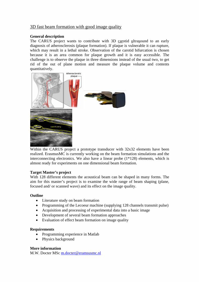
3D fast beam formation with good image quality General description The CARUS project wants to contribute with 3D carotid ultrasound to an early diagnosis of atherosclerosis (plaque formation). If plaque is vulnerable it can rupture, which may result in a lethal stroke. Observation of the carotid bifurcation is chosen because it is an area common for plaque growth and it is easy accessible. The challenge is to observe the plaque in three dimensions instead of the usual two, to get rid of the out of plane motion and measure the plaque volume and contents quantitatively.
Within the CARUS project a prototype transducer with 32x32 elements have been realized. ErasmusMC is currently working on the beam formation simulations and the interconnecting electronics. We also have a linear probe (1*128) elements, which is almost ready for experiments on one dimensional beam formation. Target Master’s project With 128 different elements the acoustical beam can be shaped in many forms. The aim for this master’s project is to examine the wide range of beam shaping (plane, focused and/ or scanned wave) and its effect on the image quality. Outline
• Literature study on beam formation • Programming of the Lecoeur machine (supplying 128 channels transmit pulse) • Acquisition and processing of experimental data into a basic image • Development of several beam formation approaches • Evaluation of effect beam formation on image quality
Requirements
• Programming experience in Matlab • Physics background
More information M.W. Docter MSc [email protected]

J.G. Bosch PhD [email protected] for Experimental Echocardiography, Department of Biomedical Engineering Room Ee23.02, Thoraxcenter, Erasmus MC P.O.Box 2040, 3000 CA Rotterdam, The Netherlands 010-7038088/ 7038525
Model-based estimation of the outflow conditions of coronary arteries for patient specific shear stress computations
Graduation project for a master student of Biomedical Engineering, Physics, Mechanical Engineering or Aerospace Technology with proven interest in fluid dynamics, modeling and/or finite element applications
Aim of the project
To obtain a model-based estimation of the outflow conditions at coro-nary artery branches, such that implementation in finite element cal-culations will result in realistic values for flow and shear stress.
Background
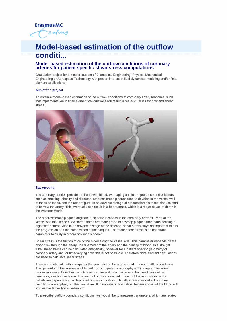
The coronary arteries provide the heart with blood. With aging and in the presence of risk factors, such as smoking, obesity and diabetes, atherosclerotic plaques tend to develop in the vessel wall of these ar-teries, see the upper figure. In an advanced stage of atherosclerosis these plaques start to narrow the artery. This eventually can result in a heart attack, which is a major cause of death in the Western World.
The atherosclerotic plaques originate at specific locations in the coro-nary arteries. Parts of the vessel wall that sense a low shear stress are more prone to develop plaques than parts sensing a high shear stress. Also in an advanced stage of the disease, shear stress plays an important role in the progression and the composition of the plaques. Therefore shear stress is an important parameter to study in athero-sclerotic research.
Shear stress is the friction force of the blood along the vessel wall. This parameter depends on the blood-flow through the artery, the di-ameter of the artery and the density of blood. In a straight tube, shear stress can be calculated analytically, however for a patient specific ge-ometry of coronary artery and for time-varying flow, this is not possi-ble. Therefore finite element calculations are used to calculate shear stress.
This computational method requires the geometry of the arteries and in, - and outflow conditions. The geometry of the arteries is obtained from computed tomography (CT) images. The artery divides in several branches, which results in several locations where the blood can exitthe geometry, see bottom figure. The amount of blood directed to each of these locations in the calculation depends on the described outflow conditions. Usually stress-free outlet boundary conditions are applied, but that would result in unrealistic flow ratios, because most of the blood will exit via the larger first side-branch
To prescribe outflow boundary conditions, we would like to measure parameters, which are related
Model-based estimation of the outflowconditi...

to flow, in the patient. However this is not possible with an image modality as CT. Thus, we need an alterna-tive method to obtain realistic flow and shear stress distribution in the coronary bifurcation. The proposed approach is to develop a model that gives a description of the vasculature behind the outflow loca-tions, which will enable us to prescribe parameters, such as pressure, resistance or flow at these locations.
For more information, please contact:
Ir. A.G. (Alina) van der Giessena. [email protected] Ir. F.J. (Frank) Gijsen [email protected] Hemodynamics, Department of Biomedical Engineering Thoraxcenter, Erasmus MC P.O.Box 2040, 3000 CA Rotterdam, The Netherlands 010-7044039
-outflow?lang=nl&view=Print
Biomedical Engineering Biomechanics Laboratory
Biomedical Engineering Biomechanics Laboratory
Contact Dr. Ir. Jolanda Wentzel
J.Wentzel@ ErasmusMC.nl 010 – 70 44 044
Wall Shear Stress Distribution in Realistic Human Coronary arteries
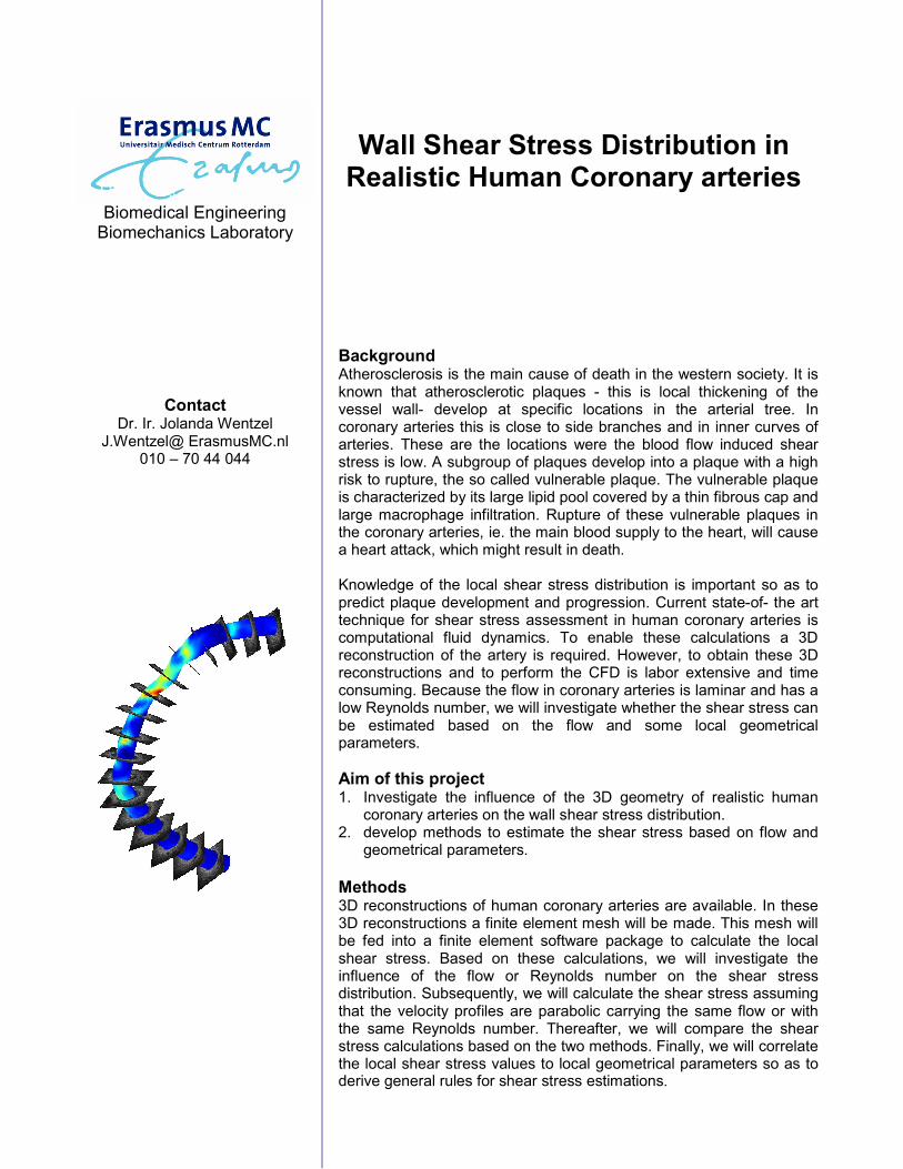
Background Atherosclerosis is the main cause of death in the western society. It is known that atherosclerotic plaques - this is local thickening of the vessel wall- develop at specific locations in the arterial tree. In coronary arteries this is close to side branches and in inner curves of arteries. These are the locations were the blood flow induced shear stress is low. A subgroup of plaques develop into a plaque with a high risk to rupture, the so called vulnerable plaque. The vulnerable plaque is characterized by its large lipid pool covered by a thin fibrous cap and large macrophage infiltration. Rupture of these vulnerable plaques in the coronary arteries, ie. the main blood supply to the heart, will cause a heart attack, which might result in death. Knowledge of the local shear stress distribution is important so as to predict plaque development and progression. Current state-of- the art technique for shear stress assessment in human coronary arteries is computational fluid dynamics. To enable these calculations a 3D reconstruction of the artery is required. However, to obtain these 3D reconstructions and to perform the CFD is labor extensive and time consuming. Because the flow in coronary arteries is laminar and has a low Reynolds number, we will investigate whether the shear stress can be estimated based on the flow and some local geometrical parameters.
Aim of this project 1. Investigate the influence of the 3D geometry of realistic human
coronary arteries on the wall shear stress distribution. 2. develop methods to estimate the shear stress based on flow and
geometrical parameters.
Methods 3D reconstructions of human coronary arteries are available. In these 3D reconstructions a finite element mesh will be made. This mesh will be fed into a finite element software package to calculate the local shear stress. Based on these calculations, we will investigate the influence of the flow or Reynolds number on the shear stress distribution. Subsequently, we will calculate the shear stress assuming that the velocity profiles are parabolic carrying the same flow or with the same Reynolds number. Thereafter, we will compare the shear stress calculations based on the two methods. Finally, we will correlate the local shear stress values to local geometrical parameters so as to derive general rules for shear stress estimations.
Biomedical Engineering Biomechanics Laboratory
Contact Dr. Ir. Frank Gijsen
[email protected] 010 – 70 44 039
Histological data from a non-ruptured
vulnerable plaque
Histological data from a non-ruptured
vulnerable plaque
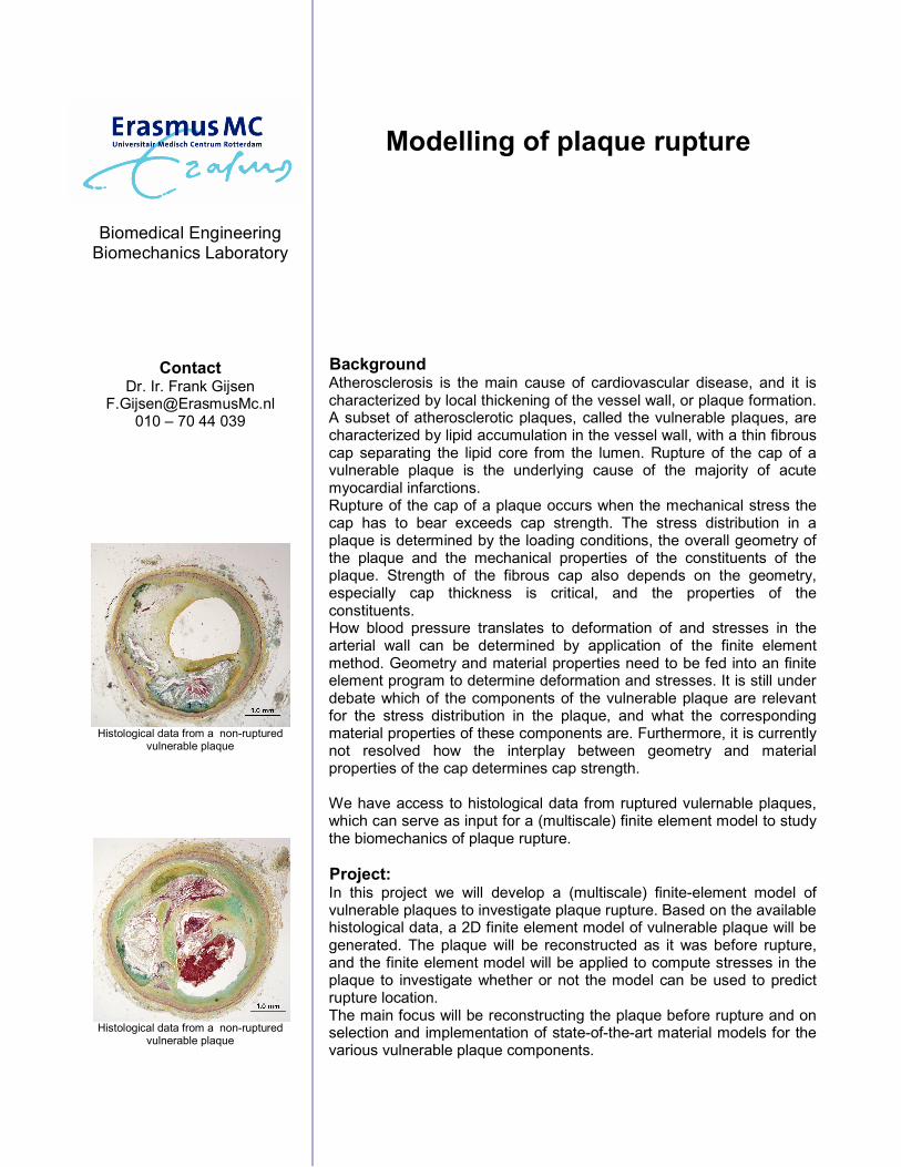
Modelling of plaque rupture
Background Atherosclerosis is the main cause of cardiovascular disease, and it is characterized by local thickening of the vessel wall, or plaque formation. A subset of atherosclerotic plaques, called the vulnerable plaques, are characterized by lipid accumulation in the vessel wall, with a thin fibrous cap separating the lipid core from the lumen. Rupture of the cap of a vulnerable plaque is the underlying cause of the majority of acute myocardial infarctions. Rupture of the cap of a plaque occurs when the mechanical stress the cap has to bear exceeds cap strength. The stress distribution in a plaque is determined by the loading conditions, the overall geometry of the plaque and the mechanical properties of the constituents of the plaque. Strength of the fibrous cap also depends on the geometry, especially cap thickness is critical, and the properties of the constituents. How blood pressure translates to deformation of and stresses in the arterial wall can be determined by application of the finite element method. Geometry and material properties need to be fed into an finite element program to determine deformation and stresses. It is still under debate which of the components of the vulnerable plaque are relevant for the stress distribution in the plaque, and what the corresponding material properties of these components are. Furthermore, it is currently not resolved how the interplay between geometry and material properties of the cap determines cap strength. We have access to histological data from ruptured vulernable plaques, which can serve as input for a (multiscale) finite element model to study the biomechanics of plaque rupture.
Project: In this project we will develop a (multiscale) finite-element model of vulnerable plaques to investigate plaque rupture. Based on the available histological data, a 2D finite element model of vulnerable plaque will be generated. The plaque will be reconstructed as it was before rupture, and the finite element model will be applied to compute stresses in the plaque to investigate whether or not the model can be used to predict rupture location. The main focus will be reconstructing the plaque before rupture and on selection and implementation of state-of-the-art material models for the various vulnerable plaque components.

Biomedical Engineering Biomechanics Laboratory
Contact Dr. Ir. Frank Gijsen
[email protected] 010 – 70 44 039
plaque imaging with MRI.
MR imaging of vulnerable plaques: pressure induced deformation
Background Atherosclerosis is the main cause of cardiovascular disease, and it is characterized by local thickening of the vessel wall, or plaque formation. A subset of atherosclerotic plaques, called the vulnerable plaques, are characterized by lipid accumulation in the vessel wall, with a thin fibrous cap separating the lipid core from the lumen. Rupture of the cap of a vulnerable plaque is the underlying cause of the majority of acute myocardial infarctions. Rupture of the cap of a plaque occurs when the mechanical stress the cap has to bear exceeds cap strength. The stress distribution in a plaque is determined by the loading conditions, the overall geometry of the plaque and the mechanical properties of the constituents of the plaque. Strength of the fibrous cap also depends on the geometry, especially cap thickness is critical, and the properties of the constituents. Both stresses in the cap and cap strength will vary locally. How blood pressure translates to deformation of and stresses in the arterial wall can be determined by application of the finite element method. Geometry and material properties need to be fed into an finite element program to determine deformation and stresses. Many research groups are currently studying the biomechanical behaviour of the vulnerable plaque, but it is still under debate which of the components of the vulnerable plaque are relevant for the stress distribution in the plaque, and what the corresponding material properties of these components are. Furthermore, it is currently not resolved how the interplay between geometry and material properties of the cap determines cap strength. Experimental data are crucial to increase our knowledge of the biomechanical behaviour of vulnerable plaques.
Project In this project will focus on in vitro MR imaging of plaques in human coronary arteries. The arteries will be obtained from autopsy material, and they will be subjected to a stepwise increase in pressure. The setup to perform these experiments was designed and validated in a previous project. MR imaging with high resolution will be performed to visualize the relevant plaque components and their displacement under influence of pressure increase. These experiments will provide us with in vitro experimental data on pressure induced deformation of human vulnerable plaques under carefully controlled experimental conditions. The emphasis in this experimental study will be on MR imaging and image analysis.
Biomedical Engineering Biomechanics Laboratory
Contact Dr. Ir. Jolanda Wentzel
J.Wentzel@ ErasmusMC.nl 010 – 70 44 044
Ir. Harald Groen [email protected]
010-703 8166
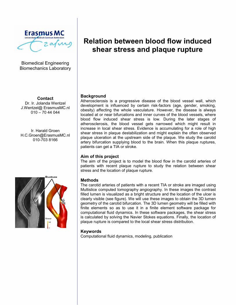
Relation between blood flow induced shear stress and plaque rupture
Background Atherosclerosis is a progressive disease of the blood vessel wall, which development is influenced by certain risk-factors (age, gender, smoking, obesity) affecting the whole vasculature. However, the disease is always located at or near bifurcations and inner curves of the blood vessels, where blood flow induced shear stress is low. During the later stages of atherosclerosis, the blood vessel gets narrowed which might result in increase in local shear stress. Evidence is accumulating for a role of high shear stress in plaque destabilization and might explain the often observed plaque ulceration at the upstream side of the plaque. We study the carotid artery bifurcation supplying blood to the brain. When this plaque ruptures, patients can get a TIA or stroke.
Aim of this project The aim of the project is to model the blood flow in the carotid arteries of patients with recent plaque rupture to study the relation between shear stress and the location of plaque rupture.
Methods The carotid arteries of patients with a recent TIA or stroke are imaged using Multislice computed tomography angiography. In these images the contrast filled lumen is visualized as a bright structure and the location of the ulcer is clearly visible (see figure). We will use these images to obtain the 3D lumen geometry of the carotid bifurcation. The 3D lumen geometry will be filled with finite elements so as to use it in a finite element software package for computational fluid dynamics. In these software packages, the shear stress is calculated by solving the Navier Stokes equations. Finally, the location of plaque rupture is compared to the local shear stress distribution.
Keywords Computational fluid dynamics, modeling, publication
Rupture
L
Hi
Shear
Biomedical Engineering Biomechanics Laboratory
Contact Dr. Ir. Jolanda Wentzel
J.Wentzel@ ErasmusMC.nl 010 – 70 44 044
Ir. Harald Groen
h.c.groen@ ErasmusMC.nl 010-703 8166
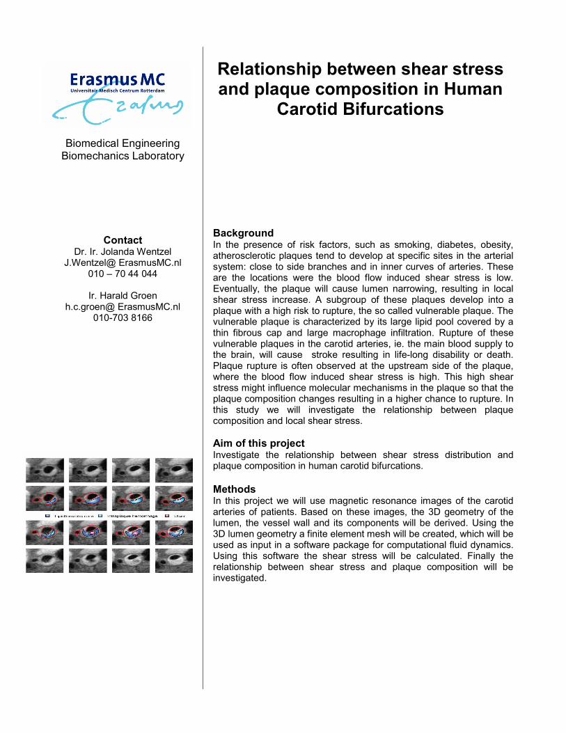
Relationship between shear stress and plaque composition in Human
Carotid Bifurcations
Background In the presence of risk factors, such as smoking, diabetes, obesity, atherosclerotic plaques tend to develop at specific sites in the arterial system: close to side branches and in inner curves of arteries. These are the locations were the blood flow induced shear stress is low. Eventually, the plaque will cause lumen narrowing, resulting in local shear stress increase. A subgroup of these plaques develop into a plaque with a high risk to rupture, the so called vulnerable plaque. The vulnerable plaque is characterized by its large lipid pool covered by a thin fibrous cap and large macrophage infiltration. Rupture of these vulnerable plaques in the carotid arteries, ie. the main blood supply to the brain, will cause stroke resulting in life-long disability or death. Plaque rupture is often observed at the upstream side of the plaque, where the blood flow induced shear stress is high. This high shear stress might influence molecular mechanisms in the plaque so that the plaque composition changes resulting in a higher chance to rupture. In this study we will investigate the relationship between plaque composition and local shear stress.
Aim of this project Investigate the relationship between shear stress distribution and plaque composition in human carotid bifurcations.
Methods In this project we will use magnetic resonance images of the carotid arteries of patients. Based on these images, the 3D geometry of the lumen, the vessel wall and its components will be derived. Using the 3D lumen geometry a finite element mesh will be created, which will be used as input in a software package for computational fluid dynamics. Using this software the shear stress will be calculated. Finally the relationship between shear stress and plaque composition will be investigated.
Biomedical Engineering Biomechanics Laboratory
Contact Dr. Ir. Jolanda Wentzel
J.Wentzel@ ErasmusMC.nl 010 – 70 44 044
Ir. Harald Groen
h.c.groen@ ErasmusMC.nl 010-703 8166
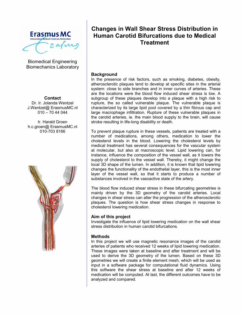
Changes in Wall Shear Stress Distribution in Human Carotid Bifurcations due to Medical
Treatment
Background In the presence of risk factors, such as smoking, diabetes, obesity, atherosclerotic plaques tend to develop at specific sites in the arterial system: close to side branches and in inner curves of arteries. These are the locations were the blood flow induced shear stress is low. A subgroup of these plaques develop into a plaque with a high risk to rupture, the so called vulnerable plaque. The vulnerable plaque is characterized by its large lipid pool covered by a thin fibrous cap and large macrophage infiltration. Rupture of these vulnerable plaques in the carotid arteries, ie. the main blood supply to the brain, will cause stroke resulting in life-long disability or death. To prevent plaque rupture in these vessels, patients are treated with a number of medications, among others, medication to lower the cholesterol levels in the blood. Lowering the cholesterol levels by medical treatment has several consequences for the vascular system at molecular, but also at macroscopic level. Lipid lowering can, for instance, influence the composition of the vessel wall, as it lowers the supply of cholesterol to the vessel wall. Thereby, it might change the local 3D shape of the lumen. In addition, it is known that lipid lowering changes the functionality of the endothelial layer, this is the most inner layer of the vessel wall, so that it starts to produce a number of substances involved in the vasoactive state of the artery. The blood flow induced shear stress in these bifurcating geometries is mainly driven by the 3D geometry of the carotid arteries. Local changes in shear stress can alter the progression of the atherosclerotic plaques. The question is how shear stress changes in response to cholesterol lowering medication.
Aim of this project Investigate the influence of lipid lowering medication on the wall shear stress distribution in human carotid bifurcations.
Methods In this project we will use magnetic resonance images of the carotid arteries of patients who received 12 weeks of lipid lowering medication. These images were taken at baseline and after treatment and will be used to derive the 3D geometry of the lumen. Based on these 3D geometries we will create a finite element mesh, which will be used as input in a software package for computational fluid dynamics. Using this software the shear stress at baseline and after 12 weeks of medication will be computed. At last, the different outcomes have to be analyzed and compared.
Biomedical Engineering Biomechanics Laboratory
Contact Ir. Harald Groen
[email protected] 010-703 8166
Dr. Ir. Jolanda Wentzel J.Wentzel@ ErasmusMC.nl
010 – 70 44 044
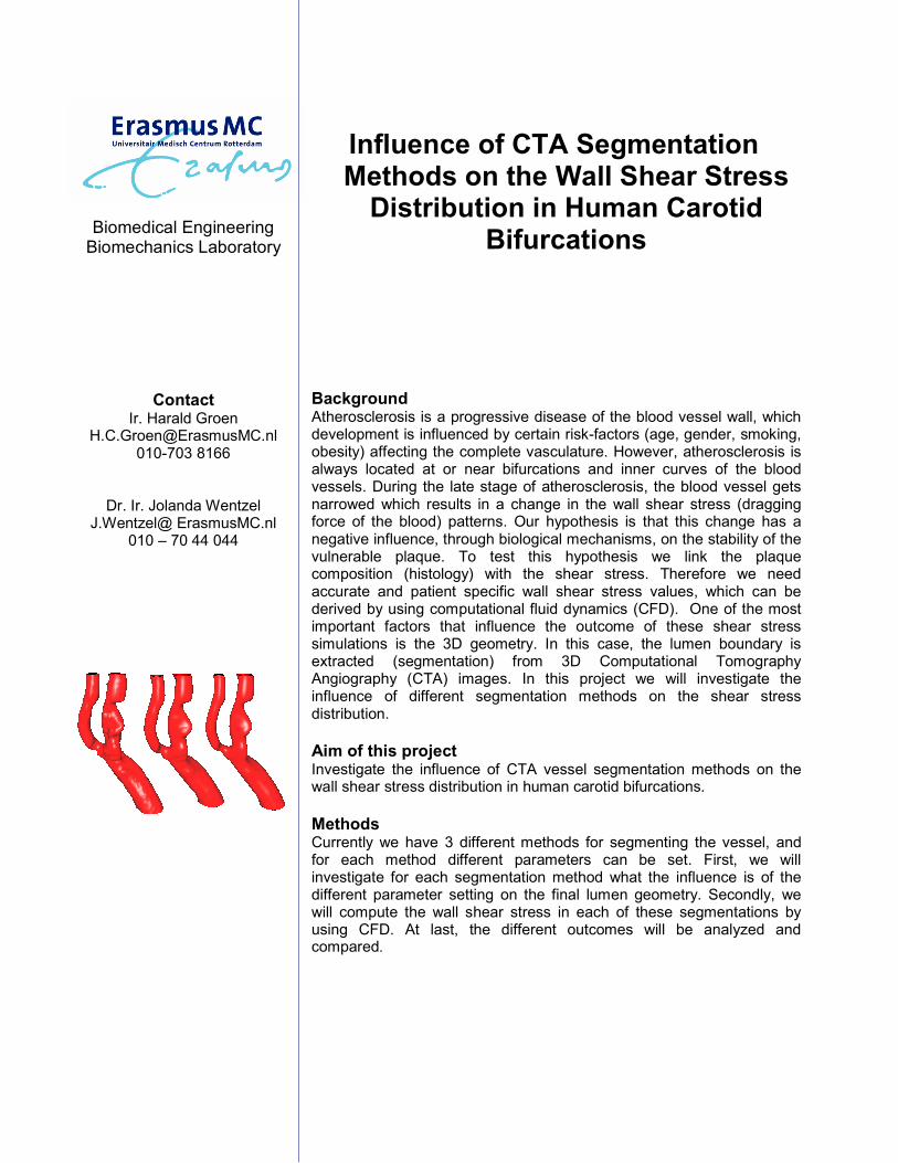
Influence of CTA Segmentation Methods on the Wall Shear Stress Distribution in Human Carotid
Bifurcations
Background Atherosclerosis is a progressive disease of the blood vessel wall, which development is influenced by certain risk-factors (age, gender, smoking, obesity) affecting the complete vasculature. However, atherosclerosis is always located at or near bifurcations and inner curves of the blood vessels. During the late stage of atherosclerosis, the blood vessel gets narrowed which results in a change in the wall shear stress (dragging force of the blood) patterns. Our hypothesis is that this change has a negative influence, through biological mechanisms, on the stability of the vulnerable plaque. To test this hypothesis we link the plaque composition (histology) with the shear stress. Therefore we need accurate and patient specific wall shear stress values, which can be derived by using computational fluid dynamics (CFD). One of the most important factors that influence the outcome of these shear stress simulations is the 3D geometry. In this case, the lumen boundary is extracted (segmentation) from 3D Computational Tomography Angiography (CTA) images. In this project we will investigate the influence of different segmentation methods on the shear stress distribution.
Aim of this project Investigate the influence of CTA vessel segmentation methods on the wall shear stress distribution in human carotid bifurcations.
Methods Currently we have 3 different methods for segmenting the vessel, and for each method different parameters can be set. First, we will investigate for each segmentation method what the influence is of the different parameter setting on the final lumen geometry. Secondly, we will compute the wall shear stress in each of these segmentations by using CFD. At last, the different outcomes will be analyzed and compared.
Tissue tracking on tendons in the hand using high-frequency ultrasound imaging
MASTER'S PROJECT ASSIGNMENT
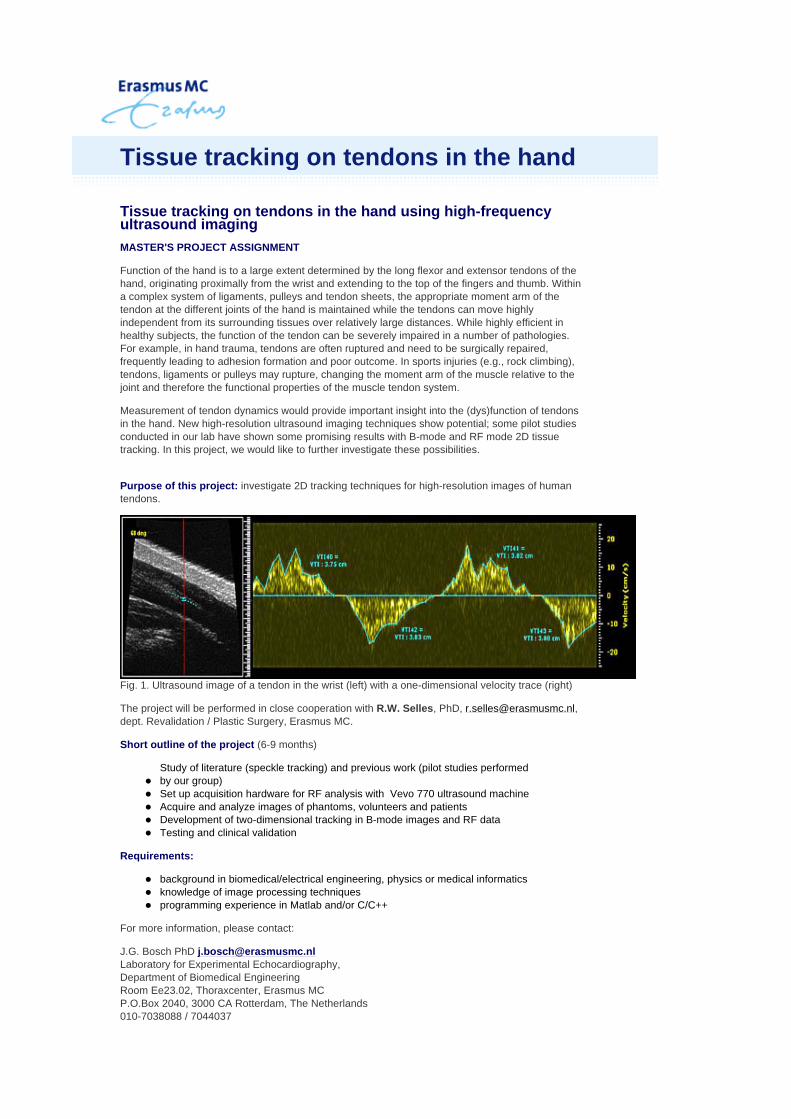
Function of the hand is to a large extent determined by the long flexor and extensor tendons of the hand, originating proximally from the wrist and extending to the top of the fingers and thumb. Within a complex system of ligaments, pulleys and tendon sheets, the appropriate moment arm of the tendon at the different joints of the hand is maintained while the tendons can move highly independent from its surrounding tissues over relatively large distances. While highly efficient in healthy subjects, the function of the tendon can be severely impaired in a number of pathologies. For example, in hand trauma, tendons are often ruptured and need to be surgically repaired, frequently leading to adhesion formation and poor outcome. In sports injuries (e.g., rock climbing), tendons, ligaments or pulleys may rupture, changing the moment arm of the muscle relative to the joint and therefore the functional properties of the muscle tendon system.
Measurement of tendon dynamics would provide important insight into the (dys)function of tendons in the hand. New high-resolution ultrasound imaging techniques show potential; some pilot studies conducted in our lab have shown some promising results with B-mode and RF mode 2D tissue tracking. In this project, we would like to further investigate these possibilities.
Purpose of this project: investigate 2D tracking techniques for high-resolution images of human tendons.
Fig. 1. Ultrasound image of a tendon in the wrist (left) with a one-dimensional velocity trace (right)
The project will be performed in close cooperation with R.W. Selles, PhD, [email protected], dept. Revalidation / Plastic Surgery, Erasmus MC.
Short outline of the project (6-9 months)
Requirements:
For more information, please contact:
J.G. Bosch PhD [email protected] Laboratory for Experimental Echocardiography, Department of Biomedical Engineering Room Ee23.02, Thoraxcenter, Erasmus MC P.O.Box 2040, 3000 CA Rotterdam, The Netherlands 010-7038088 / 7044037
Study of literature (speckle tracking) and previous work (pilot studies performed by our group) Set up acquisition hardware for RF analysis with Vevo 770 ultrasound machine Acquire and analyze images of phantoms, volunteers and patients Development of two-dimensional tracking in B-mode images and RF data Testing and clinical validation
background in biomedical/electrical engineering, physics or medical informatics knowledge of image processing techniques programming experience in Matlab and/or C/C++
Tissue tracking on tendons in the hand
Modeling of Intravascular Photo-acoustic imaging
MASTER'S PROJECT ASSIGNMENT
Background
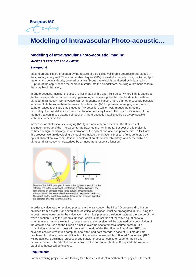
Most heart attacks are preceded by the rupture of a so-called vulnerable atherosclerotic plaque in the coronary artery wall. These vulnerable plaques (VPs) consist of a necrotic core, containing lipid material and cellular debris, covered by a thin fibrous cap which is weakened by inflammation. Rupture of the cap releases the necrotic material into the bloodstream, causing a thrombus to form, that may block the artery.
In photo-acoustic imaging, the tissue is illuminated with a short light pulse. Where light is absorbed, the tissue expands thermo-elastically, generating a pressure pulse that can be detected with an ultrasound transducer. Some vessel wall components will absorb more than others, so it is possible to differentiate between them. Intravascular ultrasound (IVUS) pulse-echo imaging is a common, catheter-based technique that is used for VP detection. While IVUS images the structure accurately, the possibilities for tissue identification are very limited. There is a clinical need for a method that can image plaque composition. Photo-acoustic imaging could be a very suitable technique to achieve this.
Intravascular photo-acoustic imaging (IVPA) is a new research theme in the Biomedical Engineering group of the Thorax center at Erasmus MC. An important aspect of this project is catheter design, particularly the optimization of the optical and acoustic parameters. To facilitate this process, we are developing a model to simulate the ultrasonic pressure field, generated by optical absorption in a computational phantom of an atherosclerotic artery, and detected by an ultrasound transducer characterized by an instrument response function.
In order to calculate the received pressure at the transducer, the initial 3D pressure distribution, obtained from a Monte Carlo simulation of optical absorption, must be propagated in time using the acoustic wave equation. In the calculations, the initial pressure distribution acts as the source of the wave equation. Using the Green’s function, which is the solution of the wave equation for a spatiotemporal impulse excitation, the pressure at the receiver will be obtained by a convolution of the obtained source and the Green’s function over the spatiotemporal source domain. This convolution is performed most efficiently with the aid of the Fast Fourier Transform (FFT), but nevertheless requires much computational effort and data storage in case of 3D time domain problems. To relieve the latter difficulties, the recently developed Fast Filtered Convolution (FFC) will be applied. Both single-processor and parallel-processor computer code for the FFC is available but must be adapted and optimized to the current application. If required, the use of a parallel computer will be invoked.
Requirements:
For this exciting project, we are looking for a Master’s student in mathematics, physics, electrical
Modeling of Intravascular Photo-acoustic...

engineering, or any other discipline related to computational science or mathematical physics. The student must have interest in the computational modeling of physical problems arising in the area of biomedics. Good programming skills are essential, and a working knowledge of Fortran is preferred.
For more information, please contact:
Gijs van Soest [email protected] Laboratory for Experimental Echocardiography, Department of Biomedical Engineering Room Ee23.02, Thoraxcenter, Erasmus MC P.O.Box 2040, 3000 CA Rotterdam, The Netherlands Tel: +31 (0)10-7044638
Martin Verweij [email protected] Laboratory of Electromagnetic Research Faculty of Electrical Engineering, Mathematics and Computer Science Delft University of Technology Delft, The Netherlands Tel: +31 (0)15-2781761