Embed Size (px)
Citation preview
Structure of human POFUT2: insights intothrombospondin type 1 repeat foldand O-fucosylation
Chun-I Chen, Jeremy J Keusch,Dominique Klein, Daniel Hess,Jan Hofsteenge and Heinz Gut*
Friedrich Miescher Institute for Biomedical Research, Basel, Switzerland
Protein O-fucosylation is a post-translational modification
found on serine/threonine residues of thrombospondin
type 1 repeats (TSR). The fucose transfer is catalysed by
the enzyme protein O-fucosyltransferase 2 (POFUT2) and
440 human proteins contain the TSR consensus sequence
for POFUT2-dependent fucosylation. To better understand
O-fucosylation on TSR, we carried out a structural and
functional analysis of human POFUT2 and its TSR sub-
strate. Crystal structures of POFUT2 reveal a variation of
the classical GT-B fold and identify sugar donor and TSR
acceptor binding sites. Structural findings are correlated
with steady-state kinetic measurements of wild-type and
mutant POFUT2 and TSR and give insight into the catalytic
mechanism and substrate specificity. By using an artificial
mini-TSR substrate, we show that specificity is not pri-
marily encoded in the TSR protein sequence but rather in
the unusual 3D structure of a small part of the TSR. Our
findings uncover that recognition of distinct conserved 3D
fold motifs can be used as a mechanism to achieve sub-
strate specificity by enzymes modifying completely folded
proteins of very wide sequence diversity and biological
function.
The EMBO Journal (2012) 31, 3183–3197. doi:10.1038/
emboj.2012.143; Published online 15 May 2012Subject Categories: proteinsKeywords: crystal structure; enzymatic mechanism; GDP-
fucose; protein O-fucosyltransferase 2; thrombospondin type 1
repeat
Introduction
Protein glycosylation is the most abundant and diverse
co- and post-translational modification in life. In eukaryotes,
450% of proteins are modified with carbohydrates
(Apweiler et al, 1999) which together regulate myriad
biological processes. Altered or defective protein
glycosylation pathways cause various developmental
defects as reflected in the rapidly growing number of
congenital disorders of glycosylation (Freeze, 2007; Jaeken
and Matthijs, 2007).
The unusual protein O-linked fucosylation has been de-
scribed on thrombospondin type 1 repeats (TSR) (Hofsteenge
et al, 2001; Gonzalez de Peredo et al, 2002) and epidermal
growth factor-like (EGF) repeats (Bjoern et al, 1991;
Buko et al, 1991; Harris et al, 1992; Nishimura et al,
1992; Harris and Spellman, 1993) and is catalysed by the
protein O-fucosyltransferase 2 (POFUT2) and protein
O-fucosyltransferase 1 (POFUT1), respectively (Harris and
Spellman, 1993; Luo et al, 2006a). Both enzymes transfer
the fucose moiety from GDP-fucose to a serine or threonine
residue of the properly folded acceptor molecule, recognizing
the consensus sequences CX2–3(S/T)CX2G (Hofsteenge et al,
2001) in TSR or CX4–5(S/T)C (Harris and Spellman, 1993) in
EGF repeats, respectively. The fucose residue on TSR can be
elongated to a glucose-b1,3-fucose disaccharide by the b1,3-glucosyltransferase (b3GlcT) (Kozma et al, 2006; Sato et al,
2006). In EGF repeats, the fucose may be extended to an
NeuAc-a2,3/a2,6-Gal-b1,4-GlcNAc-b1,3-Fuc tetrasaccharide
catalysed by the sequential enzymatic activity of Fringe,
b1,4-galactosyltransferase 1 and a2,3/a2,6-sialyltransferase(Nishimura et al, 1992; Harris and Spellman, 1993; Stanley,
2007; Luther and Haltiwanger, 2009; Rana and Haltiwanger,
2011). Both TSR and EGF repeats are small cysteine-rich,
layered structural motifs with three conserved disulphide
bonds and little secondary structural elements. TSR and
EGF repeat proteins are sequence-wise very diverse with
only a few structural key residues being conserved. The
glycosyltransferases involved in the O-fucosylation
pathways of TSR and EGF repeats are specific and do not
crossreact (Luo et al, 2006b).
The importance of protein glycosylation on EGF repeats
has been extensively studied in the Notch signalling pathway
(Luther and Haltiwanger, 2009) where the EGF modification
was shown to regulate embryonic development and tissue
renewal by controlling the ligand specificity of Notch
(Stanley, 2007; Stahl et al, 2008). Crystal structures of
C. elegans POFUT1 alone and in complex with GDP-fucose
or GDP have been solved recently and give insight into
overall protein structure and the enzymatic mechanism
(Lira-Navarrete et al, 2011). The role of POFUT2-dependent
fucosylation of TSR on the other hand is less clear. Progress
was made recently by Du et al (2010) using Pofut2 knockout
mice where they found that O-fucosylation of TSR is critical
for restricting epithelial-to-mesenchymal transition, correct
patterning of the mesoderm, and localization of the
endoderm in embryonic development. In C. elegans,
POFUT2-dependent TSR fucosylation was found to be
involved in the regulation of distal tip cell migration
(Canevascini et al, 2006). TSR proteins are expressed in the
secretory pathway with O-fucosylation occurring within the
endoplasmic reticulum. In cell culture experiments, mutation
of fucosylation sites on TSR of ADAMTS13 (Ricketts et al,
2007) and Punctin-1 (Wang et al, 2007) reduced or
completely abolished secretion of the proteins, indicating
*Corresponding author. Friedrich Miescher Institute for BiomedicalResearch, Maulbeerstrasse 66, 4058 Basel, Switzerland.Tel.: þ 41 61 696 70 38; Fax: þ 41 61 697 39 76;E-mail: [email protected]
Received: 12 January 2012; accepted: 23 April 2012; publishedonline: 15 May 2012
The EMBO Journal (2012) 31, 3183–3197 | & 2012 European Molecular Biology Organization |All Rights Reserved 0261-4189/12
www.embojournal.org
EMBO
THE
EMBOJOURNAL
THE
EMBOJOURNAL
3183&2012 European Molecular Biology Organization The EMBO Journal VOL 31 | NO 14 | 2012

that POFUT2-dependent O-fucosylation on TSR might be
required for optimal secretion of these proteins. No disorder
has yet been directly linked to a genetic defect of the Pofut2
locus in humans. However, mutations in the B3GALTL gene
that encodes the b3GlcT enzyme responsible for glucose
transfer onto O-fucosylated TSR cause the autosomal
recessive disorder Peters Plus syndrome (Lesnik Oberstein
et al, 2006; Hess et al, 2008). This disorder is characterized by
anterior-eye-chamber abnormalities, disproportionate short
stature and developmental delay.
Protein O-fucosylation raises three fundamental questions
about the interaction between glycosyltransferases and their
protein substrate: How does a glycosyltransferase accommo-
date a fully folded protein substrate in its active site? Which
structural features are used to discriminate between the
different families of protein substrates and how can specifi-
city be achieved in the case of sequence-wise degenerated
protein substrates? We have addressed these questions by
determining the structure of human POFUT2 (alone and in
complex with the sugar donor GDP-fucose) and steady-state
kinetic analysis of wild-type and mutant transferase. To
investigate further how POFUT2 interacts with its TSR
sugar acceptor, we have analysed O-fucosylation of wild-
type and mutant TSR in an in-vitro assay and in mammalian
HEK293T cells. The crystal structure shows that POFUT2
belongs to the classical GT-B fold family of glycosyltrans-
ferases with two closely interacting Rossmann-like domains.
The C-terminal domain binds the GDP-fucose moiety while
the TSR substrate is recognized by a large cavity in the centre
of the bilobal structure. Based on our structural data and
steady-state kinetic measurements, we suggest that the con-
served E54 residue acts as the catalytic base, and describe key
catalytic residues located in the active site. Structural and
biochemical knowledge was used to clarify why only TSR
modules can bind to the sugar acceptor site and to design an
artificial minimal TSR module which we show to be sufficient
as sugar acceptor for common TSR glycan modifications
(O-fucose-glucosylation and C-mannosylation). Furthermore,
we investigated how POFUT2 substrate specificity is achieved
despite the large sequence diversity present in TSR containing
the CX2–3(S/T)CX2G fucosylation motif. We present the struc-
ture of a protein glycosyltransferase modifying a completely
folded protein substrate and propose a novel mechanism of
enzyme-protein substrate specificity, based on recognition of
a small conserved 3D structural motif. It explains how site-
specific modifications can take place in the absence of a
conserved protein sequence.
Results
Crystal structure of human POFUT2
We have expressed and purified human D21-POFUT2 from
mammalian cell culture and have determined its crystal
structure at 3.0 A resolution. The protein crystallized in
space group P3221 with two molecules in the asymmetric
unit (a.u.) and the structure was solved by the single iso-
morphous replacement with anomalous scattering (SIRAS)
method using a platinum derivative. Data collection, phasing
and refinement statistics are presented in Table I. The refined
POFUT2 crystal structure displays clear electron density for
residues 41–429 (out of 22–429) and the two molecules in the
a.u. are almost identical with an r.m.s.d. of only 0.49 A. The
structure of POFUT2 is composed of two Rossmann-like
domains with b/a/b topology typical of the GT-B fold of
glycosyltransferases (Figure 1A). N- and C-terminal domains
encompass residues 22–242 and 243–429, respectively. The
two domains interact closely with each other (buried surface
area of 1416 A2) forming an extended protein unit. Fully
structured loops originating from both the N-terminal
(Q141–V156, E158–N189) and C-terminal domain (T407–
Y429, L293–L309) form a large central cavity in the molecule
with two disulphide bonds stabilizing loop conformations in
each domain (C161–C192 and C412–C419). A second nar-
rower cleft is present in the C-terminal domain, formed
by helices a13 and a14, loop Q93–Q99 and the N-terminal
tip of helix a1 (E54–N57). Electron density for three
N-acetylglucosamine (GlcNAc) moieties is present at residues
N189, 209, and 259 revealing all predicted N-glycosylation
sites occupied. The quality of the electron density allowed
model building of GlcNAc moieties at N189 and N259.
In order to identify functional POFUT2 regions involved in
catalysis and substrate binding, we mapped conserved resi-
dues onto the protein surface and also analysed the electro-
static surface potential (Figure 1B–D). Martinez-Duncker et al
(2003) identified three conserved peptide motifs, which are
shared among all four families of fucosyltransferases. These
peptide motifs (I, II, and III) map onto the bottom and one
wall of the narrow cleft in the C-terminal domain that
branches away from the central large cavity (Figure 1B).
The fact that this cavity also shows a highly positive electro-
static potential at its entrance up to the middle (Figure 1C)
and that superposition of the C. elegans POFUT1 GDP-fucose
complex placed the nucleotide sugar in the same region,
made it very likely that it harbours the GDP-fucose binding
site. Additional conserved residues (Figure 1B and D)
mapped onto a second extended surface patch located at
the bottom of the large cavity formed by N- and C-terminal
loops in the centre of the two domains. Considering the shape
and dimensions of this cavity, we hypothesized the TSR
substrate to bind in this central area.
We searched the Protein Data Bank (PDB) to identify
structurally closely related proteins using DALI (Holm and
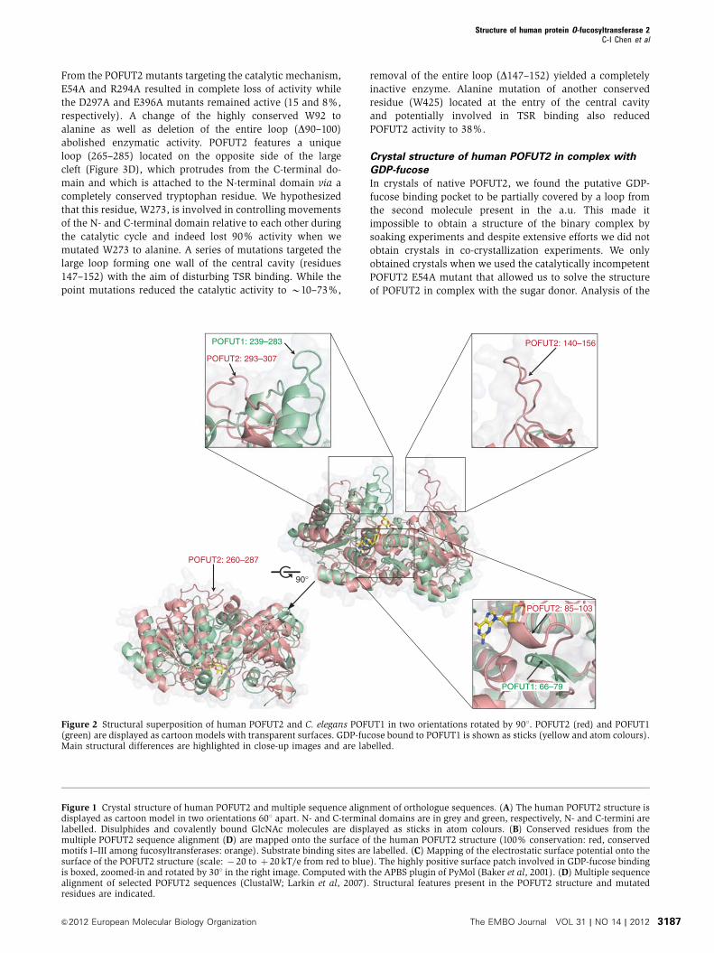
Rosenstrom, 2010; Supplementary Table SI; Figure 2;
Supplementary Figure S1). A search with the entire
POFUT2 structure revealed the structure of C. elegans
POFUT1 to be most similar (PDB 3ZY2; Lira-Navarrete
et al, 2011) followed by the nodulation fucosyltransferase
NODZ (PDB 2HHC) (Brzezinski et al, 2007), the
lipopolysaccharide heptosyltransferase I WaaC (PDB 2H1H)
(Grizot et al, 2006), and the a1,6-fucosyltransferase FUT8
(PDB 2DE0) (Ihara et al, 2007). If the N-terminal domain
alone was used in the search, then structures of POFUT1 and
NODZ gave the highest Z-scores followed by very distantly
related Rossmann-like fold proteins with low scores. A search
with the C-terminal domain alone on the other hand yielded
POFUT1, NODZ, WaaC, and FUT8 as close structural
neighbours. C. elegans POFUT1 and human POFUT2 (21%
sequence identity) have a very similar core structure in the
two Rossmann fold domains and also share the same
arrangement of N- and C-terminal domains but differ
significantly in many surface exposed structural elements
(Figure 2; Supplementary Figure S1). N-terminally, the
POFUT2 loop 85–103 that is in a coiled conformation is
replaced by an additional short b-hairpin in POFUT1. The
Structure of human protein O-fucosyltransferase 2C-I Chen et al
3184 The EMBO Journal VOL 31 | NO 14 | 2012 &2012 European Molecular Biology Organization

two structures differ dramatically in the POFUT2 region
140–200 where the long structured POFUT2 loop comprising
residues 140–156 is missing in POFUT1. In the C-terminal
domain, three striking structural differences can be identified.
First, the long POFUT2 loop (260–287) that reaches over to
the N-terminal domain opposite of the substrate binding cleft
is not present in the C. elegans POFUT1 structure. Second, the
POFUT2 loop 293–307 that builds the C-terminal wall of the
central protein cleft is replaced by an additional small domain
in POFUT1 (239–283) formed by three short helices that
together restrict access to the central POFUT1 protein cavity.
Third, the POFUT1 sequence is much shorter and the struc-
ture ends after the last C-terminal b strand where POFUT2
continues with a large disulphide linked turn followed by a
long stretch of residues in a rippled conformation defining the
entry to the central protein cavity on the C-terminal side.
Superposition of C-terminal domains of POFUT2, NODZ, and
FUT8 reveals these domains to be similar with the central
b-sheet and surrounding helices superimposing very well
(r.m.s.d. 3.1 and 2.7 A, respectively). Nevertheless, more
detailed analyses identify structural differences: Again, the
POFUT2 loop 260–287 opposite of the substrate binding cleft
is not present and also the two last strands of the central
b-sheet of NODZ and FUT8 differ by forming a regular b-sheetwhile they are in a rippled conformation with an SS-bridge
connecting the two strands in POFUT2. Superposition of the
entire POFUT2 structure with DALI hits that rank after
POFUT1 show that the N-terminal domains of FUT8 and
WaaC are structurally very different. Only the first and last
helix of the N-terminal domain and some strands of the
central b-sheet overlap well with NODZ and WaaC.
POFUT2 enzymatic activity
To validate our model of POFUT2 interaction with the GDP-
fucose sugar donor and the TSR sugar acceptor, we estab-
lished an LC-MS-based enzyme activity assay and tested the
capability of wild-type and mutant POFUT2 to fucosylate
TSR4 from rat F-spondin. In an initial set of experiments,
we analysed the effect of two different N-terminal boundaries
(D21- and D36-POFUT2) and of varying N-glycan structures,
as well as the influence of different divalent cations on the
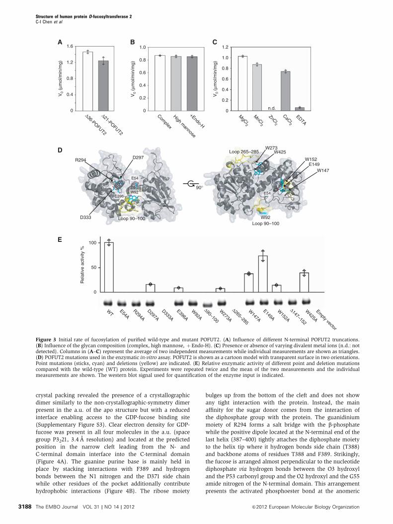
enzymatic activity of wild-type POFUT2 (Figure 3). While
neither changing the N-terminal boundary nor having a
different glycoconjugate composition had an effect on the
enzymatic activity, we found that different metal ions influ-
ence catalytic activity in different ways. Mg2þ , followed by
Mn2þ and Ca2þ , activated the enzyme in decreasing order
(100, 90, and 80% relative activity) but Zn2þ completely
abolished its activity. The enzyme was still active in the
presence of EDTA, albeit at a very low level (B5% relative
activity). Having a sensitive enzymatic activity assay avail-
able that monitors directly TSR fucosylation, we determined
the steady-state kinetic parameters for GDP-fucose and
TSR4 using wild-type high mannose type D21-POFUT2(Supplementary Figure S2). POFUT2 is an efficient enzyme
with KM values of 9.8 and 29.5 mM for GDP-fucose and TSR4,
Table I Data collection and refinement statistics
POFUT2 native POFUT2 Pt derivative POFUT2 GDP-fucose complex
Data collectionSpace group P3221 P3221 P3221Cell constants a, b, c (A) 118.6, 118.6, 196.2 118.5, 118.5, 195.0 153.0, 153.0, 185.7Wavelength l (A) 1.000 0.890 1.000Resolution range (A)a 30.0–3.0 (3.11–3.00) 20.0–5.5 (5.69–5.50) 40.0–3.4 (3.63–3.40)Unique reflections 31 375 9737 32751Completeness (%)a 96.4 (71.6) 100 (99.9) 93.3 (93.6)Multiplicity 11.3 10.4 4.7Rsym (%)a,b 12.4 (39.7) 22.4 (43.4) 16.6 (76.6)I/s(I)a 21.0 (2.4) 11.3 (3.7) 11.9 (2.1)Phasing power iso/ano 1.04/0.53
RefinementResolution range (A) 30.0–3.0 40.0–3.4Reflections (all) 31 318 32745Reflections (test set) 1593 (5.1%) 1622 (5.0)Rcrys (%) 17.4 19.3Rfree (%) 23.6 23.8
r.m.s.d.Bond lengths (A) 0.008 0.011Bond angles (deg) 1.28 1.31Wilson B-factor (A2) 50.9 —
Mean B-factor (A2)Protein 54.6 102.2Ligand — 113.4
Ramachandran plot (%)Favoured 97.0 97.0Allowed 3.0 3.0Outliers 0 0
a
Values in parentheses refer to the highest resolution shell.b
Rsym¼ShklSj|Ij,hkl�/IhklS|/ShklSjIj,hkl where /IhklS is the average of the intensity Ij,hkl over j¼ 1,y, N observations of symmetry equivalentreflections hkl.
Structure of human protein O-fucosyltransferase 2C-I Chen et al
3185&2012 European Molecular Biology Organization The EMBO Journal VOL 31 | NO 14 | 2012

respectively, and a kcat of 144 per minute. Based on our
structural results, we designed 14 mutations targeting specific
putative functional residues of the enzyme and tested enzy-
matic activity (Figure 3D and E). Out of 14 mutations, 2 did
not yield any soluble protein pointing to a critical function of
these residues in the folding pathway of the protein (D333A
and D265–285). All other mutants expressed and purified
well and equal amounts were used for the activity assay.
1
4 5
10
13
11 12
8 9
21
5
9
13 14 15
10 11 12
76 8
2
30°
A
B
C
D
60°
GlcNAc-Asn259
GlcNAc-Asn189
GlcNAc-Asn189
Asn209
60°
GDP-fucosebinding
TSRbinding
N
N
C
C
W147 W152
W273 R294
W425
D333
W92E54
Motif IIMotif I
Motif III E396
N-glycans
Deletion
Disulphide bond
α-Helix
β-Sheet
43
Q9W589 D. melanogasterQ8WR51 C. elegansQ00P38 C. savignyiQ00P33 D. rerioQ00P39 X. tropicalisQ0R343 B. taurusQ8VHI3 M. musculusQ9Y2G5 H. sapiens
Q9W589 D. melanogasterQ8WR51 C. elegansQ00P38 C. savignyiQ00P33 D. rerioQ00P39 X. tropicalisQ0R343 B. taurusQ8VHI3 M. musculusQ9Y2G5 H. sapiens
Q9W589 D. melanogasterQ8WR51 C. elegansQ00P38 C. savignyiQ00P33 D. rerioQ00P39 X. tropicalisQ0R343 B. taurusQ8VHI3 M. musculusQ9Y2G5 H. sapiens
Q9W589 D. melanogasterQ8WR51 C. elegansQ00P38 C. savignyiQ00P33 D. rerioQ00P39 X. tropicalisQ0R343 B. taurusQ8VHI3 M. musculusQ9Y2G5 H. sapiens
Structure of human protein O-fucosyltransferase 2C-I Chen et al
3186 The EMBO Journal VOL 31 | NO 14 | 2012 &2012 European Molecular Biology Organization
From the POFUT2 mutants targeting the catalytic mechanism,
E54A and R294A resulted in complete loss of activity while
the D297A and E396A mutants remained active (15 and 8%,
respectively). A change of the highly conserved W92 to
alanine as well as deletion of the entire loop (D90–100)abolished enzymatic activity. POFUT2 features a unique
loop (265–285) located on the opposite side of the large
cleft (Figure 3D), which protrudes from the C-terminal do-
main and which is attached to the N-terminal domain via a
completely conserved tryptophan residue. We hypothesized
that this residue, W273, is involved in controlling movements
of the N- and C-terminal domain relative to each other during
the catalytic cycle and indeed lost 90% activity when we
mutated W273 to alanine. A series of mutations targeted the
large loop forming one wall of the central cavity (residues
147–152) with the aim of disturbing TSR binding. While the
point mutations reduced the catalytic activity to B10–73%,
removal of the entire loop (D147–152) yielded a completely
inactive enzyme. Alanine mutation of another conserved
residue (W425) located at the entry of the central cavity
and potentially involved in TSR binding also reduced
POFUT2 activity to 38%.
Crystal structure of human POFUT2 in complex with
GDP-fucose
In crystals of native POFUT2, we found the putative GDP-
fucose binding pocket to be partially covered by a loop from
the second molecule present in the a.u. This made it
impossible to obtain a structure of the binary complex by
soaking experiments and despite extensive efforts we did not
obtain crystals in co-crystallization experiments. We only
obtained crystals when we used the catalytically incompetent
POFUT2 E54A mutant that allowed us to solve the structure
of POFUT2 in complex with the sugar donor. Analysis of the
90°
POFUT1: 239–283 POFUT2: 140–156
POFUT2: 293–307
POFUT2: 85–103
POFUT2: 260–287
POFUT1: 66–79
Figure 2 Structural superposition of human POFUT2 and C. elegans POFUT1 in two orientations rotated by 901. POFUT2 (red) and POFUT1(green) are displayed as cartoon models with transparent surfaces. GDP-fucose bound to POFUT1 is shown as sticks (yellow and atom colours).Main structural differences are highlighted in close-up images and are labelled.
Figure 1 Crystal structure of human POFUT2 and multiple sequence alignment of orthologue sequences. (A) The human POFUT2 structure isdisplayed as cartoon model in two orientations 601 apart. N- and C-terminal domains are in grey and green, respectively, N- and C-termini arelabelled. Disulphides and covalently bound GlcNAc molecules are displayed as sticks in atom colours. (B) Conserved residues from themultiple POFUT2 sequence alignment (D) are mapped onto the surface of the human POFUT2 structure (100% conservation: red, conservedmotifs I–III among fucosyltransferases: orange). Substrate binding sites are labelled. (C) Mapping of the electrostatic surface potential onto thesurface of the POFUT2 structure (scale: � 20 to þ 20 kT/e from red to blue). The highly positive surface patch involved in GDP-fucose bindingis boxed, zoomed-in and rotated by 301 in the right image. Computed with the APBS plugin of PyMol (Baker et al, 2001). (D) Multiple sequencealignment of selected POFUT2 sequences (ClustalW; Larkin et al, 2007). Structural features present in the POFUT2 structure and mutatedresidues are indicated.
Structure of human protein O-fucosyltransferase 2C-I Chen et al
3187&2012 European Molecular Biology Organization The EMBO Journal VOL 31 | NO 14 | 2012
crystal packing revealed the presence of a crystallographic
dimer similarly to the non-crystallographic-symmetry dimer
present in the a.u. of the apo structure but with a reduced
interface enabling access to the GDP-fucose binding site
(Supplementary Figure S3). Clear electron density for GDP-
fucose was present in all four molecules in the a.u. (space
group P3221, 3.4 A resolution) and located at the predicted
position in the narrow cleft leading from the N- and
C-terminal domain interface into the C-terminal domain
(Figure 4A). The guanine purine base is mainly held in
place by stacking interactions with F389 and hydrogen
bonds between the N1 nitrogen and the D371 side chain
while other residues of the pocket additionally contribute
hydrophobic interactions (Figure 4B). The ribose moiety
bulges up from the bottom of the cleft and does not show
any tight interaction with the protein. Instead, the main
affinity for the sugar donor comes from the interaction of
the diphosphate group with the protein. The guanidinium
moiety of R294 forms a salt bridge with the b-phosphatewhile the positive dipole located at the N-terminal end of the
last helix (387–400) tightly attaches the diphosphate moiety
to the helix tip where it hydrogen bonds side chain (T388)
and backbone atoms of residues T388 and F389. Strikingly,
the fucose is arranged almost perpendicular to the nucleotide
diphosphate via hydrogen bonds between the O3 hydroxyl
and the P53 carbonyl group and the O2 hydroxyl and the G55
amide nitrogen of the N-terminal domain. This arrangement
presents the activated phosphoester bond at the anomeric
Δ36-POFUT2
Δ21-POFUT2
1.6
0.8
0.4
0
1.2V
0 (μ
mol
/min
/mg)
V0
(μm
ol/m
in/m
g)
V0
(μm
ol/m
in/m
g)
Complex
High mannose
+Endo-H
0
0.2
0.4
0.6
0.8
1.0
MgCl2
MnCl2
ZnCl2
CaCl2
EDTA
0
0.2
0.4
0.6
0.8
1.2
1.0
n.d.
Loop 265–285
W92
W147E149
W152
Loop 90–100
W425W273
D333
100
R294 D297
Loop 90–100
W92E396
E54
E54
BA C
D
E
90°
WT
E54AR294A
D297A
D333A
E396A
W92A
Δ90–100
W273A
Δ265–285
W147A
E149A
W152A
Δ147–152
W425A
Empty vector
50
Rel
ativ
e ac
tivity
%
0
Figure 3 Initial rate of fucosylation of purified wild-type and mutant POFUT2. (A) Influence of different N-terminal POFUT2 truncations.(B) Influence of the glycan composition (complex, high mannose, þEndo-H). (C) Presence or absence of varying divalent metal ions (n.d.: notdetected). Columns in (A–C) represent the average of two independent measurements while individual measurements are shown as triangles.(D) POFUT2 mutations used in the enzymatic in-vitro assay. POFUT2 is shown as a cartoon model with transparent surface in two orientations.Point mutations (sticks, cyan) and deletions (yellow) are indicated. (E) Relative enzymatic activity of different point and deletion mutationscompared with the wild-type (WT) protein. Experiments were repeated twice and the mean of the two measurements and the individualmeasurements are shown. The western blot signal used for quantification of the enzyme input is indicated.
Structure of human protein O-fucosyltransferase 2C-I Chen et al
3188 The EMBO Journal VOL 31 | NO 14 | 2012 &2012 European Molecular Biology Organization

carbon to the large open channel where the TSR substrate is
postulated to bind. Both the 1C4 and 4C1 fucose ring con-
formations (C1-O1 bond in equatorial and in axial position,
respectively) have been refined against the low resolution
data (Supplementary Figure S4) and since the 1C4 conforma-
tion resulted in a slightly better fit to the experimental data
we included it in the final model of the complex. The overall
GDP-fucose binding mode in POFUT1 and POFUT2 is similar
but a detailed analysis uncovers important differences which
likely have an impact on the catalytic mechanism (Lira-
Navarrete et al, 2011). In POFUT2, the fucose moiety is
freely accessible from the large central protein cavity where
we expect TSR to bind. This is not the case in POFUT1 where
the additional small helical domain (239–283), that provides
the F261 residue holding the fucose in place, blocks access
together with F199 from the N-terminal domain. In addition,
POFUT1 residue R40 completely covers with its side chain
GDP-fucose from the top thereby limiting access from the
other side. This residue is replaced by G55 in POFUT2 and
GDP-fucose is solvent exposed. In general, GDP-fucose is
much more buried in POFUT1 compared with POFUT2 also
leading to different dihedral angles of the diphosphate group.
Although the sugar donor could only be modelled with
limited accuracy in POFUT2 due to limited resolution, the
binding mode clearly differs in several details.
Structural restraints in TSR for productive fucose
attachment
Our structure of the POFUT2 GDP-fucose complex together
with the structures of fucosylated TSR2-TSR3 of human TSP-1
(Tan et al, 2002) and fucosylated TSR1 of ADAMTS13
(Akiyama et al, 2009) enabled us to build a model of the
full enzyme substrate ternary complex. Superposition of
anomeric fucose carbons in these structures with the
anomeric carbon in the POFUT2 GDP-fucose complex,
followed by manual adjustment, yielded the overall TSR
position. This initial model was used to overlay the TSR4
structure from rat F-spondin (PDB 1VEX) (Paakkonen et al,
2006) and energy minimization of the full complex in CNS
(Brunger et al, 1998). We obtained a plausible model of the
ternary complex in which the elongated TSR unit lies in the
deep interdomain cavity of POFUT2 spanning across the
glycosyltransferase (Figure 5A). The TSR module contacts
the highly conserved POFUT2 surface via its flat hydrophobic
side, opposite of the SS-bond pattern and the tryptophan-
arginine stacking (CWR layer), where the B and C strand
show a regular antiparallel b-sheet. In addition, the rippled A
strand contacts the bottom and the side wall of the cleft. The
entire AB loop harbouring the CX2–3(S/T)CX2G motif is in
close contact with POFUT2. Only half of the TSR module was
predicted to interact with POFUT2 whereas the N-terminal
S387
T388
D371
P53
R294F389
S387
T388
D371
P53
R294F389
A
B
TSRbinding
Figure 4 GDP-fucose binding in human POFUT2. (A) Structure of the POFUT2 GDP-fucose complex displayed as a cartoon model in grey withtransparent surface. GDP-fucose is shown as sticks in atom colours. (B) Stereo figure of the GDP-fucose (cyan sticks) binding mode in POFUT2(white sticks). NCS averagedmFo-DFc electron density calculated after molecular replacement and one round of refinement (in absence of GDP-fucose) is shown as mesh in blue (3s). Hydrogen bonds are displayed as green dotted lines. The orientation is different than in (A) for betterclarity.
Structure of human protein O-fucosyltransferase 2C-I Chen et al
3189&2012 European Molecular Biology Organization The EMBO Journal VOL 31 | NO 14 | 2012

part of the A strand and the BC loop including the jar handle
are solvent exposed. Interestingly, we find a second con-
served POFUT2 surface patch located on the 90–100 loop that
could interact with an additional TSR domain (e.g., TSR2–
TSR3 in TSP-1) or with other protein domains on the
C-terminal side of the TSR unit (e.g., EGF repeat in TSP-1
or CA domain in ADAMTS13).
In order to validate our model of TSR binding, we ex-
pressed a series of F-spondin TSR4 mutants in HEK293Tcells,
purified the secreted protein from the medium and analysed
its fucosylation state by mass spectrometry (Figure 5B and C).
Changes to the conserved WXXWXXW motif in the A strand
(in the case of TSR4 of F-spondin L4XXW7XXW10), the key
element of the TSR fold that forms the multi-layered deloca-
lized p-system with the conserved arginine residues from the
B strand (WR of CWR), reduced the efficiency of fucosylation
significantly. Single mutations of W7 and W10 to alanine
produced only 24 and 31% of fucosylated TSR4, double
mutations on both sites further reduced the fucosylation to
14% compared with wild type whereas the L4A mutation did
not have any influence. Next, we introduced mutations in the
AB loop close to the threonine (T*16) that undergoes fuco-
sylation: replacing the valine (V15) next to it with an iso-
leucine or changing the glycine (G20) at the end of the turn to
alanine had only minor effects (86, 75% fucosylation). On the
other hand, when we introduced two additional glycine
residues right before the threonine (GGT*, mimicking the
fucosylation sequon in EGF repeats) to create a larger loop
between the disulphide forming cysteine and the threonine,
fucosylation was completely abolished. The disulphide bond
pattern is a hallmark in the fold of TSR therefore we inves-
tigated how fucosylation is affected by removing SS-bonds.
Mutation of the cysteines forming the SS-bond between the A
and C strand, where we postulated no interaction with
POFUT2, was well tolerated with no reduction in fucosylation
levels (C2,35A). To our surprise, the removal of the second
SS-bond between the A and C strand had no dramatic effect
with 80% fucosylated product (C13,46A). On the other hand,
removal of the SS-bond that connects the AB loop (C17,51A)
with the C-terminal end of the C strand resulted in 55%
reduced fucosylation levels. When we removed both SS-
bonds, which together stabilize the 3D conformation of the
AB loop, we could not detect any fucosylated TSR product.
We superimposed all available structures of TSR modules
(PDB 1LSL, Tan et al, 2002; 1VEX and 1SZL, Paakkonen et al,
2006; 3GHM, Akiyama et al, 2009; 2BBX, Tossavainen et al,
2006) at the predicted substrate binding site of POFUT2 and
analysed structural and sequence restraints potentially
involved in substrate recognition. The structural
information was compared with the profile hidden Markov
model (HMM) of the Pfam entry of the thrombospondin type
1 domain (PF00090, http://pfam.sanger.ac.uk; Supplemen-
tary Figure S5) and multiple sequence alignments of all
human TSR type 1 domains present in the Uniprot database
(http://www.uniprot.org) that contain the putative fucosyla-
tion motif (CX2–3(S/T)CX2GG). The sequence data revealed
the enormous sequence diversity besides only a few con-
served residues that seem to be absolutely necessary for the
TSR fold. Taking into account our experimental data about
the contribution of selected TSR structural elements to
POFUT2 fucosylation efficiency, we hypothesized that sub-
strate specificity is not primarily encoded in the protein
sequence but rather in the unusual 3D structure of one half
of the TSR module.
Consequently, we investigated whether a minimal TSR
module containing only seven conserved structural residues
of the TSR hallmark elements (disulphide pattern and side-
chain stacking) would be sufficient as a POFUT2 sugar
acceptor. We designed and expressed a truncated TSR module
based on F-spondin TSR4 consisting of approximately half
Fuc
osyl
atio
n ef
ficie
ncy
(%)
WT
GG
-insertion
W7A
W10A
L4A
G20A
C(2,35)A
C(13,46)A
C(17,51)A
C(13,46,17,51)A
V15I
W(7,10)A
BA
C
A
B
C
L4
W7W10 V15
G20
C2
C35
C13
C46
C17C51
T*16
N
C
Loop 90–100
A
100
50
0 n.d. n.d.
B
C
Figure 5 Model of the POFUT2 GDP-fucose TSR complex and POFUT2 enzymatic activity using mutant TSR as substrate. (A) Surfacerepresentation of the POFUT2 GDP-fucose structure with POFUT2 residues coloured as in Figure 1B and GDP-fucose as sticks. TSR4 from ratF-spondin (PDB 1VEX) is displayed as a cartoon model in cyan. (B) Structure of TSR4 from rat F-spondin. Residues that were mutated forfucosylation measurements are numbered. (C) Relative fucosylation efficiency of TSR mutants. Wild-type and mutant TSR was expressed andpurified from HEK293T cells and analysed by mass spectrometry for fucosylation efficiency. n.d.: not detected. Experiments were repeatedtwice and the mean of two measurements and the individual measurements are displayed.
Structure of human protein O-fucosyltransferase 2C-I Chen et al
3190 The EMBO Journal VOL 31 | NO 14 | 2012 &2012 European Molecular Biology Organization

the length and tested fucosylation by mass spectrometry. The
first TSR residue of this artificial minimal TSR corresponds to
W10 in the A strand (Figure 5B) and includes the entire AB
loop where the consensus sequence is located. Residues
predicted not to be involved in POFUT2 interaction connect-
ing the B and C strand were deleted and replaced with a short
GSG linker (Figure 6A). This artificial minimal TSR com-
prised only 29 residues compared with 55 for wild-type TSR
and is referred to as mini-TSR in this article.
Wild-type TSR4 and mini-TSR were expressed and isolated
from E. coli and used as acceptor substrates for purified
POFUT2 in a qualitative in-vitro fucosyltransferase assay.
The reaction products were monitored for fucose incorpora-
tion by mass spectrometry. An increase in mass of 146Da
corresponding to the addition of one fucose moiety was
observed for both the wild-type (Figure 6B) and mini-TSR
substrate (Figure 6C). Fucosylation was only observed in
reactions that included POFUT2. Thus, POFUT2 recognizes
and modifies both wild-type and truncated artificial mini-
TSR. We further validated our findings by expressing mini-
TSR with an N-terminal His–FLAG tag in HEK293T cells. The
secreted mini-TSR protein was purified from the culture
medium and analysed by mass spectrometry. We found a
homogenous species of 7004.98Da corresponding to the
tagged mini-TSR with two hexoses and one deoxyhexose
attached (Figure 6D). To further analyse the glycosylation
state of the mini-TSR, secreted and purified protein was
digested with human rhinovirus 3C to remove the tag,
reduced and alkylated, and digested with Lys-C protease
yielding a peptide covering the predicted glycosylation sites.
MS/MS analysis confirmed the sequence (GPWSDCSVTCGK)
and revealed the glycan modifications (Figure 6E). Loss of
one hexose and one hexose-deoxyhexose disaccharide from
the parent ion (m/z 912.36) was observed in the MS spectrum
(m/z 831.33 and 758.30, respectively). In addition, the mass
difference between the b2 and b3 ion corresponded to one
WT-TSR4 TIPCLLSPWSEWSDCSVTCGKGMRTRQRMLKSLAELGDCNEDLEQAEKCMLPECP
Mini-TSR4 --------------------WSDCSVTCGKGMRTR---------------GSG---------------QAEKCMLPECP
B strandA strand C strand
Rel
ativ
e ab
unda
nce
100
06000 70002000 4000 50003000
6335.84
6481.91
Rel
ativ
e ab
unda
nce
100
06000 70002000 4000 50003000
3267.40
3413.44
012 00010 000 14 0002000 6000 80004000
Rel
ativ
e ab
unda
nce
Rel
ativ
e ab
unda
nce
1007004.98
1515.59, z =1
758.30, z =2
912.36, z =2
1013.39
831.33, z =2
503.21
698.28
465.21
z =2 [M+H-308.1]+
[M+2H]2+
GPWSDCSVTCGK
b2
y4 b3
y9
A
CB
ED
[M]
[M]
[M-146]
[M-146]
Δ60, z =2
155.08
b2b3
m/zm/z
m/zm/z
100
012001000 1400 1600200 400 600 800
N
CN
C
GSG
120
Mannose
Glucose
Fucose
308x25
Figure 6 O-fucosylation of mini-TSR. (A) Amino-acid sequences of wild-type TSR4 from rat F-spondin and engineered mini-TSR are compared.Amino acids that form the three strands are indicated. Wild-type TSR and mini-TSR were expressed and isolated from E. coli. These moduleswere used as acceptor substrates in the POFUT2 in-vitro assay and the reaction products analysed by mass spectrometry. Both the wild-typeTSR (B) and mini-TSR (C) show two peaks that differ by 146Da, indicating the presence or absence of one fucose molecule. Wild-type andmini-TSR are drawn as cartoons with disulphide bonds in yellow and fucose as a red triangle. Mini-TSR is approximately half the size of wild-type TSR, with a truncated A strand and the deleted BC loop replaced by a three residue linker, GSG. (D) An N-terminal His6–FLAG-3C-taggedmini-TSR was expressed in mammalian HEK293Tcells, isolated from the medium and analysed by mass spectrometry. The mono-isotopic mass(7004.98) represents the intact, tagged mini-TSR with two hexoses and one deoxyhexose. (E) Secreted and purified mini-TSR from HEK293Tcell expression was digested with human rhinovirus 3C and Lys-C protease and analysed by mass spectrometry. MS/MS analysis confirmed thesequence (GPWSDCSVTCGK) and revealed the glycan modifications. Loss of one hexose and one hexose-deoxyhexose disaccharide from theparent ion (m/z 912.36) was observed in the MS spectrum (m/z 831.33 and 758.30, respectively) and the mass difference between the b2 andb3 ion corresponded to one hexose molecule attached to the tryptophan. The characteristic � 120Da fragment (m/z 698.28) represents thetypical signature motif for C-hexosylation.
Structure of human protein O-fucosyltransferase 2C-I Chen et al
3191&2012 European Molecular Biology Organization The EMBO Journal VOL 31 | NO 14 | 2012

hexose molecule attached to the tryptophan and the char-
acteristic � 120Da fragment (m/z 698.28) revealed the tryp-
tophan hexosylation to be C-linked (Hofsteenge et al, 1994).
In summary, the mass spectrometry analysis demonstrates
that mini-TSR is modified with an O-linked fucose-glucose
disaccharide and a C-linked mannose on the tryptophan
as it is the case for wild-type TSR4 (Hofsteenge et al, 2001).
These experiments confirmed that mini-TSR is a substrate for
POFUT2 both in HEK293T cells and in our in-vitro
fucosyltransferase assay.
Discussion
POFUT2 protein structure and TSR substrate
recognition
Here, we present the crystal structure of human POFUT2 that
together with orthologues forms the GT68 family of inverting
protein O-fucosyltransferases of the GT-B fold (Cantarel et al,
2009). Although many structures of glycosyltransferases are
solved there is only very limited structural information
available for glycosyltransferases that transfer the sugar
moiety to a peptide or protein acceptor. For the three
glycosyltransferase folds (GT-A, B, C) only structures of
GALNT2 and GALNT10 (GT-A), AglB and PglB (GT-C), and
MurG, OGT, and POFUT1 (GT-B) have been solved (Hu et al,
2003; Fritz et al, 2006; Kubota et al, 2006; Maita et al, 2010;
Lazarus et al, 2011; Lira-Navarrete et al, 2011; Lizak et al,
2011). Interestingly, all of these enzymes (except POFUT1)
use flexible solvent exposed protein regions as sugar acceptor
whereas POFUT2 was shown to fucosylate only properly
folded TSR (Luo et al, 2006b). The POFUT2 structure now
gives insight into how substrate recognition, specificity, and
catalysis are achieved with the special requirements of a
properly folded 3D protein sugar acceptor that transiently
forms a protein–protein interface with a glycosyltransferase.
Our data suggest that POFUT2 recognizes key 3D structural
TSR elements formed by the disulphide pattern and side-
chain stacking common to sequence-wise degenerated TSR
modules. A search of the PDB using DALI with coordinates of
a minimal TSR poly-alanine module (regions predicted to
interact with POFUT2) identified only known structures of
TSR domains without discovering this structural motif in any
other protein. Therefore, substrate specificity seems to be
achieved by the structural complementarity of a part of the
TSR fold with the POFUT2 binding site and the wide TSR
sequence diversity does not play a role as long as the critical
TSR fold motif is intact. From our experimental data and the
structural models, we conclude that disrupting the conforma-
tion of the rippled A strand (formed by the WXXWXXW
motif, LXXWXXW in TSR4 from F-spondin) and of the AB
loop (defined by the second and third disulphides) impairs
TSR substrate recognition and fucosylation efficiency
(Figure 5C). Our data of the mini-TSR also show that starting
the A strand directly at the third tryptophan of the
WXXWXXW motif has no negative effect on fucosylation as
the conformation of the shortened A strand is still intact.
Proposed key interactions are located at the entry of the large
TSR binding cavity where conserved POFUT2 residues W152
from the N-terminal domain and W425 from the very
C-terminal part define the most narrow part of the cleft
(B15 A) allowing only space for a two stranded b-sheet toenter the cavity (Figure 7A). C-terminal residues seem to lock
the position of the bound TSR module by interacting with the
backbone bulge formed by the second and third tryptophan
of the LXXWXXW motif of the rippled A strand. Thereby,
C-terminal POFUT2 residues act as a ruler to position the S/T
residue undergoing modification exactly at the right position
for E54-dependent deprotonation and nucleophilic attack at
the anomeric GDP-fucose carbon. This model is supported
by our observation that fucose attachment is reduced by 90
and 61% for the W152A and W425A mutation, respectively
(Figure 3E), and that exchange of the second and third
F-spondin TSR4 tryptophan (responsible for bulge formation)
to alanine results in 75 and 69% reduction of fucosylated
product, respectively (W7, W10 in Figure 5C). The second key
interaction is predicted to take place between the AB loop
(where the consensus motif CX2–3(S/T)CX2G is located) and
the conserved POFUT2 residues Asn51, Pro52, Pro53, Glu54,
Leu58, Asp61, and Glu221 (Figure 7A). These residues,
together with L224 that inserts its side chain exactly where
the C strand starts to crossover the B strand, ensure via TSR
backbone interactions that only the unique 3D motif at the
very tip of the TSR module can undergo fucosylation. This
structural motif is mainly defined by the length and confor-
mation of the AB loop, which is held in place by the two
disulphide bonds and which is encoded in the CX2–3(S/
T)CX2G sequon. Our observation explains experimental
data where insertion of two additional glycines before
the threonine (CX2–3GG(S/T)CX2G, changing the length of
the loop) as well as removing the two SS-bonds, which
together are responsible for pulling the C strand over
the B strand and stabilization of the AB-loop conformation,
completely abolished fucose attachment (Figure 5C).
Rational design of a minimal POFUT2 substrate resulted in
the artificial mini-TSR molecule which we predicted to con-
tain all necessary structural features for folding into the
correct TSR fold needed for productive fucosylation. We
found mini-TSR to be modified with the common glycan
structures known from wild-type TSR, thereby confirming
that it indeed can fold into the correct 3D AB-loop TSR
structure and act as a POFUT2 substrate. This result not
only defines a minimal POFUT2 substrate and validates our
proposed binding mode but also brings new insight into
folding and stability of TSR molecules. It shows that the
correct disulphide bond pattern needed for the proper AB-
loop conformation can be established with a minimal side-
chain stacking unit composed of one tryptophan and one
arginine residue only.
Having realized that POFUT2 substrate recognition is likely
to be driven by the conserved TSR residues responsible for
the unique layered TSR fold, we wondered how the substrate
binding site is able to accept the large charge and size
variation of amino acids on the remaining B40 sequence
positions. Strikingly, we found in our model of the POFUT2
TSR complex that out of the B30 TSR residues building the
upper half of the TSR (predicted to interact with POFUT2) 10
are conserved determining the TSR fold (in the central layer
of stacking residues and SS-bonds) or are part of the con-
sensus motif for fucosylation. Another nine residues are
solvent exposed and likely not involved in POFUT2 interac-
tion. At the 11 remaining TSR positions where wide sequence
diversity is present we find large cavities in POFUT2, ready
to accommodate side chains of different lengths or with
different physicochemical properties (Figure 7B).
Structure of human protein O-fucosyltransferase 2C-I Chen et al
3192 The EMBO Journal VOL 31 | NO 14 | 2012 &2012 European Molecular Biology Organization

Our model of TSR–POFUT2 interaction is also compatible
with tryptophan C-mannosylation present on many TSR
(Hofsteenge et al, 2001; Tan et al, 2002) as the CWR layer
with a potentially attached a-mannosyl residue is facing
solvent and would therefore not be involved in POFUT2
interaction. Finally, our structural data also explain why
POFUT2 is specific for TSR modules and why EGF repeats,
the other known protein module to contain O-fucose
modifications, are not accepted as substrate. EGF repeats
simply do not have the critical 3D TSR elements (e.g.,
AB-loop conformation) needed for binding to the POFUT2
active site.
Catalytic mechanism of POFUT2
Structural and biochemical data of wild-type and mutant
POFUT2 allowed us to suggest a catalytic mechanism for
the fucosyltransferase reaction (Figure 8). POFUT2 belongs
to the GT-B family of inverting glycosyltransferases where
the key catalytic residue acts as a general base responsible
for deprotonation of the nucleophile functional group of the
sugar acceptor (Lairson et al, 2008). Only the fully conserved
residues E54 and D297 are in close proximity of the TSR S/T
hydroxyls that undergo fucose attachment. Both residues are
located right at the entry of the GDP-fucose binding site, E54
on a surface exposed loop of the N-terminal domain, and
D297 in a long loop in the C-terminal domain. The E54
carboxylate side chain is closer to the anomeric carbon and
is freely accessible while D297 is located slightly further away
and its side chain is sandwiched between the two
guanidinium groups of R294 and R303 in the native
POFUT2 structure reducing its mobility and, importantly,
lowering its pKa. Complete loss of enzymatic activity for
the E54A POFUT2 mutant (the D297A mutant retained
B16% activity), and the fact that we were able to obtain a
structure of the non-hydrolysed sugar donor complex with
the same catalytically inactive E54A mutant, are strong
arguments for E54 to be the catalytic base of POFUT2.
D297, on the other hand, seems to be needed to correctly
orient the R294 side chain for binding of the GDP-fucose
diphosphate group. Positioning of aspartate, glutamate, or
histidine as the catalytic base on the N-terminal domain
facing the sugar donor binding site (as seen in POFUT2) is
known from other inverting GT-B family members like WaaC
(Grizot et al, 2006), T4 phage glucosyltransferase BGT
(Lariviere et al, 2003), H. pylori fucosyltransferase a1,3-FucT (Sun et al, 2007), E. coli MurG (Hu et al, 2003), or
VvGT1 (Offen et al, 2006) of which structures have been
solved and the catalytic base residue has been identified
(Supplementary Figure S6). During E54-dependent deproto-
nation, the TSR S/T nucleophile can attack the anomeric
A B
W425
T423W152
L224
BC A BC A
Figure 7 Structural details of the proposed TSR binding mode in human POFUT2. (A) Surface representation (grey) of POFUT2 with boundTSR4 (F-spondin) as ribbons (cyan) and the GDP-fucose substrate as sticks. POFUT2 residues predicted to recognize key elements of the TSRfold are coloured: W152 and W425 (red) scan the width of the substrate; T423 (orange) senses the bulge in the A strand and acts as a ruler;L224 (pink) scans the crossing over of the C strand; N51, P52, P53, E54, L58, D61, E221 (all in blue) recognize the correct conformation of theAB loop. Key structural residues of the TSR fold are displayed as sticks in cyan (atom colours). (B) The wide TSR sequence diversity of POFUT2substrates can be explained by the proposed TSR binding mode. POFUT2 provides large cavities (solid surface, blue) for highly variable TSRresidues (green) thereby tolerating side chains of different size and physicochemical properties at these positions. TSR side chains can also varyat positions predicted to be solvent exposed (purple) while TSR residues important for the fold and the fucosylation motif are strictly conserved(orange).
E54
R294TSR-OH
E54
R294
GDP-fucose
GDP-fucoseTSR
Figure 8 Proposed enzymatic mechanism for POFUT2-dependentTSR fucosylation. Chemical drawing of the enzymatic reaction. Thecarboxylate side chain of E54 deprotonates the TSR S/T hydroxylgroup that attacks as a nucleophile the anomeric carbon of the GDP-fucose sugar donor. R294 activates the labile bond by forming a saltbridge with the b-phosphate group. The arrangement ofcritical catalytic residues in the model of the ternary POFUT2GDP-fucose TSR complex is shown. Distances: E54 carboxylate toTSR-OH 3.1 A, TSR-OH to GDP-fucose anomeric carbon 3.3 A.
Structure of human protein O-fucosyltransferase 2C-I Chen et al
3193&2012 European Molecular Biology Organization The EMBO Journal VOL 31 | NO 14 | 2012

carbon of the GDP-fucose to form the new glycosidic bond
with inverted configuration followed by release of the two
products. The fucose ring has been modelled in the 1C4
conformation resulting in a better fit to the 3.4 A electron
density if compared with the 4C1 conformation. Interestingly,
the two conformations differ only in the ring flip and anB381conformational change around the P1-O2P bond. Both ring
conformations would need considerable distortion for in-line
nucleophilic attack geometry seen in other inverting glyco-
syltransferases such as VvGT1 (Offen et al, 2006). Therefore,
the reactive fucose ring conformation will most likely be
established upon binding of the TSR sugar acceptor.
Activation of the labile phosphoester bond of the sugar
donor is mainly achieved by the charged residue R294 that
binds the diphosphate group and by the positive dipoles of
two helices near the diphospho and the pentose moiety.
Mutation of R294 to alanine in the human protein as well
as the corresponding mutation in the C. elegans orthologue
(Canevascini et al, 2006) resulted in complete loss of
enzymatic activity consistent with its role in directly
binding the GDP-fucose diphosphates. A multiple sequence
alignment of different fucosyltransferases reveals that R294 is
also conserved in POFUT1 and FUT8 (Martinez-Duncker
et al, 2003) where mutation of this residue also abolishes
enzymatic activity (Takahashi et al, 2000; Okajima et al,
2005). While the vast majority of inverting GT-B family
glycosyltransferases are metal-ion independent there are
three family members for which metal ions significantly
enhance activity: T4 phage BGT (Morera et al, 2001),
hamster POFUT1 (Wang and Spellman, 1998), as well as
human POFUT2 which we described here (Figure 3C).
Despite many soaking and co-crystallization trials with
Mn2þ we were not able to localize the cation in anomalous
difference Fourier electron density maps in the POFUT2 apo
structure. In addition, we only obtained crystals of the GDP-
fucose complex when we added EDTA to the crystallization
buffer to remove all remaining metal ions from the protein.
These findings, together with the fact that the enzyme retains
B5% enzymatic activity in the presence of EDTA, point to a
role of metal ions in product release. This is known from
other GT-B fold glycosyltransferases like T4 phage BGTwhere
Mn2þ complexes the pyrophosphate group of the UDP pro-
duct at the place occupied by the sugar moiety in the UDP-Glc
complex structure (Morera et al, 2001). Similarly, the Mn2þ
binding site has been identified in the crystal structure of a
C. elegans POFUT1 GDP Mn2þ complex where the ion also
binds the pyrophosphate of the GDP product exactly where
the fucose moiety is placed in the GDP-fucose structure.
However, in the latter case the authors do not relate this
finding to the metal-dependent enzymatic activity (Lira-
Navarrete et al, 2011). Other GT-B fold glycosyltransferases
like FUT8 seem to have metal-independent ways of
nucleotide diphosphate release and do not need divalent
cations to reach full activity (Ihara et al, 2006). Kinetic
experiments yielded a kcat of 144 per minute for the
POFUT2 enzyme, a value that is comparable with published
results from other glycosyltransferases (Ihara et al, 2006; Sun
et al, 2007). KM values on the other hand differ with 9.8 mMfor GDP-fucose and 29.5 mM for TSR4. A KM in the low
micromolar range for the sugar donor is common for GT-B fold
glycosyltransferases (Jeanneau et al, 2004; Grizot et al, 2006;
Ihara et al, 2006) while the KM value for TSR4 is rather low.
In summary, POFUT2 seems to utilize a well-established
catalytic mechanism for GT-B fold inverting glycosyltrans-
ferases with E54 acting as general base. This is in contrast to
the suggested mechanism in C. elegans POFUT1 where no
residue acting as catalytic base could be identified and the
reaction after cleavage of the glycosidic bond (facilitated by
R240) proceeds via an oxocarbenium-phosphate ion pair
transition state and subsequent attack of the acceptor OH
group at the anomeric carbon (Lira-Navarrete et al, 2011).
Substrate specificity of protein glycosyltransferases
The mechanism of glycan transfer to a protein or peptide
acceptor has for a long time been poorly understood. It was
largely unknown how short sequence motifs present in
polypeptides of wide sequence diversity can be modified by
a position-specific enzyme. It was only recently that crystal
structures of glycosyltransferases in complex with acceptor
peptides gave insight into substrate specificity and how a few
key elements present in the recognition sequons enable
glycosylation of specific residues. Structures of acceptor
peptide complexes are now available for all glycosyltransfer-
ase families and reveal surprising similarities: GALNT10
(GT-A) (Fritz et al, 2006), OGT (GT-B) (Lazarus et al, 2011),
and PglB (GT-C) (Lizak et al, 2011) all recognize glycosylation
sequons in flexible unstructured protein regions and bind the
substrate peptide mainly via backbone interactions. Many
structured water molecules are present providing an
adaptable protein interface ready to accommodate a wide
range of polypeptides with side chains of different size,
charge, and polarity. Sequon specificity is most clearly
defined in PglB (Asn-X-Ser/Thr) where a WWD protein
motif binds the Ser/Thr residue side chain at the þ 2
position and thereby positions the asparagine correctly for
N-linked glycosylation. A similar mode is used in GALNT2
where the proline at the þ 3 position is specifically bound to
position the Ser/Thr correctly in the active site. For OGT on
the other hand, no O-GlcNAcylation motif has been identified
so far but a preference for residues that form an extended
peptide conformation near the glycosylation site can be
explained by the binding mode of the peptide as seen in the
crystal structures.
Here, we present a completely novel mode of substrate
recognition for protein glycosyltransferases that explains why
the specific fucosylation consensus motif CX2–3(S/T)CX2G
(Hofsteenge et al, 2001) can only be modified in the context
of a properly folded TSR protein domain and how these
structural constraints are not in conflict with the wide
sequence diversity present on fucosylated TSR. POFUT2 has
evolved to specifically recognize unique 3D structural TSR
elements, which are defined by a few strictly conserved
residues and the consensus motif itself. This allows for
wide sequence diversity at all the other TSR positions,
probably reflecting the diverse biological functions of
proteins containing the TSR module.
Materials and methodsA detailed description of expression and purification of wild-typeand mutant POFUT2 and TSR proteins, enzymatic assays anddetection of TSR fucosylation states by mass spectrometry isgiven in Supplementary data.
Structure of human protein O-fucosyltransferase 2C-I Chen et al
3194 The EMBO Journal VOL 31 | NO 14 | 2012 &2012 European Molecular Biology Organization

Crystallization, data collection and structure determinationPOFUT2 crystals were grown at 41C by the vapour diffusion methodin 96-well crystallization plates by mixing 0.1ml of POFUT2 proteinsolution (7.5mg/ml) with 0.1ml of crystallization buffer (20mMTris–HCl, pH 8.5, 12% PEG 20000). For native data collection,crystals were soaked in mother liquor containing 25% ethyleneglycol and frozen in liquid nitrogen. For heavy atom derivatization,crystals were soaked in mother liquor containing 5mM of K2PtCl4for 6min. Diffraction data were collected at beamlines X06DA andX10SA at the Swiss Light Source synchrotron in Villigen,Switzerland. Diffraction images were processed and scaled withHKL-2000 (Otwinowski et al, 1997). The structure of POFUT2 wassolved by the SIRAS method using two platinum sites per moleculeidentified in SHELXD (Sheldrick, 2008). Heavy atom sites were usedfor phase calculation and refinement of sites in Sharp (Bricogneet al, 2003) followed by density modification using Solomon(Abrahams and Leslie, 1996). Phases from density modificationwere then used for automatic model building in PHENIX (Adamset al, 2010) and in BUCCANEER (Cowtan, 2006) followed by manualcompletion of the model using COOT (Emsley et al, 2010).Structures were refined by the crystallographic simulatedannealing routine followed by individual B-factor refinement inPHENIX including NCS restraints.
For crystals of the POFUT2 GDP-fucose complex, 13.6mg/mlof high mannose type E54A POFUT2 was incubated in proteinbuffer containing 3.5mM EDTA and 1mM GDP-fucose (Sigma) for30min on ice before setting up the crystallization experiment.Crystals were grown at 201C by the vapour diffusion method in96-well crystallization plates by mixing 0.25ml of protein solutionwith 0.25ml of crystallization buffer (20% PEG 3350, 0.2M NaSCN).Crystals of the complex were cryoprotected and frozen as describedfor native crystals. The structure of the POFUT2 GDP-fucosecomplex was solved by molecular replacement using PHASER(McCoy et al, 2007) with the native POFUT2 structure as search
model and subsequent refinement in PHENIX. Clear mFo-DFcdifference electron density for the missing GDP-fucose moietieswas visible in the active sites for all four molecules in the a.u.The structures of native POFUT2 and of the E54A-POFUT2 GDP-fucose complex were refined by several rounds of manualrebuilding in COOT followed by refinement in PHENIX. Of thefour E54A-POFUT2 molecules present in the a.u. only chains A, B,and C have full occupancy whereas chain D is partially occupied (orhas high mobility). This results in less well-defined 2mFo-DFcelectron density for chain D. All crystal structures were validatedusing MolProbity (Chen et al, 2010) and COOT. Structural images forfigures were prepared with PyMOL (http://pymol.sourceforge.net/).Atomic coordinates and structure factors have been deposited in thePDB with entry codes 4AP5 (apo) and 4AP6 (GDP-fucose complex).
Supplementary dataSupplementary data are available at The EMBO Journal Online(http://www.embojournal.org).
Acknowledgements
We thank Ragna Sack from the Protein Analysis Facility for supportin mass spectrometry experiments and the staff at the Swiss LightSource (Villigen, Switzerland) for support in X-ray data collection.The Friedrich Miescher Institute for Biomedical Research is a part ofthe Novartis Research Foundation.Author contributions: HG, CC, and JH designed the experiments.
CC, JK, DK, DH, JH, and HG carried out experiments and analysedthe data. HG and CC wrote the paper.
Conflict of interest
The authors declare that they have no conflict of interest.
ReferencesAbrahams JP, Leslie AGW (1996) Methods used in the structure
determination of bovine mitochondrial F1 ATPase. ActaCrystallogr D Biol Crystallogr 52: 30–42
Adams PD, Afonine PV, Bunkoczi G, Chen VB, Davis IW, Echols N,Headd JJ, Hung L-W, Kapral GJ, Grosse-Kunstleve RW, McCoy AJ,Moriarty NW, Oeffner R, Read RJ, Richardson DC, Richardson JS,Terwilliger TC, Zwart PH (2010) PHENIX: a comprehensivePython-based system for macromolecular structure solution.Acta Crystallogr D Biol Crystallogr 66: 213–221
Akiyama M, Takeda S, Kokame K, Takagi J, Miyata T (2009) Crystalstructures of the noncatalytic domains of ADAMTS13 revealmultiple discontinuous exosites for von Willebrand factor. ProcNatl Acad Sci USA 106: 19274–19279
Apweiler R, Hermjakob H, Sharon N (1999) On the frequency ofprotein glycosylation, as deduced from analysis of the SWISS-PROT database. Biochim Biophys Acta 1473: 4–8
Baker NA, Sept D, Joseph S, Holst MJ, McCammon JA (2001)Electrostatics of nanosystems: application to microtubules andthe ribosome. Proc Natl Acad Sci USA 98: 10037–10041
Bjoern S, Foster DC, Thim L, Wiberg FC, Christensen M, KomiyamaY, Pedersen AH, Kisiel W (1991) Human plasma and recombinantfactor VII. Characterization of O-glycosylations at serine residues52 and 60 and effects of site-directed mutagenesis of serine 52 toalanine. J Biol Chem 266: 11051–11057
Bricogne G, Vonrhein C, Flensburg C, Schiltz M, Paciorek W (2003)Generation, representation and flow of phase information instructure determination: recent developments in and aroundSHARP 2.0. Acta Crystallogr D Biol Crystallogr 59: 2023–2030
Brunger AT, Adams PD, Clore GM, DeLano WL, Gros P, Grosse-Kunstleve RW, Jiang JS, Kuszewski J, Nilges M, Pannu NS, ReadRJ, Rice LM, Simonson T, Warren GL (1998) Crystallography &NMR system: a new software suite for macromolecular structuredetermination. Acta Crystallogr D Biol Crystallogr 54: 905–921
Brzezinski K, Stepkowski T, Panjikar S, Bujacz G, Jaskolski M(2007) High-resolution structure of NodZ fucosyltransferaseinvolved in the biosynthesis of the nodulation factor. ActaBiochim Pol 54: 537–549
Buko AM, Kentzer EJ, Petros A, Menon G, Zuiderweg ER, Sarin VK(1991) Characterization of a posttranslational fucosylation in thegrowth factor domain of urinary plasminogen activator. Proc NatlAcad Sci USA 88: 3992–3996
Canevascini S, Kozma K, Grob M, Althaus J, Klein D, Chiquet-Ehrismann R, Hofsteenge J (2006) Protein O-Fucosylation isImportant for Normal Distal Tip Migration In:European WormMeeting, Hersonissos, Crete, Greece
Cantarel BL, Coutinho PM, Rancurel C, Bernard T, Lombard V,Henrissat B (2009) The Carbohydrate-Active EnZymes database(CAZy): an expert resource for Glycogenomics. Nucleic Acids Res37: D233–D238
Chen VB, Arendall III WB, Headd JJ, Keedy DA, Immormino RM,Kapral GJ, Murray LW, Richardson JS, Richardson DC(2010) MolProbity: all-atom structure validation for macromole-cular crystallography. Acta Crystallogr D Biol Crystallogr 66:12–21
Cowtan K (2006) The Buccaneer software for automated modelbuilding. 1. Tracing protein chains. Acta Crystallogr D BiolCrystallogr 62: 1002–1011
Du J, Takeuchi H, Leonhard-Melief C, Shroyer KR, Dlugosz M,Haltiwanger RS, Holdener BC (2010) O-fucosylation of thrombos-pondin type 1 repeats restricts epithelial to mesenchymal transi-tion (EMT) and maintains epiblast pluripotency during mousegastrulation. Dev Biol 346: 25–38
Emsley P, Lohkamp B, Scott WG, Cowtan K (2010) Featuresand development of coot. Acta Crystallogr D Biol Crystallogr 66:486–501
Freeze HH (2007) Congenital disorders of glycosylation: CDG-I,CDG-II, and beyond. Curr Mol Med 7: 389–396
Fritz TA, Raman J, Tabak LA (2006) Dynamic associationbetween the catalytic and lectin domains of human UDP-GalNAc:Polypeptide a-N-acetylgalactosaminyltransferase-2. JBiol Chem 281: 8613–8619
Gonzalez de Peredo A, Klein D, Macek B, Hess D, Peter-Katalinic J,Hofsteenge J (2002) C-mannosylation and O-fucosylation ofthrombospondin type 1 repeats. Mol Cell Proteomics 1: 11–18
Structure of human protein O-fucosyltransferase 2C-I Chen et al
3195&2012 European Molecular Biology Organization The EMBO Journal VOL 31 | NO 14 | 2012

Grizot S, Salem M, Vongsouthi V, Durand L, Moreau F, Dohi H,Vincent S, Escaich S, Ducruix A (2006) Structure of theEscherichia coli heptosyltransferase WaaC: binary complexeswith ADP and ADP-2-deoxy-2-fluoro heptose. J Mol Biol 363:383–394
Harris RJ, Ling VT, Spellman MW (1992) O-linked fucose is presentin the first epidermal growth factor domain of factor XII but notprotein C. J Biol Chem 267: 5102–5107
Harris RJ, Spellman MW (1993) O-linked fucose and other post-translational modifications unique to EGF modules. Glycobiology3: 219–224
Hess D, Keusch JJ, Oberstein SA, Hennekam RC, Hofsteenge J(2008) Peters Plus syndrome is a new congenital disorder ofglycosylation and involves defective O-glycosylation of throm-bospondin type 1 repeats. J Biol Chem 283: 7354–7360
Hofsteenge J, Huwiler KG, Macek B, Hess D, Lawler J, Mosher DF,Peter-Katalinic J (2001) C-mannosylation and O-fucosylationof the Thrombospondin Type 1 Module. J Biol Chem 276:6485–6498
Hofsteenge J, Mueller DR, de Beer T, Loeffler A, Richter WJ,Vliegenthart JFG (1994) New type of linkage between a carbohy-drate and a protein: C-glycosylation of a specific tryptophanresidue in human RNase Us. Biochemistry 33: 13524–13530
Holm L, Rosenstrom P (2010) Dali server: conservation mapping in3D. Nucleic Acids Res 38: W545–W549
Hu Y, Chen L, Ha S, Gross B, Falcone B, Walker D, Mokhtarzadeh M,Walker S (2003) Crystal structure of the MurG:UDP-GlcNAccomplex reveals common structural principles of a superfamilyof glycosyltransferases. Proc Natl Acad Sci USA 100: 845–849
Ihara H, Ikeda Y, Taniguchi N (2006) Reaction mechanism andsubstrate specificity for nucleotide sugar of mammalian a1,6-fucosyltransferase–a large-scale preparation and characterizationof recombinant human FUT8. Glycobiology 16: 333–342
Ihara H, Ikeda Y, Toma S, Wang X, Suzuki T, Gu J, Miyoshi E,Tsukihara T, Honke K, Matsumoto A, Nakagawa A, Taniguchi N(2007) Crystal structure of mammalian a1,6-fucosyltransferase,FUT8. Glycobiology 17: 455–466
Jaeken J, Matthijs G (2007) Congenital disorders of glycosylation: arapidly expanding disease family. Annu Rev Genomics Hum Genet8: 261–278
Jeanneau C, Chazalet V, Auge C, Soumpasis DM, Harduin-Lepers A,Delannoy P, Imberty A, Breton C (2004) Structure-function ana-lysis of the human sialyltransferase ST3Gal I. J Biol Chem 279:13461–13468
Kozma K, Keusch JJ, Hegemann B, Luther KB, Klein D, Hess D,Haltiwanger RS, Hofsteenge J (2006) Identification and character-ization of a b1,3-glucosyltransferase that synthesizes the Glc-b1,3-Fuc disaccharide on thrombospondin type 1 repeats. J BiolChem 281: 36742–36751
Kubota T, Shiba T, Sugioka S, Furukawa S, Sawaki H, Kato R,Wakatsuki S, Narimatsu H (2006) Structural basis of carbohy-drate transfer activity by human UDP-GalNAc: polypeptide a-N-acetylgalactosaminyltransferase (pp-GalNAc-T10). J Mol Biol 359:708–727
Lairson LL, Henrissat B, Davies GJ, Withers SG (2008)Glycosyltransferases: structures, functions, and mechanisms.Annu Rev Biochem 77: 521–555
Lariviere L, Gueguen-Chaignon V, Morera S (2003) Crystal struc-tures of the T4 phage b-glucosyltransferase and the D100Amutant in complex with UDP-glucose: glucose binding andidentification of the catalytic base for a direct displacementmechanism. J Mol Biol 330: 1077–1086
Larkin MA, Blackshields G, Brown NP, Chenna R, McGettigan PA,McWilliam H, Valentin F, Wallace IM, Wilm A, Lopez R,Thompson JD, Gibson TJ, Higgins DG (2007) Clustal W andClustal X version 2.0. Bioinformatics 23: 2947–2948
Lazarus MB, Nam Y, Jiang J, Sliz P, Walker S (2011) Structure ofhuman O-GlcNAc transferase and its complex with a peptidesubstrate. Nature 469: 564–567
Lesnik Oberstein SA, Kriek M, White SJ, Kalf ME, Szuhai K, denDunnen JT, Breuning MH, Hennekam RC (2006) Peters Plussyndrome is caused by mutations in b3GALTL, a putativeglycosyltransferase. Am J Hum Genet 79: 562–566
Lira-Navarrete E, Valero-Gonzalez J, Villanueva R, Martınez-JulvezM, Tejero T, Merino P, Panjikar S, Hurtado-Guerrero R (2011)
Structural insights into the mechanism of protein O-fucosylation.PLoS One 6: e25365
Lizak C, Gerber S, Numao S, Aebi M, Locher KP (2011) X-raystructure of a bacterial oligosaccharyltransferase. Nature 474:350–355
Luo Y, Koles K, Vorndam W, Haltiwanger RS, Panin VM (2006a)Protein O-fucosyltransferase 2 adds O-fucose to thrombospondintype 1 repeats. J Biol Chem 281: 9393–9399
Luo Y, Nita-Lazar A, Haltiwanger RS (2006b) Two distinct pathwaysfor O-fucosylation of epidermal growth factor-like or thrombos-pondin type 1 repeats. J Biol Chem 281: 9385–9392
Luther KB, Haltiwanger RS (2009) Role of unusual O-glycans inintercellular signaling. Int J Biochem Cell Biol 41: 1011–1024
Maita N, Nyirenda J, Igura M, Kamishikiryo J, Kohda D (2010)Comparative structural biology of eubacterial and archaeal oligo-saccharyltransferases. J Biol Chem 285: 4941–4950
Martinez-Duncker I, Mollicone R, Candelier JJ, Breton C, Oriol R(2003) A new superfamily of protein-O-fucosyltransferases,a2-fucosyltransferases, and a6-fucosyltransferases: phylogenyand identification of conserved peptide motifs. Glycobiology 13:1C–5C
McCoy AJ, Grosse-Kunstleve RW, Adams PD, Winn MD, Storoni LC,Read RJ (2007) Phaser crystallographic software. J ApplCrystallogr 40: 658–674
Morera S, Lariviere L, Kurzeck J, Aschke-Sonnenborn U, FreemontPS, Janin J, Ruger W (2001) High resolution crystal structures ofT4 phage b-glucosyltransferase: induced fit and effect of substrateand metal binding. J Mol Biol 311: 569–577
Nishimura H, Takao T, Hase S, Shimonishi Y, Iwanaga S(1992) Human factor IX has a tetrasaccharide O-glycosidicallylinked to serine 61 through the fucose residue. J Biol Chem 267:17520–17525
Offen W, Martinez-Fleites C, Yang M, Kiat-Lim E, Davis BG, TarlingCA, Ford CM, Bowles DJ, Davies GJ (2006) Structure of aflavonoid glucosyltransferase reveals the basis for plant naturalproduct modification. EMBO J 25: 1396–1405
Okajima T, Xu A, Lei L, Irvine KD (2005) Chaperone activity ofprotein O-fucosyltransferase 1 promotes notch receptor folding.Science 307: 1599–1603
Otwinowski Z, Minor W (1997) Processing of X-ray diffraction datacollected in oscillation mode. InMethods in Enzymology, Vol. 276:Macromolecular Crystallography, Carter Jr CW, Sweet RM (eds)Part A, pp 307–326. New York: Academic Press
Paakkonen K, Tossavainen H, Permi P, Rakkolainen H, Rauvala H,Raulo E, Kilpelainen I, Guntert P (2006) Solution structuresof the first and fourth TSR domains of F-spondin. Proteins 64:665–672
Rana NA, Haltiwanger RS (2011) Fringe benefits: functional andstructural impacts of O-glycosylation on the extracellular domainof Notch receptors. Curr Opin Struct Biol 21: 583–589
Ricketts LM, Dlugosz M, Luther KB, Haltiwanger RS, Majerus EM(2007) O-fucosylation is required for ADAMTS13 secretion. J BiolChem 282: 17014–17023
Sato T, Sato M, Kiyohara K, Sogabe M, Shikanai T, Kikuchi N,Togayachi A, Ishida H, Ito H, Kameyama A, Gotoh M, NarimatsuH (2006) Molecular cloning and characterization of anovel human b1,3-glucosyltransferase, which is localized at theendoplasmic reticulum and glucosylates O-linked fucosylglycanon thrombospondin type 1 repeat domain. Glycobiology 16:1194–1206
Sheldrick G (2008) A short history of SHELX. Acta Crystallogr A 64:112–122
Stahl M, Uemura K, Ge C, Shi S, Tashima Y, Stanley P (2008) Rolesof Pofut1 and O-fucose in mammalian Notch signaling. J BiolChem 283: 13638–13651
Stanley P (2007) Regulation of Notch signaling by glycosylation.Curr Opin Struct Biol 17: 530–535
Sun HY, Lin SW, Ko TP, Pan JF, Liu CL, Lin CN, Wang AH, Lin CH(2007) Structure and mechanism of Helicobacter pylori fucosyl-transferase. A basis for lipopolysaccharide variation and inhibitordesign. J Biol Chem 282: 9973–9982
Takahashi T, Ikeda Y, Tateishi A, Yamaguchi Y, Ishikawa M,Taniguchi N (2000) A sequence motif involved in the donorsubstrate binding by a1,6-fucosyltransferase: the role of theconserved arginine residues. Glycobiology 10: 503–510
Structure of human protein O-fucosyltransferase 2C-I Chen et al
3196 The EMBO Journal VOL 31 | NO 14 | 2012 &2012 European Molecular Biology Organization

Tan K, Duquette M, Liu JH, Dong Y, Zhang R, Joachimiak A, LawlerJ, Wang JH (2002) Crystal structure of the TSP-1 type 1 repeats: anovel layered fold and its biological implication. J Cell Biol 159:373–382
Tossavainen H, Pihlajamaa T, Huttunen TK, Raulo E, Rauvala H,Permi P, Kilpelainen I (2006) The layered fold of the TSR domainof P. falciparum TRAP contains a heparin binding site. Protein Sci15: 1760–1768
Wang LW, Dlugosz M, Somerville RPT, Raed M, Haltiwanger RS,Apte SS (2007) O-fucosylation of thrombospondin type 1repeats in ADAMTS-like-1/Punctin-1 regulates secretion:implications for the ADAMTS superfamily. J Biol Chem 282:17024–17031
Wang Y, Spellman MW (1998) Purification and characterization of aGDP-fucose:polypeptide fucosyltransferase from Chinese hamsterovary cells. J Biol Chem 273: 8112–8118
Structure of human protein O-fucosyltransferase 2C-I Chen et al
3197&2012 European Molecular Biology Organization The EMBO Journal VOL 31 | NO 14 | 2012