Embed Size (px)
Citation preview
Article
Bicaudal-D1 regulates the intracellular sorting andsignalling of neurotrophin receptorsMarco Terenzio1,‡,†,**, Matthew Golding1,§,†, Matthew R G Russell2, Krzysztof B Wicher3,¶,
Ian Rosewell4, Bradley Spencer-Dene5, David Ish-Horowicz3 & Giampietro Schiavo1,6,*
Abstract
We have identified a new function for the dynein adaptor BicaudalD homolog 1 (BICD1) by screening a siRNA library for genes affect-ing the dynamics of neurotrophin receptor-containing endosomesin motor neurons (MNs). Depleting BICD1 increased theintracellular accumulation of brain-derived neurotrophic factor(BDNF)-activated TrkB and p75 neurotrophin receptor (p75NTR) bydisrupting the endosomal sorting, reducing lysosomal degradationand increasing the co-localisation of these neurotrophin receptorswith retromer-associated sorting nexin 1. The resulting re-routingof active receptors increased their recycling to the plasmamembrane and altered the repertoire of signalling-competent TrkBisoforms and p75NTR available for ligand binding on the neuronalsurface. This resulted in attenuated, but more sustained, AKT acti-vation in response to BDNF stimulation. These data, together withour observation that Bicd1 expression is restricted to the develop-ing nervous system when neurotrophin receptor expression peaks,indicate that BICD1 regulates neurotrophin signalling by modulatingthe endosomal sorting of internalised ligand-activated receptors.
Keywords Bicd1; intracellular sorting; neurotrophin signalling; p75NTR;
TrkB
Subject Categories Membrane & Intracellular Transport; Neuroscience
DOI 10.15252/embj.201387579 | Received 3 December 2013 | Revised 14 March
2014 | Accepted 23 April 2014 | Published online 11 June 2014
The EMBO Journal (2014) 33: 1582–1598
Introduction
Neurons have complex axonal and dendritic arborisations,
which are paramount for the function of the nervous system. The
development and maturation of these extended neuronal networks
require tight regulation of the intracellular transport of organelles
and cargoes, such as mRNA, mitochondria, growth factor receptors
and their signalling adaptor molecules, which in turn are necessary
to ensure neuronal growth, differentiation and survival (Salinas
et al, 2008; Hirokawa et al, 2010; Ascano et al, 2012).
Neurotrophins are essential for the development and mainte-
nance of the nervous system (Ascano et al, 2012). The neurotrophin
family consists of four structurally related growth factors: nerve
growth factor (NGF), brain-derived neurotrophic factor (BDNF),
NT3 and NT4/5 (Bibel & Barde, 2000; Huang & Reichardt, 2001).
These ligands bind and activate the tropomyosin-related kinases
TrkA, TrkB and TrkC either alone or in combination with p75
neurotrophin receptor (p75NTR), which lacks enzymatic activity
(Simi & Ibanez, 2010). Neurotrophin receptors have different bind-
ing specificities for their ligands: TrkA binds preferentially to NGF;
TrkB to BDNF and NT4/5; and TrkC to NT-3, whilst p75NTR can
bind all four neurotrophins, and their precursors, the pro-neurotro-
phins (Teng et al, 2010). The considerable crosstalk in the binding
of neurotrophins to their receptors generates a variety of signalling
outputs resulting in diverse cellular responses, which are initiated
both by ligand availability and the relative abundance of each
neurotrophin receptor at specific locations on the neuronal surface
(Reichardt, 2006; Ascano et al, 2012). This signalling diversity is
further enhanced because the same neurotrophin can have opposing
effects, depending on whether it is presented to the neuron as the
precursor or mature form (Teng et al, 2010). It is therefore impor-
tant that neurotrophin signalling is tightly controlled both spatially
and temporally, particularly in the developing nervous system when
neurotrophins and their receptors are abundantly expressed.
The signalling cascades activated by neurotrophins in the
neuronal periphery are regulated by the endocytosis, sorting and
trafficking of neurotrophin receptors along the axon or dendrites
1 Molecular NeuroPathobiology Laboratory, Cancer Research UK London Research Institute, London, UK2 Electron Microscopy Laboratory, Cancer Research UK London Research Institute, London, UK3 Developmental Genetics Laboratory, Cancer Research UK London Research Institute, London, UK4 Transgenic Services laboratory, Cancer Research UK London Research Institute, London, UK5 Experimental Histopathology Laboratory, Cancer Research UK London Research Institute, London, UK6 Sobell Department of Motor Neuroscience & Movement Disorders, UCL-Institute of Neurology, University College London, London, UK
*Corresponding author. Tel: +44 7918 738393; Fax: +44 20 7813 3107; E-mail: [email protected]**Corresponding author. Tel: +972 8934 4265; Fax: +972 8934 4112; E-mail: [email protected]†Contributed equally to this work‡Present address: Department of Biological Chemistry, Weizmann Institute of Science, Rehovot, Israel§Present address: Centre for Microvascular Research, William Harvey Research Institute, Barts & The London School of Medicine and Dentistry, Queen Mary University ofLondon, London, UK¶Present address: Medimmune, Department of Antibody Discovery and Protein Engineering, Cambridge, UK
The EMBO Journal Vol 33 | No 14 | 2014 ª 2014 The Authors1582
Published online: June 11, 2014
towards the cell body, where they elicit transcriptional responses
(Ibanez, 2007; Ascano et al, 2012; Schmieg et al, 2014). We have
previously shown that the binding fragment of tetanus neurotoxin
(HCT) is a reliable probe to monitor neurotrophin receptor uptake
and intracellular trafficking (Bercsenyi et al, 2013). Indeed, HCT
and p75NTR bound to NGF accumulate in clathrin-coated pits and
are internalised by a clathrin-mediated endocytic pathway in
motor neurons (MNs) (Deinhardt et al, 2006a, 2007). Further-
more, HCT shares the same axonal retrograde transport organelles
with neurotrophins and their receptors (Lalli & Schiavo, 2002;
Deinhardt et al, 2006b).
Since a comprehensive understanding of the molecular mecha-
nisms controlling neurotrophin receptor internalisation and traffick-
ing is still lacking, we sought to identify new genes involved in this
pathway by performing an small interfering RNA (siRNA) screen in
MNs derived from transgenic HB9-GFP embryonic stem (ES) cells
using HCT and an antibody directed against the extracellular domain
of p75NTR (ap75NTR) as fluorescent reporters (Terenzio et al, 2014).
Importantly, expression of green fluorescent protein (GFP) driven
by the Hb9 homeobox gene enhancer (Wichterle et al, 2002) facili-
tated the unequivocal identification of MNs generated from these ES
cells upon differentiation (Terenzio et al, 2014).
Using this approach, we identified a small cohort of genes affect-
ing the intracellular accumulation of HCT and ap75NTR when
silenced (see Supplementary Table S1). Knockdown of one gene in
particular, Bicaudal D homolog 1 (Bicd1), revealed an increased
internalisation phenotype for HCT and was selected for further
analyses, which demonstrated that BICD1 depletion also increased
the intracellular accumulation of ligand-bound p75NTR and TrkB
(Terenzio et al, 2014).
Bicaudal D homolog 1 is known to participate in endosomal traf-
ficking and dynein-mediated processes (Hoogenraad et al, 2001;
Matanis et al, 2002; Bianco et al, 2010; Aguirre-Chen et al, 2011),
including retrograde transport in neuronal cells (Wanschers et al,
2007). Furthermore, BICD1 has important roles in the development
and function of the Drosophila and Caenorhabditis elegans nervous
systems (Li et al, 2010). We now show that BICD1 is a key regulator
of the intracellular trafficking of neurotrophin receptors and that it
performs this role by controlling the BDNF-triggered sorting and
progression of p75NTR and TrkB through the endosomal pathway.
Thus, BICD1 depletion disrupted this mechanism to favour p75NTR
and TrkB recycling over lysosomal degradation, which in turn
affected signalling responses to BDNF stimulation.
Our data identify a novel function for BICD1 as a modulator of
neurotrophin receptor dynamics and signalling, which may also be
relevant for other receptor tyrosine kinases in different cellular
systems.
Results
Bicd1 is strongly expressed in the developing central andperipheral nervous systems
One of the most prominent candidates from our siRNA screen was
BICD1 (Terenzio et al, 2014). Since BICD1 and related proteins are
cytoplasmic dynein adaptors (Hoogenraad et al, 2001; Matanis et al,
2002), and BICD1 has been postulated to function in retrograde
transport (Wanschers et al, 2007), we decided to focus our efforts
on addressing the role played by BICD1 in the regulation of neuro-
trophin receptor trafficking in our model system.
We used a Bicd1 gene-trapped ES cell clone (Bicd1gt/+; gene trap
RRP227) for the functional characterisation of BICD1. This gene trap
vector expressed a b-galactosidase cassette, allowing us to deter-
mine Bicd1 expression patterns in chimeric embryos generated from
Bicd1gt/+ ES cells (Fig 1A). X-gal histochemistry of Bicd1gt/+ ES
cell-derived embryos revealed that at embryonic day 12 (E12), Bicd1
was highly and almost exclusively expressed in ventral horn MNs
of the developing spinal cord (Fig 1A0–A‴), dorsal root ganglia
(DRG; Fig 1A″–A‴) and brain (Fig 1A0 and A″). These X-gal-stained
embryos were then paraffin embedded, cross-sectioned and
immunostained to reveal that Bicd1 expression was highest in HB9-
positive ventral horn MNs (Fig 1B), a sub-population of DRG
neurons (Fig 1D) and in the nerve tracts emanating from these
structures (Fig 1A‴,C). High Bicd1 expression in the developing
nervous tissue closely matched the pattern of immunoreactivity for
BDNF, Trk receptors and p75NTR (Supplementary Fig S1A–C). Alto-
gether, these observations suggest that BICD1 plays a role in the
developing nervous system at a time when neurotrophins and their
receptors are highly expressed (Davies, 1994; Klein, 1994; Ernfors,
2001).
However, only one developmental day later, the expression
pattern of Bicd1 had dramatically changed: at E14.5, Bicd1-LacZ had
been lost from the brain (Supplementary Fig S1E) and spinal cord
(Supplementary Fig S1F), yet was still strongly retained in DRG
(Supplementary Fig S1F and G) and was most notably upregulated
in skin, skeletal muscle and heart ventricles, but interestingly, not
in the atria (Supplementary Fig S1H).
Bicd1gt/gt ES-cell-derived MNs are a reliable in vitro modelsystem to study BICD1 function
We had planned to use Bicd1gt/+ × C57/Bl6 mouse chimeras to
generate a Bicd1gt/+ founder colony. However, mating of male
chimeras with approximately 75% ES cell contribution to wild-type
females produced no mutant offspring, indicating that germline trans-
mission of the Bicd1gt/+ allele had failed to take place. We therefore
selected for loss of heterozygosity in culture using high G418 concen-
trations (Lefebvre et al, 2001). Resultant homozygous Bicd1gt/gt ES
cells were isolated, expanded and differentiated into MNs.
BICD1 transcript and protein levels were reduced by approxi-
mately 70% in Bicd1gt/gt MNs compared to wild-type cells (Fig 1E
and F; Supplementary Fig S1D). Complete ablation of Bicd1 expres-
sion was not expected since gene trap insertions are prone to unpre-
dicted downstream mRNA splicing events (Voss et al, 1998). In
contrast, Bicd2 mRNA levels were found to be approximately 80%
higher in Bicd1gt/gt MNs compared to wild-type controls (Supple-
mentary Fig S1D), suggesting that BICD2 might compensate for the
partial loss of BICD1. However, this scenario is unlikely since BICD2
protein levels were not increased in Bicd1gt/gt MNs (Supplementary
Fig S2A).
BICD1 depletion had no significant effect on the expression of
the MN markers Hb9 and ChAT (choline acetyltransferase; Supple-
mentary Fig S1D), or p75NTR (Fig 1E and F; Supplementary
Fig S1D), whilst Trk protein levels showed an approximate 30%
decrease using a pan-Trk antibody (Fig 1F). Since MNs do not
ª 2014 The Authors The EMBO Journal Vol 33 | No 14 | 2014
Marco Terenzio et al BICD1 regulates neurotrophin receptor signalling The EMBO Journal
1583
Published online: June 11, 2014
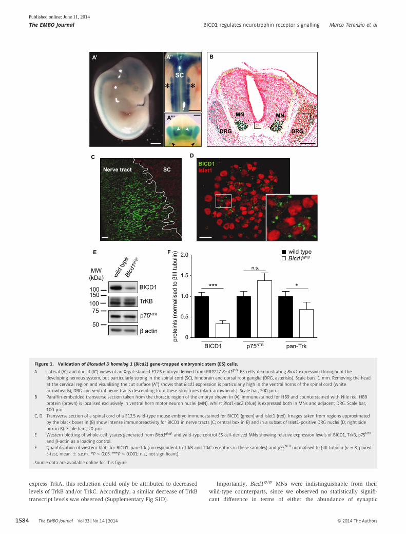
express TrkA, this reduction could only be attributed to decreased
levels of TrkB and/or TrkC. Accordingly, a similar decrease of TrkB
transcript levels was observed (Supplementary Fig S1D).
Importantly, Bicd1gt/gt MNs were indistinguishable from their
wild-type counterparts, since we observed no statistically signifi-
cant difference in terms of either the abundance of synaptic
A’ A’’
A’’’
DRG DRG
MNMN
SC
SCNerve tractBICD1Islet1
* *
B
C D
E
MW(kDa)
100 BICD1
BICD1 pan-Trk
wild type
TrKB
0prot
einl
s (n
orm
alis
ed to
βIII
tubu
lin)
0.5
1.0
1.5
***
2.0
β actin
p75NTR
p75NTR
wild
type
Bicd1g
t/gt Bicd1gt/gt
10075
50
150
F
n.s.
*
Figure 1. Validation of Bicaudal D homolog 1 (Bicd1) gene-trapped embryonic stem (ES) cells.
A Lateral (A0) and dorsal (A″) views of an X-gal-stained E12.5 embryo derived from RRP227 Bicd1gt/+ ES cells, demonstrating Bicd1 expression throughout thedeveloping nervous system, but particularly strong in the spinal cord (SC), hindbrain and dorsal root ganglia (DRG, asterisks). Scale bars, 1 mm. Removing the headat the cervical region and visualising the cut surface (A‴) shows that Bicd1 expression is particularly high in the ventral horns of the spinal cord (whitearrowheads), DRG and ventral nerve tracts descending from these structures (black arrowheads). Scale bar, 200 lm.
B Paraffin-embedded transverse section taken from the thoracic region of the embryo shown in (A), immunostained for HB9 and counterstained with Nile red. HB9protein (brown) is localised exclusively in ventral horn motor neuron nuclei (MN), whilst Bicd1-lacZ (blue) is expressed both in MNs and adjacent DRG. Scale bar,100 lm.
C, D Transverse section of a spinal cord of a E12.5 wild-type mouse embryo immunostained for BICD1 (green) and Islet1 (red). Images taken from regions approximatedby the black boxes in (B) show intense immunoreactivity for BICD1 in nerve tracts (C; central box in B) and in a subset of Islet1-positive DRG nuclei (D; right sidebox in B). Scale bars, 20 lm.
E Western blotting of whole-cell lysates generated from Bicd1gt/gt and wild-type control ES cell-derived MNs showing relative expression levels of BICD1, TrkB, p75NTR
and b-actin as a loading control.F Quantification of western blots for BICD1, pan-Trk (correspondent to TrkB and TrkC receptors in these samples) and p75NTR normalised to bIII tubulin (n = 3, paired
t-test, mean � s.e.m., *P < 0.05, ***P < 0.001; n.s., not significant).
Source data are available online for this figure.
The EMBO Journal Vol 33 | No 14 | 2014 ª 2014 The Authors
The EMBO Journal BICD1 regulates neurotrophin receptor signalling Marco Terenzio et al
1584
Published online: June 11, 2014
boutons (Supplementary Fig S2B and C) or the morphology of the
neurite network (Supplementary Fig S2D–G). Taken together, these
results demonstrated that RRP227 gene trap ES-cell-derived MNs
were a suitable model system with which to investigate the func-
tional consequences of BICD1 depletion on the trafficking of
neurotrophin receptors.
BICD1 depletion affects the internalisation and intracellular fateof HCT
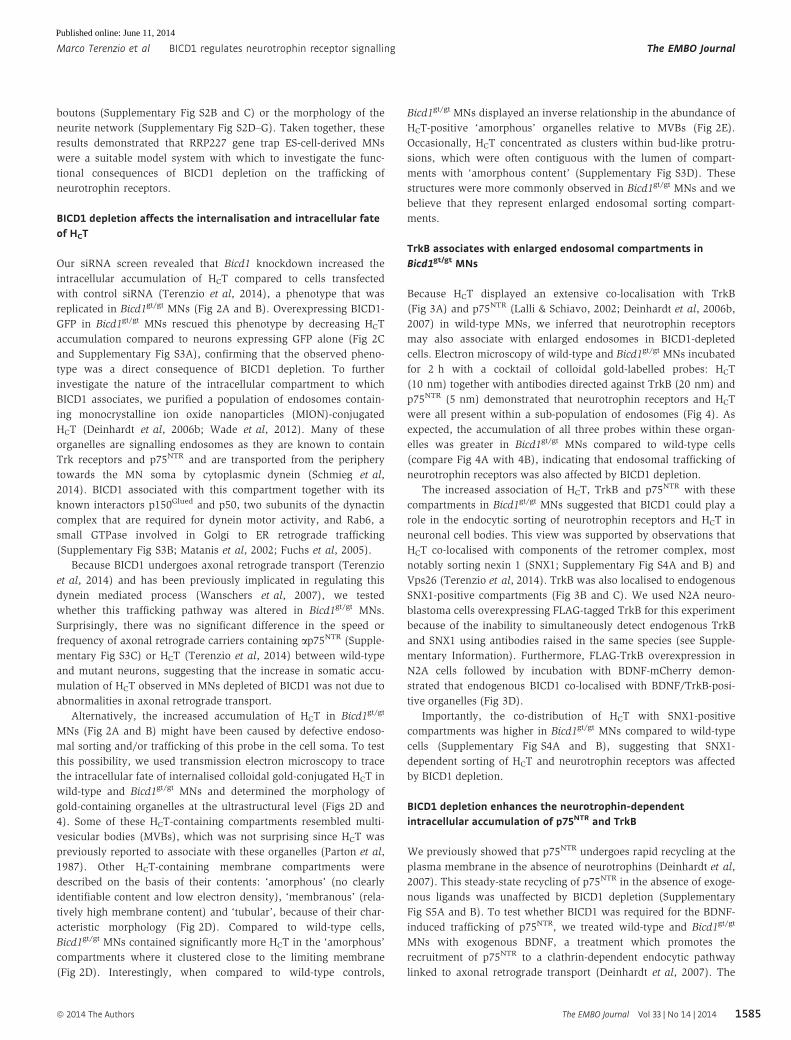
Our siRNA screen revealed that Bicd1 knockdown increased the
intracellular accumulation of HCT compared to cells transfected
with control siRNA (Terenzio et al, 2014), a phenotype that was
replicated in Bicd1gt/gt MNs (Fig 2A and B). Overexpressing BICD1-
GFP in Bicd1gt/gt MNs rescued this phenotype by decreasing HCT
accumulation compared to neurons expressing GFP alone (Fig 2C
and Supplementary Fig S3A), confirming that the observed pheno-
type was a direct consequence of BICD1 depletion. To further
investigate the nature of the intracellular compartment to which
BICD1 associates, we purified a population of endosomes contain-
ing monocrystalline ion oxide nanoparticles (MION)-conjugated
HCT (Deinhardt et al, 2006b; Wade et al, 2012). Many of these
organelles are signalling endosomes as they are known to contain
Trk receptors and p75NTR and are transported from the periphery
towards the MN soma by cytoplasmic dynein (Schmieg et al,
2014). BICD1 associated with this compartment together with its
known interactors p150Glued and p50, two subunits of the dynactin
complex that are required for dynein motor activity, and Rab6, a
small GTPase involved in Golgi to ER retrograde trafficking
(Supplementary Fig S3B; Matanis et al, 2002; Fuchs et al, 2005).
Because BICD1 undergoes axonal retrograde transport (Terenzio
et al, 2014) and has been previously implicated in regulating this
dynein mediated process (Wanschers et al, 2007), we tested
whether this trafficking pathway was altered in Bicd1gt/gt MNs.
Surprisingly, there was no significant difference in the speed or
frequency of axonal retrograde carriers containing ap75NTR (Supple-
mentary Fig S3C) or HCT (Terenzio et al, 2014) between wild-type
and mutant neurons, suggesting that the increase in somatic accu-
mulation of HCT observed in MNs depleted of BICD1 was not due to
abnormalities in axonal retrograde transport.
Alternatively, the increased accumulation of HCT in Bicd1gt/gt
MNs (Fig 2A and B) might have been caused by defective endoso-
mal sorting and/or trafficking of this probe in the cell soma. To test
this possibility, we used transmission electron microscopy to trace
the intracellular fate of internalised colloidal gold-conjugated HCT in
wild-type and Bicd1gt/gt MNs and determined the morphology of
gold-containing organelles at the ultrastructural level (Figs 2D and
4). Some of these HCT-containing compartments resembled multi-
vesicular bodies (MVBs), which was not surprising since HCT was
previously reported to associate with these organelles (Parton et al,
1987). Other HCT-containing membrane compartments were
described on the basis of their contents: ‘amorphous’ (no clearly
identifiable content and low electron density), ‘membranous’ (rela-
tively high membrane content) and ‘tubular’, because of their char-
acteristic morphology (Fig 2D). Compared to wild-type cells,
Bicd1gt/gt MNs contained significantly more HCT in the ‘amorphous’
compartments where it clustered close to the limiting membrane
(Fig 2D). Interestingly, when compared to wild-type controls,
Bicd1gt/gt MNs displayed an inverse relationship in the abundance of
HCT-positive ‘amorphous’ organelles relative to MVBs (Fig 2E).
Occasionally, HCT concentrated as clusters within bud-like protru-
sions, which were often contiguous with the lumen of compart-
ments with ‘amorphous content’ (Supplementary Fig S3D). These
structures were more commonly observed in Bicd1gt/gt MNs and we
believe that they represent enlarged endosomal sorting compart-
ments.
TrkB associates with enlarged endosomal compartments inBicd1gt/gt MNs
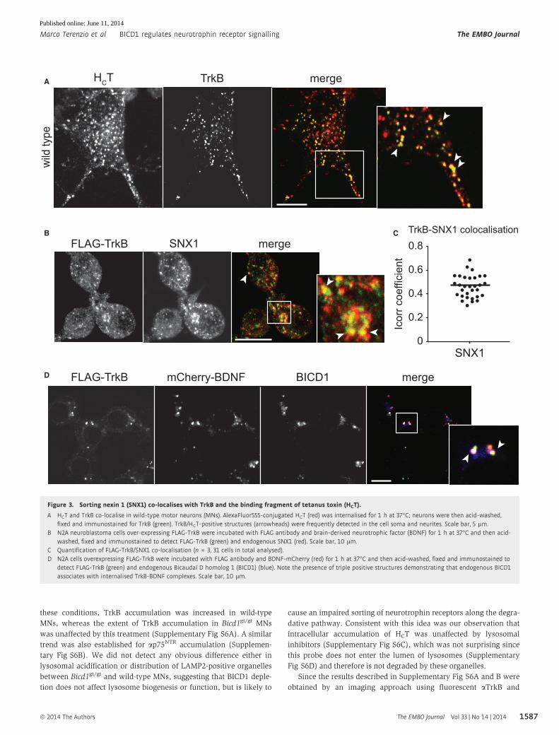
Because HCT displayed an extensive co-localisation with TrkB
(Fig 3A) and p75NTR (Lalli & Schiavo, 2002; Deinhardt et al, 2006b,
2007) in wild-type MNs, we inferred that neurotrophin receptors
may also associate with enlarged endosomes in BICD1-depleted
cells. Electron microscopy of wild-type and Bicd1gt/gt MNs incubated
for 2 h with a cocktail of colloidal gold-labelled probes: HCT
(10 nm) together with antibodies directed against TrkB (20 nm) and
p75NTR (5 nm) demonstrated that neurotrophin receptors and HCT
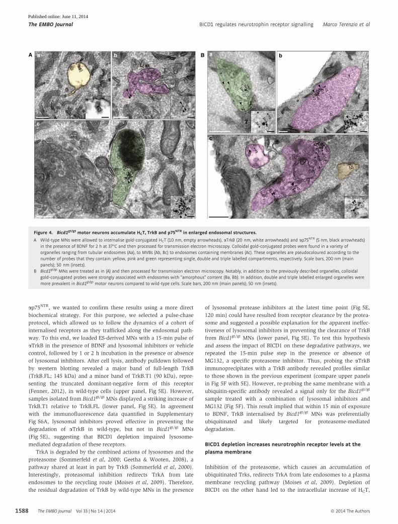
were all present within a sub-population of endosomes (Fig 4). As
expected, the accumulation of all three probes within these organ-
elles was greater in Bicd1gt/gt MNs compared to wild-type cells
(compare Fig 4A with 4B), indicating that endosomal trafficking of
neurotrophin receptors was also affected by BICD1 depletion.
The increased association of HCT, TrkB and p75NTR with these
compartments in Bicd1gt/gt MNs suggested that BICD1 could play a
role in the endocytic sorting of neurotrophin receptors and HCT in
neuronal cell bodies. This view was supported by observations that
HCT co-localised with components of the retromer complex, most
notably sorting nexin 1 (SNX1; Supplementary Fig S4A and B) and
Vps26 (Terenzio et al, 2014). TrkB was also localised to endogenous
SNX1-positive compartments (Fig 3B and C). We used N2A neuro-
blastoma cells overexpressing FLAG-tagged TrkB for this experiment
because of the inability to simultaneously detect endogenous TrkB
and SNX1 using antibodies raised in the same species (see Supple-
mentary Information). Furthermore, FLAG-TrkB overexpression in
N2A cells followed by incubation with BDNF-mCherry demon-
strated that endogenous BICD1 co-localised with BDNF/TrkB-posi-
tive organelles (Fig 3D).
Importantly, the co-distribution of HCT with SNX1-positive
compartments was higher in Bicd1gt/gt MNs compared to wild-type
cells (Supplementary Fig S4A and B), suggesting that SNX1-
dependent sorting of HCT and neurotrophin receptors was affected
by BICD1 depletion.
BICD1 depletion enhances the neurotrophin-dependentintracellular accumulation of p75NTR and TrkB
We previously showed that p75NTR undergoes rapid recycling at the
plasma membrane in the absence of neurotrophins (Deinhardt et al,
2007). This steady-state recycling of p75NTR in the absence of exoge-
nous ligands was unaffected by BICD1 depletion (Supplementary
Fig S5A and B). To test whether BICD1 was required for the BDNF-
induced trafficking of p75NTR, we treated wild-type and Bicd1gt/gt
MNs with exogenous BDNF, a treatment which promotes the
recruitment of p75NTR to a clathrin-dependent endocytic pathway
linked to axonal retrograde transport (Deinhardt et al, 2007). The
ª 2014 The Authors The EMBO Journal Vol 33 | No 14 | 2014
Marco Terenzio et al BICD1 regulates neurotrophin receptor signalling The EMBO Journal
1585
Published online: June 11, 2014
addition of BDNF resulted in a marked increase in the intracellular
accumulation of ap75NTR, which was significantly higher in Bicd1gt/gt
MNs compared to wild-type controls (Supplementary Fig S5C and
D), indicating that BICD1 is required for the trafficking of BDNF-
bound p75NTR, but does not influence the neurotrophin-independent
trafficking of this receptor. This effect was unlikely to have resulted
from any alteration in the axonal retrograde transport of ap75NTR,as this process was not affected by BICD1 depletion (Supplementary
Fig S3C).
A similar ligand-dependent accumulation phenotype was also
observed for TrkB in Bicd1gt/gt MNs (Fig 5A and D). Like many
receptor tyrosine kinases, activated TrkB-BDNF complexes traffic to
the late endosomal pathway and are ultimately delivered to lyso-
somes for degradation (Chen et al, 2005). We tested whether the
increased accumulation of ligand-activated TrkB in Bicd1gt/gt MNs
was caused by the defective sorting of antibody-bound TrkB to
lysosomes, by inhibiting lysosomal proteases using a cocktail of
well-characterised inhibitors (leupeptin, E64D and pepstatin A). Under
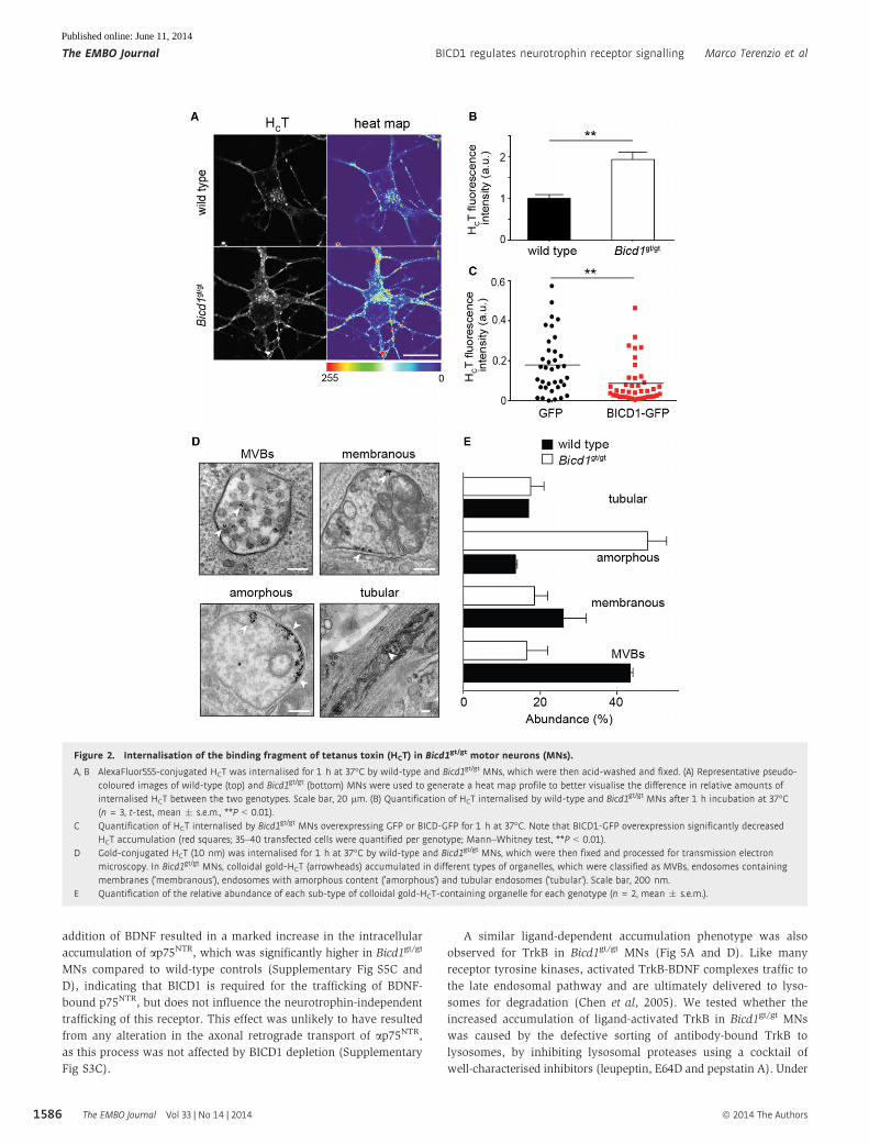
Figure 2. Internalisation of the binding fragment of tetanus toxin (HCT) in Bicd1gt/gt motor neurons (MNs).
A, B AlexaFluor555-conjugated HCT was internalised for 1 h at 37°C by wild-type and Bicd1gt/gt MNs, which were then acid-washed and fixed. (A) Representative pseudo-coloured images of wild-type (top) and Bicd1gt/gt (bottom) MNs were used to generate a heat map profile to better visualise the difference in relative amounts ofinternalised HCT between the two genotypes. Scale bar, 20 lm. (B) Quantification of HCT internalised by wild-type and Bicd1gt/gt MNs after 1 h incubation at 37°C(n = 3, t-test, mean � s.e.m., **P < 0.01).
C Quantification of HCT internalised by Bicd1gt/gt MNs overexpressing GFP or BICD-GFP for 1 h at 37°C. Note that BICD1-GFP overexpression significantly decreasedHCT accumulation (red squares; 35–40 transfected cells were quantified per genotype; Mann–Whitney test, **P < 0.01).
D Gold-conjugated HCT (10 nm) was internalised for 1 h at 37°C by wild-type and Bicd1gt/gt MNs, which were then fixed and processed for transmission electronmicroscopy. In Bicd1gt/gt MNs, colloidal gold-HCT (arrowheads) accumulated in different types of organelles, which were classified as MVBs, endosomes containingmembranes (‘membranous’), endosomes with amorphous content (‘amorphous’) and tubular endosomes (‘tubular’). Scale bar, 200 nm.
E Quantification of the relative abundance of each sub-type of colloidal gold-HCT-containing organelle for each genotype (n = 2, mean � s.e.m.).
The EMBO Journal Vol 33 | No 14 | 2014 ª 2014 The Authors
The EMBO Journal BICD1 regulates neurotrophin receptor signalling Marco Terenzio et al
1586
Published online: June 11, 2014
these conditions, TrkB accumulation was increased in wild-type
MNs, whereas the extent of TrkB accumulation in Bicd1gt/gt MNs
was unaffected by this treatment (Supplementary Fig S6A). A similar
trend was also established for ap75NTR accumulation (Supplemen-
tary Fig S6B). We did not detect any obvious difference either in
lysosomal acidification or distribution of LAMP2-positive organelles
between Bicd1gt/gt and wild-type MNs, suggesting that BICD1 deple-
tion does not affect lysosome biogenesis or function, but is likely to
cause an impaired sorting of neurotrophin receptors along the degra-
dative pathway. Consistent with this idea was our observation that
intracellular accumulation of HCT was unaffected by lysosomal
inhibitors (Supplementary Fig S6C), which was not surprising since
this probe does not enter the lumen of lysosomes (Supplementary
Fig S6D) and therefore is not degraded by these organelles.
Since the results described in Supplementary Fig S6A and B were
obtained by an imaging approach using fluorescent aTrkB and
B
D FLAG-TrkB mCherry-BDNF BICD1 merge
Icor
r coe
ffici
ent
FLAG-TrkB SNX1 merge
0
0.2
0.4
0.6
0.8
SNX1
A TrkB mergeHCT
wild
type
C TrkB-SNX1 colocalisation
Figure 3. Sorting nexin 1 (SNX1) co-localises with TrkB and the binding fragment of tetanus toxin (HCT).
A HCT and TrkB co-localise in wild-type motor neurons (MNs). AlexaFluor555-conjugated HCT (red) was internalised for 1 h at 37°C; neurons were then acid-washed,fixed and immunostained for TrkB (green). TrkB/HCT-positive structures (arrowheads) were frequently detected in the cell soma and neurites. Scale bar, 5 lm.
B N2A neuroblastoma cells over-expressing FLAG-TrkB were incubated with FLAG antibody and brain-derived neurotrophic factor (BDNF) for 1 h at 37°C and then acid-washed, fixed and immunostained to detect FLAG-TrkB (green) and endogenous SNX1 (red). Scale bar, 10 lm.
C Quantification of FLAG-TrkB/SNX1 co-localisation (n = 3, 31 cells in total analysed).D N2A cells overexpressing FLAG-TrkB were incubated with FLAG antibody and BDNF-mCherry (red) for 1 h at 37°C and then acid-washed, fixed and immunostained to
detect FLAG-TrkB (green) and endogenous Bicaudal D homolog 1 (BICD1) (blue). Note the presence of triple positive structures demonstrating that endogenous BICD1associates with internalised TrkB-BDNF complexes. Scale bar, 10 lm.
ª 2014 The Authors The EMBO Journal Vol 33 | No 14 | 2014
Marco Terenzio et al BICD1 regulates neurotrophin receptor signalling The EMBO Journal
1587
Published online: June 11, 2014
ap75NTR, we wanted to confirm these results using a more direct
biochemical strategy. For this purpose, we selected a pulse-chase
protocol, which allowed us to follow the dynamics of a cohort of
internalised receptors as they trafficked along the endosomal path-
way. To this end, we loaded ES-derived MNs with a 15-min pulse of
aTrkB in the presence of BDNF and lysosomal inhibitors or vehicle
control, followed by 1 or 2 h incubation in the presence or absence
of lysosomal inhibitors. After cell lysis, antibody pulldown followed
by western blotting revealed a major band of full-length TrkB
(TrkB.FL; 145 kDa) and a minor band of TrkB.T1 (90 kDa), repre-
senting the truncated dominant-negative form of this receptor
(Fenner, 2012), in wild-type cells (upper panel, Fig 5E). However,
samples isolated from Bicd1gt/gt MNs displayed a striking increase of
TrkB.T1 relative to TrkB.FL (lower panel, Fig 5E). In agreement
with the immunofluorescence data quantified in Supplementary
Fig S6A, lysosomal inhibitors proved effective in preventing the
degradation of aTrkB in wild-type, but not in Bicd1gt/gt MNs
(Fig 5E), suggesting that BICD1 depletion impaired lysosome-
mediated degradation of these receptors.
TrkA is degraded by the combined actions of lysosomes and the
proteasome (Sommerfeld et al, 2000; Geetha & Wooten, 2008), a
pathway shared at least in part by TrkB (Sommerfeld et al, 2000).
Interestingly, proteasomal inhibition redirects TrkA from late
endosomes to the recycling route (Moises et al, 2009). Therefore,
the residual degradation of TrkB by wild-type MNs in the presence
of lysosomal protease inhibitors at the latest time point (Fig 5E,
120 min) could have resulted from receptor clearance by the protea-
some and suggested a possible explanation for the apparent ineffec-
tiveness of lysosomal inhibitors in preventing the clearance of TrkB
from Bicd1gt/gt MNs (lower panel, Fig 5E). To test this hypothesis
and assess the impact of BICD1 on these degradative pathways, we
repeated the 15-min pulse step in the presence or absence of
MG132, a specific proteasome inhibitor. Thus, probing the aTrkBimmunoprecipitates with a TrkB antibody revealed profiles similar
to those shown in the previous experiment (compare upper panels
in Fig 5F with 5E). However, re-probing the same membrane with a
ubiquitin-specific antibody revealed a signal only for the Bicd1gt/gt
sample treated with a combination of lysosomal inhibitors and
MG132 (Fig 5F). This result implied that within 15 min of exposure
to BDNF, TrkB internalised by Bicd1gt/gt MNs was preferentially
ubiquitinated and likely targeted for proteasome-mediated
degradation.
BICD1 depletion increases neurotrophin receptor levels at theplasma membrane
Inhibition of the proteasome, which causes an accumulation of
ubiquitinated Trks, redirects TrkA from late endosomes to a plasma
membrane recycling pathway (Moises et al, 2009). Depletion of
BICD1 on the other hand led to the intracellular increase of HCT,
A Ba b
c
a b
c
Figure 4. Bicd1gt/gt motor neurons accumulate HCT, TrkB and p75NTR in enlarged endosomal structures.
A Wild-type MNs were allowed to internalise gold-conjugated HCT (10 nm, empty arrowheads), aTrkB (20 nm, white arrowheads) and ap75NTR (5 nm, black arrowheads)in the presence of BDNF for 2 h at 37°C and then processed for transmission electron microscopy. Colloidal gold-conjugated probes were found in a variety oforganelles ranging from tubular endosomes (Aa), to MVBs (Ab, Bc) to endosomes containing membranes (Ac). These organelles are pseudocoloured according to thenumber of probes that they contain: yellow, pink and green representing single, double and triple labelled compartments, respectively. Scale bars, 200 nm (mainpanels); 50 nm (insets).
B Bicd1gt/gt MNs were treated as in (A) and then processed for transmission electron microscopy. Notably, in addition to the previously described organelles, colloidalgold-conjugated probes were strongly associated with endosomes with “amorphous” content (Ba, Bb). In addition, double and triple labelled enlarged organelles weremore prevalent in Bicd1gt/gt motor neurons compared to wild-type cells. Scale bars, 200 nm (main panels); 50 nm (insets).
The EMBO Journal Vol 33 | No 14 | 2014 ª 2014 The Authors
The EMBO Journal BICD1 regulates neurotrophin receptor signalling Marco Terenzio et al
1588
Published online: June 11, 2014
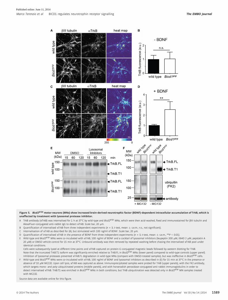
Figure 5. Bicd1gt/gt motor neurons (MNs) show increased brain-derived neurotrophic factor (BDNF)-dependent intracellular accumulation of TrkB, which isunaffected by treatment with lysosomal protease inhibitor.
A TrkB antibody (aTrkB) was internalised for 1 h at 37°C by wild-type and Bicd1gt/gt MNs, which were then acid-washed, fixed and immunostained for bIII tubulin andAlexaFluor-conjugated anti-rabbit IgG to detect aTrkB. Scale bar, 20 lm.
B Quantification of internalised aTrkB from three independent experiments (n = 3, t-test, mean � s.e.m.; n.s., not significant).C Internalisation of aTrkB as described for (A), but stimulated with 100 ng/ml of BDNF. Scale bar, 20 lm.D Quantification of internalised aTrkB in the presence of BDNF from three independent experiments (n = 3, t-test, mean � s.e.m., **P < 0.01).E Wild-type and Bicd1gt/gt MNs were co-incubated with aTrkB, 100 ng/ml of BDNF and a cocktail of lysosomal inhibitors (leupeptin 200 lM, E64D 2 lM, pepstatin A
20 lM) or DMSO vehicle control for 15 min at 37°C. Unbound antibody was then removed by repeated washing before chasing the internalised aTrkB pool underidentical conditions.Cells were subsequently lysed at different time points and aTrkB captured on protein-G-conjugated magnetic beads followed by western blotting for TrkB.Note that the truncated TrkB.T1 isoform was significantly enriched relative to TrkB.FL in Bicd1gt/gt MNs (lower panel) compared to wild-type controls (upper panel).Inhibition of lysosomal proteases prevented aTrkB.FL degradation in wild-type MNs (compare with DMSO-treated samples), but was ineffective in Bicd1gt/gt cells.
F Wild-type and Bicd1gt/gt MNs were co-incubated with aTrkB, 100 ng/ml of BDNF and lysosomal inhibitors as described in (E) for 15 min at 37°C in the presence orabsence of 10 lM MG132. Upon cell lysis, aTrkB was captured as above. Immunoprecipitated samples were probed for TrkB (upper panels), with the FK2 antibody,which targets mono- and poly-ubiquitinated proteins (middle panels), and with horseradish peroxidase-conjugated anti-rabbit immunoglobulins in order todetect internalised aTrkB. TrkB.T1 was enriched in Bicd1gt/gt MNs in both conditions, but TrkB ubiquitination was detected only in Bicd1gt/gt MN samples treatedwith MG132.
Source data are available online for this figure.
ª 2014 The Authors The EMBO Journal Vol 33 | No 14 | 2014
Marco Terenzio et al BICD1 regulates neurotrophin receptor signalling The EMBO Journal
1589
Published online: June 11, 2014
TrkB and p75NTR in large vacuoles often connected to structures
resembling tubular endosomes (Supplementary Fig S3D). Further
insights into the nature of these organelles were provided by the co-
localisation of SNX1 with TrkB and HCT (Fig 3B and Supplementary
Fig S4A and B), and the increase in SNX1-associated HCT-positive
structures in Bicd1gt/gt MNs (Supplementary Fig S4A and B). Based
on these findings and the notion that TrkB was preferentially
ubiquitinated when BICD1 was depleted (Fig 5F), we decided to
test whether the altered neurotrophin receptor trafficking observed
in Bicd1gt/gt MNs resulted in an increased recycling of these
receptors back to the plasma membrane.
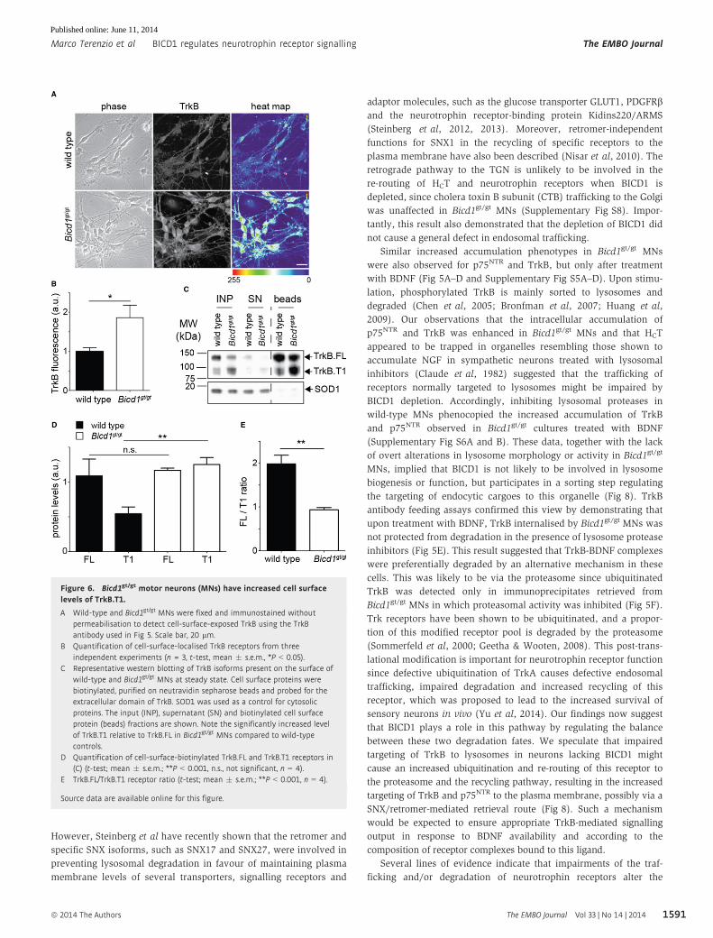
In spite of TrkB-FL transcript (Supplementary Fig S1D) and Trk
protein levels (Fig 1E and F) being reduced in Bicd1gt/gt MNs
relative to wild-type cells, cell surface immunofluorescence and
biotinylation experiments both demonstrated that significantly
more TrkB was present on the plasma membrane of Bicd1gt/gt
neurons compared to wild-type controls (Fig 6A–E), with a similar
trend also observed for p75NTR (Supplementary Fig S7A–D).
TrkB signalling is impaired in Bicd1gt/gt MNs
The most striking finding from the previous set of experiments was
the substantial increase in TrkB.T1 localisation to the surface of
Bicd1gt/gt MNs (Fig 6C–E). TrkB.T1 heterodimerises with TrkB.FL to
inhibit auto-phosphorylation and downstream signalling of the full-
length receptor (Eide et al, 1996; Fenner, 2012). Therefore, the
substantially decreased ratio of TrkB.FL to TrkB.T1 (Fig 6E) on the
plasma membrane of Bicd1gt/gt MNs, together with an increased cell
surface localisation of p75NTR (Supplementary Fig S7A–D), would
be expected to reduce phosphoinositide-3 kinase (PI3K) and Ras-
mediated signal activation following treatment with BDNF.
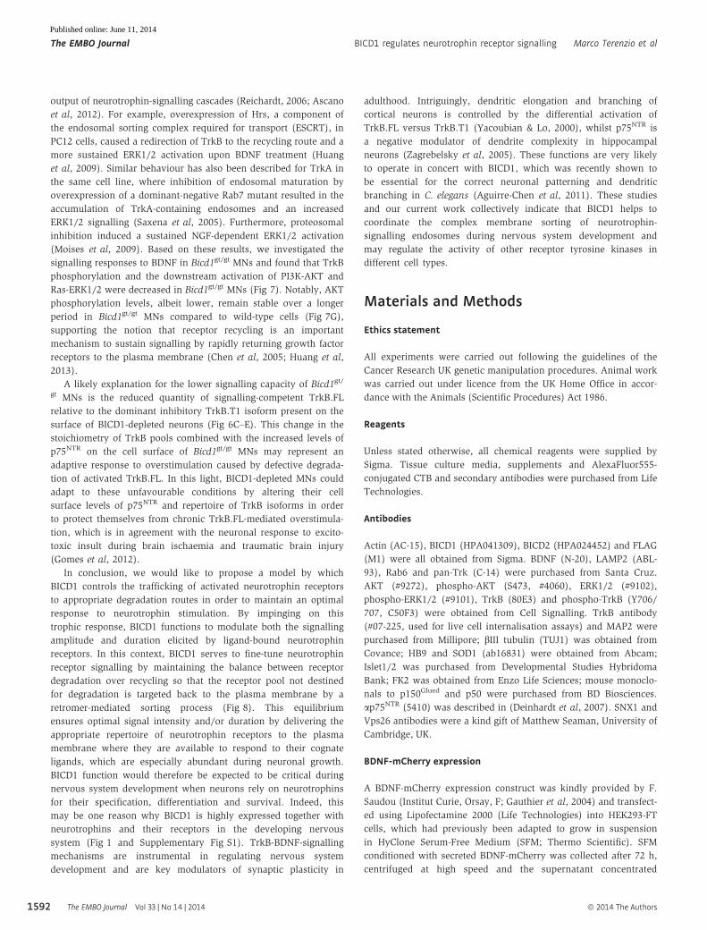
To test this hypothesis, we stimulated MNs with BDNF for vari-
ous time points before assaying lysates by western blotting for
phosphorylated TrkB, AKT and ERK1/2. In wild-type cultures,
both AKT and ERK1/2 phosphorylation peaked at 10 min followed
by a progressive attenuation of this response over the following
40 min. The same overall trend was observed for Bicd1gt/gt MNs,
but phosphorylation of both AKT and ERK1/2 was lower at all
time points tested (Fig 7A–D). Thus, even though more cell
surface TrkB and p75NTR were available for ligand binding in
Bicd1gt/gt MNs, this was accompanied by decreased activation of
AKT and ERK1/2 following BDNF treatment. This reduced signal-
ling response might be a consequence of decreased TrkB activa-
tion caused by the lower TrkB.FL to TrkB.T1 ratio present in
Bicd1gt/gt MNs. This hypothesis was supported by data shown in
Fig 7E–F, where phosphorylation of TrkB upon stimulation with
BDNF for 10 min was lower in mutant MNs compared to wild-
type controls.
Proteosomal inhibition is known to induce sustained ERK1/2
activation following NGF stimulation (Moises et al, 2009), which
was likely to result from enhanced TrkA recycling back to the
plasma membrane. We now show that Bicd1gt/gt MNs respond to
BDNF/TrkB activation through sustained PI3K/AKT signalling.
Thus, whereas the phospho-AKT (pAKT) profile tailed off at later
time points in wild-type cells, BICD1-depleted MNs displayed a
more prolonged AKT activation during 1 h of BDNF stimulation
(Fig 7A–B). This trend was substantiated by prolonged stimulation
experiments when phospho-AKT levels, albeit lower, remained
relatively stable over the entire time frame of the experiment in
Bicd1gt/gt MNs (Fig 7G).
Discussion
Using a novel siRNA-screening approach designed to identify new
players involved in the trafficking of neurotrophin receptors and
neurotropic virulence factors in MNs (Terenzio et al, 2014), we
identified several genes that affected the intracellular accumulation
of HCT and ap75NTR. Because of our long-standing interest in
axonal retrograde transport, the most intriguing candidate revealed
by this screen was Bicd1. This cytoplasmic dynein adaptor and
closely related proteins, such as BICD2 and BICDR1, play diverse
roles in nervous system development and maintenance (Matanis
et al, 2002; Schlager et al, 2010). For instance, BICD2 is required
for neuronal migration (Jaarsma et al, 2014), whereas BICD1 co-
operates with dynein and kinesins in C. elegans nervous system
patterning (Aguirre-Chen et al, 2011). Furthermore, Drosophila
BicD recycles clathrin heavy chain back to the plasma membrane
during synaptic stimulation (Li et al, 2010) and controls distribution
of Fragile X mental retardation protein (Bianco et al, 2010). Our in
vivo observations now expand this list of functions by showing that
BICD1 very likely plays an important role in the developing mouse
nervous system, at least during the period when BDNF and neuro-
trophin receptors are highly expressed (Fig 1 and Supplementary
Fig S1).
Silencing of Bicd1 increased the intracellular accumulation of
HCT, and this was confirmed in MNs expressing the RRP227
Bicd1gt/gt hypomorphic allele. The intracellular distribution of HCT
in Bicd1gt/gt MNs differed markedly from that observed in wild-type
cells (Fig 2D and E). In mutant cells, HCT clustered near the limiting
membrane of a population of enlarged organelles with amorphous
content, which lacked intra-luminal vesicles typical of MVBs
(Fig 2D) and was found in protrusions emanating from such struc-
tures (Supplementary Fig S3D). These observations suggested that
endosomal sorting and/or maturation of these HCT-containing
compartments were perturbed in Bicd1gt/gt MNs. Because HCT is co-
transported with p75NTR and TrkB, we inferred that these receptors
were also likely to be present in at least a sub-population of the
enlarged HCT-labelled compartments present in Bicd1gt/gt MNs. This
notion was supported by the findings that colloidal gold-labelled
HCT, aTrkB and ap75NTR all accumulated in the same enlarged
organelles (Fig 4), which strongly suggested that these structures
represented sorting compartments common to all three probes.
Furthermore, the increased occurrence of such organelles in
Bicd1gt/gt MNs indicated that BICD1 depletion affected the endoso-
mal sorting of HCT, TrkB and p75NTR.
This hypothesis was supported by the observation that HCT-
positive organelles in Bicd1gt/gt MNs also displayed an increased
co-localisation with SNX1, a retromer component (Supplementary
Fig S4A and B; Terenzio et al, 2014). Retromer is a large protein
complex, which controls the endosome membrane re-sculpturing
process essential for the formation of transport carriers (Bonifacino
& Hurley, 2008; Cullen & Korswagen, 2012) and the selection of
specific cargoes (Harrison et al, 2014). Classically, this complex has
been shown to mediate retrograde transport of membrane-bound
cargoes from endosomes to the trans-Golgi network (TGN).
The EMBO Journal Vol 33 | No 14 | 2014 ª 2014 The Authors
The EMBO Journal BICD1 regulates neurotrophin receptor signalling Marco Terenzio et al
1590
Published online: June 11, 2014
However, Steinberg et al have recently shown that the retromer and
specific SNX isoforms, such as SNX17 and SNX27, were involved in
preventing lysosomal degradation in favour of maintaining plasma
membrane levels of several transporters, signalling receptors and
adaptor molecules, such as the glucose transporter GLUT1, PDGFRband the neurotrophin receptor-binding protein Kidins220/ARMS
(Steinberg et al, 2012, 2013). Moreover, retromer-independent
functions for SNX1 in the recycling of specific receptors to the
plasma membrane have also been described (Nisar et al, 2010). The
retrograde pathway to the TGN is unlikely to be involved in the
re-routing of HCT and neurotrophin receptors when BICD1 is
depleted, since cholera toxin B subunit (CTB) trafficking to the Golgi
was unaffected in Bicd1gt/gt MNs (Supplementary Fig S8). Impor-
tantly, this result also demonstrated that the depletion of BICD1 did
not cause a general defect in endosomal trafficking.
Similar increased accumulation phenotypes in Bicd1gt/gt MNs
were also observed for p75NTR and TrkB, but only after treatment
with BDNF (Fig 5A–D and Supplementary Fig S5A–D). Upon stimu-
lation, phosphorylated TrkB is mainly sorted to lysosomes and
degraded (Chen et al, 2005; Bronfman et al, 2007; Huang et al,
2009). Our observations that the intracellular accumulation of
p75NTR and TrkB was enhanced in Bicd1gt/gt MNs and that HCT
appeared to be trapped in organelles resembling those shown to
accumulate NGF in sympathetic neurons treated with lysosomal
inhibitors (Claude et al, 1982) suggested that the trafficking of
receptors normally targeted to lysosomes might be impaired by
BICD1 depletion. Accordingly, inhibiting lysosomal proteases in
wild-type MNs phenocopied the increased accumulation of TrkB
and p75NTR observed in Bicd1gt/gt cultures treated with BDNF
(Supplementary Fig S6A and B). These data, together with the lack
of overt alterations in lysosome morphology or activity in Bicd1gt/gt
MNs, implied that BICD1 is not likely to be involved in lysosome
biogenesis or function, but participates in a sorting step regulating
the targeting of endocytic cargoes to this organelle (Fig 8). TrkB
antibody feeding assays confirmed this view by demonstrating that
upon treatment with BDNF, TrkB internalised by Bicd1gt/gt MNs was
not protected from degradation in the presence of lysosome protease
inhibitors (Fig 5E). This result suggested that TrkB-BDNF complexes
were preferentially degraded by an alternative mechanism in these
cells. This was likely to be via the proteasome since ubiquitinated
TrkB was detected only in immunoprecipitates retrieved from
Bicd1gt/gt MNs in which proteasomal activity was inhibited (Fig 5F).
Trk receptors have been shown to be ubiquitinated, and a propor-
tion of this modified receptor pool is degraded by the proteasome
(Sommerfeld et al, 2000; Geetha & Wooten, 2008). This post-trans-
lational modification is important for neurotrophin receptor function
since defective ubiquitination of TrkA causes defective endosomal
trafficking, impaired degradation and increased recycling of this
receptor, which was proposed to lead to the increased survival of
sensory neurons in vivo (Yu et al, 2014). Our findings now suggest
that BICD1 plays a role in this pathway by regulating the balance
between these two degradation fates. We speculate that impaired
targeting of TrkB to lysosomes in neurons lacking BICD1 might
cause an increased ubiquitination and re-routing of this receptor to
the proteasome and the recycling pathway, resulting in the increased
targeting of TrkB and p75NTR to the plasma membrane, possibly via a
SNX/retromer-mediated retrieval route (Fig 8). Such a mechanism
would be expected to ensure appropriate TrkB-mediated signalling
output in response to BDNF availability and according to the
composition of receptor complexes bound to this ligand.
Several lines of evidence indicate that impairments of the traf-
ficking and/or degradation of neurotrophin receptors alter the
Figure 6. Bicd1gt/gt motor neurons (MNs) have increased cell surfacelevels of TrkB.T1.
A Wild-type and Bicd1gt/gt MNs were fixed and immunostained withoutpermeabilisation to detect cell-surface-exposed TrkB using the TrkBantibody used in Fig 5. Scale bar, 20 lm.
B Quantification of cell-surface-localised TrkB receptors from threeindependent experiments (n = 3, t-test, mean � s.e.m., *P < 0.05).
C Representative western blotting of TrkB isoforms present on the surface ofwild-type and Bicd1gt/gt MNs at steady state. Cell surface proteins werebiotinylated, purified on neutravidin sepharose beads and probed for theextracellular domain of TrkB. SOD1 was used as a control for cytosolicproteins. The input (INP), supernatant (SN) and biotinylated cell surfaceprotein (beads) fractions are shown. Note the significantly increased levelof TrkB.T1 relative to TrkB.FL in Bicd1gt/gt MNs compared to wild-typecontrols.
D Quantification of cell-surface-biotinylated TrkB.FL and TrkB.T1 receptors in(C) (t-test; mean � s.e.m.; **P < 0.001, n.s., not significant, n = 4).
E TrkB.FL/TrkB.T1 receptor ratio (t-test; mean � s.e.m.; **P < 0.001, n = 4).
Source data are available online for this figure.
ª 2014 The Authors The EMBO Journal Vol 33 | No 14 | 2014
Marco Terenzio et al BICD1 regulates neurotrophin receptor signalling The EMBO Journal
1591
Published online: June 11, 2014
output of neurotrophin-signalling cascades (Reichardt, 2006; Ascano
et al, 2012). For example, overexpression of Hrs, a component of
the endosomal sorting complex required for transport (ESCRT), in
PC12 cells, caused a redirection of TrkB to the recycling route and a
more sustained ERK1/2 activation upon BDNF treatment (Huang
et al, 2009). Similar behaviour has also been described for TrkA in
the same cell line, where inhibition of endosomal maturation by
overexpression of a dominant-negative Rab7 mutant resulted in the
accumulation of TrkA-containing endosomes and an increased
ERK1/2 signalling (Saxena et al, 2005). Furthermore, proteosomal
inhibition induced a sustained NGF-dependent ERK1/2 activation
(Moises et al, 2009). Based on these results, we investigated the
signalling responses to BDNF in Bicd1gt/gt MNs and found that TrkB
phosphorylation and the downstream activation of PI3K-AKT and
Ras-ERK1/2 were decreased in Bicd1gt/gt MNs (Fig 7). Notably, AKT
phosphorylation levels, albeit lower, remain stable over a longer
period in Bicd1gt/gt MNs compared to wild-type cells (Fig 7G),
supporting the notion that receptor recycling is an important
mechanism to sustain signalling by rapidly returning growth factor
receptors to the plasma membrane (Chen et al, 2005; Huang et al,
2013).
A likely explanation for the lower signalling capacity of Bicd1gt/
gt MNs is the reduced quantity of signalling-competent TrkB.FL
relative to the dominant inhibitory TrkB.T1 isoform present on the
surface of BICD1-depleted neurons (Fig 6C–E). This change in the
stoichiometry of TrkB pools combined with the increased levels of
p75NTR on the cell surface of Bicd1gt/gt MNs may represent an
adaptive response to overstimulation caused by defective degrada-
tion of activated TrkB.FL. In this light, BICD1-depleted MNs could
adapt to these unfavourable conditions by altering their cell
surface levels of p75NTR and repertoire of TrkB isoforms in order
to protect themselves from chronic TrkB.FL-mediated overstimula-
tion, which is in agreement with the neuronal response to excito-
toxic insult during brain ischaemia and traumatic brain injury
(Gomes et al, 2012).
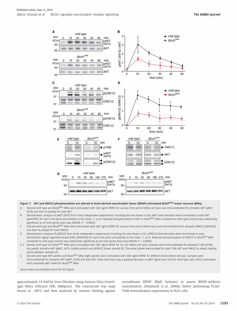
In conclusion, we would like to propose a model by which
BICD1 controls the trafficking of activated neurotrophin receptors
to appropriate degradation routes in order to maintain an optimal
response to neurotrophin stimulation. By impinging on this
trophic response, BICD1 functions to modulate both the signalling
amplitude and duration elicited by ligand-bound neurotrophin
receptors. In this context, BICD1 serves to fine-tune neurotrophin
receptor signalling by maintaining the balance between receptor
degradation over recycling so that the receptor pool not destined
for degradation is targeted back to the plasma membrane by a
retromer-mediated sorting process (Fig 8). This equilibrium
ensures optimal signal intensity and/or duration by delivering the
appropriate repertoire of neurotrophin receptors to the plasma
membrane where they are available to respond to their cognate
ligands, which are especially abundant during neuronal growth.
BICD1 function would therefore be expected to be critical during
nervous system development when neurons rely on neurotrophins
for their specification, differentiation and survival. Indeed, this
may be one reason why BICD1 is highly expressed together with
neurotrophins and their receptors in the developing nervous
system (Fig 1 and Supplementary Fig S1). TrkB-BDNF-signalling
mechanisms are instrumental in regulating nervous system
development and are key modulators of synaptic plasticity in
adulthood. Intriguingly, dendritic elongation and branching of
cortical neurons is controlled by the differential activation of
TrkB.FL versus TrkB.T1 (Yacoubian & Lo, 2000), whilst p75NTR is
a negative modulator of dendrite complexity in hippocampal
neurons (Zagrebelsky et al, 2005). These functions are very likely
to operate in concert with BICD1, which was recently shown to
be essential for the correct neuronal patterning and dendritic
branching in C. elegans (Aguirre-Chen et al, 2011). These studies
and our current work collectively indicate that BICD1 helps to
coordinate the complex membrane sorting of neurotrophin-
signalling endosomes during nervous system development and
may regulate the activity of other receptor tyrosine kinases in
different cell types.
Materials and Methods
Ethics statement
All experiments were carried out following the guidelines of the
Cancer Research UK genetic manipulation procedures. Animal work
was carried out under licence from the UK Home Office in accor-
dance with the Animals (Scientific Procedures) Act 1986.
Reagents
Unless stated otherwise, all chemical reagents were supplied by
Sigma. Tissue culture media, supplements and AlexaFluor555-
conjugated CTB and secondary antibodies were purchased from Life
Technologies.
Antibodies
Actin (AC-15), BICD1 (HPA041309), BICD2 (HPA024452) and FLAG
(M1) were all obtained from Sigma. BDNF (N-20), LAMP2 (ABL-
93), Rab6 and pan-Trk (C-14) were purchased from Santa Cruz.
AKT (#9272), phospho-AKT (S473, #4060), ERK1/2 (#9102),
phospho-ERK1/2 (#9101), TrkB (80E3) and phospho-TrkB (Y706/
707, C50F3) were obtained from Cell Signalling. TrkB antibody
(#07-225, used for live cell internalisation assays) and MAP2 were
purchased from Millipore; bIII tubulin (TUJ1) was obtained from
Covance; HB9 and SOD1 (ab16831) were obtained from Abcam;
Islet1/2 was purchased from Developmental Studies Hybridoma
Bank; FK2 was obtained from Enzo Life Sciences; mouse monoclo-
nals to p150Glued and p50 were purchased from BD Biosciences.
ap75NTR (5410) was described in (Deinhardt et al, 2007). SNX1 and
Vps26 antibodies were a kind gift of Matthew Seaman, University of
Cambridge, UK.
BDNF-mCherry expression
A BDNF-mCherry expression construct was kindly provided by F.
Saudou (Institut Curie, Orsay, F; Gauthier et al, 2004) and transfect-
ed using Lipofectamine 2000 (Life Technologies) into HEK293-FT
cells, which had previously been adapted to grow in suspension
in HyClone Serum-Free Medium (SFM; Thermo Scientific). SFM
conditioned with secreted BDNF-mCherry was collected after 72 h,
centrifuged at high speed and the supernatant concentrated
The EMBO Journal Vol 33 | No 14 | 2014 ª 2014 The Authors
The EMBO Journal BICD1 regulates neurotrophin receptor signalling Marco Terenzio et al
1592
Published online: June 11, 2014
approximately 15-fold by force filtration using Amicon Ultra Centrif-
ugal filters (Ultracel 30K; Millipore). The concentrate was snap
frozen at �80°C and then analysed by western blotting against
recombinant BDNF (R&D Systems) to assess BDNF-mCherry
concentration (Deinhardt et al, 2006b) before performing FLAG-
TrkB internalisation experiments in N2A cells.
Figure 7. AKT and ERK1/2 phosphorylation are altered in brain-derived neurotrophic factor (BDNF)-stimulated Bicd1gt/gt motor neurons (MNs).
A Starved wild-type and Bicd1gt/gt MNs were stimulated with 100 ng/ml BDNF for various time points before cell lysis and immunoblotted for phospho-AKT (pAKT;S473) and then re-probed for total AKT.
B Densitometric analysis of pAKT (S473) from three independent experiments including the one shown in (A). pAKT band densities were normalised to total AKT(pAKT/AKT) for each time point and plotted as the mean � s.e.m. Reduced phosphorylation of AKT in Bicd1gt/gt MNs compared to wild-type controls was statisticallysignificant at all time points (two-way ANOVA, P < 0.0001).
C Starved wild-type and Bicd1gt/gt MNs were stimulated with 100 ng/ml BDNF for various time points before lysis and immunoblotted for phospho-ERK1/2 (pERK1/2)and then re-probed for total ERK1/2.
D Densitometric analysis of pERK1/2 from three independent experiments including the one shown in (C). pERK1/2 band densities were normalised to totalextracellular signal-regulated kinase (ERK; pERK/ERK) for each time point and plotted as the mean � s.e.m. Reduced phosphorylation of ERK1/2 in Bicd1gt/gt MNscompared to wild-type controls was statistically significant at all time points (two-way ANOVA, P < 0.0001).
E, F Starved wild-type and Bicd1gt/gt MNs were stimulated with 100 ng/ml BDNF for 10 min before cell lysis. Samples were immunoblotted for phospho-TrkB (pTrkB;top panel), phospho-AKT (pAKT; S473; middle panels) and pERK1/2 (lower panels) (E). The same lysates were probed for total TrkB, AKT and ERK1/2 to assess loadingparity between samples (F).
G Starved wild-type (left panels) and Bicd1gt/gt MNs (right panels) were stimulated with 100 ng/ml BDNF for different times before cell lysis. Samples wereimmunoblotted for phospho-AKT (pAKT; S473) and total AKT. Note that there was a gradual decrease in pAKT signal over time for wild-type cells, which contrastedwith sustained pAKT levels for Bicd1gt/gt MNs.
Source data are available online for this figure.
ª 2014 The Authors The EMBO Journal Vol 33 | No 14 | 2014
Marco Terenzio et al BICD1 regulates neurotrophin receptor signalling The EMBO Journal
1593
Published online: June 11, 2014
Bicd1gt/+ chimeric embryos and lacZ expression analysis
10–15 RRP227 ES cells were injected into blastocyst stage embryos
collected from superovulated C57BL/6J female mice that had been
mated to C57BL/6J male mice. Embryos were transferred to pseudo-
pregnant (2.5 days post-coitum) recipient mice according to stan-
dard protocols (Nagy et al, 2003). To assess Bicd1-lacZ expression
patterns during mouse development, E11.5-E14.5 embryos were
fixed at room temperature for 15–30 min in 0.1 M sodium phos-
phate, pH 7.3, 0.4% paraformaldehyde (PFA), 5 mM EGTA, 2 mM
MgCl2, washed three times in 0.1 M sodium phosphate pH 7.3,
2 mM MgCl2, 0.1% sodium deoxycholate, 0.02% NP-40 and finally
transferred into developing solution (100 mM sodium phosphate,
pH 7.3, 2 mM MgCl2, 0.01% sodium deoxycholate, 0.02% NP-40,
5 mM K3Fe(CN)6, 5 mM K4Fe(CN)6, 1 mg/ml X-gal (Roche), for
30 min to 5 h at 37°C or overnight in the dark. The reaction was
quenched by rinsing several times in PBS, and embryos were then
post-fixed in 4% PFA and stored in 70% ethanol prior to imaging.
After imaging, some samples were paraffin embedded, sectioned,
DAB immunostained and counterstained with Nile red.
Embryonic stem-cell-derived MNs
Embryonic stem cells were grown on fish skin gelatin-coated flasks
in Glasgow Minimal Essential Medium (GMEM), 5% ES cell-
qualified foetal bovine serum FBS, 5% knockout serum replacement
(KSR), 1% GLUTAMAX, 0.1 mM 2-mercaptoethanol and 1,000
units/ml of leukaemia inhibitory factor (ESGRO, Millipore). To
generate MNs, 1.5 × 10⁶ ES cells were grown in suspension on a
10-cm non-tissue culture-treated Petri dish containing differentiation
(DFNK) medium: 45% neurobasal, 45% DMEM/Ham’s-F12, 10%
KSR, 1% GLUTAMAX and 0.1 mM 2-mercaptoethanol. The follow-
ing day, embryoid bodies (EBs) were gently centrifuged and re-
suspended in 10 ml of fresh DFNK medium and plated on a new
Petri dish. The following day, the greatly enlarged EBs were allowed
to sediment by gravity and re-suspended in fresh DFNK medium
supplemented with 1 lM all-trans retinoic acid (RA) and 333 nM
Smoothened Agonist (SAG; Enzo Life Sciences). EBs were main-
tained under these conditions for a further 4 days (medium changed
every other day) and then dissociated with 0.025% porcine pancre-
atic trypsin in 1 ml PBS for 7 min at 37°C and processed as described
previously for the dissociation of mouse E13.5 spinal cord MNs
(Hafezparast et al, 2003). Cells were plated onto poly-D-ornithine and
laminin-coated dishes in MN growth medium: neurobasal medium
supplemented with 2% B27, 2% heat-inactivated horse serum, 1%
GLUTAMAX, 25 lM 2-mercaptoethanol, 10 ng/ml rat ciliary
neurotrophic factor (CNTF; R&D Systems), 100 pg/ml rat glial
cell line-derived neurotrophic factor (GDNF; R&D Systems) and
1 lMRA.
Generation of homozygous RRP227 ES cells
Mouse ES cells with a gene trap insertion in the first intron of
Bicd1 (RRP227; http://www.informatics.jax.org/allele/key/544886)
were obtained from the Mutant Mouse Regional Resource Center.
Homozygous Bicd1gt/gt cells were generated as previously described
(Lefebvre et al, 2001). Briefly, heterozygous cells were seeded at 30%
confluence and maintained under standard feeder-free ES cell culture
Figure 8. Proposed role of BICD1 in neurotrophin receptor traffickingand signalling.In wild-type motor neurons (MNs), brain-derived neurotrophic factor (BDNF)binds to and activates TrkB. Ligand–receptor complexes are internalised atsynaptic sites located in the periphery (1); note that for clarity, internalisation ofthese complexes from the plasma membrane of the cell body is not depicted.Ligand–receptor complexes are sorted to signalling endosomes (2), retrogradelytransported in a cytoplasmic dynein-dependent process (3), towards the cellsoma where they associate with somatic sorting endosomes (4) decorated bysorting nexin 1 (SNX1) and other retromer components. Different neurotrophinreceptor pools are then trafficked towardsMVB/lysosomes (5a) or the proteasome(5b) for degradation, or recycled back to the plasma membrane (5c). Impairmentof the lysosomal targeting of TrkB in cells lacking BICD1 is envisaged to impair theflow of the receptor from somatic sorting endosomes towards lysosomes andredirect them either to the recycling route back to the plasma membrane or tothe proteasome for ubiquitin-mediated degradation. The main consequence ofthese mis-sorting steps is the increased accumulation of neurotrophin receptorson the cell surface at steady state. Such a chronic imbalance in receptor recyclingover receptor degradation in Bicd1gt/gt MNs is predicted to result in prolongedreceptor activation after internalisation and/or overstimulation from repeatedrecycling to the plasma membrane. The increased levels of cell surface TrkB.T1 inBicd1gt/gt MNs (6) may be an adaptive response to overstimulation and serves toreduce BDNF-mediated activation of TrkB.FL and associated AKT (6) and ERK1/2(7) signalling pathways.
The EMBO Journal Vol 33 | No 14 | 2014 ª 2014 The Authors
The EMBO Journal BICD1 regulates neurotrophin receptor signalling Marco Terenzio et al
1594
Published online: June 11, 2014
conditions in medium containing 1.5 mg/ml G418 until distinct clones
of antibiotic-resistant cells appeared. Several clones were picked up
into a drop of 0.025% trypsin solution and seeded into separate wells
of a 24-well plate and grown in the presence of 500 ng/ml G418. Loss
of heterozygosity was assessed by semi-quantitative real-time PCR
using the One-Step RT-PCR kit (Life Technologies) and primers
specific for the wild-type Bicd1 cDNA (forward: ggc tgg tgg tgc tgg
agg aga a; reverse: gtg gac act agt ttc tgc aat gtg a).
The G418-resistant ES cell clone that showed the most marked
reduction in PCR product relative to the heterozygous parent cell line
was then selected for further quantification by quantitative real-time
PCR, which confirmed an approximately 70% reduction in Bicd1
expression relative to wild-type ES cells.
Quantitative real-time PCR
Total RNA was extracted from ES cell-derived MN cultures 4–5 days
after the plating of disaggregated EBs, using either Trizol (Life Tech-
nologies) or RNeasy kits (Qiagen). One to 2 lg of total RNA was
used to synthesise cDNA using the Superscript-VILO cDNA synthe-
sis kit (Life Technologies). cDNA was diluted tenfold, and PCR
amplified on a 7500 Fast Real-Time PCR (Applied Biosystems) using
intron spanning primers (Supplementary Table S2) and EXPRESS
SYBR Green ER master mix (Life Technologies).
BICD1-GFP and FLAG-TrkB overexpression in N2A cells
N2A cells were transfected with FLAG-TrkB constructs (kindly
provided by Francis Lee, Weill Cornell Medical College, NY, USA)
and BICD1-GFP using Lipofectamine 2000 according to the manufac-
turer’s instructions. Approximately 13 h after transfection, antibody
uptake experiments were performed on N2A cells by incubation
with FLAG-tag antibody (clone M1, 1:1,000) in the presence either of
recombinant BDNF (100 ng/ml) or purified mCherry-BDNF for 1 h
at 37°C. Cells were then acid-washed for 2 min, washed in PBS and
fixed with 4% PFA for 20 min. Cells were then immunostained with
AlexaFluor555- or AlexaFluor488-conjugated anti-mouse antibodies
to detect internalised FLAG-TrkB and primary antibodies targeting
endogenous SNX1 or BICD1.
Immunofluorescence and immunohistochemistry
Motor neurons were seeded onto poly-D-ornithine and laminin-
coated coverslips, maintained under standard culture conditions for
4–5 days before fixation with 4% PFA for 15 min at room tempera-
ture. Fixed cells were washed and blocked in 2% bovine serum
albumin (BSA) in PBS with or without 0.2% Triton X-100 for
20 min at room temperature prior to incubation for 1 h with
primary antibodies (see above) followed by AlexaFluor-conjugated
secondary antibodies (1:500). Samples were then counterstained
with DRAQ5 (Biostatus), post-fixed with PFA and mounted with
Mowiol.
E11.5–14.5 embryos were fixed in neutral-buffered formalin
overnight, processed for paraffin embedding and then sectioned.
Sections (4 lm) were microwaved for 15 min in 0.01 M sodium
citrate buffer, pH 6.0 for antigen retrieval, blocked with 10%
normal donkey serum, 1% BSA and stained overnight at 4°C
with a combination of mouse anti-bIII tubulin and either rabbit
anti-BDNF (N-20), rabbit anti-pan-Trk (C-14) or ap75NTR (5410).
Following extensive washing in PBS, sections were stained with
biotinylated horse anti-rabbit IgG (Vector Labs) and AlexaFlu-
or488-conjugated donkey anti-mouse IgG for 1 h. After washing,
sections were incubated in AlexaFluor555-conjugated streptavidin
(Life Technologies) for 45 min at room temperature, washed, incu-
bated for 30 min in 0.1% Sudan Black dissolved in 70% ethanol
to quench auto-fluorescence and then mounted in Hardset Mount
containing DAPI (Vector). The same protocol was used for horse-
radish peroxidase (HRP) immunohistochemistry except that
sections were firstly quenched with 1.6% H2O2/PBS before block-
ing and staining with a single primary antibody, followed by
biotinylated secondary antibody and ABC reagent (Vector Labs),
developed with DAB (Vector Labs) and counterstained with
haematoxylin.
Cell surface biotinylation
The pool of neurotrophin receptors on the plasma membrane was
retrieved using a cell surface biotinylation kit (Thermo Scientific)
according to the manufacturer’s instructions. Briefly, wild-type
and Bicd1gt/gt MNs were assayed under steady-state conditions
4–5 days after plating at high density onto 6-cm dishes, by cool-
ing on wet ice before removing the growth medium, washing
with ice-cold PBS and crosslinking cell-surface-exposed proteins
with sulfo-NHS-SS-biotin for 30 min on ice. After quenching, cells
were scraped, pelleted, washed twice with ice-cold Tris-buffered
saline (TBS) and lysed on ice for 30 min. Insoluble proteins were
pelleted by centrifugation and supernatants adjusted to the same
protein concentration. Biotinylated proteins from 100 lg of total
lysate were isolated with neutravidin agarose beads prior to
western blot analysis.
Endosome isolation and Western blotting
MION-conjugated HCT was used to purify HCT-positive endo-
somes, which were isolated as previously described (Wade et al,
2012). SDS-PAGE was performed using 4–12% NuPAGE Bis-Tris
gradient gels (Life Technologies) according to the manufacturer’s
instructions and blotted onto polyvinylidene fluoride (PVDF)
membranes. Membranes were blocked in 5% skimmed milk or
5% BSA dissolved in TBS containing 0.05% Tween-20 (TBST)
for 1 h at room temperature and then incubated with primary
antibodies diluted 1:1,000 or 1:2,000 in TBST for 1 h at room
temperature or overnight at 4°C. Blots were then washed and
incubated with appropriate HRP-conjugated secondary antibod-
ies (GE Healthcare). Immunoreactivity was detected using Lumi-
nata or Crescendo ECL substrates (Millipore) and ECL-Hyperfilm
(GE Healthcare).
Internalisation assays
Internalisation assays for HCT, ap75NTR and aTrkB (Upstate,
1:1,000) were performed on wild-type and Bicd1gt/gt motor neurons
seeded onto coverslips. Experiments designed to rescue the Bicd1
depletion phenotypes were carried out on Bicd1gt/gt motor neurons
transfected with a full length mouse Bicd1 cDNA (MGC-27566, LGC
Promochem) cloned into EcoR1/BamH1 sites of EGFP-N1 (Clontech).
ª 2014 The Authors The EMBO Journal Vol 33 | No 14 | 2014
Marco Terenzio et al BICD1 regulates neurotrophin receptor signalling The EMBO Journal
1595
Published online: June 11, 2014
This BICD1-GFP construct (see Terenzio et al., 2014) was transfect-
ed into motor neurons by Magnetofection using Neuromag (OZ
Biosciences) following manufacturer’s specifications. Cells were
assayed within 16 h of transfection.
Pharmacological experiments
HCT and ap75NTR and aTrkB internalisation/accumulation assays
were performed as described in the main text, but in the presence of
lysosomal inhibitors (leupeptin 200 lM, E64D 2 lM, pepstatin A
20 lM) or an equivalent volume of DMSO. Cells were allowed to
internalise the probes for 1 h at 37°C, acid-washed for 2 min,
washed in PBS and fixed with 4% PFA for 20 min. To detect inter-
nalised ap75NTR or aTrkB, the cells were immunostained with anti-
rabbit AlexaFluor555-conjugated IgG.
For biochemical assessments of internalised receptor fate, a
pulse-chase antibody feeding assay was performed as follows: MN
cultures were incubated with aTrkB and BDNF in the presence or
absence of lysosomal inhibitors in complete growth medium as
described above, but instead of a 1-h continuous feed, a 15-min
internalisation pulse was performed. This was followed by wash-
ing the cells three times with fresh antibody-free medium and
incubation for a further 1 or 2 h as above, but in the absence of
aTrkB. To assess TrkB ubiquitination, the same protocol was used
with the addition of MG132 (10 lM), which was present through-
out the pulse and chase periods. The quantity of initially interna-
lised aTrkB/TrkB complex (15 min pulse) and non-degraded
aTrkB/TrkB remaining after the chase periods was determined by
immunoprecipitating the antibody/receptor complex from cell
lysates prepared as described below for the signalling assays.
Lysates were then incubated for 1 h at 4°C with 10 ll of pre-
washed protein-G coated Dynabeads (Life Technologies) per
sample. TrkB-bound beads were then washed three times in lysis
buffer and captured antibodies eluted with Laemmlli sample buffer
followed by SDS-PAGE and western blotting. The top half of these
blots was first probed using the rabbit anti-TrkB (80E3) antibody.
Some blots were then stripped and re-probed for ubiquitin using
the mouse FK2 antibody. The lower half of these same blots were
probed separately with HRP-conjugated anti-rabbit immunoglobu-
lins in order to detect the immunoglobulin chains of internalised
aTrkB.
Cholera toxin B subunit accumulation assay
Wild-type and Bicd1gt/gt MNs were allowed to internalise
AlexaFluor555-CTB (1 lg/ml) for 1 h at 37°C, acid-washed for
2 min, washed in PBS and fixed with 4% PFA for 20 min.
Samples were immunostained for bIII tubulin and imaged. The
amount of CTB accumulation in the Golgi area was quantified
using ImageJ.
Neurite outgrowth assay
Wild-type and Bicd1gt/gt MNs were plated at equal density, fixed
48 h after plating with 4% PFA for 20 min, immunostained for bIIItubulin and imaged. Quantification of total outgrowth, maximum
process length and number of branches was performed using Meta-
morph (Molecular Devices).
Signalling assays
Wild-type and Bicd1gt/gt MNs were starved for 5 h at 37°C in neuro-
basal medium and then stimulated with 100 ng/ml of BDNF in the
same medium. At the appropriate time points, cells were placed on
ice immediately after removing the growth medium and then lysed
in 10 mM Tris-HCl pH 8.0, 150 mM NaCl, 1% NP-40, 1 mM EDTA
containing HALT protease and phosphatase inhibitors (Thermo
Scientific) for 30 min. Insoluble proteins were pelleted and protein
concentration assayed in the supernatants before addition of sample
buffer. Samples were heated for 10 min at 70°C and 5–15 lg of
protein per sample loaded in SDS-PAGE prior to western blotting for
pTrkB, phospho-AKT and pERK1/2. Phosphorylation levels were
quantified either by re-probing the same membranes, or in some
cases, by running the same lysates in parallel and immunoblotting
for the respective total proteins.
Axonal retrograde transport assay
Axonal retrograde transport kinetics of HCT and ap75NTR in wild-
type and in Bicd1gt/gt MNs was performed as previously described
(Lalli & Schiavo, 2002; Deinhardt et al, 2006b) and quantified using
Motion Analysis software (Kinetic Imaging; Bohnert & Schiavo,
2005; Deinhardt et al, 2006b).
Transmission electron microscopy
HCT was conjugated to colloidal gold by mixing 250 lg of purified
HCT in 0.5 ml of 2 mM sodium tetraborate to 1 ml of colloidal gold
particles (10 nm; British Biocell) previously adjusted to pH 6.0.
ap75NTR (#5411; 200 lg in 0.2 ml) and aTrkB (#07-225; 60 lg in
0.2 ml) were dialysed against 2 mM sodium tetraborate and incu-
bated with 2 ml (5 nm) and 0.2 ml (20 nm), respectively, of colloi-
dal gold previously adjusted to pH 9.0. Samples were stirred at
room temperature for 10 min. BSA was added to a final concentra-
tion of 1%, and the mixture stirred for a further 10 min. The
suspension was finally pelleted at 45,000 g for 45 min (ap75NTR),30 min (HCT) and 10 min (aTrkB), resuspended in the original
volume of 20 mM Tris-NaOH pH 8.2, 150 mM NaCl, 1% BSA and
stored at 4°C for a maximum of 2–3 weeks.
Motor neurons plated on coverslips were incubated with 20 nM
nanogold-conjugated HCT for 2 h at 37°C prior to washing. Cells
were fixed in 2.5% glutaraldehyde, 4% PFA in Sorensen’s phos-
phate buffer at room temperature for 20 min, post-fixed in osmium
tetroxide, stained with tannic acid and dehydrated progressively up
to 100% ethanol. Finally, coverslips were embedded in an Epon
epoxy resin, sectioned (70–75 nm) and stained with lead citrate.
Images were acquired using a Tecnai Spirit Biotwin (FEI) transmis-
sion electron microscope. Random grids were visually scanned for
the presence of nanogold-containing organelles by two independent
operators and classified as described in the main text.
Data quantification
ImageJ was used for the quantification of western blots and immuno-
fluorescence staining in all experiments. ImageJ was used to thresh-
old the fluorescence staining of interest and quantify the thresholded
voxels. Immunostaining for neuronal-specific proteins, such as bΙΙΙ
The EMBO Journal Vol 33 | No 14 | 2014 ª 2014 The Authors
The EMBO Journal BICD1 regulates neurotrophin receptor signalling Marco Terenzio et al
1596
Published online: June 11, 2014
tubulin, was used as object masks to quantify both immunostaining
intensity as well as fluorescent probe binding and internalisation.
Supplementary information for this article is available online:
http://emboj.embopress.org
AcknowledgementsWe thank Michael Parkinson and Ken Blight for help with electron microscopy
sample preparation and analyses, Frederic Saudou for BDNF-mCherry expres-
sion vector, Francis Lee for the FLAG-TrkB construct, Matthew Seaman for
anti-SNX1 and Vps26 antibodies and members of the Molecular Neuropathobi-
ology Laboratory for constructive comments. This work was supported by
Cancer Research UK, the Marie Curie Actions Human Resources and Mobility
Training Network ‘Endocyte’ (MRTN-CT-2006-035528; to MT), the FP7-HEALTH-
2007-B grant ‘BrainCAV’ (FP7-222992; to GS) and Weizmann-UK ‘Making
Connections’ grant (to GS).
Author contributionsMT designed, optimised and performed the siRNA screen, validated the result-
ing hits and analysed and interpreted the data. MG performed embryo analy-
sis, biotinylation assays, antibody capture and signalling experiments and
interpreted the data. BS-D carried out immunohistochemistry and tissue
analyses. IR generated embryos from Bicd1gt/+ ES cells. MG, MT and KW
contributed to the remaining work. GS designed the study, contributed to data
analyses and interpretation. DI-H provided critical comments and contributed
to revision of the text. MG, MT and GS wrote the manuscript and assembled
the figures.
Conflict of interestThe authors declare that they have no conflict of interest.
References
Aguirre-Chen C, Bulow HE, Kaprielian Z (2011) C. elegans bicd-1, homolog
of the Drosophila dynein accessory factor Bicaudal D, regulates the
branching of PVD sensory neuron dendrites. Development 138:
507 – 518
Ascano M, Bodmer D, Kuruvilla R (2012) Endocytic trafficking of
neurotrophins in neural development. Trends Cell Biol 22: 266 – 273
Bercsenyi K, Giribaldi F, Schiavo G (2013) The elusive compass of clostridial
neurotoxins: deciding when and where to go? Curr Top Microbiol Immunol
364: 91 – 113
Bianco A, Dienstbier M, Salter HK, Gatto G, Bullock SL (2010) Bicaudal-D
regulates fragile X mental retardation protein levels, motility, and function
during neuronal morphogenesis. Curr Biol 20: 1487 – 1492
Bibel M, Barde YA (2000) Neurotrophins: key regulators of cell fate and cell
shape in the vertebrate nervous system. Genes Dev 14: 2919 – 2937
Bohnert S, Schiavo G (2005) Tetanus toxin is transported in a novel neuronal
compartment characterized by a specialized pH regulation. J Biol Chem
280: 42336 – 42344
Bonifacino JS, Hurley JH (2008) Retromer. Curr Opin Cell Biol 20: 427 – 436
Bronfman FC, Escudero CA, Weis J, Kruttgen A (2007) Endosomal transport of
neurotrophins: roles in signaling and neurodegenerative diseases. Dev
Neurobiol 67: 1183 – 1203
Chen ZY, Ieraci A, Tanowitz M, Lee FS (2005) A novel endocytic recycling
signal distinguishes biological responses of Trk neurotrophin receptors.
Mol Biol Cell 16: 5761 – 5772
Claude P, Hawrot E, Dunis DA, Campenot RB (1982) Binding, internalization,
and retrograde transport of 125I-nerve growth factor in cultured rat
sympathetic neurons. J Neurosci 2: 431 – 442
Cullen PJ, Korswagen HC (2012) Sorting nexins provide diversity for
retromer-dependent trafficking events. Nat Cell Biol 14: 29 – 37
Davies AM (1994) The role of neurotrophins in the developing nervous
system. J Neurobiol 25: 1334 – 1348
Deinhardt K, Berninghausen O, Willison HJ, Hopkins CR, Schiavo G (2006a)
Tetanus toxin is internalized by a sequential clathrin-dependent
mechanism initiated within lipid microdomains and independent of
epsin1. J Cell Biol 174: 459 – 471
Deinhardt K, Salinas S, Verastegui C, Watson R, Worth D, Hanrahan S, Bucci
C, Schiavo G (2006b) Rab5 and Rab7 control endocytic sorting along the
axonal retrograde transport pathway. Neuron 52: 293 – 305
Deinhardt K, Reversi A, Berninghausen O, Hopkins CR, Schiavo G (2007)
Neurotrophins Redirect p75NTR from a clathrin-independent to a
clathrin-dependent endocytic pathway coupled to axonal transport. Traffic
8: 1736 – 1749
Eide FF, Vining ER, Eide BL, Zang K, Wang XY, Reichardt LF (1996)
Naturally occurring truncated trkB receptors have dominant inhibitory
effects on brain-derived neurotrophic factor signaling. J Neurosci 16:
3123 – 3129
Ernfors P (2001) Local and target-derived actions of neurotrophins during
peripheral nervous system development. Cell Mol Life Sci 58: 1036 – 1044
Fenner BM (2012) Truncated TrkB: beyond a dominant negative receptor.
Cytokine Growth Factor Rev 23: 15 – 24
Fuchs E, Short B, Barr FA (2005) Assay and properties of rab6 interaction with
dynein-dynactin complexes. Methods Enzymol 403: 607 – 618
Gauthier LR, Charrin BC, Borrell-Pages M, Dompierre JP, Rangone H,
Cordelieres FP, De Mey J, MacDonald ME, Lessmann V, Humbert S, Saudou
F (2004) Huntingtin controls neurotrophic support and survival of
neurons by enhancing BDNF vesicular transport along microtubules. Cell
118: 127 – 138
Geetha T, Wooten MW (2008) TrkA receptor endolysosomal degradation is
both ubiquitin and proteasome dependent. Traffic 9: 1146 – 1156
Gomes JR, Costa JT, Melo CV, Felizzi F, Monteiro P, Pinto MJ, Inacio AR,
Wieloch T, Almeida RD, Graos M, Duarte CB (2012) Excitotoxicity
downregulates TrkB.FL signaling and upregulates the neuroprotective
truncated TrkB receptors in cultured hippocampal and striatal neurons. J
Neurosci 32: 4610 – 4622
Hafezparast M, Klocke R, Ruhrberg C, Marquardt A, Ahmad-Annuar A,
Bowen S, Lalli G, Witherden AS, Hummerich H, Nicholson S, Morgan PJ,
Oozageer R, Priestley JV, Averill S, King VR, Ball S, Peters J, Toda T,
Yamamoto A, Hiraoka Y et al (2003) Mutations in dynein link motor
neuron degeneration to defects in retrograde transport. Science 300:
808 – 812
Harrison MS, Hung CS, Liu TT, Christiano R, Walther TC, Burd CG (2014) A
mechanism for retromer endosomal coat complex assembly with cargo.
Proc Natl Acad Sci USA 111: 267 – 272
Hirokawa N, Niwa S, Tanaka Y (2010) Molecular motors in neurons: transport
mechanisms and roles in brain function, development, and disease.
Neuron 68: 610 – 638
Hoogenraad CC, Akhmanova A, Howell SA, Dortland BR, De Zeeuw CI,
Willemsen R, Visser P, Grosveld F, Galjart N (2001) Mammalian
Golgi-associated Bicaudal-D2 functions in the dynein-dynactin pathway
by interacting with these complexes. EMBO J 20: 4041 – 4054
Huang EJ, Reichardt LF (2001) Neurotrophins: roles in neuronal development
and function. Annu Rev Neurosci 24: 677 – 736
ª 2014 The Authors The EMBO Journal Vol 33 | No 14 | 2014
Marco Terenzio et al BICD1 regulates neurotrophin receptor signalling The EMBO Journal
1597
Published online: June 11, 2014
Huang SH, Wang J, Sui WH, Chen B, Zhang XY, Yan J, Geng Z, Chen ZY (2013)
BDNF-dependent recycling facilitates TrkB translocation to postsynaptic
density during LTP via a Rab11-dependent pathway. J Neurosci 33:
9214 – 9230
Huang SH, Zhao L, Sun ZP, Li XZ, Geng Z, Zhang KD, Chao MV, Chen ZY
(2009) Essential role of Hrs in endocytic recycling of full-length TrkB
receptor but not its isoform TrkB.T1. J Biol Chem 284: 15126 – 15136
Ibanez CF (2007) Message in a bottle: long-range retrograde signaling in the
nervous system. Trends Cell Biol 17: 519 – 528
Jaarsma D, van den Berg R, Wulf PS, van Erp S, Keijzer N, Schlager MA, de
Graaff E, De Zeeuw CI, Pasterkamp RJ, Akhmanova A, Hoogenraad CC
(2014) A role for Bicaudal-D2 in radial cerebellar granule cell migration.
Nat Commun 5: 3411
Klein R (1994) Role of neurotrophins in mouse neuronal development. FASEB J
8: 738 – 744
Lalli G, Schiavo G (2002) Analysis of retrograde transport in motor neurons
reveals common endocytic carriers for tetanus toxin and neurotrophin
receptor p75NTR. J Cell Biol 156: 233 – 239
Lefebvre L, Dionne N, Karaskova J, Squire JA, Nagy A (2001) Selection for
transgene homozygosity in embryonic stem cells results in extensive loss
of heterozygosity. Nat Genet 27: 257 – 258
Li X, Kuromi H, Briggs L, Green DB, Rocha JJ, Sweeney ST, Bullock SL (2010)
Bicaudal-D binds clathrin heavy chain to promote its transport and
augments synaptic vesicle recycling. EMBO J 29: 992 – 1006
Matanis T, Akhmanova A, Wulf P, Del Nery E, Weide T, Stepanova T, Galjart
N, Grosveld F, Goud B, De Zeeuw CI, Barnekow A, Hoogenraad CC (2002)
Bicaudal-D regulates COPI-independent Golgi-ER transport by recruiting
the dynein-dynactin motor complex. Nat Cell Biol 4: 986 – 992
Moises T, Wuller S, Saxena S, Senderek J, Weis J, Kruttgen A (2009)
Proteasomal inhibition alters the trafficking of the neurotrophin receptor
TrkA. Biochem Biophys Res Commun 387: 360 – 364
Nagy A, Gertsensten M, Vintersten K, Behringer R (2003) Manipulating the
Mouse Embryo; A Laboratory manual, 3rd edn. New York: Cold Spring
Harbor Press
Nisar S, Kelly E, Cullen PJ, Mundell SJ (2010) Regulation of P2Y1 receptor
traffic by sorting Nexin 1 is retromer independent. Traffic 11: 508 – 519
Parton RG, Ockleford CD, Critchley DR (1987) A study of the mechanism of
internalisation of tetanus toxin by primary mouse spinal cord cultures. J
Neurochem 49: 1057 – 1068
Reichardt LF (2006) Neurotrophin-regulated signalling pathways. Philos Trans
R Soc Lond B Biol Sci 361: 1545 – 1564
Salinas S, Bilsland LG, Schiavo G (2008) Molecular landmarks along the
axonal route: axonal transport in health and disease. Curr Opin Cell Biol
20: 445 – 453
Saxena S, Bucci C, Weis J, Kruttgen A (2005) The small GTPase Rab7 controls
the endosomal trafficking and neuritogenic signaling of the nerve growth
factor receptor TrkA. J Neurosci 25: 10930 – 10940
Schlager MA, Kapitein LC, Grigoriev I, Burzynski GM, Wulf PS, Keijzer N,
de Graaff E, Fukuda M, Shepherd IT, Akhmanova A, Hoogenraad CC (2010)
Pericentrosomal targeting of Rab6 secretory vesicles by Bicaudal-D-related
protein 1 (BICDR-1) regulates neuritogenesis. EMBO J 29: 1637 – 1651
Schmieg N, Menendez G, Schiavo G, Terenzio M (2014) Signalling endosomes
in axonal transport: travel updates on the molecular highway. Semin Cell
Dev Biol 27: 32 – 43
Simi A, Ibanez CF (2010) Assembly and activation of neurotrophic factor
receptor complexes. Dev Neurobiol 70: 323 – 331
Sommerfeld MT, Schweigreiter R, Barde YA, Hoppe E (2000) Down-regulation
of the neurotrophin receptor TrkB following ligand binding. Evidence for
an involvement of the proteasome and differential regulation of TrkA and
TrkB. J Biol Chem 275: 8982 – 8990
Steinberg F, Heesom KJ, Bass MD, Cullen PJ (2012) SNX17 protects integrins
from degradation by sorting between lysosomal and recycling pathways.
J Cell Biol 197: 219 – 230
Steinberg F, Gallon M, Winfield M, Thomas EC, Bell AJ, Heesom KJ, Tavare JM,
Cullen PJ (2013) A global analysis of SNX27-retromer assembly and cargo
specificity reveals a function in glucose and metal ion transport. Nat Cell
Biol 15: 461 – 471
Teng KK, Felice S, Kim T, Hempstead BL (2010) Understanding
proneurotrophin actions: recent advances and challenges. Dev Neurobiol
70: 350 – 359
Terenzio M, Golding M, Schiavo G (2014) SiRNA screen of ES cell-derived
motor neurons identifies novel regulators of tetanus toxin and
neurotrophin receptor trafficking. Front Cell Neurosci 8: 140
Voss AK, Thomas T, Gruss P (1998) Efficiency assessment of the gene trap
approach. Dev Dyn 212: 171 – 180
Wade A, Thomas C, Kalmar B, Terenzio M, Garin J, Greensmith L, Schiavo G
(2012) Activated leukocyte cell adhesion molecule modulates neurotrophin
signaling. J Neurochem 121: 575 – 586
Wanschers BF, van de Vorstenbosch R, Schlager MA, Splinter D, Akhmanova A,
Hoogenraad CC, Wieringa B, Fransen JA (2007) A role for the Rab6B
Bicaudal-D1 interaction in retrograde transport in neuronal cells. Exp Cell
Res 313: 3408 – 3420
Wichterle H, Lieberam I, Porter JA, Jessell TM (2002) Directed differentiation
of embryonic stem cells into motor neurons. Cell 110: 385 – 397
Yacoubian TA, Lo DC (2000) Truncated and full-length TrkB receptors regulate
distinct modes of dendritic growth. Nat Neurosci 3: 342 – 349
Yu T, Calvo L, Anta B, Lopez-Benito S, Lopez-Bellido R, Vicente-Garcia C,
Tessarollo L, Rodriguez RE, Arevalo JC (2014) In vivo regulation of
NGF-mediated functions by Nedd4-2 ubiquitination of TrkA. J Neurosci 34:
6098 – 6106
Zagrebelsky M, Holz A, Dechant G, Barde YA, Bonhoeffer T, Korte M (2005)
The p75 neurotrophin receptor negatively modulates dendrite
complexity and spine density in hippocampal neurons. J Neurosci 25:
9989 – 9999
The EMBO Journal Vol 33 | No 14 | 2014 ª 2014 The Authors
The EMBO Journal BICD1 regulates neurotrophin receptor signalling Marco Terenzio et al
1598
Published online: June 11, 2014