Embed Size (px)
Citation preview
Structural basis for the preferential recognitionof immature flaviviruses by a fusion-loop antibody
Mickael V Cherrier1, Barbel Kaufmann1,Grant E Nybakken2, Shee-Mei Lok1,4,Julia T Warren2, Beverly R Chen2,Christopher A Nelson2, Victor AKostyuchenko1, Heather A Holdaway1,Paul R Chipman1, Richard J Kuhn1,Michael S Diamond3, Michael GRossmann1,* and Daved H Fremont2,*1Department of Biological Sciences, Purdue University, West Lafayette,IN, USA, 2Departments of Pathology and Immunology, Biochemistry andMolecular Biophysics, Washington University School of Medicine,St Louis, MO, USA and 3Departments of Medicine, MolecularMicrobiology, Pathology and Immunology, Washington UniversitySchool of Medicine, St Louis, MO, USA
Flaviviruses are a group of human pathogens causing severe
encephalitic or hemorrhagic diseases that include West Nile,
dengue and yellow fever viruses. Here, using X-ray crystal-
lography we have defined the structure of the flavivirus
cross-reactive antibody E53 that engages the highly con-
served fusion loop of the West Nile virus envelope glycopro-
tein. Using cryo-electron microscopy, we also determined
that E53 Fab binds preferentially to spikes in noninfectious,
immature flavivirions but is unable to bind significantly to
mature virions, consistent with the limited solvent exposure
of the epitope. We conclude that the neutralizing impact of
E53 and likely similar fusion-loop-specific antibodies de-
pends on its binding to the frequently observed immature
component of flavivirus particles. Our results elucidate how
fusion-loop antibodies, which comprise a significant fraction
of the humoral response against flaviviruses, can function to
control infection without appreciably recognizing mature
virions. As these highly cross-reactive antibodies are often
weakly neutralizing they also may contribute to antibody-
dependent enhancement and flavi virus pathogenesis there-
by complicating development of safe and effective vaccines.
The EMBO Journal (2009) 28, 3269–3276. doi:10.1038/
emboj.2009.245; Published online 27 August 2009
Subject Categories: microbiology & pathogens; structural
biology
Keywords: antibodies; flaviviruses; fusion loop; partially
immature virus; structure
Introduction
Flaviviruses are a group of human pathogenic, enveloped
RNA viruses, which include dengue (DENV), yellow fever
and West Nile viruses (WNV), and cause severe hemorrhagic
or encephalitic disease of global impact. They are composed
of an outer protein shell with 180 copies of the envelope (E)
protein and membrane (M) protein, a lipid bilayer and an
inner core that contains the infectious RNA genome and
capsid (C) protein. X-ray crystal structures (Rey et al,
1995; Modis et al, 2003; Zhang et al, 2004; Nybakken et al,
2006) have established that the E glycoprotein of flaviviruses
has three domains, DI, DII and DIII, with a flexible
‘hinge’ between DI and DII (Zhang et al, 2007). At the tip
of domain DII is the conserved ‘fusion loop’, which is
required for acid-catalysed type II fusion with the host
endosomal membrane (Allison et al, 2001; Bressanelli et al,
2004; Modis et al, 2004).
In the flavivirus life cycle, formation of immature virus
occurs in the endoplasmic reticulum. The virus matures
while being transported to the plasma membrane through
the trans-Golgi network (TGN). The low pH environment of
the TGN causes the heterodimeric complex of E with the
premembrane protein (prM) on the viral surface to reorganize
into E homodimers (Wengler and Wengler, 1989; Li et al,
2008; Yu et al, 2008). This structural rearrangement leads
to the exposure of a furin cleavage site on the prM molecule.
After cleavage, the pr molecules remain associated with
the hydrophobic fusion loop of the E proteins while still
in a low pH environment thus preventing fusion of the
virus with membranes in the host cell. As the virus is released
into the neutral pH extracellular environment, the pr
peptides disassociate from the viral surface. Subsequently,
after virus attachment and internalization through poorly
characterized cellular receptors, a series of structural transi-
tions, triggered by the low pH of the endosomes, convert the
mature virion into a fusion-competent particle that promotes
the release of the viral genome into the cytoplasm of the
host cell.
In the immature virion, the E and the prM glycoproteins
form heterodimers grouped into 60 trimeric spikes (Zhang
et al, 2003) (Figure 1A). In the mature virus, the E glycopro-
tein associates into 90 homodimers, forming a smooth viral
surface (Kuhn et al, 2002) (Figure 1B). Both immature and
mature viruses have icosahedral symmetry with an external
diameter of about 600 and 500 A, respectively.
Although human vaccines are available for yellow fever
virus, tick-borne encephalitis virus and Japanese encephalitis
virus, at present none are approved for use against DENV or
WNV (Widman et al, 2008). Vaccine development for DENV,
which has four serologically distinct serotypes, has been
particularly problematic because of the possibility of anti-
body-dependent enhancement (ADE) in myeloid cells
expressing activating Fc-g receptors (Brandt, 1982). Poorly
neutralizing cross-reactive antibodies that are generatedReceived: 6 May 2009; accepted: 30 July 2009; published online:27 August 2009
*Corresponding authors. MG Rossmann, Department of BiologicalSciences, Purdue University, 915 W. State Street, West Lafayette,IN 47907-2054, USA. Tel.: þ 1 765 494 4911; Fax: þ 1 765 496 1189;E-mail: [email protected] or DH Fremont, Departments of Pathologyand Immunology, Biochemistry and Molecular Biophysics, WashingtonUniversity School of Medicine, 660 S Euclid Avenue, St Louis,MO 63110, USA. Tel.: þ 1 314 747 6547; Fax: þ 1 314 362 8888;E-mail: [email protected] address: Duke-NUS Graduate Medical School, 30 MedicalDrive, Brenner Centre for Molecular Medicine, 117609, Singapore
The EMBO Journal (2009) 28, 3269–3276 | & 2009 European Molecular Biology Organization | All Rights Reserved 0261-4189/09
www.embojournal.org
&2009 European Molecular Biology Organization The EMBO Journal VOL 28 | NO 20 | 2009
EMBO
THE
EMBOJOURNAL
THE
EMBOJOURNAL
3269
during primary infection can enhance replication of a hetero-
logous DENV strain during a subsequent secondary chal-
lenge. The majority of these antibodies are generated
against the E glycoprotein, which is the major antigenic and
immunodominant protein on the virus surface.
Antibodies that recognize the conserved flavivirus fusion
loop are generally cross-reactive and comprise a significant
fraction of the humoral response to WNV in humans
(Oliphant et al, 2007; Roberson et al, 2007), although they
have weak neutralizing activity (Crill and Chang, 2004;
Oliphant et al, 2006, 2007; Stiasny et al, 2006). Mapping studies
have shown that the monoclonal antibody (MAb) E53 recog-
nizes an epitope within the fusion loop of the WNV E glyco-
protein. Initial functional studies showed that MAb E53 protects
mice from lethal WNV infection when administered prophylac-
tically (Oliphant et al, 2006) and blocks Vero cell infection
primarily by inhibiting cellular attachment (Nybakken et al,
2005). Subsequent investigations showed that E53 had de-
creased inhibitory activity when pr was completely cleaved off
the virus particle, showing that the neutralizing activity is
dependent on the maturation state of the virion (Nelson et al,
2008). The E53 epitope was mapped by yeast surface display to
include residues in the fusion loop and adjacent bc-loop in
domain II of E (Oliphant et al, 2006) (Figure 1).
Here, using X-ray crystallography of the E53–WNV E glyco-
protein complex and cryo-electron microscopic (cryoEM) recon-
structions of WNVand DENV complexed with Fab fragments, we
show that the E53 epitope is largely inaccessible in mature
virions and the antibody binds preferentially to the spike in
immature virions. The structural description of an antibody that
neutralizes infectivity by binding to an immature virus particle
strongly supports the earlier hypothesis (Nelson et al, 2008) that
hybrid mature/immature particles contribute to virus infectivity
and pathogenesis.
Results
Crystal structure of the E53 Fab fragment in complex
with WNV E glycoprotein
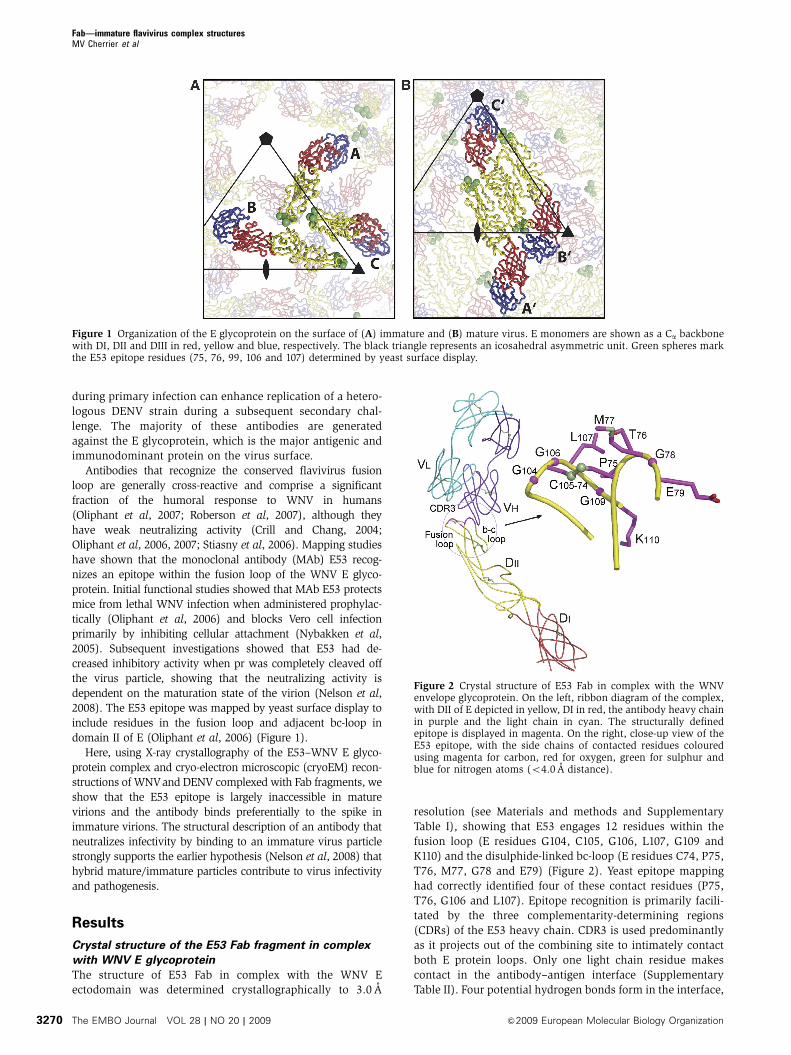
The structure of E53 Fab in complex with the WNV E
ectodomain was determined crystallographically to 3.0 A
resolution (see Materials and methods and Supplementary
Table I), showing that E53 engages 12 residues within the
fusion loop (E residues G104, C105, G106, L107, G109 and
K110) and the disulphide-linked bc-loop (E residues C74, P75,
T76, M77, G78 and E79) (Figure 2). Yeast epitope mapping
had correctly identified four of these contact residues (P75,
T76, G106 and L107). Epitope recognition is primarily facili-
tated by the three complementarity-determining regions
(CDRs) of the E53 heavy chain. CDR3 is used predominantly
as it projects out of the combining site to intimately contact
both E protein loops. Only one light chain residue makes
contact in the antibody–antigen interface (Supplementary
Table II). Four potential hydrogen bonds form in the interface,
Figure 1 Organization of the E glycoprotein on the surface of (A) immature and (B) mature virus. E monomers are shown as a Ca backbonewith DI, DII and DIII in red, yellow and blue, respectively. The black triangle represents an icosahedral asymmetric unit. Green spheres markthe E53 epitope residues (75, 76, 99, 106 and 107) determined by yeast surface display.
Figure 2 Crystal structure of E53 Fab in complex with the WNVenvelope glycoprotein. On the left, ribbon diagram of the complex,with DII of E depicted in yellow, DI in red, the antibody heavy chainin purple and the light chain in cyan. The structurally definedepitope is displayed in magenta. On the right, close-up view of theE53 epitope, with the side chains of contacted residues colouredusing magenta for carbon, red for oxygen, green for sulphur andblue for nitrogen atoms (o4.0 A distance).
Fab—immature flavivirus complex structuresMV Cherrier et al
The EMBO Journal VOL 28 | NO 20 | 2009 &2009 European Molecular Biology Organization3270
three of which involve exposed backbone oxygen and nitro-
gen atoms of the E protein fusion loop. The binding interface
has a low degree of shape complementarity (Sc¼ 0.49)
(Sundberg and Mariuzza, 2002) and occludes approximately
800 A2 of surface area. The entire sequence of the E53 epitope
is conserved between WNV and all four DENV serotypes with
the exception of residue M77, which is a glutamine in DENV.
This difference might explain the stronger binding and neu-
tralization activity of this antibody against WNV (DH
Fremont and MS Diamond, unpublished data).
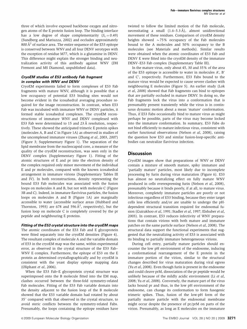
CryoEM studies of E53 antibody Fab fragment
in complex with WNV and DENV
CryoEM experiments failed to form complexes of E53 Fab
fragments with mature WNV, although it is possible that a
low occupancy of potential E53 binding sites would not
become evident in the icosahedral averaging procedure re-
quired for the image reconstruction. In contrast, when E53
Fab was incubated with immature WNV or DENV particles, it
formed stable icosahedral complexes. The cryoEM recon-
structions of immature WNV and DENV complexed with
E53 Fab were determined to 15 and 23 A resolution, respec-
tively. These showed the anticipated trimeric E protein spikes
(molecules A, B and C in Figure 1A) as observed in studies of
the uncomplexed immature viruses (Zhang et al, 2003, 2007)
(Figure 3; Supplementary Figure 1). The separation of the
lipid membrane from the nucleocapsid core, a measure of the
quality of the cryoEM reconstruction, was seen only in the
DENV complex (Supplementary Figure 1). Fitting of the
atomic structures of E and pr into the electron density of
the complex required only minor movement of the individual
E and pr molecules, compared with the known icosahedral
arrangement in immature virions (Supplementary Tables III
and IV). In both reconstructions, density representing the
bound E53 Fab molecules was associated with the fusion
loops on molecules A and B, but not with molecule C (Figure
3B and C). Indeed, in immature flavivirus particles, the fusion
loops on molecules A and B (Figure 1A) are marginally
accessible to water (accessible surface areas (Hubbard and
Thornton, 1993) are 678 and 596 A2, respectively), but the
fusion loop on molecule C is completely covered by the pr
peptide and neighbouring E proteins.
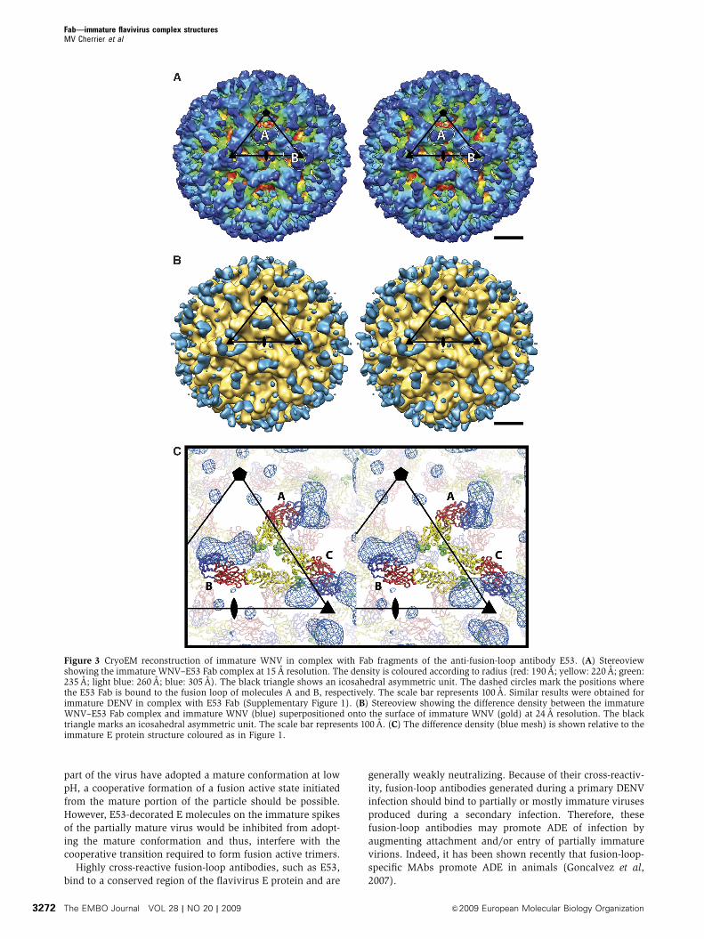
Fitting of the E53 crystal structure into the cryoEM maps
The atomic coordinates of the E53 Fab and E glycoprotein
were fitted separately into the cryoEM densities (Figure 4).
The resultant complex of molecule A and the variable domain
of E53 in the cryoEM map was the same, within experimental
error, as observed in the crystal structure of the E53 Fab–
WNV E complex. Furthermore, the epitope of E53 on the E
protein as determined crystallographically and by cryoEM is
consistent with the yeast display epitope mapping data
(Oliphant et al, 2006).
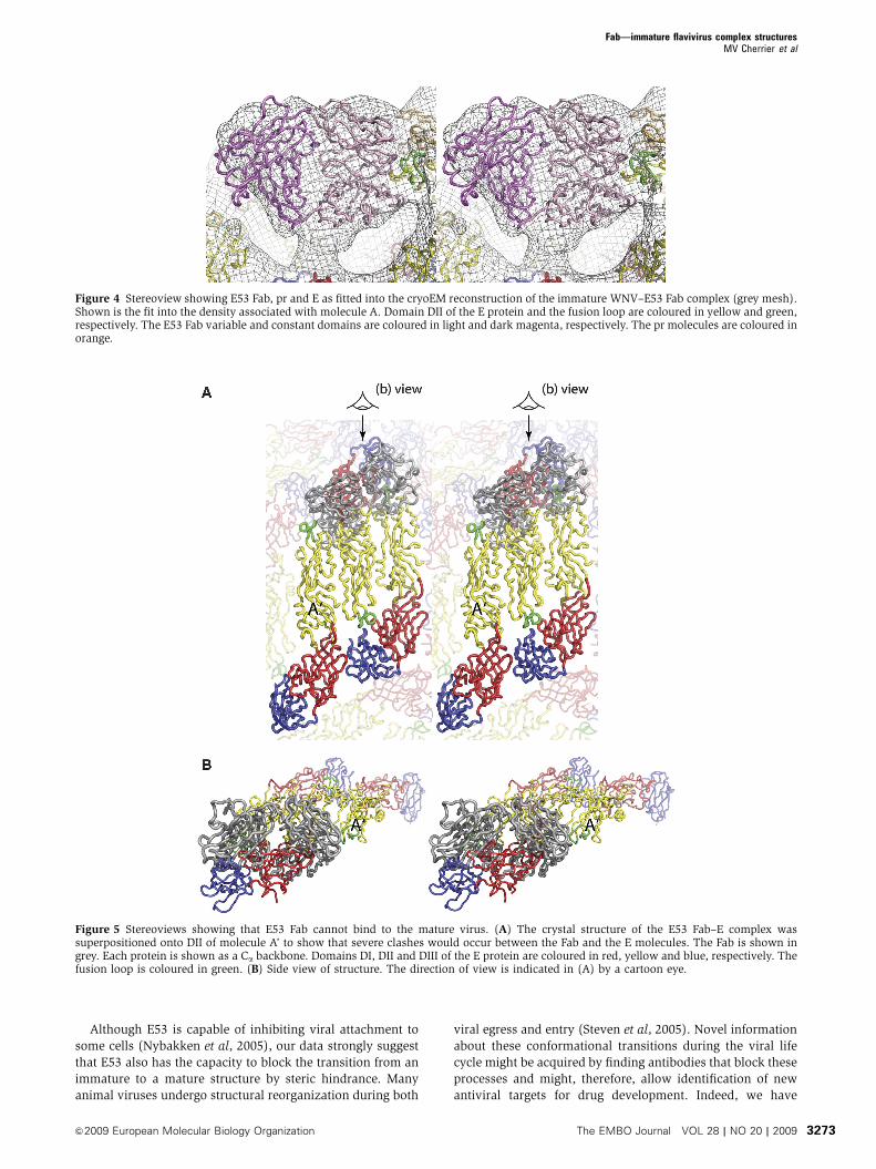
When the E53 Fab–E glycoprotein crystal structure was
superimposed onto the B molecule fitted into the EM map,
clashes occurred between the three-fold symmetry-related
Fab molecules. Fitting of the E53 Fab variable domain into
the density adjacent to the fusion loop of the B molecule
showed that the E53 variable domain had rotated by about
351 compared with that observed in the crystal structure, to
avoid steric conflicts between the symmetry-related Fabs.
Presumably, the loops containing the epitope residues have
twisted to follow the limited motion of the Fab molecule,
necessitating a small (1.6–5.3 A), almost unidirectional
movement of these residues. Comparison of cryoEM density
heights showed B75% occupancy of the Fab molecules
bound to the A molecules and 50% occupancy to the B
molecules (see Materials and methods). Similar results
were obtained when the atomic coordinates of E53 Fab and
DENV E were fitted into the cryoEM density of the immature
DENV–E53 Fab complex (Supplementary Table III).
In the mature virus, only about 45, 30 and 35% of the area
of the E53 epitope is accessible to water in molecules A’, B’
and C’, respectively. Furthermore, E53 Fabs bound to the
mature virus would be expected to cause severe clashes with
neighbouring E molecules (Figure 5). An earlier study (Lok
et al, 2008) showed that Fab fragments can bind to epitopes
that are partially occluded in mature DENV. In doing so, the
Fab fragments lock the virus into a conformation that is
presumably present transiently while the virus is in contin-
uous dynamic motion about a mean ‘mature’ conformation.
Thus, if E53 Fabs occasionally bind to mature virus as might
perhaps be possible, parts of the virus may become locked
into the immature conformation. In summary, E53 Fabs do
not bind efficiently to mature infectious virus, consistent with
earlier functional observations (Nelson et al, 2008), raising
the question how E53 and similar fusion-loop-specific anti-
bodies can neutralize flavivirus infection.
Discussion
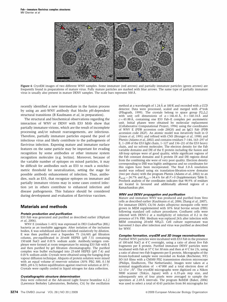
CryoEM images show that preparations of WNV or DENV
contain a mixture of smooth mature, spiky immature and
‘partially mature’ particles, most likely due to incomplete
processing by furin during virus maturation (Figure 6). E53
has almost no neutralizing effect on fully mature WNV
produced in cells overexpressing furin (Nelson et al, 2008),
presumably because it binds poorly, if at all, to mature virus.
Moreover, completely immature particles are poorly or not
infectious regardless of E53 binding, because they enter target
cells less efficiently and/or are unable to undergo the pH-
dependent structural transition required for endosomal fu-
sion (Guirakhoo et al, 1991; Stadler et al, 1997; Elshuber et al,
2003). In contrast, E53 reduces infectivity of WNV prepara-
tions that contain virions with both mature and immature
patches on the same particle surface (Nelson et al, 2008). The
structural data support the functional experiments that sug-
gested that the neutralizing activity of E53 is associated with
its binding to partially immature heterogeneous virions.
During cell entry, partially mature particles should en-
counter the low pH environment of the endosome, inducing
a conformational rearrangement of the E protein in the
immature portion of the virion, similar to the structural
changes described for virus maturation during viral egress
(Yu et al, 2008). Even though furin is present in the endosome
and could cleave prM, dissociation of the pr peptide would be
unlikely because of the mildly acidic environment (Li et al,
2008; Yu et al, 2008). Conversely, the mature part of the virus
lacks bound pr and thus, in the low pH environment of the
endosome, can change its conformation to form fusogenic
trimeric spikes. Thus, fusion of the low-pH form of the
partially mature particle with the endosomal membrane
might occur despite the presence of pr/prM on parts of the
virion. Presumably, as long as E molecules on the immature
Fab—immature flavivirus complex structuresMV Cherrier et al
&2009 European Molecular Biology Organization The EMBO Journal VOL 28 | NO 20 | 2009 3271
part of the virus have adopted a mature conformation at low
pH, a cooperative formation of a fusion active state initiated
from the mature portion of the particle should be possible.
However, E53-decorated E molecules on the immature spikes
of the partially mature virus would be inhibited from adopt-
ing the mature conformation and thus, interfere with the
cooperative transition required to form fusion active trimers.
Highly cross-reactive fusion-loop antibodies, such as E53,
bind to a conserved region of the flavivirus E protein and are
generally weakly neutralizing. Because of their cross-reactiv-
ity, fusion-loop antibodies generated during a primary DENV
infection should bind to partially or mostly immature viruses
produced during a secondary infection. Therefore, these
fusion-loop antibodies may promote ADE of infection by
augmenting attachment and/or entry of partially immature
virions. Indeed, it has been shown recently that fusion-loop-
specific MAbs promote ADE in animals (Goncalvez et al,
2007).
Figure 3 CryoEM reconstruction of immature WNV in complex with Fab fragments of the anti-fusion-loop antibody E53. (A) Stereoviewshowing the immature WNV–E53 Fab complex at 15 A resolution. The density is coloured according to radius (red: 190 A; yellow: 220 A; green:235 A; light blue: 260 A; blue: 305 A). The black triangle shows an icosahedral asymmetric unit. The dashed circles mark the positions wherethe E53 Fab is bound to the fusion loop of molecules A and B, respectively. The scale bar represents 100 A. Similar results were obtained forimmature DENV in complex with E53 Fab (Supplementary Figure 1). (B) Stereoview showing the difference density between the immatureWNV–E53 Fab complex and immature WNV (blue) superpositioned onto the surface of immature WNV (gold) at 24 A resolution. The blacktriangle marks an icosahedral asymmetric unit. The scale bar represents 100 A. (C) The difference density (blue mesh) is shown relative to theimmature E protein structure coloured as in Figure 1.
Fab—immature flavivirus complex structuresMV Cherrier et al
The EMBO Journal VOL 28 | NO 20 | 2009 &2009 European Molecular Biology Organization3272
Although E53 is capable of inhibiting viral attachment to
some cells (Nybakken et al, 2005), our data strongly suggest
that E53 also has the capacity to block the transition from an
immature to a mature structure by steric hindrance. Many
animal viruses undergo structural reorganization during both
viral egress and entry (Steven et al, 2005). Novel information
about these conformational transitions during the viral life
cycle might be acquired by finding antibodies that block these
processes and might, therefore, allow identification of new
antiviral targets for drug development. Indeed, we have
Figure 4 Stereoview showing E53 Fab, pr and E as fitted into the cryoEM reconstruction of the immature WNV–E53 Fab complex (grey mesh).Shown is the fit into the density associated with molecule A. Domain DII of the E protein and the fusion loop are coloured in yellow and green,respectively. The E53 Fab variable and constant domains are coloured in light and dark magenta, respectively. The pr molecules are coloured inorange.
Figure 5 Stereoviews showing that E53 Fab cannot bind to the mature virus. (A) The crystal structure of the E53 Fab–E complex wassuperpositioned onto DII of molecule A’ to show that severe clashes would occur between the Fab and the E molecules. The Fab is shown ingrey. Each protein is shown as a Ca backbone. Domains DI, DII and DIII of the E protein are coloured in red, yellow and blue, respectively. Thefusion loop is coloured in green. (B) Side view of structure. The direction of view is indicated in (A) by a cartoon eye.
Fab—immature flavivirus complex structuresMV Cherrier et al
&2009 European Molecular Biology Organization The EMBO Journal VOL 28 | NO 20 | 2009 3273
recently identified a new intermediate in the fusion process
by using an anti-WNV antibody that blocks pH-dependent
structural transitions (B Kaufmann et al, in preparation).
The structural and biochemical observations regarding the
interaction of WNV or DENV with E53 MAb show that
partially immature virions, which are the result of incomplete
processing and/or subunit rearrangements, are infectious.
Therefore, partially immature particles expand the pool of
infectious virus and likely contribute to the pathogenesis of
flavivirus infection. Exposing mature and immature surface
features on the same particle may be important for evading
recognition by some antibodies or other immune system
recognition molecules (e.g. lectins). Moreover, because of
the variable number of epitopes on mixed particles, it may
be difficult for antibodies such as E53 to reach a stoichio-
metric threshold for neutralization, setting the stage for
possible antibody enhancement of infection. Thus, antibo-
dies, such as E53, that recognize epitopes on immature and
partially immature virions may in some cases control infec-
tion yet in others contribute to enhanced infection and
disease pathogenesis. This balance should be considered
during development and evaluation of flavivirus vaccines.
Materials and methods
Protein production and purificationE53 Fab was generated and purified as described earlier (Oliphantet al, 2006).
WNV E (residues 1–415) was expressed in DE3 CodonPlus (RIL)bacteria as an insoluble aggregate. After isolation of the inclusionbodies, E was solubilized and then refolded oxidatively by dilution.E was then purified over a Superdex 75 (16/60) gel filtrationcolumn, pre-equilibrated in 20 mM HEPES (pH 7.5) containing150 mM NaCl and 0.01% sodium azide. Antibody/antigen com-plexes were formed at room temperature by mixing E53 Fab with Eand then purified by gel filtration chromatography. The purifiedcomplex was buffer exchanged into 25 mM HEPES (pH 7.4) and0.01% sodium azide. Crystals were obtained using the hanging dropvapour diffusion technique. Aliquots of protein solution were mixedwith an equal volume of reservoir solution (30% PEG 300, 0.1 MMES, pH 6.5) before equilibration against reservoir solution at 201C.Crystals were rapidly cooled in liquid nitrogen for data collection.
Crystallographic structure determinationData were collected at the Advanced Light Source beamline 4.2.2(Lawrence Berkeley Laboratories, Berkeley, CA) by the oscillation
method at a wavelength of 1.24 A at 100 K and recorded with a CCDdetector. Data were processed, scaled and merged with d*trek(Pflugrath, 1999). The crystals belong to space group P21212with unit cell dimensions of a¼ 146.41 A, b¼ 160.14 A andc¼ 43.88 A, containing one E53 Fab–E complex per asymmetricunit. Initial phases were obtained by molecular replacement(Collaborative Computational Project, 1994) using the coordinatesof WNV E (PDB accession code 2HG0) and an IgG1 Fab (PDBaccession code 2IGF). An atomic model was iteratively built in O(Jones et al, 1991) and refined with CNS (Brunger et al, 1998) andPhenix (Adams et al, 2002) and contains residues 7–144, 163–297 ofE, 1–208 of the E53 light chain, 1–127 and 136–211 of the E53 heavychain, and no solvent molecules. The electron density for the Fabvariable domains and DII of the E protein including the fusion andAB-loop epitope were of good quality, while significant regions ofthe Fab constant domains and E protein DI and DII regions distalfrom the combining site were of very poor quality. Electron densitycorresponding to DIII was highly ambiguous and no coordinates forthis region have been incorporated. The final 3.0-A resolutionmodel was refined using six translation-libration-screw domains(two per chain) with the program Phenix (Adams et al, 2002) to anRwork¼ 24.7% and Rfree¼ 34.4% for all F40 (Supplementary Table I).Procheck analysis of the coordinates indicates that 98.9% of residuesare located in favoured and additionally allowed regions of aRamachandran plot.
WNV and DENV propagation and purificationMature and immature WNV was produced and purified from Verocells as described earlier (Kaufmann et al, 2006; Zhang et al, 2007).For immature DENV, C6/36 Aedes albopictus mosquito cells weregrown in MEM supplemented with 10% fetal bovine serum (FBS)following standard cell culture procedures. Confluent cells wereinfected with DENV-2 at a multiplicity of infection of 0.2 in thepresence of 5% FBS. Medium was replaced 24 h after infection withMEM containing 20 mM NH4Cl. Cell culture supernatant washarvested 3 days after infection and virus was purified as describedfor WNV.
Complex formation, cryoEM and 3D image reconstructionsPurified WNV particles were incubated with E53 Fab in the presenceof 100 mM NaCl at 41C overnight, using a ratio of about five Fabfragments per E protein. Purified immature DENV particles wereincubated with Fab at 371C for 30 min and then at 41C for 2 h, usinga ratio of about two Fab fragments per E protein. Micrographs of thefrozen-hydrated sample were recorded on Kodak (Rochester, NY)SO-163 films with a CM300 FEG transmission electron microscope(Philips, Eindhoven, The Netherlands). Images were taken at anominal magnification of � 47000 and a total electron dose of12–15 e�/A2. The cryoEM micrographs were digitized on a Nikon9000 scanner (Tokyo, Japan) with a 6.35-mm step size, andsubsequently sets of four pixels were averaged to sample thespecimen at 2.69 A intervals. The program RobEM (Baker, 2004)was used to select a total of 4143 particles from 84 micrographs for
Figure 6 CryoEM images of two different WNV samples. Some immature (red arrows) and partially immature particles (green arrows) arefrequently found in preparations of mature virus. Fully mature particles are marked with blue arrows. The same type of partially immaturevirus is usually also present in mature DENV samples. The scale bars represent 500 A.
Fab—immature flavivirus complex structuresMV Cherrier et al
The EMBO Journal VOL 28 | NO 20 | 2009 &2009 European Molecular Biology Organization3274
the immature WNV–E53 Fab complex and a total of 2741 particlesfrom 23 micrographs for the complex of immature DENV with E53Fab. The defocus level was determined by fitting the theoreticalmicroscope contrast transfer functions (CTFs) to the incoherentsum of the Fourier transforms of all particle images fromeach micrograph. The 3D reconstruction was computed using CTFphase-corrected images. The reconstruction was initiated by using acryoEM density map of immature WNV as a model. The particleorientations were determined with SPIDER (Frank et al, 1996),and the 3D electron density map was calculated with a modifiedversion of XMIPP (Sorzano et al, 2004) assuming icosahedralsymmetry. Only 3927 and 2741 particles of the WNV andDENV complex, respectively, were selected to calculate the final3D electron density maps. Selection was based on correlationwith the model projections and stability of the particle centreposition used. The resolution of the resultant map was estimatedby comparing structure factors for the virus shell computedfrom two independent half-data sets. The estimated resolutionwas based on determining the spacing frequency at whichthe correlation between the two independent data sets became lessthan 0.5.
One measure of the map quality is the resolution of the lipidleaflets. The above procedure did not give a good representation ofthe lipid bilayer in the immature WNV–E53 Fab reconstruction(Supplementary Figure 1). Thus, as an alternative reconstructiontechnique, the Polar Fourier Transform (PFT) (Baker and Cheng,1996) reciprocal space procedure was used for both WNV andDENV. This gave considerably better representations of themembrane region of these viruses, but the quality of the densityrepresenting the glycoprotein was reduced. This might suggest thatthe PFT method is the better procedure indicating that theinterpretation of the Fab density in the XMIPP reconstruction couldbe inaccurate. However, the excellent agreement of the cryoEMdensity of the E53 Fab–virus complex with the crystal structure ofthe soluble E ectodomain in complex with E53 Fab showed that thereconstruction based on the modified XMIPP procedure wasaccurate. Thus, the lack of a clear separation of the lipid bilayersin the membrane of the reconstructions is not the result of disorderin the lipid due to the E53 binding, but a limitation of thereconstruction techniques.
Fitting of X-ray coordinates into the cryoEM electron densityThe fitting of the E and E53 Fab atomic coordinates to the immaturevirusFab complexes followed earlier procedures (Kaufmann et al,2006) using the program EMfit (Rossmann et al, 2001). First, the Eglycoprotein (PDB codes 3C6D (Li et al, 2008) and 2OF6 (Zhanget al, 2007) of DENV and WNV, respectively) and pr (PDB code3C5X (Li et al, 2008) for DENV and a model for the pr of WNVgenerated by SWISS-MODEL (Arnold et al, 2006)) were fittedindependently into the cryoEM density (Supplementary Table III).Each E molecule was divided into two rigid bodies, DIþDIII(residues 1–51þ133–196þ 281–400 for WNV and residues 1–49þ132–192þ 280–395 for DENV) and DIIþpr. The position andorientation of each rigid body was determined one at a time bymaking a complete 3D angular search, followed by rotational andtranslational refinement. The density corresponding to the best fitwas set to zero before fitting the next rigid body. Domain DIþDIIIwas fitted by using a maximum distance restraint of 10 A betweenthe termini of strands connecting DI and DII. The results showedthat the general arrangement of the E ectodomain and pr in thevirus–E53 Fab complexes did not significantly change in compar-ison to the structure of the unliganded immature virus.
Similarly, the atomic coordinates of E53 Fab, extracted from thecrystal structure of the E53 Fab–WNV E protein complex, werefitted into the cryoEM density of the complex. For independentfitting, the Fab was divided into two rigid bodies, the variabledomain (residues 1–121 chain H and 1–107 chain L) and theconstant domain (residues 122–221 chain H and 108–213 chain L).Although the variable domain fitted well into the density associatedwith molecule A (Figure 1A), the constant domain required someadjustment, accounting for a small change of the elbow angle from1441 to 1531 compared with the crystal structure (Figure 4).Furthermore, a good fit of the E53 variable domain into the cryoEMdensity was obtained when DII and the Fab variable domain of thecrystal structure were fitted as one rigid body, verifying the
conservation of the mode of binding between the E protein andthe variable domain of E53.
However, fitting of the E53 Fab variable domain into thedifference density adjacent to the B molecule fusion loop showedthat the E53 variable domain had rotated by about 35º relative tothe E protein in comparison with the crystal structure. When thecrystal structure of the E53 Fab–E complex was superpositionedonto the B molecule in the EM map there were clashes between thethree-fold symmetry-related Fab molecules. Thus, the immatureWNV structure, the complex of E53 Fab with the B molecule, isslightly different than that observed in the crystal structure and withmolecule A. Moreover, to avoid steric hindrance, the elbow angleof the Fab associated with the B molecule changed from 1441 to1711.
A rough measure of the Fab occupancies was obtained bycomparing the average height of the densities (‘sumf’ (Rossmannet al, 2001)) between the DIIþpr and the Fab variable domain. Theheight of density associated with the Fab molecules was lowercompared with the density of DII of E (73 and 57% of the variabledomain density for molecules A and B, respectively). This is mostlikely due to incomplete occupancy of the binding sites by the Fabmolecules. The Fab bound to the B molecule has a lower occupancycompared with molecule A, probably as a result of steric hindrance.The height of the density for the more distant Fab constantdomains is even further diminished (61 and 83% of the variabledomain density in the A and B molecules, respectively). Thissuggests that either the elbow angle in the Fab molecule issomewhat flexible.
The elbow angle between Fab variable and constant domainswas calculated using the program Elbow Angle Calculation (http://proteinmodel.org/AS2TS/RBOW/index.html).
Data depositionAtomic coordinates and structure factors for the reported crystalstructure have been deposited with the Protein Data Bank underaccession code 3I50 (rcsb053971). The cryoEM density maps weredeposited in the EM databank under accession number EMD-5103(immature WNV–E53 Fab) and EMD-5102 (immature DENV–E53Fab). The fitted complex structures were deposited in the ProteinData Bank under accession codes 3IXX (for WNV–E53 Fab) and3IXY (for DENV–E53 Fab).
Figure preparationFigures were created using the programs PYMOL (DeLano, 2002)and CHIMERA (Pettersen et al, 2004).
Supplementary dataSupplementary data are available at The EMBO Journal Online(http://www.embojournal.org).
Acknowledgements
We are grateful to Sheryl Kelly and Carol J Greski for help in thepreparation of the paper. The work was supported by the NIH grantsRO1-AI76331 (MGR), R01-AI073755 (MSD), U01-AI061373 (MSD),and PDVI grants TR-17 (RJK and MGR) and DR-5 (DHF). We aregrateful to the Keck Foundation grant for the purchase of the CM300field emission gun electron microscope used in this study. MVCprocessed and analysed cryoEM data, fitted X-ray structures incryoEM maps and wrote the paper. BK prepared Fab complexes ofmature and immature WNV. SL prepared Fab complex with im-mature DENV. GEN, BRC, CAN, JTW and DHF produced the WNVE–Fab complex crystals and performed the crystallographic analy-sis. VAK provided computational support for cryoEM reconstruc-tion. HAH and PRC collected cryoEM data. RJK was involved instudy design. MSD produced E53 Fab for cryoEM. MGR wasinvolved in study design, data analysis and paper writing. MVC,BK, SL, RJK, MSD, MGR and DHF discussed and analysed theresults and edited the paper.
Conflict of interestThe authors declare that they have no conflict of interest.
Fab—immature flavivirus complex structuresMV Cherrier et al
&2009 European Molecular Biology Organization The EMBO Journal VOL 28 | NO 20 | 2009 3275
References
Adams PD, Grosse-Kunstleve RW, Hung LW, Ioerger TR, McCoy AJ,Moriarty NW, Read RJ, Sacchettini JC, Sauter NK, Terwilliger TC(2002) PHENIX: building new software for automated crystal-lographic structure determination. Acta Crystallogr D BiolCrystallogr 58: 1948–1954
Allison SL, Schalich J, Stiasny K, Mandl CW, Heinz FX (2001)Mutational evidence for an internal fusion peptide in flavivirusenvelope protein E. J Virol 75: 4268–4275
Arnold K, Bordoli L, Kopp J, Schwede T (2006) The SWISS-MODELworkspace: a web-based environment for protein structurehomology modelling. Bioinformatics 22: 195–201
Baker TS (2004) RobEM imaging and visualization program(http://cryoem.ucsd.edu/programDocs/runRobem.txt )
Baker TS, Cheng RH (1996) A model-based approach for determin-ing orientations of biological macromolecules imaged bycryoelectron microscopy. J Struct Biol 116: 120–130
Brandt W (1982) Infection enhancement of dengue type 2 virus inthe U-937 human monocyte cell line by antibodies to flaviviruscross-reactive determinants. Infect Immun 36: 1036–1041
Bressanelli S, Stiasny K, Allison SL, Stura EA, Duquerroy S, LescarJ, Heinz FX, Rey FA (2004) Structure of a flavivirus envelopeglycoprotein in its low-pH-induced membrane fusion conforma-tion. EMBO J 23: 728–738
Brunger AT, Adams PD, Clore GM, DeLano WL, Gros P,Grosse-Kunstleve RW, Jiang JS, Kuszewski J, Nilges M, PannuNS, Read RJ, Rice LM, Simonson T, Warren GL (1998)Crystallography & NMR system: a new software suite for macro-molecular structure determination. Acta Crystallogr D BiolCrystallogr 54: 905–921
Collaborative Computational Project N (1994) The CCP4 suite:programs for protein crystallography. Acta Crystallogr D BiolCrystallogr 50: 760–763
Crill WD, Chang GJ (2004) Localization and characterization offlavivirus envelope glycoprotein cross-reactive epitopes. J Virol78: 13975–13986
DeLano WL (2002) The PyMOL Molecular Graphics System. SanCarlos, CA, USA: DeLano Scientific (http://wwwpymolorg)
Elshuber S, Allison SL, Heinz FX, Mandl CW (2003) Cleavage ofprotein prM is necessary for infection of BHK-21 cells bytick-borne encephalitis virus. J Gen Virol 84: 183–191
Frank J, Radermacher M, Penczek P, Zhu J, Li Y, Ladjadj M, Leith A(1996) SPIDER and WEB: processing and visualization of imagesin 3D electron microscopy and related fields. J Struct Biol 116:190–199
Goncalvez A, Engle R, St Claire M, Purcell R, Lai C (2007)Monoclonal antibody-mediated enhancement of dengue virusinfection in vitro and in vivo and strategies for prevention. ProcNatl Acad Sci USA 104: 9422–9427
Guirakhoo F, Heinz FX, Mandl CW, Holzmann H, Kunz C (1991)Fusion activity of flaviviruses: comparison of mature and im-mature (prM-containing) tick-borne encephalitis virions. J GenVirol 72: 1323–1329
Hubbard SJ, Thornton JM (1993) NACCESS. Computer Program,Department of Biochemistry and Molecular Biology, UniversityCollege London (http://www.bioinf.manchester.ac.uk/naccess/)
Jones TA, Zou JY, Cowan SW, Kjeldgaard M (1991) Improvedmethods for building protein models in electron density mapsand the location of errors in these models. Acta Crystallogr A 47:110–119
Kaufmann B, Nybakken GE, Chipman PR, Zhang W, Diamond MS,Fremont DH, Kuhn RJ, Rossmann MG (2006) West Nile virus incomplex with the Fab fragment of a neutralizing monoclonalantibody. Proc Natl Acad Sci USA 103: 12400–12404
Kuhn RJ, Zhang W, Rossmann MG, Pletnev SV, Corver J, Lenches E,Jones CT, Mukhopadhyay S, Chipman PR, Strauss EG, Baker TS,Strauss JH (2002) Structure of dengue virus: implications forflavivirus organization, maturation, and fusion. Cell 108: 717–725
Li L, Lok SM, Yu IM, Zhang Y, Kuhn RJ, Chen J, Rossmann MG(2008) The flavivirus precursor membrane-envelope protein com-plex: structure and maturation. Science 319: 1830–1834
Lok SM, Kostyuchenko V, Nybakken GE, Holdaway HA, Battisti AJ,Sukupolvi-Petty S, Sedlak D, Fremont DH, Chipman PR, RoehrigJT, Diamond MS, Kuhn RJ, Rossmann MG (2008) Binding of aneutralizing antibody to dengue virus alters the arrangement ofsurface glycoproteins. Nat Struct Mol Biol 15: 312–317
Modis Y, Ogata S, Clements D, Harrison SC (2003) A ligand-bindingpocket in the dengue virus envelope glycoprotein. Proc Natl AcadSci USA 100: 6986–6991
Modis Y, Ogata S, Clements D, Harrison SC (2004) Structure of thedengue virus envelope protein after membrane fusion. Nature427: 313–319
Nelson S, Jost CA, Xu Q, Ess J, Martin JE, Oliphant T, Whitehead SS,Durbin AP, Graham BS, Diamond MS, Pierson TC (2008)Maturation of West Nile virus modulates sensitivity to anti-body-mediated neutralization. PLoS Pathog 4: e1000060
Nybakken GE, Nelson CA, Chen BR, Diamond MS, Fremont DH(2006) Crystal structure of the West Nile virus envelope glyco-protein. J Virol 80: 11467–11474
Nybakken GE, Oliphant T, Johnson S, Burke S, Diamond MS,Fremont DH (2005) Structural basis of West Nile virus neutraliza-tion by a therapeutic antibody. Nature 437: 764–769
Oliphant T, Nybakken GE, Austin SK, Xu Q, Bramson J, Loeb M,Throsby M, Fremont DH, Pierson TC, Diamond MS (2007)Induction of epitope-specific neutralizing antibodies againstWest Nile virus. J Virol 81: 11828–11839
Oliphant T, Nybakken GE, Engle M, Xu Q, Nelson CA, Sukupolvi-Petty S, Marri A, Lachmi BE, Olshevsky U, Fremont DH, PiersonTC, Diamond MS (2006) Antibody recognition and neutralizationdeterminants on domains I and II of West Nile Virus envelopeprotein. J Virol 80: 12149–12159
Pettersen EF, Goddard TD, Huang CC, Couch GS, Greenblatt DM,Meng EC, Ferrin TE (2004) UCSF Chimera–a visualization systemfor exploratory research and analysis. J Comput Chem 25:1605–1612
Pflugrath JW (1999) The finer things in X-ray diffraction datacollection. Acta Crystallogr D Biol Crystallogr 55: 1718–1725
Rey FA, Heinz FX, Mandl C, Kunz C, Harrison SC (1995) Theenvelope glycoprotein from tick-borne encephalitis virus at 2 Aresolution. Nature 375: 291–298
Roberson JA, Crill WD, Chang GJ (2007) Differentiation of West Nileand St Louis encephalitis virus infections by use of noninfectiousvirus-like particles with reduced cross-reactivity. J Clin Microbiol45: 3167–3174
Rossmann MG, Bernal R, Pletnev SV (2001) Combining electronmicroscopic with X-ray crystallographic structures. J Struct Biol136: 190–200
Sorzano CO, Marabini R, Velazquez-Muriel J, Bilbao-Castro JR,Scheres SH, Carazo JM, Pascual-Montano A (2004) XMIPP: anew generation of an open-source image processing package forelectron microscopy. J Struct Biol 148: 194–204
Stadler K, Allison SL, Schalich J, Heinz FX (1997) Proteolyticactivation of tick-borne encephalitis virus by furin. J Virol 71:8475–8481
Steven AC, Heymann JB, Cheng N, Trus BL, Conway JF (2005) Virusmaturation: dynamics and mechanism of a stabilizing structuraltransition that leads to infectivity. Curr Opin Struct Biol 15: 227–236
Stiasny K, Kiermayr S, Holzmann H, Heinz FX (2006) Crypticproperties of a cluster of dominant flavivirus cross-reactive anti-genic sites. J Virol 80: 9557–9568
Sundberg EJ, Mariuzza RA (2002) Molecular recognition in anti-body-antigen complexes. Adv Protein Chem 61: 119–160
Wengler G, Wengler G (1989) Cell-associated West Nile flavivirus iscovered with E+pre-M protein heterodimers which are destroyedand reorganized by proteolytic cleavage during virus release.J Virol 63: 2521–2526
Widman DG, Frolov I, Mason PW (2008) Third-generation flavi-virus vaccines based on single-cycle, encapsidation-defectiveviruses. Adv Virus Res 72: 77–126
Yu IM, Zhang W, Holdaway HA, Li L, Kostyuchenko VA, ChipmanPR, Kuhn RJ, Rossmann MG, Chen J (2008) Structure of theimmature dengue virus at low pH primes proteolytic maturation.Science 319: 1834–1837
Zhang Y, Corver J, Chipman PR, Zhang W, Pletnev SV, Sedlak D,Baker TS, Strauss JH, Kuhn RJ, Rossmann MG (2003) Structuresof immature flavivirus particles. EMBO J 22: 2604–2613
Zhang Y, Kaufmann B, Chipman PR, Kuhn RJ, Rossmann MG (2007)Structure of immature West Nile virus. J Virol 81: 6141–6145
Zhang Y, Zhang W, Ogata S, Clements D, Strauss JH, Baker TS, KuhnRJ, Rossmann MG (2004) Conformational changes of the flavi-virus E glycoprotein. Structure 12: 1607–1618
Fab—immature flavivirus complex structuresMV Cherrier et al
The EMBO Journal VOL 28 | NO 20 | 2009 &2009 European Molecular Biology Organization3276


![Structural basis of sterol recognition and nonvesicular ...myweb.chonnam.ac.kr/~stbiochm/Publications/PDF/[2018 PNAS] LAM.pdf · Structural basis of sterol recognition and nonvesicular](https://img.pdfslide.us/doc/110x75/5c95af6109d3f2de7d8d04e3/structural-basis-of-sterol-recognition-and-nonvesicular-myweb-stbiochmpublicationspdf2018.jpg)