Embed Size (px)
Citation preview
SECTION C • Gene Transfer: Conjugation
Structure and Function of the F Factor and Mechanism of Conjugation
NEVILLE FIRTH, KARIN IPPEN-IHLER, AND RONALD A. SKURRAY
126
INTRODUCTION
Conjugation, a process which promotes DNA transfer from a donor to a recipient cell mediated byphysical contact (49, 139), occurs among both gram-negative and gram-positive bacteria andstreptomycetes (for reviews of specific systems, see the book Bacterial Conjugation [55]). Donor abilityis conferred by the presence of an infectious DNA element which disseminates to other cells.Commonly, genes encoding conjugative-transfer functions are associated with an extrachromosomalreplicon, termed a self-transmissible or conjugative plasmid. In addition to self-transfer, the transfersystems of conjugative plasmids often facilitate the independent transfer of nonconjugative, mobilizableplasmids that are coresident in the donor cell. DNA sequences that become cointegrate with theconjugative plasmid can also be transferred; thus, integration and other recombinational rearrangementscan result in transmission of sequences from the bacterial chromosome, of transposons, and ofnonmobilizable plasmids. As a means of genetic exchange among individual cells and populations, bothwithin and between bacterial species, conjugation is a phenomenon of fundamental evolutionary andecological consequence (for a review, see reference 236). The significance of this process has beenfurther highlighted by evidence that conjugation systems can also facilitate interkingdom transmission ofgenetic material. Ti plasmid-mediated T-DNA transfer from Agrobacterium species to plants appears torepresent a novel form of bacterial conjugation (177, 211, 324, 364, 372). Transfer of both broad- andnarrow-host-range plasmids (R751 and F, respectively) from Escherichia coli to Saccharomycescerevisiae has also been demonstrated (142, 143).
Responsible for the earliest observation of genetic transfer (206), the F (fertility) factor of E. coli K-12 was the first plasmid to be described (49) and, as both subject and tool, has been studied ever since.The insight and ingenuity of early investigators allowed principal characteristics of F, F-mediated DNAtransfer, and the circular configuration of plasmid and chromosomal DNAs to be determined solelythrough analysis of genetic crosses involving chromosomal markers (for an interesting account, see TheGenetics of Bacteria and their Viruses [140]). Key deductions were (i) that F is a circular “episome”able either to replicate autonomously or to integrate in the bacterial chromosome and (ii) that theefficient transfer of chromosomal markers from Hfr (high-frequency recombinant) donor strains reflectsa stable integration of F. The order and time of entry of markers transferred by Hfr donors could then beseen to reflect the position and orientation of a specific site, the F origin of transfer (oriT). It wasperceived that transfer of DNA must always begin at this site and proceed unidirectionally around thecircular genome. That a single strand of DNA (56, 124, 269) was transferred in the 5′ → 3′ direction(152, 269) was subsequently demonstrated. These appear to be basic precepts for conjugation, and oriTsites on many other subsequently studied conjugative elements have been found to function similarly indirecting the transfer of contiguous DNA.
The observation of conjugation and the synchronous Hfr crosses necessary to time-of-entryexperiments also depended on the efficient way in which F donors contact recipients. “Mating pairs”could form quickly in liquid suspensions, persist during gentle dilution, and be disrupted by severe
agitation. F pili, the filaments that extend from the donor cell surface to initiate these contacts, werediscovered after bacteriophages that infected F+ but not F– cells were isolated. The adsorption of RNAphages along the length of F pili distinguished them from other fimbrial appendages (60). Expression ofa pilus filament has also proven to be essential for other enteric and pseudomonad plasmid conjugationsystems (36, 38, 100, 161, 286), and all conjugative-plasmid transfer among these gram-negativeorganisms is thought to depend on contacts created by these structures.
STRUCTURE OF THE F PLASMID
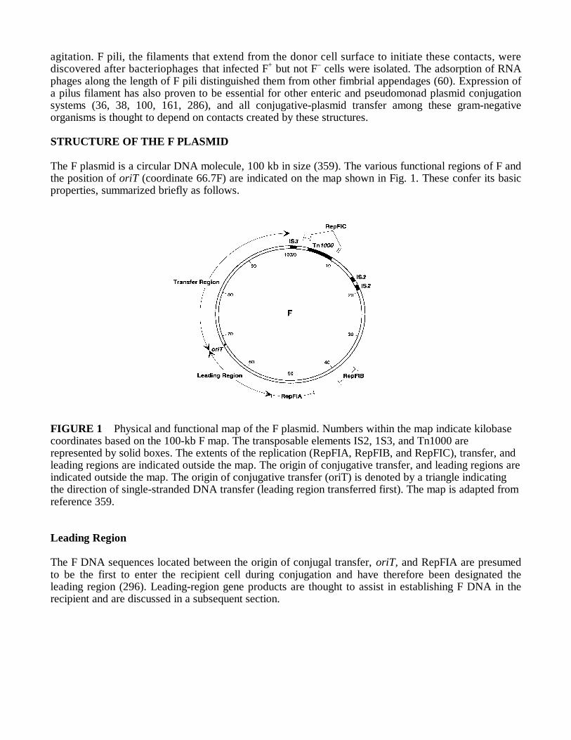
The F plasmid is a circular DNA molecule, 100 kb in size (359). The various functional regions of F andthe position of oriT (coordinate 66.7F) are indicated on the map shown in Fig. 1. These confer its basicproperties, summarized briefly as follows.
FIGURE 1 Physical and functional map of the F plasmid. Numbers within the map indicate kilobasecoordinates based on the 100-kb F map. The transposable elements IS2, 1S3, and Tn1000 arerepresented by solid boxes. The extents of the replication (RepFIA, RepFIB, and RepFIC), transfer, andleading regions are indicated outside the map. The origin of conjugative transfer, and leading regions areindicated outside the map. The origin of conjugative transfer (oriT) is denoted by a triangle indicatingthe direction of single-stranded DNA transfer (leading region transferred first). The map is adapted fromreference 359.
Leading Region
The F DNA sequences located between the origin of conjugal transfer, oriT, and RepFIA are presumedto be the first to enter the recipient cell during conjugation and have therefore been designated theleading region (296). Leading-region gene products are thought to assist in establishing F DNA in therecipient and are discussed in a subsequent section.

Autonomous Replication
The RepFIA region, believed to be primarily responsible for the typical replication properties of F,contains both unidirectional (oriS) and bidirectional (oriV) replication origins (201). The maintenancecharacteristics of mini-F plasmids that include only the F-EcoRI fragment f5 (44.6 to 53.7F) closelyresemble those of F. Stringent regulation of RepFIA and associated maintenance and partitioningmechanisms act in concert to sustain the plasmid at one to two copies per cell (57, 97).
The secondary replication region, RepFIB, is independently functional and can sustain plasmidreplication in the absence of RepFIA. The RepFIC region includes an incomplete remnant of areplication system that is used by some other related plasmids (31). The complexities of F replicationhave been reviewed elsewhere (184, 185, 359).
Transposable Elements
The F sequence includes a single copy of Tn1000 (also known as γδ) and IS2 and two copies of IS3(Fig. 1) (129, 147–149) (see chapters 111 and 124 for descriptions of transposable elements). Tn1000appears to have interrupted RepFIC (300), whereas IS3 inactivation of the transfer region regulatorygene, finO, is responsible for the constitutively high levels of conjugative transfer exhibited by F (52,371). These elements also mediate the F-chromosomal integration events that form Hfr donors (seechapters 127 and 128) which, through imprecise excision, can subsequently generate F-prime (F′)plasmids (see chapter 129). The insertion of transposable elements into an F-plasmid progenitor wasfortuitous for the efficient transfer of chromosomal markers crucial to the original detection ofconjugation and to the subsequent utility of F (353; also see chapter 137).
Conjugative Transfer
Including the oriT site (map position 66.7F) and extending to the HindIII restriction cleavage site in IS3(coordinate 100/0F), the transfer (tra) region encodes all of the F loci known to be required for efficientconjugative transfer; its 33.3-kb nucleotide sequence has recently been compiled (103). The mechanismof the transfer process and the characteristics and functions of tra products are detailed in subsequentsections.
Insertion of the 41-kb BamHI-HindIII F fragment (coordinates 59.3 to 100F) which includes thetransfer region causes other replicons to become self-transmissible (174, 295). The circularized 55-kbHindIII fragment which includes RepFIA, as well as the tra and leading regions of F, forms a usefulplasmid, pOX38 (130), that has F transfer and maintenance properties but lacks transposable elements,RepFIC and RepFIB sequences, and other loci between map positions 100/0F and 45F. The latterinclude the “killer gene” srnB (116) and pif loci responsible for inhibition of bacteriophage T7development (249). The map positions of these and other named F loci have been tabulated previously (359).
F-LIKE PLASMIDS
The F-transfer system is the prototype for the various conjugation systems expressed by a large group ofconjugative plasmids known collectively as F-like. The relatedness of these plasmids was initially indicatedby the morphological and serological similarity of the pili they expressed and the bacteriophage sensitivitiesthese conferred (66, 204). Relationships among F-like plasmids have been further subdivided on the basis ofincompatibility (Inc), resulting in the seven Inc groups, IncFI through IncFVII. This subdivision is generallyassociated with a plasmid’s replicon(s), since plasmids are placed in the same Inc group if they cannot stablycoexist in the same host cell (66). Incompatibility, which forms the basis of both F- and non-F-like plasmidclassification, usually eventuates if plasmids share similar replication functions (30). The relatedness of F-like plasmids was further demonstrated by heteroduplex analysis (311) and, more recently, by DNAsequencing (for a review, see reference 103). In addition to clinically significant determinants, which includethose for antibiotic resistances and production of hemolysins and toxins, F-like plasmids have been found to

encode a range of ecologically important factors, such as colicins and metabolic activities (see reference 164for a tabulation). F-like plasmids are found throughout the family Enterobacteriaceae (171).
The classification of other conjugative plasmids from the family Enterobacteriaceae and the genusPseudomonas, not known to have F-like conjugation systems, has been based primarily on incompatibility.However, as such transfer systems are examined, indications of broader similarities are emerging. The twostructural types of conjugative pili detected have suggested that all conjugative plasmids in these Inc groupscould belong to two evolutionary families. Whereas many, like F, express long, flexible pili, transferefficiently in liquid cultures, and are frequently associated with phage f1 (M13, fd) and/or J sensitivity (e.g.,IncF complex, IncD, IncC, and IncJ plasmids), a second group produces short, rigid pili, transfers moreefficiently among cells on surfaces (unless assisted by other pili), and often confers sensitivity to phages PR4and/or X (e.g., IncP, IncW, IncN, and IncI plasmids) (36–38; reviewed in references 100, 161, and 286).Analysis of transfer gene organization and DNA sequences has generally supported this two-family grouping(103, 128) and indicated that Agrobacterium Ti plasmid transfer systems also resemble those of the IncP,IncW, IncN, and IncI group of plasmids (210, 211, 213, 282, 344; S. Bolland, Ph.D. thesis, University ofCantabria, Spain, 1991). The characteristics of some plasmids blur even this distinction, however. Asindicated in subsequent sections, evidence for relationships that span the two families has also begun toemerge (178, 212, 213, 283, 312, 313).
F-CONJUGATION PROCESS
The proficiency of the F-conjugative system in liquid matings has allowed the physiology of F-mediated cellcontacts to be studied. Figure 2 depicts the stages of intercellular contact and DNA transfer thought to occurduring F-mediated conjugation. These are as follows. Contact between F+ donor and F– recipient cells isbelieved to be instigated by an interaction between the tip of an F pilus and the recipient cell surface (144,264, 274, 276). There is evidence for the occurrence of DNA transfer between cells that were not in surfacecontact (134, 276), but there is also both indirect (9, 279) and direct (78) evidence that conjugating cells aretypically aggregated in close wall-wall association (5, 275). When DNA transfer is completed, mating cellsactively disaggregate (9, 78) to yield two cells capable of donor activity.
The first surface association of donors and recipients in aggregates is thought to reflect pilus retraction,mediated by depolymerization of the pilus subunit, into the donor and/or recipient cell envelope(s) (63, 162,265, 315). These donor and recipient contacts then become stabilized in a manner that renders the aggregatemore resistant to shear forces (5, 9, 230). In thin sections, the cells exhibit relatively large, electron-denseregions of envelope association, termed conjugative junctions (78). The biochemical reactions involved andthe pathway for DNA transport remain unclear. DNA entry into the recipient has been suggested to occurthrough a direct passage to the recipient cytoplasm (359) or via the recipient periplasm, where recipienttransport components might facilitate DNA uptake (78).
A large number of the F products required for conjugative transfer are involved in F-pilus synthesis andaggregate stabilization. Recipient cells carrying mutations altering the structure of the lipopolysaccharide(20) or the outer membrane protein OmpA (232) also affect F-donor cell interactions, but it is unclearwhether these cell surface components are involved in initial contacts with the pilus or at a subsequentstabilization stage (for reviews, see references 10, 161, and 358). The effects are plasmid specific, sinceompA mutants which act as poor recipients for F+ donors mate efficiently with cells harboring R100-1 orR136 (231, 320) and since F, ColB2, and R100-1 transfers are affected differently by mutations in individuallipopolysaccharide core biosynthesis (rfa) genes (20). Furthermore, these specificities do not depend on thepilin subunit (20), and the transfer defects associated with conjugation-defective recipients can often bebypassed if matings are undertaken on a solid surface rather than in liquid media (137, 138).
F DNA transfer processes are believed to be precipitated by a “mating signal” generated by functional-pairformation (183, 277). Many aspects of the DNA-related events necessary for conjugative transfer have beencharacterized (for reviews, see references 202 and 349). Briefly, a protein complex (oriT complex) isassociated with the origin of transfer of transmissible and mobilizable plasmids. One DNA strand in the oriTsite is “nicked” by a relaxase that catalyzes the covalent attachment of the 5′ end of the DNA to the protein.The F-plasmid relaxase, TraI, is also a helicase and can unwind the nicked strand in the 5′ → 3′ direction.

During transfer, this protein is suggested to be associated with the site of intercellular connection throughwhich this single strand of DNA is passed; because it is attached to the 5′ end of oriT, it may also catalyzerecircularization of the transported strand to terminate transfer (202, 234, 235, 297, 349, 359, 360).Replacement strand synthesis in the donor and complementary-strand synthesis in the recipient depend onhost enzymes (183, 350) and are not essential for DNA transfer per se (183, 304). Figure 2 shows synthesisin the donor by a rolling-circle mechanism (349); a variation in which both the 5′ and 3′ oriT ends remainassociated with TraI has also been suggested (359, 360). The F-TraI protein forms a covalent linkage onlywith the 5′ oriT end; whether the 3′ end remains bound in some other persistent association is not yet clear(235).
Proteins encoded by the leading region may also contribute to establishing the plasmid DNA in therecipient, but F does not appear to encode or to transfer a primase such as that associated with IncI and IncPplasmid conjugation systems (202, 349).

ORGANIZATION OF THE TRANSFER REGION
The nucleotide sequence of the entire 33.3-kb F-tra region has revealed its genetic structure at maximalresolution (GenBank accession number U01159) (103). Figure 3 shows the organization and functionalclassification of the 36 open reading frames (ORFs) known or deemed likely to encode products. With oneexception, artA, all translated genes are encoded on the same DNA strand. The finO sequence is interruptedby a copy of the transposable element IS3 (52, 371).
The functional classes depicted in Fig. 3 reflect the phenotype of the relevant mutation and determine thecategory in which each individual tra region gene product and activity is discussed in this chapter. Classicalgenetic techniques were initially used to define F-plasmid-encoded transfer functions (12, 13, 92, 160, 270,271). Recombinant DNA techniques subsequently confirmed the autonomy of the region for conjugativeDNA transfer (174, 214, 295) and facilitated the refinement of the genetic and physical map (11, 225,229, 257, 321, 334). The involvement of loci that had not been represented in early mutant collectionshas, in most cases, been tested by insertion mutagenesis employing resistance gene cassettes prior togene replacement via homologous recombination (180).
A variety of gene expression and protein analysis methods have now also been applied toward thedetection of tra region gene products. In most cases, and particularly with genes known to be involved inconjugation, the products encoded have been identified and the subcellular location of many of these proteinshas also been examined (Table 1). Figure 4 correlates the known or predicted location of tra region productswith their size and function.
STRUCTURE AND BIOGENESIS OF F PILI
One to three F pili are typically visualized extending 1 to 2 mm from the surface of a donor cell (10, 162).Production of a thick, flexible filament similar to an F pilus in appearance has been associated with thecapacity of F and F-like plasmid donors to conjugate efficiently in liquid (39, 100). Analysis of purified F pilihas detected only a single type of protein subunit, F pilin (21, 42, 65, 144). Optical and X-ray diffractionstudies indicate that these arrange helically to form a cylindrical pilus structure 8 nm in diameter, with a 2-nm axial hole (233). The basic helix contains 25 subunits in two turns and has a pitch of 16 nm. The unit riseis 1.28 nm, and the crystallographic repeat is 32 nm. Pilin subunits in F pili are related by fivefold symmetryaround the pilus axis. Pili can therefore be envisioned as consisting of doughnut-like discs, composed of fivesegments that each correspond to a pilin subunit; the discs are stacked such that each is rotated 28.8° withrespect to the disc below (100, 278). An alternative view is that five strands of polymerized pilin are woundtogether in a helical fashion. Treatments with Triton X-100 or sodium PPi have been used to visualize veryfine fibers of pilin (42, 241).
In addition to their requirement for conjugal DNA transfer, F pili are utilized as receptors by male-specificbacteriophages; RNA phages, such as R17 (e.g., f2, MS2, and Qβ), attach to the sides of the pilus (60),whereas filamentous single-stranded DNA phages, like f1 (e.g., M13 and fd), adsorb to its tip (47). Theinhibition of mating-pair formation by F-specific DNA phages provides evidence that the pilus tip is crucialfor initial contacts between donor and recipient cells (159, 264), although nonspecific interactions involvingthe pilus side have also been suggested (274).
Synthesis of the F-Pilus Subunit
The products of three F genes, traA, traQ, and traX (Table 1), distinctly spaced within the tra region(Fig. 3), are involved in synthesis of F-pilin subunits. The traA gene encodes the 121-amino-acid (aa)precursor of the pilus subunit, propilin (TraA) (Fig. 5), a 12.8-kDa polypeptide that requires proteolyticprocessing to yield the 7.2-kDa pilin polypeptide (106, 163, 247). Mature F pilin has a sequencecorresponding to that of the last 70 aa in TraA (Fig. 5) and possesses an acetylated N terminus, whichforms its major antigenic determinant (87, 104). The processing and Nα-acetylation steps in propilinmaturation depend on expression of traQ (163, 180, 256) and traX (252), respectively.

TraQ, a 94-aa inner membrane protein of 10.9 kDa (367, 369), appears to provide an efficient, secA-independent, and ethanol-resistant pathway for rapid insertion of propilin into the inner membrane (N.Majdalani and K. Ippen-Ihler, unpublished data; N. Majdalani, D. Moore, S. Maneewanakul, andK. Ippen-Ihler, unpublished data). The 51-aa propilin leader peptide is then removed in a single cleavagestep which depends on signal peptidase I (Majdalani et al., unpublished). Although unusually long, thepropilin leader does contain all of the features typical of signal sequences cleaved by this enzyme:positively charged residues are followed by a hydrophobic core region and a processing sequence, Ala–3-Met-Ala51- Ala+1, which conforms to the –3,–1 rule (Fig. 5) (106, 272, 342). In the absence of TraQ,propilin folding is thought to interfere with its membrane translocation; the traA product is rapidlydegraded, and only a very small percentage becomes processed (223). The unusual N-terminal sequenceand length of the propilin leader peptide are not, however, responsible for these effects. Recent resultsshow that maturation of an altered traA product with a foreshortened leader sequence equivalent topropilin residues 28 to 51 is still TraQ dependent. Furthermore, when the 52-aa propilin leader sequenceand processing site was joined to the mature portion of either β-lactamase or alkaline phosphatase,processing of the fusion protein and secretion of the enzyme into the periplasm occurred efficiently andentirely independently of TraQ (Majdalani et al., unpublished). Thus, sequences within the matureportion of propilin appear to dictate the TraQ dependence of pilin subunit maturation.

TABLE 1 Transfer region genes involved in conjugation
Gene Functional groupa Product length (aa)b Product size (kDa)b Product locationc Reference(s)finOd Regulation 186 21.0 Cytoplasm 59, 371finP Regulation 78 nte Cytoplasm 88, 339traA Pilus biogenesis 121 [70] 12.8 [7.2] Inner membrane and
extracellularly106, 252, 255, 278
traB Pilus biogenesis 475 50.5 Inner membrane 257; Frost, unpublished data cited inreference 103
traC Pilus biogenesis 875 99.2 Cytoplasm/innermembranef
306, 307
traD DNA metabolism 717 81.7 Inner membrane 173, 280, 370traE Pilus biogenesis 188 21.1 Inner membrane 7, 106, 163, 200traF Pilus biogenesis 247 [228] 28.0 [25.9] Periplasm 368traGg Pilus biogenesis and
aggregate stabilization 938 102.4 Inner membrane 94, 230, 254, 356
traH Pilus biogenesis 458 [434] 50.2 [47.8] Periplasm 94, 132, 230traIg DNA metabolism 1,756 192.0 Cytoplasm 2, 40traJ Regulation 229 27.0 Cytoplasm 62, 328traK Pilus biogenesis 242 [221] 25.6 [23.3] Periplasm 287traL Pilus biogenesis 91 10.4 Inner membrane 103, 106traM DNA metabolism 127 14.5 Cytoplasm 73, 328traN Aggregate stabilization 602 [584] 65.7 [63.8] Outer membrane 224traQ Pilus biogenesis 94 10.9 Inner membrane 223, 367, 369traS Surface exclusion 149 16.9 Inner membrane 7, 172traT Surface exclusion 244 [233] 26.0 [23.8] Outer membrane 6, 172, 245, 246, 248, 288traU Pilus biogenesis 330 [308] 36.8 [34.2] Periplasm 253traV Pilus biogenesis 171 [153] 18.6 [16.6] Outer membrane 76, 257traW Pilus biogenesis 210 [193] 23.6 [21.7] Periplasm 225, 227traX Pilus biogenesis 248 27.5 Inner membrane 59, 222, 252traY DNA metabolism 131 15.2 Cytoplasm 95, 157, 199, 261trbC Pilus biogenesis 212 [191] 23.4 [21.2] Periplasm 226trbI Pilus biogenesis 128 14.1 Inner membrane 227aPrimary references for allocation of genes to functional groups are shown in the text.bSizes and lengths are calculated from the deduced amino acid sequence of each product. Sizes and lengths of processed products are shown in brackets. Values forprocessed products based on predicted cleavage sites (i.e., not N-terminal sequencing) are italicized.cProduct locations have been determined experimentally unless shown italicized, in which case the predicted location is listed.dThe finO gene of F is inactivated by an inserted IS3 element, and its complementation class is based on the phenotype of finO alleles from other F-like plasmids. The finOproduct characteristics are hypothetical values calculated after the IS3 sequence is removed (see text).eSince finP encodes an antisense RNA molecule, its product length is in nucleotides.fTraC is a cytoplasmic protein that fractionates with the inner membrane in the presence of other tra products (see the text).gIn addition to the products shown, traG and traI have been found to encode the smaller products TraG* and TraI*, respectively (see the text).


Expression of the traX gene is required for Nα-acetylation of the F-pilin polypeptide (252). Thepredicted product of the traX ORF is 248 aa in length and appears to be a polytopic inner membraneprotein (59). In vivo and in vitro analyses have detected two traX products, TraX1 (24 kDa) and TraX2(22 kDa), which associate with the inner membrane (222, 223a) but appear significantly smaller than the27.5 kDa calculated from the sequence (59). Although both products seem to be translated from the traXORF, there may be more than one translation initiation site. traX codons 29 to 225 encode a regionessential to pilin acetylation activity (Maneewannakul et al., unpublished). Nα-acetylation of pilin is aproperty common to all of the F-like systems characterized thus far (87, 104, 365, 366) but appears notto be essential for F-pilus biogenesis or function. Under typical laboratory conditions, an F-traX mutantwas found to be phenotypically normal for both conjugal DNA transfer and phage sensitivity (252).However, the antibody-binding characteristics of pili elaborated in the absence of TraX differ from thoseof wild-type pili (126, 127, 252).
Amino acid sequence similarity has been detected between TraX, TrbP of the IncPα plasmid RP4(282), the product of the Dichelobacter nodosus (formerly Bacteroides nodosus) fimC gene (145), and thededuced product of an ORF from the filamentous single-stranded DNA phage Cf1c of Xanthomonascampestris pv. citri (94a, 197). Paralleling what has been observed for traX, two polypeptides have beenassociated with expression of fimC, which is located immediately downstream of the major fimbrial subunitgene, fimA, in some D. nodosus serotypes (145).
Attempts to confirm the suggestion that F pilin might be phosphorylated or glycosylated (42, 65, 144)have failed to identify any such covalent modification (22, 101). However, it has recently been shown that aminor proportion of pilin subunits, within both an inner membrane pool and assembled pili, do bear anuncharacterized modification which causes them to migrate on sodium dodecyl sulfate-polyacrylamide gelelectrophoresis (SDS-PAGE) slightly more slowly than the majority of subunits (223, 252). Thismodification, which is presumed to be performed by host-encoded products, appears to occur after signalpeptide cleavage and does not interfere with, but is inhibited by, Nα-acetylation (223). Both forms of F pilinwere found to exhibit considerable stability in vivo (223).

Topology of the F-Pilin Subunit
Under normal circumstances, the majority of pilin subunits in an F+ cell seem to be associated with the innermembrane rather than with assembled pili (127, 254, 255, 323). The topology of F pilin in the innermembrane has been analyzed by traA′-′phoA gene fusions, which indicated that the protein contains twotransmembrane segments and is oriented such that its N- and C-terminal residues are located in theperiplasm (278). Immunogold labeling of spheroplasts has suggested that F-pilin subunits cluster atdiscrete locations in the inner membrane (278).
The sequences of F-traA mutations affecting phage sensitivity and of the traA genes carried by F-likeconjugative plasmids (Fig. 5) have provided insights into the topology of assembled pilin andspecificities associated with these filaments (100, 102, 103, 286). Both the long propilin signal sequenceand the structure of pilin seem to be highly conserved among F-like plasmids (Fig. 5). Among F-likeplasmids, differences in the N-terminal sequence of the mature pilin polypeptide alter the dominantepitope and confer plasmid specificities. However, this region of the protein, although exposed inmembrane pilin and the basal knobs found on free pili, appears to be masked in the assembled filamentexcept possibly at the tip (104, 278, 365). Residues at or near the pilin C terminus and in a regionincluding mature pilin residues 12 to 22 (Fig. 5) appear to be important to RNA-phage binding and havebeen suggested to be available on the outside of the pilus (100, 105). Comparison of the rather differentdeduced pilin sequences of F and pED208 (IncFV), which both confer bacteriophage f1 sensitivity, hassuggested that the binding site of these filamentous DNA phages could involve residues in the region ofgreatest identity (F-pilin residues 16 to 24 [Fig. 5]) (84). Another suggestion, however, is that residuescloser to the F-pilin N terminus (near M9) are important to f1 binding at the pilus tip (100); antibody-binding studies indicate that both an F-pilin epitope including these residues and the N-terminal epitopeof the pED208 pilin sequence are masked along the length of F pili but may be exposed at the end of thefilament (104, 365). F-pilin residues K46 to K49 have been suggested to be located in the cytoplasm formembrane pilin and in the lumen of the assembled filament (278).
F-Pilus Assembly
Exhaustive genetic studies have demonstrated that the majority of genes in the F-tra region areinvolved in pilus assembly (Fig. 3 and 4). As discussed above, the propilin gene, traA, and the traQand traX products involved in its maturation, accomplish the synthesis of membrane F pilin.However, mutations in traL, traE, traK, traB, traV, traC, traW, traU, traF, traH, traG, trbC, or trbIhave each also been shown to have effects on piliation-associated phenotypes (13, 226, 227, 243,252, 362). Since such mutations allow the accumulation of membrane F pilin (252, 255), theproducts of these genes are presumed to be involved in the assembly of subunits into the pilusfilament. The known or predicted characteristics of these proteins are summarized in Table 1.Although little is known concerning their specific functions, each of these proteins appears to beassociated with the cell envelope, lending credence to the notion that they constitute a pilusassembly complex (162, 200, 359). As Fig. 4 indicates, subcellular localization studies suggest thatthis complex could connect the inner and outer membranes of the cell.
The products of traW, traU, traF, and trbC possess typical N-terminal peptidase I signal sequences,and their cleavage and translocation to the periplasm have been experimentally demonstrated (Table 1)(226, 227, 253, 368). TraK and TraH are likewise thought to be periplasmically located, as the deducedamino acid sequence of each contains an identifiable N-terminal peptidase I signal peptide (132, 287).Globomycin inhibition of signal peptidase II processing has demonstrated that traV encodes alipoprotein, as suggested by the characteristic signal peptide evident at the N terminus of the deducedTraV sequence (76). Lipid modification of TraV is presumed to account for the larger than predictedsizes of mature TraV and its precursor (16.6 and 18.6 kDa, respectively [Table 1]) calculated from theirmigration upon SDS-PAGE (20 and 21.5 kDa, respectively) (76, 257). As a lipoprotein, mature TraV islikely to be tethered to the outer membrane via covalent lipid modification and, as such, may representthe only tra product required for pilus biogenesis to be so located (Fig. 4) (76).

Cellular fractionation studies and/or hydropathy analyses of the additional six products encodedby traB, traC, traE, traG, traL, and trbI suggest that with the exception of TraC, these are integralinner membrane proteins (Fig. 4; Table 1) (103). Whereas TraC appeared to be localized in thecytoplasm when synthesized in isolation, Schandel et al. (306, 307) found that it fractionated withthe inner membrane in the presence of other tra region products. It is therefore presumed that TraCnormally associates with one or more of the tra inner membrane proteins (307), although its specificinteractions have not been identified. Type A ATP/GTP-binding-site motifs which may be importantto the energetics of pilus assembly have been identified in the deduced amino acid sequence of TraCand in that of one periplasmic assembly protein, TraH (103). Although the significance of thesesequences is yet to be established experimentally, it should be noted that the agrobacterial T-DNAtransfer protein, VirB4, which shares amino acid sequence similarity with TraC, has been shown toexhibit ATPase activity (313).
Whereas mutations in most of the genes of the pilus assembly class typically result in donor cells thatlack pili and are completely resistant to pilus-specific phages and transfer deficient, some cause differentphenotypes which may provide functional insights. Although all traC mutations appear to abolish F-pilus outgrowth, the traC1044 missense mutation was found to cause only partial defects in mating-aggregate formation and filamentous single-stranded DNA phage infection, suggesting that at least thepilus tip is exposed in such a mutant (305, 306). Deletion/insertion mutations which eliminateexpression of the periplasmic protein TrbC have been found to exhibit a similar phenotype in thatsignificant f1 sensitivity is retained but F-pilus filaments are not detected (226). Donor cells unable toexpress traU were found to synthesize reduced numbers of otherwise apparently normal F pili (253).Mutation of trbI also allowed pilus production, and such mutations also had no discernible effect onDNA transfer efficiency but were found to alter male-specific phage sensitivities, indicative of alteredpilus function; overproduction of TrbI was found to cause the same effect (227). Some trbI mutants alsosynthesize unusually long pili (227).
The traG gene is unique in that mutations in this gene fall into either of two phenotypic classes.Whereas all traG mutations result in transfer deficiency, only the N-terminal region of the gene productis essential to pilus biogenesis. Mutation or deletion of the C-terminal region does not affect piliation(11–13, 160), and the traG function essential to filament outgrowth can be expressed from a SmaIfragment carrying only traG codons 1 to 534 (94). The N-terminal region relevant to piliation issuggested to include a large periplasmic domain, anchored in the inner membrane by the surroundingmembrane-spanning regions (94). Transfer deficiency resulting from mutation of the C-terminalportion of the traG product is believed to reflect a defect in mating-aggregate stabilization (230;also see below). Present at 500 to 600 copies per F+ cell (230), TraG is therefore believed to bebifunctional (Fig. 3 and 4) (11, 13).
Electron microscopy of thin sections has indicated that F-pilus outgrowth occurs at regions ofadhesion between inner and outer membranes (27). Although the existence and nature of such adhesionzones (also known as Bayer junctions) remain controversial (28, 181), the envelope locations associatedwith pilus assembly proteins (Fig. 4; Table 1) suggest that they could interact with each other and withpilin to create such connections, forming a site for filament extension and perhaps also a route for DNA.
Assembly of the filament is presumed to be energy dependent, since respiratory poisons, such ascyanide and arsenate, result in pilus retraction (265, 266). As suggested above, the ATP-binding sites onassembly proteins, TraC and TraH, could be important to this process. Although the possibility that F-pilin subunits add to the tip of the filament has been raised (315), there are data demonstrating that thethick, flexible conjugative pili of an IncH plasmid lengthen by subunit addition at the base (220),suggesting that F pilin may also be polymerized in this way. The analyses by Sowa et al. (323)suggested that F-pilin monomers in the membrane pool can be transiently and reversibly assembled intoF pili. These authors proposed a model in which pilin is conserved in an inner membrane pool andrecycled via pilus outgrowth and subsequent retraction (323). Filament assembly can be viewed as atransport process, engaged in the secretion and uptake of pilin, that is, as an energy-dependent “pump”which can move pilin from the inner membrane through the periplasm, excrete it as a polymer, and takeit up again. Another view is to consider pilin assembly proteins to form a filament-organizing center

and, like proteins that mediate polymerization of actin and tubulin, to organize, activate, and deactivatethe polymerization of pilin. There is evidence suggesting that filaments may also be involved in othertypes of macromolecular transport in bacteria (292). The similarities detected among proteins involvedin conjugation, as well as their possible implications, are discussed at the end of this chapter.
MATING-AGGREGATE STABILIZATION
Cells carrying F derivatives bearing mutations in the promoterdistal portion of traG or within traNelaborate apparently normal pili. However, they fail to transfer DNA (13, 230, 243), even thoughconjugal DNA metabolism can be initiated (183). This, together with the observation that such mutantsform aggregates with recipient cells inefficiently, has led to the assignment of these genes to the mating-aggregate stabilization stage of the conjugative process (Fig. 3 and 4) (230). This stage, classicallydefined by the mating-aggregate phenotype of recipient ompA mutants (230), is believed to represent theconversion of initial unstable contacts between donor and recipient cells to a form which is resistant todisruption by shear forces (137, 138, 230). However, unlike transfer to recipient ompA mutants, transferfrom traG donors shows no increase in efficiency when matings are conducted on a solid surface, and atraN donor defect is suppressed only to a very limited extent under such conditions (230). The inabilityof piliate traG donor cells to form stable mating aggregates was found not to be due to a defect inpilus retraction (265).
The traN product was found to be a protein which is expressed as a precursor, undergoes signalsequence processing, and fractionates with the outer membrane in its mature form (Table 1). This geneencodes a 65.7-kDa, 602-residue polypeptide, with signal peptidase I cleavage predicted to remove 18aa and to yield a 63.8-kDa product (224). Protease susceptibility experiments have also demonstratedthat a portion of TraN is exposed extracellularly, raising the possibility that this protein interacts directlywith a surface component of the recipient cell envelope (224). A large segment of the polypeptide is,however, resistant to external proteolytic digestion and may include a periplasmic domain(s). Thededuced TraN amino acid sequence contains a type A ATP/GTP-binding site motif, although thesignificance of this site is unknown (103, 224). Thus far, TraN function has been defined by phenotypiccharacterization of only one traN mutant plasmid, in which an amber mutation truncates the product atresidue 130 (224).
As indicated above, traG appears to be bifunctional, with sequences at the 5′ end of the gene beingessential to piliation and those at the 3′ end of the gene being dispensable to the pilus assembly functionbut essential for its role in aggregate stabilization (Fig. 3 and 4; Table 1) (11, 13, 230). On the basis ofprotease susceptibility experiments and DNA sequence-based structural predictions, Firth and Skurray(94) have suggested a topology in which TraG is divided into two large periplasmic domains involved inperformance of its dual roles: that nearest the N terminus is sufficient for the TraG pilus assemblyfunction, and the second, composed of approximately half of the polypeptide length and including the Cterminus, is additionally or separately required for stabilization. The detection of TraG* (Fig. 4), a 50-kDa periplasmic protein reactive with antibody raised against TraG C-terminal region sequences andsuggested to be released from TraG by proteolytic cleavage, has raised the possibility that this portion ofTraG functions in stabilization as an independent protein (94). Whether the N-terminal domain of TraGis also necessary to aggregate stabilization and whether TraG* release is influenced by early contactstage events and/or is necessary for aggregate stabilization is not yet clear. However, the phenotypes oftraN and traG mutants, together with the envelope positions of TraN and TraG/TraG*, suggest that aninteraction between periplasmic domains of these proteins might be needed to form a stable andfunctional connection between conjugating cells. Such an interaction may be associated with theformation of electron-dense conjugation junctions observed in thin sections (Fig. 2) (78).
COMPONENTS INVOLVED IN THE DNA TRANSFER STAGES OF CONJUGATION
Once mating contacts are made, the F-plasmid DNA undergoes processing and replicative events (Fig.2) that result in the establishment of complete plasmid copies in both donor and recipient cells. Nickingof one DNA strand occurs as a prelude to these events; that strand is displaced and enters the recipientcell in a 5′ → 3′ direction (152). The dual nicking and DNA-unwinding activities demonstrated for FTraI indicate that its role is central to these events. However, there is evidence that the products of the F-traY, -traM, and -traD genes are also involved in the oriT nicking, strand displacement, and DNA transferevents that occur during conjugation (Table 1) (81, 183). As indicated above, synthesis of a replacement forthe transferred strand (donor conjugal DNA synthesis) and generation of the complementary strand in therecipient (recipient conjugal DNA synthesis) are thought to occur concomitantly with conjugative DNAtransfer (183). Since these processes appear to be undertaken by host-encoded activities and thetransmission of DNA can apparently occur even if such replication is inhibited (183, 304, 350), they willnot be discussed in detail here. Wilkins and Lanka (349) have recently reviewed the subject in depth.
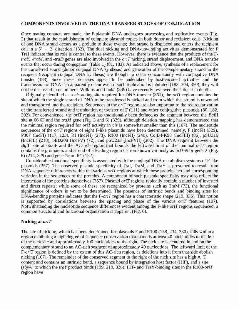
Originally identified as a cis-acting site required for DNA transfer (361), the oriT region contains thesite at which the single strand of DNA to be transferred is nicked and from which this strand is unwoundand transported into the recipient. Sequences in the oriT region are also important to the recircularizationof the transferred strand and termination of transfer of F (111) and other conjugative plasmids (80, 182,202). For convenience, the oriT region has traditionally been defined as the segment between the BglIIsite at 66.6F and the traM gene (Fig. 3 and 6) (329), although deletion mapping has demonstrated thatthe minimal region required for oriT activity in cis is somewhat smaller than this (107). The nucleotidesequences of the oriT regions of eight F-like plasmids have been determined, namely, F (IncFI) (329),P307 (IncFI) (117, 123), RI (IncFII) (273), R100 (IncFII) (240), ColB4-K98 (IncFIII) (86), pSU316(IncFIII) (218), pED208 (IncFV) (72), and pSU233 (IncFVII) (302). The DNA segment between theBglII site at 66.6F and the AC-rich region that bounds the leftward limit of the minimal oriT regioncontains the promoters and 5′ end of a leading region cistron known variously as orf169 or gene X (Fig.6) (214, 329) and gene 19 on R1 (122).
Considerable functional specificity is associated with the conjugal DNA metabolism systems of F-likeplasmids (357). The observed plasmid specificity of TraI, TraM, and TraY is presumed to result fromDNA sequence differences within the various oriT regions at which these proteins act and correspondingvariation in the sequences of the proteins. A component of such plasmid specificity may also reflect theinteraction of the proteins themselves (357). Plasmid oriT regions typically contain a number of invertedand direct repeats; while some of these are recognized by proteins such as TraM (73), the functionalsignificance of others is yet to be determined. The presence of intrinsic bends and binding sites forDNA-bending proteins indicates that the F-oriT region has a characteristic shape (219, 336). This notionis supported by correlations between the spacing and phase of the various oriT features (107).Notwithstanding the nucleotide sequence differences evident among the F-like oriT regions sequenced, acommon structural and functional organization is apparent (Fig. 6).
Nicking at oriT
The site of nicking, which has been determined for plasmids F and R100 (158, 234, 330), falls within aregion exhibiting a high degree of sequence conservation that extends at least 40 nucleotides to the leftof the nick site and approximately 100 nucleotides to the right. The nick site is centered in and on thecomplementary strand to an AC-rich segment of approximately 40 nucleotides. The leftward limit of theF-oriT region is defined by the extent of this AC-rich region, as deletions into it from that side abolishnicking (107). The remainder of the conserved segment to the right of the nick site has a high A+Tcontent and contains an intrinsic bend, a sequence bound by integration host factor (IHF), and a site(sbyA) to which the traY product binds (199, 219, 336); IHF- and TraY-binding sites in the R100-oriTregion have
also been identified (154, 156). Removal of F sequences from the right has shown that only the left halfof the sbyA TraY-binding sequence is essential for oriT nicking (107, 199). It was recently shown thatbound TraY induces bending and that this protein can bind to additional sequences to the left of sbyA;the site, sbyC, overlaps the IHF-binding sequence (219).
The traI genes of plasmids F and R100 both encode a 1,756-aa polypeptide (40, 370); type AATP/GTP-binding motifs occur in both the N-terminal and C-terminal regions of the deduced proteinsequences (40). Present at about 600 copies per F+ cell (186), the cytoplasmically located 192-kDa Fprotein, TraI, corresponds to the well-characterized enzyme E. coli DNA helicase I (2) and exhibits the5′ → 3′ DNA unwinding activity appropriate for displacement of the transferred strand (1, 199). Assayswith purified components have now demonstrated that the TraI protein of both F and R100 isresponsible for introducing the single-strand break at oriT (158, 234, 297). A phosphodiester transferase,TraI becomes covalently linked to the 5′ terminus of the nicked DNA strand (Fig. 2 and 6); no suchlinkage to the 3′ end of the nick has been detected (155, 235, 297). F-oriT-nicking function has beenshown to depend on the N-terminal sequences of TraI, some of which are dispensable to the unwindingfunction of the protein (335). Amino acid sequence similarity has been detected between this region ofTraI and the TrwC, TraI, and VirD2 proteins, which are DNA-nicking proteins encoded by R388, RP4,and agrobacterial Ti plasmids, respectively (213, 282, 283; also see below); these proteins all belong to asuperfamily of DNA-nicking enzymes (153, 189, 213). TraI*, an 88-kDa protein, originally known as 2b andthen known as TraZ, was previously thought to derive from a genetically undefined cistron assigned thename traZ, and once suggested to function in oriT nicking (7, 81). However, this product appears to derivefrom a second translational start site within the traI reading frame and to lack the N-terminal TraI domainsessential to nicking (334, 335). Thus, the significance of TraI* is unknown.
No F- or R100-encoded proteins other than TraI are essential to the oriT-nicking reaction in vitro(155, 234, 297). Although no F-TraY protein effect on the kinetics of the F-TraI-mediated in vitroreaction was detected (261), recent studies with purified R100 components have found the R100-nicking reaction to be stimulated by R100-TraY protein and IHF (155, 158). Other assays indicate thatthere is also an accessory requirement for F TraY in the in vivo reaction of TraI with oriT.
Expression of both traI and traY was necessary for the original observation of nicking in vivo (81) andfor detection of recombination events stimulated by F oriT (48). In addition, the purified F- and R100-traY products have both been shown to bind to oriT region sites near the site of nicking (Fig. 6) (156,199, 219, 261), and the major sequence determinants defined for TraY binding within the F site, sbyA,have been shown to be required for efficient nicking in vivo (111, 219). That bound TraY has beendemonstrated to induce DNA bending at this sequence (219) indicates that TraY can affect oriTcomplex conformation.
The F- and R100-TraY polypeptides are cytoplasmic proteins of 15.2 and 8.5 kDa, respectively.Comparison of the deduced amino acid sequence of F TraY (95, 157) with itself and with alleles encoded byother IncFI (P307) (123), IncFII (R1 and R100) (85, 157, 193), IncFIII (ColB4-K98) (86), and IncFV(pED208) (84) plasmids has revealed that F traY has resulted from a gene duplication event (157); F TraY is131 aa in length, whereas the products of other alleles range in size from 71 to 77 aa for the P307 andpED208 genes, respectively. Suggestive of a degree of regulation at the translational level, all of the traYgenes sequenced thus far have been found to initiate translation at a GUG or UUG triplet rather than at themore frequently utilized AUG start codon (157).
Profile analysis has revealed that TraY is a member of a class of DNA-binding proteins that includesthe Mnt and Arc repressors of phage P22 and the E. coli methionine repressor, MetJ (35, 41). Structuralanalyses of Arc and MetJ have indicated that two dimers of these proteins bind at their cognate operatorsequences by using an N-terminal anti-parallel β-sheet structure (41, 322). The duplicate nature of FTraY led to the suggestion that dimerization might not be required for binding (261). Consistent withthis notion, F TraY was found to exist as a monomer in solution (261), and binding data indicate thattwo F-TraY monomers bind separately to sbyA (219). The structural relationship to transcriptionrepressors and the demonstration of TraY binding in the vicinity of the PtraY promoter on both F (Fig. 3)and R100, albeit at lower affinity than at the oriT region site, sby, have raised the possibility that thisprotein negatively regulated its own transcription and hence that of most tra region genes (156, 261).
Nicking at oriT represents the initial step in the processing of plasmid DNA during conjugation, butthere is no evidence to suggest that this is the decisive event precipitated by recognition of a matingcontact. By definition, the oriT nicking demonstrated in vitro occurred in the absence of any signalgenerated by mating-pair formation. Likewise, a proportion of nicked molecules have been isolated fromdonors in the absence of recipients and from cells carrying mutations in tra genes required for theformation of mating aggregates (81). Conceivably, conditions used to detect nicked DNA could sidestepa signal requirement. However, the data imply an equilibrium of open-circular and covalently closedcircular plasmid DNA in donor cell populations; thus, it was suggested that the signal may precipitatesubsequent steps in conjugative DNA metabolism by triggering the initiation of DNA unwinding (81).
DNA Strand Displacement and Transfer
The existence of a signal was deduced from the finding that replacement strand synthesis occurred onlyin donors which could elaborate F pili and which had been mixed with recipient cells (183). Suchsynthesis, which should reflect the progression of unwinding, was found also to depend on traM and to alesser extent on traD, as well as on traI (183). The traM gene is dispensable to pilus assembly andaggregate stabilization, as well as to oriT nicking per se. As TraM was necessary for DNA transfer andaccompanying donor strand replacement synthesis, it was suggested that TraM might transduce thesignal that commits the oriT nicking machinery to unwind the DNA (81, 360). Alternatively, TraM maybe required to anchor the oriT region to a transfer apparatus in the inner membrane (3) or to alterconformational properties of the DNA (103). These possibilities, which are not mutually exclusive, areconsistent with the finding that a proportion of the otherwise cytoplasmically located products of the F-,R100- and pED208-traM genes is detected in inner membrane preparations; such a fractionation pattern issuggestive of an interaction with an intrinsic inner membrane protein (3, 72, 73).
Although the deduced 127-aa product of F-traM has a predicted size of 14.5 kDa (328), TraMmigrates as a 10-kDa protein upon SDS-PAGE (73). In the form of a tetramer, TraM has been found tobind to three sites within the oriT region (Fig. 6) (73). The two binding sites closest to the F-traM gene
overlap the two traM promoter sequences identified by transcript analysis, and expression of this F genehas been shown to be subject to negative autoregulation (S. S. Penfold and L. S. Frost, unpublisheddata). It appears that the presence of TraY protein may determine which TraM-binding sites can beoccupied; expression of both TraY and IHF contributed to maximal traM product expression (Penfoldand Frost, unpublished). The traM products of R1, R100, and pED208 also bind to multiple sites withintheir respective oriT regions (3, 72, 309), and autoregulation of the R1- and R100-traM alleles has beendemonstrated (4, 310). TraM binding has also been suggested to regulate transcription of a promoter forgene 19 on plasmid R1 (192). As a mutant traM product that lacks the eight C-terminal amino acidresidues of the wild-type protein has been shown to form tetramers but to be unable to repress traM or tobind to DNA, a role for the C-terminal region of the protein in DNA binding has been suggested(Penfold and Frost, unpublished). Other studies suggest that amino acid residues in the N-terminalregion of TraM may also be critical to DNA-binding activity (310).
Whereas the minimal oriT region required for nicking is the approximately 100-nucleotide segmentdelimited by the leftward boundary of the AC-rich region and the first half of the TraY-binding region,additional rightward sequences, including TraM-binding sites, are required for efficient transfer (Fig.6) (107, 111). The segment between the AT-rich region and the traM gene shows considerablesequence divergence among the characterized F-like plasmids and, in addition to sites that bind TraM,includes another intrinsic bend and a second IHF-binding site (73, 154, 156, 336). DNA sequencedeletions extending from the right into this region result in decreasing transfer efficiencies as thelength of the deletion increases; those affecting the left half of the TraM-binding site closest to oriTcompletely abolish transfer (73, 107).
The characteristics of F TraI that contribute to strand displacement became apparent with thediscovery that this protein corresponded to DNA helicase I (2), which exhibits ATP-dependent 5′ →3′ DNA-unwinding activity (1, 64). An amino acid sequence motif characteristic of ATP-dependenthelicases is present in the C-terminal region of the deduced protein sequence (370). Reports relatingto the number of TraI molecules required for maximal strand-unwinding activity have been conflicting;estimates have ranged from 5 to 90 per aggregate, and even a DNA/protein ratio of 1:1 has been foundto result in significant unwinding (29, 64, 195, 198, 348). TraI has been estimated to unwind DNA at arate of approximately 1,200 bp/s (195). The ATP-dependent 5′ → 3′ helicase activity of TraI has led tothe further suggestion that TraI might also energize the transmission of the DNA strand duringconjugation (315, 360). Such models assume that TraI is in some way immobilized, perhaps ininteraction with a tra-encoded or chromosomally encoded protein associated with the innermembrane (Fig. 2) (315); it has been suggested that TraD might provide such an association (64).In the absence of TraI-mediated unwinding activity but not nicking activity, some other helicasecan apparently substitute, albeit at a lower transfer efficiency (335).
The F-traD gene encodes an 81.7-kDa inner membrane protein (280). The gene sequence indicatesthat TraD is 717 aa in length (40, 173). The deduced R100-TraD amino acid sequence is homologous tothat of F but contains in its C terminus the three-residue sequence Gln-Gln-Pro reiterated 10 timesinstead of the single occurrence in F TraD (370). Although traD mutants elaborate apparently normalpili, form mating aggregates with recipients, and trigger conjugal DNA metabolism, they fail to transferDNA (183, 279, 354). Furthermore, it appears that functional TraD is required only after mating-aggregate formation (279). These characteristics have led to the suggestion that TraD is involved in thetransportation of single-stranded DNA across the cell envelope into the recipient (183). Severalobservations support the notion that TraD plays a role in transmembrane conveyance of nucleic acid.First, RNA phages, such as MS2, are able to adsorb to the pili of traD mutants but infection is abortedbecause of a defect in RNA penetration (284, 308). Second, traD is required for mobilization of theplasmid ColE1 and related plasmids, unlike the other DNA metabolism genes traM, traI, and probablytraY (355). Finally, purified TraD has been found to bind DNA in a nonspecific manner (280).
Recently, amino acid sequence similarity has been detected between TraD and polypeptides encodedby transfer systems previously thought to be distinct from that of F, namely, TraG of RP4, TrwB ofR388, and VirD4 of agrobacterial Ti plasmids (212, 213; also see below). Furthermore, it is likely thatsimilar proteins are also involved in gram-positive DNA transfer mechanisms, because a member of this
protein family is encoded by the transfer region of conjugative staphylococcal plasmids (93, 258).Purified TraD has been shown to possess DNA-dependent ATPase activity (280). Consistent with thisobservation, the deduced F- and R100-TraD amino acid sequences contain both type A (370) and type B(212) nucleoside triphosphate-binding-site motifs. The type B motif is particularly well conserved inother members of the protein family to which TraD appears to belong (93, 212). It has been proposedthat proteins of this family may link the conjugal DNA metabolism machinery to the DNA transportapparatus (202, 345).
Termination of F Transfer
The properties of TraI have suggested that this protein is also the mediator of transfer termination, andplasmids carrying two directly repeated oriT sites have been employed to test the DNA sequencerequirements associated with this process (111, 202). Completion of transfer has been proposed todepend on recognition of the oriT site reconstituted by replacement strand synthesis, cleavage, andligation of the newly generated 3′ end to the 5′ end of the transferred strand (Fig. 2) (202, 349). Insupport of this model, Gao et al. (111) have obtained data suggesting that termination can occur at F-oriT sequences which do not contain a preexisting nick. The sequence required for termination extendsno more than 36 bases to the right of the nick site and includes phased poly(A) tracts which specify asequence-determined bend (111). Since point mutations affecting the fourth and ninth base pairs to theright of the nick site affect both nicking and termination, a step involving TraI is suggested for bothprocesses (111). Sequences preceding orf169 (Fig. 6) may also be involved in the termination step ofconjugation, since sequence deletions in this region resulted in the transfer of plasmids of greater thanunit length (107).
SURFACE EXCLUSION
The F-plasmid transfer region encodes two genes responsible for surface exclusion (Sfx). This propertylimits the host cell capacity to act as a recipient for the same or a closely related plasmid. The traS andtraT genes (Fig. 3; Table 1) appear to be responsible for independent aspects of the phenomenon, actingin concert to reduce transfer efficiencies by several orders of magnitude (6, 8, 10). The expression ofsurface exclusion is probably a fundamental requisite of donor ability, since in its absence, donor cellpopulations would bear the metabolic cost of continuous and futile recipient-donor activity (54).
Five surface exclusion specificity classes, SfxI to SfxV, represented by F, ColB2-K98, R1, R100, andpED208, respectively, have been identified (89, 357). The two traS genes so far sequenced, those of F(172) and pED208 (89), encode products which differ markedly in primary sequence (103). Thiscontrasts with the available data for the F (172), ColB2-K98 (325), R100 (268), and pED208 (89) traTalleles, in which only one or two amino acid differences appear to define the observed phenotypicspecificities (135).
The traS gene encodes a 16.9-kDa inner membrane protein (Fig. 4), which, when present in therecipient, appears to prevent triggering of donor conjugal DNA metabolism (6, 172, 248). It hastherefore been suggested that TraS blocks the transmission of a mating signal (230). Present at anestimated 20,000 to 30,000 copies per cell (6, 246), the 26-kDa lipoprotein product of the traT gene(172, 288) constitutes a major outer membrane component of F-containing cells (Fig. 4). Exposed onthe cell surface (228), TraT is thought to span the outer membrane (325) in a multimeric form (135,228, 245, 248). TraT appears to function by inhibiting the formation of mating aggregates (6). This,together with the observation that purified TraT could reduce transfer efficiency, led Minkley andWilletts (248) to propose that TraT may interact with the tip of the sex pilus, perhaps competing with thenormal cellular receptor. However, the specificity between traT alleles and the corresponding transfersystem that it inhibits has recently been found not to be associated with the cognate pilin sequence(20). It has also been suggested that TraT may exert its effect by masking a region of OmpA (298).
There is circumstantial evidence that the traT gene product may play a role in bacterial pathogenesisin addition to its surface exclusion function (for a review, see reference 325). Several studies have

demonstrated that at least in some hosts, TraT may contribute to bacterial serum complement resistance(250, 267, 268). It is suspected that TraT inhibits the functioning of complement membrane attackcomplex (291, 333). Reduced susceptibility to phagocytosis has also been attributed to the presence ofTraT (14). Although surveys of clinical isolates have failed to establish an unambiguous link betweentraT and pathogenesis (34, 179, 251), the identification of traT homologs on nonconjugative virulence-associated Salmonella and Yersinia plasmids has lent credence to the proposition that the product of thisgene may act as a virulence factor (53, 325).
TRANSFER GENE EXPRESSION
Transcription of the tra Region and Its Regulation
Transcriptional promoters have been identified preceding the F-plasmid traM, traJ, traY, trbF, traS, traT, andtraD genes (Fig. 3) (96, 132, 328). In addition, two promoters, PfinP and PartA, have been found to initiatetranscription in the opposite direction to that of the majority of the tra region genes (Fig. 3) (260, 367). Thepicture of tra region transcription that is emerging, while not complete, is consistent with a model in whichPtraY is responsible for the initiation of a polycistronic mRNA that may encode 34 genes, from traY throughfinO, inclusively. This would represent an operon of approximately 32 kb. Although the 3′ extremity of thetranscript originating from PtraY has not been precisely determined, recent analyses which have indicated thatthis promoter is required for normal levels of expression of the distal genes, traD and traI, support thisnotion (222). The promoters PtrbF, PtraS, PtraT, and PtraD may, in combination with a proposed transcriptionterminator identified after the traT gene (131), serve to modulate and/or differentially regulate expression ofdistal tra operon genes. The expression of traT, in particular, appears to be somewhat independent ofregulatory factors affecting transcription from PtraY (51, 294). From the accumulated data, the likelytranscriptional units evident in the tra region are summarized in Fig. 3. Analysis of transcripts encoding traKhas suggested that the unusual polarity of the amber mutation traK4 is due to the presence of a rho-dependent transcription termination element (287). Computer analysis suggests that such rho-dependenttermination sequences are positioned at a number of sites within the tra region (287); transcription istherefore likely to be influenced by translational efficiency.
Two promoters located between the oriT nick site and the traM gene, which apparently direct thetranscription of gene 19, have recently been identified on R1 (192). These promoters are within theconserved segment of the oriT region (Fig. 6) and are in addition to tandem promoters locatedimmediately upstream of orf169 and gene 19 of F and R1, respectively (192, 214).
PtraY appears to be regulated, either directly or indirectly, by several plasmid-encoded andchromosomally encoded proteins. The traJ gene (Fig. 3; Table 1) encodes a 27-kDa cytoplasmic product(62) that positively regulates transcription originating at the tra operon promoter, PtraY (109, 260, 318,319, 351). A 229-aa protein (95), TraJ is estimated to be present at approximately 2,000 copies per F+
cell (62). The F-, P307-, R1-, R100-, and pED208-encoded traJ products are quite distinct, the greatestdegree of similarity being at their N termini (72, 85, 95, 123, 157), a region that has been predicted toform a helix-turn-helix DNA-binding domain (74, 285). Zone sedimentation data indicate that TraJ maybe dimeric (62).
PtraY has been shown to be utilized in vitro by the E. coli σ70 RNA polymerase (115). However, RNApolymerase was found to form a stable complex at PtraY only when the promoter sequence was in asupercoiled conformation (115). From this finding and the observed involvement of upstream sequencesin the down-regulation of PtraY in the absence of TraJ (319), Gaudin and Silverman (115) have suggestedthat in the absence of a transcriptional activator, the sequences preceding PtraY contribute to localrelaxation of the promoter region. In such a model, TraJ may invoke transcription from PtraY byfacilitating the restoration of normal superhelicity (115). Assays employing galK transcriptional fusionshave indicated that in the presence of TraJ, PtraY is a powerful promoter (260).
Purified TraY derived from both F and R100 has recently been found to bind to sequencesoverlapping the transcription initiation sites of their cognate PtraY promoters (156, 261). It is thereforelikely that transcription of traY is autoregulated (156). If this is so, transcription of most tra genes is

subject to both positive and negative regulation mediated by tra region-encoded products, namely, TraJand TraY, respectively.
TraJ has also been implicated in enhanced transcription from PtrbF, whereas PtraS, PtraT, and PtraDappear to be traJ-independent promoters (131, 172). Findings concerning the effect of TraJ ontranscription of traM have been conflicting. Analyses employing lacZ transcriptional fusions indicatedthat PtraM was stimulated by TraJ (109), yet analogous experiments utilizing galK as a reporter generevealed no such effect (260). Two initiation sites responsible for transcription of F traM (Penfold andFrost, unpublished) and for the traM alleles of plasmids R1 and R100 (4, 194) have been identified.Whereas binding of TraM affects both F-traM transcripts (Penfold and Frost, unpublished), only themore upstream of the R100-traM promoters is repressed by TraM binding; the second, weaker promoterappears to function constitutively (4). The R1-traM gene is also autoregulated (310).
FinOP Fertility Inhibition
In donor cells harboring most F-like plasmids, tra gene expression, and hence conjugative transfer itself,is repressed by a phenomenon known as fertility inhibition (fin) (90, 242). In combination, a smallantisense RNA molecule, FinP, and a polypeptide encoded by the most distal tra gene, finO, inhibit theexpression of the regulatory gene, traJ (Fig. 3) (99, 207, 351). The absence of TraJ, in turn, precludestranscription from the major transfer region promoter PtraY described above. Transfer of the F plasmid isderepressed as a result of insertional inactivation of the finO gene by the transposable element IS3 (Fig.3) (52, 371). However, expression of a finO gene from a compatible coresident plasmid can repress Ftransfer in trans (92).
Approximately 78 nucleotides in length (339), the FinP RNA molecule is transcribed constitutivelyfrom a promoter, PfinP, located within and in opposite orientation to the nontranslated leader of the traJtranscript (Fig. 3) (88, 260). Complementary base pairing between FinP and the traJ mRNA is thoughtto preclude translation of that mRNA into TraJ, because the duplex formed overlaps the translationinitiation signals of traJ (83, 88). However, recent evidence indicates that RNase III-mediated cleavageof the duplex might be responsible for inactivation of traJ mRNA (339). FinP and the complementaryregion of the traJ transcript each fold into two stem-loop structures (339). Sequences within thesecondary structure loops of the traJ/FinP RNA molecules appear to be critical to interactions betweenthe RNA species (191) and appear to constitute the basis of the observed plasmid specificities (357).
The finO genes of F, ColB2, R6-5, and R100 have been sequenced and, after removal of interveningIS3 sequences and the target duplication from the F allele, found to code for highly homologous 186-aaproducts (59, 239, 337, 370, 371). The 21-kDa finO product is hydrophilic and therefore is presumed toreside in the cytoplasm, a location consistent with its regulatory role (Table 1) (332, 337, 371). FinOseems to exert its coregulatory effect on traJ expression by stabilizing the FinP antisense RNA (207).The FinO-mediated extension of FinP half-life, from ≈2 min to >40 min, occurs even in the absence ofthe complementary traJ messenger (207). This and the inability of FinO to protect FinP transcriptsbearing a fisO mutation (91) have suggested that the FinO protein and FinP RNA might interact directly(99, 207). Recently, it was demonstrated that FinO is, in fact, an RNA-binding protein that interacts withone of the two stem-loops in FinP (Fig. 4) and with the complementary structure in the traJ mRNA(338). Allelic specificities attributed to the finO gene (357) have been found to result from differentialexpression by F-like plasmids (337).
The high-level fertility inhibition exhibited by plasmids such as R6-5 and R100 correlates with thepresence on these plasmids of a gene known as orfC or orf286, located between traI and finO (59, 370).F-like plasmids such as ColB2 (and presumably the F progenitor) lack the sequences that encode orf286and express lower levels of fertility inhibition (59, 337, 357, 370). Cotranscription of orf286 and finOleads to the synthesis of FinO at levels far in excess of that observed in the absence of orf286 sequencesand is believed to result from an increase in finO mRNA half-life (337). This enhancement of finOexpression occurs only in cis, indicating that it is not mediated by the orf286 translational product (337).Indeed, this stabilizing effect, resulting from cotranscription with the upstream gene, appears to beindependent of orf286 translation (337). Secondary structure in the mRNA as a result of base pairing
between sequences within orf286 and finO is thought to increase the resistance of the transcript toribonucleolytic degradation (337).
A slightly different mechanism has been proposed for the regulation of the tra region of R100.Termed latch relay, this model is based on competition between transcripts initiating upstream ofand within traM, and the untranslated traJ leader, for the antisense FinP molecules (69). Under sucha scheme, the normal steady-state condition is “off”; any event that can affect this state may result inswitching on of tra region transcription and hence transient derepression (70). Dempsey (68) hasalso described a second, upstream finP promoter on R100 and has identified a third potential stem-loop structure unique to the R100-FinP RNA molecule. A similar mechanism may operate on R1,since traM transcripts are also believed to regulate FinP activity expressed by this plasmid (194).
FinOP-mediated transfer repression is thought to reflect an evolutionary penalty associated withconstitutive expression of conjugative functions. In addition to enduring the metabolic overheadassociated with constitutive expression, host cells harboring derepressed F-like plasmids are vulnerableto infection by pilus-specific phages (141). Repressed plasmids are thought to escape such costs whilemaintaining transfer potential, because transient derepression in an individual donor may lead to“infectious spread” through a recipient population (43, 358). Such spread results from the high-frequency transfer exhibited by cells which have recently received the plasmid, because of the lag timerequired for synthesis of FinO and FinP to inhibitory levels and the ensuing dilution of TraJ and othertra products (337, 363). It has also been suggested that conjugative transfer may be required only tointroduce a plasmid into a population, because any selected advantage conferred by that plasmid wouldfacilitate its subsequent establishment via preferential host cell survival and growth (61).
Other Fertility Inhibition Systems
In addition to the normally endogenously encoded FinOP system, five other fertility inhibition systemshave been identified on plasmids which, when coresident with F, reduce the efficiency of F transfer.These fertility inhibition systems also affect, to various degrees, the mating efficiency of other F-likeplasmids (114). As well as the benefits associated with repression listed above, it has been suggestedthat plasmids encoding one of these trans-acting mechanisms may be able to compete more successfullyfor new hosts (109, 113). However, it is also possible that the repression caused by these systems iscoincidental (162). The inhibition of the IncP plasmid RP4 transfer by F appears to represent an exampleof such an inadvertent interaction (326); the product of the autoregulated repC gene of F (also known aspifC), which regulates replication from oriV (184, 327), is thought to be responsible for thisphenomenon (244).
The FinQ and FinW fertility inhibition systems are believed to act independently of traJ at the levelof transcription (109, 112). FinQ is encoded by IncI1 (formerly IncIα) plasmids such as R62, R820a,TP102, and TP108 (109, 114). The finQ gene from R820a encodes a 40-kDa product, which is proposedto cause rho-independent termination of the transcript initiating at PtraY at several sites between traC andtraD (109, 114, 133; L. M. Ham and R. A. Skurray, unpublished data). Encoded by the IncFI plasmidR455, FinW appears to act by reducing the transcription of traM (109, 113).
The FinC, FinU, and FinV fertility inhibition systems are thought to act posttranscriptionally. FinC isexpressed by copy number mutants of the mobilizable bacteriocinogenic plasmid CloDF13, such as JN62and JN77 (352). In addition to reducing the level of F transfer, FinC inhibition renders F-containing cellsresistant to infection by RNA phage f2 but not to the filamentous DNA phage f1, a phenotype characteristicof F-traD mutants (352). FinC fertility inhibition results from overexpression of the CloDF13 mobA or rpigenes (262, 340). The product of mobA is a 58-kDa protein, MobB, involved in mobilization, whereas theonly phenotype so far attributed to the 16-kDa rpi product is the FinC-mediated resistance to F-specific RNAphage infection described above (262). As transcription of traD was found to be unaffected by FinC, Willetts(352) suggested that this mechanism inhibits TraD. This contention has been strengthened by the recognition

of the CloDF13 MobB protein as a TraD homolog (26, 46; also see below). The presence of a gene encodinga TraD-like protein may also explain why traD is not required for mobilization of CloDF13 but is essentialfor transfer of the related plasmid ColE1, which encodes a different protein from an analogously locatedposition in its genome (341, 352).
The basis of the FinU and FinV transfer inhibition systems, encoded by the plasmids JR66a (IncI1) andR485 (IncX), respectively, is unknown (113). FinU inhibits both pilus assembly and surface exclusion andwas therefore suspected to affect tra region transcription (113). Although the presence of JR66a was found toreduce transcription of distal F-tra cistrons, the extent of the reduction was disproportional to the 7,000-foldtransfer inhibition specified by the FinU system (109). On the basis of these findings, Gaffney et al. (109)suggested that although the effect on transcription may be responsible for the observed reduction in surfaceexclusion, the primary target of FinU inhibition was more likely to be the translation and/or function of oneor more tra genes. However, the synthesis of all 11 tra products detected in the study was unaffected by FinU(109).
The FinV fertility inhibition system encoded by plasmid R485 inhibits F piliation and hence transfer butdoes not reduce surface exclusion, indicating that an effect on tra region transcription is unlikely (113).Subsequent transcriptional studies supported this contention (109). FinV is therefore thought to prevent theproper translation and/or function of one or more of the tra products required for pilus biogenesis; Gaffney etal. (109) did in fact note a reduction in the amount of a 30-kDa protein synthesized in the presence of R485,although the origin of this polypeptide was unclear.
Environmental and Host Cell Factors InfluencingF-Plasmid Transfer
In addition to host-encoded activities required for the DNA synthesis that accompanies transfer, anumber of environmental and host-specified factors have been found to influence the donor ability of F+
cells, reinforcing the notion that conjugation is a cellular process. F pili are believed to retract whencultures are cooled below 25°C (265). Furthermore, the synthesis of the pilin subunit itself was found todiminish as the incubation temperature was lowered (265, 323). Parallel reductions in the synthesis ofseveral other tra products led Sowa et al. (323) to speculate that transcription of the tra region may beregulated by temperature.
The early finding that F– phenocopies of donor cells (i.e., F+ or Hfr cell cultures with recipient ability)are produced by growth into late-stationary phase (205) suggests that expression of surface exclusion isaffected by starvation or growth phase (6). Cultures of F– phenocopies also failed to elaborate normalnumbers of F pili (42). It is therefore likely that growth into stationary phase has a general effect on traoperon expression and may be associated with host-encoded cellular regulators, such as those discussedbelow (see also other chapters in this volume on growth and regulation of gene expression, e.g., chapter93).
Intracellular levels of cyclic AMP (cAMP) have also been reported to influence the expression oftransfer-related activities from F-like plasmids, although the data available are very limited. Harwoodand Meynell (136) found that addition of exogenous cAMP to cya mutants carrying various derepressedF-like plasmids reduced piliation, although this effect was not evident in cells harboring F. In contrast,the observed reduction in transfer-related properties expressed by cya and crp mutant hosts led Kumarand Srivastava (196) to suggest that F-tra expression is dependent on cAMP and its receptor protein.The identification of a sequence in the vicinity of PtraJ with similarity to the binding-site motif of the crpproduct, catabolite activator protein, lends credence to this proposal (285).
Several other chromosomal genes appear to play regulatory roles in the expression of F-transferfunctions. The products of cpxA (mapping at 88.34 min on the E. coli chromosome; also called ecfB, ssd,and eup) (16, 17, 293) and sfrA (99.95 min; alternatively known as arcA, fex, cpxC, msp, and dye) (44,165, 166, 208, 209, 314) represent transducer and effector homologs, respectively, of the two-component regulator protein families (18, 77, 346; for reviews, see references 125, 151, and 343). TheSfrA+ phenotype is in fact an activity of the 29-kDa product (45, 77) encoded by the arcA gene, which is
a transcriptional regulator of genes involved in aerobic metabolism (45, 77, 166, 167). The activity ofArcA is modulated, via histidine phosphorylation, by the signal transducer encoded by arcB (72.11min), which senses the redox state of the cell (165, 168–170).
It has been shown that the Arc+ and Sfr+ functions of ArcA are genetically and physiologicallyseparable (316). While it is possible that SfrA (ArcA) is acted on by CpxA in addition to ArcB, thisrelationship is yet to be clearly established (165). Evidence indicates that the 52-kDa cpxA product isrequired primarily for efficient traJ expression (18, 317, 346), whereas SfrA appears to be an activatorof the PtraY promoter (109, 318). It is therefore possible that sfrA and cpxA act independently of eachother. Several key questions relating to SfrA and CpxA remain to be answered, namely, the identity ofthe effector protein acted upon by CpxA, the possibility of a second transducer affecting SfrA, and thenature of the stimuli to which these systems respond. Encoded by a gene upstream of cpxA andexhibiting amino acid sequence similarity to regulatory proteins of other two component systems, theproduct of the recently identified cpxR is a candidate for the CpxA effector (75), as is the yet to beidentified product of another chromosomal gene required for efficient tra expression, cpxB (41.01 min)(237, 238, 303, 317).
The sfrB gene (86.64 min; also known as rfaH and hlyT) encodes an 18-kDa product, whichsuppresses premature rho-dependent termination of tra region transcription (24, 33, 109). Two sites ofSfrB-associated transcription termination have been localized within the tra region, between traC andtraG and between traG and traS (33, 109). Mutations in sfrB are pleiotropic, also affecting flagellumformation, bacteriophage and antibiotic sensitivity, and lipopolysaccharide and hemolysin synthesis (24,32, 290).
The sequence-specific, histone-like DNA-binding protein IHF is involved in a number of DNA-associated processes in E. coli, including replication, transposition, site-specific recombination, and geneexpression (for reviews, see reference 98 and chapter 125). IHF has been shown to be necessary forefficient transcription of tra region genes from both F and R100 (67, 110; Penfold and Frost,unpublished); the amount and length of gene X, traM, and traJ transcripts synthesized by R100 werefound to be affected in IHF– hosts (71). Transcription initiation from PtraY of F also appears to bereduced in such mutants (318), although this may represent an indirect effect due to reduced traJexpression.
IHF-binding sites have been localized in the oriT region, between the nick region and traM, from Fand R100 (Fig. 6) (71, 156). The presence of these binding sites within the oriT region suggests that IHFalso plays a role in the processes that lead to nicking and ultimately DNA transfer, perhaps via aninteraction with TraM (4, 336). The IHF protein contributes to the nicking of R100 oriT in vitro (155)and can partially repress the most upstream R100-traM promoter (4). IHF binding has also been detectedin the vicinity of PtraJ (71).
Posttranscriptional Modulation of tra Gene Expression
In addition to the regulatory controls governing translation of the traJ and finO mRNAs,posttranscriptional mechanisms involving mRNA processing and stability are believed to be involved inthe regulation of expression of the long tra operon transcript. It has been proposed that 5′endoribonucleolytic cleavage and protection from 3′ exoribonucleases leads to the accumulation ofsmall, stable R1–19 mRNA species which derive from the polycistronic mRNA initiating at PtraY butencode only the traA gene product (propilin) (190). The inverted repeats presumed to form the RNAstem-loop secondary structures responsible for exoribonuclease resistance of the R1–19-traA transcriptsare conserved on F (103). Like traA, the F-traT gene is also expressed in abundance and has similarlybeen found to be located on stable mRNAs (131). The expression of R1 gene 19 has been shown to becontrolled by the endoribonuclease RNase III. A transcript initiating upstream of and in the oppositeorientation to traM seems to be similarly processed (192).
The translation initiation codons of the majority of the genes that are thought to be coded for by themajor tra transcript initiating at PtraY overlap with or are in close proximity to the stop codon of theprevious cistron (40, 76, 94, 103, 131, 173, 224, 225, 227, 253, 306, 367, 368, 370). Such an
arrangement is suggestive of translational coupling (15). Although active coupling within the tra regionhas not been detected, juxtapositioning of translation control signals probably serves to maximizeexpression via translation reinitiation.
OTHER PLASMID-ENCODED GENES POTENTIALLYINVOLVED IN CONJUGATION
Nucleotide sequencing within the tra region revealed a number of ORFs presumed to correspond topreviously undetected genes (40, 76, 103, 132, 224, 227, 367). The involvement in the conjugativeprocess of many of these cistrons and several other tra genes identified only on the basis of productdetection has been investigated by using resistance-gene cassette mutagenesis followed by homologousrecombination into F-plasmid derivatives (180, 221, 222, 224, 226, 227, 253). Subsequent analyses haveindicated that several of these mutants exhibit phenotypes indiscernible from that of wild-type F, asjudged by standard laboratory assays. Characteristics of the tra region genes of unknown function areshown in Table 2. It has been suggested that such apparently dispensable genes may contribute toconjugative transfer from different hosts or under alternative conditions (164). It is also possible thatsome of these genes encode products that perform functions unrelated to DNA transfer; the presence ofan additional gene, orfE, between traT and traD and the absence of trbH in the R100-tra region wouldseem to support this view (40, 370).
Although the autonomy of the F-tra region has been demonstrated by the transfer proficiency of arecombinant plasmid containing the segment 66.6 to 100F (214), there is accumulating evidence thatgenes in the 13-kb leading region (Fig. 1) may also normally play a role in conjugation. In addition tobeing conserved on F-like plasmids (121, 214, 311), leading-region sequences have been found tohybridize to non-F-like plasmids (118, 119, 146, 214, 311). A number of ORFs have been identified inthe leading region, but only three functions have so far been associated with this DNA segment (forreviews, see references 164 and 349), namely, a plasmid maintenance system designated flm (F leadingregion maintenance; formerly called parL and also known as stm, and homologous to the parB hok/soksystem of R1) (216, 217), a single-stranded DNA-binding protein encoded by the ssb gene (also knownas ssf) (50, 187), and an inhibitor of the RecA-mediated SOS response, expressed by the psiB gene (25,79, 215). Roles have been envisaged for all of these functions in the recipient subsequent to conjugativetransfer (164).
Supporting the notion that the leading region may play a role in conjugative transfer, expression of thessb and psiB genes of the conjugative IncI1 plasmid, ColIb-P9, has been shown to be induced upontransfer into a recipient cell (175). The psiB gene of F also appears to be zygotically induced (23).Furthermore, the ssb gene of R1 is believed to be coordinately regulated with the tra genes of thatplasmid (120). Genes encoding single-stranded DNA-binding proteins have been identified on a numberof conjugative plasmids (see below and reference 176).
Although the leading-region gene orf169 (Fig. 6) does not appear to be essential for F conjugation(214), experiments with the F-like R1-plasmid transfer system suggest that the corresponding gene 19product is required for efficient transfer of that plasmid (26a). The gene 19 product is synthesized as aprecursor which is subsequently processed, presumably by signal peptidase I, to a 17-kDa mature form(192, 214). Amino acid sequence similarity has been detected between the deduced product oforf169/gene 19 and IpgF encoded by the mxi locus of the Shigella flexneri virulence plasmid, pWR100(19) and, more recently, with R64 PilT, pMK101 TraL, RP4 TrbN, Ti VirB1 (Table 3), and Slt70, thesoluble lytic transglycosylase of E. coli (26a; G. Koraimann, personal communication). In addition toother ORFs of unknown function in the leading region (122, 214), two single-stranded initiationsequences, designated ssiD and ssiE, have been identified in the F-leading region; these sites are likelyto play a role in recipient conjugal DNA synthesis (263).

BACTERIAL CONJUGATION: A MACROMOLECULAR TRANSPORT MECHANISM
Similarities have recently been noted between proteins encoded by genes from a number of bacterialDNA transfer systems (Table 3). Most striking is the homology evident between the T-DNA transfersystem encoded by the vir genes of agrobacterial Ti and Ri plasmids and the conjugation systems of theIncN, IncP, and IncW plasmids pMK101, RP4, and R388, respectively. The likeness of these systems,which extends to the levels of protein function and genetic organization, has given strong support to thehypothesis that the transfer of T-DNA to plants represents a modification of bacterial conjugation (324).Furthermore, amino acid sequence similarities have also been detected between products from thesesystems and those from IncF and IncI plasmids and even with transfer-associated proteins encoded byplasmids from gram-positive organisms (illustrated by pSK41 and pS194 in Table 3). The products ofplasmids such as pED208 (IncFV) (103) and R751 (IncPβ) (128), which would appear to have divergedrelatively recently from the lineages represented by F and RP4, respectively, are omitted from Table 3because of constraints of space, but they serve to remind of the extent of variation that exists. In somecases, the homology evident between DNA metabolism proteins from different systems is paralleled byrecognizable similarity in the nucleotide sequences of their respective nick sites; the oriT regions ofR64, RP4, pTF-FC2, and pS194 and Ti-plasmid border sequences are examples of this duality (202, 281,282, 299). The similarities described above have led to the realization that DNA transfer mechanismsfrom seemingly diverse host origins share genes of common evolutionary ancestry.
The role of the F pilus in the conjugative process has been the subject of debate for several decades(10, 42, 63, 353). The hypothesis that the pilus acts as the conduit for the passage of DNA from donor torecipient has gradually given way to the prevalent notion that the pilus acts as a “grappling hook” thatretracts to bring mating cells into close physical contact, thereby enabling the establishment ofconjugation junctions (78), the sites at which DNA transfer is presumed to occur. However, there areseveral reasons to think that the latter suggestion may represent an oversimplification of pilus functionand that that the pilus or a vestige thereof remaining after retraction may indeed represent a specializedpore which facilites DNA transmission across the donor and possibly the recipient cell envelope(s).Such a structure might include at least some of the tra products currently defined as pilus assemblyproteins (Fig. 4; Table 1) and may correspond to structures vizualized recently by freeze-fracture andfreeze-etch electron microscopy (301).First, the observed mating, albeit at low efficiency, between physically separated cells indicates that thelumen of the pilus can support the transmission of single-stranded plasmid DNA (134, 275). Second,mutations in the traD gene, which is believed to be involved in the transmission of single-strandedplasmid DNA (see above), result in a slightly increased level of host cell piliation (21), suggesting a linkbetween the conjugal DNA processing machinery and pilus function. Indeed, TraD homologs from otherDNA transfer systems (Table 3) have been suggested to mediate an interaction between the conjugalDNA metabolism proteins and the DNA transfer pore (202, 345). Third, the apparent complexity of F-pilus biogenesis may reflect a multifunctional role for the pilus and associated proteins. No less than 16gene products appear to be involved in the elaboration of the F pilus, and representatives of theseproteins are believed to be associated with the cytoplasm, inner membrane, periplasm, and outermembrane, with the pilus itself extending into the external environment of the cell (Fig. 4). In contrast,only about half this number of proteins seem to be directly involved in the elaboration of nonconjugativeE. coli P and type 1 pili, and a number of those represent subunits of the pilus itself (150).

TABLE 2 Transfer region genes of unknown function
Gene Product length (aa)a Product size (kDa)a Product locationb Commentsc and reference(s)artA 104 12.1 Inner membrane Nonessential product yet to be identified (180); translation
supported by artA′-′lacZ fusion (367).traP 196 22.0 Inner membrane Probable product identified but function unknown (257;
Frost, unpublished data cited in reference 103)traR 73 8.3 Cytoplasm Identified product found to be nonessential (221, 257);
deduced product shares amino acid sequence similaritywith the E. coli dosage-dependent dnaK suppressor, DksA,and coliphage 186 and P2 proteins of unknown function(76)
trbA 115 12.9 Inner membrane Identified product found to be nonessential (180, 367, 369)trbB 179 [159] 19.5 [17.4] Periplasm Identified product found to be nonessential (180, 367, 369)trbD 65 7.1 Cytoplasm Product of unknown function yet to be identified (Frost,
unpublished data cited in reference 103)trbE 86 9.9 Inner membrane Identified product found to be nonessential (224)trbF 126 14.5 Inner membrane Product of unknown function yet to be identified (132);
translation supported by trbF′-′phoA fusions (Y. N. Lin, J.Tennent, N. Firth, and R. A. Skurray, unpublished data)
trbG 83 9.1 Cytoplasm Product of unknown function yet to be identified (76)trbH 239 26.3 Inner membrane Nonessential product yet to be identified (40, 221)trbJ 93 10.2 Inner membrane Identified product found to be nonessential (221, 367)aSizes and lengths are calculated from the deduced amino acid sequence of each product. Sizes and lengths of processed products are shown inbrackets. Values for processed products are based on predicted cleavage sites.bProduct locations have been determined experimentally unless shown italicized, in which case the predicted location is listed.cThe term “nonessential” indicates that a mutation in this gene was found not to cause any identifiable effect on the transfer-related phenotypesexamined.
Compelling support for the above contention that pilus components form a mating channel has beenprovided by sequence similarities to the F-pilus assembly proteins TraB, TraC, TraE, and TraL and theF-pilus subunit precursor itself, TraA (Table 3). Kado and his colleagues (178, 313) not only identifiedamino acid sequence similarity between these proteins and VirB products believed to form an envelope-spanning mating structure that facilitates agrobacterial T-DNA transfer to plant cells (331) but alsodemonstrated that VirB2 is processed in an analogous fashion to F propilin (312). These evolutionaryrelationships have been extended to include products known or suspected to be involved in pilusbiogenesis/mating-pair formation encoded by other conjugative plasmids, such as pMK101, RP4, andR388 (Table 3) (210, 289; Bolland, Ph.D. thesis). Since no traditional pilus-like structure has beenattributed to agrobacterial Ti plasmids (82), the relationships between “pilus-associated” proteins andthe T-DNA transfer system are particularly intriguing. Perhaps even more provocative, however, havebeen the similarities detected between such pilus-associated proteins and the Ptl proteins responsible forthe export of Bordetella pertussis multiple-subunit toxin (Table 3) (58, 347). The detection of anevolutionarily and functionally related apparatus involved in the export of a molecule other than DNA(namely, B. pertussis toxin) has led to the realization that the DNA transfer mechanisms discussed here,including that encoded by F, represent members of a family of pilus-related macromolecular transportsystems.

TABLE 3 Similarities between proteins of macromolecular transport systemsa
Conjugation Mobilization Proteinsecretion(Ptl)
Reference(s)
F (IncF1) R64 (IncI1) pMK101(IncN)
RP4(IncPα)
R388(IncW)
Ti pSK41
CloDF13 pTF-FC2 pS194
Orf169 (p19)b TraL TrbN VirB1 26a, 289TraA TraM TrbC TrwLc VirB2 PtlA 58, 177, 210,
211, 289,312
TraL TraA TrbD TrwMc VirB3 PtlB 58, 177, 210,211, 289,312, 347
TraC TraB TrbE TrwKc VirB4 PtlC 58, 178, 210,211, 289,313, 347
TraE TraC TrbF TrwJc VirB5 178, 210,211, 289
TraD TrbL TrwIc VirB6 PtlD 178, 211,289, 347
TraN TrwHc VirB7 178, 289 TraE TrwGc VirB8 PtlE 178, 211,
289, 347 TraO TrwFc VirB9 PtlF 178, 211,
289, 347raB TraF TrbI TrwEc VirB10 PtlG 178, 210,
211, 289,347
TraG TrbB TrwDc VirB11 PtlH 178, 210,211, 259,289, 347
TraL VirC1 MobD 188, 282,299, 349
TraM VirC2 MobE 282, 299 NikA TraJ TrwA VirD1 MobB 108, 178,
213, 282,299
TraI NikB TraI TrwC VirD2 MobA R1x 26, 108, 153,178, 189,211, 213,282, 283,299
TraH VirD3 282TraD TraG TrwB VirD4
TraKMobB 26, 46, 93,
178, 212,213, 282,373
TraK MobC 282, 299TraX TrbP 94a
ArdB KlcA 203Ssb Ssb Ssb
TraM93, 176
aThe proteins shown are from the gram-negative conjugation systems of plasmids F, R64, pMK101, RP4, and R388; the T-DNA transfer system of Agrobacteriumtumefaciens Ti plasmids; the gram-positive conjugation system of the Staphylococcus aureus plasmid pSK41; the mobilization systems of the gram-negativeplasmid CloDF13, the Thiobacillus ferrooxidans plasmid pTF-FC2, and the gram-positive plasmid pS194; and the Pt1 toxin secretion system of Bordetellapertussis. The table is adapted and extended from those of Kado (177, 178).bSimilarities were noted to the deduced gene 19 product (p19) encoded by the IncFII plasmid, R1 (26a); gene 19 is homologous to F orf169 (see the text).cThe data for the relationships between R388 Trw and Ti VirB proteins are from Bolland (Ph.D. thesis), kindly provided by F. de la Cruz.

In recent years, considerable advancements have been made in our understanding of the biochemicalbasis of conjugal DNA metabolism, as exemplified by F and related DNA transfer systems. However,insights into the mechanistic basis of cell-cell DNA transport have so far proven more elusive. It ishoped that the recently identified relationships between F-pilus-associated proteins and those of othermacromolecular transport mechanisms, which highlight a role for the pilus in the transmission of DNA,will stimulate progress on this key biological process in ensuing years. Studies of the F factor willdoubtless remain central to these efforts.
DEDICATION
Karin Ippen-Ihler died on 17 March 1995 at the age of 53, shortly after the completion of this chapter,following a courageous battle with cancer. Karin’s intellect, strength, and determination were a hallmarkof her scientific endeavors and were no less evident in the preparation of this chapter. Her commitmentwas, and is, an inspiration to all her colleagues. Karin is remembered with the highest regard and deepestaffection.
ACKNOWLEDGMENTS
We thank our colleagues, in particular Fernando de la Cruz, Laura Frost, Günther Koraimann, andErich Lanka, for providing manuscripts and data prior to publication, and we thank Sandra Meiras-Colley for assistance with the preparation of the manuscript. We are especially indebted to NadimMajdalani for assistance with figures and electronic communications. Work in our laboratories issupported by a grant from the Australian Research Council (to R.A.S.) and by U.S. Public HealthService grant GM51120 from the National Institutes of Health (to K.I.I.).
LITERATURE CITED
1. Abdel-Monem, M., H. Durwald, and H. Hoffmann-Berling. 1976. Enzymic unwinding of DNA.II. Chain separation by an ATP-dependent DNA unwinding enzyme. Eur. J. Biochem. 65:441–449.
2. Abdel-Monem, M., G. Taucher-Scholz, and M. Q. Klinkert. 1983. Identification of Escherichiacoli DNA helicase I as the traI gene product of the F sex factor. Proc. Natl. Acad. Sci. USA 80:4659–4663.
3. Abo, T., S. Inamoto, and E. Ohtsubo. 1991. Specific DNA binding of the TraM protein to the oriTregion of plasmid R100. J. Bacteriol. 173:6347–6354.
4. Abo, T., and E. Ohtsubo. 1993. Repression of the traM gene of plasmid R100 by its own productand integration host factor at one of the two promoters. J. Bacteriol. 175:4466–4474.
5. Achtman, M. 1975. Mating aggregates in Escherichia coli conjugation. J. Bacteriol. 123:505–515. 6. Achtman, M., N. Kennedy, and R. Skurray. 1977. Cell-cell interactions in conjugating
Escherichia coli: role of traT protein in surface exclusion. Proc. Natl. Acad. Sci. USA 74:5104–5108. 7. Achtman, M., P. A. Manning, C. Edelbluth, and P. Herrlich. 1979. Export without proteolytic
processing of inner and outer membrane proteins encoded by F sex factor tra cistrons in Escherichiacoli minicells. Proc. Natl. Acad. Sci. USA 76:4837–4841.
8. Achtman, M., P. A. Manning, B. Kusecek, S. Schwuchow, and N. Willetts. 1980. A geneticanalysis of F sex factor cistrons needed for surface exclusion in Escherichia coli. J. Mol. Biol.138:779–795.
9. Achtman, M., G. Morelli, and S. Schwuchow. 1978. Cell-cell interactions in conjugatingEscherichia coli: role of F pili and fate of mating aggregates. J. Bacteriol. 135:1053–1061.
10. Achtman, M., and R. Skurray. 1977. A redefinition of the mating phenomenon in bacteria, p.234–279. In J. L. Reissig (ed.), Microbial Interactions: Receptors and Recognition, ser. B, vol. 3.Chapman & Hall, Ltd., London.
11. Achtman, M., R. A. Skurray, R. Thompson, R. Helmuth, S. Hall, L. Beutin, and A. J. Clark.
1978. Assignment of tra cistrons to EcoRI fragments of F sex factor DNA. J. Bacteriol. 133:1383–1392.
12. Achtman, M., N. Willetts, and A. J. Clark. 1971. Beginning a genetic analysis of conjugationaltransfer determined by the F factor in Escherichia coli by isolation and characterization of transfer-deficient mutants. J. Bacteriol. 106:529–538.
13. Achtman, M., N. Willetts, and A. J. Clark. 1972. Conjugational complementation analysis oftransfer-deficient mutants of Flac in Escherichia coli. J. Bacteriol. 110:831–842.
14. Aguero, M. E., L. Aron, A. G. DeLuca, K. N. Timmis, and F. C. Cabello. 1984. A plasmid-encoded outer membrane protein, TraT, enhances resistance of Escherichia coli to phagocytosis.Infect. Immun. 46:740–746.
15. Aksoy, S., C. L. Squires, and C. Squires. 1984. Translational coupling of the trpB and trpA genesin the Escherichia coli tryptophan operon. J. Bacteriol. 157:363–367.
16. Albin, R., and P. M. Silverman. 1984. Identification of the Escherichia coli K-12 cpxA locus as asingle gene: construction and analysis of biologically-active cpxA gene fusions. Mol. Gen. Genet.197:272–279.
17. Albin, R., and P. M. Silverman. 1984. Physical and genetic structure of the glpK-cpxA interval ofthe Escherichia coli K-12 chromosome. Mol. Gen. Genet. 197:261–271.
18. Albin, R., R. Weber, and P. M. Silverman. 1986. The Cpx proteins of Escherichia coli K12.Immunologic detection of the chromosomal cpxA gene product. J. Biol. Chem. 261:4698–4705.
19. Allaoui, A., R. Ménard, P. J. Sansonetti, and C. Parsot. 1993. Characterization of the Shigellaflexneri ipgD and ipgF genes, which are located in the proximal part of the mxi locus. Infect. Immun.61:1707–1714.
20. Anthony, K. G., C. Sherburne, R. Sherburne, and L. S. Frost. 1994. The role of the pilus inrecipient cell recognition during bacterial conjugation mediated by F-like plasmids. Mol. Microbiol.13:939–953.
21. Armstrong, G. D., L. S. Frost, P. A. Sastry, and W. Paranchych. 1980. Comparativebiochemical studies on F and EDP208 conjugative pili. J. Bacteriol. 141:333–341.
22. Armstrong, G. D., L. S. Frost, H. J. Vogel, and W. Paranchych. 1981. Nature of thecarbohydrate and phosphate associated with ColB2 and EDP208 pilin. J. Bacteriol. 145:1167–1176.
23. Bagdasarian, M., A. Bailone, J. F. Angulo, P. Scholz, M. Bagdasarian, and R. Devoret. 1992.PsiB, an anti-SOS protein, is transiently expressed by the F sex factor during its transmission to anEscherichia coli K-12 recipient. Mol. Microbiol. 6:885–893.
24. Bailey, M. J., V. Koronakis, T. Schmoll, and C. Hughes. 1992. Escherichia coli HlyT protein, atranscriptional activator of haemolysin synthesis and secretion, is encoded by the rfaH (sfrB) locusrequired for expression of sex factor and lipopolysaccharide genes. Mol. Microbiol. 6:1003–1012.
25. Bailone, A., A. Bäckman, S. Sommer, J. Célérier, M. M. Bagdasarian, M. Bagdasarian, andR. Devoret. 1988. PsiB polypeptide prevents activation of RecA protein in Escherichia coli. Mol.Gen. Genet. 214:389–395.
26. Balzer, D., W. Pansegrau, and E. Lanka. 1994. Essential motifs of relaxase (TraI) and TraGproteins involved in conjugative transfer of plasmid RP4. J. Bacteriol. 176:4285–4295.
26a.Bayer, M., R. Eferl, G. Zellnig, K. Teferle, A. Dijkstra, G. Koraimann, and G. Högenauer.1995. Gene 19 of plasmid R1 is required for both efficient conjugative DNA transfer andbacteriophage R17 infection. J. Bacteriol. 177:4279–4288.
27. Bayer, M. E. 1976. Role of adhesion zones in bacterial cell-surface function and bio-genesis, p.393–427. In A. Tzagoloff (ed.), Membrane Biogenesis. Plenum Publishing Corp., New York.
28. Bayer, M. E. 1991. Zones of membrane adhesion in the cryofixed envelope of Escherichia coli. J.Struct. Biol. 107:268–280.
29. Benz, I., and H. Müller. 1990. Escherichia coli DNA helicase I. Characterization of the proteinand of its DNA-binding properties. Eur. J. Biochem. 189:267–276.
30. Bergquist, P. L. 1987. Incompatibility, p. 37–78. In K. G. Hardy (ed.), Plasmids: a PracticalApproach. IRL Press, Oxford.
31. Bergquist, P. L., S. Saadi, and W. K. Maas. 1986. Distribution of basic replicons havinghomology with RepFIA, RepFIB, and RepFIC among IncF group plasmids. Plasmid 15:19–34.
32. Beutin, L., and M. Achtman. 1979. Two Escherichia coli chromosomal cistrons, sfrA and sfrB,which are needed for expression of F factor tra functions. J. Bacteriol. 139:730–737.
33. Beutin, L., P. A. Manning, M. Achtman, and N. Willetts. 1981. sfrA and sfrB products ofEscherichia coli K-12 are transcriptional control factors. J. Bacteriol. 145:840–844.
34. Bitter-Suermann, D., H. Peters, M. Jurs, R. Nehrbass, M. Montenegro, and K. N. Timmis.1984. Monoclonal antibody detection of IncF group plasmid-encoded TraT protein in clinical isolatesof Escherichia coli. Infect. Immun. 46:308–313.
35. Bowie, J. U., and R. T. Sauer. 1990. TraY proteins of F and related episomes are members of theArc and Mnt repressor family. J. Mol. Biol. 211:5–6.
36. Bradley, D. E. 1980. Determination of pili by conjugative bacterial drug resistance plasmids ofincompatibility groups B, C, H, J, K, M, V, and X. J. Bacteriol. 141:828–837.
37. Bradley, D. E. 1980. Morphological and serological relationships of conjugative pili. Plasmid4:155–169.
38. Bradley, D. E. 1983. Specification of the conjugative pili and surface mating systems ofPseudomonas plasmids. J. Gen. Microbiol. 129:2545–2556.
39. Bradley, D. E., D. E. Taylor, and D. R. Cohen. 1980. Specification of surface mating systemsamong conjugative drug resistance plasmids in Escherichia coli K-12. J. Bacteriol. 143:1466–1470.
40. Bradshaw, H. D., Jr., B. A. Traxler, E. G. Minkley, Jr., E. W. Nester, and M. P. Gordon.1990. Nucleotide sequence of the traI (helicase I) gene from the sex factor F. J. Bacteriol. 172:4127–4131.
41. Breg, J. N., J. H. van Opheusden, M. J. Burgering, R. Boelens, and R. Kaptein. 1990. Structureof Arc repressor in solution: evidence for a family of β-sheet DNA-binding proteins. Nature (London)346:586–589.
42. Brinton, C. C., Jr. 1971. The properties of sex pili, the viral nature of “conjugal” genetic transfersystems, and some possible approaches to the control of bacterial drug resistance. Crit. Rev.Microbiol. 1:105–160.
43. Broda, P. 1975. Transience of the donor state in an Escherichia coli K-12 strain carrying arepressed R factor. Mol. Gen. Genet. 138:65–69.
44. Buxton, R. S., and L. S. Drury. 1983. Cloning and insertional inactivation of the dye (sfrA) gene,mutation of which affects sex factor F expression and dye sensitivity of Escherichia coli K-12. J.Bacteriol. 154:1309–1314.
45. Buxton, R. S., and L. S. Drury. 1984. Identification of the dye gene product, mutational loss ofwhich alters envelope protein composition and also affects sex factor F expression in Escherichia coliK-12. Mol. Gen. Genet. 194:241–247.
46. Cabezón, E., E. Lanka, and F. de la Cruz. 1994. Requirements for mobilization of plasmidsRSF1010 and ColE1 by the IncW plasmid R388: trwB and RP4 traG are interchangeable. J. Bacteriol.176:4455–4458.
47. Caro, L. G., and M. Schnös. 1966. The attachment of the male-specific bacteriophage f1 tosensitive strains of Escherichia coli. Proc. Natl. Acad. Sci. USA 56:126–132.
48. Carter, J. R., and R. D. Porter. 1991. traY and traI are required for oriT-dependent enhancedrecombination between lac-containing plasmids and λplac5. J. Bacteriol. 173:1027–1034.
49. Cavalli, L. L., E. Lederberg, and J. M. Lederberg. 1953. An effective factor controlling sexcompatibility in Bacterium coli. J. Gen. Microbiol. 8:89–103.
50. Chase, J. W., B. M. Merrill, and K. R. Williams. 1983. F sex factor encodes a single-strandedDNA binding protein (SSB) with extensive sequence homology to Escherichia coli SSB. Proc. Natl.
Acad. Sci. USA 80:5480–5484. 51. Cheah, K.-C., A. Ray, and R. Skurray. 1986. Expression of F plasmid traT: independence of
traY→ Z promoter and traJ control. Plasmid 16:101–107. 52. Cheah, K.-C., and R. Skurray. 1986. The F plasmid carries an IS3 insertion within finO. J. Gen.
Microbiol. 132:3269–3275. 53. China, B., T. Michiels, and G. R. Cornelis. 1990. The pYV plasmid of Yersinia encodes a
lipoprotein, YlpA, related to TraT. Mol. Microbiol. 4:1585–1593. 54. Clark, A. J. 1985. Conjugation and its aftereffects in E. coli, p. 47–68. In H. O. Halvorson and A.
Monroy (ed.), The Origins and Evolutions of Sex. Alan R. Liss, Inc., New York. 55. Clewell, D. B. (ed.). 1993. Bacterial Conjugation. Plenum Publishing Corp., New York. 56. Cohen, A., W. D. Fisher, R. Curtiss III, and H. I. Adler. 1968. DNA isolated from Escherichia
coli minicells mated with F+ cells. Proc. Natl. Acad. Sci. USA 61:61–68. 57. Collins, J., and R. H. Pritchard. 1973. Relationship between chromosome replication and F′lac
episome replication in Escherichia coli. J. Mol. Biol. 78:143–155. 58. Covacci, A., and R. Rappuoli. 1993. Pertussis toxin export requires accessory genes located
downstream from the pertussis toxin operon. Mol. Microbiol. 8:429–434. 59. Cram, D. S., S. M. Loh, K.-C. Cheah, and R. A. Skurray. 1991. Sequence and conservation of
genes at the distal end of the transfer region on plasmids F and R6–5. Gene 104:85–90. 60. Crawford, E. M., and R. F. Gesteland. 1964. The adsorption of bacteriophage R-17. Virology
22:165–167. 61. Cullum, J., J. F. Collins, and P. Broda. 1978. The spread of plasmids in model populations of
Escherichia coli K12. Plasmid 1:545–556. 62. Cuozzo, M., and P. M. Silverman. 1986. Characterization of the F plasmid TraJ protein
synthesized in F′ and Hfr strains of Escherichia coli K-12. J. Biol. Chem. 261:5175–5179. 63. Curtiss, R., III. 1969. Bacterial conjugation. Annu. Rev. Microbiol. 23:69–136. 64. Dash, P. K., B. A. Traxler, M. M. Panicker, D. D. Hackney, and E. G. Minkley, Jr. 1992.
Biochemical characterization of Escherichia coli DNA helicase I. Mol. Microbiol. 6:1163–1172. 65. Date, T., M. Inuzuka, and M. Tomoeda. 1977. Purification and characterization of F pili from
Escherichia coli. Biochemistry 16:5579–5585. 66. Datta, N. 1975. Epidemiology and classification of plasmids, p. 9–15. In D. Schlessinger (ed.),
Microbiology— 1974. American Society for Microbiology, Washington, D.C. 67. Dempsey, W. B. 1987. Integration host factor and conjugative transfer of the antibiotic resistance
plasmid R100. J. Bacteriol. 169:4391–4392. 68. Dempsey, W. B. 1987. Transcript analysis of the plasmid R100 traJ and finP genes. Mol. Gen.
Genet. 209:533–544. 69. Dempsey, W. B. 1989. Sense and antisense transcripts of traM, a conjugal transfer gene of the
antibiotic resistance plasmid R100. Mol. Microbiol. 3:561–570. 70. Dempsey, W. B. 1993. Key regulatory aspects of transfer of F-related plasmids, p. 53–74. In D. B.
Clewell (ed.), Bacterial Conjugation. Plenum Publishing Corp., New York. 71. Dempsey, W. B., and B. E. Fee. 1990. Integration host factor affects expression of two genes at
the conjugal transfer origin of plasmid R100. Mol. Microbiol. 4:1019–1028. 72. Di Laurenzio, L., L. S. Frost, B. B. Finlay, and W. Paranchych. 1991. Characterization of the
oriT region of the IncFV plasmid pED208. Mol. Microbiol. 5:1779–1790. 73. Di Laurenzio, L., L. S. Frost, and W. Paranchych. 1992. The TraM protein of the conjugative
plasmid F binds to the origin of transfer of the F and ColE1 plasmids. Mol. Microbiol. 6:2951–2959. 74. Dodd, I. B., and J. B. Egan. 1990. Improved detection of helix-turn-helix DNA-binding motifs in
protein sequences. Nucleic Acids Res. 18:5019–5026. 75. Dong, J., S. Iuchi, H. S. Kwan, Z. Lu, and E. C. C. Lin. 1993. The deduced amino-acid sequence
of the cloned cpxR gene suggests the protein is the cognate regulator for the membrane sensor, CpxA,

in a two-component signal transduction system of Escherichia coli. Gene 136:227–230. 76. Doran, T. J., S. M. Loh, N. Firth, and R. A. Skurray. 1994. Molecular analysis of the F plasmid
traVR region: traV encodes a lipoprotein. J. Bacteriol. 176:4182–4186. 77. Drury, L. S., and R. S. Buxton. 1985. DNA sequence analysis of the dye gene of Escherichia coli
reveals amino acid homology between the Dye and OmpR proteins. J. Biol. Chem. 260:4236–4242. 78. Dürrenberger, M. B., W. Villiger, and T. Bächi. 1991. Conjugational junctions: morphology of
specific contacts in conjugating Escherichia coli bacteria. J. Struct. Biol. 107:146–156. 79. Dutreix, M., A. Bäckman, J. Célérier, M. M. Bagdasarian, S. Sommer, A. Bailone, R. Devoret,
and M. Bagdasarian. 1988. Identification of psiB genes of plasmids F and R6–5. Molecular basis forpsiB enhanced expression in plasmid R6–5. Nucleic Acids Res. 16:10669–10679.
80. Erickson, M. J., and R. J. Meyer. 1993. The origin of greater-than-unit-length plasmids generatedduring bacterial conjugation. Mol. Microbiol. 7:289–298.
81. Everett, R., and N. Willetts. 1980. Characterisation of an in vivo system for nicking at the originof conjugal DNA transfer of the sex factor F. J. Mol. Biol. 136:129–150.
82. Farrand, S. K. 1993. Conjugal transfer of Agrobacterium plasmids, p. 255–292. In D. B. Clewell(ed.), Bacterial Conjugation. Plenum Publishing Corp., New York.
83. Fee, B. E., and W. B. Dempsey. 1986. Cloning, mapping, and sequencing of plasmid R100 traMand finP genes. J. Bacteriol. 167:336–345.
84. Finlay, B. B., L. S. Frost, and W. Paranchych. 1986. Nucleotide sequence of the traYALE regionfrom IncFV plasmid pED208. J. Bacteriol. 168:990–998.
85. Finlay, B. B., L. S. Frost, and W. Paranchych. 1986. Nucleotide sequences of the R1–19 plasmidtransfer genes traM, finP, traJ, and traY and the traYZ promoter. J. Bacteriol. 166:368–374.
86. Finlay, B. B., L. S. Frost, and W. Paranchych. 1986. Origin of transfer of IncF plasmids andnucleotide sequences of the type II oriT, traM, and traY alleles from ColB4-K98 and the type IV traYallele from R100-1. J. Bacteriol. 168:132–139.
87. Finlay, B. B., L. S. Frost, W. Paranchych, J. M. R. Parker, and R. S. Hodges. 1985. Majorantigenic determinants of F and ColB2 pili. J. Bacteriol. 163:331–335.
88. Finlay, B. B., L. S. Frost, W. Paranchych, and N. S. Willetts. 1986. Nucleotide sequences of fiveIncF plasmid finP alleles. J. Bacteriol. 167:754–757.
89. Finlay, B. B., and W. Paranchych. 1986. Nucleotide sequence of the surface exclusion genes traSand traT from the IncF0lac plasmid pED208. J. Bacteriol. 166:713–721.
90. Finnegan, D., and N. Willetts. 1972. The nature of the transfer inhibitor of several F-likeplasmids. Mol. Gen. Genet. 119:57–66.
91. Finnegan, D., and N. Willetts. 1973. The site of action of the F transfer inhibitor. Mol. Gen.Genet. 127:307–316.
92. Finnegan, D. J., and N. S. Willetts. 1971. Two classes of Flac mutants insensitive to transferinhibition by an F-like R factor. Mol. Gen. Genet. 111:256–264.
93. Firth, N., K. P. Ridgway, M. E. Byrne, P. D. Fink, L. Johnson, I. T. Paulsen, and R. A.Skurray. 1993. Analysis of a transfer region from the staphylococcal conjugative plasmid pSK41.Gene 136:13–25.
94. Firth, N., and R. Skurray. 1992. Characterization of the F plasmid bifunctional conjugation gene,traG. Mol. Gen. Genet. 232:145–153.
94a.Firth, N., and R. Skurray. 1995. A protein family associated with filament biogenesis in bacteria.Mol. Microbiol. 17:1218–1219.
95. Fowler, T., L. Taylor, and R. Thompson. 1983. The control region of the F plasmid transferoperon: DNA sequence of the traJ and traY genes and characterisation of the traY-Z promoter. Gene26:79–89.
96. Fowler, T., and R. Thompson. 1986. Shadow promoters in the F plasmid transfer operon. Mol.Gen. Genet. 202:509–511.
97. Frame, R., and J. O. Bishop. 1971. The number of sex-factors per chromosome in Escherichiacoli. Biochem. J. 121:93–103.
98. Freundlich, M., N. Ramani, E. Mathew, A. Sirko, and P. Tsui. 1992. The role of integrationhost factor in gene expression in Escherichia coli. Mol. Microbiol. 6:2557–2563.
99. Frost, L., S. Lee, N. Yanchar, and W. Paranchych. 1989. finP and fisO mutations in FinP anti-sense RNA suggest a model for FinOP action in the repression of bacterial conjugation by the Flacplasmid JCFL0. Mol. Gen. Genet. 218:152–160.
100. Frost, L. S. 1993. Conjugative pili and pilus-specific phages, p. 189–223. In D. B. Clewell (ed.),Bacterial Conjugation. Plenum Publishing Corp., New York.
101. Frost, L. S., and D. P. Bazett-Jones. 1991. Examination of the phosphate in conjugative F-likepili by use of electron spectroscopic imaging. J. Bacteriol. 173:7728–7731.
102. Frost, L. S., B. B. Finlay, A. Opgenorth, W. Paranchych, and J. S. Lee. 1985. Characterizationand sequence analysis of pilin from F-like plasmids. J. Bacteriol. 164:1238–1247.
103. Frost, L. S., K. Ippen-Ihler, and R. A. Skurray. 1994. Analysis of the sequence and geneproducts of the transfer region of the F sex factor. Microbiol. Rev. 58:162–210.
104. Frost, L. S., J. S. Lee, D. G. Scraba, and W. Paranchych. 1986. Two monoclonal antibodiesspecific for different epitopes within the amino-terminal region of F pilin. J. Bacteriol. 168:192–198.
105. Frost, L. S., and W. Paranchych. 1988. DNA sequence analysis of point mutations in traA, the Fpilin gene, reveals two domains involved in F-specific bacteriophage attachment. Mol. Gen. Genet.213:134–139.
106. Frost, L. S., W. Paranchych, and N. S. Willetts. 1984. DNA sequence of the F traALE regionthat includes the gene for F pilin. J. Bacteriol. 160:395–401.
107. Fu, Y.-H. F., M.-M. Tsai, Y. N. Luo, and R. C. Deonier. 1991. Deletion analysis of the Fplasmid oriT locus. J. Bacteriol. 173:1012–1020.
108. Furuya, N., T. Nisioka, and T. Komano. 1991. Nucleotide sequence and functions of the oriToperon in IncI1 plasmid R64. J. Bacteriol. 173:2231–2237.
109. Gaffney, D., R. Skurray, and N. Willetts. 1983. Regulation of the F conjugation genes studied byhybridization and tra-lacZ fusion. J. Mol. Biol. 168:103–122.
110. Gamas, P., L. Caro, D. Galas, and M. Chandler. 1987. Expression of F transfer functionsdepends on the Escherichia coli integration host factor. Mol. Gen. Genet. 207:302–305.
111. Gao, Q., Y. Luo, and R. C. Deonier. 1994. Initiation and termination of DNA transfer at Fplasmid oriT. Mol. Microbiol. 11:449–458.
112. Gasson, M., and N. Willetts. 1976. Transfer gene expression during fertility inhibition of theEscherichia coli K12 sex factor F by the I-like plasmid R62. Mol. Gen. Genet. 149:329–333.
113. Gasson, M. J., and N. S. Willetts. 1975. Five control systems preventing transfer of Escherichiacoli K-12 sex factor F. J. Bacteriol. 122:518–525.
114. Gasson, M. J., and N. S. Willetts. 1977. Further characterization of the F fertility inhibitionsystems of “unusual” Fin+ plasmids. J. Bacteriol. 131:413–420.
115. Gaudin, H. M., and P. M. Silverman. 1993. Contributions of promoter context and structure toregulated expression of the F plasmid traY promoter in Escherichia coli K-12. Mol. Microbiol. 8:335–342.
116. Gerdes, K., A. Nielsen, P. Thorsted, and E. G. H. Wagner. 1992. Mechanism of killer geneactivation. Antisense RNA-dependent RNase III cleavage ensures rapid turn-over of the stable hok,srnB and pndA effector messenger RNAs. J. Mol. Biol. 226:637–649.
117. Göldner, A., H. Graus, and G. Högenauer. 1987. The origin of transfer of P307. Plasmid 18:76–83.
118. Golub, E., A. Bailone, and R. Devoret. 1988. A gene encoding an SOS inhibitor is present indifferent conjugative plasmids. J. Bacteriol. 170:4392–4394.
119. Golub, E. I., and K. B. Low. 1985. Conjugative plasmids of enteric bacteria from many different
incompatibility groups have similar genes for single-stranded DNA-binding proteins. J. Bacteriol.162:235–241.
120. Golub, E. I., and K. B. Low. 1986. Derepression of single-stranded DNA-binding protein geneson plasmids derepressed for conjugation, and complementation of an E. coli ssb– mutation by thesegenes. Mol. Gen. Genet. 204:410–416.
121. Golub, E. I., and K. B. Low. 1986. Unrelated conjugative plasmids have sequences which arehomologous to the leading region of the F factor. J. Bacteriol. 166:670–672.
122. Graus, H., A. Hödl, P. Wallner, and G. Högenauer. 1990. The sequence of the leading region ofthe resistance plasmid R1. Nucleic Acids Res. 18:1046.
123. Graus-Göldner, A., H. Graus, T. Schlacher, and G. Högenauer. 1990. The sequences of genesbordering oriT in the enterotoxin plasmid P307: comparison with the sequences of plasmids F and R1.Plasmid 24:119–131.
124. Gross, J. D., and L. G. Caro. 1966. DNA transfer in bacterial conjugation. J. Mol. Biol. 16:269–284.
125. Gross, R., B. Arico, and R. Rappuoli. 1989. Families of bacterial signal-transducing proteins.Mol. Microbiol. 3:1661–1667.
126. Grossman, T. H., L. S. Frost, and P. M. Silverman. 1990. Structure and function of conjugativepili: monoclonal antibodies as probes for structural variants of F pili. J. Bacteriol. 172:1174–1179.
127. Grossman, T. H., and P. M. Silverman. 1989. Structure and function of conjugative pili:inducible synthesis of functional F pili by Escherichia coli K-12 containing a lac-tra operon fusion. J.Bacteriol. 171:650–656.
128. Guiney, D. G. 1993. Broad host range conjugative and mobilizable plasmids in Gram-negativebacteria, p. 75–104. In D. B. Clewell (ed.), Bacterial Conjugation. Plenum Publishing Corp., NewYork.
129. Guyer, M. S. 1978. The γδ sequence of F is an insertion sequence. J. Mol. Biol. 126:347–365.130. Guyer, M. S., R. R. Reed, J. A. Steitz, and K. B. Low. 1980. Identification of a sex-factor
affinity site in E. coli as γδ. Cold Spring Harbor Symp. Quant. Biol. 45:135–140.131. Ham, L. M., D. Cram, and R. Skurray. 1989. Transcriptional analysis of the F plasmid surface
exclusion region: mapping of traS, traT, and traD transcripts. Plasmid 21:1–8.132. Ham, L. M., N. Firth, and R. Skurray. 1989. Nucleotide sequence of the F plasmid transfer
gene, traH: identification of a new gene and a promoter within the transfer operon. Gene 75:157–165.133. Ham, L. M., and R. Skurray. 1989. Molecular analysis and nucleotide sequence of finQ, a
transcriptional inhibitor of the F plasmid transfer genes. Mol. Gen. Genet. 216:99–105.134. Harrington, L. C., and A. C. Rogerson. 1990. The F pilus of Escherichia coli appears to support
stable DNA transfer in the absence of wall-to-wall contact between cells. J. Bacteriol. 172:7263–7264.
135. Harrison, J. L., I. M. Taylor, K. Platt, and C. D. O’Connor. 1992. Surface exclusion specificityof the TraT lipoprotein is determined by single alterations in a five-amino-acid region of the protein.Mol. Microbiol. 6:2825–2832.
136. Harwood, C. R., and E. Meynell. 1975. Cyclic AMP and the production of sex pili by E. coli K-12 carrying derepressed sex factors. Nature (London) 254:628–660.
137. Havekes, L. M., and W. P. M. Hoekstra. 1976. Characterization of an Escherichia coli K-12 F–
Con– mutant. J. Bacteriol. 126:593–600.138. Havekes, L. M., B. J. J. Lugtenberg, and W. P. M. Hoekstra. 1976. Conjugation deficient E.
coli K12 F– mutants with heptose-less lipopolysaccharide. Mol. Gen. Genet. 146:43–50.139. Hayes, W. 1953. Observations on a transmissible agent determining sexual differentiation in
Bacterium coli. J. Gen. Microbiol. 8:72–88.140. Hayes, W. 1968. The Genetics of Bacteria and Their Viruses. Blackwell Scientific Publications,
Oxford.

141. Hedges, R. W., N. Datta, J. N. Coetzee, and S. Dennison. 1973. R factors from Proteusmorganii. J. Gen. Microbiol. 77:249–259.
142. Heinemann, J. A. 1991. Genetics of gene transfer between species. Trends Genet. 7:181–185.143. Heinemann, J. A., and G. F. Sprague, Jr. 1989. Bacterial conjugative plasmids mobilize DNA
transfer between bacteria and yeast. Nature (London) 340:205–209.144. Helmuth, R., and M. Achtman. 1978. Cell-cell interactions in conjugating Escherichia coli:
purification of F pili with biological activity. Proc. Natl. Acad. Sci. USA 75:1237–1241.145. Hobbs, M., B. P. Dalrymple, P. T. Cox, S. P. Livingstone, S. F. Delaney, and J. S. Mattick.
1991. Organization of the fimbrial gene region of Bacteroides nodosus: class I and class II strains.Mol. Microbiol. 5:543–560.
146. Howland, C. J., C. E. D. Rees, P. T. Barth, and B. M. Wilkins. 1989. The ssb gene of plasmidColIb-P9. J. Bacteriol. 171:2466–2473.
147. Hu, S., E. Ohtsubo, and N. Davidson. 1975. Electron microscopic heteroduplex studies ofsequence relations among plasmids of Escherichia coli: structure of F13 and related F-primes. J.Bacteriol. 122:749–763.
148. Hu, S., E. Otsubo, N. Davidson, and H. Saedler. 1975. Electron microscope heteroduplexstudies of sequence relations among bacterial plasmids: identification and mapping of the insertionsequences IS1 and IS2 in F and R plasmids. J. Bacteriol. 122:764–775.
149. Hu, S., K. Ptashne, S. N. Cohen, and N. Davidson. 1975. αβ sequence of F is IS3. J. Bacteriol.123:687–692.
150. Hultgren, S. J., S. Normark, and S. N. Abraham. 1991. Chaperone-assisted assembly andmolecular architecture of adhesive pili. Annu. Rev. Microbiol. 45:383–415.
151. Igo, M. M., J. M. Slauch, and T. J. Silhavy. 1990. Signal transduction in bacteria: kinases thatcontrol gene expression. New Biol. 2:5–9.
152. Ihler, G., and W. D. Rupp. 1969. Strand-specific transfer of donor DNA during conjugation in E.coli. Proc. Natl. Acad. Sci. USA 63:138–143.
153. Ilyina, T. V., and E. V. Koonin. 1992. Conserved sequence motifs in the initiator proteins forrolling circle DNA replication encoded by diverse replicons from eubacteria, eucaryotes andarchaebacteria. Nucleic Acids Res. 20:3279–3285.
154. Inamoto, S., T. Abo, and E. Ohtsubo. 1990. Binding sites of integration host factor in oriT ofplasmid R100. J. Gen. Appl. Microbiol. 36:287–293.
155. Inamoto, S., H. Fukuda, T. Abo, and E. Ohtsubo. 1994. Site- and strand-specific nicking at oriTof plasmid R100 in a purified system: enhancement of the nicking activity of TraI (helicase I) withTraY and IHF. J. Biochem. 116:838–844.
156. Inamoto, S., and E. Ohtsubo. 1990. Specific binding of the TraY protein to oriT and thepromoter region for the traY gene of plasmid R100. J. Biol. Chem. 265:6461–6466.
157. Inamoto, S., Y. Yoshioka, and E. Ohtsubo. 1988. Identification and characterization of theproducts from the traJ and traY genes of plasmid R100. J. Bacteriol. 170:2749–2757.
158. Inamoto, S., Y. Yoshioka, and E. Ohtsubo. 1991. Site- and strand-specific nicking in vitro atoriT by the traY-traI endonuclease of plasmid R100. J. Biol. Chem. 266:10086–10092.
159. Ippen, K. A., and R. C. Valentine. 1967. The sex hair of E. coli as sensory fiber, conjugationtube, or mating arm? Biochem. Biophys. Res. Commun. 27:674–680.
160. Ippen-Ihler, K., M. Achtman, and N. Willetts. 1972. Deletion map of the Escherichia coli K-12sex factor F: the order of eleven transfer cistrons. J. Bacteriol. 110:857–863.
161. Ippen-Ihler, K., and S. Maneewannakul. 1991. Conjugation among enteric bacteria: matingsystems dependent on expression of pili, p. 35–69. In M. Dworkin (ed.), Microbial Cell-CellInteractions. American Society for Microbiology, Washington, D.C.
162. Ippen-Ihler, K., and E. G. Minkley, Jr. 1986. The conjugation system of F, the fertility factor ofEscherichia coli. Annu. Rev. Genet. 20:593–624.

163. Ippen-Ihler, K., D. Moore, S. Laine, D. A. Johnson, and N. S. Willetts. 1984. Synthesis of F-pilin polypeptide in the absence of F traJ product. Plasmid 11:116–129.
164. Ippen-Ihler, K. A., and R. A. Skurray. 1993. Genetic organisation of transfer-relateddeterminants on the sex factor F and related plasmids, p. 23–52. In D. B. Clewell (ed.), BacterialConjugation. Plenum Publishing Corp., New York.
165. Iuchi, S., D. Furlong, and E. C. C. Lin. 1989. Differentiation of arcA, arcB, and cpxA mutantphenotypes of Escherichia coli by sex pilus formation and enzyme regulation. J. Bacteriol. 171:2889–2893.
166. Iuchi, S., and E. C. C. Lin. 1988. arcA (dye), a global regulatory gene in Escherichia colimediating repression of enzymes in aerobic pathways. Proc. Natl. Acad. Sci. USA 85:1888–1892.
167. Iuchi, S., and E. C. C. Lin. 1991. Adaptation of Escherichia coli to respiratory conditions:regulation of gene expression. Cell 66:5–7.
168. Iuchi, S., and E. C. C. Lin. 1992. Mutational analysis of signal transduction by ArcB, amembrane sensor protein responsible for anaerobic repression of operons involved in the centralaerobic pathways in Escherichia coli. J. Bacteriol. 174:3972–3980.
169. Iuchi, S., and E. C. C. Lin. 1992. Purification and phosphorylation of the Arc regulatorycomponents of Escherichia coli. J. Bacteriol. 174:5617–5623.
170. Iuchi, S., Z. Matsuda, T. Fujiwara, and E. C. C. Lin. 1990. The arcB gene of Escherichia coliencodes a sensor-regulator protein for anaerobic repression of the arc modulon. Mol. Microbiol.4:715–727.
171. Jacob, A. E., J. A. Shapiro, L. Yamamoto, D. L. Smith, S. N. Cohen, and D. Berg. 1977.Plasmids studied in Escherichia coli and other enteric bacteria, p. 607–638. In A. I. Bukhari, J. A.Shapiro, and S. L. Adhya (ed.), DNA Insertion Elements, Plasmids and Episomes. Cold Spring HarborLaboratory, Cold Spring Harbor, N.Y.
172. Jalajakumari, M. B., A. Guidolin, H. J. Buhk, P. A. Manning, L. M. Ham, A. L. M. Hodgson,K.-C. Cheah, and R. A. Skurray. 1987. Surface exclusion genes traS and traT of the F sex factor ofEscherichia coli K-12. Determination of the nucleotide sequence and promoter and terminatoractivities. J. Mol. Biol. 198:1–11.
173. Jalajakumari, M. B., and P. A. Manning. 1989. Nucleotide sequence of the traD region in theEscherichia coli F sex factor. Gene 81:195–202.
174. Johnson, D. A., and N. S. Willetts. 1980. Construction and characterization of multicopyplasmids containing the entire F transfer region. Plasmid 4:292–304.
175. Jones, A. L., P. T. Barth, and B. M. Wilkins. 1992. Zygotic induction of plasmid ssb and psiBgenes following conjugative transfer of IncI1 plasmid ColIb-P9. Mol. Microbiol. 6:605–613.
176. Jovanovic, O. S., E. K. Ayres, and D. H. Figurski. 1992. The replication initiator operon ofpromiscuous plasmid RK2 encodes a gene that complements an Escherichia coli mutant defective insingle-stranded DNA-binding protein. J. Bacteriol. 174:4842–4846.
177. Kado, C. I. 1993. Agrobacterium-mediated transfer and stable incorporation of foreign genes inplants, p. 243–254. In D. B. Clewell (ed.), Bacterial Conjugation. Plenum Publishing Corp., NewYork.
178. Kado, C. I. 1994. Promiscuous DNA transfer system of Agrobacterium tumefaciens: role of thevirB operon in sex pilus assembly and synthesis. Mol. Microbiol. 12:17–22.
179. Kanukollu, U., S. Bieler, S. Hull, and R. Hull. 1985. Contribution of the traT gene to serumresistance among clinical isolates of enterobacteriaceae. J. Med. Microbiol. 19:61–67.
180. Kathir, P., and K. Ippen-Ihler. 1991. Construction and characterization of derivatives carryinginsertion mutations in F plasmid transfer region genes, trbA, artA, traQ, and trbB. Plasmid 26:40–54.
181. Kellenberger, E. 1990. The ‘Bayer bridges’ confronted with results from improved electronmicroscopy methods. Mol. Microbiol. 4:697–705.
182. Kim, K., and R. J. Meyer. 1989. Unidirectional transfer of broad host-range plasmid R1162

during conjugative mobilization. Evidence for genetically distinct events at oriT. J. Mol. Biol.208:501–505.
183. Kingsman, A., and N. Willetts. 1978. The requirements for conjugal DNA synthesis in the donorstrain during Flac transfer. J. Mol. Biol. 122:287–300.
184. Kline, B. C. 1985. A review of mini-F plasmid maintenance. Plasmid 14:1–16.185. Kline, B. C. 1988. Aspects of plasmid F maintenance in Escherichia coli. Can. J. Microbiol.
34:526–535.186. Klinkert, M. Q., A. Klein, and M. Abdel-Monem. 1980. Studies on the functions of DNA
helicase I and DNA helicase II of Escherichia coli. J. Biol. Chem. 255:9746–9752.187. Kolodkin, A. L., M. A. Capage, E. I. Golub, and K. B. Low. 1983. F sex factor of Escherichia
coli K-12 codes for a single-stranded DNA binding protein. Proc. Natl. Acad. Sci. USA 80:4422–4426.
188. Koonin, E. V. 1993. A superfamily of ATPases with diverse functions containing either classicalor deviant ATP-binding motif. J. Mol. Biol. 229:1165–1174.
189. Koonin, E. V., and T. V. Ilyina. 1993. Computer-assisted dissection of rolling circle DNAreplication. BioSystems 30:241–268.
190. Koraimann, G., and G. Högenauer. 1989. A stable core region of the tra operon mRNA ofplasmid R1–19. Nucleic Acids Res. 17:1283–1298.
191. Koraimann, G., C. Koraimann, V. Koronakis, S. Schlager, and G. Högenauer. 1991.Repression and derepression of conjugation of plasmid R1 by wild-type and mutated finP antisenseRNA. Mol. Microbiol. 5:77–87.
192. Koraimann, G., C. Schroller, H. Graus, D. Angerer, K. Teferle, and G. Högenauer. 1993.Expression of gene 19 of the conjugative plasmid R1 is controlled by RNase III. Mol. Microbiol.9:717–727.
193. Koronakis, V., and G. Högenauer. 1986. The sequences of the traJ gene and the 5′ end of thetraY gene of the resistance plasmid R1. Mol. Gen. Genet. 203:137–142.
194. Koronakis, V. E., E. Bauer, and G. Högenauer. 1985. The traM gene of the resistance plasmidR1: comparison with the corresponding sequence of the Escherichia coli F factor. Gene 36:79–86.
195. Kuhn, B., M. Abdel-Monem, H. Krell, and H. Hoffmann-Berling. 1979. Evidence for twomechanisms for DNA unwinding catalyzed by DNA helicases. J. Biol. Chem. 254:11343–11350.
196. Kumar, S., and S. Srivastava. 1983. Cyclic AMP and its receptor protein are required forexpression of transfer genes of conjugative plasmid F in Escherichia coli. Mol. Gen. Genet. 190:27–34.
197. Kuo, T.-T., M.-S. Tan, M.-T. Su, and M.-K. Yang. 1991. Complete nucleotide sequence offilamentous phage Cf1c from Xanthomonas campestris pv. citri. Nucleic Acids Res. 19:2498.
198. Lahue, E. E., and S. W. Matson. 1988. Escherichia coli DNA helicase I catalyzes aunidirectional and highly processive unwinding reaction. J. Biol. Chem. 263:3208–3215.
199. Lahue, E. E., and S. W. Matson. 1990. Purified Escherichia coli F-factor TraY protein bindsoriT. J. Bacteriol. 172:1385–1391.
200. Laine, S., D. Moore, P. Kathir, and K. Ippen-Ihler. 1985. Genes and gene products involved inthe synthesis of F-pili, p. 535–553. In D. R. Helinski, S. N. Cohen, D. B. Clewell, D. A. Jackson, andA. Hollaender (ed.), Plasmids in Bacteria. Plenum Publishing Corp., New York.
201. Lane, H. E. D. 1981. Replication and incompatibility of F and plasmids in the IncFI group.Plasmid 5:100–126.
202. Lanka, E., and B. M. Wilkins. 1995. DNA processing reactions in bacterial conjugation. Annu.Rev. Biochem. 64:141–169.
203. Larsen, M. H., and D. H. Figurski. 1994. Structure, expression, and regulation of the kilC operonof promiscuous IncPα plasmids. J. Bacteriol. 176:5022–5032.
204. Lawn, A. M., G. G. Meynell, E. Meynell, and N. Datta. 1967. Sex pili and the classification of

sex factors in the Enterobacteriaceae. Nature (London) 216:343–346.205. Lederberg, J., L. L. Cavalli, and E. M. Lederberg. 1952. Sex compatibility in Escherichia coli.
Genetics 37:720–730.206. Lederberg, J., and E. Tatum. 1946. Gene recombination in E. coli. Nature (London) 158:558.207. Lee, S. H., L. S. Frost, and W. Paranchych. 1992. FinOP repression of the F plasmid involves
extension of the half-life of FinP antisense RNA by FinO. Mol. Gen. Genet. 235:131–139.208. Lerner, T. J., and N. D. Zinder. 1979. Chromosomal regulation of sexual expression in
Escherichia coli. J. Bacteriol. 137:1063–1065.209. Lerner, T. J., and N. D. Zinder. 1982. Another gene affecting sexual expression of Escherichia
coli. J. Bacteriol. 150:156–160.210. Lessl, M., D. Balzer, W. Pansegrau, and E. Lanka. 1992. Sequence similarities between the RP4
Tra2 and the Ti VirB region strongly support the conjugation model for T-DNA transfer. J. Biol.Chem. 267:20471–20480.
211. Lessl, M., and E. Lanka. 1994. Common mechanisms in bacterial conjugation and Ti-mediatedT-DNA transfer to plant cells. Cell 77:321–324.
212. Lessl, M., W. Pansegrau, and E. Lanka. 1992. Relationship of DNA-transfer-systems: essentialtransfer factors of plasmids RP4, Ti and F share common sequences. Nucleic Acids Res. 20:6099–6100.
213. Llosa, M., S. Bolland, and F. de la Cruz. 1994. Genetic organization of the conjugal DNAprocessing region of the IncW plasmid R388. J. Mol. Biol. 235:448–464.
214. Loh, S., D. Cram, and R. Skurray. 1989. Nucleotide sequence of the leading region adjacent tothe origin of transfer on plasmid F and its conservation among conjugative plasmids. Mol. Gen. Genet.219:177–186.
215. Loh, S., R. Skurray, J. Célérier, M. Bagdasarian, A. Bailone, and R. Devoret. 1990.Nucleotide sequence of the psiA (plasmid SOS inhibition) gene located on the leading region ofplasmids F and R6–5. Nucleic Acids Res. 18:4597.
216. Loh, S. M., D. S. Cram, and R. A. Skurray. 1988. Nucleotide sequence and transcriptionalanalysis of a third function (Flm) involved in F-plasmid maintenance. Gene 66:259–268.
217. Loh, S. M., A. Ray, D. S. Cram, L. E. O’Gorman, and R. A. Skurray. 1986. Location of asecond partitioning region (ParL) on the F plasmid. FEMS Microbiol. Lett. 37:179–182.
218. López, J., L. Salazar, I. Andrés, J. M. Ortíz, and J. C. Rodríguez. 1991. Nucleotide sequenceof the oriT-traM-finP region of the haemolytic plasmid pSU316: comparison to F. Nucleic Acids Res.19:3451.
219. Luo, Y., Q. Gao, and R. C. Deonier. 1994. Mutational and physical analysis of F plasmid traYprotein binding to oriT. Mol. Microbiol. 11:459–469.
220. Maher, D., R. Sherburne, and D. E. Taylor. 1993. H-pilus assembly kinetics determined byelectron microscopy. J. Bacteriol. 175:2175–2183.
221. Maneewannakul, K., and K. Ippen-Ihler. 1993. Construction and analysis of F plasmid traR,trbJ, and trbH mutants. J. Bacteriol. 175:1528–1531.
222. Maneewannakul, K., S. Maneewannakul, and K. Ippen-Ihler. 1992. Sequence alterationsaffecting F plasmid transfer gene expression: a conjugation system dependent on transcription by theRNA polymerase of phage T7. Mol. Microbiol. 6:2961–2973.
223. Maneewannakul, K., S. Maneewannakul, and K. Ippen-Ihler. 1993. Synthesis of F pilin. J.Bacteriol. 175:1384–1391.
223a.Maneewannakul, K., S. Maneewannakul, and K. Ippen-Ihler. 1995. Characterization of traX,the F plasmid locus required for acetylation of F-pilin subunits. J. Bacteriol. 177:2957–2964.
224. Maneewannakul, S., P. Kathir, and K. Ippen-Ihler. 1992. Characterization of the F plasmidmating aggregation gene traN and of a new F transfer region locus trbE. J. Mol. Biol. 225:299–311.
225. Maneewannakul, S., P. Kathir, D. Moore, L.-A. Le, J. H. Wu, and K. Ippen-Ihler. 1987.

Location of F plasmid transfer operon genes traC and traW and identification of the traW product. J.Bacteriol. 169:5119–5124.
226. Maneewannakul, S., K. Maneewannakul, and K. Ippen-Ihler. 1991. Characterization of trbC, anew F plasmid tra operon gene that is essential to conjugative transfer. J. Bacteriol. 173:3872–3878.
227. Maneewannakul, S., K. Maneewannakul, and K. Ippen-Ihler. 1992. Characterization,localization, and sequence of F transfer region products: the pilus assembly gene product TraW and anew product, TrbI. J. Bacteriol. 174:5567–5574.
228. Manning, P. A., L. Beutin, and M. Achtman. 1980. Outer membrane of Escherichia coli:properties of the F sex factor traT protein which is involved in surface exclusion. J. Bacteriol.142:285–294.
229. Manning, P. A., B. Kusecek, G. Morelli, C. Fisseau, and M. Achtman. 1982. Analysis of thepromoter-distal region of the tra operon of the F sex factor of Escherichia coli K-12 encoded byEcoRI restriction fragments f17, f19, and f2. J. Bacteriol. 150:76–88.
230. Manning, P. A., G. Morelli, and M. Achtman. 1981. traG protein of the F sex factor ofEscherichia coli K-12 and its role in conjugation. Proc. Natl. Acad. Sci. USA 78:7487–7491.
231. Manning, P. A., and P. Reeves. 1976. Outer membrane of Escherichia coli K-12: differentiationof proteins 3A and 3B on acrylamide gels and further characterization of con (tolG) mutants. J.Bacteriol. 127:1070–1079.
232. Manoil, C., and J. P. Rosenbusch. 1982. Conjugation-deficient mutants of Escherichia colidistinguish classes of functions of the outer membrane OmpA protein. Mol. Gen. Genet. 187:148–156.
233. Marvin, D. A., and W. Folkhard. 1986. Structure of F-pili: reassessment of the symmetry. J.Mol. Biol. 191:299–300.
234. Matson, S. W., and B. S. Morton. 1991. Escherichia coli DNA helicase I catalyzes a site- andstrand-specific nicking reaction at the F plasmid oriT. J. Biol. Chem. 266:16232–16237.
235. Matson, S. W., W. C. Nelson, and B. S. Morton. 1993. Characterization of the reaction productof the oriT nicking reaction catalyzed by Escherichia coli DNA helicase I. J. Bacteriol. 175:2599–2606.
236. Mazodier, P., and J. Davies. 1991. Gene transfer between distantly related bacteria. Annu. Rev.Genet. 25:147–171.
237. McEwen, J., and P. Silverman. 1980. Chromosomal mutations of Escherichia coli that alterexpression of conjugative plasmid functions. Proc. Natl. Acad. Sci. USA 77:513–517.
238. McEwen, J., and P. Silverman. 1980. Genetic analysis of Escherichia coli K-12 chromosomalmutants defective in expression of F-plasmid functions: identification of genes cpxA and cpxB. J.Bacteriol. 144:60–67.
239. McIntire, S. A., and W. B. Dempsey. 1987. Fertility inhibition gene of plasmid R100. NucleicAcids Res. 15:2029–2042.
240. McIntire, S. A., and W. B. Dempsey. 1987. oriT sequence of the antibiotic resistance plasmidR100. J. Bacteriol. 169:3829–3832.
241. Meynell, E. 1978. Experiments with sex pili: an investigation of the characters and function of F-like and I-like sex pili based on their reactions with antibody and phage, p. 207–233. In D. E. Bradley,E. Raizen, P. Fives-Taylor, and J. Ou (ed.), Pili. International Conferences on Pili, Washington, D.C.
242. Meynell, E., G. G. Meynell, and N. Datta. 1968. Phylogenetic relationships of drug-resistancefactors and other transmissible bacterial plasmids. Bacteriol. Rev. 32:55–83.
243. Miki, T., T. Horiuchi, and N. S. Willetts. 1978. Identification and characterization of four newtra cistrons on the E. coli K12 sex factor F. Plasmid 1:316–323.
244. Miller, J. F., E. Lanka, and M. H. Malamy. 1985. F factor inhibition of conjugal transfer ofbroad-host-range plasmid RP4: requirement for the protein product of pif operon regulatory gene pifC.J. Bacteriol. 163:1067–1073.

245. Minkley, E. G., Jr. 1984. Purification and characterization of pro-TraTp, the signal sequence-containing precursor of a secreted protein encoded by the F sex factor. J. Bacteriol. 158:464–473.
246. Minkley, E. G., Jr., and K. Ippen-Ihler. 1977. Identification of a membrane protein associatedwith expression of the surface exclusion region of the F transfer operon. J. Bacteriol. 129:1613–1622.
247. Minkley, E. G., Jr., S. Polen, C. C. Brinton, Jr., and K. Ippen-Ihler. 1976. Identification of thestructural gene for F-pilin. J. Mol. Biol. 108:111–121.
248. Minkley, E. G., Jr., and N. S. Willetts. 1984. Overproduction, purification and characterizationof the F traT protein. Mol. Gen. Genet. 196:225–235.
249. Molineux, I. J. 1991. Host-parasite interactions: recent developments in the genetics of abortivephage infections. New Biol. 3:230–236.
250. Moll, A., P. A. Manning, and K. N. Timmis. 1980. Plasmid-determined resistance to serumbactericidal activity: a major outer membrane protein, the traT gene product, is responsible forplasmid-specified serum resistance in Escherichia coli. Infect. Immun. 28:359–367.
251. Montenegro, M. A., S. D. Bitter, J. K. Timmis, M. E. Aguero, F. C. Cabello, S. C. Sanyal, andK. N. Timmis. 1985. traT gene sequences, serum resistance and pathogenicity-related factors inclinical isolates of Escherichia coli and other gram-negative bacteria. J. Gen. Microbiol. 131:1511–1521.
252. Moore, D., C. M. Hamilton, K. Maneewannakul, Y. Mintz, L. S. Frost, and K. Ippen-Ihler.1993. The Escherichia coli K-12 F plasmid gene traX is required for acetylation of F pilin. J.Bacteriol. 175:1375–1383.
253. Moore, D., K. Maneewannakul, S. Maneewannakul, J. H. Wu, K. Ippen-Ihler, and D. E.Bradley. 1990. Characterization of the F-plasmid conjugative transfer gene traU. J. Bacteriol.172:4263–4270.
254. Moore, D., B. A. Sowa, and K. Ippen-Ihler. 1981. The effect of tra mutations on the synthesis ofthe F-pilin membrane polypeptide. Mol. Gen. Genet. 184:260–264.
255. Moore, D., B. A. Sowa, and K. Ippen-Ihler. 1981. Location of an F-pilin pool in the innermembrane. J. Bacteriol. 146:251–259.
256. Moore, D., B. A. Sowa, and K. Ippen-Ihler. 1982. A new activity in the F tra operon which isrequired for F-pilin synthesis. Mol. Gen. Genet. 188:459–464.
257. Moore, D., J. H. Wu, P. Kathir, C. M. Hamilton, and K. Ippen-Ihler. 1987. Analysis oftransfer genes and gene products within the traB-traC region of the Escherichia coli fertility factor, F.J. Bacteriol. 169:3994–4002.
258. Morton, T. M., D. M. Eaton, J. L. Johnston, and G. L. Archer. 1993. DNA sequence and unitsof transcription of the conjugative transfer gene complex (trs) of Staphylococcus aureus plasmidpGO1. J. Bacteriol. 175:4436–4447.
259. Motallebi-Veshareh, M., D. Balzer, E. Lanka, G. Jagur-Burdzy, and C. M. Thomas. 1992.Conjugative transfer functions of broad-host-range plasmid RK2 are coregulated with vegetativereplication. Mol. Microbiol. 6:907–920.
260. Mullineaux, P., and N. Willetts. 1985. Promoters in the transfer region of plasmid F, p. 605–614.In D. R. Helinski, S. N. Cohen, D. B. Clewell, D. A. Jackson, and A. Hollaender (ed.), Plasmids inBacteria. Plenum Publishing Corp., New York.
261. Nelson, W. C., B. S. Morton, E. E. Lahue, and S. W. Matson. 1993. Characterization of theEscherichia coli F factor traY gene product and its binding sites. J. Bacteriol. 175:2221–2228.
262. Nijkamp, H. J. J., R. de Lang, A. R. Stuitje, P. J. M. van den Elzen, E. Veltkamp, and A. J.van Putten. 1986. The complete nucleotide sequence of the bacteriocinogenic plasmid CloDF13.Plasmid 16:135–160.
263. Nomura, N., H. Masai, M. Inuzuka, C. Miyazaki, E. Ohtsubo, T. Itoh, S. Sasamoto, M.Matsui, R. Ishizaki, and K.-I. Arai. 1991. Identification of eleven single-strand initiation sequences(ssi) for priming of DNA replication in the F, R6K, R100 and ColE2 plasmids. Gene 108:15–22.

264. Novotny, C., W. S. Knight, and C. C. Brinton, Jr. 1968. Inhibition of bacterial conjugation byribonucleic acid and deoxyribonucleic acid male-specific bacteriophages. J. Bacteriol. 95:314–326.
265. Novotny, C. P., and P. Fives-Taylor. 1974. Retraction of F pili. J. Bacteriol. 117:1306–1311.266. O’Callaghan, R. J., L. Bundy, R. Bradley, and W. Paranchych. 1973. Unusual arsenate
poisoning of the F pili of Escherichia coli. J. Bacteriol. 115:76–81.267. Ogata, R. T., and R. P. Levine. 1980. Characterization of complement resistance in Escherichia
coli conferred by the antibiotic resistance plasmid R100. J. Immunol. 125:1494–1498.268. Ogata, R. T., C. Winters, and R. P. Levine. 1982. Nucleotide sequence analysis of the
complement resistance gene from plasmid R100. J. Bacteriol. 151:819–827.269. Ohki, M., and J. Tomizawa. 1968. Asymmetric transfer of DNA strands in bacterial conjugation.
Cold Spring Harbor Symp. Quant. Biol. 33:651–658.270. Ohtsubo, E. 1970. Transfer-defective mutants of sex factors in Escherichia coli. II. Deletion
mutants of an F-prime and deletion mapping of cistrons involved in genetic transfer. Genetics 64:189–197.
271. Ohtsubo, E., Y. Nishimura, and Y. Hirota. 1970. Transfer-defective mutants of sex factors inEscherichia coli. I. Defective mutants and complementation analysis. Genetics 64:173–188.
272. Oliver, D. 1985. Protein secretion in Escherichia coli. Annu. Rev. Microbiol. 39:615–648.273. Ostermann, E., F. Kricek, and G. Högenauer. 1984. Cloning the origin of transfer region of the
resistance plasmid R1. EMBO J. 3:1731–1735.274. Ou, J. T. 1973. Inhibition of formation of Escherichia coli mating pairs by f1 and MS2
bacteriophages as determined with a Coulter counter. J. Bacteriol. 114:1108–1115.275. Ou, J. T., and T. F. Anderson. 1970. Role of pili in bacterial conjugation. J. Bacteriol. 102:648–
654.276. Ou, J. T., and T. F. Anderson. 1972. Effect of Zn2+ on bacterial conjugation: inhibition of mating
pair formation. J. Bacteriol. 111:177–185.277. Ou, J. T., and R. L. Reim. 1978. F– mating materials able to generate a mating signal in mating
with HfrH dnaB(Ts) cells. J. Bacteriol. 133:442–445.278. Paiva, W. D., T. Grossman, and P. M. Silverman. 1992. Characterization of F-pilin as an inner
membrane component of Escherichia coli K12. J. Biol. Chem. 267:26191–26197.279. Panicker, M. M., and E. G. Minkley, Jr. 1985. DNA transfer occurs during a cell surface contact
stage of F sex factor-mediated bacterial conjugation. J. Bacteriol. 162:584–590.280. Panicker, M. M., and E. G. Minkley, Jr. 1992. Purification and properties of the F sex factor
TraD protein, an inner membrane conjugal transfer protein. J. Biol. Chem. 267:12761–12766.281. Pansegrau, W., and E. Lanka. 1991. Common sequence motifs in DNA relaxases and nick
regions from a variety of DNA transfer systems. Nucleic Acids Res. 19:3455.282. Pansegrau, W., E. Lanka, P. T. Barth, D. H. Figurski, D. G. Guiney, D. Haas, D. R. Helinski,
H. Schwab, V. A. Stanisich, and C. M. Thomas. 1994. Complete nucleotide sequence ofBirmingham IncPα plasmids. Compilation and comparative analysis. J. Mol. Biol. 239:623–663.
283. Pansegrau, W., W. Schröder, and E. Lanka. 1994. Concerted action of three distinct domains inthe DNA cleaving-joining reaction catalyzed by relaxase (TraI) of conjugative plasmid RP4. J. Biol.Chem. 269:2782–2789.
284. Paranchych, W. 1975. Attachment, ejection and penetration stages of the RNA phage infectiousprocess, p. 85–111. In N. Zinder (ed.), RNA Phages. Cold Spring Harbor Laboratory, Cold SpringHarbor, N.Y.
285. Paranchych, W., B. B. Finlay, and L. S. Frost. 1986. Studies on the regulation of IncF plasmidtransfer operon expression, p. 117–129. In S. B. Levy and R. P. Novick (ed.), Antibiotic ResistanceGenes: Ecology, Transfer, and Expression. Cold Spring Harbor Laboratory, Cold Spring Harbor, N.Y.
286. Paranchych, W., and L. S. Frost. 1988. The physiology and biochemistry of pili. Adv. Microb.Physiol. 29:53–114.

287. Penfold, S. S., K. Usher, and L. S. Frost. 1994. The nature of the traK4 mutation in the F sexfactor of Escherichia coli. J. Bacteriol. 176:1924–1931.
288. Perumal, N. B., and E. G. Minkley, Jr. 1984. The product of the F sex factor traT surfaceexclusion gene is a lipoprotein. J. Biol. Chem. 259:5357–5360.
289. Pohlman, R. F., H. D. Genetti, and S. C. Winans. 1994. Common ancestry between IncNconjugal transfer genes and macromolecular export systems of plant and animal pathogens. Mol.Microbiol. 14:655–668.
290. Pradel, E., and C. A. Schnaitman. 1991. Effect of rfaH (sfrB) and temperature on expression ofrfa genes of Escherichia coli K-12. J. Bacteriol. 173:6428–6431.
291. Pramoonjago, P., M. Kaneko, T. Kinoshita, E. Ohtsubo, J. Takeda, K. S. Hong, R. Inagi, andK. Inoue. 1992. Role of TraT protein, an anticomplementary protein produced in Escherichia coli byR100 factor, in serum resistance. J. Immunol. 148:827–836.
292. Pugsley, A. P. 1993. The complete general secretory pathway in gram-negative bacteria.Microbiol. Rev. 57:50–108.
293. Rainwater, S., and P. M. Silverman. 1990. The Cpx proteins of Escherichia coli K-12: evidencethat cpxA, ecfB, ssd, and eup mutations all identify the same gene. J. Bacteriol. 172:2456–2461.
294. Rashtchian, A., J. H. Crooks, and S. B. Levy. 1983. traJ independence in expression of traT onF. J. Bacteriol. 154:1009–1012.
295. Ray, A., K.-C. Cheah, and R. Skurray. 1986. An F-derived conjugative cosmid: analysis of trapolypeptides in cosmid-infected cells. Plasmid 16:90–100.
296. Ray, A., and R. Skurray. 1983. Cloning and polypeptide analysis of the leading region in Fplasmid DNA transfer. Plasmid 9:262–272.
297. Reygers, U., R. Wessel, H. Müller, and H. Hoffmann-Berling. 1991. Endonuclease activity ofEscherichia coli DNA helicase I directed against the transfer origin of the F factor. EMBO J.10:2689–2694.
298. Riede, I., and M.-L. Eschbach. 1986. Evidence that TraT interacts with OmpA of Escherichiacoli. FEBS Lett. 205:241–245.
299. Rohrer, J., and D. E. Rawlings. 1992. Sequence analysis and characterization of the mobilizationregion of a broad-host-range plasmid, pTF-FC2, isolated from Thiobacillus ferrooxidans. J. Bacteriol.174:6230–6237.
300. Saadi, S., W. K. Maas, D. F. Hill, and P. L. Bergquist. 1987. Nucleotide sequence analysis ofRepFIC, a basic replicon present in IncFI plasmids P307 and F, and its relation to the RepA repliconof IncFII plasmids. J. Bacteriol. 169:1836–1846.
301. Sabelnikov, A. G. 1994. Nucleic acid transfer through cell membranes— towards the underlyingmechanisms. Prog. Biophys. Mol. Biol. 62:119–152.
302. Salazar, L., J. López, I. Andrés, J. M. Ortíz, and J. C. Rodríguez. 1992. Characterization andnucleotide sequence of the oriT-traM-finP region of the IncFVII plasmid pSU233. Mol. Gen. Genet.234:442–448.
303. Sambucetti, L., L. Eoyang, and P. M. Silverman. 1982. Cellular control of conjugation inEscherichia coli K12. Effect of chromosomal cpx mutations on F-plasmid gene expression. J. Mol.Biol. 161:13–31.
304. Sarathy, P. V., and O. Siddiqi. 1973. DNA synthesis during bacterial conjugation. II. Is DNAreplication in the Hfr obligatory for chromosome transfer? J. Mol. Biol. 78:443–451.
305. Schandel, K. A., S. Maneewannakul, K. Ippen-Ihler, and R. E. Webster. 1987. A traC mutantthat retains sensitivity to f1 bacteriophage but lacks F pili. J. Bacteriol. 169:3151–3159.
306. Schandel, K. A., S. Maneewannakul, R. A. Vonder-Haar, K. Ippen-Ihler, and R. E. Webster.1990. Nucleotide sequence of the F plasmid gene, traC, and identification of its product. Gene96:137–140.
307. Schandel, K. A., M. M. Muller, and R. E. Webster. 1992. Localization of TraC, a protein

involved in assembly of the F conjugative pilus. J. Bacteriol. 174:3800–3806.308. Schoulaker-Schwarz, R., and H. Engelberg-Kulka. 1983. Effect of an Escherichia coli traD (ts)
mutation on MS2 RNA replication. J. Gen. Virol. 64:207–210.309. Schwab, M., H. Gruber, and G. Högenauer. 1991. The TraM protein of plasmid R1 is a DNA-
binding protein. Mol. Microbiol. 5:439–446.310. Schwab, M., H. Reisenzein, and G. Högenauer. 1993. TraM of plasmid R1 regulates its own
expression. Mol. Microbiol. 7:795–803.311. Sharp, P. A., S. N. Cohen, and N. Davidson. 1973. Electron microscope heteroduplex studies of
sequence relations among plasmids of Escherichia coli. II. Structure of drug resistance (R) factors andF factors. J. Mol. Biol. 75:235–255.
312. Shirasu, K., and C. I. Kado. 1993. Membrane location of the Ti plasmid VirB proteins involvedin the biosynthesis of a pilin-like conjugative structure on Agrobacterium tumefaciens. FEMSMicrobiol. Lett. 111:287–294.
313. Shirasu, K., Z. Koukolikova-Nicola, B. Hohn, and C. I. Kado. 1994. An inner-membrane-associated virulence protein essential for T-DNA transfer from Agrobacterium tumefaciens to plantsexhibits ATPase activity and similarities to conjugative transfer genes. Mol. Microbiol. 11:581–588.
314. Silverman, P., K. Nat, J. McEwen, and R. Birchman. 1980. Selection of Escherichia coli K-12chromosomal mutants that prevent expression of F-plasmid functions. J. Bacteriol. 143:1519–1523.
315. Silverman, P. M. 1987. The structural basis of prokaryotic DNA transfer, p. 277–310. In M.Inouye (ed.), Bacterial Outer Membranes as Model Systems. John Wiley & Sons, Inc., New York.
316. Silverman, P. M., S. Rother, and H. Gaudin. 1991. Arc and Sfr functions of the Escherichia coliK-12 arcA gene product are genetically and physiologically separable. J. Bacteriol. 173:5648–5652.
317. Silverman, P. M., L. Tran, R. Harris, and H. M. Gaudin. 1993. Accumulation of the F plasmidTraJ protein in cpx mutants of Escherichia coli. J. Bacteriol. 175:921–925.
318. Silverman, P. M., E. Wickersham, and R. Harris. 1991. Regulation of the F plasmid traYpromoter in Escherichia coli by host and plasmid factors. J. Mol. Biol. 218:119–128.
319. Silverman, P. M., E. Wickersham, S. Rainwater, and R. Harris. 1991. Regulation of the Fplasmid traY promoter in Escherichia coli K12 as a function of sequence context. J. Mol. Biol.220:271–279.
320. Skurray, R. A., R. E. Hancock, and P. Reeves. 1974. Con mutants: class of mutants inEscherichia coli K-12 lacking a major cell wall protein and defective in conjugation and adsorption ofa bacteriophage. J. Bacteriol. 119:726–735.
321. Skurray, R. A., H. Nagaishi, and A. J. Clark. 1976. Molecular cloning of DNA from F sexfactor of Escherichia coli K-12. Proc. Natl. Acad. Sci. USA 73:64–68.
322. Somers, W. S., and S. E. V. Phillips. 1992. Crystal structure of the met repressor-operatorcomplex at 2.8 Å resolution reveals DNA recognition by β-strands. Nature (London) 359:387–393.
323. Sowa, B. A., D. Moore, and K. Ippen-Ihler. 1983. Physiology of F-pilin synthesis andutilization. J. Bacteriol. 153:962–968.
324. Stachel, S. E., and P. C. Zambryski. 1986. Agrobacterium tumefaciens and the susceptible plantcell: a novel adaptation of extracellular recognition and DNA conjugation. Cell 47:155–157.
325. Sukupolvi, S., and C. D. O’Connor. 1990. TraT lipoprotein, a plasmid-specified mediator ofinteractions between gram-negative bacteria and their environment. Microbiol. Rev. 54:331–341.
326. Tanimoto, K., and T. Iino. 1983. Transfer inhibition of RP4 by F factor. Mol. Gen. Genet.192:104–109.
327. Tanimoto, K., and T. Iino. 1984. An essential gene for replication of the mini-F plasmid fromorigin I. Mol. Gen. Genet. 196:59–63.
328. Thompson, R., and L. Taylor. 1982. Promoter mapping and DNA sequencing of the F plasmidtransfer genes traM and traJ. Mol. Gen. Genet. 188:513–518.
329. Thompson, R., L. Taylor, K. Kelly, R. Everett, and N. Willetts. 1984. The F plasmid origin of

transfer: DNA sequence of wild-type and mutant origins and location of origin-specific nicks. EMBOJ. 3:1175–1180.
330. Thompson, T. L., M. B. Centola, and R. C. Deonier. 1989. Location of the nick at oriT of the Fplasmid. J. Mol. Biol. 207:505–512.
331. Thorstenson, Y. R., G. A. Kuldau, and P. C. Zambryski. 1993. Subcellular localization ofseven VirB proteins of Agrobacterium tumefaciens: implications for the formation of a T-DNAtransport structure. J. Bacteriol. 175:5233–5241.
332. Timmis, K. N., I. Andrés, and M. Achtman. 1978. Fertility repression of F-like conjugativeplasmids: physical mapping of the R6–5 finO and finP cistrons and identification of the finO protein.Proc. Natl. Acad. Sci. USA 75:5836–5840.
333. Timmis, K. N., G. J. Boulnois, S. D. Bitter, and F. C. Cabello. 1985. Surface components ofEscherichia coli that mediate resistance to the bactericidal activities of serum and phagocytes. Curr.Top. Microbiol. Immunol. 118:197–218.
334. Traxler, B. A., and E. G. Minkley, Jr. 1987. Revised genetic map of the distal end of the Ftransfer operon: implications for DNA helicase I, nicking at oriT, and conjugal DNA transport. J.Bacteriol. 169:3251–3259.
335. Traxler, B. A., and E. G. Minkley, Jr. 1988. Evidence that DNA helicase I and oriT site-specificnicking are both functions of the F TraI protein. J. Mol. Biol. 204:205–209.
336. Tsai, M.-M., Y.-H. F. Fu, and R. C. Deonier. 1990. Intrinsic bends and integration host factorbinding at F plasmid oriT. J. Bacteriol. 172:4603–4609.
337. van Biesen, T., and L. S. Frost. 1992. Differential levels of fertility inhibition among F-likeplasmids are related to the cellular concentration of finO mRNA. Mol. Microbiol. 6:771–780.
338. van Biesen, T., and L. S. Frost. 1994. The FinO protein of IncF plasmids binds FinP antisenseRNA and its target, traJ mRNA, and promotes duplex formation. Mol. Microbiol. 14:427–436.
339. van Biesen, T., F. Soderbom, E. G. H. Wagner, and L. S. Frost. 1993. Structural and functionalanalysis of the FinP antisense RNA regulatory system of the F conjugative plasmid. Mol. Microbiol.6:771–780.
340. van de Pol, H., E. Veltkamp, and H. J. J. Nijkamp. 1979. CloDF13 plasmid genes affectingFlac transfer and propagation of male specific RNA phages. Mol. Gen. Genet. 168:309–317.
341. van Putten, A. J., G. J. Jochems, R. de Lang, and H. J. J. Nijkamp. 1987. Structure andnucleotide sequence of the region encoding the mobilization proteins of plasmid CloDF13. Gene51:171–178.
342. von Heijne, G. 1984. How signal sequences maintain cleavage specificity. J. Mol. Biol. 173:243–251.
343. Wanner, B. L. 1992. Is cross regulation by phosphorylation of two-component response regulatorproteins important in bacteria? J. Bacteriol. 174:2053–2058.
344. Waters, V. L., K. H. Hirata, W. Pansegrau, E. Lanka, and D. G. Guiney. 1991. Sequenceidentity in the nick regions of IncP plasmid transfer origins and T-DNA borders of Agrobacterium Tiplasmids. Proc. Natl. Acad. Sci. USA 88:1456–1460.
345. Waters, V. L., B. Strack, W. Pansegrau, E. Lanka, and D. G. Guiney. 1992. Mutationalanalysis of essential IncPα plasmid transfer genes traF and traG and involvement of traF in phagesensitivity. J. Bacteriol. 174:6666–6673.
346. Weber, R. F., and P. M. Silverman. 1988. The Cpx proteins of Escherichia coli K12. Structureof the cpxA polypeptide as an inner membrane component. J. Mol. Biol. 203:467–478.
347. Weiss, A. A., F. D. Johnson, and D. L. Burns. 1993. Molecular characterization of an operonrequired for pertussis toxin secretion. Proc. Natl. Acad. Sci. USA 90:2970–2974.
348. Wessel, R., H. Müller, and H. Hoffmann-Berling. 1990. Electron microscopic analysis of DNAforks generated by Escherichia coli DNA helicase II. Eur. J. Biochem. 192:695–701.
349. Wilkins, B., and E. Lanka. 1993. DNA processing and replication during plasmid transfer
between Gram-negative bacteria, p. 105–136. In D. B. Clewell (ed.), Bacterial Conjugation. PlenumPublishing Corp., New York.
350. Wilkins, B. M., and S. E. Hollom. 1974. Conjugational synthesis of Flac+ and Col I DNA in thepresence of rifampicin and in Escherichia coli K12 mutants defective in DNA synthesis. Mol. Gen.Genet. 134:143–156.
351. Willetts, N. 1977. The transcriptional control of fertility in F-like plasmids. J. Mol. Biol. 112:141–148.
352. Willetts, N. 1980. Interactions between the F conjugal transfer system and CloDF13::TnAplasmids. Mol. Gen. Genet. 180:213–217.
353. Willetts, N. 1993. Bacterial conjugation: a historical perspective, p. 1–22. In D. B. Clewell (ed.),Bacterial Conjugation. Plenum Publishing Corp., New York.
354. Willetts, N., and M. Achtman. 1972. Genetic analysis of transfer by the Escherichia coli sex factorF, using P1 transductional complementation. J. Bacteriol. 110:843–851.
355. Willetts, N., and J. Maule. 1979. Investigations of the F conjugation gene traI: traI mutants andλtraI transducing phages. Mol. Gen. Genet. 169:325–336.
356. Willetts, N., and J. Maule. 1980. Characterisation of a λ transducing phage carrying the Fconjugation gene traG. Mol. Gen. Genet. 178:675–680.
357. Willetts, N., and J. Maule. 1985. Specificities of IncF plasmid conjugation genes. Genet. Res. 47:1–11.
358. Willetts, N., and R. Skurray. 1980. The conjugation system of F-like plasmids. Annu. Rev. Genet.14:41–76.
359. Willetts, N., and R. Skurray. 1987. Structure and function of the F factor and mechanism ofconjugation, p. 1110–1133. In F. C. Neidhardt, J. L. Ingraham, K. B. Low, B. Magasanik, M. Schaechter, andH. E. Umbarger (ed.), Escherichia coli and Salmonella typhimurium: Cellular and Molecular Biology.American Society for Microbiology, Washington, D.C.
360. Willetts, N., and B. Wilkins. 1984. Processing of plasmid DNA during bacterial conjugation.Microbiol. Rev. 48:24–41.
361. Willetts, N. S. 1972. Location of the origin of transfer of the sex factor F. J. Bacteriol. 112:773–778.362. Willetts, N. S. 1973. Characterization of the F transfer cistron, traL. Genet. Res. 21:205–213.363. Willetts, N. S. 1974. The kinetics of inhibition of Flac transfer by R100 in E. coli. Mol. Gen. Genet.
129:123–130.364. Winans, S. C. 1992. Two-way chemical signaling in Agrobacterium-plant interactions. Microbiol.
Rev. 56:12–31.365. Worobec, E. A., L. S. Frost, P. Pieroni, G. D. Armstrong, R. S. Hodges, J. M. R. Parker, B. B.
Finlay, and W. Paranchych. 1986. Location of the antigenic determinants of conjugative F-like pili.J. Bacteriol. 167:660–665.
366. Worobec, E. A., W. Paranchych, J. M. R. Parker, A. K. Taneja, and R. S. Hodges. 1985.Antigen-antibody interaction. The immunodominant region of EDP208 pili. J. Biol. Chem. 260:938–943.
367. Wu, J. H., and K. Ippen-Ihler. 1989. Nucleotide sequence of traQ and adjacent loci in theEscherichia coli K-12 F-plasmid transfer operon. J. Bacteriol. 171:213–221.
368. Wu, J. H., P. Kathir, and K. Ippen-Ihler. 1988. The product of the F plasmid transfer operongene, traF, is a periplasmic protein. J. Bacteriol. 170:3633–3639.
369. Wu, J. H., D. Moore, T. Lee, and K. Ippen-Ihler. 1987. Analysis of Escherichia coli K-12 Ffactor transfer genes: traQ, trbA, and trbB. Plasmid 18:54–69.
370. Yoshioka, Y., Y. Fujita, and E. Ohtsubo. 1990. Nucleotide sequence of the promoter-distalregion of the tra operon of plasmid R100, including traI (DNA helicase I) and traD genes. J. Mol.Biol. 214:39–53.
371. Yoshioka, Y., H. Ohtsubo, and E. Ohtsubo. 1987. Repressor gene finO in plasmids R100 and F:

constitutive transfer of plasmid F is caused by insertion of IS3 into F finO. J. Bacteriol. 169:619–623.372. Zambryski, P. 1988. Basic processes underlying Agrobacterium-mediated DNA transfer to plant
cells. Annu. Rev. Genet. 22:1–30.373. Ziegelin, G., W. Pansegrau, B. Strack, D. Balzer, M. Kroger, V. Kruft, and E. Lanka. 1991.
Nucleotide sequence and organization of genes flanking the transfer origin of promiscuous plasmidRP4. DNA Seq. 1:303–327.