Embed Size (px)
Citation preview
1
Structural Requirements for Assembly of the CSL/Intracellular Notch1/Mastermind-like 1
Transcriptional Activation Complex†
Yunsun Nam, §Andrew P. Weng,§ Jon C. Aster§ and and Stephen C. Blacklow§*
§Department of Pathology, Brigham and Women’s Hospital and Harvard Medical School, 75 Francis St.,Boston, MA 02115.
Running title: Notch transcriptional activation complex
†Supported by NIH grants HL61001 and CA92433 (to S.C.B.) and CA82308 (to J.CA.) S.C.B. is a PewScholar in the Biomedical Sciences and an Established Investigator of the American Heart Association.
*To whom correspondence should be addressed:Tel: (617) 732-5799Fax: (617) 264-5296e-mail: [email protected].
Copyright 2003 by The American Society for Biochemistry and Molecular Biology, Inc.
JBC Papers in Press. Published on March 18, 2003 as Manuscript M301567200 by guest on Septem
ber 6, 2018http://w
ww
.jbc.org/D
ownloaded from
2
Summary
Ligand binding by Notch receptors triggers a series of proteolytic cleavages that liberate the intracellular
portion of Notch (ICN) from the cell membrane, permitting it to translocate to the nucleus. Nuclear ICN
binds to a highly conserved DNA-binding transcription factor called CSL and recruits mastermind-like
transcriptional co-activators to form a transcriptional activation complex. Using bioinformatics tools, we
identified a Rel-homology region (RHR) within CSL that was used as a guide to determine the minimal
protein requirements for ternary complex formation. The RHR of CSL contains both the N- and C-
terminal beta-sheet domains (RHR-n and RHR-c) of typical Rel transcription factors, as judged by circular
dichroism spectra. Binding of monomeric CSL to DNA requires the entire RHR of CSL and an additional
125-residue N-terminal sequence, whereas binding to ICN requires only the RHR-n domain. Although the
RAM domain of ICN is flexible and relatively unstructured as an isolated polypeptide in solution, it
associates stably with CSL on DNA. Recruitment of Mastermind-like 1 (MAML1) to CSL/ICN
complexes on DNA requires inclusion of the ankyrin-repeat (ANK) domain of ICN, and N- and C-
terminal sequences of CSL extending beyond the DNA-binding region. The requirement for cooperative
assembly of the MAML1/ICN/CSL/DNA complex suggests that a primary function of ICN is to render
CSL competent for MAML loading. Based on our results, we present a working structural model for the
organization of the MAML1/ICN/CSL/DNA complex.
by guest on September 6, 2018
http://ww
w.jbc.org/
Dow
nloaded from
3
Introduction
Notch proteins act as receptors in an evolutionarily conserved signaling pathway that controls
differentiation and proliferation in response to ligands expressed on neighboring cells (reviewed in (1)).
Ligand binding to the extracellular region of Notch receptors causes activation of a signal by triggering a
series of proteolytic cleavages that release the intracellular portion of Notch (ICN) from the plasma
membrane. ICN then migrates to the nucleus, where it activates the transcription of target genes.
ICNs are modular polypeptides comprised of a N-terminal RAM domain of ~110 amino acids, a set of
seven iterated ankyrin repeats (2,3), and less conserved C-terminal regions that include a transcriptional
activation domain and a far C-terminal PEST sequence. The ankyrin repeat (ANK) domain is the most
highly conserved region of ICN, and is essential for all known Notch functions (1).
The primary target of ICN in the nucleus is the highly conserved DNA-binding transcription factor CSL
(also known as RBP-Jk, CBF1, Suppressor of Hairless, and Lag-1) (4-6). Previous analysis of CSL
proteins using primary sequence alignment tools has failed to detect similarity to other known
transcription factors. In the absence of ICN, CSL represses transcription by virtue of interactions with a
number of co-repressors, including SMRT/N-CoR, CIR, and KyoT2 (7-9).
Binding of ICNs not only displaces co-repressors from CSL proteins, but also recruits transcriptional co-
activators to CSL-ICN complexes on DNA. Several studies indicate that Mastermind-like polypeptides
such as Lag-3 in worms (10) and mammalian Mastermind-like-1 (MAML1) are core components of a
by guest on September 6, 2018
http://ww
w.jbc.org/
Dow
nloaded from

4
higher-order transcriptional activation complex on DNA (11). The existence of a CSL/ICN/Mastermind
signaling complex was foreshadowed by genetic screens in the fly revealing that mutations in each of these
genes cause similar “neurogenic,” loss-of-function phenotypes (12-14).
Although the level of ICN in the nucleus of most cells undergoing physiologic Notch signaling is too low
to be detected readily, certain T cell leukemias associated with chromosomal translocations involving the
human NOTCH1 gene express nuclear ICN1-like polypeptides at high levels (15). Capobianco’s group
exploited a cell line derived from a NOTCH1 leukemia, SUPT-1, to demonstrate the presence of
endogenous high molecular weight (>1 MDa) nuclear complexes containing ICN1, MAML1, and CSL
(16). These studies, and prior genetic and functional data, suggest that CSL/ICN/MAML1 is an essential
sub-complex within a larger protein assembly required for transcription of Notch target genes.
MAML1 and other members of the MAML family are polypeptides of ~1,000 amino acid residues
predicted to be of low structural complexity, suggesting they function as scaffolds for further recruitment
of additional co-activators and/or the transcription machinery. The N-terminal region of MAML1 and
related polypeptides contains a sequence that associates with ICN and CSL (11,17). When expressed as a
truncated moiety, the N-terminal ICN-interaction region of MAML1 is a potent dominant negative
inhibitor of Notch signals, indicating C-terminal sequences are also needed for proper function (11).
Recent studies indicate these C-terminal sequences bind the transcriptional co-activator p300 as well as
other unknown factors that are needed for activation of transcription from chromatinized templates in
vitro (18,19).
by guest on September 6, 2018
http://ww
w.jbc.org/
Dow
nloaded from

5
At present there is limited information about the structure of CSL and its higher order complexes with
ICN and MAML1. Here, using bioinformatics tools, we have located the two canonical domains of a
previously unidentified Rel-homology region (RHR) within CSL, suggesting CSL is a distant relative of
the Rel-homology family of transcription factors. With these domains as a guide, we define the regions of
CSL required for formation of subcomplexes and for assembly of ternary complexes on DNA, showing
that both ICN and CSL must be present to recruit MAMLl. These mapping studies support the
classification of CSL as a member of the Rel-homology family of transcription factors. The requirement
for cooperative assembly of the ternary complex on DNA, combined with the CSL mapping studies,
suggests a primary function of ICN1 is to render CSL competent for MAML1 loading.
Experimental Procedures
DNA constructs. All CSL constructs are derived from isoform 3 of human CSL. CSL cDNAs used to
produce polypeptides using in vitro transcription/translation (IVT) reactions were amplified by PCR and
purified by gel extraction. IVT reactions were performed using TNT® T7 Quick for PCR-DNA
(Promega) according to the manufacturer’s instructions. For bacterial expression, cDNAs encoding GST-
CSL RHR (residues K156-G435), GST-CSL RHR-n (residues K156-P332), and GST-CSL RHR-c
(residues A335-G435) were assembled in the vector pGEX-4T-1. Each construct contained a tobacco etch
virus (TEV) protease recognition site (ENLYFQG) at the junction between GST and the CSL sequence.
cDNAs encoding the polypeptides R1761-G2127 (RAMANK) and M1873-G2127 (ANK) of human
Notch1 were inserted in frame downsteam of glutathione-S-transferase (GST) and TEV cleavage site
coding sequences in a modified version of the vector pDEST15 using GATEWAY technology
by guest on September 6, 2018
http://ww
w.jbc.org/
Dow
nloaded from
6
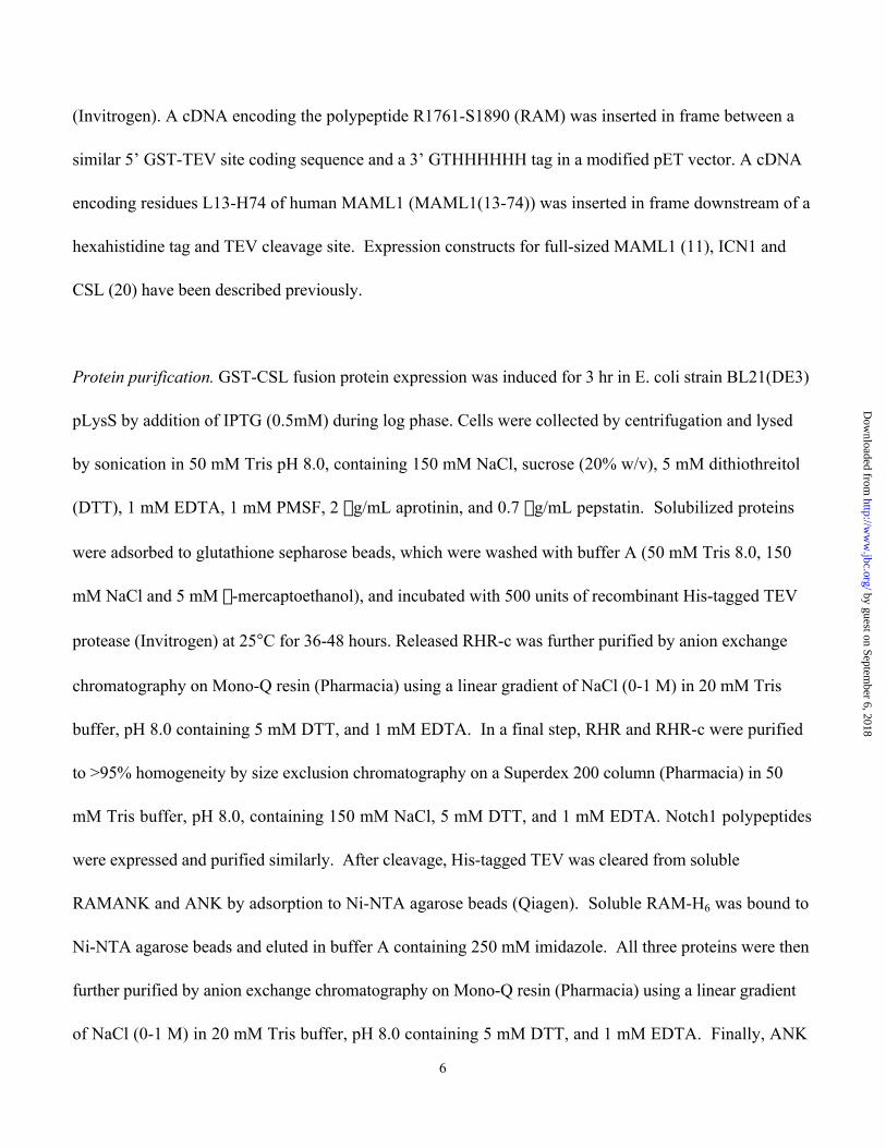
(Invitrogen). A cDNA encoding the polypeptide R1761-S1890 (RAM) was inserted in frame between a
similar 5’ GST-TEV site coding sequence and a 3’ GTHHHHHH tag in a modified pET vector. A cDNA
encoding residues L13-H74 of human MAML1 (MAML1(13-74)) was inserted in frame downstream of a
hexahistidine tag and TEV cleavage site. Expression constructs for full-sized MAML1 (11), ICN1 and
CSL (20) have been described previously.
Protein purification. GST-CSL fusion protein expression was induced for 3 hr in E. coli strain BL21(DE3)
pLysS by addition of IPTG (0.5mM) during log phase. Cells were collected by centrifugation and lysed
by sonication in 50 mM Tris pH 8.0, containing 150 mM NaCl, sucrose (20% w/v), 5 mM dithiothreitol
(DTT), 1 mM EDTA, 1 mM PMSF, 2 mg/mL aprotinin, and 0.7 mg/mL pepstatin. Solubilized proteins
were adsorbed to glutathione sepharose beads, which were washed with buffer A (50 mM Tris 8.0, 150
mM NaCl and 5 mM b-mercaptoethanol), and incubated with 500 units of recombinant His-tagged TEV
protease (Invitrogen) at 25°C for 36-48 hours. Released RHR-c was further purified by anion exchange
chromatography on Mono-Q resin (Pharmacia) using a linear gradient of NaCl (0-1 M) in 20 mM Tris
buffer, pH 8.0 containing 5 mM DTT, and 1 mM EDTA. In a final step, RHR and RHR-c were purified
to >95% homogeneity by size exclusion chromatography on a Superdex 200 column (Pharmacia) in 50
mM Tris buffer, pH 8.0, containing 150 mM NaCl, 5 mM DTT, and 1 mM EDTA. Notch1 polypeptides
were expressed and purified similarly. After cleavage, His-tagged TEV was cleared from soluble
RAMANK and ANK by adsorption to Ni-NTA agarose beads (Qiagen). Soluble RAM-H6 was bound to
Ni-NTA agarose beads and eluted in buffer A containing 250 mM imidazole. All three proteins were then
further purified by anion exchange chromatography on Mono-Q resin (Pharmacia) using a linear gradient
of NaCl (0-1 M) in 20 mM Tris buffer, pH 8.0 containing 5 mM DTT, and 1 mM EDTA. Finally, ANK
by guest on September 6, 2018
http://ww
w.jbc.org/
Dow
nloaded from

7
and RAMANK were further purified by size exclusion chromatography on a Superdex 200 column
(Pharmacia) in 50 mM Tris buffer, pH 8.0, containing 150 mM NaCl, 5 mM DTT, and 1 mM EDTA.
Expression of H6-tagged MAML1(13-74) was induced as above for CSL and ICN polypeptides. H6-
MAML13-74 was purified from inclusion bodies by solubilization in 50 mM Tris buffer, pH 8.0,
containing 6 M guanidinium HCl, and 150 mM NaCl, followed by affinity chromatography on a Ni-NTA
sepharose column. After extensive dialysis against 5% acetic acid, the protein was lyophilized, dissolved
in 50 mM sodium citrate buffer, pH 4.9, containing 5 mM DTT, and 0.5 mM EDTA, and cleaved with
TEV protease. Free MAML1(13-74) polypeptide was purified further by cation exchange
chromatography (MonoS 5/5 HR, Pharmacia) using a gradient of NaCl (0-1 M) in 20 mM sodium
phosphate buffer, pH 6.8, containing 5 mM DTT, and 1 mM EDTA. Purified MAML1(13-74) was
dialyzed against 5% acetic acid and lyophilized. Before use, MAML1(13-74) was dissolved in 50 mM
TrisHCl pH 8.0 buffer with 150 mM NaCl and 5 mM DTT.
Bioinformatics. Multiple sequence alignments were performed using CLUSTAL-W
(www.ebi.ac.uk/clustalw). Secondary structure prediction for all protein sequences was carried out using
the program PHD (21,22). The structural homology of CSL proteins (human CSL and Drosophila
Suppressor of Hairless) to Rel proteins of known three-dimensional structure was detected using the 3D-
PSSM fold-recognition program (www.sbg.bio.ic.ac.uk/~3dpssm/) (23,24).
Biophysical studies. Circular dichroism (CD) spectra were acquired on an Aviv 62DS spectropolarimeter
equipped with a thermoelectric temperature controller. Protein concentrations were determined according
by guest on September 6, 2018
http://ww
w.jbc.org/
Dow
nloaded from
8
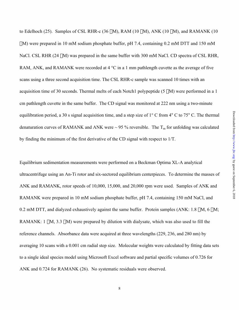
to Edelhoch (25). Samples of CSL RHR-c (36 mM), RAM (10 mM), ANK (10 mM), and RAMANK (10
mM) were prepared in 10 mM sodium phosphate buffer, pH 7.4, containing 0.2 mM DTT and 150 mM
NaCl. CSL RHR (24 mM) was prepared in the same buffer with 300 mM NaCl. CD spectra of CSL RHR,
RAM, ANK, and RAMANK were recorded at 4 °C in a 1 mm pathlength cuvette as the average of five
scans using a three second acquisition time. The CSL RHR-c sample was scanned 10 times with an
acquisition time of 30 seconds. Thermal melts of each Notch1 polypeptide (5 mM) were performed in a 1
cm pathlength cuvette in the same buffer. The CD signal was monitored at 222 nm using a two-minute
equilibration period, a 30 s signal acquisition time, and a step size of 1° C from 4° C to 75° C. The thermal
denaturation curves of RAMANK and ANK were ~ 95 % reversible. The Tm for unfolding was calculated
by finding the minimum of the first derivative of the CD signal with respect to 1/T.
Equilibrium sedimentation measurements were performed on a Beckman Optima XL-A analytical
ultracentrifuge using an An-Ti rotor and six-sectored equilibrium centerpieces. To determine the masses of
ANK and RAMANK, rotor speeds of 10,000, 15,000, and 20,000 rpm were used. Samples of ANK and
RAMANK were prepared in 10 mM sodium phosphate buffer, pH 7.4, containing 150 mM NaCl, and
0.2 mM DTT, and dialyzed exhaustively against the same buffer. Protein samples (ANK: 1.8 mM, 6 mM;
RAMANK: 1 mM, 3.3 mM) were prepared by dilution with dialysate, which was also used to fill the
reference channels. Absorbance data were acquired at three wavelengths (229, 236, and 280 nm) by
averaging 10 scans with a 0.001 cm radial step size. Molecular weights were calculated by fitting data sets
to a single ideal species model using Microsoft Excel software and partial specific volumes of 0.726 for
ANK and 0.724 for RAMANK (26). No systematic residuals were observed.
by guest on September 6, 2018
http://ww
w.jbc.org/
Dow
nloaded from

9
Electrophoretic Mobility Shift Assay. Electrophoretic mobility shift assay (EMSAs) were performed using
a 32P-labeled CSL specific oligonucleotide probe as described previously (11).
GST Bead-binding Assays. Glutathione sepharose beads were mixed at 4°C for 1 hr with cleared bacterial
lysates containing GST fusion polypeptides. After five washes with 50 mM TrisHCl buffer, pH 8.0,
containing 150 mM NaCl, 5 mM DTT, and 0.5% NP-40 (v/v), beads with GST-fusion proteins (20-30
mL/experiment) were incubated with potential binding partners for 1 hr at 4° C, followed by five washes
in the same buffer. Bound proteins were released by heating (100o C) for 10 min in SDS-PAGE loading
buffer. After electrophoresis in SDS-polyacrylamide gels, proteins were visualized by Coomassie blue-
staining.
Results
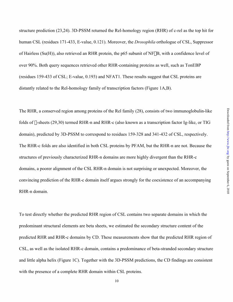
Detection of the Rel homology regions of CSL. The major isoform of human CSL encodes a protein of 486
amino acids (Figure 1A). Known biochemical activities of CSL include sequence-specific DNA binding
(27), association with ICN polypeptides (4), and formation of higher order complexes with MAML
polypeptides (10,11). To investigate the structural basis for these activities, we used a bioinformatic
approach to guide the design of CSL constructs. Because significant primary sequence homology between
CSL proteins and other proteins in the database was not detectable, we searched for evidence of homology
to protein domains of known structure using the 3D-PSSM fold-recognition program, which compares
input sequences to the existing protein fold library using structure-based sequence profiles and secondary
by guest on September 6, 2018
http://ww
w.jbc.org/
Dow
nloaded from
10
structure prediction (23,24). 3D-PSSM returned the Rel-homology region (RHR) of c-rel as the top hit for
human CSL (residues 171-433, E-value, 0.121). Moreover, the Drosophila orthologue of CSL, Suppressor
of Hairless (Su(H)), also retrieved an RHR protein, the p65 subunit of NFkB, with a confidence level of
over 90%. Both query sequences retrieved other RHR-containing proteins as well, such as TonEBP
(residues 159-433 of CSL; E-value, 0.193) and NFAT1. These results suggest that CSL proteins are
distantly related to the Rel-homology family of transcription factors (Figure 1A,B).
The RHR, a conserved region among proteins of the Rel family (28), consists of two immunoglobulin-like
folds of b-sheets (29,30) termed RHR-n and RHR-c (also known as a transcription factor Ig-like, or TIG
domain), predicted by 3D-PSSM to correspond to residues 159-328 and 341-432 of CSL, respectively.
The RHR-c folds are also identified in both CSL proteins by PFAM, but the RHR-n are not. Because the
structures of previously characterized RHR-n domains are more highly divergent than the RHR-c
domains, a poorer alignment of the CSL RHR-n domain is not surprising or unexpected. Moreover, the
convincing prediction of the RHR-c domain itself argues strongly for the coexistence of an accompanying
RHR-n domain.
To test directly whether the predicted RHR region of CSL contains two separate domains in which the
predominant structural elements are beta sheets, we estimated the secondary structure content of the
predicted RHR and RHR-c domains by CD. These measurements show that the predicted RHR region of
CSL, as well as the isolated RHR-c domain, contains a predominance of beta-stranded secondary structure
and little alpha helix (Figure 1C). Together with the 3D-PSSM predictions, the CD findings are consistent
with the presence of a complete RHR domain within CSL proteins.
by guest on September 6, 2018
http://ww
w.jbc.org/
Dow
nloaded from

11
The predicted RHR domain boundaries were combined with additional information to guide subsequent
structure/function studies. Truncation points outside of the RHR domain boundaries were chosen based
on primary sequence homology and secondary structure prediction. Homology between CSL and RBP-L,
a related protein that binds DNA but not ICNs (31), starts at residue 31. C-terminal to the RHR of CSL,
residue 452 marks the end of a stretch of 21 residues (432-452) predicted by PHD (21,22) to contain
helical structure. The C-terminal 34 residues (453-486) consist of low-complexity sequence. Finally, sites
chosen for intradomain truncations (e.g. residues 79 and 400) lie within regions predicted by PHD to lack
secondary structure. Guided by the informatics results, we therefore made CSL expression constructs
with N-termini starting at residues 31, 79, and 156, and C-terminal truncations at positions 332, 400, 435,
and 452.
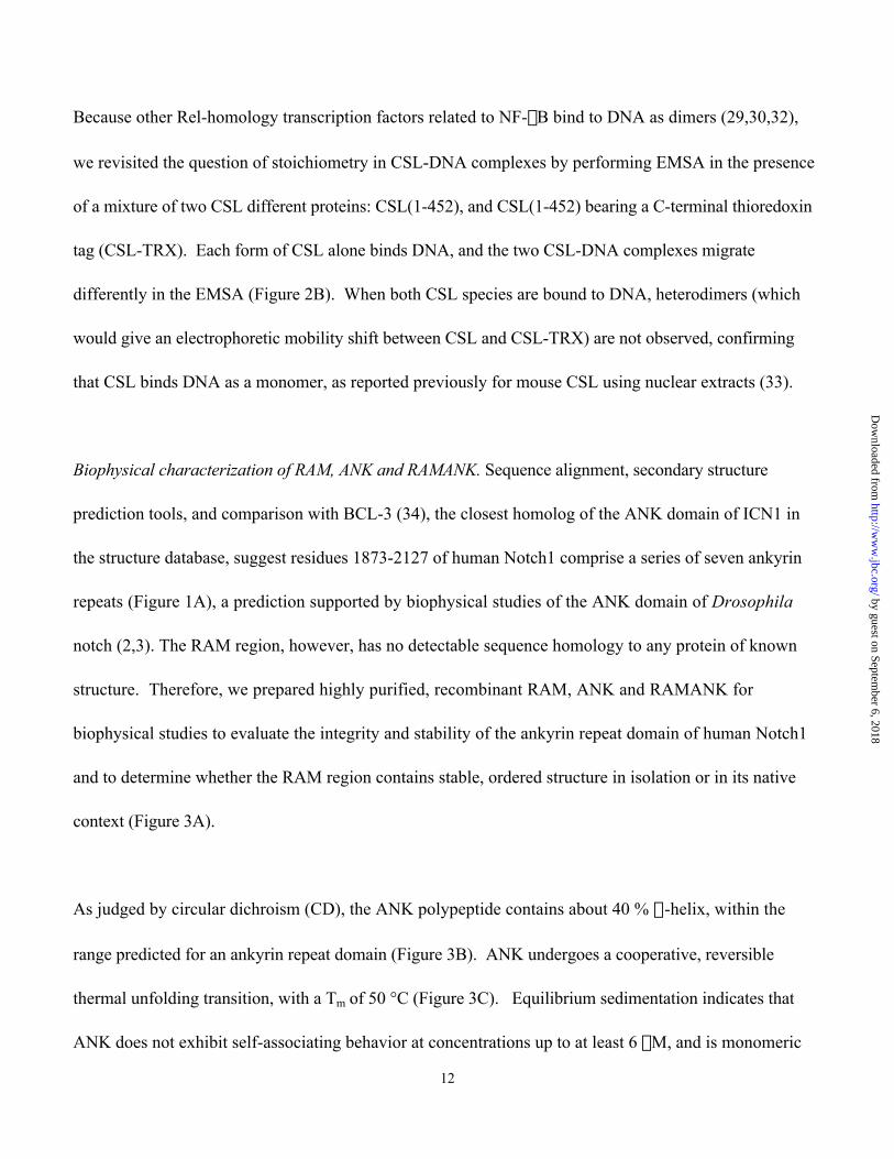
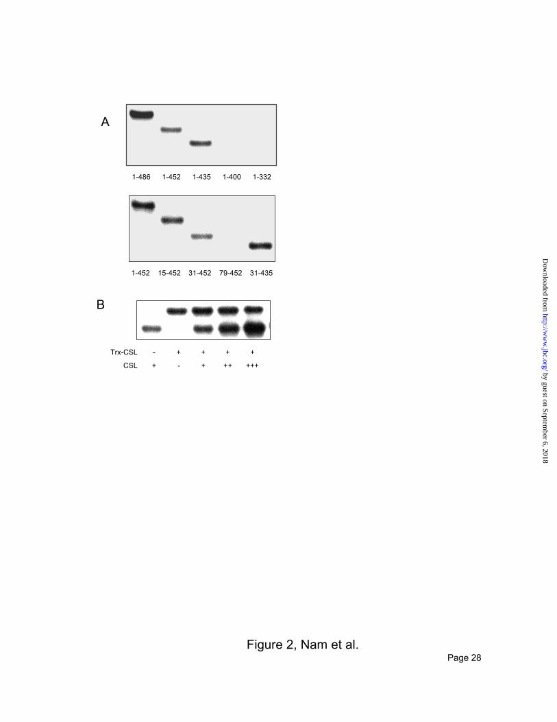
CSL requires residues 31-435 to bind DNA as a monomer. Using these constructs, we first investigated
the requirements for binding of CSL to DNA using an electrophoretic mobility shift assay (EMSA; Figure
2A). Removal of the first 31 residues of CSL does not interfere with DNA binding, but constructs with
N-termini beginning at residue 79 or 156 fail to bind DNA (Figure 3A), indicating that the RHR region
alone is insufficient. C-terminal truncation of CSL to residue 452 or 435 does not interfere with DNA
binding, but further truncations to residue 400 or 332, which interrupt or delete the RHR-c domain,
abrogate DNA binding. Thus, CSL residues 31-435, which encompass the complete RHR and an
additional N-terminal extension of 125 amino acids, are both necessary and sufficient to bind DNA.
by guest on September 6, 2018
http://ww
w.jbc.org/
Dow
nloaded from
12
Because other Rel-homology transcription factors related to NF-kB bind to DNA as dimers (29,30,32),
we revisited the question of stoichiometry in CSL-DNA complexes by performing EMSA in the presence
of a mixture of two CSL different proteins: CSL(1-452), and CSL(1-452) bearing a C-terminal thioredoxin
tag (CSL-TRX). Each form of CSL alone binds DNA, and the two CSL-DNA complexes migrate
differently in the EMSA (Figure 2B). When both CSL species are bound to DNA, heterodimers (which
would give an electrophoretic mobility shift between CSL and CSL-TRX) are not observed, confirming
that CSL binds DNA as a monomer, as reported previously for mouse CSL using nuclear extracts (33).
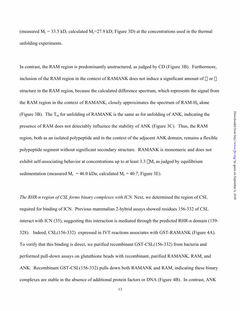
Biophysical characterization of RAM, ANK and RAMANK. Sequence alignment, secondary structure
prediction tools, and comparison with BCL-3 (34), the closest homolog of the ANK domain of ICN1 in
the structure database, suggest residues 1873-2127 of human Notch1 comprise a series of seven ankyrin
repeats (Figure 1A), a prediction supported by biophysical studies of the ANK domain of Drosophila
notch (2,3). The RAM region, however, has no detectable sequence homology to any protein of known
structure. Therefore, we prepared highly purified, recombinant RAM, ANK and RAMANK for
biophysical studies to evaluate the integrity and stability of the ankyrin repeat domain of human Notch1
and to determine whether the RAM region contains stable, ordered structure in isolation or in its native
context (Figure 3A).
As judged by circular dichroism (CD), the ANK polypeptide contains about 40 % a-helix, within the
range predicted for an ankyrin repeat domain (Figure 3B). ANK undergoes a cooperative, reversible
thermal unfolding transition, with a Tm of 50 °C (Figure 3C). Equilibrium sedimentation indicates that
ANK does not exhibit self-associating behavior at concentrations up to at least 6 mM, and is monomeric
by guest on September 6, 2018
http://ww
w.jbc.org/
Dow
nloaded from
13
(measured Mr = 33.5 kD, calculated Mr=27.9 kD; Figure 3D) at the concentrations used in the thermal
unfolding experiments.
In contrast, the RAM region is predominantly unstructured, as judged by CD (Figure 3B). Furthermore,
inclusion of the RAM region in the context of RAMANK does not induce a significant amount of a or b
structure in the RAM region, because the calculated difference spectrum, which represents the signal from
the RAM region in the context of RAMANK, closely approximates the spectrum of RAM-H6 alone
(Figure 3B). The Tm for unfolding of RAMANK is the same as for unfolding of ANK, indicating the
presence of RAM does not detectably influence the stability of ANK (Figure 3C). Thus, the RAM
region, both as an isolated polypeptide and in the context of the adjacent ANK domain, remains a flexible
polypeptide segment without significant secondary structure. RAMANK is monomeric and does not
exhibit self-associating behavior at concentrations up to at least 3.3 mM, as judged by equilibrium
sedimentation (measured Mr = 46.0 kDa; calculated Mr = 40.7; Figure 3E).
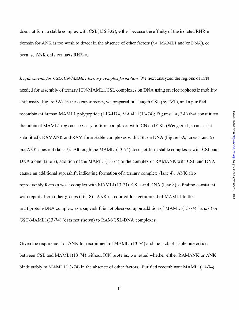
The RHR-n region of CSL forms binary complexes with ICN. Next, we determined the region of CSL
required for binding of ICN. Previous mammalian 2-hybrid assays showed residues 156-332 of CSL
interact with ICN (35), suggesting this interaction is mediated through the predicted RHR-n domain (159-
328). Indeed, CSL(156-332) expressed in IVT reactions associates with GST-RAMANK (Figure 4A).
To verify that this binding is direct, we purified recombinant GST-CSL(156-332) from bacteria and
performed pull-down assays on glutathione beads with recombinant, purified RAMANK, RAM, and
ANK. Recombinant GST-CSL(156-332) pulls down both RAMANK and RAM, indicating these binary
complexes are stable in the absence of additional protein factors or DNA (Figure 4B). In contrast, ANK
by guest on September 6, 2018
http://ww
w.jbc.org/
Dow
nloaded from
14
does not form a stable complex with CSL(156-332), either because the affinity of the isolated RHR-n
domain for ANK is too weak to detect in the absence of other factors (i.e. MAML1 and/or DNA), or
because ANK only contacts RHR-c.
Requirements for CSL/ICN/MAML1 ternary complex formation. We next analyzed the regions of ICN
needed for assembly of ternary ICN/MAML1/CSL complexes on DNA using an electrophoretic mobility
shift assay (Figure 5A). In these experiments, we prepared full-length CSL (by IVT), and a purified
recombinant human MAML1 polypeptide (L13-H74, MAML1(13-74); Figures 1A, 3A) that constitutes
the minimal MAML1 region necessary to form complexes with ICN and CSL (Weng et al., manuscript
submitted). RAMANK and RAM form stable complexes with CSL on DNA (Figure 5A, lanes 3 and 5)
but ANK does not (lane 7). Although the MAML1(13-74) does not form stable complexes with CSL and
DNA alone (lane 2), addition of the MAML1(13-74) to the complex of RAMANK with CSL and DNA
causes an additional supershift, indicating formation of a ternary complex (lane 4). ANK also
reproducibly forms a weak complex with MAML1(13-74), CSL, and DNA (lane 8), a finding consistent
with reports from other groups (16,18). ANK is required for recruitment of MAML1 to the
multiprotein-DNA complex, as a supershift is not observed upon addition of MAML1(13-74) (lane 6) or
GST-MAML1(13-74) (data not shown) to RAM-CSL-DNA complexes.
Given the requirement of ANK for recruitment of MAML1(13-74) and the lack of stable interaction
between CSL and MAML1(13-74) without ICN proteins, we tested whether either RAMANK or ANK
binds stably to MAML1(13-74) in the absence of other factors. Purified recombinant MAML1(13-74)
by guest on September 6, 2018
http://ww
w.jbc.org/
Dow
nloaded from
15
did not bind GST-RAMANK or GST-ANK (Figure 5B), indicating that recruitment of the MAML1
polypeptide into a multiprotein-DNA complex requires the presence of both ICN and CSL.
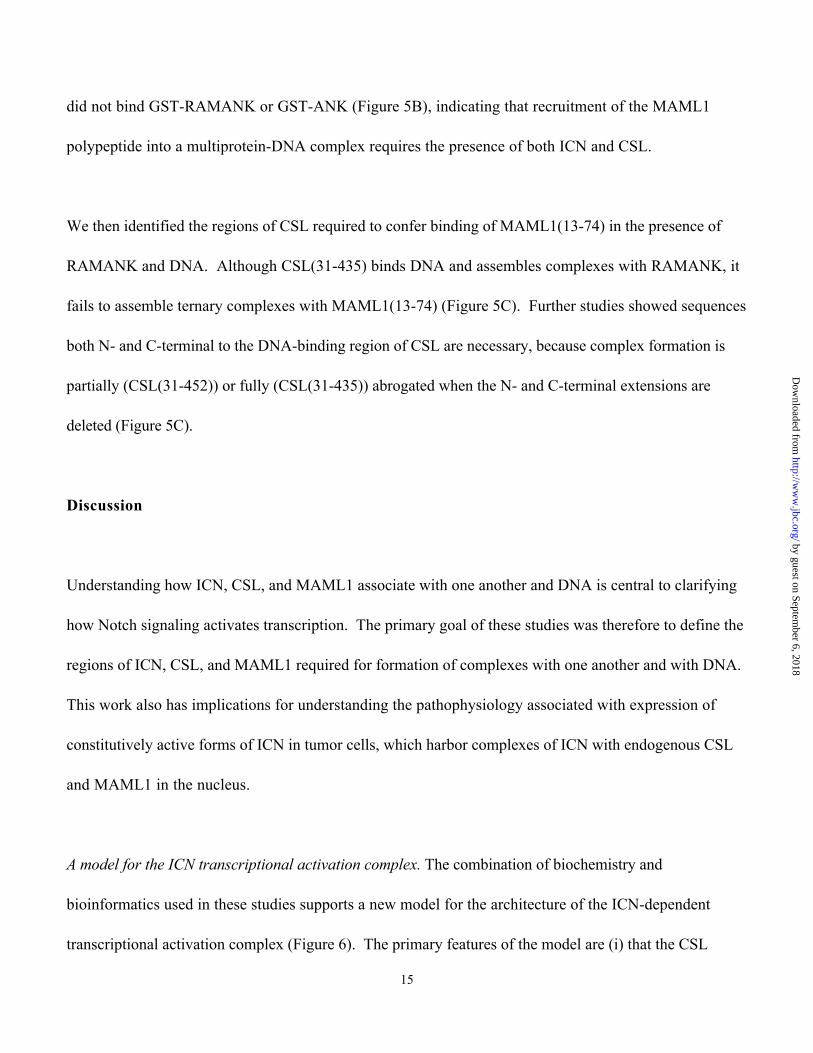
We then identified the regions of CSL required to confer binding of MAML1(13-74) in the presence of
RAMANK and DNA. Although CSL(31-435) binds DNA and assembles complexes with RAMANK, it
fails to assemble ternary complexes with MAML1(13-74) (Figure 5C). Further studies showed sequences
both N- and C-terminal to the DNA-binding region of CSL are necessary, because complex formation is
partially (CSL(31-452)) or fully (CSL(31-435)) abrogated when the N- and C-terminal extensions are
deleted (Figure 5C).
Discussion
Understanding how ICN, CSL, and MAML1 associate with one another and DNA is central to clarifying
how Notch signaling activates transcription. The primary goal of these studies was therefore to define the
regions of ICN, CSL, and MAML1 required for formation of complexes with one another and with DNA.
This work also has implications for understanding the pathophysiology associated with expression of
constitutively active forms of ICN in tumor cells, which harbor complexes of ICN with endogenous CSL
and MAML1 in the nucleus.
A model for the ICN transcriptional activation complex. The combination of biochemistry and
bioinformatics used in these studies supports a new model for the architecture of the ICN-dependent
transcriptional activation complex (Figure 6). The primary features of the model are (i) that the CSL
by guest on September 6, 2018
http://ww
w.jbc.org/
Dow
nloaded from

16
transcription factor is structurally related to proteins in the Rel homology family of transcription factors,
and (ii) that ICN and CSL combine to create the MAML-1 recruitment site.
The prediction that CSL proteins are structurally related to the Rel-homology family of transcription
factors emerged from analysis of the CSL and Su(H) amino acid sequences using the 3D-PSSM structure-
prediction server. The Rel-homology family of DNA-binding proteins encompasses a wide range of
transcription factors including the Rel/NF-kB proteins, NFAT proteins, and osmotic stress-response
proteins such as TonEBP (27). Rel-homology regions in these transcription factors typically span
roughly 250-275 amino acids and consist of two immunoglobulin-like domains (RHR-n and RHR-c)
connected by a short linker sequence. Based on the 3D-PSSM prediction, the RHR-n domain of human
CSL encompasses residues 159-328, and the RHR-c domain spans residues 341-432.
CSL structure-function relationships echo the RHR domain boundaries identified by 3D-PSSM. Several
experimental findings support the 3D-PSSM prediction that CSL contains both RHR-n and RHR-c
structural units. Most importantly, the complete RHR region, as well as the isolated RHR-c domain,
contains a predominance of beta-stranded secondary structure and little alpha helix. Furthermore, our
studies confirm that a construct encompassing only the isolated RHR-n domain constitutes the high-
affinity binding site for RAM. In addition, the C-terminal end of the region of CSL required for high-
affinity binding of cognate DNA coincides almost precisely with the C-terminal end of the predicted
RHR-c domain.
by guest on September 6, 2018
http://ww
w.jbc.org/
Dow
nloaded from

17
Our mixing studies using different-sized CSL variants are consistent with previous conclusions that the
functional DNA-binding unit of CSL is a monomer (33). Binding of DNA by CSL requires a remarkably
extensive region of the protein (residues 31-435), spanning the entire RHR domain and an additional 125
residues toward the N-terminus. This requirement that the entire RHR participate in DNA-binding is
shared by the other Rel-homology transcription factors, which exhibit a variety of structural arrangements
in DNA-binding complexes. In high-resolution structures of complexes between RHR transcription
factors and DNA, both the RHR-n and RHR-c domains participate in the DNA binding interface. Whereas
the Rel/ NF-kB (29,30) and TonEBP (32) proteins bind DNA as dimers, with the primary dimerization
interface provided by the RHR-c domain, the RHR domain of NFAT1 binds to DNA as a monomer, in
which coooperativity with the Fos/Jun hereodimer enables high-affinity binding to cognate DNA (36) .
By making additional contacts with the DNA, the extra N-terminal region of CSL may effectively obviate
the need for dimerization (e.g Rel/NFkB and TonEBP) or for additional factors (e.g. cooperativity
between NFAT1 and Fos/Jun) to achieve high-affinity DNA binding. By binding DNA as a monomer,
CSL most closely resembles the NFAT proteins, which also bind to DNA as monomers, cooperating with
the Fos/Jun heterodimer to form high-affinity protein-DNA complexes.
The RHR-n domain of CSL binds directly to RAMANK. After ICN is translocated into the nucleus, binding
of ICN to CSL-DNA complexes is likely to be the first step required for activation of transcription of
Notch-responsive genes. Previous studies have reported the existence of a strong CSL binding site within
the RAM region, and a weaker binding site in the ankyrin repeat (ANK) domain (37).
by guest on September 6, 2018
http://ww
w.jbc.org/
Dow
nloaded from
18
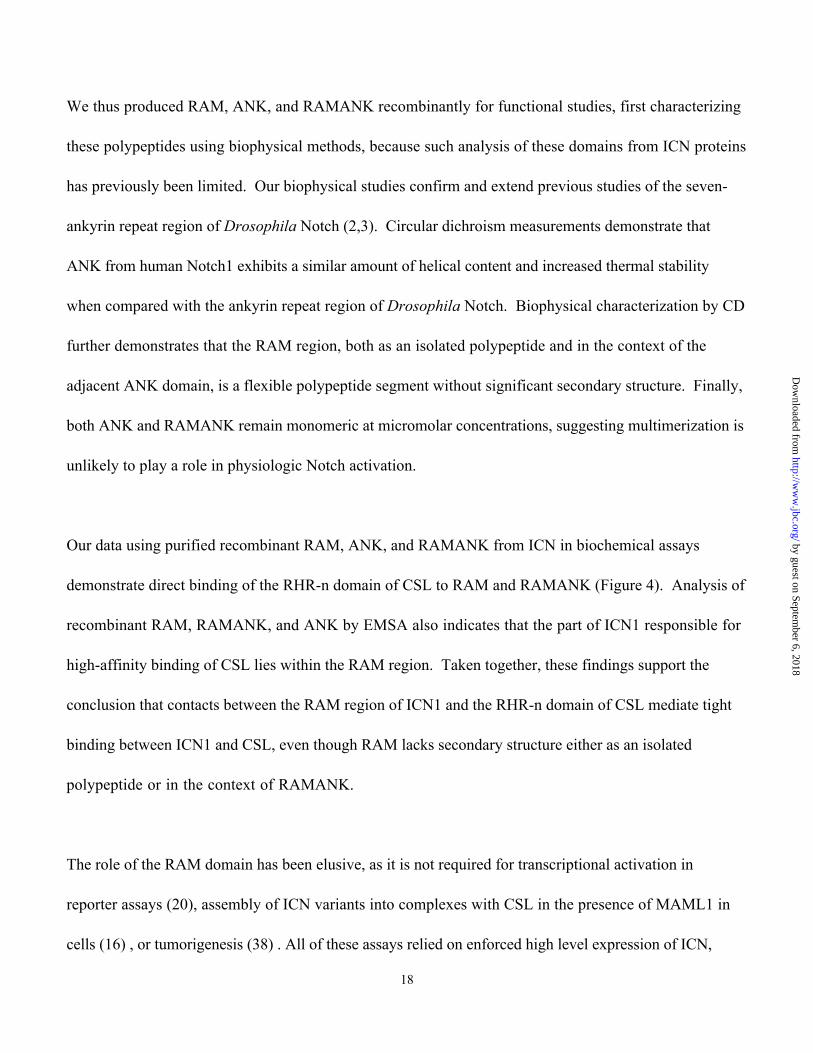
We thus produced RAM, ANK, and RAMANK recombinantly for functional studies, first characterizing
these polypeptides using biophysical methods, because such analysis of these domains from ICN proteins
has previously been limited. Our biophysical studies confirm and extend previous studies of the seven-
ankyrin repeat region of Drosophila Notch (2,3). Circular dichroism measurements demonstrate that
ANK from human Notch1 exhibits a similar amount of helical content and increased thermal stability
when compared with the ankyrin repeat region of Drosophila Notch. Biophysical characterization by CD
further demonstrates that the RAM region, both as an isolated polypeptide and in the context of the
adjacent ANK domain, is a flexible polypeptide segment without significant secondary structure. Finally,
both ANK and RAMANK remain monomeric at micromolar concentrations, suggesting multimerization is
unlikely to play a role in physiologic Notch activation.
Our data using purified recombinant RAM, ANK, and RAMANK from ICN in biochemical assays
demonstrate direct binding of the RHR-n domain of CSL to RAM and RAMANK (Figure 4). Analysis of
recombinant RAM, RAMANK, and ANK by EMSA also indicates that the part of ICN1 responsible for
high-affinity binding of CSL lies within the RAM region. Taken together, these findings support the
conclusion that contacts between the RAM region of ICN1 and the RHR-n domain of CSL mediate tight
binding between ICN1 and CSL, even though RAM lacks secondary structure either as an isolated
polypeptide or in the context of RAMANK.
The role of the RAM domain has been elusive, as it is not required for transcriptional activation in
reporter assays (20), assembly of ICN variants into complexes with CSL in the presence of MAML1 in
cells (16) , or tumorigenesis (38) . All of these assays relied on enforced high level expression of ICN,
by guest on September 6, 2018
http://ww
w.jbc.org/
Dow
nloaded from
19
conditions under which the lower affinity CSL binding site within ANK may suffice for assembly of
transcriptional activation complexes. In support of this possibility, high concentrations of ANK alone
supershift CSL-DNA complexes in the presence of MAML-1 (Figure 5A), and prior in vivo observations
indicate the RAM region is not absolutely necessary for formation of complexes in transformed cells with
increased levels of ICN (16). However, physiologically relevant levels of ICNs produced through ligand-
stimulated Notch activation are much lower, typically being below the limits of immunologic or
biochemical detection. At these levels, the high-affinity interaction between RAM and RHR-n may take
on much greater importance.
The complex between RAMANK and CSL exhibits both similarities and differences when compared with
the complex between IkB and NFkB, the only example of a complex between ANK and RHR proteins for
which high-resolution structural information is available (39,40). In the NFkB-IkB complex, the interface
between the IkB ankyrin repeats and NFkB encompasses both RHR-c domains of the NFkB heterodimer.
Because CSL binds to DNA as a monomer, it does not present as extensive an RHR-c interface for binding
to the ANK domain of ICN1. Contacts between the ANK domain of ICN and the RHR region of CSL
thus appear to confer substantially weaker binding affinity (perhaps because only half of the interface
seen in the IkB-NFkB complex would be present between ANK and RHR-c of CSL), and ICN uses the
strong binding affinity of RAM for the RHR-n domain as an anchor to stabilize complexes of ICN with
CSL.
by guest on September 6, 2018
http://ww
w.jbc.org/
Dow
nloaded from
20
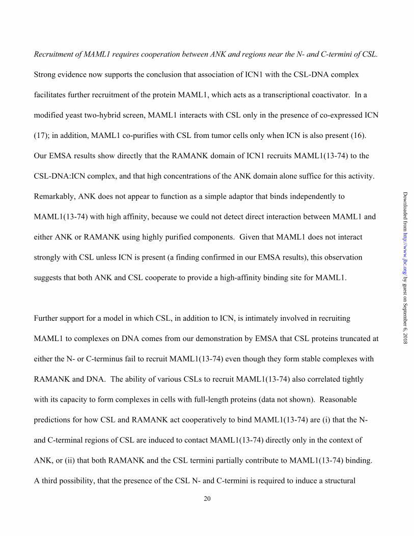
Recruitment of MAML1 requires cooperation between ANK and regions near the N- and C-termini of CSL.
Strong evidence now supports the conclusion that association of ICN1 with the CSL-DNA complex
facilitates further recruitment of the protein MAML1, which acts as a transcriptional coactivator. In a
modified yeast two-hybrid screen, MAML1 interacts with CSL only in the presence of co-expressed ICN
(17); in addition, MAML1 co-purifies with CSL from tumor cells only when ICN is also present (16).
Our EMSA results show directly that the RAMANK domain of ICN1 recruits MAML1(13-74) to the
CSL-DNA:ICN complex, and that high concentrations of the ANK domain alone suffice for this activity.
Remarkably, ANK does not appear to function as a simple adaptor that binds independently to
MAML1(13-74) with high affinity, because we could not detect direct interaction between MAML1 and
either ANK or RAMANK using highly purified components. Given that MAML1 does not interact
strongly with CSL unless ICN is present (a finding confirmed in our EMSA results), this observation
suggests that both ANK and CSL cooperate to provide a high-affinity binding site for MAML1.
Further support for a model in which CSL, in addition to ICN, is intimately involved in recruiting
MAML1 to complexes on DNA comes from our demonstration by EMSA that CSL proteins truncated at
either the N- or C-terminus fail to recruit MAML1(13-74) even though they form stable complexes with
RAMANK and DNA. The ability of various CSLs to recruit MAML1(13-74) also correlated tightly
with its capacity to form complexes in cells with full-length proteins (data not shown). Reasonable
predictions for how CSL and RAMANK act cooperatively to bind MAML1(13-74) are (i) that the N-
and C-terminal regions of CSL are induced to contact MAML1(13-74) directly only in the context of
ANK, or (ii) that both RAMANK and the CSL termini partially contribute to MAML1(13-74) binding.
A third possibility, that the presence of the CSL N- and C-termini is required to induce a structural
by guest on September 6, 2018
http://ww
w.jbc.org/
Dow
nloaded from
21
rearrangement of ANK that enables high-affinity association between ANK and MAML1(13-74), is less
likely because the ANK domain is well-structured as an isolated domain in solution. Clearly, a high-
resolution structure of the complex will clarify crucial details about the arrangement of the protein
components in the transcriptional activation complex.
Because both CSL and RAMANK combine to recruit MAML1, an intrinsic control mechanism exists to
prevent premature activation of Notch target genes, and ensure that off-pathway genes are unaffected
when Notch becomes activated. First, because MAML1 cannot bind to CSL sites in the absence of
activated Notch, target genes downstream of CSL sites continue to be repressed by co-repressors bound
to CSL. Moreover, because ICN1 and MAML1 do not form a stable complex without CSL, cross-
reactivity of ICN with DNA-binding proteins other than CSL will not inadvertently cause expression of
“off-target” gene products. Finally, recent evidence suggests that MAML1 may not only serve as an
effector for transcription of Notch target genes but may also simultaneously commit the active complex
for subsequent proteasomal degradation (18). By coordinating the timing of Notch activation with respect
to its subsequent destruction, MAML1 would thus also enable a mechanism for tight control of the
duration and timing of events driven by activated Notch.
Acknowledgments
We thank Vytas Patriub for excellent technical support.
by guest on September 6, 2018
http://ww
w.jbc.org/
Dow
nloaded from
22
References
1. Artavanis-Tsakonas, S., Rand, M. D., and Lake, R. J. (1999) Science 284, 770-7762. Zweifel, M. E., and Barrick, D. (2001) Biochemistry 40, 14344-14356.3. Zweifel, M. E., and Barrick, D. (2001) Biochemistry 40, 14357-14367.4. Fortini, M. E., and Artavanis-Tsakonas, S. (1994) Cell 79, 273-282.5. Jarriault, S., Brou, C., Logeat, F., Schroeter, E. H., Kopan, R., and Israel, A. (1995) Nature 377,
355-358.6. Christensen, S., Kodoyianni, V., Bosenberg, M., Friedman, L., and Kimble, J. (1996) Development
122, 1373-13837. Hsieh, J. J., Zhou, S., Chen, L., Young, D. B., and Hayward, S. D. (1999) Proc Natl Acad Sci U S
A 96, 23-288. Kao, H. Y., Ordentlich, P., Koyano-Nakagawa, N., Tang, Z., Downes, M., Kintner, C. R., Evans,
R. M., and Kadesch, T. (1998) Genes Dev 12, 2269-22779. Taniguchi, Y., Furukawa, T., Tun, T., Han, H., and Honjo, T. (1998) Mol Cell Biol 18, 644-654.10. Petcherski, A. G., and Kimble, J. (2000) Nature 405, 364-36811. Wu, L., Aster, J. C., Blacklow, S. C., Lake, R., Artavanis-Tsakonas, S., and Griffin, J. D. (2000)
Nat Genet 26, 484-489.12. Schweisguth, F., and Posakony, J. W. (1992) Cell 69, 1199-121213. Yedvobnick, B., Smoller, D., Young, P., and Mills, D. (1988) Genetics 118, 483-497.14. Artavanis-Tsakonas, S., Muskavitch, M. A., and Yedvobnick, B. (1983) Proc Natl Acad Sci U S A
80, 1977-1981.15. Aster, J., Pear, W., Hasserjian, R., Erba, H., Davi, F., Luo, B., Scott, M., Baltimore, D., and Sklar,
J. (1994) Cold Spring Harb Symp Quant Biol 59, 125-13616. Jeffries, S., Robbins, D. J., and Capobianco, A. J. (2002) Mol Cell Biol 22, 3927-394117. Petcherski, A. G., and Kimble, J. (2000) Curr Biol 10, R471-473.18. Fryer, C. J., Lamar, E., Turbachova, I., Kintner, C., and Jones, K. A. (2002) Genes Dev 16, 1397-
141119. Wallberg, A. E., Pedersen, K., Lendahl, U., and Roeder, R. G. (2002) Mol Cell Biol 22, 7812-781920. Aster, J. C., Robertson, E. S., Hasserjian, R. P., Turner, J. R., Kieff, E., and Sklar, J. (1997) J Biol
Chem 272, 11336-1134321. Rost, B., and Sander, C. (1993) J Mol Biol 232, 584-59922. Rost, B., and Sander, C. (1994) Proteins 19, 55-7223. Fischer, D., Barret, C., Bryson, K., Elofsson, A., Godzik, A., Jones, D., Karplus, K. J., Kelley, L.
A., MacCallum, R. M., Pawowski, K., Rost, B., Rychlewski, L., and Sternberg, M. (1999)Proteins 3, 209-217
24. Kelley, L. A., MacCallum, R. M., and Sternberg, M. J. (2000) J Mol Biol 299, 499-52025. Edelhoch, H. (1967) Biochemistry 6, 1948-195426. Laue, T. M., Shah, B. D., Ridgeway, T. M., and Pelletier, S. L. (1992) in Analytical
Ultracentrifugation in Biochemistry and Polymer Science (Harding, S. E., Rowe, A. J., and Horton,J. C., eds), pp. 90-125, Cambridge: Royal Society of Chemistry
27. Tun, T., Hamaguchi, Y., Matsunami, N., Furukawa, T., Honjo, T., and Kawaichi, M. (1994)Nucleic Acids Res 22, 965-971.
by guest on September 6, 2018
http://ww
w.jbc.org/
Dow
nloaded from

23
28. Ghosh, S., May, M. J., and Kopp, E. B. (1998) Annu Rev Immunol 16, 225-26029. Muller, C. W., Rey, F. A., Sodeoka, M., Verdine, G. L., and Harrison, S. C. (1995) Nature 373,
311-317.30. Ghosh, G., van Duyne, G., Ghosh, S., and Sigler, P. B. (1995) Nature 373, 303-310.31. Minoguchi, S., Taniguchi, Y., Kato, H., Okazaki, T., Strobl, L. J., Zimber-Strobl, U., Bornkamm,
G. W., and Honjo, T. (1997) Mol Cell Biol 17, 2679-268732. Stroud, J. C., Lopez-Rodriguez, C., Rao, A., and Chen, L. (2002) Nat Struct Biol 9, 90-94.33. Chung, C. N., Hamaguchi, Y., Honjo, T., and Kawaichi, M. (1994) Nucleic Acids Res 22, 2938-
2944.34. Michel, F., Soler-Lopez, M., Petosa, C., Cramer, P., Siebenlist, U., and Muller, C. W. (2001)
Embo J 20, 6180-619035. Hsieh, J. J., Henkel, T., Salmon, P., Robey, E., Peterson, M. G., and Hayward, S. D. (1996) Mol
Cell Biol 16, 952-95936. Chen, L., Glover, J. N., Hogan, P. G., Rao, A., and Harrison, S. C. (1998) Nature 392, 42-48.37. Kato, H., Taniguchi, Y., Kurooka, H., Minoguchi, S., Sakai, T., Nomura-Okazaki, S., Tamura, K.,
and Honjo, T. (1997) Development 124, 4133-414138. Aster, J. C., Xu, L., Karnell, F. G., Patriub, V., Pui, J. C., and Pear, W. S. (2000) Mol Cell Biol 20,
7505-751539. Huxford, T., Huang, D. B., Malek, S., and Ghosh, G. (1998) Cell 95, 759-77040. Chen, F. E., Huang, D. B., Chen, Y. Q., and Ghosh, G. (1998) Nature 391, 410-413.41. Sreerama, N., and Woody, R. W. (2000) Anal Biochem 287, 252-260
by guest on September 6, 2018
http://ww
w.jbc.org/
Dow
nloaded from
24
Figure legends
FIG 1. A. Domain organization of CSL, the intracellular portion of human Notch1 (ICN), and mastermind-
like1 (MAML1). CSL: The homology of human CSL with Drosophila Su(H) starts at residue 15, and
with RBP-L at residue 31. The RHR-n and RHR-c domains predicted by 3D-PSSM are shown. ICN1:
NLS, nuclear localization sequence (arrows); TA transactivation region. MAML1: Residues 13-74
correspond to the minimum-length polypeptide necessary and sufficient to form ternary complexes with
ICN, CSL, and DNA. Residues 75-301 of MAML1 are implicated in recruitment of CBP/p300 (18). B.
Alignment of the RHR regions of CSL and Su(H) with their structural homologues. 3D-PSSM alignments
of CSL with c-rel (top) and Su(H) with NFkB (bottom). Residues are colored by predicted (CSL and
Su(H)) or actual (c-rel and NFkB) secondary structure (blue = b strands; orange = a helices). C. Circular
dichroism (CD) spectra of the entire RHR of CSL and of CSL RHR-c. Entire RHR: open circles. RHR-c:
closed circles. The indicated estimates of protein secondary structure content were calculated with the
program CONTINLL (41).
Figure 2. CSL requires residues 31-435 to bind DNA as a monomer. A. CSL variants corresponding to the
indicated amino acid residues were tested for binding to a [32P]-labeled oligonucleotide containing a
consensus CSL binding site in an electrophoretic mobility shift assay (EMSA). DNA/proteins complexes
were detected by autoradiography. B. CSL(1-452) and CSL-TRX, polypeptides of differing size, were
tested alone and in combination by EMSA, as described in (A).
by guest on September 6, 2018
http://ww
w.jbc.org/
Dow
nloaded from
25
FIG 3. Purification and characterization of RAMANK, RAM, ANK, and MAML1(13-74). A. Coomassie
staining of purified RAM, ANK, RAMANK, and MAML 13-74 after SDS-PAGE.
B. Circular dichroism (CD) spectra of RAM (crosses), ANK (open circles) and RAMANK (closed
circles). Spectra were recorded at 4°C in physiologic buffer (10 mM sodium phosphate pH 7.4, 150mM
NaCl, 0.2mM DTT). A predicted CD spectrum of the RAM region (open triangles) was calculated from
the difference between the RAMANK and ANK spectra. C. Thermal dependence of the CD signal at
222 nm for ANK (open circles) and RAMANK (closed circles) in physiologic buffer. D and E.
Equilibrium centrifugation of 6mM ANK (D) and 3.3mM RAMANK (E) at 4°C in physiologic buffer (10
mM sodium phosphate pH 7.4, 150 mM NaCl, 0.2 mM DTT). Upper panels: log of the concentration
distribution of protein as a function of the square of the radial position at 15 000 rpm (ANK) or 10 000
rpm (RAMANK). The best fit line describes a single homogeneous species of molecular mass 33 818 Da
(ANK) or 46 008 Da (RAMANK). Lower panels: residuals to the fit. No systematic trend is observed.
FIG 4. Requirements for formation of CSL-ICN complexes. A. Glutathione beads with bound GST-
RAMANK, GST-ANK, or GST were incubated with various forms of [35S]-labeled CSL prepared by in
vitro transcription/translation (IVT). Bound CSL was then detected by autoradiography after SDS-PAGE.
The faint bands of lower molecular weight in the upper panel likely represent truncated translation
products from the IVT reaction. B. Glutathione beads with bound GST-CSL(156-332) or GST alone were
incubated with RAMANK, ANK or RAM, as indicated. Bound ICN proteins were released by heating in
SDS buffer, resolved by SDS-PAGE, and then detected by staining with Coomassie blue. Lanes marked
input contain 20% of the total ICN protein used for each pull-down.
by guest on September 6, 2018
http://ww
w.jbc.org/
Dow
nloaded from

26
FIG 5. Requirements for formation of stable ternary CSL/ICN/MAML1 complexes on DNA. A.
Electrophoretic mobility shift analysis (EMSA). The indicated combinations of proteins were incubated
with a [32P]-labeled oligonucleotide with a CSL consensus binding site, resolved by EMSA, and visualized
by autoradiography. The concentration of ANK in lane 8 was 300-fold greater than the concentration of
RAMANK used in lane 3. B. MAML1(13-74) does not bind to ANK or RAMANK alone. GST, GST-
ANK and GST-RAMANK were each captured on glutathione beads and then incubated with purified
recombinant MAML113-74. Proteins bound to the beads were released by heating in SDS buffer, resolved
by SDS-PAGE, and then detected by staining with Coomassie blue. C. Sequences both N- and C-terminal
to the DNA-binding region of CSL are necessary for ternary complex formation on DNA. Each panel
represents a series of EMSAs performed as in (A) with the indicated CSL variant and either DNA alone
(left), DNA and RAMANK (center), or DNA, RAMANK and MAML1(13-74) (right).
FIG 6. Model for the organization of the CSL/ICN/MAML1 ternary complex on DNA. The cartoon
illustrates contacts identified experimentally by mapping the interactions of CSL 1-452 with DNA,
RAMANK, and MAML1(13-74). The structures used to create ribbon diagrams for the RHR domain
within CSL and the ankyrin repeat domain within ICN are from the structures of chicken c-rel on DNA
and human Bcl-3, respectively. The N-terminal region of CSL also required for DNA-binding is
represented as a sphere with an unknown fold. Both RAM and MAML1(13-74) are schematically
illustrated as unstructured polypeptides.
by guest on September 6, 2018
http://ww
w.jbc.org/
Dow
nloaded from

1
-6000
-5000
-4000
-3000
-2000
-1000
0
1000
2000
200 210 220 230 240 250 260
Wavelength (nm)
Mea
n R
esid
ue E
llipt
icity
(deg
cm
2 dm
ol-1
)
A
Figure 1, Nam et al.
B
PEST
NLS
Ankyrinrepeats
RAM TAICN1
486159 328 341 433
15 31 452
RHR-n RHR-cCSL
13 741 1016301Maml1
C
Estimated: RHR (°) RHR-c(l)
% helix 4.5 5.4% sheet 38.9 35.7% turns 23.2 24.2% coil 33.4 34.7
RHR
Required for ternary complex
P300-binding
hCSL TKVALFNRLR SQTVSTRYLH VEGGNFHASS QQWGAFFIHL LDDDESEGEE FTVRDVYIH. YGQTCKLVCS VTGMALPRLI 249cRel PYIEIFEQPR QRGMRFRY.K CEGRS..... .......AGS IPGEHSTDNN KTFPSIQILN YFGKVKIRT. .......... 62 hCSL IMKVDKHTAL LDADDPVSQL HKCAFY...L KDT....... .ERMYLCLSQ ERII...... .........Q FQATPCPKEP 303cRel ........TL VTKNEPY.KP HPHDLVGKDC RDGYYEAEFG PERRVLSFQN LGIQCVKKKD LKESISLRIS KKINP.FNVP 132 hCSL NKEM..INDG ASWTIISTDK AEYT.....F YEGMGPVL.. ......APVT PVPVVESL.. ...QLNGGGD VAMLELTGQN 363cRel EEQLHNIDEY DLNVVRLCFQ AFLPDEHGNY TLALPPLISN PIYDNRAPNT AELRICRVNK NCGSVKGGDE IFIL...CDK 209 hCSL FT.PNLRV.. ....W..FGD VEAETMYRCG ESMLCVVPDI SAFREGWRWV RQPVQVPVTL VRNDGIIYST SLTFTYTPE 433cRel VQKDDIEVRF VLDNWEAKGS FSQADVHR.Q VAIVFRTPP. ..FLRD...I TEPITVKMQL RRPSDQEVSE PMDFRYLPD 280 Su(H) NVALFNRLRS QTVSTRYLHV ENGHFHAS.. .....STQWG AFTIHLLDDN E......... SESEEFQV.. ........RD 320NFkB YVEIIEQPKQ RGMRFRY..K CEGRSAGSIP GERSTDTTKT HPTIKINGYT GPGTVRISLV TKDPPHRPHP HELVGKDCRD 97 Su(H) GYIHY..GAT VKLVC.SVTG M.ALPRLIIR KVDKQMALLE AD...DPVSQ LHKCAFYMKD TD..RMYLCL SQEKIIQFQA 384NFkB GFYEAELCPD RCIHSFQNLG IQCVKKRDLE QAISQRIQTN NNPFQVPIEE QRG......D YDLNAVRLC. .......FQV 169 Su(H) TPCPKEPNKE MINDGACWTI ISTDKAEYQF YEGMGPVASP VTPVPI.VNS LNLNGGGDVA MLELSGDNFT .PHLQV.... 462NFkB T.VRDPSGRP LRLPPVLPHP I......... FDNRAPNTAE LKICRVNRNS GSCLGGDEIF LL...CDKVQ KEDIEVYFTG 230 Su(H) ..W..FGDVE AETMYRCTET LLCVVPEISQ FRGEWLWVRQ PTQVPISLVR .NDGIIYATG LTFTYTPE 521NFkB PGWEARGSFS QADVHR.QVA IVFRTPP... YADPS..LQA PVRVSMQLRR PSDRE.LSEP MEFQYLPD 291
sheet helix
171
260
Page 27
by guest on September 6, 2018
http://ww
w.jbc.org/
Dow
nloaded from
1-486 1-4001-4351-452 1-332
1-452 79-45231-45215-452 31-435
A
B
Trx-CSL - + + + +
CSL + - + ++ +++
Figure 2, Nam et al.Page 28
by guest on September 6, 2018
http://ww
w.jbc.org/
Dow
nloaded from
-10
-9
-8
-7
-6
-5
-4
0 20 40 60 80Temperature (°C)
Elli
ptic
ity (
10
3 d
eg
cm
2 d
mo
l-1)
RAMANK
ANK
A
Figure 3, Nam et al.
-2
-1.5
-1
-0.5
41 41.5 42 42.5 43 43.5ln (
ab
sorb
an
ce),
23
4n
m
-0.1-0.05
00.050.1
Radius squared
De
via
tion
DC
RA
M
AN
K
RA
MA
NK
Ma
m1
3-7
4
ANK
-1.8
-1.6
-1.4
-1.2
-1
41 41.5 42 42.5 43 43.5ln (
Ab
sorb
an
ce),
23
4n
m
-0.1-0.05
00.050.1
Radius squared
De
via
tion
ERAMANK
-20
-10
0
10
190 200 210 220 230 240 250 260
Wavelength (nm)
Elli
ptic
ity (
103 d
eg c
m2 d
mol
-1)
• RAMANKo ANK+ RAMD Difference
B
Page 29
by guest on September 6, 2018
http://ww
w.jbc.org/
Dow
nloaded from
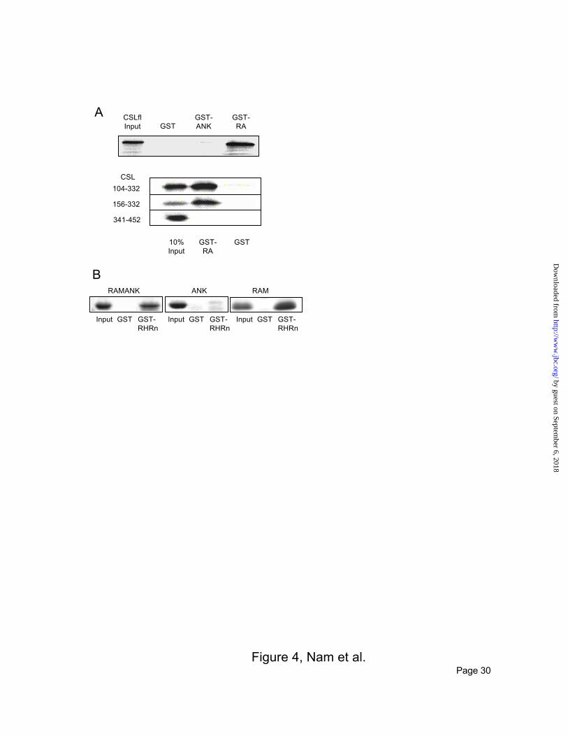
Figure 4, Nam et al.
CSLflInput GST
GST-ANK
GST-RA
104-332
156-332
341-452
CSL
GSTGST-RA
10%Input
B
A
RAMANK RAMANK
Input GST GST-RHRn
Input GST GST-RHRn
Input GST GST-RHRn
Page 30
by guest on September 6, 2018
http://ww
w.jbc.org/
Dow
nloaded from
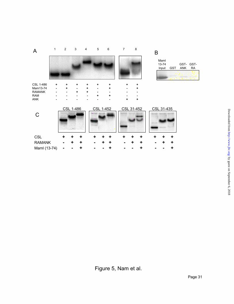
Figure 5, Nam et al.
CSL + + + + + + + + + + + +RAMANK - + + - + + - + + - + +Maml (13-74) - - + - - + - - + - - +
CSL 1-486 CSL 1-452 CSL 31-435CSL 31-452
CSL 1-486 + + + + + + + +Mam13-74 - + - + - + - +RAMANK - - + + - - - -RAM - - - - + + - -ANK - - - - - - + +
1 2 3 4 5 6 7 8A
GSTGST-ANK
GST-RA
Maml13-74Input
B
C
Page 31
by guest on September 6, 2018
http://ww
w.jbc.org/
Dow
nloaded from
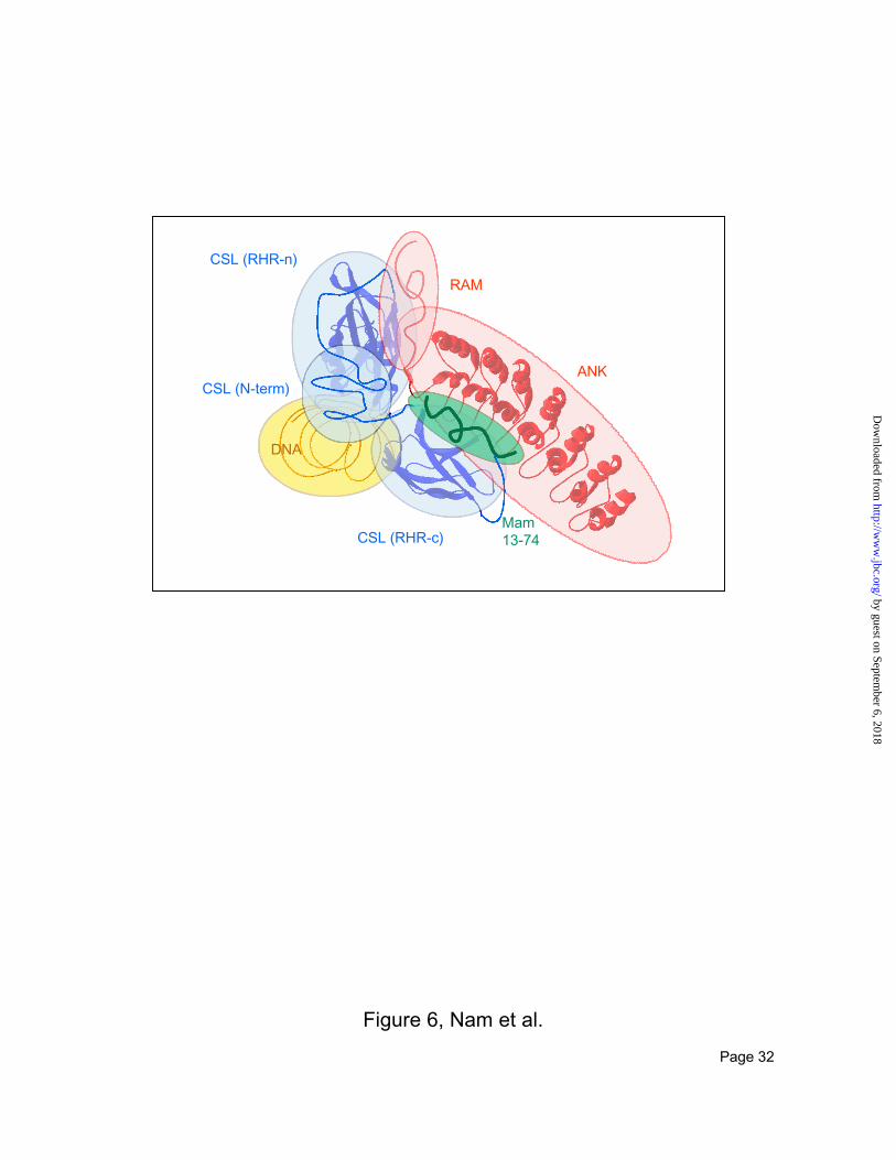
Figure 6, Nam et al.
RAM
ANK
CSL (RHR-n)
CSL (N-term)
DNA
CSL (RHR-c)Mam13-74
Page 32
by guest on September 6, 2018
http://ww
w.jbc.org/
Dow
nloaded from
Yunsun Nam, Andrew P. Weng, Jon C. Aster and Stephen C. Blacklownotch1/mastermind-like 1 transcriptional activation complexStructural requirements for assembly of the CSL/intracellular
published online March 18, 2003J. Biol. Chem.
10.1074/jbc.M301567200Access the most updated version of this article at doi:
Alerts:
When a correction for this article is posted•
When this article is cited•
to choose from all of JBC's e-mail alertsClick here
by guest on September 6, 2018
http://ww
w.jbc.org/
Dow
nloaded from