Embed Size (px)
Citation preview
doi: 10.1098/rspa.2008.0462, 1003-1027 first published online 13 January 2009465 2009 Proc. R. Soc. A
Antonio Tilocca molecular dynamics simulationsStructural models of bioactive glasses from
Referenceshtml#ref-list-1http://rspa.royalsocietypublishing.org/content/465/2104/1003.full.
This article cites 112 articles, 2 of which can be accessed free
This article is free to access
Subject collections (128 articles)materials science �
Articles on similar topics can be found in the following collections
Email alerting service herethe box at the top right-hand corner of the article or click Receive free email alerts when new articles cite this article - sign up in
http://rspa.royalsocietypublishing.org/subscriptions go to: Proc. R. Soc. ATo subscribe to
This journal is © 2009 The Royal Society
on July 27, 2010rspa.royalsocietypublishing.orgDownloaded from
on July 27, 2010rspa.royalsocietypublishing.orgDownloaded from
REVIEW
Structural models of bioactive glasses frommolecular dynamics simulations
BY ANTONIO TILOCCA*
Department of Chemistry and Materials Simulation Laboratory,University College London, London WC1H 0AJ, UK
The bioactive mechanism, by which living tissues attach to and integrate with anartificial implant through stable chemical bonds, is at the core of many current medicalapplications of biomaterials, as well as of novel promising applications in tissueengineering. Having been employed in these applications for almost 40 years, soda-limephosphosilicate glasses such as 45S5 represent today the paradigm of bioactive materials.Despite their strategical importance in the field, the relationship between the structureand the activity of a glass composition in a biological environment has not been studiedin detail. This fundamental gap negatively affects further progress, for instance, toimprove the chemical durability and tailor the biodegradability of these materials forspecific applications. This paper reviews recent advances in computer modelling ofbioactive glasses based on molecular dynamics simulations, which are starting to unveilkey structural features of these materials, thus contributing to improve our fundamentalunderstanding of how bioactive materials work.
Keywords: molecular dynamics simulations; bioactivity; silicate glasses;structural properties
*a.t
RecAcc
1. Introduction
The discovery by Hench and co-workers that a range of compositions of modifiedphosphosilicate glasses has the ability to form a stable chemical bond with livingtissues (bone, ligament and muscle) opened a completely new field inbiomedicine, ca 40 years ago (Hench et al. 1971). Since then, many artificialbiomaterials based on, or inspired by, Hench’s glasses have been developed andsuccessfully employed in clinical applications for repairing and replacing parts ofthe human body. This field is continuously expanding: new processing routeshave extended the range of applications towards new exciting directions inbiomedicine (Hench & Polak 2002), many of which still rely on the originalHench’s base formulation, 45S5 Bioglass, which has now become the paradigm ofbioactive materials. However, the growing technological importance of thesematerials has not been supported by a corresponding growth in fundamental
Proc. R. Soc. A (2009) 465, 1003–1027
doi:10.1098/rspa.2008.0462
Published online 13 January 2009
eived 11 November 2008epted 8 December 2008 1003 This journal is q 2009 The Royal Society
A. Tilocca1004
on July 27, 2010rspa.royalsocietypublishing.orgDownloaded from
understanding of the nature of their bioactivity, and trial-and-error approachesstill represent the most common way to systematically optimize new applicationsof bioactive glasses. The availability of increasingly more powerful compu-tational methods and resources now makes computer simulations an attractivealternative to experimental techniques, to obtain an atomistic view into thebioactive behaviour of bioglasses and fill the gap in fundamental knowledge.In particular, molecular dynamics (MD) simulations can now effectively tacklecomplex multicomponent amorphous systems such as the Hench’s glasses, andprovide an unprecedented high-resolution view into their atomistic structure(Takada & Cormack 2008).
The scope of the present short review is to illustrate the recent applicationsof MD simulations to study bioactive silicate glasses. First, the strategic roleof bioactive glasses in the current generation of biomaterials is introduced, andthe importance of investigating their structure is discussed; then, after a shortdescription of computational methods commonly employed to model glasses,the main highlights of recent MD simulations are discussed, focusing on structuralfeatures which can have a direct impact on the special properties of bioactive glasses.
2. Biomaterial types: bioinert, resorbable and bioactive
Materials for repairing and replacing injured, deteriorated or defective parts ofthe human musculoskeletal system have considerably contributed to improve thequality of life over the last decades, thanks to huge technological advances inthe development of orthopaedic and dental implants and devices. The demandfor these biomaterials to repair bones, joints and teeth is continuously increasing,owing to longer life expectancy and increase in musculoskeletal pathologies in themodern society (Hench 1998; Kokubo et al. 2003; Vallet-Regı et al. 2003; Hertz &Bruce 2007; Cerruti & Sahai 2006). Synthetic biomaterials can be generallyclassified into three separate categories, on the basis of the strength of theresponse that the exposure of the implant to a biological medium elicits inthe living environment (Hench 1998).
First-generation biomaterials, developed during the 1960s and 1970s, are metalsor alloys (titanium, stainless steel, cobalt–chrome) and dense or porous ceramics(Al2O3 or alumina and ZrO2 or zirconia). They are bioinert, owing to their absent orweak interaction with living tissues upon implant: in the past, the minimal responsefrom the host was in fact considered a desirable feature of any biomaterial for useinside the human body. Following contact with biological fluids, inert biomaterialsare encapsulated within a non-adherent, fibrous layer of variable thickness.The implant–tissue interaction is essentially of mechanical nature, as the materialcan be cast and pressed in place to yield a suitably tight fit: this results in a very thinfibrous layer, which is crucial for the success of inert implants, as it reduces relativemovement at the implant–tissue interface. Interfacial movement under externalstress leads to loosening and deterioration of the mechanical fit, which causes painand eventually leads to clinical failure of bioinert implants (Hench 1998). Whileporous ceramics can achieve a better mechanical fit thanks to tissue ingrowth in themicropores (Hulbert 1993), the absence of a real chemical adhesion with the tissues,combined with the reduction in strength inherent to the porous structure, limitstheir long-term durability in porous ceramic implants as well, and they have found
Proc. R. Soc. A (2009)
1005Review. Simulations of bioactive glasses
on July 27, 2010rspa.royalsocietypublishing.orgDownloaded from
applications mostly as bone spacers and scaffolds. While several millions ofsuccessful implants made from inert biomaterials have played and still play animportant role in biomedicine, the maximum lifetime of these implants is generallyless than 20 years, which is no longer adequate in a modern society with increasinglylonger life expectancy.
The opposite case of bioinert materials is represented by resorbable orcompletely biodegradable materials, such as calcium phosphates, either crystal-line or amorphous, which are rapidly dissolved upon exposure to physiologicalfluids, and gradually replaced with living tissues (Bouler et al. 2000; Knowles2003). This procedure is, in principle, ideal, being based on the natural ability ofbone and muscles to self-heal and regenerate, but is complicated by the need tomatch the rates of implant dissolution and tissue growth; while the implant isbeing replaced by new tissue, its strength will progressively decrease, and avery unstable interface will result, requiring immobilization of the patient forlong periods.
Surface-active or bioactive materials represent an ideal compromise betweenthese two limits: after implant, a bioactive material rapidly forms a stable bondwith the tissues through a reactive surface layer (see below). The chemical(as opposed to mechanical ) nature of this bond considerably enhances theinterfacial adhesion of the implant; at the same time, the reactivity of bioactivematerials is limited to their surface, owing to progressive passivation by thesurface layer, which does not allow further degradation to progress rapidly tothe bulk material, and therefore a stable interface is maintained long enoughto favour further cellular interaction and induce controlled growth of maturetissues. Several biomaterials belong to this category: the first bioactive materialswere discovered by Hench et al. (1971) ca 40 years ago; they showed that specificcompositions of melt-derived Na2O–CaO–SiO2–P2O5 glasses can form a strongbond with hard (bone) and in some cases soft (muscle and tendons) connectivetissues, through a sequence of rapid chemical processes that follow their implantin vivo or the in vitro contact with a physiological test solution. The key feature,which leads to the bone-bonding ability of bioactive glasses (and of bioactivematerials in general), is the formation of a film of crystalline hydroxyapatite(HA) on their surface: as HA is the mineral component of bone, its presence onthe glass surface promotes further cellular steps, such as incorporation of collagenand interaction with other biomolecules and tissue growth factors, which thenfavour the development of a biological bond with the tissues (Hench & Paschall1973; Hench & Andersson 1993; Hench 1998; Kokubo et al. 2003). The overallthickness of the bonding HA layer is approximately 100–200 mm in bioactivecompositions (Hench & Andersson 1993); this layer creates a strong bridgebetween the natural tissues and the artificial implanted material, thus promotingthe integration of the bioactive implant; at the same time, it passivates the glasssurface against further degradation, which would otherwise extend to the rest ofthe glass. This crucial double role of the HA film explains the close correlationobserved between the rate of HA formation and the bone-bonding ability of aglass composition: an exceedingly slow rate of HA formation will, in fact, resultin no bonding to tissue (Ogino et al. 1980). On the other hand, the most bioactivecomposition of this class, the 45S5 Bioglass, containing 45 wt% SiO2, crystallizesHA in few hours after implant, and achieves bonding to both hard and soft tissuesin approximately a week (Hench & Andersson 1993; Hench 1998). Higher silica
Proc. R. Soc. A (2009)
A. Tilocca1006
on July 27, 2010rspa.royalsocietypublishing.orgDownloaded from
compositions, up to 60 wt%, require a few days to form the HA film and onlybond to bone, but not to soft tissues. Glasses containing more than 60 wt% SiO2
do not form the HA surface layer and behave essentially as bioinert materials,with no bonding to bone and encapsulation in fibrous tissue. Given this closerelationship, compositional and other effects on the bioactivity are often assessedby simply measuring the in vitro rate of HA crystallization, rather than theactual bone-bonding ability in vivo (Ogino et al. 1980; Pereira et al. 1994; Choet al. 1996; Peitl et al. 2001; Kokubo et al. 2003; Cerruti et al. 2005a).
The high effectiveness of the bioactive fixation process, by which the implant isattached to the living tissues through the HA–collagen interface, reflects the highstrength of adhesion between the interface and the implant and with the tissues; infact, failure of bioactive implants in load-bearing application can occur in either theimplant or the bone, but not at their interface. Failure in the implant reflectsthe weak mechanical strength (i.e. brittleness and limited fracture toughness) ofbioactive glasses, whereas fracture in the bone is often related to stress-shieldingeffects (Hench 1998); owing to the lower elastic modulus of bone compared with45S5 glass, the latter supports most of the applied loads whereas the bone is exposedto lower loads, leading to its progressive weakening. The relatively poor mechanicalproperties limit clinical uses of melt-derived bioactive glasses to low-load-bearingapplications, such as otolaryngological, maxillofacial, dental and periodontalimplants (Yamamuro 1990; Hench et al. 1991; Wilson & Low 1992; Wilson et al.1993, 1995). These applications are generally based on the 45S5 composition; itsunique ability to bond to both bone and soft connective tissue is of key importance inmiddle-ear replacement prostheses (Wilson et al. 1993, 1995), whereas the ability toinduce a formation of new bone very shortly after implant is crucial in the mostcommon clinical applications of 45S5, i.e. to treat periodontal disease (Wilson &Low 1992); moreover, even though compact bioactive glass cannot be directlyemployed in high-load-bearing orthopaedic implants, its excellent osteoconductiveability can still be exploited to stimulate bone regeneration, by filling bone defectswith 45S5 Bioglass in powder form (Hench & West 1996).
Synthetic HA and glass-ceramics biomaterials also induce a similar bioactiveresponse upon implant. Synthetic HA ceramics is employed in powder orporous form, mostly in dental applications, and as coating to provide bioactivebonding to metallic prostheses (Ducheyne et al. 1980). As for bioactive glasses,the poor mechanical performances of HA biomaterials limit their application to non-load-bearing implants only; the HA response in these applications can be less efficient,because of its faster resorption,which reduces the stability of the interface in the earlierstages of the implant. The use of nanotechnology is currently being explored toimprove the mechanical toughness and bioactivity of HA (Kalita et al. 2007).Enhanced mechanical properties can be achieved by partial crystallization, as inbioactive glass ceramicswhere a toughceramicphase reinforces theglass.For instance,A/W glass ceramics, composed of apatite and wollastonite crystals embedded in acalcium-silicate amorphous phase, has very good mechanical properties while stillbeing able to forma strongbondwithbone (Nakamura et al. 1985;Kokubo et al. 2003);therefore, it canbe successfully used inbone repair prostheses (Yamamuro 1993), eventhough application to high-load cases such as femoral and tibial bones is still notpossible, and processing of glass-ceramics biomaterials can be difficult and expensive.
In §3, we focus on Hench’s bioglasses, which represent the prototype of theclass of bioactive materials.
Proc. R. Soc. A (2009)
1007Review. Simulations of bioactive glasses
on July 27, 2010rspa.royalsocietypublishing.orgDownloaded from
3. Bioactive glasses
Melt-derived bioactive glasses are obtained by quenching a mixture of the oxideprecursors (SiO2, Na2CO3, CaCO3 and Na3PO4) previously melted at hightemperatures, followed by grinding and sieving the quenched glass to produce thedesired particle size. The presence of network modifiers such as sodium andcalcium cations, which open the silicate network by breaking Si–O–Si bridges(Greaves 1985; Greaves & Sen 2007), reduces the melting temperature of theinitial mixture, but temperatures above 13008C are still needed, which negativelyaffect the processing costs. Much lower processing temperatures (below 6008C)are accessible when the glass is produced by the sol–gel synthesis route, firstintroduced for bioactive glasses in 1991 (Li et al. 1991). This technique involveshydrolysis and polycondensation of tetraethylorthosilicate, tetraethylphosphateand calcium nitrate or methoxyethoxide precursors in an aqueous environment,followed by mild thermal treatment. No source of sodium is needed to reduce themelting temperature in this case: bioactive sol–gel glasses have been synthesizedwith SiO2 : CaO : P2O5 or even SiO2 : CaO binary compositions, containingbetween 60 and 90 per cent SiO2 (Pereira et al. 1994; Martınez et al. 2000; Balaset al. 2001). In fact, besides lower processing temperatures, the sol–gel techniquealso permits to expand the relatively narrow bioactivity range of melt-derivedglasses; while melt-derived compositions containing more than 60 per cent are nolonger bioactive, the upper limit is shifted to 90 per cent SiO2 for sol–gel glasses.The enhanced bioactivity of sol–gel glasses is due to the much larger surface area(more than 100 times larger than melt-derived glasses), which reflect their poroushydrated structure, and leads to faster HA deposition rates; a higher surface areais also known to enhance the adhesion of biomolecules to the glass surface (Levyet al. 2007). In the last years, these favourable features have encouraged asignificant shift in the research on bioactive silicate glasses, towards highlyporous materials. In particular, recent work has shown the high potential ofbioactive glasses for third-generation tissue-engineering applications (Hench &Polak 2002), where a highly porous, biodegradable scaffold combined with tissuecells hosts the in vitro growth of immature bone-like material, which is thenimplanted in vivo, where the tissue-engineered construct adapts to the livingenvironment and stable mature bone is formed (Hench & Polak 2002; Hing 2004;Jones et al. 2007).
It is remarkable that the base composition for many of these advancedapplications is still the original Hench’s 45S5, which can be made into a porousscaffold by sintering of powders (Boccaccini et al. 2007): the 45S5 compositionturns out to activate genes, which stimulate healing of living tissues and controlcellular repair (Xynos et al. 2000). This could be related to the release of specificamounts of Si, P, Na and Ca ions, whose local concentration in the physiologicalenvironment can approach critical values needed to stimulate the cellularactivity (Xynos et al. 2000, 2001; Hench & Polak 2002; Day 2005; Boccacciniet al. 2007).
These observations highlight the central role still played by the original glasscompositions introduced by Hench in the bioactivity of many currentbiomaterials, and it is likely that this key role will be maintained in the future.In order to tackle new challenges in this field, fundamental research onbiomaterials needs a major shift towards more rational investigations: the
Proc. R. Soc. A (2009)
A. Tilocca1008
on July 27, 2010rspa.royalsocietypublishing.orgDownloaded from
structural relationship between glass composition and bioactivity, the nature ofthe interactions between the biomaterial and the biological environment and theeffect of surface texture on the adhesion to living tissues are some of the basicissues whose investigation is needed to support further progress in this field(Anderson 2006; Levy et al. 2007). In other words, before turning to devise newmaterials with potential biological applications, it is essential to gain a morecritical understanding of how current biomaterials work. Given the key roleplayed by Hench’s glasses in current biomaterials, and the large amount ofexperimental data available on their bioactivity, they represent an excellentreference to support new fundamental investigations, whose conclusions will betransferable to most bioactive systems, not only those based on the 45S5 orrelated compositions, but also, in general, to any surface-active biomaterial withthe ability to bond to bone via an HA layer.
In §4, we thus examine the current understanding on the properties of bioactivesilicate glasses, and illustrate in detail how the results of recent atomisticsimulations are starting to provide a new, more rational view of these systems.
4. The bioactive mechanism and its origin: key facts
Following contact with a physiological fluid, the deposition of HA on the surface ofbioactive glasses occurs through a sequence of five consecutive stages (Clark et al.1976): (i) NaC ions are rapidly released and replaced by HC from the solution,(ii) the corresponding increase in local pH promotes breaking of surface Si–O–Sibonds and release of soluble silica to the solution, (iii) some of the surface silanolgroups formed in steps (i) and (ii) condense to form a hydrated silica-rich layer onthe surface, depleted in modifier cations, (iv) calcium and phosphate ions arereleased through the surface silica layer, and incorporate other Ca2C and PO3K
4 fromsolution to form an amorphous calcium phosphate phase deposited on the surface,and (v) the latter amorphous film incorporates additional carbonate ions fromsolution and crystallizes to hydroxycarbonate apatite. Incorporation of collagenmolecules in the HA layer and further interaction with other biomolecules and cellsthen lead to the formation of new tissue (Hench 1998). The HA formationmechanism was originally identified through the surface compositional profilesmeasured byAuger electron spectroscopy (Clark et al. 1976), and fully confirmed bya large number of subsequent investigations with different analytical techniques,ranging from vibrational and X-ray photoelectron spectroscopies to characterize thedynamical changes in the surface of the sample exposed to a physiological solution(Ogino et al. 1980; Peitl et al. 2001; Cerruti et al. 2005a), to optical emissionspectroscopy techniques to analyse the composition of the contact solution atdifferent times (Sepulveda et al. 2002; Arcos et al. 2003), to scanning andtransmission electron microscopy to study the morphology and composition of thesample particles after immersion in body fluids (Kokubo 1990; Padilla et al. 2005).
As mentioned earlier, because the rate of HA formation determines the bone-bonding ability of bioactive materials, fundamental investigations only need tofocus on the initial stages leading to HA crystallization. Several importantobservations about this process have been reported: the fact that the HAlayer can be formed by silicate glasses containing no calcium or phosphorusis especially meaningful, because it highlights the central role of the hydrated
Proc. R. Soc. A (2009)
1009Review. Simulations of bioactive glasses
on July 27, 2010rspa.royalsocietypublishing.orgDownloaded from
silica-rich surface layer in attracting and steering the deposition of calciumphosphate from the solution (Ohura et al. 1991; Kokubo et al. 2003). A reducedability to form this reactive layer, as a result of a slower dissolution rate, could bethe main factor responsible for the loss of bioactivity in compositions containingmore than 60 per cent SiO2 (Ogino et al. 1980). Several authors have attributedthe key role of the silica-rich layer to its high density of exposed surface silanol(Si–OH) groups, which are needed for HA formation in a physiologicalenvironment (Cho et al. 1998; Kokubo et al. 2003); the silanol groups will bedeprotonated at physiological pH, and the negatively charged surface will attractCa2C ions, which has been proposed as the first step of the calcium phosphatedeposition (Li & Zhang 1990; Sahai & Anseau 2005). Even though for someparticular compositions containing a very low silica fraction, a high rate ofcalcium phosphate precipitation (steps (iv) and (v)) can result in no apparentformation of an intermediate silica-rich layer (Aguiar et al. 2008), the formationof this layer exposing silanol groups appears essential for most bioactivecompositions. To sum up, a rapid HA formation must reflect a fast dissolution ofthe glass network, with partial degradation of the silicate network, hydrolysis ofSi–O–Si bridges and release of soluble silica in solution. This conclusion is alsocorroborated by the possible direct participation of dissolved silica species in theoverall bioactive mechanism, not only as nucleation centres for the precipitationof calcium phosphate (Pereira et al. 1994; Cerruti et al. 2005b), but also inactivating genes that induce osteoblast proliferation (Xynos et al. 2001). Theimportant role of released silicon also contributes to the higher bioactivity ofbioactive glasses compared with synthetic, Si-free HA biomaterials (§2).
5. Computational methods: classical and ab initio molecular dynamics
Classical MD simulations using empirical force fields represent one of the mostpowerful techniques to probe the complex potential energy landscape of amorphousmaterials and silicate glasses in particular, as proven by many applications in thelast three decades (Woodcock et al. 1976; Huang & Cormack 1991; Vessal et al.1992; Smith et al. 1995; Horbach et al. 1998; Lammert & Heuer 2005; Mead &Mountjoy 2006; Tilocca et al. 2006). The fact that, despite their wide technologicalrelevance, only a small fraction of these simulations concerned glasses includingthree or more base oxides (e.g. Cormack & Du 2001) reflects the intrinsic difficultyto model multicomponent compositions using empirical potentials. While standardforce fields commonly used to model crystalline oxides, such as the BKS potential(van Beest et al. 1990), are adequate to model pure and binary silicate glasses(Vollmayr et al. 1996; Yuan & Cormack 2001; Machacek & Gedeon 2003), theirapplication to ternary and quaternary glasses is generally not as straightforward.This difficulty is mainly related to the mixed character of Si–O and P–O bonds,whose ionic–covalent balance depends on their local environment, such as thecomposition and geometry of the coordination shell of network-modifying cations inclose proximity to the oxygen, and the bridging/non-bridging nature of the latter.The effect of this diverse and often quite distorted local environment can hardly bereproduced by mean-field approaches using fixed partial charges, as in the BKSpotential and in many other rigid-ion (RI) force fields. A practical and much moreaccurate way to take the molecular environment into account in classical MD
Proc. R. Soc. A (2009)
A. Tilocca1010
on July 27, 2010rspa.royalsocietypublishing.orgDownloaded from
simulations is via a shell-model (SM) approach, where the atomic polarizability isexplicitly incorporated in the model by replacing polarizable atoms (typically theoxide ions for silicates) with core-shell dipoles consisting of two opposite chargesconnected by a harmonic spring (Yu & van Gunsteren 2005). In this way, thecharge distribution in silicate and phosphate groups is always consistent withthe local electric field, and a more adequate description of distorted bonded andnon-bonded geometries, as well as of dynamical fluctuations at finite temperature,can be achieved. Even though any empirical potential, typically fitted to crystallinestructures, is, in principle, not suitable to reproduce defects and coordinationenvironments significantly different from those found in the crystal, SM andpolarizable force fields represent a consistent step forward from this perspective. Infact, an SM scheme has been successful in a large variety of condensed-phasesystems, ranging from ionic solids and liquids and their interfaces, to aqueoussolutions (O’Sullivan & Madden 1991; Kerisit & Parker 2004; Spangberg &Hermansson 2004; Rosen et al. 2007), and was recently extended to model silicateglasses (Tilocca et al. 2006) and calcium phosphates (Pedone et al. 2007).
A higher level of accuracy is achieved in ab initio (AI) MD simulations, whereionic forces are calculated using quantum mechanics methods: this parameter-free,first-principles approach clearly represents the best possible way to includepolarization and other electronic effects in the MD model, and has a much widerapplicability than classical MD (Marx & Hutter 2000). Starting with Sarntheinet al.’s (1995) pioneering study on amorphous silica, in the last two decades,efficient AIMD methods such as the Car–Parrinello approach (Car & Parrinello1985) have enabled researchers to look at the properties of silicate glasses withvery high accuracy (Ispas et al. 2001; Charpentier et al. 2004; Donadio et al. 2004;Pohlmann et al. 2004; Tilocca & de Leeuw 2006a,b). Despite the limited time scale(10–100 ps) and system size (100–300 atoms) imposed by their high computationalrequirements, the parameter-free nature of AIMD simulations and the built-inexplicit treatment of electronic structure make them extremely useful to tackleproblems and systems which pose a serious challenge to empirical potentials, suchas reactivity at surfaces or dynamical processes in melts.
A common approach in first-principles simulations of glasses is to obtain aninitial structure through classical MD with an empirical potential and thenswitch to the ab initio treatment using the classical structure as ‘starting guess’(Ispas et al. 2001; Donadio et al. 2004; Tilocca & de Leeuw 2006a; Malavasi et al.2007). This allows a partial relaxation of the structure at the ab initio level, butsome important structural features, such as the intertetrahedral connectivity andthe composition of the ionic coordination shells, will hardly be changed uponswitching to the ab initio treatment, and therefore will be completely determinedby the quality of the empirical potential used to construct the initial structure.This limits the scope of these mixed classical/ab initio approaches to investigateshort-range features, or properties which turn out to have mainly local character(at least for these systems), such as the electronic structure and the vibrationalspectrum (Tilocca & de Leeuw 2006a,b; Malavasi et al. 2007; Corno et al. 2008).For instance, the vibrational (power) spectrum obtained from a Car-Parrinello(CP) MD trajectory has been employed to identify and assign specific vibrationalfeatures in the overall vibrational spectrum of the 45S5 glass, such as thosepertaining to phosphate and silicate groups in different coordinations (Tilocca &de Leeuw 2006b); the excellent agreement found with the phonon frequencies
Proc. R. Soc. A (2009)
1011Review. Simulations of bioactive glasses
on July 27, 2010rspa.royalsocietypublishing.orgDownloaded from
computed from the eigenvalues of the Hessian matrix in the harmonicapproximation (Corno et al. 2008) shows that anharmonic effects (included inthe CPMD power spectrum) are small for this system.
A completely ab initio approach (Sarnthein et al. 1995; Tilocca 2007), wherebythe glass structure is produced by ab initioMDmelt-and-quench, is more suitable toobtain an unbiased view of some structural and dynamical features of bioactiveglasses, such as the role of coordination defects in the glass-forming process andthe mechanism of interchange between Qn species, as well as to assess the actualoccurrence of specific structural features such as Si–O–P bonds in the glass (Tilocca2007). Compared with the mixed approach, the full ab initio procedure is clearlymuch more demanding in terms of computer resources, especially if the melt mustbe cooled down to room temperature at a reasonable rate of 10–20 K psK1
(Vollmayr et al. 1996); this explains why these investigations have only started toappear very recently (Giacomazzi et al. 2007; Tilocca 2007). It can be anticipatedthat unbiased computational studies of glasses will become more common inthe future, with the increasing availability of high-performance supercomputingresources, and the development/implementation of efficient computationalmethodologies in optimized codes.
6. The structure of bioactive glasses
Based on the observations discussed in §4, in order to achieve a deeperunderstanding of the biological activity of Hench’s glasses, we need to focus onthose properties of the glass which may impact:
(i) the partial dissolution of the silicate network and(ii) the reactivity of the glass surface.
The basic information needed to begin any rational study of these effects isthe bulk structure of the glass; despite its obvious importance, and the relativelylong history of successful applications of bioactive glasses, investigations on theatomistic structure of bioactive glasses have only started to appear very recently.This is undoubtedly related to the highly disordered and multicomponent natureof bioglass systems, which represents a serious challenge to standardexperimental and computational techniques to unveil their atomistic structure.However, prompted by recent advances in experimental and computationalmethods and available resources, in the last few years, several groups havestarted to focus their investigations on the structure of these complex systems(FitzGerald et al. 2007; Tilocca et al. 2007a; Linati et al. 2008). These studieshave produced interesting new insight on the medium-range arrangement andother structural features, which are discussed in the following sections, in relationto their possible impact on the dissolution and reactivity of bioactive glasses.
(a ) Modified silicate glasses: general structural features
Silicate glasses (Shelby 2005) are amorphous solids, characterized by anetwork of covalent SiO4 tetrahedral building blocks, linked together by bridgingoxygen (BO) atoms, each BO shared by two Si. While the short-range orderwithin the tetrahedra is similar to their crystalline counterparts, no long range
Proc. R. Soc. A (2009)
Si
O
Si
O–O–
–O
–O
O–
O
Si O–
O––O
Ca2+Na+
Na+
Na+
Ca2+
Na+
Si
O
Si
OO–
–O
–O
O
O
Si O–
O––O
Na+
Ca2+Na+
Si
O
O–
O–
SiSi
O
Ca2+
O–O
O–
O
Na+
Na+
Na+
Na+glass
body fluid
(a) (b)
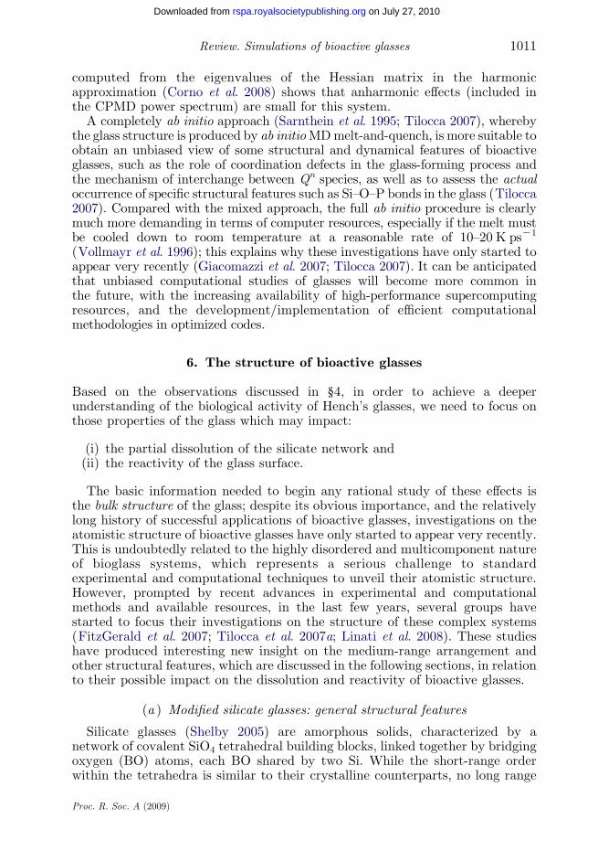
Figure 1. General scheme of the chemical structure of bioactive glasses; BOs are marked in red. Thefragment in (a) is a three-membered silicate chain, with no covalent links to the rest of the structure,whose dissolution will be relatively fast, compared with the fragment in (b): the latter is part of afive-membered ring and covalently cross-linked through the additional Si–O bonds coloured in blue.
A. Tilocca1012
on July 27, 2010rspa.royalsocietypublishing.orgDownloaded from
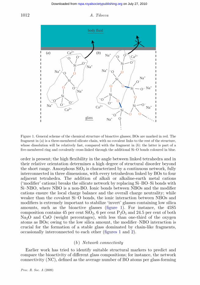
order is present; the high flexibility in the angle between linked tetrahedra and intheir relative orientation determines a high degree of structural disorder beyondthe short range. Amorphous SiO2 is characterized by a continuous network, fullyinterconnected in three dimensions, with every tetrahedron linked by BOs to fouradjacent tetrahedra. The addition of alkali or alkaline-earth metal cations(‘modifier’ cations) breaks the silicate network by replacing Si–BO–Si bonds withSi–NBO, where NBO is a non-BO. Ionic bonds between NBOs and the modifiercations ensure the local charge balance and the overall charge neutrality; whileweaker than the covalent Si–O bonds, the ionic interaction between NBOs andmodifiers is extremely important to stabilize ‘invert’ glasses containing low silicaamounts, such as the bioactive glasses (figure 1). For instance, the 45S5composition contains 45 per cent SiO2, 6 per cent P2O5 and 24.5 per cent of bothNa2O and CaO (weight percentages), with less than one-third of the oxygenatoms as BOs; owing to the low silica amount, the modifier–NBO interaction iscrucial for the formation of a stable glass dominated by chain-like fragments,occasionally interconnected to each other (figures 1 and 2).
(b ) Network connectivity
Earlier work has tried to identify suitable structural markers to predict andcompare the bioactivity of different glass compositions; for instance, the networkconnectivity (NC), defined as the average number of BO atoms per glass-forming
Proc. R. Soc. A (2009)
Figure 2. The silicate network of 45S5 Bioglass as obtained from SM MD (Tilocca 2008); Na andCa ions are not shown for clarity. Ball-and-stick visualization is used to highlight an individualsilicate chain fragment, with Si atoms coloured in green, and its interconnections to otherfragments, with Si atoms coloured in light blue.
1013Review. Simulations of bioactive glasses
on July 27, 2010rspa.royalsocietypublishing.orgDownloaded from
species, was introduced to describe the structural basis of the bioactivity (Strnad1992). For instance, NCZ4 for pure silica glass with its full three-dimensionalnetwork, whereas NCZ2 denotes chain-like structures, such as a linear polymer.By comparing the NC of glass compositions with different bioactivities(measured either as the rate of formation of the HA layer or as the bone-bonding ability), an empirical upper limit around NCZ3 was proposed,separating bioactive (NC!3) from bio-inactive (NCO3) glasses (Kim et al.1992; Strnad 1992). The qualitative interpretation of this threshold is based onthe correlation between solubility and NC; low NCs denote open and fragmentedglass structures, whose rapid partial dissolution in an aqueous physiologicalenvironment will lead to HA formation and bone bonding in a shorter time,compared with glasses with a more interconnected network. The empiricalobservation that glasses with NCO3 are bio-inactive entails that the hydrolysisof silica units with an average of three or more BOs bears an excessively highenergetic cost, which slows down the overall bioactive mechanism up to a pointwhere bone bonding can no longer be achieved (figure 1). As a reference, the NCfor the 45S5 composition would be 1.9 (Strnad 1992). However, while an usefulqualitative guideline, a classification based on the NC is not always accurate,as its predictive power rapidly decreases when a wider range of compositionsis considered (Hill 1996). The reason is that the NC estimated from theglass composition is based on the assumptions of regular coordination for allnetwork-forming ions and of homogeneous glass structure, which do notnecessarily hold true for these compositions (see below). A less crude estimateof the NC can be obtained by atomistic modelling, which enables the direct andunambiguous calculation of the average number of BOs per network-forming
Proc. R. Soc. A (2009)
A. Tilocca1014
on July 27, 2010rspa.royalsocietypublishing.orgDownloaded from
atom, as well as the analysis of the different network-forming ability/role ofdifferent species (Tilocca & Cormack 2007). This kind of analysis is based on thepremise that the theoretical framework (empirical potential and simulationset-up) is adequate and produces a reliable structural model of the glass,generally verified for the MD based on SM potentials. In fact, in the first attemptto model amorphous silicates using an SM approach, the comparison between thestructural models of soda- and soda-lime silicate glasses obtained using RI andSM potentials did show a better agreement with the experiment of the inter-tetrahedral structure predicted using the SM (Tilocca et al. 2006); in particular,considerable improvements were obtained in the Qn distribution (where Qn isa network-forming atom bonded to n BOs) of a common sodium silicatecomposition modelled with the SM potential. An accurate Qn distribution iscrucial, particularly when bioactive glasses are the target of the simulations,because, as discussed above, the dissolution process and the bioactivity of theseglasses are strongly affected by their structure in the medium range.
The application of the SM potential to model Hench’s soda-lime phospho-silicate glasses does confirm the reliability of this approach, which yields Qn
distributions of both Si and P in closer agreement with the availableexperimental data, compared with RI potentials (Tilocca 2008). It should beremarked that the current experimental picture of the Qn speciation in 45S5Bioglass is still somewhat incomplete; while a binary model with only Q2 and Q3
species is sometimes assumed (FitzGerald et al. 2007), a three-component modelwith Q1, Q2 and Q3 silicates was proposed to fit very recent nuclear magneticresonance (NMR) data (Linati et al. 2008), and the presence of more than twoQn(Si) species in this glass had already been inferred from previous Ramanspectra (Lin et al. 2005). Furthermore, given that the coexistence of severalQn(Si) species in depolymerized alkali silicate glasses is well established(Maekawa et al. 1991; Zhang et al. 1996; Mysen 2003), and that the presenceof other species besides Q1, Q2 and Q3 in 45S5 is less likely, the ternary Qn(Si)distribution as in the SM model of 45S5, centred on nZ2, appears mostadequate. While the 53 per cent fraction of Q2(Si) in the SM structure may stillbe slightly on the lower side with respect to the experiments, there is a clearimprovement with respect to other potentials; similar improvement is alsoobtained in the phosphate speciation (see below).
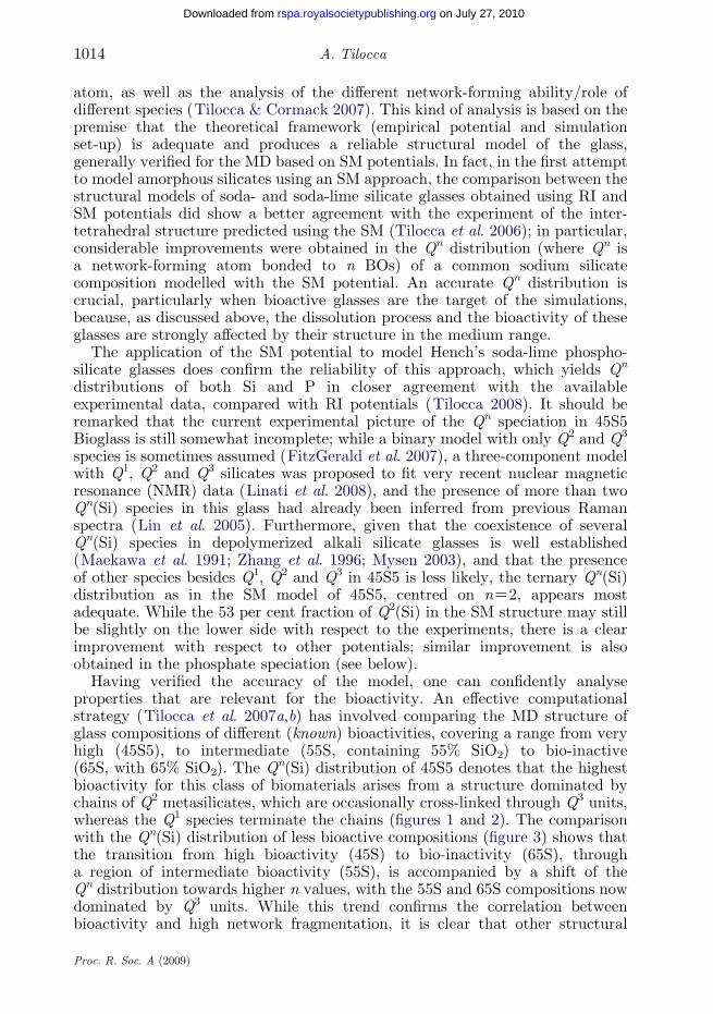
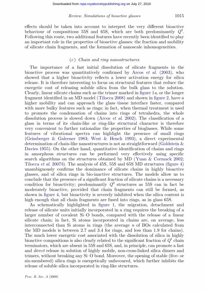
Having verified the accuracy of the model, one can confidently analyseproperties that are relevant for the bioactivity. An effective computationalstrategy (Tilocca et al. 2007a,b) has involved comparing the MD structure ofglass compositions of different (known) bioactivities, covering a range from veryhigh (45S5), to intermediate (55S, containing 55% SiO2) to bio-inactive(65S, with 65% SiO2). The Qn(Si) distribution of 45S5 denotes that the highestbioactivity for this class of biomaterials arises from a structure dominated bychains of Q2 metasilicates, which are occasionally cross-linked through Q3 units,whereas the Q1 species terminate the chains (figures 1 and 2). The comparisonwith the Qn(Si) distribution of less bioactive compositions (figure 3) shows thatthe transition from high bioactivity (45S) to bio-inactivity (65S), througha region of intermediate bioactivity (55S), is accompanied by a shift of theQn distribution towards higher n values, with the 55S and 65S compositions nowdominated by Q3 units. While this trend confirms the correlation betweenbioactivity and high network fragmentation, it is clear that other structural
Proc. R. Soc. A (2009)
1015Review. Simulations of bioactive glasses
on July 27, 2010rspa.royalsocietypublishing.orgDownloaded from
effects should be taken into account to interpret the very different bioactivebehaviour of compositions 55S and 65S, which are both predominantly Q3.Following this route, two additional features have recently been identified to playan important role in the properties of bioactive glasses: the fraction and mobilityof silicate chain fragments, and the formation of nanoscale inhomogeneities.
(c ) Chain and ring nanostructures
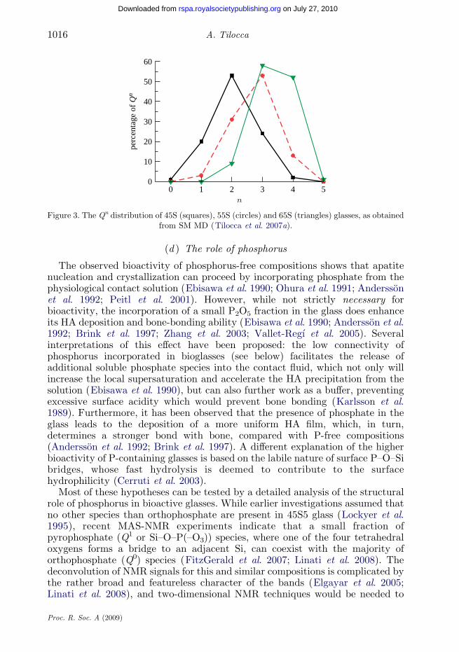
The importance of a fast initial dissolution of silicate fragments in thebioactive process was quantitatively confirmed by Arcos et al. (2003), whoshowed that a higher bioactivity reflects a lower activation energy for silicarelease. It is therefore interesting to focus on structural features that reduce theenergetic cost of releasing soluble silica from the bulk glass to the solution.Clearly, linear silicate chains such as the trimer marked in figure 1a, or the longerfragment identified in an MD model (Tilocca 2008) and shown in figure 2, have ahigher mobility and can approach the glass–tissue interface faster, comparedwith more bulky features such as rings; in fact, when thermal treatment is usedto promote the condensation of chains into rings of tetrahedra, the wholedissolution process is slowed down (Arcos et al. 2002). The classification of aglass in terms of its chain-like or ring-like structural character is thereforevery convenient to further rationalize the properties of bioglasses. While somefeatures of vibrational spectra can highlight the presence of small rings(Geissberger & Galeener 1983; West & Hench 1993), a direct experimentaldetermination of chain-like nanostructures is not as straightforward (Goldstein &Davies 1955). On the other hand, quantitative identification of chains and ringsin amorphous structures can be performed very effectively using specificsearch algorithms on the structures obtained by MD (Yuan & Cormack 2002;Tilocca et al. 2007b). The analysis of 45S, 55S and 65S MD structures (figure 4)unambiguously confirms the dominance of silicate chains in highly bioactiveglasses, and of silica rings in bio-inactive structures. The models allow us toconclude that the presence of a significant fraction of silicate chains is a necessarycondition for bioactivity; predominantly Q3 structures as 55S can in fact bemoderately bioactive, provided that chain fragments can still be formed, asshown in figure 4, but bioactivity is severely inhibited when the silica content ishigh enough that all chain fragments are fused into rings, as in glass 65S.
As schematically highlighted in figure 1, the migration, detachment andrelease of silicate units initially incorporated in a ring requires the breaking of alarger number of covalent Si–O bonds, compared with the release of a linearsilicate chain; in fact, Si atoms incorporated in chains are, on average, lessinterconnected than Si atoms in rings (the average n of BOs calculated fromthe MD models is between 2.7 and 3.4 for rings, and less than 1.9 for chains).The much lower energetic cost associated with the dissolution of silica in highlybioactive compositions is also clearly related to the significant fraction of Q1 chainterminators, which are absent in 55S and 65S, and, in principle, can promote a fastand direct release in solution of highly mobile, non-cross-linked silica dimers andtrimers, without breaking any Si–O bond. Moreover, the opening of stable (five- orsix-membered) silica rings is energetically unfavoured, which further inhibits therelease of soluble silica incorporated in ring-like structures.
Proc. R. Soc. A (2009)
0 1 2 3 4 50
10
20
30
40
50
60
perc
enta
ge o
f Q
n
Figure 3. The Qn distribution of 45S (squares), 55S (circles) and 65S (triangles) glasses, as obtainedfrom SM MD (Tilocca et al. 2007a).
A. Tilocca1016
on July 27, 2010rspa.royalsocietypublishing.orgDownloaded from
(d ) The role of phosphorus
The observed bioactivity of phosphorus-free compositions shows that apatitenucleation and crystallization can proceed by incorporating phosphate from thephysiological contact solution (Ebisawa et al. 1990; Ohura et al. 1991; Anderssonet al. 1992; Peitl et al. 2001). However, while not strictly necessary forbioactivity, the incorporation of a small P2O5 fraction in the glass does enhanceits HA deposition and bone-bonding ability (Ebisawa et al. 1990; Andersson et al.1992; Brink et al. 1997; Zhang et al. 2003; Vallet-Regı et al. 2005). Severalinterpretations of this effect have been proposed: the low connectivity ofphosphorus incorporated in bioglasses (see below) facilitates the release ofadditional soluble phosphate species into the contact fluid, which not only willincrease the local supersaturation and accelerate the HA precipitation from thesolution (Ebisawa et al. 1990), but can also further work as a buffer, preventingexcessive surface acidity which would prevent bone bonding (Karlsson et al.1989). Furthermore, it has been observed that the presence of phosphate in theglass leads to the deposition of a more uniform HA film, which, in turn,determines a stronger bond with bone, compared with P-free compositions(Andersson et al. 1992; Brink et al. 1997). A different explanation of the higherbioactivity of P-containing glasses is based on the labile nature of surface P–O–Sibridges, whose fast hydrolysis is deemed to contribute to the surfacehydrophilicity (Cerruti et al. 2003).
Most of these hypotheses can be tested by a detailed analysis of the structuralrole of phosphorus in bioactive glasses. While earlier investigations assumed thatno other species than orthophosphate are present in 45S5 glass (Lockyer et al.1995), recent MAS-NMR experiments indicate that a small fraction ofpyrophosphate (Q1 or Si–O–P(–O3)) species, where one of the four tetrahedraloxygens forms a bridge to an adjacent Si, can coexist with the majority oforthophosphate (Q0) species (FitzGerald et al. 2007; Linati et al. 2008). Thedeconvolution of NMR signals for this and similar compositions is complicated bythe rather broad and featureless character of the bands (Elgayar et al. 2005;Linati et al. 2008), and two-dimensional NMR techniques would be needed to
Proc. R. Soc. A (2009)
45S
55S
65S
ringschains
Figure 4. Chain and small ring fragments identified in structural models of 45S, 55S and 65Sglasses, as obtained from SM MD (Tilocca et al. 2007a,b).
1017Review. Simulations of bioactive glasses
on July 27, 2010rspa.royalsocietypublishing.orgDownloaded from
improve the spectral resolution, especially with a very low P2O5 fraction present(Coelho et al. 2006), but no such investigations have concerned Hench’sbioglasses as yet. The prevalence of P–O–Si over P–O–P linkages in mesoporousbioactive glasses was very recently highlighted through cross-polarization NMR(Leonova et al. 2008); this may point out that Si–O–P linkages actually replaceP–O–P in some of the signals assigned to pyro- and metaphosphate units inbioactive silicate glasses (Dupree et al. 1989). Therefore, while their exactamount is currently not known, the presence of a small fraction of non-orthophosphate species in Hench’s Bioglass appears now established.
How do the models describe this aspect? As already seen for the silicate, theSM potential also considerably improves the description of the phosphatespeciation, compared with standard fixed-charge potentials (Tilocca 2008).Owing to the low total number of P tetrahedra in common Hench’s compositions,
Proc. R. Soc. A (2009)
A. Tilocca1018
on July 27, 2010rspa.royalsocietypublishing.orgDownloaded from
the convergence of the Qn(P) distribution with the modelled system size is slowerthan for the Qn(Si). The latter is already well converged for Nw1500 atoms inthe simulation box (Tilocca et al. 2007a), whereas a converged Qn(P)distribution requires Nw10 000 atoms (Tilocca 2008). With the latter systemsize, the SM MD yields a fraction of orthophosphate units in 45S5 approximately82 per cent, whereas fixed-charge potentials lead to a much lower orthophosphatefraction, which are replaced by pyro- and even metaphosphate (Q2) species(Linati et al. 2008; Lusvardi et al. 2008; Tilocca 2008). An SM approach isthus required for a satisfactory description of the phosphate speciation andconnectivity, so that one can confidently analyse other structural propertiesextracted from the SM structure to rationalize the role of phosphorus inthese materials.
A systematic analysis of the structural changes induced by replacing SiO2 byP2O5 in bioactive compositions (Tilocca & Cormack 2007) has highlighted thatincreasing the P2O5 fraction leads to repolymerization of the silicate network.This is in agreement with experimental evidence (Lockyer et al. 1995) that wasinterpreted on the basis of the higher affinity of Na and Ca cations for phosphategroups, which are able to strip the modifier cations out of the silicate network,thus inducing its repolymerization. While the MD models do confirm the lattereffect, they also highlight an additional factor that leads to an increase inthe silicate connectivity upon phosphate inclusion: phosphorus can replace NaC
or Ca2C in balancing Si–NBO bonds. Therefore, the silicate networkrepolymerization can occur through either Si–O–Si or Si–O–P new links.A similar effect was also highlighted by Lusvardi et al. (2008), whose MDsimulations highlighted the formation of Si–O–P links when fluorine removes Naand Ca from the silicate network of bioactive glasses. Because a higher silicateNC inhibits the overall bioactive process, this would explain why substantialP2O5 fractions have a negative impact on the bioactivity (Hench 1998); however,the experiments discussed before also indicated that the inclusion of a small P2O5
amount enhances bioactivity. This remarkable inversion in the effect ofphosphorus can be explained by taking into account that, while some of theadded phosphorus forms P–O–Si links which reduce the bioactivity (negativeeffect), some other is found as free orthophosphate, whose relatively fast initialrelease accelerates the deposition of HA and boosts the bioactive process(positive effect). The balance between these opposite effects will ultimatelydetermine the bioactivity of the P-containing composition. Based on thebioactivity data of the compositions modelled, Tilocca & Cormack (2007)concluded that the positive effect prevails for low P2O5 fractions, whereas thenegative effect prevails for higher (above 10 mol% P2O5) fractions.
An additional feature highlighted by a statistical analysis of the MDsimulations is the increasingly stronger affinity between Ca and phosphategroups in glasses containing an increasing P2O5 fraction (Tilocca & Cormack2007; Tilocca et al. 2007a). The Ca–phosphate aggregation becomes increasinglystronger than the one between phosphate and Na ions, and for a 12 mol% P2O5
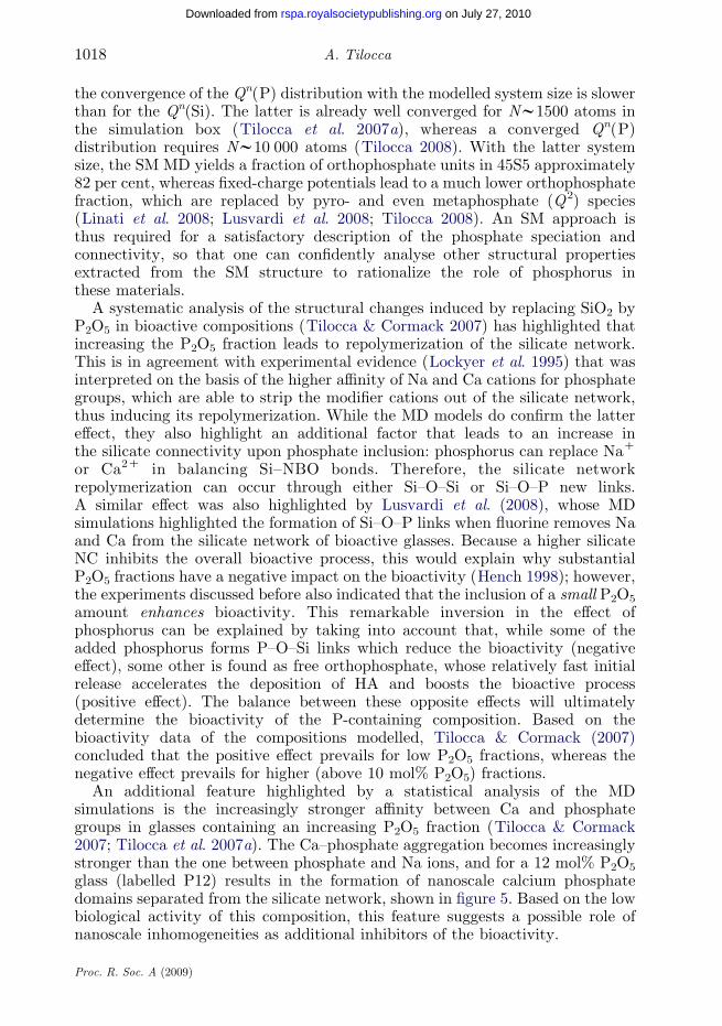
glass (labelled P12) results in the formation of nanoscale calcium phosphatedomains separated from the silicate network, shown in figure 5. Based on the lowbiological activity of this composition, this feature suggests a possible role ofnanoscale inhomogeneities as additional inhibitors of the bioactivity.
Proc. R. Soc. A (2009)
1019Review. Simulations of bioactive glasses
on July 27, 2010rspa.royalsocietypublishing.orgDownloaded from
(e ) Clustering and bioactivity
The association between nanoscale inhomogeneity and a reduction inbioactivity is interesting, given the widespread interest in controlling thedissolution rate of these compositions in order to match the natural growth rateof tissues, as discussed earlier. A possible explanation of this association is thereduced mobility of calcium and phosphate enclosed in these domains: glass-in-glass phase separation of a silica-rich and a phosphate-rich phase is known toenhance the resistance to dissolution of modified silicate glasses, owing to theincreased viscosity of the phase-separated glass (Wu et al. 1997; El-Ghannamet al. 2001). While realistic modelling of phase separation processes lies beyondthe current capabilities of atomistic MD simulations, these models are still ableto highlight a tendency of some compositions to develop local inhomogeneities(Huang & Cormack 1991; Lusvardi et al. 2005), which may well represent theprecursor of a phase-separated glass. The clustering of non-network-forming ionsin silicate glasses can be quantitatively explored by a statistical analysis based onthe ion coordination numbers calculated by MD simulations (Mead & Mountjoy2006; Tilocca & Cormack 2007; Lusvardi et al. 2008). Based on this kind ofanalysis, it can be shown that the distribution of modifier cations in bioactive45S5 is rather uniform, whereas bio-inactive 65S is characterized by markedclustering and formation of small calcium phosphate nanoaggregates (Tiloccaet al. 2007a), similar to the larger size inhomogeneities found in P12 glass.The presence of phosphorus appears important to induce the separation ofnanoaggregates, owing to the very favourable interaction with calcium ions(Tilocca & Cormack 2007, in press): phosphorus further enhances the tendencyof modifier cations to aggregate and concentrate in cluster regions, above whatwould be expected, for instance, on the basis of modified random network modelsof silicate glasses (Greaves 1985; Huang & Cormack 1991), which would predictclustering only for compositions containing a significantly lower fraction ofmodifier cations. In fact, recent MD simulations by Mead & Mountjoy (2006)have highlighted that calcium clustering does occur for P-free (CaO)x(SiO2)1Kx
glasses, but only for a silica fraction greater than 80 per cent, as opposedto only approximately 60 per cent SiO2 for the P-containing glasses (Tiloccaet al. 2007a).
A different driving force for nanosegregation in bioactive glasses has beenproposed by Lusvardi et al. (2008); their MD simulations showed that theincorporation of fluorine, which has high affinity for Na and Ca modifier cations,determines the separation of a highly polymerized phosphosilicate matrix froman ionic phase rich in Na, Ca and F. While fluorine-free bioglasses favour theseparation of a phosphate-rich phase from a silica-rich one, incorporation ofF leads instead to enhanced P–Si interactions, which are found in the samephase, well separated from an ionic phase with low viscosity.
Clustering of mobile cations in glasses is known to affect ionic transport, whichis thought to proceed through ion-conducting channels populated primarily bythese cations (Lammert & Heuer 2005). In the case of Hench’s bioglasses,clustering does not seem to promote ion migration, given the known reducedbioactivity of compositions with marked clustering. However, it is hard toseparate the possible contribution to this reduction owing to the moreinterconnected silicate network from the (presumed) lower ionic mobility in
Proc. R. Soc. A (2009)
Figure 5. Separation of nanoscale calcium phosphate domains in a 24.4 Na2O–27.1CaO–36.3 SiO2–12.2 P2O5 system (red, oxygen; blue, silicon; green, phosphorus; white, sodium;black, calcium). Snapshot extracted from the MD simulation (Tilocca & Cormack 2007).
A. Tilocca1020
on July 27, 2010rspa.royalsocietypublishing.orgDownloaded from
calcium phosphate clusters, and direct MD simulations of ionic migration wouldprobably represent the only way to assess the relative weight of these effects.
(f ) The bioactive interface
While establishing the relationship between bulk structure and bioactivity is akey starting point, it is at the glass surface that the bioactive process starts and iseventually completed, and a direct investigation of this region is thereforeessential. As for the bulk, molecular simulations can not only assist in theinterpretation of the results of surface analytical techniques (Cerruti & Sahai2006), but also provide a high-resolution probe into the features of the surface.Adsorption and reactivity at surfaces involves significant structural rearrange-ments, bond breaking and shifts in electronic density, whose modelling requiresab initio techniques. While ab initio cluster models can offer some interestinginsight into the local properties of hypothetical surface sites (Cerruti et al. 2005b;Cerruti & Sahai 2006), the activity of a realistic, extended model of the 45S5Bioglass dry surface has very recently been explored by CPMD, by examiningthe different ability of selected sites (such as two- and three-membered rings andcoordinative defects) to adsorb and dissociate a water molecule (Tilocca &Cormack 2008). A very interesting finding of these calculations is that theopening and hydroxylation of small rings exposed on the glass surface, whilethermodynamically favourable, is hindered by a significant kinetic barrier, whichmay result in a fraction of these rings to remain stable even after immersionof the glass in an aqueous environment. Therefore, the CPMD calculationsfully support the possibility that small silicate rings can provide active sitesfor calcium phosphate nucleation and deposition in the early stages of thebone-bonding mechanism (Sahai & Anseau 2005).
Proc. R. Soc. A (2009)
1021Review. Simulations of bioactive glasses
on July 27, 2010rspa.royalsocietypublishing.orgDownloaded from
7. Final remarks
The powerful combination of the atomistic insight provided by MD models withexperimental data on structure, solubility and bone-bonding ability of selectedcompositions enables a much deeper understanding of the bioactivity. Whileimportant correlations have already been identified between bulk structuralfeatures and the glass performance in biological applications, much more work isneeded towards a rationalization of the properties of these materials. A crucialshift in the focus of simulations, from the bulk glass to the processes occurring atthe interface between the glass and the physiological environment, can beenvisaged to lead to further progress. Ab initio models of gas-phase adsorptionhave already started to yield valuable information on the surface properties of45S5 bioactive glass (Tilocca & Cormack 2008) and of HA (Rimola et al. 2008).As the available computational power steadily grows, it will become possible tofurther extend these ab initio approaches to study the interface of bioactivesystems on a large scale, and thus complete the model with important effectssuch as full surface hydration and interactions with biomolecules.
A.T. is a Royal Society University Research Fellow. The author would like to acknowledgecollaboration and interaction with Prof. A. N. Cormack (Alfred University, USA) and Prof. N. H.de Leeuw (UCL), as well as fruitful exchanges with Dr A. Pedone (University of Modena andReggio Emilia, Italy).
References
Aguiar, H., Solla, E. L., Serra, J., Gonzalez, P., Leon, B., Malz, F. & Jager, C. 2008 Raman andNMR study of bioactive Na2O–MgO–CaO–P2O5–SiO2 glasses. J. Non-Cryst. Solids 354,5004–5008. (doi:10.1016/j.jnoncrysol.2008.07.033)
Anderson, J. M. 2006 The future of biomedical materials. J. Mater. Sci. Mater. Med. 17,1025–1028. (doi:10.1007/s10856-006-0439-5)
Andersson, O. H., Liu, G., Kangasniemi, J. & Juhanoja, J. 1992 Evaluation of the acceptance ofglass in bone. J. Mater. Sci. Mater. Med. 3, 145–150. (doi:10.1007/BF00705283)
Arcos, D., Greenspan, D. & Vallet-Regı, M. 2002 Influence of the stabilization temperature ontextural and structural features and ion release in SiO2–CaO–P2O5 sol–gel glasses. Chem.Mater. 14, 1515–1522. (doi:10.1021/cm011119p)
Arcos, D., Greenspan, D. & Vallet-Regı, M. 2003 A new quantitative method to evaluate thein vitro bioactivity of melt and sol–gel-derived silicate glasses. J. Biomed. Mater. Res. A 65,344–351. (doi:10.1002/jbm.a.10503)
Balas, F., Arcos, D., Perez-Pariente, J. & Vallet-Regı, M. 2001 Textural properties of SiO2 CaOP2O5 glasses prepared by the sol–gel method. J. Mater. Res. 16, 1345–1348. (doi:10.1557/JMR.2001.0187)
Boccaccini, A. R., Chen, W., Lefebvre, L., Gremillard, L. & Chevalier, J. 2007 Sintering,crystallisation and biodegradation behaviour of Bioglass-derived glass-ceramics. FaradayDiscuss. 136, 27. (doi:10.1039/b616539g)
Bouler, J.-M., LeGeros, R. Z. & Daculsi, G. 2000 Biphasic calcium phosphates: influence of threesynthesis parameters on the HA/b-TCP ratio. J. Biomed. Mater. Res. 51, 680–684. (doi:10.1002/1097-4636(20000915)51:4!680::AID-JBM16O3.0.CO;2-#)
Brink, M., Turunen, T., Happonen, R.-P. & Yli-Urpo, A. 1997 Compositional dependence ofbioactivity of glasses in the system Na2O–K2O–MgO–CaO–B2O3–P2O5–SiO2. J. Biomed.Mater. Res. 37, 114–121. (doi:10.1002/(SICI)1097-4636(199710)37:1!114::AID-JBM14O3.0.CO;2-G)
Proc. R. Soc. A (2009)
A. Tilocca1022
on July 27, 2010rspa.royalsocietypublishing.orgDownloaded from
Car, R. & Parrinello, M. 1985 Unified approach for molecular dynamics and density-functional
theory. Phys. Rev. Lett. 55, 2471–2474. (doi:10.1103/PhysRevLett.55.2471)Cerruti, M. & Sahai, N. 2006 Silicate biomaterials for orthopaedic and dental implants. Rev.
Minerol. Geochem. 64, 283. (doi:10.2138/rmg.2006.64.9)Cerruti, M., Magnacca, G., Bolis, V. & Morterra, C. 2003 Characterization of sol–gel bioglasses
with the use of simple model systems: a surface-chemistry approach. J. Mater. Chem. 13,1279–1286. (doi:10.1039/b300961k)
Cerruti, M., Greenspan, D. & Powers, K. 2005a Effect of pH and ionic strength on the reactivity ofBioglass 45S5. Biomaterials 26, 1665–1674. (doi:10.1016/j.biomaterials.2004.07.009)
Cerruti, M., Morterra, C. & Ugliengo, P. 2005b Surface features of P-doped silica explored with
CD3CN adsorption: can Si atoms act as Lewis centers? Chem. Mater. 17, 1416–1423. (doi:10.1021/cm0480420)
Charpentier, T., Ispas, S., Profeta, M., Mauri, F. & Pickard, C. J. 2004 First-principles calculationof 17O, 29Si, and 23Na NMR spectra of sodium silicate crystals and glasses. J. Phys. Chem. B108, 4147–4161. (doi:10.1021/jp0367225)
Cho, S. B., Miyaji, F., Kokubo, T., Nakanishi, K., Soga, N. & Nakamura, T. 1996 Apatite-formingability of silicate ion dissolved from silica gels. J. Mater. Sci. Mater. Med. 32, 375–381. (doi:10.
1002/(SICI)1097-4636(199611)32:3!375::AID-JBM10O3.0.CO;2-G)Cho, S. B., Miyaji, F., Kokubo, T., Nakanishi, K., Soga, N. & Nakamura, T. 1998 Apatite
formation on silica gel in simulated body fluid: effects of structural modification with solvent-exchange. J. Mater. Sci. Mater. Med. 9, 279–284. (doi:10.1023/A:1008808828567)
Clark, A. R., Pantano, C. G. & Hench, L. L. 1976 Auger spectroscopic analysis of Bioglasscorrosion films. J. Am. Ceram. Soc. 59, 37–39. (doi:10.1111/j.1151-2916.1976.tb09382.x)
Coelho, C., Babonneau, F., Azais, T., Bonhomme-Coury, L., Maquet, J., Laurent, G. &Bonhomme, C. 2006 Chemical bonding in silicophosphate gels: contribution of dipolar and
J-derived solid state NMR techniques. J. Sol–Gel Sci. Technol. 40, 181–189. (doi:10.1007/s10971-006-7431-x)
Cormack, A. N. & Du, J. 2001 Molecular dynamics simulations of soda-lime silicate glasses.J. Non-Cryst. Solids 293, 283–289. (doi:10.1016/S0022-3093(01)00831-6)
Corno, M., Pedone, A., Dovesi, R. & Ugliengo, P. 2008 B3LYP simulation of the full vibrationalspectrum of 45S5 bioactive silicate glass compared to v-silica. Chem. Mater. 20, 5610–5621.
(doi:10.1021/cm801164u)Day, R. M. 2005 Bioactive glass stimulates the secretion of angiogenic growth factors and
angiogenesis in vitro. Tissue Eng. 11, 768–777. (doi:10.1089/ten.2005.11.768)Donadio, D., Bernasconi, M. & Tassone, F. 2004 Photoelasticity of sodium silicate glass from first
principles. Phys. Rev. B 70, 214205. (doi:10.1103/PhysRevB.70.214205)Ducheyne, P., Hench, L. L., Kagan, A., Martens, M., Bursens, A. & Mulier, J. C. 1980 Effect of
hydroxyapatite impregnation on skeletal bonding of porous coated implants. J. Biomed. Mater.Res. 114, 225–237. (doi:10.1002/jbm.820140305)
Dupree, R., Holland, D., Mortuza, M. G., Collins, J. A. & Lockyer, M. W. G. 1989 Magic anglespinning NMR of alkali phospho-alumino-silicate glasses. J. Non-Cryst. Solids 112, 111–119.
(doi:10.1016/0022-3093(89)90504-8)Ebisawa, Y., Ohura, K., Kokubo, T. & Nakamura, T. 1990 Bioactivity of CaO–SiO2-based glasses:
in vitro evaluation. J. Biomed. Mater. Res. 1, 239–244.Elgayar, I., Aliev, A. E., Boccaccini, A. R. & Hill, R. 2005 Structural analysis of bioactive glasses.
J. Non-Cryst. Solids 351, 173–183. (doi:10.1016/j.jnoncrysol.2004.07.067)El-Ghannam, A., Hamazawy, E. & Yehia, A. 2001 Effect of thermal treatment on bioactive glass
microstructure, corrosion behavior, z potential, and protein adsorption. J. Biomed. Mater. Res.55, 387–395. (doi:10.1002/1097-4636(20010605)55:3!387::AID-JBM1027O3.0.CO;2-V)
FitzGerald, V., Pickup, D. M., Greenspan, D., Sarkar, G., Fitzgerald, J. J., Wetherall, K. M.,
Moss, R. M., Jones, J. R. & Newport, R. J. 2007 A neutron and X-ray diffraction study ofBioglassw with reverse Monte Carlo modelling. Adv. Funct. Mater. 17, 3746–3753. (doi:10.1002/adfm.200700433)
Proc. R. Soc. A (2009)
1023Review. Simulations of bioactive glasses
on July 27, 2010rspa.royalsocietypublishing.orgDownloaded from
Geissberger, A. E. & Galeener, F. L. 1983 Raman studies of vitreous SiO2 versus fictive
temperature. Phys. Rev. B 28, 3266–3271. (doi:10.1103/PhysRevB.28.3266)
Giacomazzi, L., Massobrio, C. & Pasquarello, A. 2007 First-principles investigation of the
structural and vibrational properties of vitreous GeSe2. Phys. Rev. B 75, 174 207. (doi:10.1103/
PhysRevB.75.174207)
Goldstein, M. & Davies, T. H. 1955 Glass fibers with oriented chain molecules. J. Am. Ceram. Soc.
38, 223–226. (doi:10.1111/j.1151-2916.1955.tb14935.x)
Greaves, G. N. 1985 EXAFS and the structure of glass. J. Non-Cryst. Solids 71, 203–217. (doi:10.
1016/0022-3093(85)90289-3)
Greaves, G. N. & Sen, S. 2007 Inorganic glasses, glass-forming liquids and amorphizing solids. Adv.
Phys. 56, 1–166. (doi:10.1080/00018730601147426)
Hench, L. L. 1998 Bioceramics. J. Am. Ceram. Soc. 81, 1705–1728. (doi:10.1111/j.1151-2916.1998.
tb02540.x)
Hench, L. L. & Andersson, O. H. 1993 Bioactive glasses. In An introduction to bioceramics (eds
L. L. Hench & J. Wilson), pp. 41–62. Republic of Singapore: World Scientific.
Hench, L. L. & Paschall, H. A. 1973 Direct chemical bond of bioactive glass-ceramic materials to
bone and muscle. J. Biomed. Mater. Res. Symp. 4, 25–42. (doi:10.1002/jbm.820070304)
Hench, L. L. & Polak, J. M. 2002 Third-generation biomedical materials. Science 295, 1014–1017.
(doi:10.1126/science.1067404)
Hench, L. L. & West, J. K. 1996 Biological applications of bioactive glasses. Life Chem. Rep. 13,
187–241.
Hench, L. L., Splinter, R. J., Allen, W. C. & Greenlee Jr, T. K. 1971 Bonding mechanisms at the
interface of ceramic prosthetic materials. J. Biomed. Mater. Res. 2, 117–141. (doi:10.1002/jbm.
820050611)
Hench, L. L., Stanley, H. R., Clark, A. E., Hall, M. & Wilson, J. 1991 Dental application of bioglass
implant. In Bioceramics, vol. 4 (eds E. Bonfield, G. W. Hastings & K. E. Tanner), pp. 232–238.
Oxford, UK: Butterworth Heinemann.
Hertz, A. & Bruce, I. J. 2007 Inorganic materials for bone repair or replacement applications.
Nanomedicine 2, 899–918. (doi:10.2217/17435889.2.6.899)
Hill, R. G. 1996 An alternative view of the degradation of bioglass. J. Mater. Sci. Lett. 15,
1122–1125. (doi:10.1007/BF00539955)
Hing, K. A. 2004 Bone repair in the twenty-first century: biology, chemistry or engineering? Phil.
Trans. R. Soc. A 362, 2821–2849. (doi:10.1098/rsta.2004.1466)
Horbach, J., Kob, W. & Binder, K. 1998 Specific heat of amorphous silica within the harmonic
approximation. J. Phys. Chem. B 103, 4104–4108. (doi:10.1021/jp983898b)
Huang, C. & Cormack, A. N. 1991 Structural differences and phase separation in alkali silicate
glasses. J. Chem. Phys. 95, 3634–3642. (doi:10.1063/1.460814)
Hulbert, S. F. 1993 The use of alumina and zirconia in surgical implants. In An introduction to
bioceramics (eds L. L. Hench & J. Wilson), pp. 25–40. London, UK: World Scientific.
Ispas, S., Benoit, M., Jund, P. & Jullien, R. 2001 Structural and electronic properties of the sodium
tetrasilicate glass Na2Si4O9 from classical and ab initio molecular dynamics simulations. Phys.
Rev. B 72, 214206. (doi:10.1103/PhysRevB.64.214206)
Jones, J. R., Tsigkou, O., Coates, E. E., Stevens, M. M., Polak, J. M. & Hench, L. L. 2007
Extracellular matrix formation and mineralization on a phosphate-free porous bioactive glass
scaffold using primary human osteoblast (HOB) cells. Biomaterials 28, 1653. (doi:10.1016/
j.biomaterials.2006.11.022)
Kalita, S. J., Bhardwaj, A. & Bhatt, H. A. 2007 Nanocrystalline calcium phosphate ceramics in
biomedical engineering. Mater. Sci. Eng. C 27, 441–449. (doi:10.1016/j.msec.2006.05.018)
Karlsson, K. H., Froerg, K. & Ringbom, T. 1989 A structural approach to bone adhering of
bioactive glasses. J. Non-Cryst. Solids 112, 69 . (doi:10.1016/0022-3093(89)90495-X)
Kerisit, S. & Parker, S. C. 2004 Free energy of adsorption of water and metal ions on the {1014}
calcite surface. J. Am. Chem. Soc. 126, 10 152–10 161. (doi:10.1021/ja0487776)
Proc. R. Soc. A (2009)

A. Tilocca1024
on July 27, 2010rspa.royalsocietypublishing.orgDownloaded from
Kim, C. Y., Clark, A. E. & Hench, L. L. 1992 Compositional dependence of calcium phosphate
layer formation in fluoride Bioglassesw. J. Biomed. Mater. Res. 26, 1147–1161. (doi:10.1002/jbm.820260905)
Knowles, J. C. 2003 Phosphate based glasses for biomedical applications. J. Mater. Chem. 13,2395–2401. (doi:10.1039/b307119g)
Kokubo, T. 1990 Surface chemistry of bioactive glass-ceramics. J. Non-Cryst. Solids 120, 138–151.(doi:10.1016/0022-3093(90)90199-V)
Kokubo, T., Kim, H.-M. & Kawashita, M. 2003 Novel bioactive materials with differentmechanical properties. Biomaterials 24, 2161–2175. (doi:10.1016/S0142-9612(03)00044-9)
Lammert, H. & Heuer, A. 2005 Contributions to the mixed-alkali effect in molecular dynamics
simulations of alkali silicate glasses. Phys. Rev. B 72, 214202. (doi:10.1103/PhysRevB.72.214202)
Leonova, E., Izquierdo-Barba, I., Arcos, D., Lopez-Noriega, A., Hedin, N., Vallet-Regı, M. & Eden,M. 2008 Multinuclear solid-state NMR studies of ordered mesoporous bioactive glasses. J. Phys.Chem. C 112, 5552–5562. (doi:10.1021/jp7107973)
Levy, S.,VanDalen,M.,Agonafer, S.&Soboyejo,W.O. 2007Cell/surface interactions andadhesion onbioactive glass 45S5. J. Mater. Sci. Mater. Med. 18, 89–102. (doi:10.1007/s10856-006-0666-9)
Li, P. & Zhang, F. 1990 The electrochemistry of a glass surface and its application to the bioactive
glass in solution. J. Non-Cryst. Solids 119, 112–118. (doi:10.1016/0022-3093(90)90247-J)Li, R., Clark, A. E. & Hench, L. L. 1991 An investigation of bioactive glass powders by sol–gel
processing. J. Appl. Biomater. 2, 231–239. (doi:10.1002/jab.770020403)Lin, C.-C., Huang, L.-C. & Shen, P. 2005 Na2CaSi2O6–P2O5 based bioactive glasses. Part 1:
elasticity and structure. J. Non-Cryst. Solids 351, 3195–3203. (doi:10.1016/j.jnoncrysol.2005.08.020)
Linati, L., Lusvardi, G., Malavasi, G., Menabue, L., Menziani, M. C., Mustarelli, P., Pedone, A. &Segre, U. 2008 Medium-range order in phospho-silicate bioactive glasses: insights from MAS-
NMR spectra, chemical durability experiments and molecular dynamics simulations. J. Non-Cryst. Solids 354, 84–89. (doi:10.1016/j.jnoncrysol.2007.06.076)
Lockyer, M. W. G., Holland, D. & Dupree, R. 1995 NMR investigation of the structure of somebioactive and related glasses. J. Non-Cryst. Solids 188, 207–219. (doi:10.1016/0022-3093(95)00188-3)
Lusvardi, G., Malavasi, G., Menabue, L., Menziani, M. C., Pedone, A. & Segre, U. 2005 Acomputational tool for the prediction of crystalline phases obtained from controlled
crystallization of glasses. J. Phys. Chem. B 109, 21 586–21 592. (doi:10.1021/jp0546857)Lusvardi, G., Malavasi, G., Cortada, M., Menabue, L., Menziani, M. C., Pedone, A. & Segre, U.
2008 Elucidation of the structural role of fluorine in potentially bioactive glasses byexperimental and computational investigation. J. Phys. Chem. B 112, 12 730–12 739. (doi:10.
1021/jp803031z)Machacek, J. & Gedeon, O. 2003 Structure of binary alkali silicate glasses—structural
modifications caused by various alkali ions. Phys. Chem. Glasses 44, 308–312.Maekawa, H., Maekawa, T., Kawamura, K. & Yokokawa, T. 1991 The structural groups of alkali
silicate glasses determined from 29Si MAS-NMR. J. Non-Cryst. Solids 127, 53–64. (doi:10.1016/0022-3093(91)90400-Z)
Malavasi, G., Menziani, M. C., Pedone, A., Civalleri, B., Corno, M. & Ugliengo, P. 2007 Acomputational multiscale strategy to the study of amorphous materials. Theor. Chem. Acc. 117,
933–942. (doi:10.1007/s00214-006-0214-1)Martınez, A., Izquierdo-Barba, I. & Vallet-Regı, M. 2000 Bioactivity of a CaO–SiO2 binary glasses
system. Chem. Mater. 12, 3080–3088. (doi:10.1021/cm001107o)Marx, D. & Hutter, J. 2000 Ab initio molecular dynamics: theory and implementation. In Modern
methods and algorithms of quantum chemistry, vol. 1 (ed. J. Grotendorst). NIC Series,pp. 301–449. Julich, Germany: John von Neumann Institute for Computing.
Mead, R. N. & Mountjoy, G. 2006 A molecular dynamics study of the atomic structure of(CaO)x(SiO2)1Kx glasses. J. Phys. Chem. B 110, 14 273–14 278. (doi:10.1021/jp0628939)
Proc. R. Soc. A (2009)
1025Review. Simulations of bioactive glasses
on July 27, 2010rspa.royalsocietypublishing.orgDownloaded from
Mysen, B. 2003 Physics and chemistry of silicate glasses and melts. Eur. J. Mineral. 15, 781–802.
(doi:10.1127/0935-1221/2003/0015-0781)
Nakamura, T., Yamamuro, T., Higashi, S., Kokubo, T. & Itoo, S. 1985 A new glass-ceramic for
bone replacement: evaluation of its bonding to bone tissue. J. Biomed. Mater. Res. 19, 685.
(doi:10.1002/jbm.820190608)
Ogino, M., Ohuchi, F. & Hench, L. L. 1980 Compositional dependence of the formation of
calcium phosphate films on bioglass. J. Biomed. Mater. Res. 14, 55 . (doi:10.1002/jbm.
820140107)
Ohura, K., Nakamura, T., Yamamuro, T., Kokubo, T., Ebisawa, Y., Kotoura, Y. & Oka, M. 1991
Bone-bonding ability of P2O5-free CaO–SiO2 glasses. J. Biomed. Mater. Res. 25, 357–365.
(doi:10.1002/jbm.820250307)
O’Sullivan, K. F. & Madden, P. A. 1991 Light scattering by alkali halides melts: a comparison of
shell-model and rigid-ion computer simulation results. J. Phys. Condens. Matter 3, 8751–8756.
(doi:10.1088/0953-8984/3/44/018)
Padilla, S., Roman, J., Carenas, A. & Vallet-Regı, M. 2005 The influence of the phosphorus
content on the bioactivity of sol–gel glass ceramics. Biomaterials 26, 475–483. (doi:10.1016/j.
biomaterials.2004.02.054)
Pedone, A., Corno, M., Civalleri, B., Malavasi, G., Menziani, M. C., Segre, U. & Ugliengo, P. 2007
An ab initio parameterized interatomic force field for hydroxyapatite. J. Mater. Chem. 17,
2061–2068. (doi:10.1039/b617858h)
Peitl, O., Zanotto, E. D. & Hench, L. L. 2001 Highly bioactive P2O5–Na2O–CaO–SiO2 glass-
ceramics. J. Non-Cryst. Solids 292, 115–126. (doi:10.1016/S0022-3093(01)00822-5)
Pereira, M. M., Clark, A. E. & Hench, L. L. 1994 Calcium phosphate formation on sol–gel-derived
bioactive glasses in vitro. J. Biomed. Mater. Res. 28, 693. (doi:10.1002/jbm.820280606)
Pohlmann, M., Benoit, M. & Kob, W. 2004 First-principles molecular-dynamics simulations of a
hydrous silica melt: structural properties and hydrogen diffusion mechanism. Phys. Rev. B 70,
184 209. (doi:10.1103/PhysRevB.70.184209)
Rimola, A., Corno, M., Zicovich-Wilson, C. M. & Ugliengo, P. 2008 Ab initio modeling of
protein/biomaterial interactions: glycine adsorption at hydroxyapatite surfaces. J. Am. Chem.
Soc. 130, 16 181–16 183. (doi:10.1021/ja806520d)
Rosen, J., Warschkow, O., McKenzie, D. R. & Bilek, M. M. M. 2007 Amorphous and crystalline
phases in thermal quench simulations of alumina. J. Chem. Phys. 126, 204709. (doi:10.1063/
1.2739538)
Sahai, N. & Anseau, M. 2005 Cyclic silicate active site and stereochemical match for apatite
nucleation on pseudowollastonite bioceramics–bone interfaces. Biomaterials 26, 5763–5770.
(doi:10.1016/j.biomaterials.2005.02.037)
Sarnthein, J., Pasquarello, A. & Car, R. 1995 Structural and electronic properties of liquid and
amorphous SiO2: an ab initio molecular dynamics study. Phys. Rev. Lett. 74, 4682–4685.
(doi:10.1103/PhysRevLett.74.4682)
Sepulveda, P., Jones, J. R. & Hench, L. L. 2002 In vitro dissolution of melt-derived 45S5 and sol–
gel derived 58S bioactive glasses. J. Biomed. Mater. Res. 61, 301–311. (doi:10.1002/jbm.10207)
Shelby, J. E. 2005 Introduction to glass science and technology, 2nd edn. London, UK: The Royal
Society of Chemistry.
Smith, W., Greaves, G. N. & Gillan, M. J. 1995 Computer simulation of sodium disilicate glass.
J. Chem. Phys. 103, 3091–3097. (doi:10.1063/1.470498)
Spangberg, D. & Hermansson, K. 2004 Many-body potentials for aqueous LiC, NaC, Mg2C, and
Al3C: comparison of effective three-body potentials and polarizable models. J. Chem. Phys. 120,
4829–4843. (doi:10.1063/1.1641191)
Strnad, Z. 1992 Role of the glass phase in bioactive glass-ceramics. Biomaterials 13, 317–321.
(doi:10.1016/0142-9612(92)90056-T)
Takada, A. & Cormack, A. N. 2008 Computer simulation models of glass structure. Phys. Chem.
Glasses 49, 127–135.
Proc. R. Soc. A (2009)
A. Tilocca1026
on July 27, 2010rspa.royalsocietypublishing.orgDownloaded from
Tilocca, A. 2007 Structure and dynamics of bioactive phosphosilicate glasses and melts from ab
initio molecular dynamics simulations. Phys. Rev. B 76, 224202. (doi:10.1103/PhysRevB.76.
224202)
Tilocca, A. 2008 Short- and medium-range structure of multicomponent bioactive glasses and
melts: an assessment of the performances of shell-model and rigid-ion potentials. J. Chem. Phys.
129, 084504. (doi:10.1063/1.2972146)
Tilocca, A. & Cormack, A. N. 2007 Structural effects of phosphorus inclusion in bioactive silicate
glasses. J. Phys. Chem. B 111, 14 256–14 264. (doi:10.1021/jp075677o)
Tilocca, A. & Cormack, A. N. 2008 Exploring the surface of bioactive glasses: water adsorption and
reactivity. J. Phys. Chem. C 112, 11 936–11 945. (doi:10.1021/jp803541j)
Tilocca, A. & Cormack, A. N. In press. In SIF Conference Proceedings.
Tilocca, A. & de Leeuw, N. H. 2006a Structural and electronic properties of modified sodium and
soda-lime silicate glasses by Car–Parrinello molecular dynamics. J. Mater. Chem. 16,
1950–1955. (doi:10.1039/b517362k)
Tilocca, A. & de Leeuw, N. H. 2006b Ab initio molecular dynamics study of 45S5 bioactive silicate
glass. J. Phys. Chem. B 110, 25 810–25 816. (doi:10.1021/jp065146k)
Tilocca, A., de Leeuw, N. H. & Cormack, A. N. 2006 Shell-model molecular dynamics calculations
of modified silicate glasses. Phys. Rev. B 73, 104209. (doi:10.1103/PhysRevB.73.104209)
Tilocca, A., Cormack, A. N. & de Leeuw, N. H. 2007a The structure of bioactive silicate glasses:
new insight from molecular dynamics simulations. Chem. Mater. 19, 95. (doi:10.1021/
cm061631g)
Tilocca, A., Cormack, A. N. & de Leeuw, N. H. 2007b The formation of nanoscale structures in
soluble phosphosilicate glasses for biomedical applications: MD simulations. Faraday Discuss.
136, 45–55. (doi:10.1039/b617540f)
Vallet-Regı, M., Ragel, C. V. & Salinas, A. J. 2003 Glasses with medical applications. Eur.
J. Inorg. Chem. 6, 1029–1043. (doi:10.1002/ejic.200390134)
Vallet-Regı, M., Salinas, A. J., Ramirez-Castellanos, J. & Gonzalez-Calbet, J. M. 2005
Nanostructure of bioactive sol–gel glasses and organic–inorganic hybrids. Chem. Mater. 17,
1874–1879. (doi:10.1021/cm047956j)
van Beest, B. W. H., Kramer, G. J. & Van Santen, R. A. 1990 Force fields for silicas and
aluminophosphates based on ab-initio calculations. Phys. Rev. Lett. 64, 1955–1958. (doi:10.
1103/PhysRevLett.64.1955)
Vessal, B., Greaves, G. N., Marten, P. T., Chadvick, A. V., Mole, R. & Houdewalter, S. 1992
Cation microsegregation and ionic mobility in mixed alkali glasses. Nature 356, 505–506.
(doi:10.1038/356504a0)
Vollmayr, K., Kob, W. & Binder, W. 1996 Cooling-rate effects in amorphous silica: a computer-
simulation study. Phys. Rev. B 54, 15 808–15 827. (doi:10.1103/PhysRevB.54.15808)
West, J. K. & Hench, L. L. 1993 Interactions of water with trisiloxane rings. I. Experimental
analysis. J. Non-Cryst. Solids 152, 101–108. (doi:10.1016/0022-3093(93)90239-T)
Wilson, J. & Low, S. B. 1992 Bioactive ceramics for periodontal treatment: comparative studies in
the Patus monkey. J. Appl. Biomater. 3, 123–169. (doi:10.1002/jab.770030208)
Wilson, J., Yli-Urpo, A. & Risto-Pekka, H. 1993 Bioactive glasses: clinical applications. In An
introduction to bioceramics (eds L. L. Hench & J. Wilson), pp. 63–74. Republic of Singapore:
World Scientific.
Wilson, J., Douek, E. & Rust, K. 1995 Bioglass middle ear devices: ten year clinical results. In
Bioceramics, vol. 8 (eds J. Wilson, L. L. Hench & D. Greenspan), pp. 239–246. Oxford, UK:
Pergamon/Elsevier.
Woodcock, L. V., Angell, C. A. & Cheeseman, P. 1976 Molecular-dynamics studies of
vitreous state—simple ionic systems and silica. J. Chem. Phys. 65, 1565–1577. (doi:10.1063/
1.433213)
Wu,H.-F., Lin, C.-C.&Shen, P. 1997 Structure and dissolution ofCaO–ZrO2–TiO2–Al2O3–B2O3–SiO2
glass (II). J. Non-Cryst. Solids 209, 76–86. (doi:10.1016/S0022-3093(96)00553-4)
Proc. R. Soc. A (2009)
1027Review. Simulations of bioactive glasses
on July 27, 2010rspa.royalsocietypublishing.orgDownloaded from
Xynos, I. D., Hukkanen, M. V. J., Batten, J. J., Buttery, L. D., Hench, L. L. & Polak, J. M. 2000Bioglassw 45S5 stimulates osteoblast turnover and enhances bone formation in vitro:implications and applications for bone tissue engineering. Calcif. Tissue Int. 67, 321–329.(doi:10.1007/s002230001134)
Xynos, I. D., Edgar, A. J., Buttery, L. D. K., Hench, L. L. & Polak, J. M. 2001 Gene-expressionprofiling of human osteoblasts following treatment with the ionic products of Bioglassw 45S5dissolution. J. Biomed. Mater. Res. 55, 151–157. (doi:10.1002/1097-4636(200105)55:2!151::AID-JBM1001O3.0.CO;2-D)
Yamamuro, T. 1990 Reconstruction of the iliac crest with bioactive glass-ceramic prostheses. InHandbook of bioactive ceramics: 1. Bioactive glasses and glass-ceramics (eds T. Yamamuro,L. L. Hench & J. Wilson), pp. 335–342. Boca Raton, FL: CRC Press.
Yamamuro, T. 1993 A/W glass-ceramic: clinical applications. In An introduction to bioceramics(eds L. L. Hench & J. Wilson), pp. 89–104. Republic of Singapore: World Scientific.
Yu, H. & van Gunsteren, W. F. 2005 Accounting for polarization in molecular simulations. Comp.Phys. Commun. 172, 69–85. (doi:10.1016/j.cpc.2005.01.022)
Yuan, X. & Cormack, A. N. 2001 Local structures of MD-modeled vitreous silica and sodiumsilicate glasses. J. Non-Cryst. Solids 283, 69–87. (doi:10.1016/S0022-3093(01)00363-5)
Yuan, X. & Cormack, A. N. 2002 Efficient algorithm for primitive ring statistics in topologicalnetworks. J. Non-Cryst. Solids 283, 69–87. (doi:10.1016/S0022-3093(01)00363-5)
Zhang, P., Dunlap, P., Florian, P., Grandinetti, P. J., Farnan, I. & Stebbins, J. F. 1996 Silicon sitedistributions in an alkali silicate glass derived by two-dimensional 29Si nuclear magneticresonance. J. Non-Cryst. Solids 204, 294–300. (doi:10.1016/S0022-3093(96)00601-1)
Zhang, K., Yan, H., Bell, D. C., Stein, A. & Francis, L. F. 2003 Effects of materials parameters onmineralization and degradation of sol–gel bioactive glasses with 3D-ordered macroporousstructures. J. Biomed. Mater. Res. A 66, 860–869. (doi:10.1002/jbm.a.10093)
Proc. R. Soc. A (2009)


![Estimating and interpreting structural equation models … · Estimating and interpreting structural equation models in Stata 12 ... and Var [ǫ] = Σ sem (y1 ... Structural equation](https://img.pdfslide.us/doc/110x75/5b286e167f8b9ae8108b4592/estimating-and-interpreting-structural-equation-models-estimating-and-interpreting.jpg)