Embed Size (px)
Citation preview

Scientific Report
Structural basis for polyspecificity in the POT familyof proton-coupled oligopeptide transportersJoseph A Lyons1,†,‡, Joanne L Parker2,‡, Nicolae Solcan2, Alette Brinth1, Dianfan Li1, Syed TA Shah1,
Martin Caffrey1,** & Simon Newstead2,*
Abstract
An enigma in the field of peptide transport is the structural basis forligand promiscuity, as exemplified by PepT1, the mammalian plasmamembrane peptide transporter. Here, we present crystal structuresof di- and tripeptide-bound complexes of a bacterial homologue ofPepT1, which reveal at least two mechanisms for peptide recognitionthat operate within a single, centrally located binding site. Thedipeptide was orientated laterally in the binding site, whereasthe tripeptide revealed an alternative vertical binding mode. Theco-crystal structures combined with functional studies reveal thatbiochemically distinct peptide-binding sites likely operate within thePOT/PTR family of proton-coupled symporters and suggest thattransport promiscuity has arisen in part through the ability of thebinding site to accommodate peptides in multiple orientations fortransport.
Keywords crystallography; major facilitator superfamily; membrane protein;
peptide binding site; POT/PTR family
Subject Category Membrane & Intracellular Transport
DOI 10.15252/embr.201338403 | Received 21 December 2013 | Revised 22 April
2014 | Accepted 12 May 2014 | Published online 10 June 2014
EMBO Reports (2014) 15: 886–893
Introduction
Membrane transporters involved in nutrient uptake are usually
substrate specific, recognizing and transporting only chemically very
similar ligands across the membrane [1]. However, a number of
mammalian nutrient transporters also recognize and transport a vari-
ety of drug molecules, in addition to their intended nutrients, with
important implications for drug pharmacokinetics and drug–drug
interactions [2]. These include the organic anion-transporting poly-
peptides (OATPs), the organic anion and cation transporters (OAT
and OCT) and the proton-coupled peptide transporters (POT/PTR
family) [3,4]. The molecular basis for this promiscuity or polyspeci-
ficity is an important biochemical question that must be addressed if
their role in drug transport is to be understood and usefully exploited.
The human POT family transporters, PepT1 (SLC15A1) and PepT2
(SLC15A2), are responsible for peptide transport across the plasma
membrane and have evolved one of the most promiscuous binding
sites in biology, capable of recognizing and transporting over 8,000
different di- and tripeptide ligands [5]. They are of increasing
pharmaceutical importance as they recognize and transport a growing
library of antibiotics, antiviral and anticancer molecules [6]. The POT
(or PTR) family belongs to the major facilitator superfamily (MFS) of
secondary active transporters that includes the OATPs, OATs and
OCTs [7–9]. The POT/PTR family contains a number of conserved
sequence motifs (Supplementary Fig S1) and a conserved architecture
consisting of 12 or 14 transmembrane a-helices arranged into
N (H1–6)- and C-terminal (H7–12) bundles [3,10–12]. A centrally
located peptide-binding site is highly conserved between prokaryote
and eukaryote members, with both operating via similar transport
mechanisms [13]. Conserved pairs of salt bridge interactions coordi-
nate structural rearrangements that lead to the vectorial co-transport
of peptides and protons across the membrane [3,11].
A key question we sought to address was how one binding site
could specifically recognize and transport such a large number of
different ligands, while retaining specificity for peptides only two
or three amino acids long. To address this question, we deter-
mined high-resolution crystal structures of a bacterial homologue
to the mammalian PepT1 protein from Streptococcus thermophilus,
PepTSt in complex with physiologically relevant di- and tripeptides
in addition to a peptide-free apo form (Table 1). POT family trans-
porters are understood to transport both di- and tripeptides [13].
However, for PepTSt and other bacterial members of the family,
there exists some selectivity for different di- and tripeptides
[14,15]. We previously showed that PepTSt displays broad specific-
ity for dipeptides, being able to transport a range of charged and
hydrophobic peptides with IC50 values in the region of 5–400 lM[3]. Tripeptides are also transported, with tri-alanine being the best
peptide tested to date with an IC50 value comparable to that of the
dipeptides (Supplementary Fig S2). PepTSt therefore represents a
good model with which to study the structural basis for substrate
selectively within the POT family. The current structures and func-
tional data reveal a binding site that can accommodate peptides in
different orientations that helps to explain the broad substrate
1 Schools of Medicine and Biochemistry & Immunology, Trinity College Dublin, Dublin, Ireland2 Department of Biochemistry, University of Oxford, Oxford, UK
*Corresponding author. Tel: +44 1865613319; Fax: +44 1865 613201; E-mail: [email protected]**Corresponding author. Tel: +353 18964253; Fax: +353 18964257; E-mail: [email protected]‡Authors contributed equally to study.†Present address: Department of Molecular Biology and Genetics, Aarhus University, Aarhus, Denmark
EMBO reports Vol 15 | No 8 | 2014 ª 2014 The Authors. Published under the terms of the CC BY 4.0 license886
Published online: June 10, 2014
specificity characteristic of this pharmaceutically relevant trans-
porter family.
Results and Discussion
Physiological dipeptide complex
A co-crystal structure with the natural dipeptide L-Ala-L-Phe (Ala-Phe)
was obtained using the in meso crystallization method [16] and
refined to a maximum resolution of 2.5 A (Materials and Methods,
Table 1 and Supplementary Fig S3). The peptide was clearly visible
in the mFo-DFc difference electron density maps and sits laterally
in the binding site (Fig 1A and B). The Ala-Phe peptide is held by
electrostatic interactions between its amino and carboxy termini and
side chains extending from both the N- and C-terminal bundles
(Fig 1B). This binding mode is similar to the previous POT family
homologues co-crystallized with the antibacterial phosphono-
peptide, alafosfalin [11,12] (Supplementary Fig S4). The peptide
amino terminus interacts with a conserved glutamate (Glu400) on
Table 1. Data collection and refinement statistics
PepTSt apo PepTSt AAAc PepTSt AF
Data collection
Space group C2221 C2221 C2221
Cell dimensions
a, b, c (Å) 102.2, 110.2, 111.0 103.4, 110.7, 110.6 102.1, 110.3, 110.7
a, b, c (°) 90.0, 90.0, 90.0 90.0, 90.0, 90.0 90.0, 90.0, 90.0
Wavelength (Å) 0.9686 1.0332 0.9686
Resolution (Å)a 39.1–2.35 (2.48–2.35) 75.56–2.52 (2.59–2.52) 51.03–2.47 (2.53–2.47)
CC1/2(%)b 98.6 (63.4) 99.6 (74.6) 99.8 (53.0)
Rmerge 15.8 (71.0) 8.0 (56.8) 7.3 (64.0)
Rpim 6.1 (37.7) 5.2 (37.0) 4.6 (40.8)
I/rI 8.3 (2.2) 10.4 (2.2) 10.9 (2.0)
Completeness (%) 99.3 (98.0) 98.7 (99.3) 97.9 (99.4)
Redundancy 6.0 (4.1) 3.2 (3.2) 4.1 (4.2)
Number of crystals 7 1 1
Refinement
Resolution (Å) 39.1–2.35 75.56–2.52 51.04–2.47
Number of reflections 26,224 21,332 22,139
Rwork/Rfree 21.3/24.2 19.8/24.2 22.2/26.6
Number of atoms
Protein 3,440 3,582 3,321
Ligand N/A 16 17
Lipid 198 132 132
Ions 5 5 5
Water 54 45 38
B-factors (Å2)
Protein 38.4 49.8 39.7
Ligand N/A 75.9c 62.8
Lipid 54.8 61.4 54.0
Ions 56.4 69.6 61.7
Water 34.2 44.1 36.9
R.m.s deviations
Bond lengths (Å) 0.006 0.006 0.006
Bond angles (°) 0.913 0.915 0.879
Ramachandran statistics favoured/outliers (%) 99.0/0 98.3/0.2 99.0/0/0
aHighest resolution shell is shown in parenthesis.bPercentage of correlation between intensities from random half-datasets, as given by XDS.cPepTSt tri-Ala complex was deposited with the carboxy terminus of the tripeptide oriented towards the extracellular side of the membrane.
ª 2014 The Authors EMBO reports Vol 15 | No 8 | 2014
Joseph A Lyons et al Structural basis for polyspecificity in the POT family EMBO reports
887
Published online: June 10, 2014
H10 and through a hydrogen bond to an asparagine (Asn328) on
H8. These residues are essential for binding and transport of
peptides in PepTSt [3]. The amide nitrogen does not interact with
the binding site, consistent with previous predictions that only the
carbonyl group of the peptide bond is recognized in PepT1. The
carbonyl group of the peptide bond is coordinated to a conserved
asparagine (Asn156) on H5, forming a ligand-coordinated bridge
between the two six-helix bundles. The C-terminal bundle in PepTSt
appears to be more flexible than the opposing N-terminal bundle, as
evidenced by the atomic displacement parameters (Fig 1C), and
consistent with this region undergoing more structural change
during transport.
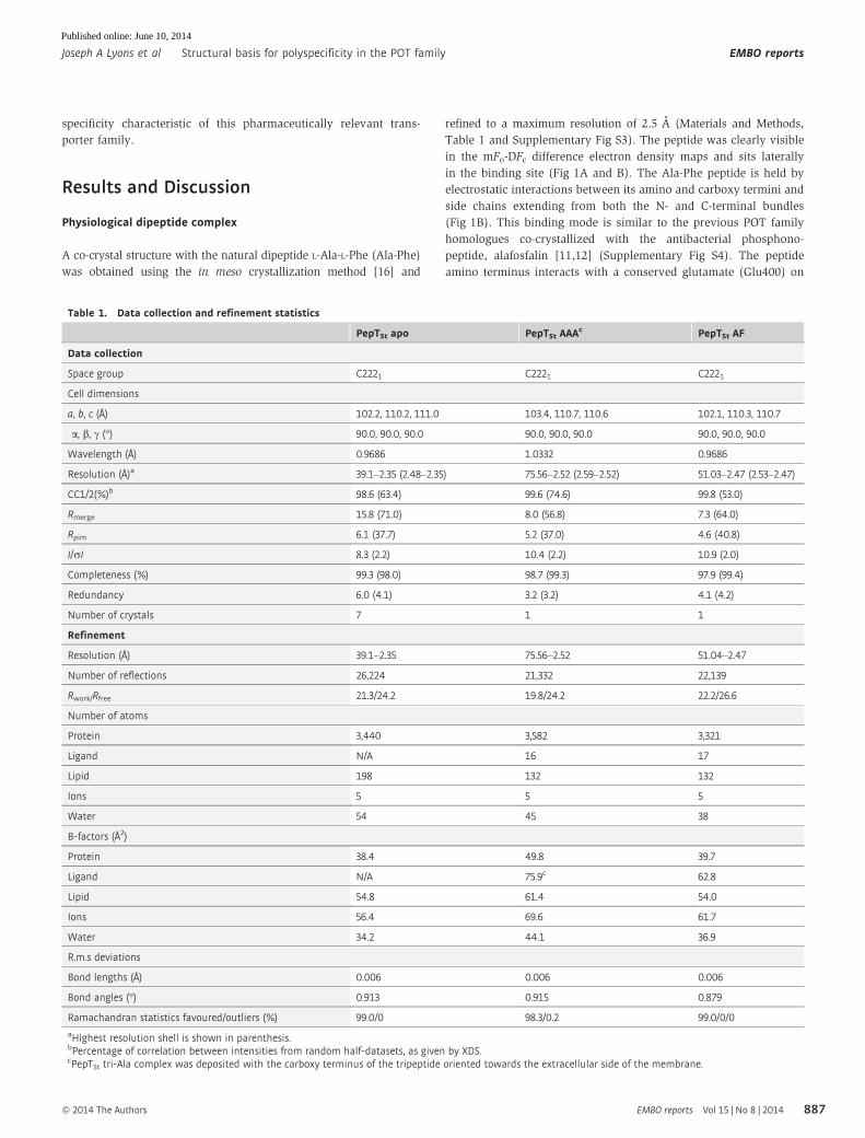
Interestingly, the phenyl ring of the peptide is accommodated
in a previously unobserved hydrophobic pocket (pocket 1)
approximately 10 × 10 × 3 A in size and formed by side chains
from H2 (Tyr68), H7 (Trp296) and H11 (Trp427, Phe428, Ser431)
(Fig 1D). The presence of such a pocket was predicted previously
based on transport and modelling data in PepT1 [7]. The side chain
of Tyr68 is positioned 3.9 A away, making a pi–pi stacking interac-
tion with the phenyl group. The close interaction of Tyr68 with the
dipeptide is also consistent with our previous study on PepTSt
showing that Tyr68 plays an important role in determining
dipeptide specificity [3]. At the N-terminal end of the ligand,
another elongated cavity, pocket 2, with dimensions approximately
16 × 7 × 11 A, is present that may accommodate a larger side chain
at this position (Supplementary Fig S5). The C-terminus of the
ligand interacts with both a conserved arginine on H1 (Arg26) and a
lysine on H4 (Lys126). High-affinity ligands of PepT1 share a
A B
C D
90o
H2H11
H7
W427
F428
W296
Y68S431
Hydrophobicpocket
C N
Ala-Phepeptide
7.8 MAGlipid
Extracellular
Intracellular
42 Ao
C
N
H11
E400
N328
N156
R26
K126
Y68 F428W427
S431
W296 3.0 A
3.0 A
3.2 A3.2 A
3.6 A
H2 H4
H1
H5
H8H10
H7
Ala-Phe
Figure 1. Structure of PepTSt with Ala-Phe dipeptide.
A Side on view of PepTSt with the 7.8 MAG lipid shown in spheres. The mFo-DFc difference electron density map (green) is shown, contoured at 3.0 r.B Extracellular view of the peptide-binding site with hydrogen bonds as dashed lines.C Flexibility in the C-terminal domain indicated by the atomic displacement-coded putty thickness and colour gradient from blue (low disorder) to red (high disorder).
The average atomic displacement parameter for the protein is 39.7 Å2.D View into the conserved hydrophobic pocket accommodating the ligand’s phenylalanine side chain in the peptide.
EMBO reports Vol 15 | No 8 | 2014 ª 2014 The Authors
EMBO reports Structural basis for polyspecificity in the POT family Joseph A Lyons et al
888
Published online: June 10, 2014

common feature of having their N- and C-terminal groups separated
by approximately 6 A [17]. Our structure suggests this requirement
is due to the positioning of these groups between the previously
observed dipole [3,10] within the binding site to orientate peptides
laterally (Fig 1B). Tripeptides by contrast are considerably longer
than dipeptides, approximately 10 A fully extended. Based on the
results to date, it was clear that extended tripeptides could not be
accommodated in the same way as the Ala-Phe ligand. We specu-
lated that tripeptides would therefore be recognized differently,
with important implications for understanding the transport of
b-lactam antibiotics, which are similar to tripeptides in size
(Supplementary Fig S6).
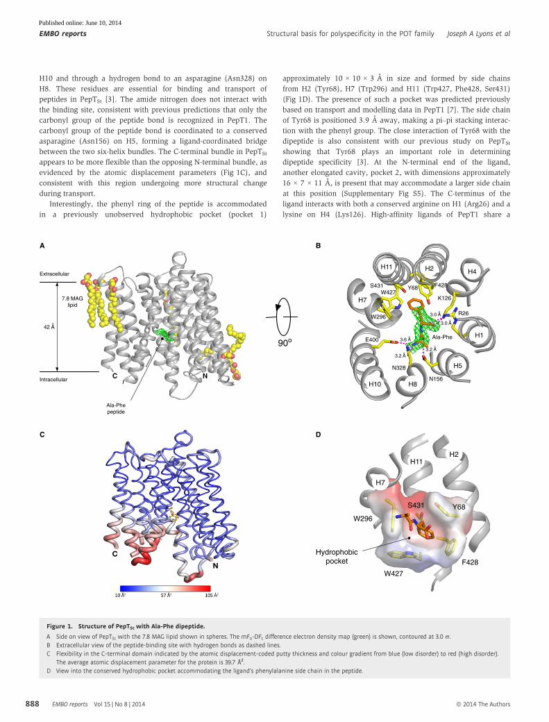
Peptides can also bind in a vertical orientation
A new crystal structure in complex with the tripeptide L-Ala-L-Ala-L-
Ala (tri-Ala), which was the only tested tripeptide to inhibit di-Ala
transport (Supplementary Fig S2), was obtained and refined to a
final resolution of 2.5 A (Materials and Methods and Table 1).
Careful analyses of the electron density maps, including the calcula-
tion of simulated annealing OMIT maps and averaged kick maps
[18], clearly showed the tri-Ala peptide sitting in a vertical orienta-
tion (Fig 2A and Supplementary Fig S7). The data, however, do not
allow us to establish how the peptide is orientated in the binding
site, with its C-terminus facing towards the cytoplasmic or periplas-
mic space. This ambiguity arises due to ill-defined electron density
that likely reflects low occupancy of the peptide in the binding site.
For the purpose of the discussion that follows, we focus on the
peptide modelled with the C-terminus facing the periplasm. This
choice is based on functional data for peptide recognition in PepT1
that suggests the C-terminus of the peptide interacts with residues at
the extracellular entrance to the binding site [19]. Compared to the
Ala-Phe complex, tri-Ala makes far fewer interactions with the
protein. This explains, in part, the disparate affinities of PepTSt for
di- and tripeptides, and unlike the dipeptide, the tri-Ala may be
protonated as it sits within hydrogen bonding distance of both the
backbone carbonyl groups of Glu299 and Glu300 on H7 and the
backbone amide nitrogen of Ser303. The tripeptide sits in an
H1 H7
H11
R33
Y30Y29
R26
S303
E300
E400
Ala-Ala-Ala
3.4 Ao
3.4 Ao
3.4 Ao
E299
3.5 Ao
2.9 Ao
A B
C D E
100 101 102 1030
50
100
Ala-Ala [µM]
% re
sidu
al 3 H
-Ala
Ala
trans
port WT: 37 µM
Y29F: 40 µMN156A: 86 µMW427F: 35 µM
101 102 103 1040
50
100
Ala-Ala-Ala [µM]
% re
sidu
al 3 H
-Ala
Ala
trans
port WT: 340 μM
Y29F: 1.4 mMN156A: 345 μM
W427F: 98 μM
Extracellular
Intracellular
WTR26KY29FY30FR33AY68F
N156AE300DS303AN328DE400D
W427FS431A
0
100
200
300
nmol
es/m
g Pe
pTSt
Figure 2. Tri-alanine binds to PepTSt in a vertical orientation.
A View of the binding site in the plane of the membrane, showing the mFo-DFc difference electron density map, contoured at 3.0 r. Hydrogen bonds are shown asdashed lines. In this panel, the peptide was modelled with the C-terminus orientated towards the apex of the cavity.
B Electrostatic surface representation of the binding site in the Ala-Phe complex structure with both peptides superimposed illustrating the relative orientations. Theside chains of key residues involved in peptide binding are outlined.
C Effect of binding site mutations on proton-driven di-alanine uptake in liposomes.D IC50 competition curves for di-alanine.E IC50 competition curves for tri-alanine showing residual uptake of [3H]-di-Ala peptide normalized to WT.
Data information: Error bars indicate the standard deviation from three independent experiments.
ª 2014 The Authors EMBO reports Vol 15 | No 8 | 2014
Joseph A Lyons et al Structural basis for polyspecificity in the POT family EMBO reports
889
Published online: June 10, 2014

elongated cavity formed by H1 (Tyr30), H5 (Asn156), H7 (Glu299,
300) and H8 (Gln325, Asn328) and above the alanine side chain
observed in the Ala-Phe complex (Fig 2B). The extracellular end of
the cavity is sealed by the packing together of H1 and H7, which
form the extracellular gate in the POT family [10]. Another notice-
able difference is the presence of a third smaller cavity (pocket 3)
that opens up opposite the third side chain in the tri-Ala peptide as
a result of the less compact structure adopted by PepTSt in the tri-
Ala complex compared to the Ala-Phe complex. The side chains of
Glu299 and Glu300 are within hydrogen bond distance to the
carbonyl of the C-terminal peptide bond. Glu299, however, is not
conserved within the wider POT family; it is a phenylalanine in
PepT1 for example (Supplementary Fig S1). It seems reasonable that
the interaction with tripeptides in this region will differ between
homologues, further explaining the differences observed in
substrate specificity with respect to di- and tripeptides discussed
above [14]. The nitrogen of the C-terminal peptide bond in tri-Ala
peptide interacts with the side chain of Tyr30. This aromatic residue
forms part of the highly conserved E22xxERFxYY motif on H1 and
was previously shown to play a role in proton binding [3,11]. The
N-terminal peptide carbonyl is within hydrogen bond distance to
Glu400. As modelled, the amino terminus of the tri-Ala peptide
makes no interaction within the binding site. This is consistent with
previous studies using PepT2 where the C-terminus of tripeptides
was shown to be more important in tripeptide versus dipeptide
transport (reviewed in [20]).
Binding modes are functionally distinct
The PepTSt complex structures in this study show that although the
di- and tripeptides are positioned differently in the binding site, the
first two side chains of both ligands are similarly orientated
(Fig 2B). If PepTSt can bind peptides in these two orientations, we
should be able to identify mutations that selectively affect only
peptides recognized in these two modes. To investigate this possi-
bility, residues observed interacting with the two peptides were
mutated and functionally tested using a proton-driven peptide
uptake assay [3] (Fig 2C). Aside from Tyr68, already discussed,
three further residues were identified that interacted with the
peptides but that did not result in reduced overall transport upon
substitution; they included Tyr29Phe, Asn156Ala and Trp427Phe.
In the di-Ala uptake study, these variants showed similar IC50
values, with one exception; Asn156Ala showed a small but signifi-
cant decrease in affinity (86 lM compared to 37 lM for the WT
transporter) (Fig 2D). This result is consistent with complex struc-
ture where Asn156 forms a hydrogen bond to the carbonyl group of
the Ala-Phe peptide (Fig 1B and D). By comparison, the same
Asn156Ala variant had no effect on tri-Ala competition (Fig 2E),
consistent with the complex structure where Asn156 does not inter-
act with the tri-Ala peptide (Fig 2A and Supplementary Fig S7).
However, Tyr29Phe did show reduced competition (IC50 of 1.4 mM
versus 340 lM for the WT) consistent with both Tyr30 and Tyr29
being important in tripeptide uptake [3]. Interestingly, Trp427Phe
showed increased affinity for tri-Ala compared to WT (IC50 98 lM).
Our structures do not provide an obvious explanation as to why
reducing the size of this side chain would result in an increased
affinity for tri-Ala peptide, but as discussed below this may indicate
that other binding modes for tri-Ala exist within the binding site
that are not revealed by our structures. However, the observation
that different binding site mutants differentially affect substrate
specificity further highlights the likelihood of multiple binding
modes.
A B
Intracellular
C
E300
Y68
Ala-Phe
F428
W427
S431
H11
H7
H2
27o
S431N156
H11H5
Ala-Phe
5o
27o
9o
H8
N328
Extracellular
Figure 3. Conformational changes in PepTSt upon Ala-Phe binding.
A Three states of PepTSt are shown superimposed; Apo (grey), tri-Ala bound (purple) and Ala-Phe bound (coloured from the N-terminus blue to C-terminus red). Thearrows indicate the major structural change observed between the crystal structures, and black dots identify the main hinge points in the N- and C-terminal bundles,respectively.
B Structural comparison between the three structures reveals that in the Ala-Phe complex helices H5, H8 and H11 close in around the peptide. The side chains of twoconserved asparagines make hydrogen bonds to the Ala-Phe peptide at the hinge points in the helices H5 (N156) and H8 (N328).
C Zoomed in view of H11 showing the formation of the hydrophobic pocket around the phenylalanine side chain of the Ala-Phe peptide (coloured helices) and thedissolution of the pocket upon adopting the more open structure observed in the Apo structure (grey).
EMBO reports Vol 15 | No 8 | 2014 ª 2014 The Authors
EMBO reports Structural basis for polyspecificity in the POT family Joseph A Lyons et al
890
Published online: June 10, 2014

Intracellular gate linked to formation of the hydrophobic pocket
Comparing the structures of PepTSt in the peptide-bound states with
a high-resolution peptide-free apo state determined to 2.35 A (Mate-
rials and Methods and Table 1) reveals that the Ala-Phe complex
assumes a more compact structure (Fig 3A). The movement appears
localized to the C-terminal bundle and occurs following intra-helical
bending at residues Ser431 (H11), Asn328 (H8) and to a lesser
extent at Asn156 (H5), the latter two side chains making direct
H-bond interactions with the Ala-Phe peptide (Figs 1D and 3B). This
is consistent with our finding that mutation of either Asn328 or
Ser431 severely reduced transport (Fig 2C). The cytoplasmic ends of
H10 and H11 form an intracellular gate with a hinge region located
around Gly407 (H10) and Trp427 (H11) [3]. The structure of the
Ala-Phe complex suggests that the closing of this gate around the
dipeptide results in the formation of the hydrophobic pocket, which
forms to accommodate the bulky phenyl side chain of the ligand
(Fig 3C). These observations are consistent with an induced fit
mechanism within the POT/PTR family with peptide binding facili-
tating structural rearrangement of the TM helices at sites of interac-
tion with the peptide ligand.
A multi-mode transport model for the POT family
This study reveals that polyspecificity in the POT family is likely to
have arisen in part through the evolution of a binding site that can
accommodate peptide ligands in at least two different binding
modes. Further, the binding site can co-opt the same or similar
pockets (hydrophobic, pockets 2 and 3) to accommodate the side
chains, linking their formation and dissolution with the different
Vertical
Lateral
Hydrophobic cavity
N C
Extracellular
Intracellular
Hydrophobic cavity& side chain pockets
disrupted
H+
H+
H+
H+
Lateraloccluded
Vertical occluded
1
1 1
1
44
44
7 7
7 7
10 10
10 10
Outward open
Inward open
E300
K126 E400
R33
R26
N328
Y30
Side chainpockets
Side chainpockets
Figure 4. A multi-site model for peptide transport in the POT family.In the outward-open state, the transporter will accept peptides and accommodate them in preferred orientations. This study has revealed two modes of binding, a lateralmode observed for the Ala-Phe peptide and a vertical mode for the tri-Ala peptide. In both cases, peptide and proton binding will drive the transporter to the occluded state,where the extracellular gate (H1-H7) is closed, stabilized by a salt bridge between Arg33 and Glu300 and the intracellular gate (H4-H10) salt bridge between Lys126 andGlu400 is disrupted [3]. The structural transition required to drive reorientation of the binding site is potentially the result of proton binding to/release from Glu300 [11].Peptide release from the transporter will occur following rearrangement of the binding site to the inward-open state characterized by a disrupted hydrophobic pocket andconcomitant proton release into the cytoplasm.
ª 2014 The Authors EMBO reports Vol 15 | No 8 | 2014
Joseph A Lyons et al Structural basis for polyspecificity in the POT family EMBO reports
891
Published online: June 10, 2014

states in the transport cycle to facilitate peptide recognition and
release (Fig 4). It has been suggested that both LacY, the lactose
permease and VMAT2, the vesicular monoamine transporter,
facilitate transport via an induced fit mechanism [21,22]. The
comparison between the structures presented here suggests a
similar mechanism may operate within the POT/PTR family. If
so, it could play an important role in driving the structural
changes required for peptide and drug transport. However, our
data do not exclude the possibility that other binding modes exist
for peptides. Indeed, it is plausible that the promiscuity of these
transporters arises in large part due to their ability to accommo-
date peptides in multiple orientations and that this study reveals
two of perhaps many different ways peptides can interact with
these transporters.
Materials and Methods
Protein production and purification
Wild-type and mutant PepTSt were purified to homogeneity as
described previously [3].
Crystallization
The protein-laden mesophase was prepared by homogenizing 7.8
monoacylglycerol and 10 mg/ml protein solution in a 1:1 ratio by
weight using a coupled syringe mixing device at 20°C [23]. Crystalli-
zation trials were carried out at 20°C in 96-well glass sandwich
plates with 50 nl mesophase and 0.8 ll precipitant solution using
an in meso robot [24]. For apo-PepTSt, crystallization solutions
consisted of 16–23% (v/v) polyethylene glycol 400, 0.1 M HEPES pH
7.0 and 0.15–0.55 M NH4H2PO4. For PepTSt-peptide complexes, the
protein was incubated with 10 mM peptide for 1 h on ice. Crystalli-
zation was carried out as described above with the screen supple-
mented with 10 mM peptide. 3-D pyramidal crystals grew to a
maximum size of 40 × 40 × 40 lm3 in 3–5 days (Supplementary Fig
S2). Wells were opened using a tungsten–carbide glasscutter, and the
crystals were harvested using 30–50 lm micromounts (MiTeGen)
[25]. Crystals were snap-cooled directly in liquid nitrogen.
Data collection and processing
X-ray diffraction data were collected on the 23-ID-B beamline (GM/
CA-CAT) at the Advanced Photon Source (APS), Argonne, IL, USA,
and the I24 beamline at the Diamond Light Source (DLS), Oxford,
UK. Data were acquired using a 10-lm minibeam at GM/CA-CAT
[26], while a 10-lm microfocus beam was used at DLS. Oscillation
data were measured in 1.0° frames with 1–2 s exposures using a 1×
or 10× attenuated beam at GM/CA-CAT or 0.2° frames with 0.2 s
exposures at DLS. All data were initially reduced in xia2 [27] using
XDS [28], XSCALE and AIMLESS [29,30] (Table 1). For the apo-
PepTSt data set, the data reduction strategy involved combining a
complete medium resolution (3.0 A) data set recorded from a single
crystal using a 10× attenuated X-ray beam with six high-resolution
(up to 2.3 A) 20° wedges of data collected from multiple crystals.
Data for the PepTSt/peptide co-crystals were collected from a single
crystal and processed in XDS, XSCALE and AIMLESS.
Structure solution and refinement
Molecular replacement search models were prepared from the
inward-open PepTSt model (PDB ID: 4APS) pruned of all side chains
and non-protein atoms using Chainsaw [31]. Initial phases were
obtained by MR using Phaser [32]. Iterative rounds of structure
refinement were performed in PHENIX [33]. The structural model
was revised in real space with the program COOT [34] using sigma-
A-weighted 2Fo-Fc and mFo-DFc electron density maps. The geometric
quality of the model was assessed with MolProbity [35]. Structures
of ligands, lipids and water molecules were determined and refined.
Peptides
For the Ala-Phe and tri-Ala PepTSt complexes, the respective peptide
molecules were identified in unbiased mFo-DFc difference electron
density maps, standard OMIT [36] maps and average kick maps
[18].
Transport assays
PepTSt was reconstituted into Escherichia coli total lipids with egg
PC liposomes and assayed using a proton-driven system as previ-
ously described [3].
Supplementary information for this article is available online:
http://embor.embopress.org
AcknowledgmentsThis research was funded primarily through the Medical Research Council
(MRC) Career Development Award grant G0900399 to SN and Science Founda-
tion Ireland (07/IN.1/B1836, 12/IA/1255), FP7 COST Action CM0902 and National
Institutes of Health (P50GM073210, U54GM094599) grants to MC. JAL is funded
by the Danish Council for Independent Research in Natural Sciences. We thank
the beamline staff at the Diamond Light Source Ltd, UK (I24) and GM/CA-CAT,
APS, USA (ID23-B). The authors would like to thank Dr. Maike Bublitz
(Department of Molecular Biology and Genetics, Aarhus University,
Denmark) for discussion. The atomic coordinates have been deposited in
the Protein Data Bank with accession codes 4D2B (Apo), 4D2C (Ala-Phe) and
4D2D (tri-Ala).
Author contributionsJAL, JLP, NS, AB, DL, STAS designed and performed experiments. JAL, JLP, MC
and SN analysed the data and wrote the paper.
Conflict of interestThe authors declare that they have no conflict of interest.
References
1. Manolescu AR, Witkowska K, Kinnaird A, Cessford T, Cheeseman C (2007)
Facilitated hexose transporters: new perspectives on form and function.
Physiology 22: 234 – 240
2. Hillgren KM, Keppler D, Zur AA, Giacomini KM, Stieger B, Cass CE, Zhang
L; International Transporter Consortium (2013) Emerging transporters of
clinical importance: an update from the International Transporter
Consortium. Clin Pharmacol Ther 94: 52 – 63
EMBO reports Vol 15 | No 8 | 2014 ª 2014 The Authors
EMBO reports Structural basis for polyspecificity in the POT family Joseph A Lyons et al
892
Published online: June 10, 2014

3. Solcan N, Kwok J, Fowler PW, Cameron AD, Drew D, Iwata S, Newstead
S (2012) Alternating access mechanism in the POT family of oligopeptide
transporters. EMBO J 31: 3411 – 3421
4. Giacomini KM, Huang S-M, Tweedie DJ, Benet LZ, Brouwer KL, Chu X,
Dahlin A, Evers R, Fischer V, Hillgren KM et al (2010) Membrane trans-
porters in drug development. Nat Rev Drug Discov 9: 215 – 236
5. Brandsch M, Knütter I, Bosse-Doenecke E (2008) Pharmaceutical and
pharmacological importance of peptide transporters. J Pharm Pharmacol
60: 543 – 585
6. Brandsch M (2013) Drug transport via the intestinal peptide transporter
PepT1. Curr Opin Pharmacol 13: 881 – 887
7. Daniel H, Adibi SA (1994) Functional separation of dipeptide transport
and hydrolysis in kidney brush border membrane vesicles. FASEB J 8:
753 – 759
8. Paulsen IT, Skurray RA (1994) The POT family of transport proteins.
Trends Biochem Sci 19: 404
9. Reddy VS, Shlykov MA, Castillo R, Sun EI, Saier MH (2012) The major
facilitator superfamily (MFS) revisited. FEBS J 279: 2022 – 2035
10. Newstead S, Drew D, Cameron AD, Postis VLG, Xia X, Fowler PW, Ingram
JC, Carpenter EP, Sansom MSP, Mcpherson MJ et al (2011) Crystal struc-
ture of a prokaryotic homologue of the mammalian oligopeptide-proton
symporters, PepT1 and PepT2. EMBO J 30: 417 – 426
11. Doki S, Kato HE, Solcan N, Iwaki M, Koyama M, Hattori M, Iwase N,
Tsukazaki T, Sugita Y, Kandori H et al (2013) Structural basis for
dynamic mechanism of proton-coupled symport by the peptide trans-
porter POT. Proc Natl Acad Sci U S A 110: 11343 – 11348
12. Guettou F, Quistgaard EM, Trésaugues L, Moberg P, Jegerschöld C, Zhu
L, Jong AJO, Nordlund P, Löw C (2013) Structural insights into substrate
recognition in proton-dependent oligopeptide transporters. EMBO Rep
14: 804 – 810
13. Daniel H, Spanier B, Kottra G, Weitz D (2006) From bacteria to man:
archaic proton-dependent peptide transporters at work. Physiology
(Bethesda, MD) 21: 93 – 102.
14. Prabhala BK, Aduri NG, Jensen JM, Ernst HA, Iram N, Rahman M, Mirza
O (2014) New insights into the substrate specificities of proton-coupled
oligopeptide transporters from E. coli by a pH sensitive assay. FEBS Lett
588: 560 – 565
15. Harder D, Stolz J, Casagrande F, Obrdlik P, Weitz D, Fotiadis D, Daniel H
(2008) DtpB (YhiP) and DtpA (TppB, YdgR) are prototypical proton-
dependent peptide transporters of Escherichia coli. FEBS J 275:
3290 – 3298
16. Caffrey M (2009) Crystallizing membrane proteins for structure determi-
nation: use of lipidic mesophases. Annu Rev Biophys 38: 29 – 51
17. Bailey P, Boyd C, Bronk J, Collier I, Meredith D, Morgan K, Temple C
(2000) How to make drugs orally active: a substrate template for
peptide transporter PepT1. Angew Chem Int Ed Engl 39: 505 – 508
18. Praznikar J, Afonine PV, Guncar G, Adams PD, Turk D (2009) Averaged
kick maps: less noise, more signal... and probably less bias. Acta
Crystallogr D Biol Crystallogr 65: 921 – 931
19. Meredith D, Temple CS, Guha N, Sword CJ, Boyd CA, Collier ID, Morgan KM,
Bailey PD (2000) Modified amino acids and peptides as substrates for
the intestinal peptide transporter PepT1. Eur J Biochem 267: 3723 – 3728
20. Biegel A, Knutter I, Hartrodt B, Gebauer S, Theis S, Luckner P, Kottra G,
Rastetter M, Zebisch K, Thondorf I et al (2006) The renal type H+/
peptide symporter PEPT2: structure-affinity relationships. Amino Acids
31: 137 – 156
21. Mirza O, Guan L, Verner G, Iwata S, Kaback HR (2006) Structural
evidence for induced fit and a mechanism for sugar/H+ symport in LacY.
EMBO J 25: 1177 – 1183
22. Yaffe D, Radestock S, Shuster Y, Forrest LR, Schuldiner S (2013) Identifi-
cation of molecular hinge points mediating alternating access in the
vesicular monoamine transporter VMAT2. Proc Natl Acad Sci U S A 110:
E1332 – E1341
23. Caffrey M, Cherezov V (2009) Crystallizing membrane proteins using
lipidic mesophases. Nat Protoc 4: 706 – 731
24. Cherezov V, Peddi A, Muthusubramaniam L, Zheng YF, Caffrey M
(2004) A robotic system for crystallizing membrane and soluble
proteins in lipidic mesophases. Acta Crystallogr D Biol Crystallogr 60:
1795 – 1807
25. Li D, Boland C, Aragao D, Walsh K, Caffrey M (2012) Harvesting and
cryo-cooling crystals of membrane proteins grown in lipidic mesophases
for structure determination by macromolecular crystallography. J Vis Exp
e4001 . DOI: 10.3791/4001.
26. Fischetti RF, Xu S, Yoder DW, Becker M, Nagarajan V, Sanishvili R,
Hilgart MC, Stepanov S, Makarov O, Smith JL (2009) Mini-beam
collimator enables microcrystallography experiments on standard beam-
lines. J Synchrotron Radiat 16: 217 – 225
27. Winter G (2010) Xia2: an expert system for macromolecular crystallogra-
phy data reduction. J Appl Crystallogr 43: 186 – 190
28. Kabsch W (2010) XDS. Acta Crystallogr D Biol Crystallogr 66: 125 – 132
29. Evans PR (2011) An introduction to data reduction: space-group deter-
mination, scaling and intensity statistics. Acta Crystallogr D Biol Crystal-
logr 67: 282 – 292
30. Winter G, Lobley CMC, Prince SM (2013) Decision making in xia2. Acta
Crystallogr D Biol Crystallogr 69: 1260 – 1273
31. Collaborative Computational Project Number 4 (1994) The CCP4 suite:
programs for protein crystallography. Acta Crystallogr D Biol Crystallogr
50: 760 – 763
32. McCoy AJ, Grosse-Kunstleve RW, Adams PD, Winn MD, Storoni LC, Read
RJ (2007) Phaser crystallographic software. J Appl Crystallogr 40:
658 – 674
33. Adams PD, Afonine PV, Bunkóczi G, Chen VB, Davis IW, Echols N, Headd
JJ, Hung L-W, Kapral GJ, Grosse-Kunstleve RW et al (2010) PHENIX: a
comprehensive Python-based system for macromolecular structure solu-
tion. Acta Crystallogr D Biol Crystallogr 66: 213 – 221
34. Emsley P, Lohkamp B, Scott WG, Cowtan K (2010) Features and develop-
ment of Coot. Acta Crystallogr D Biol Crystallogr 66: 486 – 501
35. Chen VB, Arendall WB, Headd JJ, Keedy DA, Immormino RM, Kapral GJ,
Murray LW, Richardson JS, Richardson DC (2010) MolProbity: all-atom
structure validation for macromolecular crystallography. Acta Crystallogr
D Biol Crystallogr 66: 12 – 21
36. Terwilliger TC, Grosse-Kunstleve RW, Afonine PV, Moriarty NW,
Adams PD, Read RJ, Zwart PH, Hung LW (2008) Iterative-build
OMIT maps: map improvement by iterative model building and refine-
ment without model bias. Acta Crystallogr D Biol Crystallogr 64:
515 – 524
License: This is an open access article under the
terms of the Creative Commons Attribution 4.0
License, which permits use, distribution and reproduc-
tion in any medium, provided the original work is
properly cited.
ª 2014 The Authors EMBO reports Vol 15 | No 8 | 2014
Joseph A Lyons et al Structural basis for polyspecificity in the POT family EMBO reports
893
Published online: June 10, 2014