Embed Size (px)
Citation preview
Review
Neurogenesis during development of the vertebratecentral nervous systemJudith TML Paridaen* & Wieland B Huttner**
Abstract
During vertebrate development, a wide variety of cell types andtissues emerge from a single fertilized oocyte. One of these tissues,the central nervous system, contains many types of neurons and glialcells that were born during the period of embryonic and post-natalneuro- and gliogenesis. As to neurogenesis, neural progenitors initiallydivide symmetrically to expand their pool and switch to asymmetricneurogenic divisions at the onset of neurogenesis. This processinvolves various mechanisms involving intrinsic as well as extrinsicfactors. Here, we discuss the recent advances and insights intoregulation of neurogenesis in the developing vertebrate centralnervous system. Topics include mechanisms of (a)symmetric celldivision, transcriptional and epigenetic regulation, and signalingpathways, using mostly examples from the developing mammalianneocortex.
Keywords central nervous system; development; neural progenitors;
neurogenesis
DOI 10.1002/embr.201438447 | Received 8 January 2014 | Revised 17 February
2014 | Accepted 17 February 2014 | Published online 17 March 2014
EMBO Reports (2014) 15, 351–364
See the Glossary for abbreviations used in this article.
Introduction
During early development of the vertebrate embryo, neural fate is
induced in the ectoderm by the underlying notochord. Subsequently,
the neural plate undergoes patterning of the future distinctive CNS
regions as well as neurulation to form the neural tube. The neural tube
wall constitutes a pseudostratified epithelium as it is made up of NECs
that move their nuclei depending on the cell cycle phase. Prior to divi-
sion, NECs move their nuclei to the ventricular surface for mitosis to
occur. At the onset of neurogenesis, these cells switch their identity
and turn into RGCs that will generate, directly or indirectly, all neurons
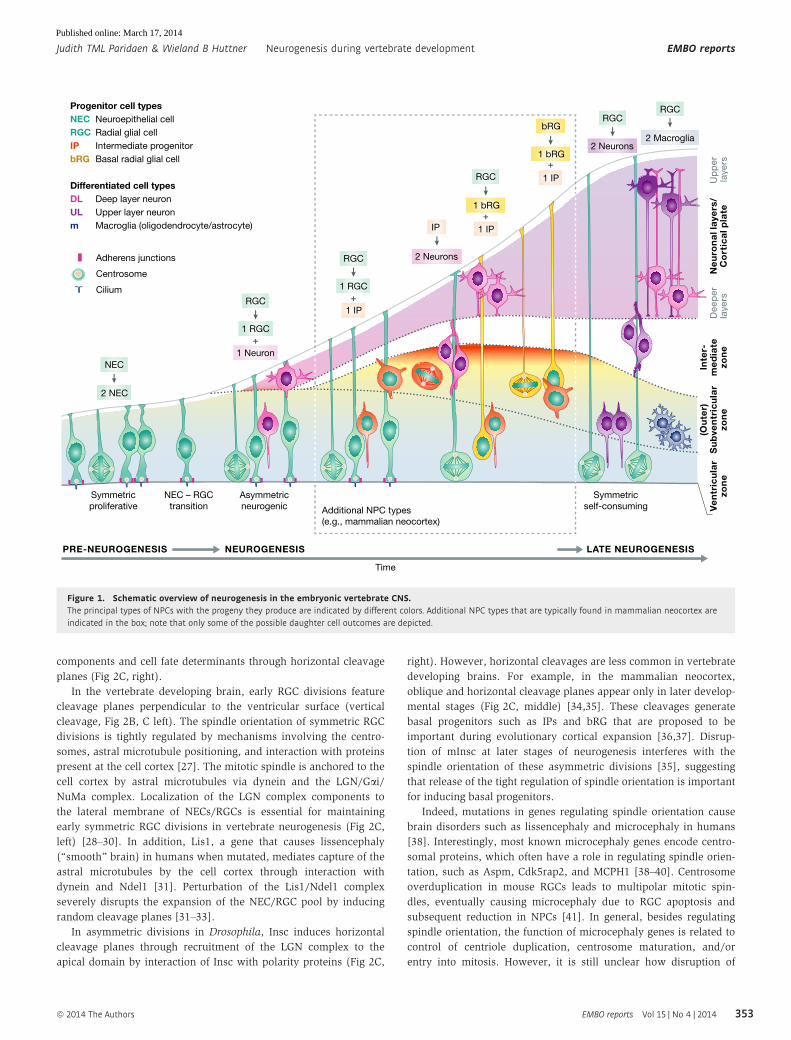
and later in development, glial cells (Fig 1).
Transition from neuroepithelial to radial glial cells
NECs and RGCs, collectively referred to as APs, portray apico-
basal polarity, with apical and basal processes that span the
neuroepithelium. As NECs turn into RGCs, they downregulate Golgi-
derived apical trafficking, lose tight junctions but maintain adherens
junctions. Also, they initiate the expression of astroglial markers
such as GLAST and BLBP. The mechanisms underlying NEC to RGC
transition are only partially understood. Expression of members of
the bHLH transcription factor Hes family, as well as transient
expression of Fgf10, is necessary for this transition [1,2].
At the onset of neurogenesis, RGCs switch from symmetric to
asymmetric divisions, giving rise to an RGC daughter cell and a
differentiating cell (Fig 2A, B). This latter cell constitutes a neuron,
or in certain areas of the brain such as the neocortex, a more fate-
restricted type of progenitor that is called IP and is one of the
types of BPs. IPs divide mainly symmetrically to yield two
neurons, thus doubling the neuron output. In some more
expanded brain regions, such as the neocortex in mammals, there
are additional BPs present with glial characteristics that are capa-
ble of self-renewal (see below). These progenitors are proposed to
mediate cortical expansion in some mammals during evolution [3]
(see below).
Cellular features of neural progenitors
Neural progenitor cells (NPCs) such as NECs and RGCs are highly
polarized, with their apical membrane exposed to the ventricle and
their basal side contacting the pial basal membrane (Fig 1).
Apical domainThe apical domain of RGCs contains several features that are impor-
tant for RGC function. Just basal to the apical and subapical plasma
membrane, the AJs mediate cell–cell adhesion. AJs consist of cadhe-
rins and catenins that connect to the intracellular actin network.
Importantly, polarity proteins such as Par3, Par6, and aPKC are
associated with the subapical cell cortex and are important for RGC
proliferation [4]. The Rho GTPases RhoA, cdc42, and Rac1 have
important roles in the maintenance of AJs and apical mitoses by
the regulation of actin [5–9]. The apical plasma membrane is
Max Planck Institute of Molecular Cell Biology and Genetics, Dresden, Germany*Corresponding author. Tel: +49 351 210 1500; Fax: +49 351 210 1600; E-mail: [email protected]**Corresponding author. Tel: +49 351 210 1500; Fax: +49 351 210 1600; E-mail: [email protected]
ª 2014 The Authors EMBO reports Vol 15 | No 4 | 2014 351
Published online: March 17, 2014
characterized by a specific composition of membrane constituents.
The resulting apical polarity is essential for NPC function.
Newborn neurogenic daughter cells need to withdraw their apical
endfoot from the apical belt of AJs in order to migrate basally and
differentiate. Proneural genes expressed in the differentiating daugh-
ter cell induce downregulation of cadherins to mediate delamination
from the ventricular surface, in a manner similar to epithelial–
mesenchymal transition in other epithelia [10,11]. An alternative
mechanism for delamination recently observed in chick and mouse
neural tube is abscission of the apical endfoot that is similarly regu-
lated by proneural genes acting upstream of cadherin and other
factors [12]. In this process, actomyosin-dependent constriction of
the apical process, preceded by dissociation of the centrosome from
the apical primary cilium, leads to abscission of the apical process
from the apical-most portion of the apical endfoot [12]. In this way,
the cell loses its apical polarity and ciliary proteins, which contrib-
utes to its subsequent cell cycle exit and differentiation.
At the apical side, the centrosome is docked at the apical plasma
membrane. Here, it functions as the basal body in nucleation of the
primary cilium, an important sensory organelle that detects signals
in the ventricular fluid/CSF such as IGF and Shh [13,14]. Primary
cilium activity is required for maintaining proper apicobasal polarity
as NECs transform into RGCs [15]. Upon disruption of Arl13b, a
small ciliary GTPase, during NEC to RGC transition, the polarity of
the cortical wall is inverted, with mitoses occurring at the pial
surface and neurons migrating to the ventricular surface [15]. After
onset of neurogenesis, primary cilium function in processing of the
transcriptional repressor Gli3R is involved in the regulation of RGC
proliferation [16] (see also below).
Basal process
The basal process of RGCs stretches all the way to the basal lamina
at the pial surface. Recent studies have shown that the basal process
is important in the maintenance of proliferative capacity through
integrin signaling from the basal lamina and via the specific basal
localization of the G1-S-phase regulator CyclinD2 [17–19]. It is
hypothesized that the presence of a basal process is involved in the
continued proliferative capacity of bRGs that are present in gyren-
cephalic brains [17].
Cell cycle kinetics of RGCsPrior to mitosis, in G2, the RGC nucleus moves to the ventricular
surface where the centrosome is docked. This nuclear movement is
part of INM in which the NEC/RGC nucleus moves in concert with
the cell cycle using actomyosin and microtubule motor proteins
[20]. It has been proposed that INM functions to maximize the
number of RGC mitoses at the small ventricular surface [20].
Another possible function of INM is to differentially expose the RGC
nucleus to signals that are present along an apical–basal gradient,
such as Delta-Notch signaling (see below). Recently, it was demon-
strated that dynein recruitment to the nuclear pore through two
consequential mechanisms is required for apical nuclear movement
and mitotic entry of rat RGCs [21]. Interestingly, nuclear pore
complexes were also necessary for the basal movement of the
centrosome, which occurs just prior to prophase [21,22].
Changes in cell cycle length have been implicated in cell fate
determination during neurogenesis [23]. The duration of the RGC cell
cycle changes during brain development, with an increased G1 phase
length being linked to neurogenic divisions [24–26]. Interestingly,
the S-phase of RGCs that undergo proliferative divisions is longer
than that of RGCs undergoing neurogenic divisions, suggesting that
careful control of DNA replication takes place during the S-phase of
expanding RGCs [24]. Conversely, one may speculate that somatic
mutations that occur in RGCs after their switch to asymmetric self-
renewing/neurogenic divisions due to the lack of correction of DNA
replication errors may be a means of increasing neuronal diversity.
Regulation of symmetric versus asymmetric divisions
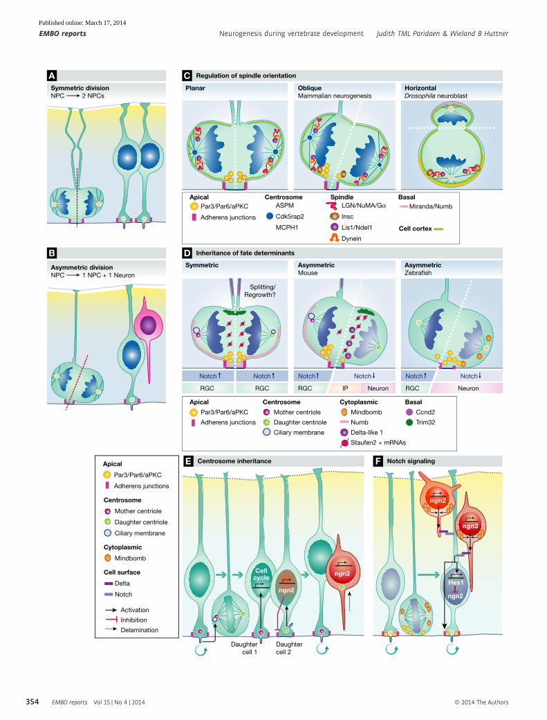
Mitotic spindle orientationAfter onset of neurogenesis, RGCs divide mainly asymmetrically
yielding one RGC daughter and a differentiating daughter cell
(Fig 2B). In invertebrates such as Drosophila, asymmetric division
has been shown to result from unequal division of cellular
Glossary
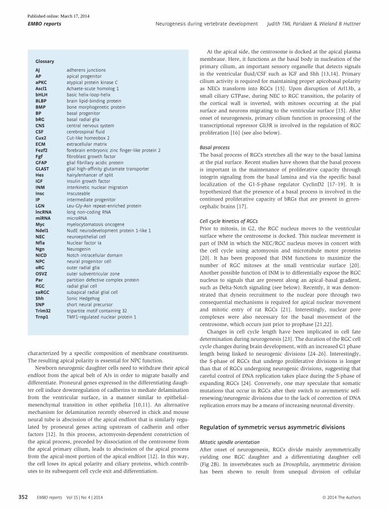
AJ adherens junctionsAP apical progenitoraPKC atypical protein kinase CAscl1 Achaete-scute homolog 1bHLH basic helix-loop-helixBLBP brain lipid-binding proteinBMP bone morphogenetic proteinBP basal progenitorbRG basal radial gliaCNS central nervous systemCSF cerebrospinal fluidCux2 Cut-like homeobox 2ECM extracellular matrixFezf2 forebrain embryonic zinc finger-like protein 2Fgf fibroblast growth factorGFAP glial fibrillary acidic proteinGLAST glial high-affinity glutamate transporterHes hairy/enhancer of splitIGF insulin growth factorINM interkinetic nuclear migrationInsc InscuteableIP intermediate progenitorLGN Leu-Gly-Asn repeat-enriched proteinlncRNA long non-coding RNAmiRNA microRNAMyc myelocytomatosis oncogeneNdel1 NudE neurodevelopment protein 1-like 1NEC neuroepithelial cellNfia Nuclear factor IaNgn NeurogeninNICD Notch intracellular domainNPC neural progenitor celloRG outer radial gliaOSVZ outer subventricular zonePar partition defective complex proteinRGC radial glial cellsaRGC subapical radial glial cellShh Sonic HedgehogSNP short neural precursorTrim32 tripartite motif containing 32Trnp1 TMF1-regulated nuclear protein 1
EMBO reports Vol 15 | No 4 | 2014 ª 2014 The Authors
EMBO reports Neurogenesis during vertebrate development Judith TML Paridaen & Wieland B Huttner
352
Published online: March 17, 2014
components and cell fate determinants through horizontal cleavage
planes (Fig 2C, right).
In the vertebrate developing brain, early RGC divisions feature
cleavage planes perpendicular to the ventricular surface (vertical
cleavage, Fig 2B, C left). The spindle orientation of symmetric RGC
divisions is tightly regulated by mechanisms involving the centro-
somes, astral microtubule positioning, and interaction with proteins
present at the cell cortex [27]. The mitotic spindle is anchored to the
cell cortex by astral microtubules via dynein and the LGN/Gai/NuMa complex. Localization of the LGN complex components to
the lateral membrane of NECs/RGCs is essential for maintaining
early symmetric RGC divisions in vertebrate neurogenesis (Fig 2C,
left) [28–30]. In addition, Lis1, a gene that causes lissencephaly
(“smooth” brain) in humans when mutated, mediates capture of the
astral microtubules by the cell cortex through interaction with
dynein and Ndel1 [31]. Perturbation of the Lis1/Ndel1 complex
severely disrupts the expansion of the NEC/RGC pool by inducing
random cleavage planes [31–33].
In asymmetric divisions in Drosophila, Insc induces horizontal
cleavage planes through recruitment of the LGN complex to the
apical domain by interaction of Insc with polarity proteins (Fig 2C,
right). However, horizontal cleavages are less common in vertebrate
developing brains. For example, in the mammalian neocortex,
oblique and horizontal cleavage planes appear only in later develop-
mental stages (Fig 2C, middle) [34,35]. These cleavages generate
basal progenitors such as IPs and bRG that are proposed to be
important during evolutionary cortical expansion [36,37]. Disrup-
tion of mInsc at later stages of neurogenesis interferes with the
spindle orientation of these asymmetric divisions [35], suggesting
that release of the tight regulation of spindle orientation is important
for inducing basal progenitors.
Indeed, mutations in genes regulating spindle orientation cause
brain disorders such as lissencephaly and microcephaly in humans
[38]. Interestingly, most known microcephaly genes encode centro-
somal proteins, which often have a role in regulating spindle orien-
tation, such as Aspm, Cdk5rap2, and MCPH1 [38–40]. Centrosome
overduplication in mouse RGCs leads to multipolar mitotic spin-
dles, eventually causing microcephaly due to RGC apoptosis and
subsequent reduction in NPCs [41]. In general, besides regulating
spindle orientation, the function of microcephaly genes is related to
control of centriole duplication, centrosome maturation, and/or
entry into mitosis. However, it is still unclear how disruption of
Symmetric
proliferative
NEC – RGC
transition
NEUROGENESIS
Asymmetric
neurogenic Additional NPC types
(e.g., mammalian neocortex)
Up
pe
rla
ye
rsD
ee
pe
rla
ye
rs
Inte
r-m
ed
iate
zon
e
(Ou
ter)
Su
bve
ntr
icu
lar
zon
eV
en
tric
ula
rzo
ne
Ne
uro
na
l lay
ers
/C
ort
ica
l pla
te
LATE NEUROGENESISPRE-NEUROGENESIS
Symmetric
self-consuming
NEC
2 NEC
2 Macroglia
Time
Progenitor cell typesNEC Neuroepithelial cell
RGC Radial glial cell
IP Intermediate progenitor
bRG Basal radial glial cell
Differentiated cell typesDL Deep layer neuron
UL Upper layer neuron
m Macroglia (oligodendrocyte/astrocyte)
RGC
RGC
1 bRG
1 bRG
bRG
1 RGC
1 Neuron
+
RGC
RGC RGC
1 RGC
1 IP
1 IP
1 IP
+
+
+
2 Neurons
2 Neurons
IP
Adherens junctions
Centrosome
Cilium
Figure 1. Schematic overview of neurogenesis in the embryonic vertebrate CNS.The principal types of NPCs with the progeny they produce are indicated by different colors. Additional NPC types that are typically found in mammalian neocortex areindicated in the box; note that only some of the possible daughter cell outcomes are depicted.
ª 2014 The Authors EMBO reports Vol 15 | No 4 | 2014
Judith TML Paridaen & Wieland B Huttner Neurogenesis during vertebrate development EMBO reports
353
Published online: March 17, 2014
Centrosome
Mother centriole
Daughter centriole
Ciliary membrane
Basal
Ccnd2
Trim32
Apical
Par3/Par6/aPKC
Adherens junctions
Symmetric divisionNPC 2 NPCs
C Regulation of spindle orientation
D Inheritance of fate determinants
F Notch signaling
Hes1
ngn2
ngn2
ngn2Centrosome
Mother centriole
Daughter centriole
Ciliary membrane
Apical
Par3/Par6/aPKC
Adherens junctions
Cytoplasmic
Mindbomb
Cell surface
Delta
Notch
Activation
Inhibition
Delamination
Cytoplasmic
Mindbomb
Numb
Delta-like 1
Staufen2 + mRNAs
Notch
Symmetric
Splitting/
Regrowth?
RGC
Notch Notch
AsymmetricZebrafish
RGC Neuron
A
ngn2Cellcycle
ngn2
Daughter
cell 1
Daughter
cell 2
E Centrosome inheritance
CentrosomeASPM
Cdk5rap2
MCPH1
ApicalPar3/Par6/aPKC
Adherens junctions
SpindleLGN/NuMA/G
Insc
Lis1/Ndel1
Dynein
BasalMiranda/Numb
Cell cortex
HorizontalDrosophila neuroblast
ObliqueMammalian neurogenesis
Planar
BAsymmetric divisionNPC 1 NPC + 1 Neuron
Notch
RGC
Notch Notch
RGC IP Neuron
AsymmetricMouse
EMBO reports Vol 15 | No 4 | 2014 ª 2014 The Authors
EMBO reports Neurogenesis during vertebrate development Judith TML Paridaen & Wieland B Huttner
354
Published online: March 17, 2014
these centrosomal functions leads to reduced brain size (see, e.g.,
[42]).
Asymmetric segregation of cellular components and
cell fate determinantsAs discussed above, the apical domain of RGCs contains important
features such as the AJs and the centrosome. One previous model
suggests that the cleavage furrow bypasses the apical domain, lead-
ing to its inheritance by only one daughter cell [34,43]. However,
recent studies have shown equal division of the apical domain even
in asymmetric divisions [28,37]. In this case, both daughter cells
have inherited an apical domain initially, but the differentiating
daughter will withdraw its apical process from the ventricular
surface (Fig 2D, middle).
The basal process is thought to be important for the mainte-
nance of NPC proliferation. In symmetric divisions occurring during
early neurogenesis, the basal process of NECs can either be split
and divided among the daughter cells [44], or inherited by one
daughter cell with the other daughter re-extending it [45]. In
contrast, in asymmetric divisions, the basal process is inherited by
one daughter cell that retains self-renewing properties [37,45]. The
daughter cell without the basal process is not able to re-establish it
and becomes a differentiating cell such as a neuron or IP
[28,37,46]. Taken together, these findings suggest that inheritance
of both the apical and basal domain is required for maintaining
RGC fate [28,37].
Recent studies have shown an intriguing link between centro-
some asymmetries, ciliogenesis, and daughter cell fate (Fig 2E). In
interphase cells, the centrosome contains one mother and one
daughter centriole. The mother centriole is the oldest centriole
within the cell and mediates nucleation of the primary cilium. Inter-
estingly, older centrioles are preferentially inherited by daughter
cells maintaining stem cell identity in the mouse neocortex [47]. A
recent study shows that in mitotic RGCs, the mother centriole is able
to retain ciliary membrane, which is subsequently asymmetrically
inherited by one daughter cell that reforms a new cilium before its
sister cell [48]. This earlier cilium reformation results in earlier cili-
ary signaling in this cell, which is proposed to contribute to its adop-
tion of RGC daughter cell fate. In addition, nascent differentiating
daughter cells show reformation of primary cilia at their basolateral
instead of their apical membranes prior to their delamination [49].
These temporal and spatial asymmetries in ciliogenesis are proposed
to lead to differential exposure of daughter cells to proliferative
signals present in the CSF, such as IGF-1 [15,50], thus leading to
asymmetrical daughter cell behavior.
In Drosophila, asymmetric division of neuroblasts is mediated
through unequal division of polarity proteins and fate determinants.
Similarly, in asymmetrically dividing RGCs of vertebrates, polarity
proteins such as Par3 are asymmetrically segregated into one
daughter cell [34,46,51,52]. At the same time, Notch signaling
components such as the Notch ligand Delta-like 1, the regulator of
Delta internalization, Mindbomb, and the Notch antagonist Numb
are differentially segregated between daughter cells, leading to
differential Notch signaling between daughter cells (Fig 2D, F) [51–
53]. Interestingly, the cell fate related to Par3 inheritance appears to
vary between species. In the mouse, Par3 segregates asymmetrically
into the daughter cell that inherits both apical domain and basal
processes and that remains an RGC (Fig 2D, middle) [51]. In
contrast, in the zebrafish brain, the daughter cell inheriting the
apical domain, including Par3, also inherits the Notch inhibitor
Mindbomb and differentiates (Fig 2D, right) [46,52]. The other
daughter cell quickly re-expresses Par3, re-establishes apical
contact, and remains an RGC. At present, the mechanisms under-
lying these differences between species are unknown.
In addition to polarity proteins, other cytoplasmic proteins also
show unequal inheritance in asymmetric divisions of neural progen-
itors. For example, the double-stranded RNA-binding protein Stau-
fen binds a range of mRNAs that induce cell cycle exit and
differentiation and segregates these into the differentiating daughter
cell during mitosis of RGCs (Fig 2D, middle) [54,55]. One of these
RNAs encodes Trim32 (Brat1 in Drosophila) that is asymmetrically
segregated in both Drosophila neuroblasts and mammalian RGCs.
Trim32 stimulates cell cycle exit through ubiquitination of c-Myc
and activation of differentiation-inducing microRNAs such as Let-7
[56] (see also below).
Regulation of daughter cell fate specification
Transcription factors
During early development, the central nervous system is subdivided
into the prospective different areas by gradients of morphogens such
as Fgfs, Wnts, Shh, and BMPs. This patterning leads to regional
expression of homeodomain and bHLH transcription factors that
instruct NPCs to produce specific cell types during neurogenesis
[57]. One of the master regulators of neurogenesis is the paired box
containing homeodomain transcription factor Pax6 that is expressed
in several CNS regions, such as the forebrain, retina, and hindbrain
[58]. In addition to the regulation of regional patterning, Pax6
promotes RGC proliferation and spindle orientation [59], but also
promotes neurogenesis through the induction of bHLH proneural
genes such as Neurogenins [60]. These partially opposing effects
appear to be mediated through alternative splicing of Pax6 [61] and
its interaction with other transcription factors such as Sox2 and
Hes1 [58,60]. Neuronal differentiation is induced through the
expression of region-specific proneural genes, Pou-homeodomain
transcription factors such as Brn1/2, and SoxC transcription factors
such as Sox4 and Sox11 that initiate specific neuronal programs and
repress other regional identities [57,62]. For example, NPCs in the
dorsal telencephalon express the bHLH proneural factors Neurogenin
Figure 2. Division types of NPCs are determined by spindle orientation and inheritance of cell fate determinants.(A, B) Symmetric division yields two NPCs, whereas asymmetric NPC division yields one NPC daughter and one differentiating daughter cell. (C) Spindle orientation insymmetric versus asymmetric divisions is regulated by centrosomal protein and spindle orientation complexes in vertical and oblique divisions of vertebrate NPCs (left andmiddle) and horizontal neuroblast divisions in Drosophila. (D) Cell fate determinants may be equally (symmetric division, left) or unequally (middle, mouse; right, zebrafish)distributed between daughter cells. (E, F) Examples of asymmetries between daughter cells that were introduced by asymmetric inheritance of differently aged centrioles andciliary membrane (E), and Par3 and Notch signaling components (F).
◂
ª 2014 The Authors EMBO reports Vol 15 | No 4 | 2014
Judith TML Paridaen & Wieland B Huttner Neurogenesis during vertebrate development EMBO reports
355
Published online: March 17, 2014
(Ngn) 1/2. These factors instruct the generation of glutamatergic pyra-
midal neurons that make up the six-layered neocortex in mammals
and repress ventral telencephalic genes. In contrast, the ventral telen-
cephalon expresses Gsh1/2, Nkx2.1, and the bHLH proneural factor
Ascl1 that instructs the generation of GABA-ergic basal ganglia
neurons and cortical interneurons, and represses dorsal identity.
The different types of neurons and glial are born sequentially
from a pool of seemingly identical RGCs. Surprisingly, there is a
significant stochasticity in RGC cell fate choices in individual RGC
lineages in the developing retina, although there is a clear temporal
order in neuronal subtype specification [63,64]. In analogy to find-
ings made in Drosophila, the temporal order of neuronal specifica-
tion by neural progenitors is thought to depend on sequential
expression of transcription factors [65]. In the developing neocortex,
neurons are born in an “inside-out” manner, with earlier-born
neurons destined for the deep layers and later-born neurons for the
upper layers. Contradicting observations with regard to the exis-
tence of fate-restricted RGCs in the developing cortex have been
reported [66,67]. One study reports that a subpopulation of Cux2+
RGCs generates only upper-layer neurons during later stages of
neurogenesis [66]. However, recently, it was reported that Fezf2+
RGCs sequentially produce deep and upper neurons, as well as
oligodendrocytes and astrocytes [67]. Also, in this work, Cux2+
RGCs contributed to both deep and upper layers. More studies
will be needed to resolve the question whether fate-restricted
RGCs constitute a relevant proportion of the progenitor pool and
contribute specifically to the diversity of produced neurons.
Epigenetic modifications
In recent years, evidence has emerged that epigenetic modifications
such as DNA methylation and histone modifications are involved
in the control of temporal and spatial gene expression during
neurogenesis, and the switch from neuronal to glial production [68].
Early-stage NPCs show high expression of regulators of epigenetic
modifications. Examples of such regulators are HMG proteins that
regulate the chromatin state and methyltransferases such as Ezh2
that function in histone modifications [69–71]. Therefore, the
chromatin of early-stage neocortical NPCs is in a more open
state (less condensed) than that of late-stage NPCs [70]. Global
chromatin condensation as well as epigenetic modification of certain
genes seems to be involved in the switch of NPC from producing
neuronal to glial progeny during neocortical development [69,70,72].
For example, DNA methylation of glial genes such as Gfap prevents
a premature switch from neuro- to gliogenesis [73]. Activated
Notch signaling induces demethylation of the Gfap promoter
through the induction of Nfia that dissociates DNA methyltransferases
[74]. Conversely, at late stages of neurogenesis, proneural genes
such as Ngn1 are repressed through the action of Polycomb proteins
[69].
The activity of specific transcription factors is also modified by
epigenetic mechanisms. In the developing cortex, Pax6 mediates
transcription of a range of genes that regulate patterning, NPC
proliferation, but also instruction of IPs and late progenitor fates.
Pax6 interacts with BAF155 and BAF170, which are components of
ATP-dependent multi-subunit mSWI/SNF nucleosome remodeling
complexes [75]. During early neurogenesis, BAF170 competes with
the BAF155 subunit and modifies euchromatin structure. This
results in the recruitment of Pax6/REST-corepressor complex to
repress expression of Pax6 target genes, such as Tbr2, Cux2, and
Tle2, that instruct the generation of IPs and late cortical progenitors
[75]. In this way, switching BAF complex subunits at some point
during neurogenesis could release the repression of Pax6 target
genes, and the generation of IPs and late cortical neuronal types
would follow. Another example of epigenetic control of transcrip-
tion factor activity is transcriptional repression of the forkhead
homeodomain transcription factor Foxg1 through the chromatin
remodeling protein Snf2 l at mid-neurogenesis. Repression of
Foxg1 leads to de-repression of the cell cycle exit regulator p21,
thereby promoting cell cycle exit and neuronal differentiation of
NPCs [76].
Post-transcriptional regulation of gene expression
Alternative pre-mRNA processing results in the generation of differ-
ent proteins from one primary transcript. Alternative splicing plays
a role in differentiation and development and has recently also
been implicated in neurogenesis [77]. For example, alternative
splicing of the transcriptional repressor REST by the splicing factor
nSR100 leads to de-repression of neuron-specific genes and neuro-
nal differentiation [78]. Furthermore, the polypyrimidine tract
RNA-binding protein Ptbp2 inhibits splicing of exons that are
typical for the splice variant expressed in adult tissues [79]. For
example, Ptbp2 induces alternative splicing of proteins that are
involved in RGC adhesion [79]. Deletion of Ptbp2 induces prema-
ture neurogenesis. Sequence-specific RNA-binding proteins such as
Rbfox3 were shown to mediate alternative splicing of Numb, an
important regulator of Notch signaling involved in the induction of
neuronal differentiation [80].
An additional post-transcriptional mechanism for regulating gene
expression in RGCs is through miRNAs, highly conserved non-
coding RNAs of 18–24 nucleotides that bind to the 30 UTR of mRNAs
to silence their expression through degradation or suppressed trans-
lation [81]. In the developing brain, groups of miRNAs regulate
either RGC proliferation or neuronal differentiation, suggesting that
miRNAs play a crucial role in determining neuron numbers. For
example, in the developing mouse cortex, miR-92 suppresses the
transition of RGC into IPs by silencing the transcription factor Tbr2
that induces IP fate [82,83]. Besides direct silencing of target genes,
some miRNAs form a regulatory loop together with their targets.
The HMG-box transcription factor Sox2 that is expressed by NPCs
and directs their self-renewal regulates expression of the RNA-bind-
ing protein LIN28 through epigenetic modifications [84]. LIN28
regulates the biogenesis of the let-7 miRNA family by inhibiting
their maturation. In turn, let-7 miRNA suppresses expression of
LIN28 and inhibits both proliferation and neuronal commitment
through silencing of the cell cycle regulators Ccnd1, Cdc25a, and
proneural genes Ngn1 and Ascl1, respectively [84].
Recently, long non-coding RNAs (lncRNAs) have been implicated
in the regulation of developmental processes including neurogenesis
[85]. LncRNA loci encode RNA transcripts of >200 nucleotides that
modulate gene expression through chromatin modifications and
translational control such as alternative splicing. The lncRNA Rmst
regulates neurogenesis in the midbrain through co-transcriptional
interaction with Sox2 to activate proneural target genes such as Ascl
and Ngn1 [86]. In RGCs that are committed to neurogenic divisions,
several lncRNAs such as Miat are expressed that regulate prolifera-
tion versus differentiation [87].
EMBO reports Vol 15 | No 4 | 2014 ª 2014 The Authors
EMBO reports Neurogenesis during vertebrate development Judith TML Paridaen & Wieland B Huttner
356
Published online: March 17, 2014
Signaling pathwaysAs already mentioned, a variety of signaling pathways triggered at
the plasma membrane, notably the Notch, Wnt, Shh, and Fgf path-
ways, are known to act during the process of neurogenesis. Many of
these signaling pathways have an effect on RGC proliferation and
undergo considerable crosstalk (see also below).
Notch The Notch signaling pathway plays essential roles in the
regulation of both embryonic and adult neurogenesis [88]. As first
elucidated in Drosophila, Delta-Notch signaling regulates neurogene-
sis through the process of lateral inhibition. The Notch ligands Delta
or Jagged activate Notch receptors on directly adjacent cells, leading
to release of NICD that mediates the transcription of Hes genes.
These in turn repress the expression of bHLH proneural genes such
as Ngn and Ascl and thus keep this cell in a proliferative state. In
the developing mouse cortex, the expression of Hes1 in RGCs oscil-
lates with 2- to 3-h periods due to an autoinhibitory feedback loop
[89]. These Hes1 oscillations induce oscillations in Delta and Ngn2
expression. Therefore, it has been proposed that the differential
expression levels of Hes1 could mediate differential responses of
RGCs to incoming signals that regulate proliferation versus differen-
tiation.
Pairs of daughter cells derived from asymmetric RGC divisions
show asymmetries in Delta-Notch signaling components and activity
(Fig 2D, F). For example, in asymmetric RGC divisions in the devel-
oping zebrafish as well as mouse telencephalon, the daughter cell
with higher Notch signaling remains an RGC, while the daughter
cell with low Notch signaling shows high expression of Delta and
proneural genes and initiates delamination from the ventricular
surface and neural differentiation (Fig 2F) [52,89,90]. In the devel-
oping mouse cortex, Notch ligands as well as the E3 ubiquitin ligase
Mindbomb that promotes Notch signaling are expressed by neurons
and IPs [91–94], which signal back to RGC via dynamic and tran-
sient processes (Fig 2F) [93]. One important question is how the
response of cells to Notch signaling changes during neurogenesis, as
Notch signaling is also active in newborn neurons. Some general
repressors of Notch have been identified, but it is unclear whether
these factors are specifically upregulated during neurogenesis
[95,96]. Recently, a transcriptional repressor, Bcl6, was identified
with increased expression during neurogenesis. Bcl6 changes the
composition of the Notch-dependent transcriptional complex at the
Hes5 promoter and leads to histone modifications that permanently
silence Hes5 through recruitment of the deacetylase Sirt1 [97]. This
epigenetic switch results in stable Hes5 inactivity despite active
Notch signaling in differentiating cells, thereby stabilizing neuronal
differentiation.
Wnt Wnt/b-catenin signaling is important in patterning of, and
regulation of proliferation and differentiation in, the developing
brain [98]. After binding of Wnt ligands to their Frizzled/LRP5/6
receptors, cytoplasmic b-catenin is stabilized and translocates to the
nucleus where it mediates gene transcription through LEF/TCF tran-
scription factor activity. Wnt signaling activity plays dual roles
during neurogenesis. During early neurogenesis, Wnt signaling
promotes symmetric RGC divisions and delays IP formation [99].
Later at neurogenesis, however, Wnt activity promotes IP formation
and neuronal differentiation through upregulation of N-myc [100–
102]. A recent study reports that N-myc is expressed in RGCs that
are undergoing neurogenic division in the chick neural tube [103].
N-myc increases non-vertical cleavage planes and represses Notch
signaling to stimulate neuronal differentiation [103]. Although it is
not yet understood how the differential Wnt signaling responses are
mediated, it is likely that the targeted genes change during neuro-
genesis through context- and cell-type-dependent mechanisms such
as epigenetic modifications.
Hedgehog Sonic hedgehog (Shh) signaling is essential for proper
dorsoventral patterning of the vertebrate central nervous system.
Shh signaling is activated through binding of Shh ligand to the
Patched receptor, followed by ciliary accumulation of Smoothened
and processing in the primary cilium of the Gli transcription factors
into their activator forms that mediate downstream gene transcrip-
tion. In the absence of Shh, the Gli proteins are processed into
repressor forms. In addition to its roles in patterning, Shh signaling
also has important roles in the regulation of the RGC cell cycle kinet-
ics through cell cycle regulators, as well as in the production of IPs
[14,16,104]. During neurogenesis, active Shh signaling decreases,
whereas activity of the Gli3 repressor increases, which is necessary
for IP production and neuronal differentiation [16].
A recent study provides mathematical modeling of spinal cord
neurogenesis to predict that decreasing Shh signaling mediates the
switch from symmetric proliferative and asymmetric self-renewing
divisions to symmetric neurogenic divisions by changing RGC cell
cycle kinetics [105]. In the developing neocortex, Shh activity
promotes symmetric proliferative divisions of RGCs through tran-
scription of the Notch transcription factor Hes1 [106], thus showing
that there is a significant crosstalk between different signaling path-
ways in the regulation of RGC proliferation.
Fgf Such interplay between pathways has also been observed for
Fgf and Notch. Fgfs are important for anterior–posterior patterning
of the brain as well as for expansion of RGCs by symmetric division
through downstream activation of Hes1-mediated transcription
[107].
NPC environmentIn addition to the above-mentioned extracellular signals, numerous
other factors in the NPC environment influence NPC behavior
(Fig 3).
At the ventricular surface, several ECM molecules such as lami-
nin and syndecan-1 are present that regulate, via integrin receptors,
the apical adhesion and proliferation of RGCs [108,109]. Apical
adhesion of RGCs and apical localization of integrin b1 are also
controlled by ephrin B1 [110]. At the basal side, the interaction of
the NPC basal process with basal lamina ECM is thought to be
important for the self-renewing potential of RGCs and bRGs [17].
Another important signal from the basal side, retinoic acid, is
produced by the meninges. Retinoic acid is essential for the switch
of RGCs from symmetric proliferative to asymmetric neurogenic
divisions at the onset of neurogenesis [111].
In addition to signals derived from the apical or basal side, envi-
ronmental cues present within the developing neural tube wall also
exert important effects on NPCs. For example, the presence of blood
vessels near IPs appears to regulate their proliferation [112,113]. This
resembles NPC regulation by blood vessels in the stem cell niche of
adult neural progenitors. In addition, non-neuronal cells such as
ª 2014 The Authors EMBO reports Vol 15 | No 4 | 2014
Judith TML Paridaen & Wieland B Huttner Neurogenesis during vertebrate development EMBO reports
357
Published online: March 17, 2014
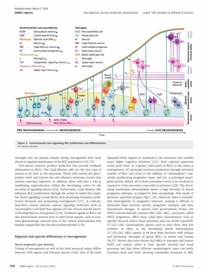
microglia that are present already during neurogenesis have been
shown to regulate maintenance of the RGC population [114,115].
Post-mitotic neurons produce molecules that provide feedback
information to RGCs. The Cajal–Retzius cells are the first type of
neuron to be born in the neocortex. These cells secrete the glyco-
protein reelin and express the cell adhesion molecules nectins that
mediate neuronal migration. In addition, these cells play a role in
modulating regionalization within the developing cortex by the
secretion of signaling factors [116]. Furthermore, Cajal–Retzius cells
influence RGC proliferation through the action of reelin that ampli-
fies Notch signaling in early RGCs, thus promoting symmetric prolif-
erative divisions and postponing neurogenesis [117]. In contrast,
later-born cortical neurons express signaling molecules such as
neurotrophin 3 and Fgf9 that regulate cell fate choices and the switch
of dividing RGCs to astrogenesis [118]. Feedback signals to RGCs are
also derived from neurons born in other brain regions, such as tran-
sient glutamatergic neurons born in the ventral telencephalon that
migrate tangentially into the dorsal telencephalon [119].
Regional and species differences in neurogenesis
Neural progenitor type diversity
Timing of neurogenesis as well as the total neuronal output differs
between CNS regions and between species [120]. One of the most
expanded brain regions in mammals is the neocortex that enables
many higher cognitive functions [121]. Such regional expansion
could result from: (i) a greater initial pool of RGCs at the onset of
neurogenesis, (ii) increased neuronal production through increased
number of RGC cell cycles or the addition of “intermediate” tran-
siently proliferating progenitor types, and (iii) a prolonged neuro-
genic period. Indeed, all of these parameters seem to be involved in
expansion of the neocortex, especially in primates [120]. The devel-
oping mammalian telencephalon shows a large diversity of neural
progenitor subtypes, as judged by their morphology, their mode of
divisions, and their progeny (Fig 1; [3]). However, there is consider-
able heterogeneity in progenitor behavior, making it difficult to
determine links between specific progenitor subtypes and their
downstream lineages. In species with gyrencephalic brains, the
OSVZ characteristically contains bRG (also oRG), previously called
OSVZ progenitors. bRGs keep radial glial characteristics such as
apically directed and/or basal processes and can divide repeatedly
[17,122–124]. Lissencephalic species such as mice show only low
numbers of bRGs in the developing dorsal telencephalon
[37,125,126]. bRGs appear to be born from divisions with oblique
and horizontal cleavages of apical RGCs in mouse and human
[36,37]. Recent data have shown that bRGs in macaque and human
OSVZ can contain either or both apically directed and basal
processes and that these different morphological types can freely
transition back and forth, showing remarkable dynamics in bRG
Up
pe
rla
ye
rsD
ee
pe
rla
ye
rs
(Ou
ter)
Su
bve
ntr
icu
lar
zon
eV
en
tric
ula
rzo
ne
Ne
uro
na
l lay
ers
/C
ort
ica
l pla
te
Inte
r-m
ed
iate
zon
e
Gliogenesis
NEUROGENESIS LATE NEUROGENESISPRE-NEUROGENESIS
Time
CR
N
IP
TM
reelin
Thalamicafferents
RA
ECM
ECM
bRG
m
UL
DLDL
UL
NEC RG
IGF2, FGF2,Wnt, Bmp, Shh,…CSF(ventricle)
Environmental cues provided by
ECM Extracellular matrix
CSF Cerebrospinal fluid
Meninges Retinoic acid (RA)
N Neuron
CR Cajal-Retzius neuron
IP Intermediate progenitor
blood vessel
μ Microglia
TM Tangentially migrating neuron
Thalamic afferents
UL Upper layer neuron
Cell types
NEC Neuroepithelial cell
RG Radial glial cell
N Neuron
CR Cajal-Retzius neuron
IP Intermediate progenitor
DL Deep layer neuron
bRG Basal radial glial cell
μ Microglia
UL Upper layer neuron
m Macroglia
Ntf3 Fgf9
Bloodvessel
Meningesμ
Figure 3. Environmental cues regulating NPC proliferation and differentiation.For details, see text.
EMBO reports Vol 15 | No 4 | 2014 ª 2014 The Authors
EMBO reports Neurogenesis during vertebrate development Judith TML Paridaen & Wieland B Huttner
358
Published online: March 17, 2014
characteristics and lineages [36,122]. Additional apical RGC types,
named short neural precursors (SNPs) [127], and subapical RGCs
(saRGCs) [128] have also been identified. SNPs divide apically like
apical RGCs, but have only short basal processes and undergo
mainly neurogenic divisions [127]. saRGCs were identified in the
developing ventral telencephalon of lissencephalic rodents and in
the dorsal telencephalon of gyrencephalic species. Therefore,
saRGCs are proposed to add to cortical expansion through increased
production of neurons [128].
These observations show that depending on the CNS region
and species, different types of neural progenitors exist with a
wide variety of morphologies, division modes, and lineages to
generate diverse neuronal outputs. Furthermore, neural progenitor
types and their lineages are by no means strictly separated and
unidirectional.
Differential molecular control of cell fate decisions
Although many general principles and mechanisms underlying
neurogenesis have been identified, it is poorly understood how
(subtle) differences in molecular mechanisms mediate the different
neuronal outputs required for distinct brain regions. For example,
only few molecular mechanisms in induction and maintenance of
the diverse types of neural progenitors in the mammalian neocortex
have been identified. Recently, it was shown that the nuclear Trnp1
protein maintains self-renewing RGCs, possibly through chromatin
remodeling [129]. Interestingly, Trnp1 expression is reduced in
areas of cortical expansion in human fetal brains. Also, deletion of
Trnp1 in mouse leads to increased horizontal cleavages and
increased bRG production [129].
As mentioned above, differences in early patterning events
induce subtle intrinsic molecular and epigenetic differences
between RGCs of different regions. Subsequently, RGCs of different
CNS regions show different responses to signals. For instance,
upon deletion of the small GTPase RhoA, RGCs in cortex,
midbrain, and spinal cord show similar RGC polarity defects and
migrate away from the ventricular surface. However, RGCs in
more expanded regions such as cortex and midbrain respond by
hyperproliferation, whereas RGCs in the spinal cord proliferate less
[6,8,9]. Within tissues, RGC proliferative capacity is modulated
through differential expression of transcription factors, possibly
influenced by dorsoventral and anterioposterior gradients of
morphogens. For example, maintained expression of the transcrip-
tion factor PLZF modulates RGC response to FGF ligands in the
central domain of the developing spinal cord through alterations in
FGF receptor and subsequent downstream signaling component
levels [130]. In this way, centrally localized RGCs maintain prolif-
erative capacity, whereas their dorsal and ventral counterparts
undergo differentiation. Future studies will certainly uncover new
mechanisms that differentially regulate initial RGC pool expansion,
regulation of cell cycle and progenitor diversity, and the length of
the neurogenic period to understand how regional and species
differences in neuronal output are mediated.
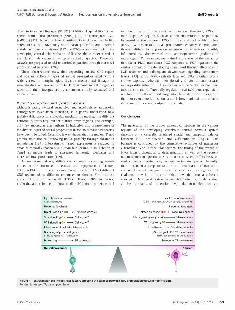
Conclusions
The generation of the proper amount of neurons in the various
regions of the developing vertebrate central nervous system
depends on a carefully regulated spatial and temporal balance
between NPC proliferation and differentiation (Fig 4). This
balance is controlled by the cumulative activities of numerous
extracellular and intracellular factors. The timing of the switch of
NPCs from proliferation to differentiation, as well as the sequen-
tial induction of specific NPC and neuron types, differs between
central nervous system regions and vertebrate species. Recently,
there has been a steep increase in the identification of molecules
and mechanisms that govern specific aspects of neurogenesis. A
challenge now is to integrate this knowledge into a coherent
concept of NPC proliferation versus differentiation, to determine,
at the cellular and molecular level, the principles that are
Neural progenitor Neuron
Inheritance of cell fate determinants
Silencing of NPC TF expression
miR, epigenetic modification
Neuronal feedback
Input from environment
CSF, meninges, blood vessels, afferents
Sequential TF expression
Shh signaling suppression Differentiation
Notch signaling OFF Proneural genes
Wnt signaling ON Differentiation
Inheritance of cell fate determinants
Silencing of proneural genes
miR, epigenetic modification
Neuronal feedback
Input from environment
CSF, meninges
Patterning TF expression
Shh signaling ON Cell cycle
Notch signaling ON Proneural genes
Wnt signaling ON Cell cycle
Figure 4. Extracellular and intracellular factors affecting the balance between NPC proliferation versus differentiation.For details, see text. TF, transcription factor.
ª 2014 The Authors EMBO reports Vol 15 | No 4 | 2014
Judith TML Paridaen & Wieland B Huttner Neurogenesis during vertebrate development EMBO reports
359
Published online: March 17, 2014
conserved in vertebrate central nervous system development, and
to identify the modifications that account for the differences
between species.
AcknowledgmentsWe apologize to colleagues whose work we could not include due to space
constraints. We thank the Huttner laboratory members and in particular
Elena Taverna and Marta Florio for useful discussions. JTMLP was supported
by an EMBO long-term fellowship. WBH was supported by grants from the
DFG (SFB 655, A2; TRR 83, Tp6) and the ERC (250197), by the DFG-funded
Center for Regenerative Therapies Dresden, and by the Fonds der Chemischen
Industrie.
Conflict of interestThe authors declare that they have no conflict of interest.
References
1. Hatakeyama J, Bessho Y, Katoh K, Ookawara S, Fujioka M, Guillemot F,
Kageyama R (2004) Hes genes regulate size, shape and histogenesis of
the nervous system by control of the timing of neural stem cell differ-
entiation. Development 131: 5539 – 5550
2. Sahara S, O’Leary DD (2009) Fgf10 regulates transition period of corti-
cal stem cell differentiation to radial glia controlling generation of
neurons and basal progenitors. Neuron 63: 48 – 62
3. Franco SJ, Muller U (2013) Shaping our minds: stem and progenitor cell
diversity in the mammalian neocortex. Neuron 77: 19 – 34
4. Costa MR, Wen G, Lepier A, Schroeder T, Gotz M (2008) Par-complex
proteins promote proliferative progenitor divisions in the developing
mouse cerebral cortex. Development 135: 11 – 22
5. Cappello S, Attardo A, Wu X, Iwasato T, Itohara S, Wilsch-Brauninger M,
Eilken HM, Rieger MA, Schroeder TT, Huttner WB et al (2006) The Rho-
GTPase cdc42 regulates neural progenitor fate at the apical surface.
Nat Neurosci 9: 1099 – 1107
6. Cappello S, Bohringer CR, Bergami M, Conzelmann KK, Ghanem A,
Tomassy GS, Arlotta P, Mainardi M, Allegra M, Caleo M et al (2012) A
radial glia-specific role of RhoA in double cortex formation. Neuron 73:
911 – 924
7. Chen L, Melendez J, Campbell K, Kuan CY, Zheng Y (2009) Rac1 defi-
ciency in the forebrain results in neural progenitor reduction and
microcephaly. Dev Biol 325: 162 – 170
8. Herzog D, Loetscher P, van Hengel J, Knusel S, Brakebusch C, Taylor V,
Suter U, Relvas JB (2011) The small GTPase RhoA is required to main-
tain spinal cord neuroepithelium organization and the neural stem cell
pool. J Neurosci 31: 5120 – 5130
9. Katayama K, Melendez J, Baumann JM, Leslie JR, Chauhan BK,
Nemkul N, Lang RA, Kuan CY, Zheng Y, Yoshida Y (2011) Loss of
RhoA in neural progenitor cells causes the disruption of adherens
junctions and hyperproliferation. Proc Natl Acad Sci USA 108:
7607 – 7612
10. Itoh Y, Moriyama Y, Hasegawa T, Endo TA, Toyoda T, Gotoh Y (2013)
Scratch regulates neuronal migration onset via an epithelial-mesen-
chymal transition-like mechanism. Nat Neurosci 16: 416 – 425
11. Rousso DL, Pearson CA, Gaber ZB, Miquelajauregui A, Li S, Portera-
Cailliau C, Morrisey EE, Novitch BG (2012) Foxp-mediated suppression
of N-cadherin regulates neuroepithelial character and progenitor
maintenance in the CNS. Neuron 74: 314 – 330
12. Das RM, Storey KG (2014) Apical abscission alters cell polarity and
dismantles the primary cilium during neurogenesis. Science 343:
200 – 204
13. Lehtinen MK, Walsh CA (2011) Neurogenesis at the brain-cerebrospinal
fluid interface. Annu Rev Cell Dev Biol 27: 653 – 679
14. Willaredt MA, Tasouri E, Tucker KL (2013) Primary cilia and forebrain
development. Mech Dev 130: 373 – 380
15. Higginbotham H, Guo J, Yokota Y, Umberger NL, Su CY, Li J, Verma N,
Hirt J, Ghukasyan V, Caspary T et al (2013) Arl13b-regulated cilia activi-
ties are essential for polarized radial glial scaffold formation. Nat
Neurosci 16: 1000 – 1007
16. Wang H, Ge G, Uchida Y, Luu B, Ahn S (2011) Gli3 is required for main-
tenance and fate specification of cortical progenitors. J Neurosci 31:
6440 – 6448
17. Fietz SA, Kelava I, Vogt J, Wilsch-Brauninger M, Stenzel D, Fish JL,
Corbeil D, Riehn A, Distler W, Nitsch R et al (2010) OSVZ progenitors of
human and ferret neocortex are epithelial-like and expand by integrin
signaling. Nat Neurosci 13: 690 – 699
18. Radakovits R, Barros CS, Belvindrah R, Patton B, Muller U (2009) Regu-
lation of radial glial survival by signals from the meninges. J Neurosci
29: 7694 – 7705
19. Tsunekawa Y, Britto JM, Takahashi M, Polleux F, Tan SS, Osumi N
(2012) Cyclin D2 in the basal process of neural progenitors is linked to
non-equivalent cell fates. EMBO J 31: 1879 – 1892
20. Taverna E, Huttner WB (2010) Neural progenitor nuclei IN motion.
Neuron 67: 906 – 914
21. Hu DJ, Baffet AD, Nayak T, Akhmanova A, Doye V, Vallee RB (2013)
Dynein recruitment to nuclear pores activates apical nuclear
migration and mitotic entry in brain progenitor cells. Cell 154:
1300 – 1313
22. Spear PC, Erickson CA (2012) Apical movement during interkinetic
nuclear migration is a two-step process. Dev Biol 370: 33 – 41
23. Dehay C, Kennedy H (2007) Cell-cycle control and cortical development.
Nat Rev Neurosci 8: 438 – 450
24. Arai Y, Pulvers JN, Haffner C, Schilling B, Nusslein I, Calegari F, Huttner
WB (2011) Neural stem and progenitor cells shorten S-phase on
commitment to neuron production. Nat Commun 2: 154
Sidebar A. In need of answers
Recent technological advances in live imaging and lineage reconstruc-tion in both “old” model animals used to study neurogenesis such asmouse, zebrafish, Drosophila, and in “new” models with gyrencephalicbrains such as macaque and ferret will hopefully allow answering ofsome of the important open questions in the field of neurogenesis:(i) How is the input from signaling pathways integrated into a
specific cell fate choice?(ii) Do morphologically and molecularly distinct progenitor types have
distinct and specific lineages? What is the level of stochasticity inthese lineages?
(iii) Which molecular mechanisms mediate the induction of differentprogenitor types and how do these differ between species andCNS subregions?
(iv) Ultimately, which genomic changes account for the greater prolif-erative capacity of neural stem and progenitor cells that underliesthe evolutionary expansion of the neocortex?
(v) What are the similarities and differences between embryonic andadult neurogenesis? What is the embryonic origin of adult neuralstem cells?
EMBO reports Vol 15 | No 4 | 2014 ª 2014 The Authors
EMBO reports Neurogenesis during vertebrate development Judith TML Paridaen & Wieland B Huttner
360
Published online: March 17, 2014
25. Lange C, Huttner WB, Calegari F (2009) Cdk4/cyclinD1 overexpression
in neural stem cells shortens G1, delays neurogenesis, and promotes
the generation and expansion of basal progenitors. Cell Stem Cell 5:
320 – 331
26. Pilaz LJ, Patti D, Marcy G, Ollier E, Pfister S, Douglas RJ, Betizeau M,
Gautier E, Cortay V, Doerflinger N et al (2009) Forced G1-phase
reduction alters mode of division, neuron number, and laminar
phenotype in the cerebral cortex. Proc Natl Acad Sci USA 106:
21924 – 21929
27. Lancaster MA, Knoblich JA (2012) Spindle orientation in mammalian
cerebral cortical development. Curr Opin Neurobiol 22: 737 – 746
28. Konno D, Shioi G, Shitamukai A, Mori A, Kiyonari H, Miyata T,
Matsuzaki F (2008) Neuroepithelial progenitors undergo LGN-dependent
planar divisions to maintain self-renewability during mammalian
neurogenesis. Nat Cell Biol 10: 93 –101
29. Morin X, Jaouen F, Durbec P (2007) Control of planar divisions by the
G-protein regulator LGN maintains progenitors in the chick neuroepi-
thelium. Nat Neurosci 10: 1440 – 1448
30. Peyre E, Jaouen F, Saadaoui M, Haren L, Merdes A, Durbec P, Morin X
(2011) A lateral belt of cortical LGN and NuMA guides mitotic spindle
movements and planar division in neuroepithelial cells. J Cell Biol 193:
141 – 154
31. Yingling J, Youn YH, Darling D, Toyo-Oka K, Pramparo T, Hirotsune S,
Wynshaw-Boris A (2008) Neuroepithelial stem cell proliferation
requires LIS1 for precise spindle orientation and symmetric division.
Cell 132: 474 – 486
32. Pawlisz AS, Mutch C, Wynshaw-Boris A, Chenn A, Walsh CA, Feng Y
(2008) Lis1-Nde1-dependent neuronal fate control determines cerebral
cortical size and lamination. Hum Mol Genet 17: 2441 – 2455
33. Xie Y, Juschke C, Esk C, Hirotsune S, Knoblich JA (2013) The phosphatase
PP4c controls spindle orientation to maintain proliferative symmetric
divisions in the developing neocortex. Neuron 79: 254 – 265
34. Kosodo Y, Roper K, Haubensak W, Marzesco AM, Corbeil D, Huttner WB
(2004) Asymmetric distribution of the apical plasma membrane during
neurogenic divisions of mammalian neuroepithelial cells. EMBO J 23:
2314 – 2324
35. Postiglione MP, Juschke C, Xie Y, Haas GA, Charalambous C, Knoblich JA
(2011) Mouse inscuteable induces apical-basal spindle orientation to
facilitate intermediate progenitor generation in the developing neocor-
tex. Neuron 72: 269 – 284
36. LaMonica BE, Lui JH, Hansen DV, Kriegstein AR (2013) Mitotic spindle
orientation predicts outer radial glial cell generation in human neocor-
tex. Nat Commun 4: 1665
37. Shitamukai A, Konno D, Matsuzaki F (2011) Oblique radial glial divi-
sions in the developing mouse neocortex induce self-renewing progeni-
tors outside the germinal zone that resemble primate outer
subventricular zone progenitors. J Neurosci 31: 3683 – 3695
38. Gilmore EC, Walsh CA (2013) Genetic causes of microcephaly and
lessons for neuronal development. Wiley Interdiscip Rev Dev Biol 2:
461 – 478
39. Fish JL, Kosodo Y, Enard W, Paabo S, Huttner WB (2006) Aspm specifi-
cally maintains symmetric proliferative divisions of neuroepithelial
cells. Proc Natl Acad Sci USA 103: 10438 – 10443
40. Gruber R, Zhou Z, Sukchev M, Joerss T, Frappart PO, Wang ZQ (2011)
MCPH1 regulates the neuroprogenitor division mode by coupling the
centrosomal cycle with mitotic entry through the Chk1-Cdc25 path-
way. Nat Cell Biol 13: 1325 – 1334
41. Marthiens V, Rujano MA, Pennetier C, Tessier S, Paul-Gilloteaux P,
Basto R (2013) Centrosome amplification causes microcephaly. Nat Cell
Biol 15: 731 – 740
42. Pulvers JN, Bryk J, Fish JL, Wilsch-Brauninger M, Arai Y, Schreier D,
Naumann R, Helppi J, Habermann B, Vogt J et al (2010) Mutations in
mouse Aspm (abnormal spindle-like microcephaly associated) cause
not only microcephaly but also major defects in the germline. Proc
Natl Acad Sci USA 107: 16595 – 16600
43. Marthiens V, ffrench-Constant C (2009) Adherens junction domains are
split by asymmetric division of embryonic neural stem cells. EMBO Rep
10: 515 – 520
44. Kosodo Y, Toida K, Dubreuil V, Alexandre P, Schenk J, Kiyokage E,
Attardo A, Mora-Bermudez F, Arii T, Clarke JD et al (2008) Cytokinesis
of neuroepithelial cells can divide their basal process before anaphase.
EMBO J 27: 3151 – 3163
45. Shitamukai A, Matsuzaki F (2012) Control of asymmetric cell division of
mammalian neural progenitors. Dev Growth Differ 54: 277 – 286
46. Alexandre P, Reugels AM, Barker D, Blanc E, Clarke JD (2010) Neurons
derive from the more apical daughter in asymmetric divisions in the
zebrafish neural tube. Nat Neurosci 13: 673 – 679
47. Wang X, Tsai JW, Imai JH, Lian WN, Vallee RB, Shi SH (2009) Asymmet-
ric centrosome inheritance maintains neural progenitors in the
neocortex. Nature 461: 947 – 955
48. Paridaen JT, Wilsch-Brauninger M, Huttner WB (2013) Asymmetric
inheritance of centrosome-associated primary cilium membrane directs
ciliogenesis after cell division. Cell 155: 333 – 344
49. Wilsch-Brauninger M, Peters J, Paridaen JT, Huttner WB (2012) Baso-
lateral rather than apical primary cilia on neuroepithelial cells
committed to delamination. Development 139: 95 – 105
50. Lehtinen MK, Zappaterra MW, Chen X, Yang YJ, Hill AD, Lun M,
Maynard T, Gonzalez D, Kim S, Ye P et al (2011) The cerebrospinal fluid
provides a proliferative niche for neural progenitor cells. Neuron 69:
893 – 905
51. Bultje RS, Castaneda-Castellanos DR, Jan LY, Jan YN, Kriegstein AR, Shi
SH (2009) Mammalian Par3 regulates progenitor cell asymmetric divi-
sion via notch signaling in the developing neocortex. Neuron 63:
189 – 202
52. Dong Z, Yang N, Yeo SY, Chitnis A, Guo S (2012) Intralineage directional
Notch signaling regulates self-renewal and differentiation of asymmet-
rically dividing radial glia. Neuron 74: 65 – 78
53. Kawaguchi D, Furutachi S, Kawai H, Hozumi K, Gotoh Y (2013) Dll1
maintains quiescence of adult neural stem cells and segregates asym-
metrically during mitosis. Nat Commun 4: 1880
54. Kusek G, Campbell M, Doyle F, Tenenbaum SA, Kiebler M, Temple S
(2012) Asymmetric segregation of the double-stranded RNA binding
protein Staufen2 during mammalian neural stem cell divisions
promotes lineage progression. Cell Stem Cell 11: 505 – 516
55. Vessey JP, Amadei G, Burns SE, Kiebler MA, Kaplan DR, Miller FD (2012)
An asymmetrically localized Staufen2-dependent RNA complex regu-
lates maintenance of mammalian neural stem cells. Cell Stem Cell 11:
517 – 528
56. Schwamborn JC, Berezikov E, Knoblich JA (2009) The TRIM-NHL protein
TRIM32 activates microRNAs and prevents self-renewal in mouse
neural progenitors. Cell 136: 913 – 925
57. Martynoga B, Drechsel D, Guillemot F (2012) Molecular control of
neurogenesis: a view from the mammalian cerebral cortex. Cold Spring
Harb Perspect Biol 4: a008359
ª 2014 The Authors EMBO reports Vol 15 | No 4 | 2014
Judith TML Paridaen & Wieland B Huttner Neurogenesis during vertebrate development EMBO reports
361
Published online: March 17, 2014
58. Osumi N, Shinohara H, Numayama-Tsuruta K, Maekawa M (2008)
Concise review: Pax6 transcription factor contributes to both embry-
onic and adult neurogenesis as a multifunctional regulator. Stem Cells
26: 1663 – 1672
59. Asami M, Pilz GA, Ninkovic J, Godinho L, Schroeder T, Huttner WB, Gotz
M (2011) The role of Pax6 in regulating the orientation and mode of
cell division of progenitors in the mouse cerebral cortex. Development
138: 5067 – 5078
60. Sansom SN, Griffiths DS, Faedo A, Kleinjan DJ, Ruan Y, Smith J, van
Heyningen V, Rubenstein JL, Livesey FJ (2009) The level of the tran-
scription factor Pax6 is essential for controlling the balance between
neural stem cell self-renewal and neurogenesis. PLoS Genet 5:
e1000511
61. Walcher T, Xie Q, Sun J, Irmler M, Beckers J, Ozturk T, Niessing D,
Stoykova A, Cvekl A, Ninkovic J et al (2013) Functional dissection of
the paired domain of Pax6 reveals molecular mechanisms of
coordinating neurogenesis and proliferation. Development 140:
1123 – 1136
62. Bergsland M, Werme M, Malewicz M, Perlmann T, Muhr J (2006) The
establishment of neuronal properties is controlled by Sox4 and Sox11.
Genes Dev 20: 3475 – 3486
63. Gomes FL, Zhang G, Carbonell F, Correa JA, Harris WA, Simons BD,
Cayouette M (2011) Reconstruction of rat retinal progenitor cell
lineages in vitro reveals a surprising degree of stochasticity in cell fate
decisions. Development 138: 227 – 235
64. He J, Zhang G, Almeida AD, Cayouette M, Simons BD, Harris WA (2012)
How variable clones build an invariant retina. Neuron 75: 786 – 798
65. Kohwi M, Doe CQ (2013) Temporal fate specification and neural
progenitor competence during development. Nat Rev Neurosci 14:
823 – 838
66. Franco SJ, Gil-Sanz C, Martinez-Garay I, Espinosa A, Harkins-Perry SR,
Ramos C, Muller U (2012) Fate-restricted neural progenitors in the
mammalian cerebral cortex. Science 337: 746 – 749
67. Guo C, Eckler MJ, McKenna WL, McKinsey GL, Rubenstein JL, Chen B
(2013) Fezf2 expression identifies a multipotent progenitor for neocorti-
cal projection neurons, astrocytes, and oligodendrocytes. Neuron 80:
1167 – 1174
68. MuhChyi C, Juliandi B, Matsuda T, Nakashima K (2013) Epigenetic
regulation of neural stem cell fate during corticogenesis. Int J Dev
Neurosci 31: 424 – 433
69. Hirabayashi Y, Suzki N, Tsuboi M, Endo TA, Toyoda T, Shinga J, Koseki
H, Vidal M, Gotoh Y (2009) Polycomb limits the neurogenic competence
of neural precursor cells to promote astrogenic fate transition. Neuron
63: 600 – 613
70. Kishi Y, Fujii Y, Hirabayashi Y, Gotoh Y (2012) HMGA regulates the
global chromatin state and neurogenic potential in neocortical precur-
sor cells. Nat Neurosci 15: 1127 – 1133
71. Pereira JD, Sansom SN, Smith J, Dobenecker MW, Tarakhovsky A,
Livesey FJ (2010) Ezh2, the histone methyltransferase of PRC2,
regulates the balance between self-renewal and differentiation in the
cerebral cortex. Proc Natl Acad Sci USA 107: 15957 – 15962
72. Tan SL, Nishi M, Ohtsuka T, Matsui T, Takemoto K, Kamio-Miura A,
Aburatani H, Shinkai Y, Kageyama R (2012) Essential roles of the
histone methyltransferase ESET in the epigenetic control of neural
progenitor cells during development. Development 139: 3806 – 3816
73. Fan G, Martinowich K, Chin MH, He F, Fouse SD, Hutnick L, Hattori D,
Ge W, Shen Y, Wu H et al (2005) DNA methylation controls the timing
of astrogliogenesis through regulation of JAK-STAT signaling. Develop-
ment 132: 3345 – 3356
74. Namihira M, Kohyama J, Semi K, Sanosaka T, Deneen B, Taga T,
Nakashima K (2009) Committed neuronal precursors confer astrocytic
potential on residual neural precursor cells. Dev Cell 16: 245 – 255
75. Tuoc TC, Boretius S, Sansom SN, Pitulescu ME, Frahm J, Livesey FJ,
Stoykova A (2013) Chromatin regulation by BAF170 controls cerebral
cortical size and thickness. Dev Cell 25: 256 – 269
76. Yip DJ, Corcoran CP, Alvarez-Saavedra M, DeMaria A, Rennick S, Mears
AJ, Rudnicki MA, Messier C, Picketts DJ (2012) Snf2 l regulates Foxg1-
dependent progenitor cell expansion in the developing brain. Dev Cell
22: 871 – 878
77. Calarco JA, Zhen M, Blencowe BJ (2011) Networking in a global world:
establishing functional connections between neural splicing regulators
and their target transcripts. RNA 17: 775 – 791
78. Raj B, O’Hanlon D, Vessey JP, Pan Q, Ray D, Buckley NJ, Miller FD,
Blencowe BJ (2011) Cross-regulation between an alternative splicing
activator and a transcription repressor controls neurogenesis. Mol Cell
43: 843 – 850
79. Licatalosi DD, Yano M, Fak JJ, Mele A, Grabinski SE, Zhang C, Darnell RB
(2012) Ptbp2 represses adult-specific splicing to regulate the generation
of neuronal precursors in the embryonic brain. Genes Dev 26:
1626 – 1642
80. Kim KK, Nam J, Mukouyama YS, Kawamoto S (2013) Rbfox3-regulated
alternative splicing of Numb promotes neuronal differentiation during
development. J Cell Biol 200: 443 – 458
81. Bian S, Xu TL, Sun T (2013) Tuning the cell fate of neurons and glia by
microRNAs. Curr Opin Neurobiol 23: 928 – 934
82. Bian S, Hong J, Li Q, Schebelle L, Pollock A, Knauss JL, Garg V, Sun T
(2013) MicroRNA cluster miR-17-92 regulates neural stem cell expan-
sion and transition to intermediate progenitors in the developing
mouse neocortex. Cell Rep 3: 1398 – 1406
83. Nowakowski TJ, Fotaki V, Pollock A, Sun T, Pratt T, Price DJ (2013)
MicroRNA-92b regulates the development of intermediate cortical
progenitors in embryonic mouse brain. Proc Natl Acad Sci USA 110:
7056 – 7061
84. Cimadamore F, Amador-Arjona A, Chen C, Huang CT, Terskikh AV (2013)
SOX2-LIN28/let-7 pathway regulates proliferation and neurogenesis in
neural precursors. Proc Natl Acad Sci USA 110: E3017 – E3026
85. Fatica A, Bozzoni I (2014) Long non-coding RNAs: new players in cell
differentiation and development. Nat Rev Genet 15: 7 – 21
86. Ng SY, Bogu GK, Soh BS, Stanton LW (2013) The long noncoding RNA
RMST interacts with SOX2 to regulate neurogenesis. Mol Cell 51:
349 – 359
87. Aprea J, Prenninger S, Dori M, Ghosh T, Monasor LS, Wessendorf E,
Zocher S, Massalini S, Alexopoulou D, Lesche M et al (2013) Transcrip-
tome sequencing during mouse brain development identifies long non-
coding RNAs functionally involved in neurogenic commitment. EMBO J
32: 3145 – 3160
88. Pierfelice T, Alberi L, Gaiano N (2011) Notch in the vertebrate nervous
system: an old dog with new tricks. Neuron 69: 840 – 855
89. Shimojo H, Ohtsuka T, Kageyama R (2008) Oscillations in notch
signaling regulate maintenance of neural progenitors. Neuron 58:
52 – 64
90. Ochiai W, Nakatani S, Takahara T, Kainuma M, Masaoka M, Minobe S,
Namihira M, Nakashima K, Sakakibara A, Ogawa M et al (2009) Peri-
ventricular notch activation and asymmetric Ngn2 and Tbr2 expression
EMBO reports Vol 15 | No 4 | 2014 ª 2014 The Authors
EMBO reports Neurogenesis during vertebrate development Judith TML Paridaen & Wieland B Huttner
362
Published online: March 17, 2014
in pair-generated neocortical daughter cells. Mol Cell Neurosci 40:
225 – 233
91. Kawaguchi D, Yoshimatsu T, Hozumi K, Gotoh Y (2008) Selection of
differentiating cells by different levels of delta-like 1 among neural
precursor cells in the developing mouse telencephalon. Development
135: 3849 – 3858
92. Mizutani K, Yoon K, Dang L, Tokunaga A, Gaiano N (2007) Differential
Notch signalling distinguishes neural stem cells from intermediate
progenitors. Nature 449: 351 – 355
93. Nelson BR, Hodge RD, Bedogni F, Hevner RF (2013) Dynamic interac-
tions between intermediate neurogenic progenitors and radial glia in
embryonic mouse neocortex: potential role in Dll1-Notch signaling. J
Neurosci 33: 9122 – 9139
94. Yoon KJ, Koo BK, Im SK, Jeong HW, Ghim J, Kwon MC, Moon JS, Miyata
T, Kong YY (2008) Mind bomb 1-expressing intermediate progenitors
generate notch signaling to maintain radial glial cells. Neuron 58:
519 – 531
95. Chi Z, Zhang J, Tokunaga A, Harraz MM, Byrne ST, Dolinko A, Xu J,
Blackshaw S, Gaiano N, Dawson TM et al (2012) Botch promotes
neurogenesis by antagonizing Notch. Dev Cell 22: 707 – 720
96. Dai Q, Andreu-Agullo C, Insolera R, Wong LC, Shi SH, Lai EC (2013)
BEND6 is a nuclear antagonist of Notch signaling during self-renewal
of neural stem cells. Development 140: 1892 – 1902
97. Tiberi L, van den Ameele J, Dimidschstein J, Piccirilli J, Gall D, Herpoel A,
Bilheu A, Bonnefont J, Iacovino M, Kyba M et al (2012) BCL6 controls
neurogenesis through Sirt1-dependent epigenetic repression of selec-
tive Notch targets. Nat Neurosci 15: 1627 – 1635
98. Harrison-Uy SJ, Pleasure SJ (2012) Wnt signaling and forebrain develop-
ment. Cold Spring Harb Perspect Biol 4: a008094
99. Wrobel CN, Mutch CA, Swaminathan S, Taketo MM, Chenn A (2007)
Persistent expression of stabilized beta-catenin delays maturation of
radial glial cells into intermediate progenitors. Dev Biol 309: 285 – 297
100. Kuwahara A, Hirabayashi Y, Knoepfler PS, Taketo MM, Sakai J, Kodama
T, Gotoh Y (2010) Wnt signaling and its downstream target N-myc
regulate basal progenitors in the developing neocortex. Development
137: 1035 – 1044
101. Munji RN, Choe Y, Li G, Siegenthaler JA, Pleasure SJ (2011) Wnt signal-
ing regulates neuronal differentiation of cortical intermediate progeni-
tors. J Neurosci 31: 1676 – 1687
102. Fang WQ, Chen WW, Fu AK, Ip NY (2013) Axin directs the amplification
and differentiation of intermediate progenitors in the developing cere-
bral cortex. Neuron 79: 665 – 679
103. Zinin N, Adameyko I, Wilhelm M, Fritz N, Uhlén P, Ernforns P, Henriks-
son MA (2014) MYC proteins promote neuronal differentiation by
controlling the mode of progenitor cell division. EMBO Rep 15:
383 – 391
104. Shikata Y, Okada T, Hashimoto M, Ellis T, Matsumaru D, Shiroishi T,
Ogawa M, Wainwright B, Motoyama J (2011) Ptch1-mediated dosage-
dependent action of Shh signaling regulates neural progenitor devel-
opment at late gestational stages. Dev Biol 349: 147 – 159
105. Saade M, Gutierrez-Vallejo I, Le Dreau G, Rabadan MA, Miguez DG,
Buceta J, Marti E (2013) Sonic hedgehog signaling switches the mode
of division in the developing nervous system. Cell Rep 4: 492 – 503
106. Dave RK, Ellis T, Toumpas MC, Robson JP, Julian E, Adolphe C, Bartlett
PF, Cooper HM, Reynolds BA, Wainwright BJ (2011) Sonic hedgehog
and notch signaling can cooperate to regulate neurogenic divisions of
neocortical progenitors. PLoS ONE 6: e14680
107. Rash BG, Lim HD, Breunig JJ, Vaccarino FM (2011) FGF signaling
expands embryonic cortical surface area by regulating Notch-depen-
dent neurogenesis. J Neurosci 31: 15604 – 15617
108. Loulier K, Lathia JD, Marthiens V, Relucio J, Mughal MR, Tang SC,
Coksaygan T, Hall PE, Chigurupati S, Patton B et al (2009) Beta1 inte-
grin maintains integrity of the embryonic neocortical stem cell niche.
PLoS Biol 7: e1000176
109. Wang Q, Yang L, Alexander C, Temple S (2012) The niche factor syndec-
an-1 regulates the maintenance and proliferation of neural progenitor
cells during mammalian cortical development. PLoS ONE 7: e42883
110. Arvanitis DN, Behar A, Tryoen-Toth P, Bush JO, Jungas T, Vitale N, Davy
A (2013) Ephrin B1 maintains apical adhesion of neural progenitors.
Development 140: 2082 – 2092
111. Siegenthaler JA, Ashique AM, Zarbalis K, Patterson KP, Hecht JH, Kane
MA, Folias AE, Choe Y, May SR, Kume T et al (2009) Retinoic acid from
the meninges regulates cortical neuron generation. Cell 139: 597 – 609
112. Javaherian A, Kriegstein A (2009) A stem cell niche for intermediate
progenitor cells of the embryonic cortex. Cereb Cortex 19(Suppl 1):
i70 – i77
113. Stubbs D, DeProto J, Nie K, Englund C, Mahmud I, Hevner R, Molnar Z
(2009) Neurovascular congruence during cerebral cortical development.
Cereb Cortex 19(Suppl 1): i32 – i41
114. Antony JM, Paquin A, Nutt SL, Kaplan DR, Miller FD (2011) Endogenous
microglia regulate development of embryonic cortical precursor cells.
J Neurosci Res 89: 286 – 298
115. Cunningham CL, Martinez-Cerdeno V, Noctor SC (2013) Microglia regu-
late the number of neural precursor cells in the developing cerebral
cortex. J Neurosci 33: 4216 – 4233
116. Griveau A, Borello U, Causeret F, Tissir F, Boggetto N, Karaz S, Pierani A
(2010) A novel role for Dbx1-derived Cajal-Retzius cells in early region-
alization of the cerebral cortical neuroepithelium. PLoS Biol 8:
e1000440
117. Lakoma J, Garcia-Alonso L, Luque JM (2011) Reelin sets the pace of
neocortical neurogenesis. Development 138: 5223 – 5234
118. Seuntjens E, Nityanandam A, Miquelajauregui A, Debruyn J, Stryjewska
A, Goebbels S, Nave KA, Huylebroeck D, Tarabykin V (2009) Sip1
regulates sequential fate decisions by feedback signaling from
postmitotic neurons to progenitors. Nat Neurosci 12: 1373 – 1380
119. Teissier A, Waclaw RR, Griveau A, Campbell K, Pierani A (2012) Tangen-
tially migrating transient glutamatergic neurons control neurogenesis
and maintenance of cerebral cortical progenitor pools. Cereb Cortex 22:
403 – 416
120. Charvet CJ, Striedter GF, Finlay BL (2011) Evo-devo and brain scaling:
candidate developmental mechanisms for variation and constancy in
vertebrate brain evolution. Brain Behav Evol 78: 248 – 257
121. Geschwind DH, Rakic P (2013) Cortical evolution: judge the brain by its
cover. Neuron 80: 633 – 647
122. Betizeau M, Cortay V, Patti D, Pfister S, Gautier E, Bellemin-Menard A,
Afanassieff M, Huissoud C, Douglas RJ, Kennedy H et al (2013) Precur-
sor diversity and complexity of lineage relationships in the outer
subventricular zone of the primate. Neuron 80: 442 –457
123. Hansen DV, Lui JH, Parker PR, Kriegstein AR (2010) Neurogenic radial
glia in the outer subventricular zone of human neocortex. Nature 464:
554 – 561
124. Reillo I, de Juan Romero C, Garcia-Cabezas MA, Borrell V (2011) A role
for intermediate radial glia in the tangential expansion of the
mammalian cerebral cortex. Cereb Cortex 21: 1674 – 1694
ª 2014 The Authors EMBO reports Vol 15 | No 4 | 2014
Judith TML Paridaen & Wieland B Huttner Neurogenesis during vertebrate development EMBO reports
363
Published online: March 17, 2014
125. Kelava I, Reillo I, Murayama AY, Kalinka AT, Stenzel D, Tomancak P,
Matsuzaki F, Lebrand C, Sasaki E, Schwamborn JC et al (2012) Abun-
dant occurrence of basal radial glia in the subventricular zone of
embryonic neocortex of a lissencephalic primate, the common marmo-
set Callithrix jacchus. Cereb Cortex 22: 469 – 481
126. Wang X, Tsai JW, LaMonica B, Kriegstein AR (2011) A new subtype of
progenitor cell in the mouse embryonic neocortex. Nat Neurosci 14:
555 – 561
127. Tyler WA, Haydar TF (2013) Multiplex genetic fate mapping reveals a
novel route of neocortical neurogenesis, which is altered in the Ts65Dn
mouse model of Down syndrome. J Neurosci 33: 5106 – 5119
128. Pilz GA, Shitamukai A, Reillo I, Pacary E, Schwausch J, Stahl R, Ninkovic
J, Snippert HJ, Clevers H, Godinho L et al (2013) Amplification of
progenitors in the mammalian telencephalon includes a new radial
glial cell type. Nat Commun 4: 2125
129. Stahl R, Walcher T, De Juan RomeroC, Pilz GA, Cappello S, Irmler M,
Sanz-Aquela JM, Beckers J, Blum R, Borrell V et al (2013) Trnp1 regu-
lates expansion and folding of the mammalian cerebral cortex by
control of radial glial fate. Cell 153: 535 – 549
130. Gaber ZB, Butler SJ, Novitch BG (2013) PLZF regulates fibroblast growth
factor responsiveness and maintenance of neural progenitors. PLoS Biol
11: e1001676
364 EMBO reports Vol 15 | No 4 | 2014 ª 2014 The Authors
EMBO reports Neurogenesis during vertebrate development Judith TML Paridaen & Wieland B Huttner
Published online: March 17, 2014