Embed Size (px)
Citation preview
hPrimpol1/CCDC111 is a human DNAprimase-polymerase required for the maintenanceof genome integrityLi Wan1*, Jiangman Lou1*, Yisui Xia2*, Bei Su1, Ting Liu1, Jiamin Cui2, Yingying Sun1, Huiqiang Lou2+
& Jun Huang1++
1Life Sciences Institute, Zhejiang University, Hangzhou, Zhejiang, and 2State key laboratory of Agro-Biotechnology,
College of Biological Sciences, Agricultural University, Beijing, China
Prim-pol is a recently identified DNA primase-polymerasebelonging to the archaeao-eukaryotic primase (AEP) superfamily.Here, we characterize a previously unrecognized prim-pol inhuman cells, which we designate hPrimpol1 (human primase-polymerase 1). hPrimpol1 possesses primase and DNA polymer-ase activities in vitro, interacts directly with RPA1 and isrecruited to sites of DNA damage and stalled replication forksin an RPA1-dependent manner. Cells depleted of hPrimpol1display increased spontaneous DNA damage and defects in therestart of stalled replication forks. Both RPA1 binding and theprimase activity of hPrimpol1 are required for its cellular functionduring DNA replication. Our results indicate that hPrimpol1 is anovel factor involved in the response to DNA replication stress.Keywords: hPrimpol1/CCDC111; primase-polymerase;RPA1; stalled replication forkEMBO reports (2013) 14, 1104–1112. doi:10.1038/embor.2013.159
INTRODUCTIONTo maintain genetic information, human cells must accuratelyreplicate billions of base pairs of DNA and repair a very widerange of DNA lesions. One of the proteins required for themaintenance of genetic information is the single-stranded DNA(ssDNA)-binding protein, replication protein A (RPA) [1]. HumanRPA is a stable three-subunit complex composed of RPA1, RPA2and RPA3 [1]. RPA1 contains four OB-folds, and RPA2 and RPA3have one OB-fold each [1]. Although each OB-fold is structurally
similar, the majority of the ssDNA binding occurs throughtwo OB-folds (DBD-A and DBD-B) centrally located in RPA1,referred to as the ssDNA-binding core [2,3]. In addition to thessDNA-binding core, RPA1 also contains an OB-fold at eachterminus. The OB-fold at the N terminus of RPA1, DBD-F,interacts with a large number of other proteins and is required forDNA repair, recombination and cell cycle regulation [3,4].DBD-C, at the C terminus of RPA1, is required forheterotrimeric complex formation and has been implicated inrecognition of some types of DNA damage [3,4]. As RPA isengaged in these diverse functions through its ssDNA-bindingactivity and its ability to interact with multiple proteins involved inthese pathways, it has thus been considered as an adaptor proteinthat facilitates various biochemical reactions that occur at ssDNAregions during DNA replication and/or DNA repair [3–10].
DNA primases are enzymes that catalyse the synthesis of shortRNA (or DNA in some organisms) sequences called primers,which serve as starting points for DNA synthesis [11–13].Structurally, most DNA primases can be divided into twoclasses [11–13]. The first class contains the DnaG familyenzymes found in bacteria and archaea [11,14]. The secondclass comprises the heterodimeric primases of the archaeao-eukaryotic primase (AEP) superfamily found in the eukarya andarchaea [11,14]. Recently, a novel family of AEPs, called the prim-pol, which is sporadically present in crenarchaeal and Gram-positive bacterial plasmids, has been described [15,16]. Membersof the prim-pol family are able to catalyse both primase and DNApolymerase reaction in vitro, hence the name prim-pols (primase-polymerases) [15–17]. The precise physiological function of theseprim-pols has remained largely unexplored.
In this study, we used an affinity purification approach to isolateRPA-containing complex and identified a novel proteinCCDC111, which we refer to as hPrimpol1 (human primase-polymerase 1). hPrimpol1 belongs to the prim-pol familyand possesses both primase and DNA polymerase activitiesin vitro. hPrimpol1 is recruited to sites of DNA damage andstalled replication forks via its direct interaction with RPA1. Cells
1Life Sciences Institute, Zhejiang University, Hangzhou, Zhejiang 310058, China2State key laboratory of Agro-Biotechnology, College of Biological Sciences,Agricultural University, Beijing 100193, China
+Corresponding author. Tel: þ 86 10 6273 4504; Fax: þ 86 10 6273 4504;E-mail: [email protected]++Corresponding author. Tel: þ 86 571 8898 1391; Fax: þ 86 571 8898 1391;E-mail: [email protected]
*These authors contributed equally to this work.
Received 25 July 2013; revised 17 September 2013; accepted 17 September 2013;published online 15 October 2013
scientificreportscientific report
1104 EMBO reports VOL 14 | NO 12 | 2013 &2013 EUROPEAN MOLECULAR BIOLOGY ORGANIZATION
with hPrimpol1 depletion display increased spontaneous DNAdamage and defects in the restart of stalled replication forks,suggesting that hPrimpol1 normally acts to stabilize stalledreplication forks. Our results provide the first evidence of theinvolvement of a human DNA primase-polymerase in thereplication stress response.
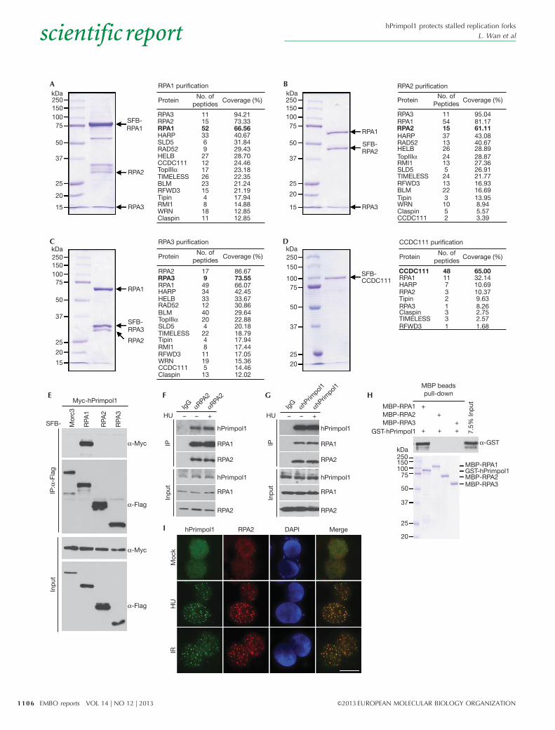
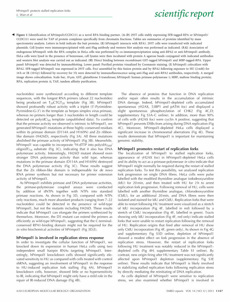
RESULTS AND DISCUSSIONIdentification of hPrimpol1/CCDC111To search for previously undetected proteins present in RPA-containing complex, we performed tandem affinity purificationusing HEK293T cells stably expressing SFB-tagged wild-typeRPA1, RPA2 or RPA3 for the identification of RPA-interactingproteins. Mass spectrometry analysis revealed several knownRPA-associated proteins, including SMARCAL1/HARP, RAD52and BLM (Fig 1A–C). Interestingly, we also repeatedly identi-fied a previously uncharacterized RPA-binding protein asCCDC111 (Fig 1A–C). To ensure that CCDC111 indeed associateswith RPA, we performed reverse tandem affinity purification usinga cell line stably expressing tagged CCDC111, and identified RPA1,RPA2 and RPA3 as major CCDC111-associated proteins (Fig 1D).These data strongly suggest that CCDC111 is a bone fide RPA-binding protein. The CCDC111 gene encodes a deduced polypep-tide of 560 amino acids with a predicted molecular mass of 65 kDa.Structure and sequence analysis revealed that CCDC111 belongs tothe prim-pol family and contains two conserved domains: an AEPdomain (residues 101–240) and a Zn ribbon-like domain (residues392–470) [15]. We have now designated this protein as hPrimpol1(for ‘human primase-polymerase 1’).
hPrimpol1 interacts with RPATo validate our tandem affinity purification results, we firstperformed co-immunoprecipitation experiments using epitope-tagged hPrimpol1 and RPA. We found that Myc-tagged hPrimpol1interacts strongly with SFB-tagged RPA1 but not with the Morc3control protein (Fig 1E). In addition, the interaction betweenhPrimpol1 and RPA was confirmed by reciprocal immuno-precipitations using antibodies against endogenous RPA2 andhPrimpol1, respectively (Fig 1F,G; supplementary Fig S1A,Bonline). Moreover, the hPrimpol1–RPA complex formation wasDNA damage-independent (Fig 1F,G). To determine whether theinteraction between RPA and hPrimpol1 is direct, we expressedand purified recombinant MBP-tagged RPA1, RPA2, RPA3 andGST-tagged hPrimpol1. Pull-down experiments revealed thathPrimpol1 binds strongly with RPA1 but not with RPA2 orRPA3 (Fig 1H), indicating that hPrimpol1 associates with RPAcomplex through RPA1.
Upon the occurrence of DNA damage or inhibition of DNAreplication, RPA could form large nuclear foci. A physicalinteraction between hPrimpol1 and RPA as demonstrated aboveraises the possibility that hPrimpol1 might colocalize with RPA atsites of DNA damage or other barriers in the cell. Indeed, discretefoci of hPrimpol1 were readily detected in cells followinghydroxyurea (HU) or IR treatment (Fig 1I). Moreover, these focicolocalize with RPA2 foci, indicating that the localization ofhPrimpol1, like that of RPA, is regulated in response to DNAdamage and DNA replication stress (Fig 1I).
RPA-binding is required for hPrimpol1 foci formationTo further define the binding between hPrimpol1 and RPA1, wesought to identify the region(s) within hPrimpol1 responsible for itsinteraction with RPA1 (Fig 2A). Co-immunoprecipitation experi-ments revealed that hPrimpol1 associated with RPA1 through itsvery C terminus, as deletion mutant lacking the C-terminal 80amino acids (D5) failed to co-precipitate with RPA1 (Fig 2B).Interestingly, the C terminus of hPrimpol1 has been highlyconserved throughout evolution, suggesting that it might carryout an important function of hPrimpol1 (Fig 2C). Indeed, althoughdistinct nuclear foci of full-length hPrimpol1 and the othermutants were readily detected in HU- or IR-treated cells, theD5 mutant, which does not bind to RPA1, failed to form nuclearfoci after HU or IR treatment (Fig 2D). Thus, the conservedC-terminal region of hPrimpol1 is important not only for itsinteraction with RPA1, but also for its proper localization inresponse to DNA damage and replication stress.
To test whether this C-terminal region of hPrimpol1 is alsosufficient to bind RPA1 and localize hPrimpol1 to sites of DNAdamage and stalled replication forks, we generated a constructencoding the C-terminal region alone (T5) (Fig 2A). As shown inFig 2E, the T5 protein localized to sites of DNA damage andstalled replication forks as efficiently as full-length hPrimpol1.Moreover, T5 retained the ability to interact with RPA1, althoughthis ability was reduced in comparison with the wild-typehPrimpol1 (Fig 2F). These results suggest that hPrimpol1 islikely to be recruited to sites of DNA damage and stalledreplication forks via an interaction between its conservedC-terminal 80 amino acids and RPA1. Consistent with thishypothesis, CtIP depletion abolished IR-induced recruitmentof RPA and the downstream hPrimpol1 to DNA damage sites(supplementary Fig S1C,D online).
The DBD-C domain of RPA1 interacts with hPrimpol1To identify the hPrimpol1-binding domain on RPA1, severalRPA1 truncation and internal deletion mutants were generated(supplementary Fig S2A online). Although wild-type and othermutants of RPA1 could be co-immunoprecipitated with hPrim-pol1, the D5 mutant, which is deleted of DBD-C domain, failedto bind to hPrimpol1 (supplementary Fig S2B online). Theseresults suggest that the DBD-C domain of RPA1 is required for itsbinding to hPrimpol1.
We next examined whether the physical interaction betweenhPrimpol1 and RPA1 might be important for RPA1 localization. Asshown in supplementary Fig S2C online, the D5 mutant was stillable to form discrete foci following HU treatment. By contrast, theDBC-A domain deletion mutant and the DBC-B domain deletionmutant totally lost their foci formation ability. These observationssuggest that the interaction between hPrimpol1 and RPA1 iscritical for hPrimpol1 focus formation, but not vice versa.
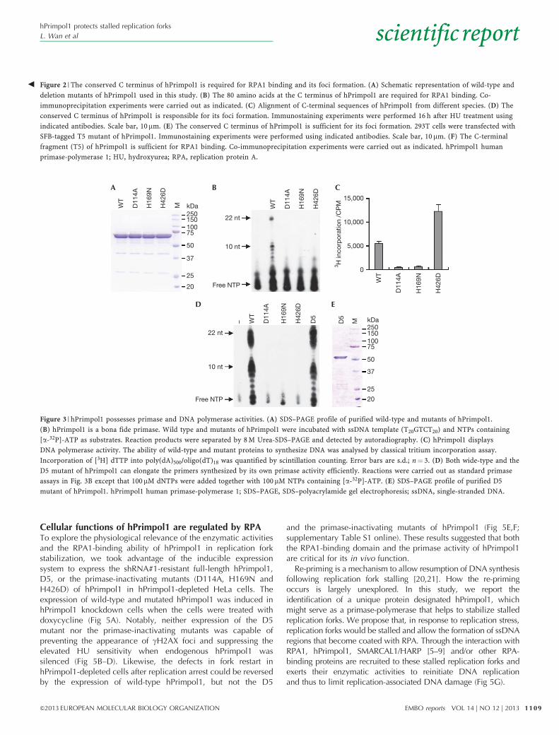
hPrimpol1 has primase and DNA polymerase activitiesAs hPrimpol1 contains a highly conserved AEP-primase domain,we tested whether hPrimpol1 is a bona fide primase. We purifiedwild-type hPrimpol1 protein (Fig 3A) and examined its primaseactivity using an assay described previously [18,19]. As shown inFig 3B, after incubation with various ssDNA templates in thepresence of NTPs containing [a-32P] ATP, hPrimpol1 shows strongprimase activity in vitro. Primers of variable size (mostly 7–11
hPrimpol1 protects stalled replication forks
L. Wan et al scientificreport
1105&2013 EUROPEAN MOLECULAR BIOLOGY ORGANIZATION EMBO reports VOL 14 | NO 12 | 2013
kDa25015010075
50
37
25
20
15
kDa25015010075
50
37
25
20
15
SFB-RPA1
RPA2
RPA3
RPA1
RPA1
RPA2
RPA2
RPA3
RPA3
CCDC111
SFB-
SFB-
SFB-
RPA1 purification
ProteinNo. of
peptidesCoverage (%)
RPA3RPA2RPA1HARPSLD5RAD52HELBCCDC111
TIMELESSBLMRFWD3
RMI1WRNClaspin
Tipin
TopIIIα
1115523369271217262315481811
94.2173.3366.5640.6731.8429.4328.7024.4623.1822.3521.2421.1917.9414.8812.8512.85
RPA3RPA2 17
9493433124020422481119513
RPA1HARP
SLD5
RAD52HELB
CCDC111
TIMELESS
BLM
RFWD3RMI1
WRN
Claspin
Tipin
TopIIIα
RPA3
RPA1HARPRPA2
RPA3ClaspinTIMELESSRFWD3
Tipin
RPA2RPA1
HARP
SLD5
RAD52HELB
CCDC111
CCDC111
TIMELESS
BLMRFWD3
RMI1
WRNClaspin
Tipin
TopIIIα
kDa25015010075
50
37
25
20
15
kDa250
150
10075
50
37
25
20
RPA3 purification
ProteinNo. of
peptidesCoverage (%)
RPA2 purification
ProteinNo. of
PeptidesCoverage (%)
CCDC111 purification
ProteinNo. of
peptidesCoverage (%)
86.6773.5566.0742.4533.6730.8629.6422.8820.1818.7917.9417.4417.0515.3614.4612.02
11541537132624135
2413223
1052
95.0481.1761.1143.0840.6728.8928.8727.3626.9121.7716.9316.6913.958.945.573.39
48117321331
65.0032.1410.6910.379.638.262.752.571.68
Myc-hPrimpol1
SFB-
α-Myc
α-Flag
hPrimpol1
Moc
kH
UIR
α-Myc
α-Flag
Mor
c3
IP:α
-Fla
gIn
put
RPA
1
RPA
2
RPA
3 HU
hPrimpol1
hPrimpol1
RPA1
RPA1
RPA2
RPA2
RPA2 DAPI Merge
hPrimpol1
MBP-RPA1MBP-RPA2MBP-RPA3
GST-hPrimpol1
kDa250150100
75
50
37
25
20
hPrimpol1
RPA1
RPA1
RPA2
RPA2
IPIn
put
HU
IPIn
put
– – + – – +Ig
G αRPA2
αRPA2
IgG +
+
+ + +
MBP-RPA1GST-hPrimpol1MBP-RPA2MBP-RPA3
α-GST
+
7.5%
Inp
ut
MBP beadspull-down
A B
C D
E F
I
G H
αhPrim
pol1
αhPrim
pol1
hPrimpol1 protects stalled replication forks
L. Wan et alscientificreport
1106 EMBO reports VOL 14 | NO 12 | 2013 &2013 EUROPEAN MOLECULAR BIOLOGY ORGANIZATION
nucleotides) were synthesized according to different templatesequences, with the longest RNA primers (about 22 nucleotides)being produced on T20CTGT20 template (Fig 3B). hPrimpol1showed profoundly robust activity with a triplet (50-Pyrimidine-Pyrimidine-G-30) in the template as herpes simplex virus primase,whereas no primers longer than 3 nucleotides in length could bedetected on poly(dC)40 template (unpublished data). To confirmthat the primase activity we observed is intrinsic to hPrimpol1, wegenerated hPrimpol1 mutations at three highly conserved residueswithin its primase domain (D114A and H169N) and Zn ribbon-like domain (H426D), respectively (Fig 3A). All three mutationsabolished the primase activity of hPrimpol1 (Fig 3B). Meanwhile,hPrimpol1 was capable to incorporate 3H-dTTP into poly(dA)500/oligo(dT)18 substrate (Fig 3C), indicating that it also has DNApolymerase activity. Interestingly, H426D mutant showed evenstronger DNA polymerase activity than wild type, whereasmutations in the primase domain (D114A and H169N) destroyedthe DNA polymerase activity (Fig 3C). These results suggestthat the Zn ribbon-like domain is indispensable for de novoRNA primer synthesis but not necessary for primer extensionactivity of hPrimpol1.
As hPrimpol1 exhibits primase and DNA polymerase activities,the primase-polymerase coupled assays were conductedby addition of dNTPs together with NTPs into standardprimase reactions. As shown in Fig 3D, compared with NTPsonly reactions, much more abundant products (ranging from 7–22nucleotides) could be detected in the presence of wild-typehPrimpol1, but not the mutants including H426D. These resultsindicate that hPrimpol1 can elongate the primers synthesized bythemselves. Moreover, the D5 mutant can extend the primers asefficiently as wild-type hPrimpol1, suggesting that the C-terminal-conserved RPA1-binding domain might not be required for thein vitro biochemical activities of hPrimpol1 (Fig 3D,E).
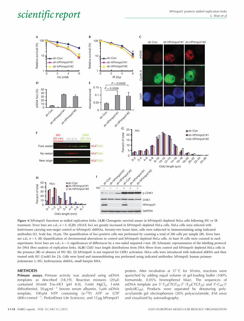
hPrimpol1 is involved in replication stress responseIn order to investigate the cellular function of hPrimpol1, weknocked down its expression in human HeLa cells using twoindependent small hairpin RNAs targeting hPrimpol1. Inter-estingly, hPrimpol1 knockdown cells showed significantly ele-vated sensitivity to HU as compared with cells treated with controlshRNA, suggesting an involvement of hPrimpol1 in the responseto HU-induced replication fork stalling (Fig 4A). hPrimpol1knockdown cells, however, showed little or no hypersensitivityto IR, indicating that hPrimpol1 might only have a mild role in therepair of IR-induced DNA damage (Fig 4B).
The absence of proteins that function in DNA replicationand/or repair often results in the accumulation of intrinsicDNA damage. Indeed, hPrimpol1-depleted cells accumulatedspontaneous gH2AX, 53BP1 and pATM foci and displayed aslight spontaneous phosphorylation of CHK2 (Fig 4C,D;supplementary Fig S3A–C online). In addition, more than 90%of cells with gH2AX foci were cyclin A positive, suggesting thathPrimpol1 prevents DSBs from arising during DNA replication (Fig4C). Moreover, hPrimpol1-depleted HeLa cells displayed asignificant increase in chromosomal aberrations (Fig 4E). Theseresults demonstrate that hPrimpol1 is important for maintaininggenomic stability.
hPrimpol1 promotes restart of replication forksThe localization of hPrimpol1 to stalled replication forks,appearance of gH2AX foci in hPrimpol1-depleted HeLa cellsand its ability to act as a primase-polymerase in vitro indicate thathPrimpol1 might normally be required during the restart of stalledreplication forks. To test this possibility, we analysed replicationfork progression on single DNA fibres. HeLa cells were pulselabelled with the modified thymidine analogue iododeoxyuridine(IdU) for 20 min, and then treated with HU for 2 h to arrestreplication fork progression. Following removal of HU, cells werelabelled with another thymidine analogue, chlorodeoxyuridine(CIdU), for an additional 20 min, and DNA fibres were thenisolated and stained for IdU and CIdU. Replication forks that wereable to restart following HU treatment were visualized as a stretchof IdU incorporation (Fig 4F, labelled in red) followed by astretch of CIdU incorporation (Fig 4F, labelled in green). Tractsshowing only IdU incorporation (Fig 4F, red only) indicate stalledforks that were unable to restart replication following the removalof HU. Replication origins that fired after removal of HU showonly CIdU incorporation (Fig 4F, green only). As shown in Fig 4Gand supplementary Fig S3D online, depletion of hPrimpol1showed a modest effect on fork progression in the absence ofreplication stress. However, the restart of replication forksfollowing HU treatment was notably reduced in the hPrimpol1-depleted cells (Fig 4H; supplementary Table S1 online). Bycontrast, new origin firing after HU treatment was not significantlyaffected upon hPrimpol1 depletion (supplementary Fig S3Eonline). These results indicate that hPrimpol1 is likely involvedin stabilizing stalled replication forks following replication stressby directly mediating the reinitiating of DNA replication.
As cells depleted of hPrimpol1 were sensitive to replicationstress, we also examined whether hPrimpol1 is involved in
Figure 1 | Identification of hPrimpol1/CCDC111 as a novel RPA-binding partner. (A–D) 293T cells stably expressing SFB-tagged-RPA or hPrimpol1/
CCDC111 were used for TAP of protein complexes specifically from chromatin fractions. Tables are summaries of proteins identified by mass
spectrometry analysis. Letters in bold indicate the bait proteins. (E) hPrimpol1 interacts with RPA1. 293T cells were transfected with indicated
plasmids. Cell lysates were immunoprecipitated with anti-Flag antibody and western blot analysis was performed as indicated. (F,G) Association of
endogenous hPrimpol1 with the RPA complex in HeLa cells was performed by co-immunoprecipitation using anti-RPA2 or anti-hPrimpol1 antibody.
HeLa cells were lysed in the presence of benzonase, cell lysates were then incubated with protein A agarose beads conjugated with indicated antibodies
and western blot analysis was carried out as indicated. (H) Direct binding between recombinant GST-tagged hPrimpol1 and MBP-tagged-RPA. Upper
panel: hPrimpol1 was detected by immunoblotting. Lower panel: Purified proteins visualized by Coomassie staining. (I) hPrimpol1 colocalizes with
RPA2. SFB-tagged hPrimpol1 was expressed in 293T cells. Foci assembled by this fusion protein and by RPA2 following exposure to HU (2 mM) for
16 h or IR (10 Gy) followed by recovery for 3 h were detected by immunofluorescence using anti-Flag and anti-RPA2 antibodies, respectively. A merged
image shows colocalization. Scale bar, 10 mm. GST, glutathione S-transferase; hPrimpol1 human primase-polymerase 1; MBP, maltose binding protein;
RPA, replication protein A; TAP, tandem affinity purification.
b
hPrimpol1 protects stalled replication forks
L. Wan et al scientificreport
1107&2013 EUROPEAN MOLECULAR BIOLOGY ORGANIZATION EMBO reports VOL 14 | NO 12 | 2013
replication checkpoint control. As shown in Fig 4I, therewas no detectable change in phospho-CHK1 level in hPrimpol1-depleted cells following HU treatment. Our results suggest
that although hPrimpol1 depletion results in destabilization ofstalled replication forks, it does not participate in replicationcheckpoint control.
560hPrimpol1
WT
D1
D2
D3
D4
D5
T5
481481480462480469479
560560555537554548554
Homo sapiensPan troglodytesBos taurusMus musculusCanis familiarisMacaca mulattaOvis aries
1
1
1
1
1
101
100 241
240
390
480
481
481
560
560
560
560
560
391
AEP Zn-ribbon
Zn-ribbon
Zn-ribbon
Zn-ribbon
Zn-ribbon
AEP
AEP
AEP
AEP
RPA1binding
Focusformation
+ +
+ +
+ +
+ +
+ +
+ +
– –
Myc-hPrimpol1
SFB-RPA1
WT D5D4D3D2
D5D4D3D2
D1
IP:α
-Fla
gIn
put
α-Myc
α-Flag
α-Myc
α-Flag
WT D1 Mock HU IR
Mer
geD
AP
IR
PA2
Mer
geD
AP
IR
PA2
T5
hPrim
pol
1
Myc-RPA1
SFB-hPrimpol1 WT D5 T5
IP:α
-Fla
gIn
put
α-Myc
α-Flag
α-Myc
α-Flag
A
C
D
F
E
B
hPrimpol1 protects stalled replication forks
L. Wan et alscientificreport
1108 EMBO reports VOL 14 | NO 12 | 2013 &2013 EUROPEAN MOLECULAR BIOLOGY ORGANIZATION
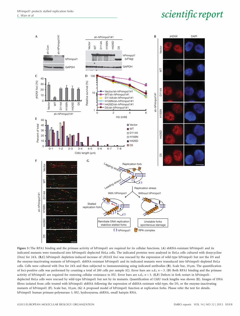
Cellular functions of hPrimpol1 are regulated by RPATo explore the physiological relevance of the enzymatic activitiesand the RPA1-binding ability of hPrimpol1 in replication forkstabilization, we took advantage of the inducible expressionsystem to express the shRNA#1-resistant full-length hPrimpol1,D5, or the primase-inactivating mutants (D114A, H169N andH426D) of hPrimpol1 in hPrimpol1-depleted HeLa cells. Theexpression of wild-type and mutated hPrimpol1 was induced inhPrimpol1 knockdown cells when the cells were treated withdoxycycline (Fig 5A). Notably, neither expression of the D5mutant nor the primase-inactivating mutants was capable ofpreventing the appearance of gH2AX foci and suppressing theelevated HU sensitivity when endogenous hPrimpol1 wassilenced (Fig 5B–D). Likewise, the defects in fork restart inhPrimpol1-depleted cells after replication arrest could be reversedby the expression of wild-type hPrimpol1, but not the D5
and the primase-inactivating mutants of hPrimpol1 (Fig 5E,F;supplementary Table S1 online). These results suggested that boththe RPA1-binding domain and the primase activity of hPrimpol1are critical for its in vivo function.
Re-priming is a mechanism to allow resumption of DNA synthesisfollowing replication fork stalling [20,21]. How the re-primingoccurs is largely unexplored. In this study, we report theidentification of a unique protein designated hPrimpol1, whichmight serve as a primase-polymerase that helps to stabilize stalledreplication forks. We propose that, in response to replication stress,replication forks would be stalled and allow the formation of ssDNAregions that become coated with RPA. Through the interaction withRPA1, hPrimpol1, SMARCAL1/HARP [5–9] and/or other RPA-binding proteins are recruited to these stalled replication forks andexerts their enzymatic activities to reinitiate DNA replicationand thus to limit replication-associated DNA damage (Fig 5G).
WT
D11
4A
H16
9N
H42
6D
M
WT
D11
4A
H16
9N
H42
6D
WT
D11
4A
H16
9N
H42
6D
WT
D11
4A
H16
9N
H42
6D
D5
D5
M
kDa250150 22 nt
10,000
5,000
3 H in
corp
orat
ion
/CP
M
15,000
0
10 nt
Free NTP
22 nt
10 nt
Free NTP
10075
50
37
25
20
kDa25015010075
50
37
25
20
–
A B
D E
C
Figure 3 | hPrimpol1 possesses primase and DNA polymerase activities. (A) SDS–PAGE profile of purified wild-type and mutants of hPrimpol1.
(B) hPrimpol1 is a bona fide primase. Wild type and mutants of hPrimpol1 were incubated with ssDNA template (T20GTCT20) and NTPs containing
[a-32P]-ATP as substrates. Reaction products were separated by 8 M Urea-SDS–PAGE and detected by autoradiography. (C) hPrimpol1 displays
DNA polymerase activity. The ability of wild-type and mutant proteins to synthesize DNA was analysed by classical tritium incorporation assay.
Incorporation of [3H] dTTP into poly(dA)500/oligo(dT)18 was quantified by scintillation counting. Error bars are s.d.; n¼ 3. (D) Both wide-type and the
D5 mutant of hPrimpol1 can elongate the primers synthesized by its own primase activity efficiently. Reactions were carried out as standard primase
assays in Fig. 3B except that 100mM dNTPs were added together with 100mM NTPs containing [a-32P]-ATP. (E) SDS–PAGE profile of purified D5
mutant of hPrimpol1. hPrimpol1 human primase-polymerase 1; SDS–PAGE, SDS–polyacrylamide gel electrophoresis; ssDNA, single-stranded DNA.
Figure 2 | The conserved C terminus of hPrimpol1 is required for RPA1 binding and its foci formation. (A) Schematic representation of wild-type and
deletion mutants of hPrimpol1 used in this study. (B) The 80 amino acids at the C terminus of hPrimpol1 are required for RPA1 binding. Co-
immunoprecipitation experiments were carried out as indicated. (C) Alignment of C-terminal sequences of hPrimpol1 from different species. (D) The
conserved C terminus of hPrimpol1 is responsible for its foci formation. Immunostaining experiments were performed 16 h after HU treatment using
indicated antibodies. Scale bar, 10mm. (E) The conserved C terminus of hPrimpol1 is sufficient for its foci formation. 293T cells were transfected with
SFB-tagged T5 mutant of hPrimpol1. Immunostaining experiments were performed using indicated antibodies. Scale bar, 10 mm. (F) The C-terminal
fragment (T5) of hPrimpol1 is sufficient for RPA1 binding. Co-immunoprecipitation experiments were carried out as indicated. hPrimpol1 human
primase-polymerase 1; HU, hydroxyurea; RPA, replication protein A.
b
hPrimpol1 protects stalled replication forks
L. Wan et al scientificreport
1109&2013 EUROPEAN MOLECULAR BIOLOGY ORGANIZATION EMBO reports VOL 14 | NO 12 | 2013
METHODSPrimase assays. Primase activity was analysed using ssDNAtemplates as described [18,19]. Reaction mixtures (20ml)contained 50 mM Tris–HCl (pH 8.0), 5 mM MgCl2, 1 mMdithiothreitol, 50mg ml� 1 bovine serum albumin, 5mM ssDNAtemplate, 100mM NTP containing [a-32P] ATP or GTP(800 ci mmol�1, PerkinElmer Life Sciences), and 15mg hPrimpol1
protein. After incubation at 37 1C for 30 min, reactions werequenched by adding equal volume of gel-loading buffer (100%formamide, 0.05% bromophenol blue). The sequences ofssDNA template are 50-T20CTGT20-30 (T20CTGT20) and 50-C40-3
0
(poly(dC)40). Products were separated by denaturing poly-acrylamide gel electrophoresis (20% polyacrylamide, 8 M urea)and visualized by autoradiography.
γH2A
X fo
ci (%
)
sh-C
on
sh-h
Primpol1
#1
sh-h
Primpol1
#2
40302010
50
0
Fork restart
No restart
New origin
IdU(20 min)
IdU/CldU
IdU/CldU
IdU/CldU
CldU(20 min)HU (2 h)
10
100
12
sh-Consh-hPrimpol1#1
sh-hPrimpol1#2
40 6HU (mM)
Rel
ativ
e su
rviv
al (%
)
10
100
10 2
sh-Con
sh-hPrimpol1#1sh-hPrimpol1#2
4 6IR (Gy)
Rel
ativ
e su
rviv
al (%
)
A B
γH2A
XC
yclin
AD
AP
I
sh-Con sh-hPrimpol1#1 sh-hPrimpol1#2
C
D
Fsh-Con
sh-hPrimpol1#1sh-hPrimpol1#2
sh-Consh-hPrimpol1#1sh-hPrimpol1#2
HU-
HU+50
40
30
20
10
0
CldU length (mm)
0–1
1–2
2–3
3–4
4–5
5–6
6–7
Per
cent
of t
otal
Per
cent
of t
otal
25
20
15
10
5
0
CldU length (μm)
0–2
2–4
4–6
6–8
8–10
10–1
2
12–1
4
14–1
6
16–1
8
18–2
0
20–2
2
22–2
4
24–2
6
G
H
sh-C
on
sh-h
Primpol1
#1
sh-h
Primpol1
#2
sh-C
on
sh-h
Primpol1
#1
sh-h
Primpol1
#2
HU – – – + + +
p-CHK1
CHK1
hPrimpol1
GAPDH
I
sh-C
on
sh-h
Primpol1
#1
sh-h
Primpol1
#2
P = 0.0435
P = 0.0306
0.1
0.05
0.15
0
Ab
erra
tions
/cel
l
E
Figure 4 | hPrimpol1 functions at stalled replication forks. (A,B) Clonogenic survival assays in hPrimpol1-depleted HeLa cells following HU or IR
treatment. Error bars are s.d.; n¼ 3. (C,D) gH2AX foci are greatly increased in hPrimpol1-depleted HeLa cells. HeLa cells were infected with
lentiviruses carrying non-target control or hPrimpol1 shRNAs. Seventy-two hours later, cells were subjected to immunostaining using indicated
antibodies (C). Scale bar, 10 mm. The quantification of foci-positive cells was performed by counting a total of 200 cells per sample (D). Error bars
are s.d.; n¼ 3. (E) Quantification of chromosomal aberrations in control and hPrimpol1-depleted HeLa cells. At least 50 cells were counted in each
experiment. Error bars are s.d.; n¼ 3; significances of differences by a two-tailed unpaired t-test. (F) Schematic representation of the labelling protocol
for DNA fibre analysis of replication forks. (G,H) CIdU tract length distributions from DNA fibres from control and hPrimpol1-depleted HeLa cells in
the presence (H) or absence of HU (G). (I) hPrimpol1 is not required for CHK1 activation. HeLa cells were introduced with indicated shRNA and then
treated with HU (2 mM) for 2 h. Cells were lysed and immunoblotting was performed using indicated antibodies. hPrimpol1 human primase-
polymerase 1; HU, hydroxyurea; shRNA, small hairpin RNA.
hPrimpol1 protects stalled replication forks
L. Wan et alscientificreport
1110 EMBO reports VOL 14 | NO 12 | 2013 &2013 EUROPEAN MOLECULAR BIOLOGY ORGANIZATION
sh-C
on
sh-h
Prim
pol
1#1
sh-h
Prim
pol
1#1
sh-h
Prim
pol
1#1
sh-hPrimpol1#1
sh-hPrimpol1#1
Vect
or
WT
D11
4A
H16
9N
H42
6D
D5
Vect
or
WT
D11
4A
H16
9N
H42
6D D5
hPrimpol1
GAPDH
hPrimpol1(αFlag)
GAPDH
γH2AX DAPI
Vect
or
Vector
WT
WT
D11
4A
D114A
H16
9N
H169N H42
6D
H426D
D5
D5
Replication fork
Vect
orW
TD
114A
H16
9NH
426D
D5
10
Rel
ativ
e su
rviv
al (%
)
100
12 40 6
HU (mM)
30
20
10
40
0
50
40
30
20
10
0
CldU length (μm)
1–2 2–3 3–4 4–5 5–6 6–70–1 7–8
Per
cent
of t
otal
γH2A
X fo
ci (%
)
Vector/sh-hPrimpol1#1WT/sh-hPrimpol1#1D114A/sh-hPrimpol1#1H169N/sh-hPrimpol1#1H426D/sh-hPrimpol1#1D5/sh-hPrimpol1#1
Lagging strand
Leading strand
With hPrimpol1 Without hPrimpol1
Replication stress
Stalledreplication fork
Reinitiate DNA replicationstabilize stalled forks
Unstable forksspontenous damage
hPrimpol1 RPA complex
A
C D
E
F G
B
Figure 5 | The RPA1 binding and the primase activity of hPrimpol1 are required for its cellular functions. (A) shRNA-resistant hPrimpol1 and its
indicated mutants were transduced into hPrimpol1-deplected HeLa cells. The indicated proteins were analysed in HeLa cells cultured with doxycycline
(Dox) for 24 h. (B,C) hPrimpol1 depletion-induced increase of gH2AX foci was rescued by the expression of wild-type hPrimpol1 but not the D5 and
the enzyme-inactivating mutants of hPrimpol1. shRNA-resistant hPrimpol1 and its indicated mutants were transduced into hPrimpol1-depleted HeLa
cells. Cells were cultured with Dox for 24 h and then subjected to immunostaining using indicated antibodies (B). Scale bar, 10mm. The quantification
of foci-positive cells was performed by counting a total of 200 cells per sample (C). Error bars are s.d.; n¼ 3. (D) Both RPA1 binding and the primase
activity of hPrimpol1 are required for restoring cellular resistance to HU. Error bars are s.d.; n¼ 3. (E,F) Defects in fork restart in hPrimpol1-
deplected HeLa cells were rescued by wild-type hPrimpol1 but not by its mutants. Quantification of CIdU track lengths was shown (E). Images of DNA
fibres isolated from cells treated with hPrimpol1 shRNA following the expression of shRNA-resistant wild-type, the D5, or the enzyme-inactivating
mutants of hPrimpol1 (F). Scale bar, 10 mm. (G) A proposed model of hPrimpol1 function at replication forks. Please refer the text for details.
hPrimpol1 human primase-polymerase 1; HU, hydroxyurea; shRNA, small hairpin RNA.
hPrimpol1 protects stalled replication forks
L. Wan et al scientificreport
1111&2013 EUROPEAN MOLECULAR BIOLOGY ORGANIZATION EMBO reports VOL 14 | NO 12 | 2013
Polymerase assays. Incorporation of 3H-dTTP into poly(dA)500/oligo(dT)18 DNA substrate was carried out as described [19].The poly(dA)500/oligo(dT)18 (1:20, template to primer chains)was annealed in 25 mM Hepes (pH 7.1), 60 mM KCl. Reactionmixtures (60 ml) consisted of 50 mM Tris-HCl (pH 7.5), 8 mMMgCl2, 120 mM NaCl, 2 mM dithiothreitol, 17mg ml�1 poly(dA)500/oligo(dT)18, 10% glycerol, 100 mg ml� 1 bovine serumalbumin, and 50 mM [3H] dTTP (100 cpm pmol� 1). The reactionswere conducted at 37 1C for 30 min. The mixtures were directlyspotted on Whatman DE81 filter paper. The filters were washedfive times with 0.5 M Na2HPO4, two times with water and thenrinsed with 95% ethanol. Radioactivity was monitored byscintillation counter Triathler.
Supplementary information is available at EMBO reports online(http://www.emboreports.org).
ACKNOWLEDGEMENTSWe thank all our colleagues in the Huang laboratory for insightfuldiscussions. This work was supported by National Basic ResearchProgram of China Grants 2012CB944402 and 2013CB911003, NationalNatural Science Foundation of China 31071095 and 31271331, and theChina’s Fundamental Research Funds for the Central Universities.
Author contributions: L.W., J.L., Y.X., B.S., T.L., J.C. and Y.S.performed the experiments; J.H. and H.L. designed the experiments,analysed the data and wrote the manuscript.
CONFLICT OF INTERESTThe authors declare that they have no conflict of interest.
REFERENCES1. Wold MS (1997) Replication protein A: a heterotrimeric, single-stranded
DNA-binding protein required for eukaryotic DNA metabolism. AnnuRev Biochem 66: 61–92
2. Arunkumar AI, Stauffer ME, Bochkareva E, Bochkarev A, Chazin WJ(2003) Independent and coordinated functions of replication protein Atandem high affinity single-stranded DNA binding domains. J Biol Chem278: 41077–41082
3. Haring SJ, Mason AC, Binz SK, Wold MS (2008) Cellular functions ofhuman RPA1. Multiple roles of domains in replication, repair, andcheckpoints. J Biol Chem 283: 19095–19111
4. Fanning E, Klimovich V, Nager AR (2006) A dynamic model forreplication protein A (RPA) function in DNA processing pathways.Nucleic Acids Res 34: 4126–4137
5. Yusufzai T, Kong X, Yokomori K, Kadonaga JT (2009) The annealinghelicase HARP is recruited to DNA repair sites via an interaction withRPA. Genes Dev 23: 2400–2404
6. Yuan J, Ghosal G, Chen J (2009) The annealing helicase HARP protectsstalled replication forks. Genes Dev 23: 2394–2399
7. Bansbach CE, Betous R, Lovejoy CA, Glick GG, Cortez D (2009) Theannealing helicase SMARCAL1 maintains genome integrity at stalledreplication forks. Genes Dev 23: 2405–2414
8. Ciccia A, Bredemeyer AL, Sowa ME, Terret ME, Jallepalli PV, Harper JW,Elledge SJ (2009) The SIOD disorder protein SMARCAL1 is an RPA-interacting protein involved in replication fork restart. Genes Dev 23:2415–2425
9. Couch FB et al (2013) ATR phosphorylates SMARCAL1 to preventreplication fork collapse. Genes Dev 27: 1610–1623
10. Xu X, Vaithiyalingam S, Glick GG, Mordes DA, Chazin WJ,Cortez D (2008) The basic cleft of RPA70N binds multiple checkpointproteins, including RAD9, to regulate ATR signaling. Mol Cell Biol 28:7345–7353
11. Frick DN, Richardson CC (2001) DNA primases. Annu Rev Biochem 70:39–80
12. Kuchta RD, Stengel G (2010) Mechanism and evolution of DNAprimases. Biochim Biophys Acta 1804: 1180–1189
13. Lao-Sirieix SH, Pellegrini L, Bell SD (2005) The promiscuous primase.Trends Genet 21: 568–572
14. Swiatek A, Macneill SA (2010) The archaeo-eukaryotic GINS proteinsand the archaeal primase catalytic subunit PriS share a common domain.Biol Direct 5: 17
15. Iyer LM, Koonin EV, Leipe DD, Aravind L (2005) Origin and evolution ofthe archaeo-eukaryotic primase superfamily and related palm-domainproteins: structural insights and new members. Nucleic Acids Res 33:3875–3896
16. Lipps G, Rother S, Hart C, Krauss G (2003) A novel type of replicativeenzyme harbouring ATPase, primase and DNA polymerase activity.EMBO J 22: 2516–2525
17. Lipps G, Weinzierl AO, von Scheven G, Buchen C, Cramer P (2004)Structure of a bifunctional DNA primase-polymerase. Nat Struct Mol Biol11: 157–162
18. Ramirez-Aguilar KA, Low-Nam NA, Kuchta RD (2002) Key role oftemplate sequence for primer synthesis by the herpes simplex virus 1helicase-primase. Biochemistry 41: 14569–14579
19. Lou H, Duan Z, Huo X, Huang L (2004) Modulation of hyperthermophilicDNA polymerase activity by archaeal chromatin proteins. J Biol Chem279: 127–132
20. Petermann E, Helleday T (2010) Pathways of mammalian replication forkrestart. Nat Rev Mol Cell Biol 11: 683–687
21. Elvers I, Johansson F, Groth P, Erixon K, Helleday T (2011) UV stalledreplication forks restart by re-priming in human fibroblasts. Nucleic AcidsRes 39: 7049–7057
hPrimpol1 protects stalled replication forks
L. Wan et alscientificreport
1112 EMBO reports VOL 14 | NO 12 | 2013 &2013 EUROPEAN MOLECULAR BIOLOGY ORGANIZATION