Embed Size (px)
Citation preview
RESEARCH ARTICLES◥
PHOTOSYNTHESIS
Structural basis for energytransfer pathways in the plantPSI-LHCI supercomplexXiaochun Qin,1,2* Michihiro Suga,2* Tingyun Kuang,1† Jian-Ren Shen1,2†
Photosynthesis converts solar energy to chemical energy by means of two largepigment-protein complexes: photosystem I (PSI) and photosystem II (PSII). In higherplants, the PSI core is surrounded by a large light-harvesting complex I (LHCI) thatcaptures sunlight and transfers the excitation energy to the core with extremely highefficiency. We report the structure of PSI-LHCI, a 600-kilodalton membrane proteinsupercomplex, from Pisum sativum (pea) at a resolution of 2.8 angstroms. The structurereveals the detailed arrangement of pigments and other cofactors—especially withinLHCI—as well as numerous specific interactions between the PSI core and LHCI. Theseresults provide a firm structural basis for our understanding on the energy transferand photoprotection mechanisms within the PSI-LHCI supercomplex.
Photosynthesis uses light energy from theSun to convert CO2 and water into carbo-hydrates and oxygen, thus sustaining allaerobic life forms on Earth. In oxygenicphotosynthesis, photosystem II (PSII) uses
light energy to split water into oxygen, protons,and electrons, whereas photosystem I (PSI) isresponsible for the reduction of nicotinamideadenine dinucleotide phosphate (NADP+) intoNADPH. In higher plants, the PSI core is sur-rounded by the light-harvesting complex I (LHCI),forming a PSI-LHCI supercomplex, to facilitateefficient energy harvesting while avoiding con-centration quenching (1). The energy absorbedby LHCI is transferred to the PSI core, where itis converted into chemical energy with a quan-tum efficiency close to 100% (2).The PSI core exists as a homotrimer in cyano-
bacteria, and its structure has been determinedat 2.5 Å resolution (3). Each monomer contains12 protein subunits, 128 cofactors [96 chloro-phylls (Chls), two phylloquinones, three Fe4S4clusters, 22 carotenoids, four lipids, and a Ca2+
ion]. By contrast, the higher plant PSI-LHCI super-complex exists in a monomeric form and con-tains four unique subunits (PsaG, PsaH, PsaN,and PsaO) in the core, as well as subunits homol-ogous to cyanobacterial PSI (4) and four Lhcasubunits (Lhca1 to Lhca4) in the LHCI complex.The structure of the higher plant PSI-LHCI super-complex has been solved at 4.4 Å to 3.3 Å res-olutions from P. sativum var. Alaska (5–7), which
revealed the presence of 17 protein subunits (13core subunits and four Lhcas) and 199 cofactors[174 chlorophyll a molecules (Chls a), 19 carote-noids, two phylloquinones, three Fe4S4 clusters,and one lipid] (PDB ID: 3LW5) with a total mo-lecular mass of 600 kD (7). However, the resolu-tions achieved so far were not high enough toreveal the detailed structure of PSI-LHCI, espe-cially with respect to the position and numberof cofactors associated with LHCI.We crystallized PSI-LHCI from P. sativum var.
Alaska and analyzed its structure at 2.8 Å reso-lution (figs. S1 to S5 and table S1) (8). This im-proved resolution reveals the detailed organizationof protein subunits and cofactors, enabling themechanisms of energy transfer, regulation, andphotoprotection within the PSI-LHCI super-complex to be examined on a more robust struc-tural basis.
Architecture of thePSI-LHCI supercomplex
The overall structure of PSI-LHCI resembles asemispherical shape similar to that reportedpreviously, with a dimension of 140 Å × 40 Å(Fig. 1, A and C). The supercomplex contains 16subunits but does not include PsaN and PsaO,which suggests that these two subunits were lostduring purification or crystallization owing totheir loose association with the PSI core (9, 10).Four LHCI subunits, Lhca1 to Lhca4, are arrangedin the form of a heterodimer of two heterodimers(Lhca1-Lhca4 and Lhca2-Lhca3) and are attachedto one side of the PSI core where PsaG, PsaF, PsaJ,and PsaK are located (Fig. 1A).In addition to the protein subunits, we as-
signed 155 Chls (143 Chls a and 12 Chls b), 35carotenoids [26 b-carotenes (BCRs), five luteins(Luts), and four violaxanthins (Vios)], 10 lipids[six phosphatidylglycerols (PGs), three monoga-
lactosyldiacylglycerols (MGDGs), and one digalac-tosyldiacylglycerol (DGDG)], three Fe4S4 clusters,two phylloquinones, and several water molecules(Fig. 1B and table S2). We identified fewer Chlsthan in the previous study (155 versus 174) (7),which may be due to ambiguities in the electrondensity of the previous lower-resolution struc-ture. The composition of the pigments we as-signed in the crystal structure largely agreeswith the results of our chemical analysis by high-performance liquid chromatography (HPLC)(table S3). [For a detailed assignment of thesecofactors and comparison with the previousstructure (6, 7), see table S2 and fig. S6.] Thereare two monomers in the crystallographic asym-metric unit; because the monomers are identi-cal, we describe the PSI-LHCI structure of themonomer whose chains are labeled by capitalletters in the PDB file.
Structure of the Lhca apoproteins
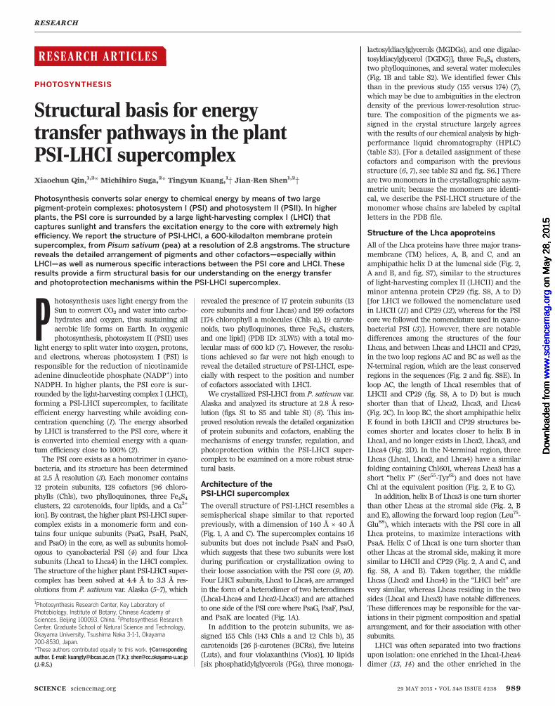
All of the Lhca proteins have three major trans-membrane (TM) helices, A, B, and C, and anamphipathic helix D at the lumenal side (Fig. 2,A and B, and fig. S7), similar to the structuresof light-harvesting complex II (LHCII) and theminor antenna protein CP29 (fig. S8, A to D)[for LHCI we followed the nomenclature usedin LHCII (11) and CP29 (12), whereas for the PSIcore we followed the nomenclature used in cyano-bacterial PSI (3)]. However, there are notabledifferences among the structures of the fourLhcas, and between Lhcas and LHCII and CP29,in the two loop regions AC and BC as well as theN-terminal region, which are the least conservedregions in the sequences (Fig. 2 and fig. S8E). Inloop AC, the length of Lhca1 resembles that ofLHCII and CP29 (fig. S8, A to D) but is muchshorter than that of Lhca2, Lhca3, and Lhca4(Fig. 2C). In loop BC, the short amphipathic helixE found in both LHCII and CP29 structures be-comes shorter and locates closer to helix B inLhca1, and no longer exists in Lhca2, Lhca3, andLhca4 (Fig. 2D). In the N-terminal region, threeLhcas (Lhca1, Lhca2, and Lhca4) have a similarfolding containing Chl601, whereas Lhca3 has ashort “helix F” (Ser55-Tyr61) and does not haveChl at the equivalent position (Fig. 2, E to G).In addition, helix B of Lhca3 is one turn shorter
than other Lhcas at the stromal side (Fig. 2, Band E), allowing the forward loop region (Leu75-Glu88), which interacts with the PSI core in allLhca proteins, to maximize interactions withPsaA. Helix C of Lhca1 is one turn shorter thanother Lhcas at the stromal side, making it moresimilar to LHCII and CP29 (Fig. 2, A and C, andfig. S8, A and B). Taken together, the middleLhcas (Lhca2 and Lhca4) in the “LHCI belt” arevery similar, whereas Lhcas residing in the twosides (Lhca1 and Lhca3) have notable differences.These differences may be responsible for the var-iations in their pigment composition and spatialarrangement, and for their association with othersubunits.LHCI was often separated into two fractions
upon isolation: one enriched in the Lhca1-Lhca4dimer (13, 14) and the other enriched in the
RESEARCH
SCIENCE sciencemag.org 29 MAY 2015 • VOL 348 ISSUE 6238 989
1Photosynthesis Research Center, Key Laboratory ofPhotobiology, Institute of Botany, Chinese Academy ofSciences, Beijing 100093, China. 2Photosynthesis ResearchCenter, Graduate School of Natural Science and Technology,Okayama University, Tsushima Naka 3-1-1, Okayama700-8530, Japan.*These authors contributed equally to this work. †Correspondingauthor. E-mail: [email protected] (T.K.); [email protected](J.-R.S.)
on
May
28,
201
5w
ww
.sci
ence
mag
.org
Dow
nloa
ded
from
o
n M
ay 2
8, 2
015
ww
w.s
cien
cem
ag.o
rgD
ownl
oade
d fr
om
on
May
28,
201
5w
ww
.sci
ence
mag
.org
Dow
nloa
ded
from
o
n M
ay 2
8, 2
015
ww
w.s
cien
cem
ag.o
rgD
ownl
oade
d fr
om
on
May
28,
201
5w
ww
.sci
ence
mag
.org
Dow
nloa
ded
from
o
n M
ay 2
8, 2
015
ww
w.s
cien
cem
ag.o
rgD
ownl
oade
d fr
om
on
May
28,
201
5w
ww
.sci
ence
mag
.org
Dow
nloa
ded
from

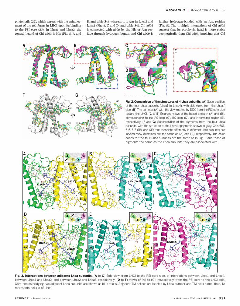
Lhca2-Lhca3 dimer (15). From our structure,the Lhca-Lhca intersubunit interactions canbe categorized into two types: (i) hydrophilicinteractions between the N- and C-terminal re-gions of one Lhca with helix C of the adjacentLhca, and (ii) hydrophobic interactions betweenChls and carotenoids from helix A of one Lhcawith those from helix C of the adjacent Lhca(Fig. 3). Our structure indicates that both typesof interactions are stronger between Lhca1 andLhca4, slightly weaker between Lhca2 and Lhca3,and much weaker between Lhca2 and Lhca4.In particular, two bridging carotenoids, BCR623and Lut624, were found in the region betweenLhca1 and Lhca4, maximizing their interactions(Fig. 3, A and D), whereas only BCR623 was foundbetween Lhca2 and Lhca4 (Fig. 3, B and E) andbetween Lhca2 and Lhca3 (Fig. 3, C and F), lead-ing to weaker interactions between them.
Arrangement and functionsof Chls in LHCI
We identified 75 cofactors in the four Lhca sub-units: 45 Chls a, 12 Chls b, four BCRs, five Luts,four Vios, three PGs, and two MGDGs (Fig. 1B,figs. S9 and S10, and tables S2 and S4). The num-bers of Chls b and carotenoids assigned are verysimilar to those determined chemically (tableS3), which gives rise to a Chl a/b ratio of 3.75,consistent with that of 3.71 from previous bio-chemical analysis (15). The Chls in LHCI are
distributed into two layers, one close to thestromal and the other one close to the lumenalsurface (Fig. 4). The stromal-side Chl layer con-tains 36 Chls (29 Chls a and 7 Chls b) with anaverage Mg-to-Mg distance of 11.3 Å (Fig. 4B).The lumenal-side Chl layer has a less dense pack-ing composed of 16 Chls a and 5 Chls b, whichare separated into two clusters in each Lhca(Fig. 4C) with an average Mg-to-Mg distanceof 10.6 Å within each cluster and 19.7 Å betweenthe clusters. The shortest distance between thestromal- and lumenal-side layers is 11.6 Å, fromChl a607 to Chl a619 in Lhca3.In comparison with LHCII, there are unique
Chls found in LHCI with either different orienta-tions or positions, most of which are located atthe gap region between LHCI and the PSI core,or in connecting regions between adjacent Lhcas(Fig. 4, fig. S8, C and D, and table S4). Among them,Chls 601 in Lhca1, Lhca2, and Lhca4 have sim-ilar Qx and Qy transition dipole moments (Fig. 2,F and G, and table S4), but different from theones in LHCII. Chl a607 in Lhca3 is unusual be-cause other Lhcas bind Chl b at this site, and it isalso closer to the PSI core than is Chl b607, whichsuggests that Chl a607 may promote energytransfer from Lhca3 to the PSI core. In addition,Chl a616 in Lhca1, Chl a617 in Lhca3 and Lhca4,Chl b618 in Lhca2 and Lhca4, and Chl a619 inLhca4 were not found in LHCII (Fig. 2, F and G,Fig. 4, fig. S8, C and D, and table S4).
Among the 12 Chls b in LHCI, two (b601, b607)are bound to Lhca1, five (b601, b606, b607, b608,and b618) to Lhca2, one (b608) to Lhca3, and four(b606, b607, b608, and b618) to Lhca4 (Fig. 4).The uneven distribution of Chl b indicates thatLhca2 and Lhca4 have a higher affinity for Chl bthan do Lhca1 and Lhca3, in agreement withresults from in vitro reconstitution experiments(16, 17). Nearly all Chls b are located in helix Cor the N-terminal loop, both of which form theinterfacial regions of two adjacent Lhca proteins(Figs. 1 and 4), which suggests that they may beimportant in mediating energy transfer and/orinteractions between adjacent Lhca subunits.A striking feature of Lhcas is the presence of
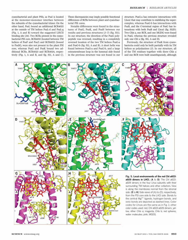
red forms of several Chls a that are importantfor energy transfer and trapping in the wholePSI complex (18), through which most of har-vested energy is transferred to the reaction centerwith high efficiencies (19–21). The a603-a609dimers in each Lhca (Fig. 5) have a close distanceand some overlap between their C and E rings,reflecting their nature as red Chls. All four Chl603-609 dimers are bound at the inside of LHCI,with their phytol tails protruding into the gapregion between LHCI and the PSI core, in goodagreement with their roles of mediating energytransfer from LHCI to the PSI core. The inter-actions between the PSI core and LHCI may af-fect the conformation of the red dimers, especiallythrough the interactions with the hydrophobic
990 29 MAY 2015 • VOL 348 ISSUE 6238 sciencemag.org SCIENCE
Fig. 1. Overall structure of the PSI-LHCI supercomplex from Pisum sativumat a resolution of 2.8 Å. (A) View along the membrane normal from the stromalside. (B) Arrangement of pigments and other cofactors (lipids, Fe4S4 clusters,and phylloquinones), with the view direction same as in (A). For clarity, the pro-tein backbones and phytol chains of Chls are omitted. (C) Side view of the PSI-LHCI supercomplex from the LHCI side, with the phytol chains of Chls omitted.Color codes for all panels: PSI core subunits: pink, PsaA; gray, PsaB; light blue,PsaC; khaki, PsaD; light green, PsaE; orange, PsaF; blue, PsaG and PsaK; red, PsaH;purple, PsaI and PsaJ; green, PsaL. Lhca proteins: green, Lhca1; cyan, Lhca2; magenta,Lhca3; yellow, Lhca4. Cofactors: green, Chls a of PSI core complex; yellow, Chls aof LHCI; magenta, Chls b of LHCI; blue, carotenoids; black, lipids; red, cofactors ofthe electron transfer chain (Chls a, phylloquinones, and Fe4S4 clusters).
RESEARCH | RESEARCH ARTICLES
phytol tails (22), which agrees with the enhance-ment of the red forms in LHCI upon its bindingto the PSI core (23). In Lhca1 and Lhca2, thecentral ligand of Chl a603 is His (Fig. 5, A and
B, and table S4), whereas it is Asn in Lhca3 andLhca4 (Fig. 5, C and D, and table S4). Chl a603is connected with a609 by the His or Asn res-idue through hydrogen bonds, and Chl a609 is
further hydrogen-bonded with an Arg residue(Fig. 5). The multiple interactions of Chl a609suggest that its porphyrin head is more stablegeometrically than Chl a603, implying that Chl
SCIENCE sciencemag.org 29 MAY 2015 • VOL 348 ISSUE 6238 991
Fig. 3. Interactions between adjacent Lhca subunits. (A to C) Side view, from LHCI to the PSI core side, of interactions between Lhca1 and Lhca4,between Lhca4 and Lhca2, and between Lhca2 and Lhca3, respectively. (D to F) Views of (A) to (C), respectively, from the PSI core to the LHCI side.Carotenoids bridging two adjacent Lhca subunits are shown as blue sticks. Adjacent TM helices are labeled by Lhca number and TM helix name; thus, 1Arepresents helix A of Lhca1.
Fig. 2. Comparison of the structures of 4 Lhca subunits. (A) Superpositionof the four Lhca subunits (Lhca1 to Lhca4), with side views from the Lhcas’side. (B) The same as (A) with the view rotated by 180°, from the PSI core sidetoward the LHCI. (C to E) Enlarged views of the boxed areas in (A) and (B),corresponding to the AC loop (C), BC loop (D), and N-terminal region (E),respectively. (F and G) Superposition of the pigments from the four Lhcasubunits, with the structure of the Lhca1 apoprotein shown in gray. Chls 601,616, 617, 618, and 619 that associate differently in different Lhca subunits arelabeled. View directions are the same as (A) and (B), respectively. The colorcodes for the four Lhca subunits are the same as in Fig. 1, and those ofpigments the same as the Lhca subunits they are associated with.
RESEARCH | RESEARCH ARTICLES

a603 may be more likely to undergo confor-mational modulation. This suggests a functionaldifference between the individual Chls in thered dimer.
Arrangement and functionof carotenoids in LHCI
Our structure reveals that each Lhca binds threecarotenoids (one Lut, one Vio, and one BCR) atthree sites (L1, L2, and N1, respectively), and an-other lutein (Lut624) is bound between Lhca1and Lhca4, giving rise to a total of 13 carotenoidsin the four Lhcas (Fig. 1B, Fig. 4, fig. S9, and tableS4). Lut620 (L1 site) and Vio621 (L2 site) are inall-trans configurations and are located in thetwo elongated grooves on the two sides of helicesA and B. The third carotenoid is an all-transb-carotene, BCR623, located at the N1 site. Thepresence of these three kinds of carotenoids isconsistent with previous and present biochem-ical analysis (15, 16, 24, 25) (table S3), and theLhcas do not bind any carotenoids at the V1site found in LHCII (although the V1 site maybe partially occupied by a BCR from an adjacentLhca). Among the three carotenoid-binding sitesof Lhca, the L1 site is conserved also in LHCIIand CP29 (fig. S8, C and D, and table S4), whereit has been suggested to function as a nonphoto-chemical quencher to promote energy dissipationfrom the nearby, lowest excited energy-bearingChl a610-a611-a612 cluster in both major (26) andminor LHCIIs (27). We observed the same orient-ation of Lut620 and the surrounding Chl a610-a611-a612 cluster in Lhcas; however, becausethe Chl a610-a611-a612 cluster is not the sitewith the lowest energy level in LHCI (thelowest energy level is located in the red Chls),the efficiency of quenching through this pathwaywould be lower in LHCI than that in LHCIIs.The occupancy of the L2 site is variable de-
pending on different proteins (i.e., Lut in LHCII,Vio in minor LHCIIs and LHCI) (table S4). Vioin LHCIIs may be changed to zeaxanthin (Zea) byVio de-epoxidase, which is important for photo-protection by quenching the 1Chl* states (28–30).In LHCI, in vivo studies also suggested the pos-sible conversion of Vio to Zea (31–33), and indeeda Zea-dependent quenching of the excitationenergy in LHCI has been observed recently (33).However, the extent and role of the xanthophyllcycle in PSI are not clear, and the de-epoxidationof LHCI may depend on affinities of the Viobinding sites, as well as the interaction betweenLHCI and the PSI core. Our structure revealedpossible functional differences between the L2sites of different Lhcas, as the Chls (a/b606, a/b607) surrounding this site are involved in adifferent hydrogen-bond network. In Lhca2 andLhca4, the central ligand for both Chl b606and Chl b607 is water (Fig. 5, B, D, F, and H),and the C7-formyl group of Chl b606 forms ahydrogen bond to the coordinated water of Chlb607, similar to the arrangement seen in LHCIIand CP29. On the other hand, the central ligandof Chl a/b607 is Gln105 in Lhca1 (Fig. 5, A and E)and Val135 in Lhca3 (Fig. 5, C and G), neither ofwhich forms hydrogen bonds with Chl a606.
Furthermore, a Glu residue located at helix Cclose to the lumenal side in all Lhcas and CP29interacts with both Chl 607 and/or Chl 606 byhydrogen bonds (Fig. 5). The differences in theircoordination pattern may suggest a differentrole of these pigments in energy absorption orquenching.
Structure of the PSI core
The PSI core contains 98 Chls a, 22 BCRs, five lip-ids (three PGs, one MGDG, and one DGDG), threeFe4S4 clusters, and two phylloquinones (Fig. 1B,fig. S10, and tables S2 and S5). Of the 22 BCRs,17 have an all-trans configuration and 5 contain
one or two cis bonds (two 9-cis, one 9,9´-cis, one9,13´-cis, and one 13-cis) (table S5). Most of theBCRs were located in the same position as thoseobserved in the cyanobacterial PSI, and the cis andtrans conformations of carotenoids are identical inthe cyanobacterial and plant PSI cores (table S5),indicating that they have been conserved over1.5 billion years of evolution. However, severalchanges were observed in the position of carote-noids between the two species. BCR4022, boundat the outside of TM helices PsaL-e, PsaL-g, andPsaI in the cyanobacterial PSI core, is not presentin the plant PSI core; this may be related to thedistinctly different environment of PsaI between
992 29 MAY 2015 • VOL 348 ISSUE 6238 sciencemag.org SCIENCE
Fig. 4. Arrangements of pigments within LHCI. (A and D) Side view, from the LHCI side, of theoverall arrangement of pigments in the four Lhcas. (B and C) View of the pigment arrangements along themembrane normal from the stromal and lumenal sides, respectively [view directions indicated by arrow-heads in (A)]. In (A) to (C), Chls a and Chls b are represented by gray and orange spheres, respectively, atthe positions of their Mg2+ ions, with Mg-to-Mg distances of adjacent Chls given in Å.The numbers of Chlsare labeled with the last one or two digits. Lhca subunits are depicted in the same colors as in Fig. 1, andcarotenoids are depicted in the same color as the Lhca subunits they are associated with. In (D), the threecarotenoid binding sites L1 (Lut620), L2 (Vio621), and N1 (BCR623) are shown in green, yellow, andmagenta, respectively. Lut624 in the L3 site of Lhca4 is shown in light blue.
RESEARCH | RESEARCH ARTICLES
cyanobacterial and plant PSIs, as PsaI is locatedat the monomer-monomer interface betweenthe subunits of the cyanobacterial trimer. On theother hand, PsaL bound an additional BCR4025at the outside of TM helices PsaL-d and PsaL-g(Fig. 1, A and B) toward the suggested LHCIIbinding site (34). Two BCRs present in the cyano-bacterial PSI core, BCR4015 (located between TMhelices of PsaF and PsaJ) and BCR4021 (boundto PsaM), were also not present in the plant PSIcore, whereas PsaG and PsaK bound two ad-ditional BCRs, BCR4023 and BCR4024, respec-tively (Fig. 1, A and B, and fig. S11, A and C).
These discrepancies may imply possible functionaldifferences of BCRs between plant and cyanobac-terial PSI cores.Notable differences were found in the struc-
tures of PsaG, PsaK, and PsaH between ourresults and previous structures (5–7) (fig. S11).In our structure, the direction of the PsaG poly-peptide was reversed, resulting in a completelyreversed location of the two TM helices PsaG-aand PsaG-b (fig. S11, A and B). A short helix wasfound between PsaG-a and PsaG-b, and a largeextramembrane loop in the lumenal side foundin the previous structure was not found in our
structure. PsaG-a has extensive interactions withLhca1 that may contribute to stabilizing the super-complex, whereas PsaG-b has interactions withPsaB, and the C-terminal region of PsaG has in-teractions with both PsaB and Lhca1 (fig. S12D).Two Chls a, one BCR, and one MGDG were foundin PsaG, whereas the previous structure revealedonly one Chl a (fig. S11, A and B).Previously, the structure of PsaK from cyano-
bacteria could only be built partially with its TMhelices as polyalanines (3). In our structure, allof the TM residues together with three Chls aand one BCR were built unambiguously, although
SCIENCE sciencemag.org 29 MAY 2015 • VOL 348 ISSUE 6238 993
Fig. 5. Local environments of the red Chl a603-a609 dimers in LHCI. (A to D) The Chl a603-a609 dimers in the four Lhca subunits with theirsurrounding TM helices and other cofactors. Viewis along the membrane normal from the stromalside. (E toH) Side views of (A) to (D), respectively,from the PSI core side to the LHCI side. Bonds tothe central Mg2+ ligands, hydrogen bonds, andionic bonds are depicted as dashed lines. Colorcodes for Lhcas are the same as in Fig. 1; othercolor codes used: red, Chl a603-a609 dimers; yel-low, other Chls a; magenta, Chls b; red spheres,water molecules; pink, MGDG.
RESEARCH | RESEARCH ARTICLES
the loop region connecting the two helices couldnot be built because of the relatively poor elec-tron density around this region (fig. S11C). Al-though the structure of helix PsaK-a was similarto that reported previously for plant PSI (6, 7),the locations of PsaK-b, together with Chls, werecompletely different, and no BCR was found inPsaK in the previous structure (6, 7) (fig. S11D).Because the sequences of plant PsaK and PsaGare similar to that of cyanobacterial PsaK, PsaGhas been suggested to arise from PsaK via geneduplication (35). Our structure shows that PsaKindeed has a structure similar to that of PsaG(fig. S11, E and F), which suggests that theymay have similar functions. Because both sub-units are required to enable energy transfer from
LHCI to the PSI core, the common structuralfeatures may reflect the prerequisites for suchfunctions.The structure of PsaH differs from the previous
model (6, 7), especially in the N-terminal region(fig. S11G). In our structure, PsaH extensively in-teracts with PsaL as well as PsaD and PsaB in thestromal side. On the lumenal side, PsaH interactswith PsaB, PsaI, and PsaL (fig. S11, H and I). Theinteractions with PsaB, PsaD, PsaI, and PsaL arein agreement with previous cross-linking results(36) and also explain PsaH’s function in stabiliz-ing PSI (37). The interaction with PsaD may ex-plain why PsaH is essential for efficient electrontransfer in PSI (37) because PsaC, PsaD, and PsaEform the docking site for ferredoxin (38) and
PsaC carries the terminal Fe4S4 clusters. Thesestructural features illustrate the importance ofPsaH in stabilizing the PSI monomer and main-taining the function of plant PSI.
Interactions and possible energy transferpathways between LHCI and the PSI core
Our structure reveals extensive and specificprotein-protein and pigment-pigment interac-tions between each Lhca and PSI core subunits(Fig. 1, A and B, and fig. S12). At the stromal side,the region between the N-terminal loop andthe beginning of helix B in Lhca1, Lhca2, andLhca4 interacts with PsaB-Pro311 and severalcofactors, the N-terminal region of PsaJ, and theregion around Phe207-Asn222 of PsaF, respectively,
994 29 MAY 2015 • VOL 348 ISSUE 6238 sciencemag.org SCIENCE
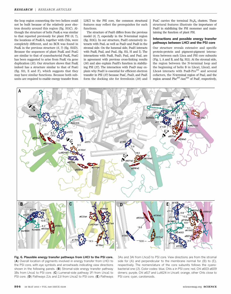
Fig. 6. Plausible energy transfer pathways from LHCI to the PSI core.(A) Overall location of pigments involved in energy transfer from LHCI tothe PSI core, with eye symbols and arrowheads indicating view directionsshown in the following panels. (B) Stromal-side energy transfer pathway1Bs from Lhca1 to PSI core. (C) Lumenal-side pathway 1Fl from Lhca1 toPSI core. (D) Pathways 2Js and 2Jl from Lhca2 to PSI core. (E) Pathways
3As and 3Al from Lhca3 to PSI core. View directions are from the stromalside for (A) and perpendicular to the membrane normal for (B) to (E),respectively. The nomenclature of the core subunits follows the cyano-bacterial one (3). Color codes: blue, Chls a in PSI core; red, Chl a603-a609dimers; purple, Chl a617 and Lut624 in Lhca4; orange, other Chls close toPSI core; cyan, carotenoids.
RESEARCH | RESEARCH ARTICLES
whereas Gln57 in helix F and residues Pro74 toIle87 at the N-terminal loop of Lhca3 interactwith several regions of PsaA (fig. S12, B to E). Inaddition, BCR623 and helix C at the peripheralregion of Lhca1 interact with MGDG and TMhelix PsaG-a (fig. S12D), and residues Ile237,Leu240, Val241, Leu266, and Chl a607 at theperipheral region of Lhca3 interact with PsaAat the lumenal side (fig. S12, E and F).Examination of nearest-neighbor relationships
between pigments revealed a number of possi-ble energy transfer pathways from Lhcas to thePSI core (table S6), among which four appear tobe most plausible; these were designated 1Bs, 1Fl,2Js, and 3As/3Al (according to Lhca number, PSIcore subunit, and stromal or lumenal side) (Fig.6 and table S6). The 1Bs pathway indicates pos-sible energy transfer from the Chl dimer a603-a609of Lhca1 to three Chls (a1218, a1219, and a1802) ofPsaB at the stromal side, with the shortest edge-to-edge distance of 7.5 Å (Fig. 6, A and B). Chla616 of Lhca1 may also guide energy to Chl a1701of PsaF at the lumenal side, providing the 1Flpathway (Fig. 6, A and C). Because of the largegap between Lhca4 and the PSI core, a directenergy transfer from Lhca4 to the PSI core wouldbe difficult, and Lhca4 may guide its energy to thePSI core via its red forms Chl a603-a609 throughthe 1Fl pathway. Chl a603 of Lhca2 is slightlydistant to accomplish a direct energy transfer toChl a1302 of PsaJ (Fig. 6, A and D), whichsuggests that the 2Js pathway may not be highlyefficient. The Chl trimer (a603-a609-a619) ofLhca3 is close to Chl a1108 and the a1110-a1118dimer of PsaA at the stromal side, forming the3As pathway (Fig. 6, A and E). Because of thestrong coupling between Chl a1110-a1118, thispathway suggests energy transfer from red formsof Lhca3 to this Chl pair in the PSI core. At thelumenal side, Chl a607 of Lhca3 is close to anotherChl a1112-a1114 dimer of PsaA, providing the 3Alpathway (Fig. 6, A and E). In addition, Chls a613and a614 of Lhca3 are close to Chl a1002 of PsaK(3Kl pathway), which may also facilitate energytransfer from Lhca3 to the PSI core.
REFERENCES AND NOTES
1. G. D. Scholes, G. R. Fleming, A. Olaya-Castro, R. van Grondelle,Nat. Chem. 3, 763–774 (2011).
2. N. Nelson, J. Nanosci. Nanotechnol. 9, 1709–1713 (2009).3. P. Jordan et al., Nature 411, 909–917 (2001).4. H. V. Scheller, P. E. Jensen, A. Haldrup, C. Lunde, J. Knoetzel,
Biochim. Biophys. Acta 1507, 41–60 (2001).5. A. Ben-Shem, F. Frolow, N. Nelson, Nature 426, 630–635 (2003).6. A. Amunts, O. Drory, N. Nelson, Nature 447, 58–63 (2007).7. A. Amunts, H. Toporik, A. Borovikova, N. Nelson, J. Biol. Chem.
285, 3478–3486 (2010).8. See supplementary materials on Science Online.9. W. Z. He, R. Malkin, FEBS Lett. 308, 298–300 (1992).10. P. E. Jensen, A. Haldrup, S. Zhang, H. V. Scheller, J. Biol. Chem.
279, 24212–24217 (2004).11. Z. Liu et al., Nature 428, 287–292 (2004).12. X. Pan et al., Nat. Struct. Mol. Biol. 18, 309–315 (2011).13. T. Y. Kuang, J. H. Argyroudi-Akoyunoglou, H. Y. Nakatani, J. Watson,
C. J. Arntzen, Arch. Biochem. Biophys. 235, 618–627 (1984).14. E. Lam, W. Ortiz, R. Malkin, FEBS Lett. 168, 10–14 (1984).15. E. Wientjes, R. Croce, Biochem. J. 433, 477–485 (2011).16. R. Croce, T. Morosinotto, S. Castelletti, J. Breton, R. Bassi,
Biochim. Biophys. Acta 1556, 29–40 (2002).17. S. Castelletti et al., Biochemistry 42, 4226–4234 (2003).18. B. Gobets, R. van Grondelle, Biochim. Biophys. Acta 1507,
80–99 (2001).
19. T. Morosinotto, J. Breton, R. Bassi, R. Croce, J. Biol. Chem.278, 49223–49229 (2003).
20. T. Morosinotto, M. Mozzo, R. Bassi, R. Croce, J. Biol. Chem.280, 20612–20619 (2005).
21. R. Croce, H. van Amerongen, Photosynth. Res. 116, 153–166 (2013).22. L. Fiedor, A. Kania, B. Myśliwa-Kurdziel, Ł. Orzeł, G. Stochel,
Biochim. Biophys. Acta 1777, 1491–1500 (2008).23. T. Morosinotto, M. Ballottari, F. Klimmek, S. Jansson, R. Bassi,
J. Biol. Chem. 280, 31050–31058 (2005).24. V. H. R. Schmid et al., J. Biol. Chem. 277, 37307–37314 (2002).25. X. Qin et al., Photosynth. Res. 90, 195–204 (2006).26. A. V. Ruban et al., Nature 450, 575–578 (2007).27. M. Mozzo, F. Passarini, R. Bassi, H. van Amerongen, R. Croce,
Biochim. Biophys. Acta 1777, 1263–1267 (2008).28. B. Demmig, K. Winter, A. Krüger, F. C. Czygan, Plant Physiol.
84, 218–224 (1987).29. A. M. Gilmore, H. Y. Yamamoto, Plant Physiol. 96, 635–643 (1991).30. K. K. Niyogi, A. R. Grossman, O. Björkman, Plant Cell 10,
1121–1134 (1998).31. A. Wehner, S. Storf, P. Jahns, V. H. R. Schmid, J. Biol. Chem.
279, 26823–26829 (2004).32. T. Morosinotto, S. Caffarri, L. Dall'Osto, R. Bassi, Physiol. Plant.
119, 347–354 (2003).33. M. Ballottari et al., Proc. Natl. Acad. Sci. U.S.A. 111, E2431–E2438
(2014).34. R. Kouřil et al., Biochemistry 44, 10935–10940 (2005).35. S. Kjaerulff, B. Andersen, V. S. Nielsen, B. L. Møller, J. S. Okkels,
J. Biol. Chem. 268, 18912–18916 (1993).36. S. Jansson, B. Andersen, H. V. Scheller, Plant Physiol. 112,
409–420 (1996).
37. H. Naver, A. Haldrup, H. V. Scheller, J. Biol. Chem. 274,10784–10789 (1999).
38. N. Nelson, C. F. Yocum, Annu. Rev. Plant Biol. 57, 521–565 (2006).
ACKNOWLEDGMENTS
We thank C. Lu, W. Wang, and G. Han for discussions and help duringsample preparation and K. Wang and C. Yang for HPLC analysis.X-ray data were collected at beamlines BL41XU and BL44XU ofSPring-8, Japan, and we thank the staff members of these beamlinesfor their extensive support. This work was supported by NationalBasic Research Program of China grants 2011CBA00901 and2015CB150101, Chinese Academy of Sciences grant KGZD-EW-T05,and JSPS KAKENHI grants 24000018 (J.-R.S.) and 26840023 (M.S.)from MEXT, Japan. The atomic coordinates have been depositedin the Protein Data Bank with accession code 4XK8. Authorcontributions: T.K. and J.-R.S. conceived the project; X.Q. prepared thesample and made the crystals; M.S. and X.Q. conducted thediffraction experiments; M.S. analyzed the structure; X.Q., M.S., andJ.-R.S. wrote the manuscript; and all authors discussed andcommented on the results and the manuscript.
SUPPLEMENTARY MATERIALS
www.sciencemag.org/content/348/6238/989/suppl/DC1Materials and MethodsFigs. S1 to S12Tables S1 to S6References (39–45)
1 March 2015; accepted 28 April 201510.1126/science.aab0214
T CELL METABOLISM
The protein LEM promotes CD8+ Tcell immunity through effects onmitochondrial respirationIsobel Okoye,1* Lihui Wang,1* Katharina Pallmer,2† Kirsten Richter,2†Takahuru Ichimura,3 Robert Haas,4 Josh Crouse,2 Onjee Choi,1 Dean Heathcote,1
Elena Lovo,1 Claudio Mauro,4 Reza Abdi,3 Annette Oxenius,2
Sophie Rutschmann,1‡ Philip G. Ashton-Rickardt1,3§
Protective CD8+ T cell–mediated immunity requires a massive expansion in cell numberand the development of long-lived memory cells. Using forward genetics in mice, weidentified an orphan protein named lymphocyte expansion molecule (LEM) that promotedantigen-dependent CD8+ Tcell proliferation, effector function, and memory cell generationin response to infection with lymphocytic choriomeningitis virus. Generation of LEM-deficient mice confirmed these results. Through interaction with CR6 interacting factor(CRIF1), LEM controlled the levels of oxidative phosphorylation (OXPHOS) complexes andrespiration, resulting in the production of pro-proliferative mitochondrial reactive oxygenspecies (mROS). LEM provides a link between immune activation and the expansion ofprotective CD8+ T cells driven by OXPHOS and represents a pathway for the restoration oflong-term protective immunity based on metabolically modified cytotoxic CD8+ T cells.
Cytotoxic CD8+ T cells (CTLs) are a centralarm of the immune system responsible forprotection from intracellular viruses andcancer because they kill infected or trans-formed cells (1). Because chronic virus in-
fection (2) and cancer (3) arewidespread diseases,it is clear that CTL immunity often fails. A majorreason for this failure is that high viral (4, 5) ortumor (6–8) load results in either deletion orfunctional inactivation (known as immune ex-haustion) of CTLs. The result is failure in bothshort-term CTL immunity and immunologicalmemory because memory CD8 T cell develop-
ment is blocked (9). Impaired expansion is animportant cause of deletion and immune ex-haustion and results in failure to produce suf-ficient numbers of protective CTLs and memorycells (5).
Retro mutant mice have increasedimmunity to chronic viral infection
Infection of wild-type C57BL/6 mice with theclone 13 variant of lymphocytic choriomeningitisvirus (LCMV C13) is an established model forhumanchronic viral infection resulting inamassiveviral load that causes both deletion and immune
SCIENCE sciencemag.org 29 MAY 2015 • VOL 348 ISSUE 6238 995
RESEARCH | RESEARCH ARTICLES
DOI: 10.1126/science.aab0214, 989 (2015);348 Science et al.Xiaochun Qin
supercomplexStructural basis for energy transfer pathways in the plant PSI-LHCI
This copy is for your personal, non-commercial use only.
clicking here.colleagues, clients, or customers by , you can order high-quality copies for yourIf you wish to distribute this article to others
here.following the guidelines
can be obtained byPermission to republish or repurpose articles or portions of articles
): May 28, 2015 www.sciencemag.org (this information is current as of
The following resources related to this article are available online at
http://www.sciencemag.org/content/348/6238/989.full.htmlversion of this article at:
including high-resolution figures, can be found in the onlineUpdated information and services,
http://www.sciencemag.org/content/suppl/2015/05/27/348.6238.989.DC1.html can be found at: Supporting Online Material
http://www.sciencemag.org/content/348/6238/989.full.html#relatedfound at:
can berelated to this article A list of selected additional articles on the Science Web sites
http://www.sciencemag.org/content/348/6238/989.full.html#ref-list-1, 16 of which can be accessed free:cites 44 articlesThis article
http://www.sciencemag.org/content/348/6238/989.full.html#related-urls1 articles hosted by HighWire Press; see:cited by This article has been
http://www.sciencemag.org/cgi/collection/biochemBiochemistry
subject collections:This article appears in the following
registered trademark of AAAS. is aScience2015 by the American Association for the Advancement of Science; all rights reserved. The title
CopyrightAmerican Association for the Advancement of Science, 1200 New York Avenue NW, Washington, DC 20005. (print ISSN 0036-8075; online ISSN 1095-9203) is published weekly, except the last week in December, by theScience
on
May
28,
201
5w
ww
.sci
ence
mag
.org
Dow
nloa
ded
from


![BASIS OF STRUCTURAL DESIGN - eurocodes.fi1].pdf · basis of structural design ... 7 basis for verification of the satisfaction of the ... 4.2 mechanical analysis iv-8](https://img.pdfslide.us/doc/110x75/5a791e687f8b9a00168d6c15/basis-of-structural-design-1pdfbasis-of-structural-design-7-basis-for-verification.jpg)