Embed Size (px)
Citation preview
ARTICLE
Received 18 Jul 2013 | Accepted 19 Nov 2013 | Published 6 Jan 2014
The structural basis for the negative regulationof thioredoxin by thioredoxin-interacting proteinJungwon Hwang1,2, Hyun-Woo Suh3, Young Ho Jeon4, Eunha Hwang5, Loi T. Nguyen2, Jeonghun Yeom6,7,
Seung-Goo Lee8, Cheolju Lee6,7, Kyung Jin Kim9, Beom Sik Kang9, Jin-Ok Jeong10, Tae-Kwang Oh2,
Inpyo Choi3, Jie-Oh Lee1 & Myung Hee Kim2,11
The redox-dependent inhibition of thioredoxin (TRX) by thioredoxin-interacting protein
(TXNIP) plays a pivotal role in various cancers and metabolic syndromes. However, the
molecular mechanism of this regulation is largely unknown. Here, we present the crystal
structure of the TRX–TXNIP complex and demonstrate that the inhibition of TRX by TXNIP is
mediated by an intermolecular disulphide interaction resulting from a novel disulphide bond-
switching mechanism. Upon binding to TRX, TXNIP undergoes a structural rearrangement
that involves switching of a head-to-tail interprotomer Cys63-Cys247 disulphide between
TXNIP molecules to an interdomain Cys63-Cys190 disulphide, and the formation of a de novo
intermolecular TXNIP Cys247-TRX Cys32 disulphide. This disulphide-switching event unex-
pectedly results in a domain arrangement of TXNIP that is entirely different from those of
other arrestin family proteins. We further show that the intermolecular disulphide
bond between TRX and TXNIP dissociates in the presence of high concentrations of
reactive oxygen species. This study provides insight into TRX and TXNIP-dependent cellular
regulation.
DOI: 10.1038/ncomms3958 OPEN
1 Department of Chemistry, Korea Advanced Institute of Science and Technology, Daejeon 305-701, Korea. 2 Infection and Immunity Research Center, KoreaResearch Institute of Bioscience and Biotechnology, Daejeon 305-806, Korea. 3 Immunotherapy Research Center, Korea Research Institute of Bioscience andBiotechnology, Daejeon 305-806, Korea. 4 College of Pharmacy, Korea University, Sejong 339-700, Korea. 5 Division of Magnetic Resonance, Korea BasicScience Institute, Ochang, Chungbuk 363-883, Korea. 6 BRI, Korea Institute of Science and Technology, Seoul 136-791, Korea. 7 Department of BiologicalChemistry, University of Science and Technology, Daejeon 305-333, Korea. 8 Biochemicals and Synthetic Biology Research Center, Korea Research Institute ofBioscience and Biotechnology, Daejeon 305-806, Korea. 9 School of Life Science and Biotechnology, Kyungpook National University, Daegu702-701, Korea. 10 Division of Cardiology, Department of Internal Medicine, Chungnam National University School of Medicine, Daejeon 301-721, Korea.11 Biosystems and Bioengineering Program, University of Science and Technology, Daejeon 305-333, Korea. Correspondence and requests for materials shouldbe addressed to M.H.K. (email: [email protected]).
NATURE COMMUNICATIONS | 5:2958 | DOI: 10.1038/ncomms3958 | www.nature.com/naturecommunications 1
& 2014 Macmillan Publishers Limited. All rights reserved.
Human thioredoxin (TRX) reduces disulphides on targetedproteins, and in that role it is crucial in modulating intra-and extracellular signalling pathways by inducing a
number of transcription factors such as Ref-1, p53, NF-kB, andAP-1 that regulate various aspects of cell growth and survival1.TRX is upregulated in a variety of human tumours, includinglung, pancreatic, colon, gastric, and breast cancer, although themolecular mechanisms by which TRX augments tumorigenesisare unclear2. Upregulation of TRX is associated with inhibition ofapoptosis, promotion of tumour cell proliferation, aggressivetumour growth, and reduced patient survival3,4. TRX directlybinds to and inhibits the proapoptotic protein apoptosis signal-regulating kinase-1 (ref. 5) and the tumour-suppressor PTEN, aprotein that hydrolyses membrane phosphatidylinositol-3-phos-phates and attenuates the activity of the phosphatidylinositol-3-kinase/Akt cell survival pathway6 in a redox-dependent manner.Due to these cell growth-promoting effects and its ability toinhibit apoptosis, TRX has emerged as an attractive moleculartarget for new anticancer drugs7,8.
Thioredoxin-interacting protein (TXNIP), also known asvitamin D3-upregulated protein-1 or TRX-binding protein-2, isan endogenous inhibitor of TRX9,10. Inhibition of TRX by TXNIPreduces the ability of TRX to interact with a number of othercellular molecules, thereby affecting cell signalling; hence, TXNIPhas emerged as an important element in the pathogenesis ofmany cancers and metabolic diseases11,12. TXNIP has a criticalfunction in cell growth and acts as a tumour suppressor13. Incontrast to TRX, which is upregulated in many cancers2, TXNIPis strongly downregulated in a variety of tumour tissues andcell lines14–17. TXNIP-deficient mice have an increasedincidence of hepatocellular carcinoma18, while xenotransplantsof HEp-2 cells overexpressing TXNIP display significantlyreduced tumorigenesis19. These tumour-suppressing functionsof TXNIP are due, in many cases, to its inhibition of TRX. TXNIPco-localizes with and reduces the activity of TRX in Jurkat T cellstreated with the tumour-suppressor lipid ceramide, leading todissociation of TRX from apoptosis signal-regulating kinase-1, ERstress, phosphorylation of p38 and JNK, and subsequent cellularapoptosis20. Similar TXNIP activity was observed in an ovariancancer cell line treated with a synthetic retinoid21. Furthermore,the histone methyltransferase inhibitor 3-deazaneplanocin Aupregulates TXNIP and inhibits TRX in cancer cell lines,thereby increasing reactive oxygen species (ROS) levels andpromoting apoptosis22. Similarly, treatment of tumour cells withsuberoylanilide hydroxamic acid, a potent inhibitor of histonedeacetylases, induces TXNIP expression, reduces TRX levels, andcauses growth arrest, differentiation and/or apoptosis17.
TXNIP is also a metabolic control protein, although it isunclear whether it acts in this context by inhibiting TRX23–26.TXNIP expression is strongly correlated with extracellularglucose levels. Glucose stimulates TXNIP transcription througha carbohydrate-response element in the TXNIP promoter27 andits association with max-like protein X and MondoA trans-cription factors28. Elevated levels of TXNIP lead to a reduction inthe number of pancreatic b-cells, insulin secretion, and peripheralglucose uptake24,28. By contrast, TXNIP deficiency protectsagainst b-cell apoptosis, enhances insulin sensitivity, andcounteracts hyperglycaemia and glucose intolerance29,30.Although the role of TXNIP in glucose metabolism maynot require interaction with TRX, TXNIP does regulate othermetabolic functions via this interaction. Binding to TRXpromotes TXNIP stability, which can block adipocytedifferentiation31. In response to increased ROS levels, TXNIP isreleased from TRX and binds to the inflammasome proteinNLRP3, which may link metabolism with the innate immuneresponse32.
Considerable efforts have been made to understand themolecular mechanism of the inhibition of TRX by TXNIP.TXNIP belongs to the a-arrestin family, which also includes thearrestin domain-containing proteins 1–5 in humans; theseproteins contain PPxY motifs in the C-terminal region33.However, TXNIP is the only a-arrestin family member thatbinds to and negatively regulates TRX. Patwari et al.demonstrated that the Cys32 residue of TRX and the Cys247residue of TXNIP form a stable mixed disulphide and proposedthat TXNIP contains an intramolecular disulphide bond betweenCys63 and Cys247 that allows it to interact with TRX34. Notably,TXNIP contains 11 cysteines, a uniquely large number, whileTRX contains five. Although important insights into theinteraction between TRX and TXNIP have been made, theexact molecular mechanism by which TXNIP interacts with andnegatively regulates TRX has not yet been elucidated. Severalessential questions still remain: because TXNIP has a distinctability to regulate TRX, should it be classified in the a-arrestinfamily? How can an intramolecular disulphide between Cys63and Cys247 in TXNIP be formed in a topological sense? What isthe role of TXNIP Cys63 in the interaction with TRX? What rolesdo other cysteines in TXNIP play in the interaction with TRX?What molecular mechanism does TXNIP employ to regulateTRX? To answer these questions, we performed structural studiesof the interaction between TRX and TXNIP.
Here we report the crystal structures of the heterodimericTRX–TXNIP complex and the N-terminal domain of TXNIP. Incombination with co-immunoprecipitation, NMR, and biochem-ical data, the structures suggest that the interaction between TRXand TXNIP is regulated by a redox-dependent disulphide bond-switching mechanism.
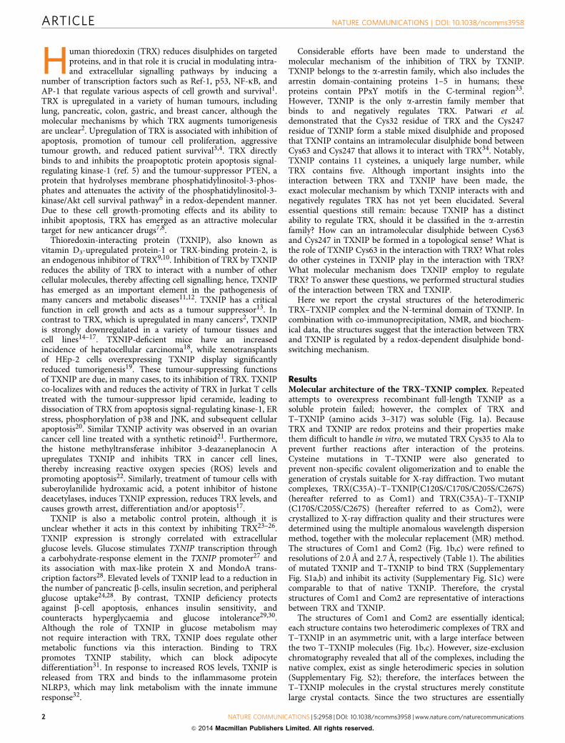
ResultsMolecular architecture of the TRX–TXNIP complex. Repeatedattempts to overexpress recombinant full-length TXNIP as asoluble protein failed; however, the complex of TRX andT–TXNIP (amino acids 3–317) was soluble (Fig. 1a). BecauseTRX and TXNIP are redox proteins and their properties makethem difficult to handle in vitro, we mutated TRX Cys35 to Ala toprevent further reactions after interaction of the proteins.Cysteine mutations in T–TXNIP were also generated toprevent non-specific covalent oligomerization and to enable thegeneration of crystals suitable for X-ray diffraction. Two mutantcomplexes, TRX(C35A)–T–TXNIP(C120S/C170S/C205S/C267S)(hereafter referred to as Com1) and TRX(C35A)–T–TXNIP(C170S/C205S/C267S) (hereafter referred to as Com2), werecrystallized to X-ray diffraction quality and their structures weredetermined using the multiple anomalous wavelength dispersionmethod, together with the molecular replacement (MR) method.The structures of Com1 and Com2 (Fig. 1b,c) were refined toresolutions of 2.0 Å and 2.7 Å, respectively (Table 1). The abilitiesof mutated TXNIP and T–TXNIP to bind TRX (SupplementaryFig. S1a,b) and inhibit its activity (Supplementary Fig. S1c) werecomparable to that of native TXNIP. Therefore, the crystalstructures of Com1 and Com2 are representative of interactionsbetween TRX and TXNIP.
The structures of Com1 and Com2 are essentially identical;each structure contains two heterodimeric complexes of TRX andT–TXNIP in an asymmetric unit, with a large interface betweenthe two T–TXNIP molecules (Fig. 1b,c). However, size-exclusionchromatography revealed that all of the complexes, including thenative complex, exist as single heterodimeric species in solution(Supplementary Fig. S2); therefore, the interfaces between theT–TXNIP molecules in the crystal structures merely constitutelarge crystal contacts. Since the two structures are essentially
ARTICLE NATURE COMMUNICATIONS | DOI: 10.1038/ncomms3958
2 NATURE COMMUNICATIONS | 5:2958 | DOI: 10.1038/ncomms3958 | www.nature.com/naturecommunications
& 2014 Macmillan Publishers Limited. All rights reserved.
identical and the TRX and T–TXNIP complex exists as a singleheterodimeric species in solution, the T–TXNIP (chain C) andTRX (chain D) complex (Fig. 1b) of Com1 was chosen as arepresentative structure and is hereafter referred to unlessotherwise specified. The final structure of the heterodimericcomplex contains residues 8–299 of the expressed protein,residues 3–317 of T–TXNIP and full-length TRX. Residues148–153 and 260–265 of T–TXNIP are not included in the finalmodel because they are not visible in the electron density map;these regions are presumably very flexible.
T–TXNIP is composed entirely of b-strands that form twodeeply curved b-sandwich domains: an N-terminal domain(N-TXNIP) consists of strands b1 to b11 (residues 8–147) anda C-terminal domain (C-TXNIP) consists of strands b12 to b20(residues 154–299) (Figs 1b and 2a). The protein folds into anelongated S-shaped domain arrangement with pseudo-2-foldsymmetry in which C-TXNIP is inverted relative to N-TXNIP.The two domains can be superimposed with a root-mean-squaredeviation (RMSD) of 1.88 for 60 Ca atoms (SupplementaryFig. S3). Although the domain arrangement of T–TXNIP in
TRX
TRX
N
C
N
C
N
C
α1
α3
α2α4
β1β2
β3β4
β1
β3 β4
β5
β6β8
β9
β10β12
β14
β13
β16β17
β15 β20
C-TXNIP
β7
β2
β11
β18
β19
N
CN-TXNIP
C-TXNIP
N-TXNIP
Com1
Com2
1 391
3
3
317
156
1 105
: T-TXNIP
: TXNIP
C32 C35C62 C73
C69
C36a
b
c
C49 C63 C120 C170 C190 C205 C247 C267 C333 C384
TRX
N-TXNIP
N-TXNIP
C-TXNIP
C-TXNIP
N-TXNIP
Figure 1 | Overall structure of the heterodimeric complex of TRX and TXNIP in the asymmetric unit. (a) Schematic representation of the TXNIP
(cyan) and TRX (yellow) constructs used in this study, showing the locations of the cysteines. (b,c) Ribbon representations of the structures of the
TRX(C35A)–T–TXNIP(C120S/C170S/C205S/C267S) complex (referred to as Com1) (b) and the TRX(C35A)–T–TXNIP(C170S/C205S/C267S) complex
(referred to as Com2) (c). The structures of Com1 and Com2 were determined at resolutions of 2.0 Å and 2.7 Å, respectively. There are two heterodimeric
complexes of TRX and TXNIP in the asymmetric unit. The b-sheets and disordered regions of TXNIP are shown in cyan and by white dashed lines,
respectively. The a-helices and b-sheets of TRX are shown in yellow. The N-terminal TXNIP (N-TXNIP) and C-terminal TXNIP (C-TXNIP) domains are
indicated.
NATURE COMMUNICATIONS | DOI: 10.1038/ncomms3958 ARTICLE
NATURE COMMUNICATIONS | 5:2958 | DOI: 10.1038/ncomms3958 | www.nature.com/naturecommunications 3
& 2014 Macmillan Publishers Limited. All rights reserved.
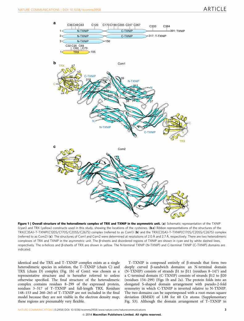
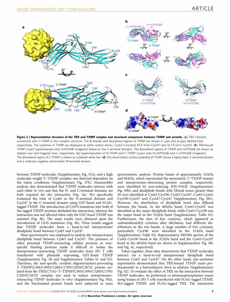
complex with TRX is completely different to that of all otherarrestin structures reported (which have o-shaped domains), theoverall structure of T–TXNIP is similar to that of proteins in thearrestin superfamily, including mouse vacuolar protein sorting-associated protein 26B (mVPS26B; Protein Data Bank accessioncode 3LH8) (Supplementary Fig. S4)35. Therefore, tandemT–TXNIP, comprising the N-TXNIP and C-TXNIP domains,could not be superimposed onto the mVPS26B structure(Fig. 2b), while the individual N-TXNIP (RMSD¼ 1.33 for 82Ca atoms; Z¼ 12.3) and C-TXNIP (RMSD¼ 1.42 for 84 Caatoms; Z¼ 12.5) domains could be superimposed onto eachN-mVPS26B and C-mVPS26B domain, respectively (Fig. 2c). Thestructural analysis also revealed that T–TXNIP is electricallypolarized (Fig. 2d). In particular, the inside section of the curvedb-sandwich of the C-TXNIP domain contains a prominent basicregion, while N-TXNIP has a negative electrostatic potential.
TRX interacts exclusively with C-TXNIP in the complexstructure. No significant structural changes were observed in thestructure of TRX complexed with T–TXNIP compared with thepreviously reported free TRX structure (Supplementary Fig. S5)36.
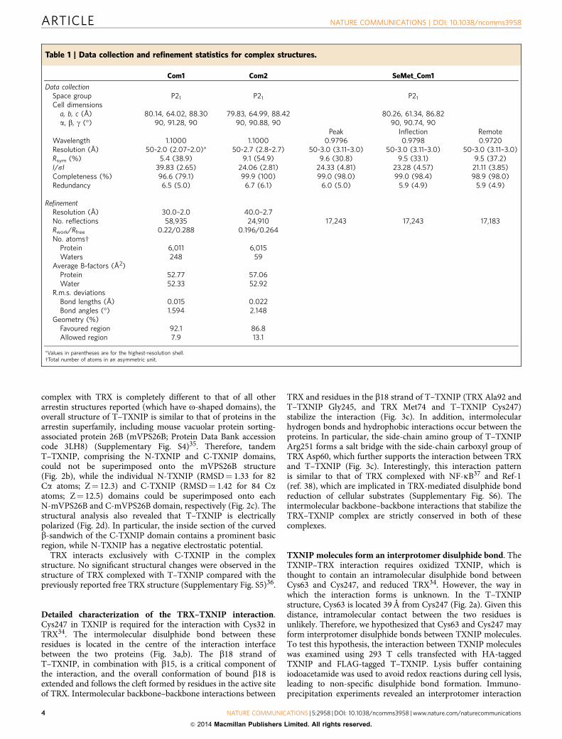
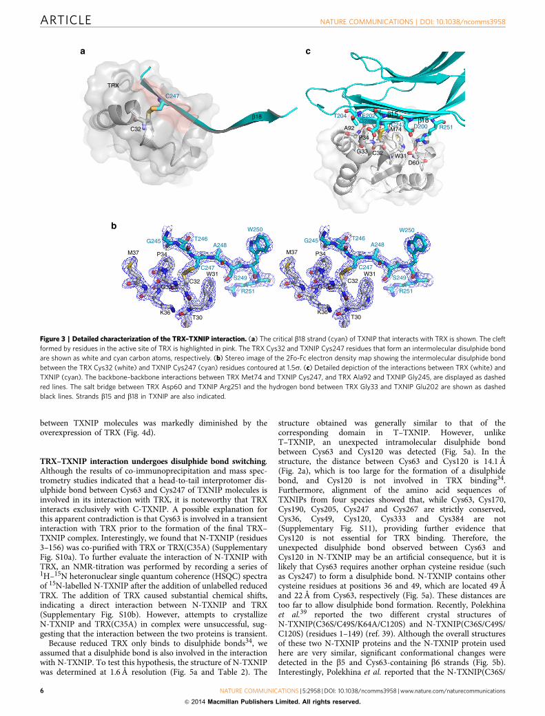
Detailed characterization of the TRX–TXNIP interaction.Cys247 in TXNIP is required for the interaction with Cys32 inTRX34. The intermolecular disulphide bond between theseresidues is located in the centre of the interaction interfacebetween the two proteins (Fig. 3a,b). The b18 strand ofT–TXNIP, in combination with b15, is a critical component ofthe interaction, and the overall conformation of bound b18 isextended and follows the cleft formed by residues in the active siteof TRX. Intermolecular backbone–backbone interactions between
TRX and residues in the b18 strand of T–TXNIP (TRX Ala92 andT–TXNIP Gly245, and TRX Met74 and T–TXNIP Cys247)stabilize the interaction (Fig. 3c). In addition, intermolecularhydrogen bonds and hydrophobic interactions occur between theproteins. In particular, the side-chain amino group of T–TXNIPArg251 forms a salt bridge with the side-chain carboxyl group ofTRX Asp60, which further supports the interaction between TRXand T–TXNIP (Fig. 3c). Interestingly, this interaction patternis similar to that of TRX complexed with NF-kB37 and Ref-1(ref. 38), which are implicated in TRX-mediated disulphide bondreduction of cellular substrates (Supplementary Fig. S6). Theintermolecular backbone–backbone interactions that stabilize theTRX–TXNIP complex are strictly conserved in both of thesecomplexes.
TXNIP molecules form an interprotomer disulphide bond. TheTXNIP–TRX interaction requires oxidized TXNIP, which isthought to contain an intramolecular disulphide bond betweenCys63 and Cys247, and reduced TRX34. However, the way inwhich the interaction forms is unknown. In the T–TXNIPstructure, Cys63 is located 39 Å from Cys247 (Fig. 2a). Given thisdistance, intramolecular contact between the two residues isunlikely. Therefore, we hypothesized that Cys63 and Cys247 mayform interprotomer disulphide bonds between TXNIP molecules.To test this hypothesis, the interaction between TXNIP moleculeswas examined using 293 T cells transfected with HA-taggedTXNIP and FLAG-tagged T–TXNIP. Lysis buffer containingiodoacetamide was used to avoid redox reactions during cell lysis,leading to non-specific disulphide bond formation. Immuno-precipitation experiments revealed an interprotomer interaction
Table 1 | Data collection and refinement statistics for complex structures.
Com1 Com2 SeMet_Com1
Data collectionSpace group P21 P21 P21
Cell dimensionsa, b, c (Å) 80.14, 64.02, 88.30 79.83, 64.99, 88.42 80.26, 61.34, 86.82a, b, g (�) 90, 91.28, 90 90, 90.88, 90 90, 90.74, 90
Peak Inflection RemoteWavelength 1.1000 1.1000 0.9796 0.9798 0.9720Resolution (Å) 50-2.0 (2.07–2.0)* 50-2.7 (2.8–2.7) 50-3.0 (3.11–3.0) 50-3.0 (3.11–3.0) 50-3.0 (3.11–3.0)Rsym (%) 5.4 (38.9) 9.1 (54.9) 9.6 (30.8) 9.5 (33.1) 9.5 (37.2)I/sI 39.83 (2.65) 24.06 (2.81) 24.33 (4.81) 23.28 (4.57) 21.11 (3.85)Completeness (%) 96.6 (79.1) 99.9 (100) 99.0 (98.0) 99.0 (98.4) 98.9 (98.0)Redundancy 6.5 (5.0) 6.7 (6.1) 6.0 (5.0) 5.9 (4.9) 5.9 (4.9)
RefinementResolution (Å) 30.0–2.0 40.0–2.7No. reflections 58,935 24,910 17,243 17,243 17,183Rwork/Rfree 0.22/0.288 0.196/0.264No. atomsw
Protein 6,011 6,015Waters 248 59
Average B-factors (Å2)Protein 52.77 57.06Water 52.33 52.92
R.m.s. deviationsBond lengths (Å) 0.015 0.022Bond angles (�) 1.594 2.148
Geometry (%)Favoured region 92.1 86.8Allowed region 7.9 13.1
*Values in parentheses are for the highest-resolution shell.wTotal number of atoms in an asymmetric unit.
ARTICLE NATURE COMMUNICATIONS | DOI: 10.1038/ncomms3958
4 NATURE COMMUNICATIONS | 5:2958 | DOI: 10.1038/ncomms3958 | www.nature.com/naturecommunications
& 2014 Macmillan Publishers Limited. All rights reserved.
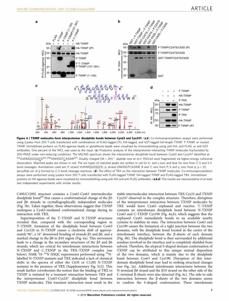
between TXNIP molecules (Supplementary Fig. S7a), and a highmolecular weight T–TXNIP complex was detected dependent onthe redox conditions (Supplementary Fig. S7b). Immunoblotanalysis also demonstrated that TXNIP molecules interact witheach other in vivo and that the N- and C-terminal domains areboth required for the interaction (Fig. 4a). We specificallyevaluated the roles of Cys63 in the N-terminal domain andCys247 in the C-terminal domain using GST-fused and FLAG-tagged TXNIP. The introduction of C247S mutations into both ofthe tagged TXNIP proteins abolished the interaction, whereas theinteraction was not affected when only the GST-fused TXNIP wasmutated (Fig. 4b). The same results were obtained upon theintroduction of C63S mutations (Fig. 4b). These results suggestthat TXNIP molecules form a head-to-tail interprotomerdisulphide bond between Cys63 and Cys247.
Mass spectrometry was employed to analyse the interprotomerdisulphide bond between Cys63 and Cys247. The presence ofother potential TXNIP-interacting cellular proteins or non-specific binding proteins made it difficult to isolate theinterprotomer-interacting TXNIP molecules from 293 T cellstransfected with plasmids expressing GST-fused TXNIP(Supplementary Fig. S8 and Supplementary Tables S1 and S2).Therefore, the non-specific covalent oligomerization-preventingT–TXNIP(C36S/C49S/C120S/C170S/C205S/C267S) protein iso-lated from the TRX(C73A)–T–TXNIP(C36S/C49S/C120S/C170S/C205S/C267S) complex was used to induce interprotomer-interacting TXNIP molecules in vitro (Supplementary Fig. S9a),and the fractionated protein bands were subjected to mass
spectrometric analysis. Protein bands of approximately 32 kDaand 60 kDa, which represented the monomeric T–TXNIP mutantand interprotomer-interacting protein complex, respectively,were identified by non-reducing SDS–PAGE (SupplementaryFig. S9b), and disulphide bonds with Dbond scores greater than20 were identified at Cys63-Cys190, Cys63-Cys247, Cys63-Cys63,Cys190-Cys247 and Cys247-Cys247 (Supplementary Fig. S9c).However, the distribution of disulphide bond sites differedbetween the bands. In the 60 kDa band, Cys63-Cys247 wasidentified as the major disulphide bond, while Cys63-Cys190 wasthe major bond in the 32 kDa band (Supplementary Table S3).Furthermore, the sites of free cysteines, which appeared ascarbamidomethyl cysteines after alkylation, had different dis-tributions in the two bands. A large number of free cysteines,particularly Cys190, were identified in the 32 kDa band(Supplementary Table S3). Representative MS/MS spectra of theCys63-Cys190 bond in the 32 kDa band and the Cys63-Cys247bond in the 60 kDa band are shown in Supplementary Fig. S9cand Fig. 4c, respectively.
Taken together, these data demonstrate that TXNIP moleculesinteract via a head-to-tail interprotomer disulphide bondbetween Cys63 and Cys247. On the other hand, size-exclusionexperiments demonstrated that TRX-bound TXNIP exists pre-dominantly in a heterodimeric form in solution (SupplementaryFig. S2). To evaluate the effect of TRX on the interaction betweenTXNIP molecules, we performed co-immunoprecipitation assaysusing lysates of 293 T cells transfected with FLAG-tagged TXNIP,HA-tagged TXNIP, and FLAG-tagged TRX. The interaction
C-TXNIP
N-TXNIP
C-mVPS26BN-mVPS26B
TRX
ab
c
d
N
C
TRX
C-TXNIP
N-TXNIP
N
C
C63
C190
C170
C205
C49
C267
C36
C120
C247
C36
C49
C120
C267
C63C190
C170 C205
C247
90°
C-TXNIP N-TXNIPC-mVPS26B N-mVPS26B
C-TXNIP
N-TXNIP 90°
90°
90°
90°90°
Figure 2 | Representative structure of the TRX and TXNIP complex and structural comparison between TXNIP and arrestin. (a) TRX interacts
exclusively with C-TXNIP in the complex structure. The b-strands and disordered regions of TXNIP are shown in cyan and as grey dashed lines,
respectively. The cysteines in TXNIP are displayed as white carbon atoms. Cys63 is located 39 Å from Cys247 and 14.1 Å from Cys120. (b) TRX-bound
TXNIP (cyan) superimposed onto mVPS26B (magenta) based on the C-terminal domains. The disordered regions of TXNIP and mVPS26B are shown as
dashed cyan and magenta lines, respectively. (c) Superimposition of N-TXNIP and C-TXNIP (cyan) onto N-mVPS26B and C-mVPS26B (magenta).
The disordered region of C-TXNIP is shown as a dashed white line. (d) The electrostatic surface potential of TXNIP shows a highly basic C-terminal domain
and a relatively negative electrostatic N-terminal domain.
NATURE COMMUNICATIONS | DOI: 10.1038/ncomms3958 ARTICLE
NATURE COMMUNICATIONS | 5:2958 | DOI: 10.1038/ncomms3958 | www.nature.com/naturecommunications 5
& 2014 Macmillan Publishers Limited. All rights reserved.
between TXNIP molecules was markedly diminished by theoverexpression of TRX (Fig. 4d).
TRX–TXNIP interaction undergoes disulphide bond switching.Although the results of co-immunoprecipitation and mass spec-trometry studies indicated that a head-to-tail interprotomer dis-ulphide bond between Cys63 and Cys247 of TXNIP molecules isinvolved in its interaction with TRX, it is noteworthy that TRXinteracts exclusively with C-TXNIP. A possible explanation forthis apparent contradiction is that Cys63 is involved in a transientinteraction with TRX prior to the formation of the final TRX–TXNIP complex. Interestingly, we found that N-TXNIP (residues3–156) was co-purified with TRX or TRX(C35A) (SupplementaryFig. S10a). To further evaluate the interaction of N-TXNIP withTRX, an NMR-titration was performed by recording a series of1H–15N heteronuclear single quantum coherence (HSQC) spectraof 15N-labelled N-TXNIP after the addition of unlabelled reducedTRX. The addition of TRX caused substantial chemical shifts,indicating a direct interaction between N-TXNIP and TRX(Supplementary Fig. S10b). However, attempts to crystallizeN-TXNIP and TRX(C35A) in complex were unsuccessful, sug-gesting that the interaction between the two proteins is transient.
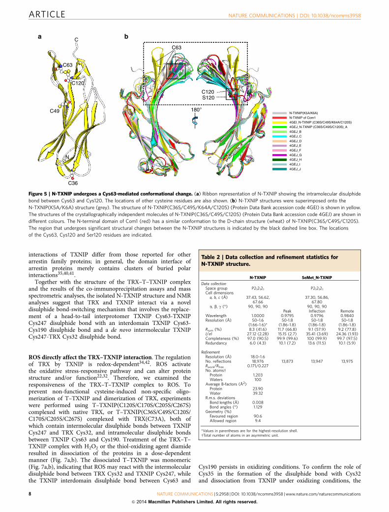
Because reduced TRX only binds to disulphide bonds34, weassumed that a disulphide bond is also involved in the interactionwith N-TXNIP. To test this hypothesis, the structure of N-TXNIPwas determined at 1.6 Å resolution (Fig. 5a and Table 2). The
structure obtained was generally similar to that of thecorresponding domain in T–TXNIP. However, unlikeT–TXNIP, an unexpected intramolecular disulphide bondbetween Cys63 and Cys120 was detected (Fig. 5a). In thestructure, the distance between Cys63 and Cys120 is 14.1 Å(Fig. 2a), which is too large for the formation of a disulphidebond, and Cys120 is not involved in TRX binding34.Furthermore, alignment of the amino acid sequences ofTXNIPs from four species showed that, while Cys63, Cys170,Cys190, Cys205, Cys247 and Cys267 are strictly conserved,Cys36, Cys49, Cys120, Cys333 and Cys384 are not(Supplementary Fig. S11), providing further evidence thatCys120 is not essential for TRX binding. Therefore, theunexpected disulphide bond observed between Cys63 andCys120 in N-TXNIP may be an artificial consequence, but it islikely that Cys63 requires another orphan cysteine residue (suchas Cys247) to form a disulphide bond. N-TXNIP contains othercysteine residues at positions 36 and 49, which are located 49 Åand 22 Å from Cys63, respectively (Fig. 5a). These distances aretoo far to allow disulphide bond formation. Recently, Polekhinaet al.39 reported the two different crystal structures ofN-TXNIP(C36S/C49S/K64A/C120S) and N-TXNIP(C36S/C49S/C120S) (residues 1–149) (ref. 39). Although the overall structuresof these two N-TXNIP proteins and the N-TXNIP protein usedhere are very similar, significant conformational changes weredetected in the b5 and Cys63-containing b6 strands (Fig. 5b).Interestingly, Polekhina et al. reported that the N-TXNIP(C36S/
W250
β18β15
R251D200
E202T204G245
C247
D60W31
M74
C32G33
P34
A92C32
β18
TRX
a c
b
T30
C32G33
P34
K36
W31
M37
S249
C247
T246G245A248
R251
T30
C32G33
P34
K36
W31
M37
W250
S249
C247
T246G245A248
R251
C247
Figure 3 | Detailed characterization of the TRX–TXNIP interaction. (a) The critical b18 strand (cyan) of TXNIP that interacts with TRX is shown. The cleft
formed by residues in the active site of TRX is highlighted in pink. The TRX Cys32 and TXNIP Cys247 residues that form an intermolecular disulphide bond
are shown as white and cyan carbon atoms, respectively. (b) Stereo image of the 2Fo-Fc electron density map showing the intermolecular disulphide bond
between the TRX Cys32 (white) and TXNIP Cys247 (cyan) residues contoured at 1.5s. (c) Detailed depiction of the interactions between TRX (white) and
TXNIP (cyan). The backbone–backbone interactions between TRX Met74 and TXNIP Cys247, and TRX Ala92 and TXNIP Gly245, are displayed as dashed
red lines. The salt bridge between TRX Asp60 and TXNIP Arg251 and the hydrogen bond between TRX Gly33 and TXNIP Glu202 are shown as dashed
black lines. Strands b15 and b18 in TXNIP are also indicated.
ARTICLE NATURE COMMUNICATIONS | DOI: 10.1038/ncomms3958
6 NATURE COMMUNICATIONS | 5:2958 | DOI: 10.1038/ncomms3958 | www.nature.com/naturecommunications
& 2014 Macmillan Publishers Limited. All rights reserved.
C49S/C120S) structure contains a Cys63-Cys63 intermoleculardisulphide bond39 that causes a conformational change of the b5and b6 strands in crystallographically independent molecules(Fig. 5b). Taken together, these observations suggest that TXNIPundergoes a Cys63-mediated conformational change during itsinteraction with TRX.
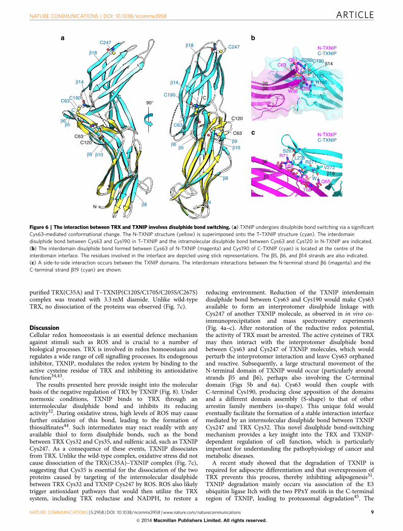
Superimposition of the T–TXNIP and N-TXNIP structuresrevealed that, compared with the corresponding region inT–TXNIP, formation of the disulphide bond between Cys63and Cys120 in N-TXNIP causes a clockwise shift of approxi-mately 96�, a 54� downward bending of strands b5 and b6, and amarked change in the b8 to b10 region (Fig. 6a). This movementleads to a change in the secondary structures of the b5 and b6strands, which are critical for interdomain interactions betweenN-TXNIP and C-TXNIP in the TRX–TXNIP complex (seebelow). NMR 1H–15N HSQC experiments performed using 15N-labelled N-TXNIP mutants and TRX indicated a lack of chemicalshifts in the spectra of either the C63S or C120S N-TXNIPmutants in the presence of TRX (Supplementary Fig. S12). Thisresult further corroborates the notion that the binding of TRX toTXNIP is initiated by a transient interaction between TRX andthe interprotomer Cys63-Cys247 disulphide bond betweenTXNIP molecules. This transient interaction must result in the
stable intermolecular interaction between TRX Cys32 and TXNIPCys247 observed in the complex structure. Therefore, disruptionof the interprotomer interaction between TXNIP molecules byTRX would leave Cys63 orphaned and reactive. T–TXNIPcontains an interdomain disulphide bond between N-TXNIPCys63 and C-TXNIP Cys190 (Fig. 6a,b), which suggests that theorphaned Cys63 immediately bonds to an available nearbycysteine to stabilize its state. The interaction between Cys63 andCys190 causes the formation of a tight junction between the twodomains, with the disulphide bond located in the centre of theinterdomain interface, between the b-sheets of each domain(Fig. 6b). The disulphide bond is then surrounded by additionalresidues involved in the interface and is completely shielded fromsolvent. Therefore, the atypical S-shaped domain conformation ofTXNIP can be attributed to this unique mutual dispositionof the two domains, which is mainly due to the disulphidebond between Cys63 and Cys190. Disruption of this inter-domain disulphide bond may lead to striking structural changes(see Fig. 2a). Additional interdomain interactions between theN-terminal b6 strand and the b19 strand on the other side of theC-terminal b-sheets were also detected (Fig. 6c). The side-to-sideinteraction between the b-sheets of the two domains seemsto confirm the S-shaped conformation. These interdomain
15
50
50
50
kDa
50
50
50
37
75
kDa
ba
c d
50
25
50
37
50
kDa
50
25
IP:F
LAG
-aga
rose
FLA
GF-
TXN
IPF-
TXN
IP (1
–155
)F-
TXN
IP(1
57–3
91)
F-T–
TXN
IP
+ + + + +
HA-TXNIP (IP)
HA-TXNIP
F-TXNIP
HA-TXNIP
F-T–TXNIPF-TXNIP (157–391)
F-TXNIP (1–155)
HA-TXNIP
HA-TXNIP
F-TXNIP
F-TRX
F-TXNIP (IP)
IP: H
AW
CL
F-TXNIP
HA-TXNIP
F-TRX
+
+
+
+ +
–
–
–
–
GST
-TXN
IP +
F-T
XNIP
GST
-C24
7S +
F-T
XNIP
GST
-C24
7S +
F-C
247S
GST
-C63
S +
F-TX
NIP
GST
-C63
S +
F-C
63S
GST
GST-TXNIP/C247S/C63S
F-TXNIP/C247S/C63S (IP)
F-TXNIP/C247S/C63S
IP: G
SH
WC
L
GST
+ F
-TXN
IP
200 400 600 800 1,000 1,200 1,400 1,600 1,800 2,000 2,200 2,400 2,600 m/z
V L W M Q G S Q Q C KB2 B3 B4 B5 B6
Y2Y3Y4Y5Y6Y8Y954 6463
G N H I S G T C A S W Ry2
b3 b4 b5 b6
y3y4y7y8y9y10240 251247
WC
L
Figure 4 | TXNIP molecules form interprotomer disulphide bonds between Cys63 and Cys247. (a,b) Co-immunoprecipitation assays were performed
using lysates from 293 T cells transfected with combinations of FLAG-tagged (F), HA-tagged, and GST-tagged full-length TXNIP, T–TXNIP, or mutant
TXNIP. Immobilized proteins on FLAG-agarose beads or glutathione beads were visualized by immunoblotting using anti-HA, anti-FLAG, or anti-GST
antibodies. One percent of the WCL was used as the input. (c) Proteomic analysis of the interprotomer-interacting TXNIP molecules fractionated by
SDS–PAGE under non-reducing conditions. The MS/MS spectrum shows the interprotomer disulphide bond between Cys63 and Cys247 identified as54VLWMQGSQQCK64-240GNHISGTCASWR251. Doubly charged [Mþ 2H]þ peptide ions at m/z 1353.64 were fragmented via higher-energy collisional
dissociation. Matched peaks are shown in red. The ion types of matched peaks are written in red for b- and y-ions and blue for ions from C-S and S-S
bond cleavages. Annotations used are: P, strand VLWMQGSQQCK; p, strand GNHISGTCASWR; B and Y, ions from P; b and y, ions from p; pþ 32,
persulfide ion of p formed by C-S bond cleavage reactions. (d) The effect of TRX on the interaction between TXNIP molecules. Co-immunoprecipitation
assays were performed using lysates from 293 T cells transfected with FLAG-tagged TXNIP, HA-tagged TXNIP and FLAG-tagged TRX. Immobilized
proteins on HA-agarose beads were visualized by immunoblotting using anti-HA and anti-FLAG antibodies. (a,b,d) The results are representative of at least
two independent experiments with similar results.
NATURE COMMUNICATIONS | DOI: 10.1038/ncomms3958 ARTICLE
NATURE COMMUNICATIONS | 5:2958 | DOI: 10.1038/ncomms3958 | www.nature.com/naturecommunications 7
& 2014 Macmillan Publishers Limited. All rights reserved.
interactions of TXNIP differ from those reported for otherarrestin family proteins; in general, the domain interface ofarrestin proteins merely contains clusters of buried polarinteractions35,40,41.
Together with the structure of the TRX–T–TXNIP complexand the results of the co-immunoprecipitation assays and massspectrometric analyses, the isolated N-TXNIP structure and NMRanalyses suggest that TRX and TXNIP interact via a noveldisulphide bond-switching mechanism that involves the replace-ment of a head-to-tail interprotomer TXNIP Cys63-TXNIPCys247 disulphide bond with an interdomain TXNIP Cys63-Cys190 disulphide bond and a de novo intermolecular TXNIPCys247-TRX Cys32 disulphide bond.
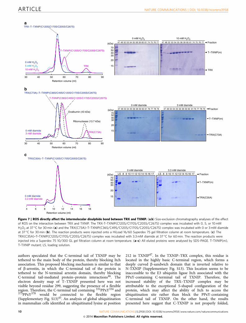
ROS directly affect the TRX–TXNIP interaction. The regulationof TRX by TXNIP is redox-dependent34,42. ROS activatethe oxidative stress-responsive pathway and can alter proteinstructure and/or function22,32. Therefore, we examined theresponsiveness of the TRX–T–TXNIP complex to ROS. Toprevent non-functional cysteine-induced non-specific oligo-merization of T–TXNIP and dimerization of TRX, experimentswere performed using T–TXNIP(C120S/C170S/C205S/C267S)complexed with native TRX, or T–TXNIP(C36S/C49S/C120S/C170S/C205S/C267S) complexed with TRX(C73A), both ofwhich contain intermolecular disulphide bonds between TXNIPCys247 and TRX Cys32, and intramolecular disulphide bondsbetween TXNIP Cys63 and Cys190. Treatment of the TRX–T–TXNIP complex with H2O2 or the thiol-oxidizing agent diamideresulted in dissociation of the proteins in a dose-dependentmanner (Fig. 7a,b). The dissociated T–TXNIP was monomeric(Fig. 7a,b), indicating that ROS may react with the intermoleculardisulphide bond between TRX Cys32 and TXNIP Cys247, whilethe TXNIP interdomain disulphide bond between Cys63 and
Cys190 persists in oxidizing conditions. To confirm the role ofCys35 in the formation of the disulphide bond with Cys32and dissociation from TXNIP under oxidizing conditions, the
C63
C
C120
C63
C120S120
180°N-TXNIP(K5A/K6A)
4GEI_N-TXNIP (C36S/C49S/K64A/C120S)4GEJ_N-TXNIP (C36S/C49S/C120S)_A
4GEJ_B4GEJ_C
4GEJ_D4GEJ_E4GEJ_F
4GEJ_G4GEJ_H
4GEJ_I4GEJ_J
N-TXNIP of Com1
C49
C36
N
a b
Figure 5 | N-TXNIP undergoes a Cys63-mediated conformational change. (a) Ribbon representation of N-TXNIP showing the intramolecular disulphide
bond between Cys63 and Cys120. The locations of other cysteine residues are also shown. (b) N-TXNIP structures were superimposed onto the
N-TXNIP(K5A/K6A) structure (grey). The structure of N-TXNIP(C36S/C49S/K64A/C120S) (Protein Data Bank accession code 4GEI) is shown in yellow.
The structures of the crystallographically independent molecules of N-TXNIP(C36S/C49S/C120S) (Protein Data Bank accession code 4GEJ) are shown in
different colours. The N-terminal domain of Com1 (red) has a similar conformation to the D-chain structure (wheat) of N-TXNIP(C36S/C49S/C120S).
The region that undergoes significant structural changes between the N-TXNIP structures is indicated by the black dashed line box. The locations
of the Cys63, Cys120 and Ser120 residues are indicated.
Table 2 | Data collection and refinement statistics forN-TXNIP structure.
N-TXNIP SeMet_N-TXNIP
Data collectionSpace group P212121 P212121
Cell dimensionsa, b, c (Å) 37.43, 56.62,
67.6637.30, 56.86,
67.80a, b, g (�) 90, 90, 90 90, 90, 90
Peak Inflection RemoteWavelength 1.0000 0.9795 0.9796 0.9840Resolution (Å) 50–1.6
(1.66–1.6)*50-1.8
(1.86–1.8)50–1.8
(1.86–1.8)50–1.8
(1.86–1.8)Rsym (%) 8.3 (41.6) 11.7 (66.8) 9.1 (57.9) 9.2 (77.8)I/sI 27.12 (2.28) 15.15 (2.7) 35.41 (3.69) 24.36 (1.93)Completeness (%) 97.0 (90.5) 99.9 (99.6) 100 (99.9) 99.7 (97.5)Redundancy 6.0 (4.3) 10.1 (7.2) 13.6 (11.5) 10.1 (5.9)
RefinementResolution (Å) 18.0–1.6No. reflections 18,976 13,873 13,947 13,975Rwork/Rfree 0.171/0.227No. atomsw
Protein 1,203Waters 100
Average B-factors (Å2)Protein 23.90Water 39.32
R.m.s. deviationsBond lengths (Å) 0.008Bond angles (�) 1.129
Geometry (%)Favoured region 90.6Allowed region 9.4
*Values in parentheses are for the highest-resolution shell.wTotal number of atoms in an asymmetric unit.
ARTICLE NATURE COMMUNICATIONS | DOI: 10.1038/ncomms3958
8 NATURE COMMUNICATIONS | 5:2958 | DOI: 10.1038/ncomms3958 | www.nature.com/naturecommunications
& 2014 Macmillan Publishers Limited. All rights reserved.
purified TRX(C35A) and T–TXNIP(C120S/C170S/C205S/C267S)complex was treated with 3.3 mM diamide. Unlike wild-typeTRX, no dissociation of the proteins was observed (Fig. 7c).
DiscussionCellular redox homoeostasis is an essential defence mechanismagainst stimuli such as ROS and is crucial to a number ofbiological processes. TRX is involved in redox homoeostasis andregulates a wide range of cell signalling processes. Its endogenousinhibitor, TXNIP, modulates the redox system by binding to theactive cysteine residue of TRX and inhibiting its antioxidativefunction34,43.
The results presented here provide insight into the molecularbasis of the negative regulation of TRX by TXNIP (Fig. 8). Undernormoxic conditions, TXNIP binds to TRX through anintermolecular disulphide bond and inhibits its reducingactivity32. During oxidative stress, high levels of ROS may causefurther oxidation of this bond, leading to the formation ofthiosulfinates44. Such intermediates may react readily with anyavailable thiol to form disulphide bonds, such as the bondbetween TRX Cys32 and Cys35, and sulfenic acid, such as TXNIPCys247. As a consequence of these events, TXNIP dissociatesfrom TRX. Unlike the wild-type complex, oxidative stress did notcause dissociation of the TRX(C35A)–TXNIP complex (Fig. 7c),suggesting that Cys35 is essential for the dissociation of the twoproteins caused by targeting of the intermolecular disulphidebetween TRX Cys32 and TXNIP Cys247 by ROS. ROS also likelytrigger antioxidant pathways that would then utilize the TRXsystem, including TRX reductase and NADPH, to restore a
reducing environment. Reduction of the TXNIP interdomaindisulphide bond between Cys63 and Cys190 would make Cys63available to form an interprotomer disulphide linkage withCys247 of another TXNIP molecule, as observed in in vivo co-immunoprecipitation and mass spectrometry experiments(Fig. 4a–c). After restoration of the reductive redox potential,the activity of TRX must be arrested. The active cysteines of TRXmay then interact with the interprotomer disulphide bondbetween Cys63 and Cys247 of TXNIP molecules, which wouldperturb the interprotomer interaction and leave Cys63 orphanedand reactive. Subsequently, a large structural movement of theN-terminal domain of TXNIP would occur (particularly aroundstrands b5 and b6), perhaps also involving the C-terminaldomain (Figs 5b and 6a). Cys63 would then couple withC-terminal Cys190, producing close apposition of the domainsand a different domain assembly (S-shape) to that of otherarrestin family members (o-shape). This unique fold wouldeventually facilitate the formation of a stable interaction interfacemediated by an intermolecular disulphide bond between TXNIPCys247 and TRX Cys32. This novel disulphide bond-switchingmechanism provides a key insight into the TRX and TXNIP-dependent regulation of cell function, which is particularlyimportant for understanding the pathophysiology of cancer andmetabolic diseases.
A recent study showed that the degradation of TXNIP isrequired for adipocyte differentiation and that overexpression ofTRX prevents this process, thereby inhibiting adipogenesis31.TXNIP degradation mainly occurs via association of the E3ubiquitin ligase Itch with the two PPxY motifs in the C-terminalregion of TXNIP, leading to proteasomal degradation45. The
β5
C63C190
D193R186
R299Q61
Q58β6
β14
W56
C-TXNIPN-TXNIP
C190C63
C63C120
β5β6
β10
β14
C247
N
C
N
C
β9
β18
β8
90°
C190
C63C120
C63
β9β6β5
β14
C247
N
C
N
C
β10
β18
β8β19β5
β6R71
E68
T66
Q65P109
S267
L270R271
V272
C-TXNIPN-TXNIP
W
a b
c
Figure 6 | The interaction between TRX and TXNIP involves disulphide bond switching. (a) TXNIP undergoes disulphide bond switching via a significant
Cys63-mediated conformational change. The N-TXNIP structure (yellow) is superimposed onto the T–TXNIP structure (cyan). The interdomain
disulphide bond between Cys63 and Cys190 in T–TXNIP and the intramolecular disulphide bond between Cys63 and Cys120 in N-TXNIP are indicated.
(b) The interdomain disulphide bond formed between Cys63 of N-TXNIP (magenta) and Cys190 of C-TXNIP (cyan) is located at the centre of the
interdomain interface. The residues involved in the interface are depicted using stick representations. The b5, b6, and b14 strands are also indicated.
(c) A side-to-side interaction occurs between the TXNIP domains. The interdomain interactions between the N-terminal strand b6 (magenta) and the
C-terminal strand b19 (cyan) are shown.
NATURE COMMUNICATIONS | DOI: 10.1038/ncomms3958 ARTICLE
NATURE COMMUNICATIONS | 5:2958 | DOI: 10.1038/ncomms3958 | www.nature.com/naturecommunications 9
& 2014 Macmillan Publishers Limited. All rights reserved.
authors speculated that the C-terminal tail of TXNIP may betethered to the main body of the protein, thereby blocking Itchassociation. This proposed blocking mechanism is similar to thatof b-arrestin, in which the C-terminal tail of the protein istethered to the N-terminal arrestin domain, thereby blockingC-terminal tail-mediated protein–protein interactions46. Theelectron density map of T–TXNIP presented here was notvisible beyond residue 299, suggesting the presence of a flexibleregion. Therefore, the C-terminal tail containing 331PPxY334 and375PPxY378 would be connected to the flexible region(Supplementary Fig. S13)45. An analysis of global ubiquitinationin mammalian cells identified an ubiquitinated lysine at position
212 in TXNIP47. In the TXNIP–TRX complex, this residue islocated in the highly basic C-terminal region, which forms adeeply curved b-sandwich domain that is inverted relative toN-TXNIP (Supplementary Fig. S13). This location seems to beinaccessible to the E3 ubiquitin ligase Itch associated with thePPxY-containing C-terminal tail of TXNIP. Therefore, theincreased stability of the TRX–TXNIP complex may beattributable to the exceptional S-shaped configuration of theprotein, which may affect the ability of Itch to access theubiquitination site rather than block the PPxY-containingC-terminal tail of TXNIP. On the other hand, the resultspresented here suggest that C-TXNIP is not properly folded,
TRX–T–TXNIP(C120S/C170S/C205S/C267S)
TRX(C73A)–T–TXNIP(C36S/C49S/C120S/C170S/C205S/C267S)
0 mM H2O2
5 mM H2O2
10 mM H2O2
b
Retention volume (ml)
Ovalbumin (43 kDa)
Ribonuclease (13.7 kDa)
30 40 50 60 70 80 90
Retention volume (ml)
30 40 50 60 70 80 90
15
20
30405070
(kDa) 47 48 52 53 54 55 58 59 60 61 74 75 76 77 47 48 52 53 54 55 58 59 60 61 74 75 76 77
0 mM H2O2 10 mM H2O2
Fraction
T–TXNIP(m)
TRX
15
20
30405070
(kDa) 47 48 53 54 55 56 58 59 60 61 76 77 78 79 47 48 53 54 55 56 58 59 60 61 76 77 78 79
0 mM diamide 3 mM diamide
0 mM diamide
LS 8 9 10 11 12 13 14 15 16 17 LS 8 9 10 11 12 13 14 15 16 17(kDa)70504030
20
15
3.3 mM diamide
Fraction
T–TXNIP(m)
TRX(C73A)
T–TXNIP(C120S/C170S/C205S/C267S)
T-TXNIP(C36S/C49S/C120S/C170S/C205S/C267S)
TRX(C73A)
TRX
Fraction
T–TXNIP(m)
TRX(C35A)
0 mM diamide3 mM diamide
6 8 10 12 14 16 184
Retention volume (ml)
0 mM diamide3.3 mM diamide
TRX(C35A)–T–TXNIP(C120S/C170S/C205S/C267S)
a
c
Figure 7 | ROS directly affect the intermolecular disulphide bond between TRX and TXNIP. (a,b) Size-exclusion chromatography analyses of the effect
of ROS on the interaction between TRX and TXNIP. The TRX-T–TXNIP(C120S/C170S/C205S/C267S) complex was incubated with 0, 5, or 10 mM
H2O2 at 37 �C for 30 min (a) and the TRX(C73A)–T–TXNIP(C36S/C49S/C120S/C170S/C205S/C267S) complex was incubated with 0 or 3 mM diamide
at 37 �C for 30 min (b). The reaction products were injected onto a HiLoad 16/60 Superdex 75 gel filtration column at room temperature. (c) The
TRX(C35A)–T–TXNIP(C120S/C170S/C205S/C267S) complex was incubated with 3.3 mM diamide at 37 �C for 60 min. The reaction products were
injected into a Superdex 75 10/300 GL gel filtration column at room temperature. (a–c) All eluted proteins were analysed by SDS–PAGE. T–TXNIP(m),
T–TXNIP mutant; LS, loading solution.
ARTICLE NATURE COMMUNICATIONS | DOI: 10.1038/ncomms3958
10 NATURE COMMUNICATIONS | 5:2958 | DOI: 10.1038/ncomms3958 | www.nature.com/naturecommunications
& 2014 Macmillan Publishers Limited. All rights reserved.
while N-TXNIP behaves as a globular protein. Accordingly, TRX-free TXNIP, including the TXNIP(C247S) mutant, should beflexible. Thus, its structure may not be S-shaped, which wouldallow Itch to access Lys212 and attach ubiquitin. We do notexclude the possibility that the inhibition of TRX by TXNIP isrequired to prevent adipogenesis. Adipogenesis is linked tooxidative stress48 and Itch is known to regulate intracellular ROSlevels45.
TXNIP is a multifunctional protein that interacts with a varietyof proteins involved in redox-independent cell signalling42. Thisfeature may be characteristic of arrestin family members, whichhave emerged as molecular scaffolds and signal integrators. Arecent review article suggested that, rather than functioningprimarily as an inhibitor of TRX, TXNIP is an a-arrestin proteinthat is regulated by binding to TRX49, as occurs duringadipogenesis31. Here, we demonstrated that TXNIP is acysteine-containing redox protein that robustly regulates TRXvia a novel disulphide bond-switching mechanism. Thismechanism underlies a number of cell signalling pathwaysinvolved in cell growth, proliferation and metabolism. Theseproperties are entirely distinct from other arrestin familymembers, including a- and b-arrestins33. In addition, thedomain assembly of TXNIP differs from that of other arrestinproteins. Therefore, rather than classifying it as an arrestinprotein, it may be prudent to describe TXNIP as a redox proteinthat contains an arrestin-like fold domain and functions in both aTRX-dependent and independent manner. TXNIP interacts withdifferent cellular substrates using a variety of binding sites. For
example, residues 1–227 of TXNIP interact with importin a1,leading to its nuclear localization50. In addition, the C-terminalregion of TXNIP (residues 253–396) is critical for its interactionwith JAB1 to stabilize p27(kip1) (ref. 51). These interactions donot involve TXNIP Cys247, suggesting a disulphide bond-switching-independent mechanism. Further experiments arerequired to confirm whether these binding partners can causeconformational changes and induce a general o-shaped arrestindomain arrangement in TXNIP.
MethodsDNA cloning and mutagenesis. TXNIP could not be expressed as a solubleprotein without TRX; therefore, plasmids co-expressing human TRX and TXNIPwere constructed using a two-promoter vector system52. Briefly, the gene encodingT–TXNIP (residues 3–317) with an N-terminal hexahistidine-tag containing anintegrated rTEV protease cleavage site was PCR amplified using the primers50-catgccatggtcaagaagatcaag-30 and 50-ataagaatgcggccgctcacattcagagctgg-30 andcloned into the pProEX HTa plasmid (Invitrogen), which expresses tag-free humanTRX under the control of the T7 promoter. A plasmid co-expressing human TRXand N-TXNIP (residues 3–156) was constructed using the same method with theprimers 50-ggaattccatatggtgaagcagat-30 and 50-ccgctcgagtcagactaattcattaat-30
for human TRX, and primers 50-catgccatggtcaagaagatcaag-30 and50-ataagaatgcggccgctcataaatcagggg-30 for N-TXNIP. N-TXNIP was also subclonedinto the pHis-Parallel1 expression vector53, which includes an N-terminalhexahistidine-tag with an rTEV protease cleavage site. For NMR experiments,C-terminal hexahistidine-tagged TRX was subcloned into the pET21 vector(Invitrogen) using the primers 50-ggaattccatatggtgaagcagat-30 and50-ccgctcgaggactaattcattaat-30 . Mutations were introduced into the plasmids usingthe QuikChange Mutagenesis Kit (Stratagene), according to the manufacturer’sinstructions. All constructs were verified by sequencing.
C247
C190C63
C32C35
Oxidative stress
ROS
Antioxidant defense(TRX reductase + NADPH)
Normoxic state
C32C35
ROS
C247
C190C63
C32C35
C190C63
C32C35
C247C190
C63
C247
C190
C63
C247
C32C35
C190
C63
C247
C32C35
C32C35
Arrest of TRX
C247
C247
C63C190
C190C63
C32C35
Figure 8 | Proposed molecular mechanism of the negative regulation of TRX by TXNIP. The inhibition of TRX by TXNIP is regulated by a redox-
dependent disulphide bond-switching mechanism. Under normoxic conditions, TXNIP binds to TRX through an intermolecular disulphide bond formed
between TXNIP Cys247 and TRX Cys32 and inhibits its reducing activity. During oxidative stress, high levels of ROS may cause further oxidation of this
bond and lead to the dissociation of TXNIP from TRX. ROS also likely trigger antioxidant pathways to restore a reducing environment. After restoration of a
reductive redox potential, the active cysteines of TRX may interact with the interprotomer disulphide bond between Cys63 and Cys247 of TXNIP
molecules. Subsequently, TXNIP molecules would undergo a structural rearrangement that involves switching of the interprotomer disulphide bond
between TXNIP molecules to an interdomain Cys63-Cys190 disulphide bond, and the formation of a de novo intermolecular TXNIP Cys247-TRX Cys32
disulphide bond. See discussion for a full description. TRX and TXNIP are shown in yellow and cyan, respectively. Cysteine residues forming disulphide
bonds are displayed in brown.
NATURE COMMUNICATIONS | DOI: 10.1038/ncomms3958 ARTICLE
NATURE COMMUNICATIONS | 5:2958 | DOI: 10.1038/ncomms3958 | www.nature.com/naturecommunications 11
& 2014 Macmillan Publishers Limited. All rights reserved.
Protein expression and purification. Expression of wild-type and mutant TRX–T–TXNIP complexes and the TRX(C35A)–N-TXNIP complex was induced inEscherichia coli Rosetta gami (DE3) cells by treatment with 0.5 mM IPTG at 21 �Cfor 40 h. Expression of the N-TXNIP(K5A/K6A) mutant in E. coli C41(DE3) cellswas induced by treatment with 0.5 mM IPTG at 21 �C for 16 h. Recombinantproteins were purified by Ni-NTA affinity chromatography, treated with rTEVprotease to remove hexahistidine-tags, and then purified by size-exclusionchromatography and additional Ni-NTA affinity chromatography. The purifiedproteins were dialyzed against 50 mM Tris-HCl (pH 7.0). A selenomethionine(SeMet)-substituted complex of TRX(C35A) and T–TXNIP(C120S/C170S/C205S/C267S), and a SeMet-substituted N-TXNIP(K5A/K6A) complex, were expressed inthe methionine auxotroph E. coli B834(DE3) strain (Novagen), which was grown inminimal medium supplemented with 50 mg ml� 1 SeMet under the same condi-tions as cells containing the native plasmid. The SeMet-substituted proteins werepurified as described for the native proteins. Expression of TRX was induced inE. coli C41 (DE3) cells by treatment with 0.5 mM IPTG at 21 �C for 18 h. Theprotein was purified by Ni-NTA agarose affinity and size-exclusion chromato-graphy, and then dialyzed against 50 mM potassium phosphate (pH 6.6) for use inNMR experiments. E. coli C41 (DE3) cells transformed with plasmids encodingN-TXNIP or its mutants were grown in M9 minimal medium enriched with(15NH4)2SO4 (99% 15N) (Cambridge Isotope Laboratory Inc.) as the sole nitrogensource. All 15N-labelled proteins were purified using Ni-NTA affinity chromato-graphy. The proteins were treated with rTEV protease to remove the hexahistidine-tags and dialyzed against 50 mM potassium phosphate (pH 6.6) as the final stepbefore NMR.
Size-exclusion chromatography. A Superdex 200 10/300 GL gel filtration column(GE Healthcare) installed on an AKTA purifier FPLC system (GE Healthcare) wasequilibrated with 50 mM Tris-HCl (pH 8.0), 500 mM NaCl and 10% glycerol at aflow rate of 0.4 ml min� 1 at room temperature. The purified TRX–T–TXNIPcomplex and its mutants were injected onto the column. Ovalbumin was used asthe molecular weight standard.
Crystallization and structure determination. Because N-TXNIP was not crys-tallized, we crystallized an engineered derivative with reduced surface entropy54,N-TXNIP(K5A/K6A), using the sitting drop vapor-diffusion method at 21 �C. Thebest crystals were obtained with 0.75 M sodium-potassium phosphate and 0.1 MHEPES-Na (pH 7.5). Diffraction data were collected at beamline 4A (PohangAccelerator Laboratory, Korea) at a resolution of 1.6 Å. SeMet-substitutedK5A/K6A crystals were grown under the same conditions as the native protein.Multiple-wavelength anomalous diffraction data for the SeMet-substituted crystalswere collected at beamline 6C at a resolution of 1.8 Å. The TRX(C35A) andT–TXNIP(C120S/C170S/C205S/C267S) complex crystals were optimized in 0.16 Mtri-sodium citrate and 16% PEG 3350, and diffraction data were collected at aresolution of 2.0 Å at beamline 4A. SeMet-substituted crystals of the complex weregrown in 0.2 M tri-sodium citrate and 20% PEG 3350. Multiple-wavelengthanomalous diffraction data were collected at a resolution of 3.0 Å at beamline 6C.Crystals of the TRX(C35A) and T–TXNIP(C170S/C205S/C267S) complex wereoptimized using conditions similar to those used for the other complexes, anddiffraction data were collected at a resolution of 2.7 Å at beamline 4A. All data wereprocessed using the HKL2000 package55. The N-TXNIP(K5A/K6A) structure wasdetermined by analysing anomalous signals from Se atoms with the programSOLVE56. Density modification and subsequent automated model building wereperformed using RESOLVE57. The native N-TXNIP(K5A/K6A) crystal structurewas solved at a resolution of 1.6 Å using the MR method with the programMOLREP58, based on the partially refined structure of the SeMet crystal. TheSeMet-substituted TRX(C35A) and T–TXNIP(C120S/C170S/C205S/C267S)structure was determined using SOLVE56, and subsequent automated modelbuilding was performed using RESOLVE57. The complex structure at a resolutionof 2.0 Å was solved by MR using the partially refined SeMet complex structure.The TRX(C35A) and T–TXNIP(C170S/C205S/C267S) complex structure wasdetermined by MR using the TRX(C35A) and T–TXNIP(C120S/C170S/C205S/C267S) complex model. All structures were revised using COOT59 and refined withREFMAC5 (ref. 60). The crystallographic data are summarized in Tables 1 and 2.
Pull-down and co-immunoprecipitation assays. To examine the in vivo inter-protomer interaction between TXNIP molecules, HEK 293 T cells were transfectedwith plasmids expressing HA-tagged TXNIP and FLAG-tagged T–TXNIP. After24 h, the cells were harvested and lysed in lysis buffer (0.5% Triton X-100, 150 mMNaCl, 10% glycerol, 1 mM NaF, 1 mM AEBSF, 2 mg ml� 1 leupeptin, 5 mg ml� 1
aprotinin and 20 mM HEPES (pH 7.2)) containing 50 mM iodoacetamide. Aftercentrifugation at 16,000� g for 20 min, the supernatant was incubated with anti-FLAG M2 agarose (Sigma) for 12 h at 4 �C. The agarose beads were collected bycentrifugation and washed three times with lysis buffer. The bound proteins werethen subjected to SDS–PAGE under reducing and non-reducing conditions. Forthe assay demonstrating the TXNIP–TXNIP interaction in detail, HEK 293 T cellswere transfected with combinations of expression plasmids, as indicated in themain text. After 24 h, the cells were harvested, lysed and centrifuged at 16,000� gfor 20 min. The supernatants were incubated for 12 h at 4 �C with mouse
monoclonal anti-FLAG M2 agarose beads (Sigma) for immunoprecipitation, orwith glutathione-conjugated sepharose beads (GE Healthcare) for pull-downassays. The immobilized proteins were collected by centrifugation and washedthree times with lysis buffer. Bound proteins or whole cell lysates (WCLs) wereeluted by boiling in SDS sample buffer. To detect HA-tagged, FLAG-tagged orGST-fused proteins, rabbit polyclonal anti-HA (1:2,000), anti-FLAG (1:2,000) andanti-GST (1:1,000) antibodies (Santa Cruz Biotechnology) were used, respectively.Horseradish peroxidase-conjugated secondary antibody (1:5,000; Santa Cruz Bio-technology) was used to visualize the specific target bands. For the immunopre-cipitation assay showing the effect of TRX on the TXNIP–TXNIP interaction,transfected HEK 293 T cells were harvested and lysed in buffer containing 0.5%Triton X-100, 150 mM NaCl, 10% glycerol, 20 mM HEPES (pH 7.2) and completeprotease inhibitor cocktail (Roche). After incubation at 4 �C for 30 min, the lysatewas centrifuged at 16,000� g for 20 min. The supernatants were pre-cleared byincubation with 20ml of protein G-sepharose beads (GE Healthcare) for 2 h at 4 �C,and then centrifuged at 10,000� g for 5 min. The pre-cleared lysate was incubatedfor 14 h at 4 �C with the rabbit polyclonal anti-HA antibody, followed by 5 h at 4 �Cwith protein G-sepharose beads. After immunoprecipitation, the beads werewashed five times with lysis buffer. Bound proteins or WCLs were eluted by boilingin LDS–PAGE loading buffer (Life Technologies). To detect HA-tagged proteins orFLAG-tagged proteins, a mouse monoclonal anti-HA antibody (1:2,000; Abcam) oranti-FLAG antibody (1:2,000; Sigma) was used, and then horseradish peroxidase-conjugated secondary antibody (1:5,000) or peroxidase-conjugated light chain-specific secondary antibody (1:3,500; Jackson ImmunoResearch Laboratories Inc.)was used to visualize the specific target bands.
TRX activity assay. TRX activity was measured using the insulin disulphidereduction assay, as described previously10. Briefly, HEK 293 T cells were mock-transfected or transiently transfected with TXNIP or mutant TXNIP expressionplasmids. The transfected cells were lysed in buffer containing 20 mM HEPES(pH 7.9), 100 mM KCl, 300 mM NaCl, 10 mM EDTA, 0.1% Nonidet P-40 andprotease inhibitors. To reduce TRX, cell extracts (20mg) were incubated for 20 minat 37 �C with 2 ml of DTT activation buffer containing 1 mM EDTA, 1 mg ml� 1
BSA, 2 mM DTT and 50 mM HEPES (pH 7.6) in a total volume of 70 ml. A 40mlaliquot of reaction mixture taken from a solution containing 200 ml of 1 M HEPES(pH 7.6), 40 ml of 0.2 M EDTA, 40ml of NADPH (40 mg ml� 1) and 500ml ofinsulin (10 mg ml� 1) was then added. The reaction was started by the addition of10 ml of rat TRX reductase (100 U ml� 1) and incubated for 20 min at 37 �C. Thereaction was stopped by the addition of 6 M guanidine-HCl and 1 mM DNTB(3-carboxy-4-nitrophenyl disulphide), and the absorbance at 412 nm was measured.
Sample preparation for mass spectrometry. For analysis by mass spectrometryin vivo, HEK 293 T cells were transfected with a plasmid expressing GST-fusedTXNIP. After 24 h, the cells were harvested and lysed in buffer containing 0.5%Triton X-100, 150 mM NaCl, 10% glycerol, 20 mM HEPES (pH 7.2) and completeprotease inhibitor cocktail. After incubation for 30 min at 4 �C, the lysates werecentrifuged at 16,000� g for 20 min. The supernatants were incubated overnight at4 �C with Glutathione Sepharose 4FF (GE Healthcare) and then centrifuged at10,000� g for 5 min. The supernatant was discarded and the resin was washed fivetimes with lysis buffer. Bound proteins were eluted by boiling in LDS–PAGEloading buffer. For analysis by mass spectrometry in vitro, the purifiedTRX(C73A)–T–TXNIP(C36S/C49S/C120S/C170S/C205S/C267S) complex wasincubated with 100 mM DTT for 1 h at room temperature and then injected into aSuperdex 200 10/300 GL gel filtration column installed on an AKTA purifier FPLCsystem to obtain T–TXNIP(C36S/C49S/C120S/C170S/C205S/C267S) andTRX(C73A). The fractions containing T–TXNIP(C36S/C49S/C120S/C170S/C205S/C267S) were dialyzed against 50 mM Tris-HCl (pH 8.0), 500 mM NaCl and 10%glycerol to induce the formation of disulphide bonds between TXNIP moleculesthrough air oxidation. Dialyzed proteins were then subjected to non-reducingSDS–PAGE.
In-gel digestion. The high molecular weight TXNIP complex-containing gelpieces were destained in 25 mM ammonium bicarbonate and 50% acetonitrile,dehydrated with 100% acetonitrile and then dried at room temperature. Freecysteines were alkylated by adding 55 mM iodoacetamide in 25 mM ammoniumbicarbonate and then incubated for 1 h at 25 �C in the dark. The gel was washedthree times with 25 mM ammonium bicarbonate and 50% acetonitrile solution,dehydrated with 100% acetonitrile and then dried at room temperature. The gelpieces were rehydrated with a solution of 12.5 ng ml� 1 sequencing-grade trypsin(Promega) in 25 mM ammonium bicarbonate and incubated for 16 h at 37 �C forprotein digestion. The supernatants were transferred to fresh tubes and theremaining peptides were sequentially extracted by incubating the gel pieces with50% acetonitrile in 25 mM ammonium bicarbonate, 50% acetonitrile in 0.5%trifluoroacetic acid and then 70% acetonitrile in 0.5% trifluoroacetic acid. Theextracted peptides were combined, dried in a miVac Duo vacuum evaporator(Genevac) and stored at � 20 �C until use61.
Mass spectrometry. The peptides were diluted with 0.4% acetic acid to achieveconcentrations of 1 mgml� 1 and an aliquot containing approximately 1 mg was
ARTICLE NATURE COMMUNICATIONS | DOI: 10.1038/ncomms3958
12 NATURE COMMUNICATIONS | 5:2958 | DOI: 10.1038/ncomms3958 | www.nature.com/naturecommunications
& 2014 Macmillan Publishers Limited. All rights reserved.
injected into a reversed-phase Magic C18aq column (15 cm� 75mm) on anEksigent nanoLC-ultra 1D plus system at a flow rate of 300 nl min� 1. Prior to use,the column was equilibrated with 95% buffer A (0.1% formic acid in H2O) and 5%buffer B (0.1% formic acid in acetonitrile). The peptides were eluted with a lineargradient from 10% buffer B to 40% buffer B over 40 min. The HPLC system wascoupled to a Q Exactive quadrupole mass spectrometer (Thermo Scientific)operated in the data dependent mode. Survey full-scan MS spectra (m/z 300–2000)were acquired in the Orbitrap with a resolution of 75,000. The MS/MS spectra ofthe 12 most intense ions from the MS1 scan with a charge state Z2 were acquiredwith the following options: resolution, 17,500; isolation width, 2 m/z; normalizedcollision energy, 27%; dynamic exclusion duration, 30 s; and ion selectionthreshold, 4.00Eþ 03 counts.
Database searching and validation. The acquired MS/MS spectra were searchedagainst the SwissProt database (August 2011) with 56,392 entries using X!Tandemopen source software (available from http://www.thegpm.org/TANDEM/). Theprecursor mass tolerance was set to±15 p.p.m. and the cleavage specificity was setto trypsin, allowing for a maximum of one missed cleavage. A variable modificationof methionine oxidation (þ 15.9949 Da) and carbamidomethylated cysteine(þ 57.0215 Da) was allowed. Peptide assignment was performed using the TransProteomics Pipeline (version 4.5 RAPTURE rev 2) provided by the Institute forSystems Biology (http://www.proteomecenter.org). From the X!Tandem searchoutput, peptides with probabilities greater than 0.9 were included in the subsequentProtein-Prophet, and proteins with probabilities greater than 0.9 were gathered.Contaminants, such as keratin and trypsin artifacts, were removed from the results.The identification of disulphide linkages was performed using DBond software(version 3.02; http://prix.hanyang.ac.kr/download/dbond.jsp)62.
NMR spectroscopy. NMR interaction experiments were performed by recording1H–15N NMR HSQC spectra of 15N-labelled N-TXNIP, or its mutants, upon theaddition of unlabelled TRX. Prior to the interaction study, TRX was reduced with5 mM DTT and dialyzed against 50 mM potassium phosphate (pH 6.6) to removeexcess DTT. The 1H–15N HSQC spectra of all proteins were measured on a Bruker900 MHz NMR spectrometer at the Korea Basic Science Institute (Ochang, Korea).The NMR measurements were performed in 50 mM potassium phosphate (pH 6.6)containing 10% D2O at 25 �C. All NMR spectra were processed with Topspin 2.1and analysed with the program SPARKY 3.1.
Analysis of the effect of ROS on the TRX–TXNIP interaction. The TRX andT–TXNIP(C120S/C170S/C205S/C267S) complex was used to circumvent non-functional cysteine-induced aggregation. The complex (0.05 mM) was incubated at37 �C with 0–10 mM H2O2 in 50 mM Tris-HCl (pH 8.0), 500 mM NaCl and 10%glycerol. The TRX(C73A) and T–TXNIP(C36S/C49S/C120S/C170S/C205S/C267S)complex was also used to avoid TRX dimerization and non-functional cysteine-induced aggregation. This complex was incubated with 0–3 mM diamide in thesame buffer at 37 �C. After 30 min, the samples were injected onto a HiLoad 16/60Superdex 75 gel filtration column (GE Healthcare) installed on an AKTA purifierFPLC system at a flow rate of 1 ml min� 1 at room temperature. To examine therole of TRX Cys35 in the dissociation of TRX and TXNIP, the TRX(C35A)–T–TXNIP(C120S/C170S/C205S/C267S) complex was incubated for 1 h at roomtemperature with 3.3 mM diamide and then injected into a Superdex 75 10/300 GLgel filtration column. All fractions were subjected to SDS–PAGE.
References1. Lillig, C. H. & Holmgren, A. Thioredoxin and related molecules--from biology
to health and disease. Antioxid. Redox Signal. 9, 25–47 (2007).2. Burke-Gaffney, A., Callister, M. E. & Nakamura, H. Thioredoxin: friend or foe
in human disease? Trends Pharmacol. Sci. 26, 398–404 (2005).3. Grogan, T. M. et al. Thioredoxin, a putative oncogene product, is overexpressed
in gastric carcinoma and associated with increased proliferation and increasedcell survival. Hum. Pathol. 31, 475–481 (2000).
4. Raffel, J. et al. Increased expression of thioredoxin-1 in human colorectalcancer is associated with decreased patient survival. J. Lab. Clin. Med. 142,46–51 (2003).
5. Saitoh, M. et al. Mammalian thioredoxin is a direct inhibitor of apoptosissignal-regulating kinase (ASK) 1. EMBO J. 17, 2596–2606 (1998).
6. Meuillet, E. J., Mahadevan, D., Berggren, M., Coon, A. & Powis, G.Thioredoxin-1 binds to the C2 domain of PTEN inhibiting PTEN’s lipidphosphatase activity and membrane binding: a mechanism for the functionalloss of PTEN’s tumor suppressor activity. Arch. Biochem. Biophys. 429,123–133 (2004).
7. Powis, G. & Kirkpatrick, D. L. Thioredoxin signaling as a target for cancertherapy. Curr. Opin. Pharmacol. 7, 392–397 (2007).
8. Mukherjee, A. & Martin, S. G. The thioredoxin system: a key target in tumourand endothelial cells. Br. J. Radiol. 81 Spec No 1, S57–S68 (2008).
9. Junn, E. et al. Vitamin D3 up-regulated protein 1 mediates oxidative stress viasuppressing the thioredoxin function. J. Immunol. 164, 6287–6295 (2000).
10. Nishiyama, A. et al. Identification of thioredoxin-binding protein-2/vitaminD(3) up-regulated protein 1 as a negative regulator of thioredoxin function andexpression. J. Biol. Chem. 274, 21645–21650 (1999).
11. Dunn, L. L., Buckle, A. M., Cooke, J. P. & Ng, M. K. The emerging role ofthe thioredoxin system in angiogenesis. Arterioscler. Thromb. Vasc. Biol. 30,2089–2098 (2010).
12. Watanabe, R., Nakamura, H., Masutani, H. & Yodoi, J. Anti-oxidative, anti-cancer and anti-inflammatory actions by thioredoxin 1 and thioredoxin-binding protein-2. Pharmacol. Ther. 127, 261–270 (2010).
13. Zhou, J., Yu, Q. & Chng, W. J. TXNIP (VDUP-1, TBP-2): a major redoxregulator commonly suppressed in cancer by epigenetic mechanisms. Int. J.Biochem. Cell Biol. 43, 1668–1673 (2011).
14. Ikarashi, M. et al. Vitamin D3 up-regulated protein 1 (VDUP1) expression ingastrointestinal cancer and its relation to stage of disease. Anticancer Res. 22,4045–4048 (2002).
15. Dutta, K. K. et al. Two distinct mechanisms for loss of thioredoxin-bindingprotein-2 in oxidative stress-induced renal carcinogenesis. Lab. Invest. 85,798–807 (2005).
16. Ohta, S. et al. Downregulation of metastasis suppressor genes in malignantpheochromocytoma. Int. J. Cancer 114, 139–143 (2005).
17. Butler, L. M. et al. The histone deacetylase inhibitor SAHA arrests cancer cellgrowth, up-regulates thioredoxin-binding protein-2, and down-regulatesthioredoxin. Proc. Natl Acad. Sci. USA 99, 11700–11705 (2002).
18. Sheth, S. S. et al. Hepatocellular carcinoma in TXNIP-deficient mice. Oncogene25, 3528–3536 (2006).
19. Shin, K. H., Kim, R. H., Kang, M. K. & Park, N. H. hnRNP G elicits tumor-suppressive activity in part by upregulating the expression of TXNIP. Biochem.Biophys. Res. Commun. 372, 880–885 (2008).
20. Chen, C. L. et al. Ceramide induces p38 MAPK and JNK activation through amechanism involving a thioredoxin-interacting protein-mediated pathway.Blood 111, 4365–4374 (2008).
21. Matsuoka, S. et al. Involvement of thioredoxin-binding protein 2 in theantitumor activity of CD437. Cancer Sci. 99, 2485–2490 (2008).
22. Zhou, J. et al. The histone methyltransferase inhibitor, DZNep, up-regulatesTXNIP, increases ROS production, and targets leukemia cells in AML. Blood118, 2830–2839 (2011).
23. Muoio, D. M. TXNIP links redox circuitry to glucose control. Cell Metab. 5,412–414 (2007).
24. Parikh, H. et al. TXNIP regulates peripheral glucose metabolism in humans.PLoS Med. 4, e158 (2007).
25. Oslowski, C. M. et al. Thioredoxin-interacting protein mediates ER stress-induced beta cell death through initiation of the inflammasome. Cell Metab. 16,265–273 (2012).
26. Wu, N. et al. AMPK-dependent degradation of TXNIP upon energy stressleads to enhanced glucose uptake via GLUT1. Mol. Cell 49, 1167–1175(2013).
27. Minn, A. H., Hafele, C. & Shalev, A. Thioredoxin-interacting protein isstimulated by glucose through a carbohydrate response element and inducesbeta-cell apoptosis. Endocrinology 146, 2397–2405 (2005).
28. Stoltzman, C. A. et al. Glucose sensing by MondoA:Mlx complexes: a role forhexokinases and direct regulation of thioredoxin-interacting proteinexpression. Proc. Natl Acad. Sci. USA 105, 6912–6917 (2008).
29. Yoshihara, E. et al. Disruption of TBP-2 ameliorates insulin sensitivity andsecretion without affecting obesity. Nat. Commun. 1, 127 (2010).
30. Chen, J. et al. Thioredoxin-interacting protein deficiency induces Akt/Bcl-xLsignaling and pancreatic beta-cell mass and protects against diabetes. FASEB J.22, 3581–3594 (2008).
31. Chutkow, W. A. & Lee, R. T. Thioredoxin regulates adipogenesis throughthioredoxin-interacting protein (TXNIP) protein stability. J. Biol. Chem. 286,29139–29145 (2011).
32. Zhou, R., Tardivel, A., Thorens, B., Choi, I. & Tschopp, J. Thioredoxin-interacting protein links oxidative stress to inflammasome activation. Nat.Immunol. 11, 136–140 (2010).
33. Alvarez, C. E. On the origins of arrestin and rhodopsin. BMC Evol. Biol. 8, 222(2008).
34. Patwari, P., Higgins, L. J., Chutkow, W. A., Yoshioka, J. & Lee, R. T. Theinteraction of thioredoxin with TXNIP. Evidence for formation of a mixeddisulfide by disulfide exchange. J. Biol. Chem. 281, 21884–21891 (2006).
35. Norwood, S. J. et al. Assembly and solution structure of the core retromerprotein complex. Traffic 12, 56–71 (2011).
36. Weichsel, A., Gasdaska, J. R., Powis, G. & Montfort, W. R. Crystal structures ofreduced, oxidized, and mutated human thioredoxins: evidence for a regulatoryhomodimer. Structure 4, 735–751 (1996).
37. Qin, J., Clore, G. M., Kennedy, W. M., Huth, J. R. & Gronenborn, A. M.Solution structure of human thioredoxin in a mixed disulfide intermediatecomplex with its target peptide from the transcription factor NF kappa B.Structure 3, 289–297 (1995).
NATURE COMMUNICATIONS | DOI: 10.1038/ncomms3958 ARTICLE
NATURE COMMUNICATIONS | 5:2958 | DOI: 10.1038/ncomms3958 | www.nature.com/naturecommunications 13
& 2014 Macmillan Publishers Limited. All rights reserved.
38. Qin, J., Clore, G. M., Kennedy, W. P., Kuszewski, J. & Gronenborn, A. M. Thesolution structure of human thioredoxin complexed with its target from Ref-1reveals peptide chain reversal. Structure 4, 613–620 (1996).
39. Polekhina, G. et al. Structure of the N-terminal domain of human thioredoxin-interacting protein. Acta Crystallogr. D Biol. Crystallogr. 69, 333–344 (2013).
40. Milano, S. K., Kim, Y. M., Stefano, F. P., Benovic, J. L. & Brenner, C. Nonvisualarrestin oligomerization and cellular localization are regulated by inositolhexakisphosphate binding. J. Biol. Chem. 281, 9812–9823 (2006).
41. Hirsch, J. A., Schubert, C., Gurevich, V. V. & Sigler, P. B. The 2.8 A crystalstructure of visual arrestin: a model for arrestin’s regulation. Cell 97, 257–269(1999).
42. Spindel, O. N., World, C. & Berk, B. C. Thioredoxin interacting protein: redoxdependent and independent regulatory mechanisms. Antioxid. Redox Signal.16, 587–596 (2012).
43. Kaimul, A. M., Nakamura, H., Masutani, H. & Yodoi, J. Thioredoxin andthioredoxin-binding protein-2 in cancer and metabolic syndrome. Free Radic.Biol. Med. 43, 861–868 (2007).
44. Giles, G. I., Tasker, K. M. & Jacob, C. Oxidation of biological thiols by highlyreactive disulfide-S-oxides. Gen. Physiol. Biophys. 21, 65–72 (2002).
45. Zhang, P. et al. The ubiquitin ligase itch regulates apoptosis by targetingthioredoxin-interacting protein for ubiquitin-dependent degradation. J. Biol.Chem. 285, 8869–8879 (2010).
46. Gurevich, V. V. & Gurevich, E. V. The structural basis of arrestin-mediatedregulation of G-protein-coupled receptors. Pharmacol. Ther. 110, 465–502 (2006).
47. Meierhofer, D., Wang, X., Huang, L. & Kaiser, P. Quantitative analysis ofglobal ubiquitination in HeLa cells by mass spectrometry. J. Proteome Res. 7,4566–4576 (2008).
48. Findeisen, H. M. et al. Oxidative stress accumulates in adipose tissue duringaging and inhibits adipogenesis. PLoS One 6, e18532 (2011).
49. Patwari, P. & Lee, R. T. An expanded family of arrestins regulate metabolism.Trends Endocrinol. Metab. 23, 216–222 (2012).
50. Nishinaka, Y. et al. Importin alpha1 (Rch1) mediates nuclear translocation ofthioredoxin-binding protein-2/vitamin D(3)-up-regulated protein 1. J. Biol.Chem. 279, 37559–37565 (2004).
51. Jeon, J. H. et al. Tumor suppressor VDUP1 increases p27(kip1) stability byinhibiting JAB1. Cancer Res. 65, 4485–4489 (2005).
52. Kim, K. J. et al. Two-promoter vector is highly efficient for overproduction ofprotein complexes. Protein Sci. 13, 1698–1703 (2004).
53. Sheffield, P., Garrard, S. & Derewenda, Z. Overcoming expression andpurification problems of RhoGDI using a family of ‘parallel’ expression vectors.Protein Expr. Purif. 15, 34–39 (1999).
54. Derewenda, Z. S. Rational protein crystallization by mutational surfaceengineering. Structure 12, 529–535 (2004).
55. Otwinowski, Z. & Minor, W. Processing of X-ray Diffraction Data Collected inOscillation Mode. Methods Enzymol. 276, 307–326 (1997).
56. Terwilliger, T. C. & Berendzen, J. Bayesian correlated MAD phasing. ActaCrystallogr. D Biol. Crystallogr. 53, 571–579 (1997).
57. Terwilliger, T. C. Maximum-likelihood density modification using patternrecognition of structural motifs. Acta Crystallogr. D Biol. Crystallogr. 57,1755–1762 (2001).
58. Vagin, A. & Teplyakov, A. MOLREP: an automated program for molecularreplacement. J. Appl. Crystallogr. 30, 1022–1025 (1997).
59. Emsley, P. & Cowtan, K. Coot: model-building tools for molecular graphics.Acta Crystallogr. D Biol. Crystallogr. 60, 2126–2132 (2004).
60. Murshudov, G. N., Vagin, A. A. & Dodson, E. J. Refinement of macromolecularstructures by the maximum-likelihood method. Acta Crystallogr. D Biol.Crystallogr. 53, 240–255 (1997).
61. Hong, H. M. et al. Endothelin-1- and isoproterenol-induced differentialprotein expression and signaling pathway in HL-1 cardiomyocytes. Proteomics11, 283–297 (2011).
62. Choi, S. et al. New algorithm for the identification of intact disulfide linkagesbased on fragmentation characteristics in tandem mass spectra. J. Proteome Res.9, 626–635 (2010).
AcknowledgementsWe thank the staff at the 4A and 6C beamlines, Pohang Accelerator Laboratory,Korea, for their help with data collection. This work was supported by the GlobalFrontier Project (NRF-2010-0029767) and the 21C Frontier Microbial Genomics andApplications Center Program funded by the Ministry of Science, ICT and FuturePlanning (MSIP), Korea; the Next-Generation BioGreen 21 Program (no. PJ00952403)funded by the Rural Development Administration, Korea; the KRIBB Research InitiativeProgram to M.H.K.; and the New Drug Target Discovery Project (M10848000352-08N4800) funded by MSIP to I.C. NMR analysis was supported by KBSI grantnumber T33226.
Author contributionsJ.H. and M.H.K. organized the study. J.H. and M.H.K. performed the X-ray crystal-lographic and biochemical studies. H.W.S., J.H. and I.C. performed the co-immuno-precipitation and TRX activity assays. Y.H.J. and E.H. performed the NMR experiments.J.Y. and C.L. performed the mass spectrometric analysis. J.H., H.W.S., Y.H.J., J.Y., C.L.,S.G.L., K.J.K., B.S.K., J.O.J., T.K.O., I.C., J.O.L. and M.H.K. analysed the data. J.H. andM.H.K. wrote the manuscript.
Additional informationAccession codes: The atomic coordinates and structure factor amplitudes of N-TXNIP,Com1, and Com2 have been deposited in the Protein Data Bank under accession codes4GFX, 4LL1 and 4LL4, respectively.
Supplementary Information accompanies this paper at http://www.nature.com/naturecommunications
Competing financial interests: The authors declare no competing financial interests.
Reprints and permission information is available online at http://npg.nature.com/reprintsandpermissions/
How to cite this article: Hwang, J. et al. The structural basis for the negativeregulation of thioredoxin by thioredoxin-interacting protein. Nat. Commun. 5:2958doi: 10.1038/ncomms3958 (2014).
This work is licensed under a Creative Commons Attribution-NonCommercial-ShareAlike 3.0 Unported License. To view a copy of
this license, visit http://creativecommons.org/licenses/by-nc-sa/3.0/
ARTICLE NATURE COMMUNICATIONS | DOI: 10.1038/ncomms3958
14 NATURE COMMUNICATIONS | 5:2958 | DOI: 10.1038/ncomms3958 | www.nature.com/naturecommunications
& 2014 Macmillan Publishers Limited. All rights reserved.