Embed Size (px)
Citation preview
HAL Id: hal-02150781https://hal.univ-lille.fr/hal-02150781
Submitted on 7 Jun 2019
HAL is a multi-disciplinary open accessarchive for the deposit and dissemination of sci-entific research documents, whether they are pub-lished or not. The documents may come fromteaching and research institutions in France orabroad, or from public or private research centers.
L’archive ouverte pluridisciplinaire HAL, estdestinée au dépôt et à la diffusion de documentsscientifiques de niveau recherche, publiés ou non,émanant des établissements d’enseignement et derecherche français ou étrangers, des laboratoirespublics ou privés.
Striking while the iron is hot: Iron metabolism andferroptosis in neurodegeneration
Shashank Masaldan, Ashley I. Bush, David Devos, Anne Sophie Rolland,Caroline Moreau
To cite this version:Shashank Masaldan, Ashley I. Bush, David Devos, Anne Sophie Rolland, Caroline Moreau.Striking while the iron is hot: Iron metabolism and ferroptosis in neurodegeneration. FreeRadical Biology and Medicine, Elsevier, 2019, Free Radical Biology and Medicine, pp.221-233.�10.1016/j.freeradbiomed.2018.09.033�. �hal-02150781�
1
Striking while the iron is hot:
Iron metabolism and Ferroptosis in neurodegeneration
Shashank Masaldan, PhD1, Ashley I. Bush, MBBS PhD
1*, David Devos MD PhD
2,3 Anne Sophie
Rolland, PhD 3, Caroline Moreau, MD PhD
2,3
1Melbourne Dementia Research Centre, The Florey Institute of Neuroscience and Mental Health, The
University of Melbourne, Parkville, Victoria 3052, Australia
2 Department of Neurology, ALS Center, Lille University, INSERM UMRS_1171, University
Hospital Center, LICEND COEN Center, Lille, France
3 Department of Medical Pharmacology, Lille University, INSERM UMRS_1171, University Hospital
Center, LICEND COEN Center, Lille, France
*For correspondence
Prof. Ashley I. Bush
Director,
Melbourne Dementia Research Centre
The Florey Institute of Neuroscience and Mental Health,
The University of Melbourne,
Parkville, Victoria 3052, Australia
Email: [email protected], Phone: +613 90356532
Running title: Ferroptosis and iron dysregulation in neurodegeneration
Keywords: Iron homeostasis, ferroptosis, neurodegeneration, Alzheimer’s disease, Parkinson’s
disease
Manuscript
Click here to view linked References
2
Abstract
Perturbations in iron homeostasis and iron accumulation are a feature of several neurodegenerative
disorders including Alzheimer’s disease (AD), Parkinson’s disease (PD) and Amyotrophic lateral
sclerosis (ALS). Proteins such as α-synuclein, tau and amyloid precursor protein that are
pathologically associated with neurodegeneration are involved in molecular crosstalk with iron
homeostatic proteins. Quantitative susceptibility mapping, an MRI based non-invasive technique,
offers proximal evaluations of iron load in regions of the brain and powerfully predicts cognitive
decline. Further, small molecules that target elevated iron have shown promise against PD and AD in
preclinical studies and clinical trials. Despite these strong links between altered iron homeostasis and
neurodegeneration the molecular biology to describe the association between enhanced iron levels and
neuron death, synaptic impairment and cognitive decline is ill defined. In this review we discuss the
current understanding of brain iron homeostasis and how it may be perturbed under pathological
conditions. Further, we explore the ramifications of a novel cell death pathway called ferroptosis that
has provided a fresh impetus to the “metal hypothesis” of neurodegeneration. While lipid peroxidation
plays a central role in the execution of this cell death modality the removal of iron through chelation
or genetic modifications appears to be sufficient to completely extinguish the ferroptotic pathway.
Conversely, tissues that harbour elevated iron may be predisposed to ferroptotic damage. These
emerging findings are of relevance to neurodegeneration where ferroptotic signalling may offer new
targets to mitigate cell death and dysfunction.
(Word count: 239)
3
Introduction
Iron is the most abundant transition metal on Earth and essential for life. Iron availability in
primordial oceans allowed for its incorporation in living organisms. Metabolic processes catalysed by
iron or by iron-sulfur clusters that could be generated in prebiotic settings may be among the first of
such processes to evolve on Earth and essential for the emergence of carbon-based life (Bonfio et al.,
2017, Varma et al., 2018). The photolysis of water by the process of photosynthesis around 2.45
billion years ago introduced a new global poison i.e. oxygen, causing what is described as the Great
Oxygenation Event (Sessions et al., 2009). The resultant oxidising environment transformed iron into
a limiting factor for life processes due to the limited solubility of the oxidised iron cation.
The ability of iron to cycle through its oxidation states and form coordination bonds is utilised
by many enzymes to carry out their catalytic function. Iron has thus emerged as an indispensable co-
factor for proteins involved in essential (respiration, DNA replication, cell division) and specialised
(oxygen transport, neurotransmission) cellular functions. Iron can serve as a potent oxidant that can
wreak havoc on biomolecules, ironically endangering the life that it helps facilitate. This conundrum
necessitated the evolution of homeostatic mechanisms to ensure the availability of this critical element
while mitigating potential oxidative damage. In the body iron levels are maintained through the
precise uptake of iron from the diet. However, the body has no specific physiological mechanism for
iron excretion. Iron thus tends to accumulate in certain tissues with age.
The brain is a major organ where iron accumulates with age, especially in regions of
pathological relevance. The study of monogenic genetic disorders that affect iron homeostasis, and
indications from dietary studies, have established that brain iron homeostasis is mostly independent of
systemic iron homeostasis (Belaidi and Bush, 2016). Furthermore, indicators of systemic iron levels
are weakly correlated with iron in the brain. Several neurodegenerative conditions including
Alzheimer's disease (AD) and Parkinson's disease (PD) are associated with increased iron levels in
affected region of the brain with levels of iron corresponding to disease severity (Belaidi and Bush,
2016). However, the iron-mediated events that may promote neurodegeneration appear to be more
intricate than iron-associated oxidative damage. Here we review the development of the “iron-
4
hypothesis” of neurodegeneration, shifting our focus beyond iron toxicity to consider the recently
(re)discovered iron-dependent programmed cell death pathway called ferroptosis.
Iron homeostasis in the brain
Overview
Iron is essential for brain health and development. Iron-dependent enzymes and proteins are required
for development of synapses, myelination, and production and turnover of neurotransmitters
(Carpenter et al., 2016). Further, the brain is a highly metabolically active organ and energetically
reliant on iron-dependent proteins involved in cellular respiration. Iron deficiency during early
development is deleterious to normal brain development and negatively impacts brain function
including IQ, cognition, motor skills, and social behaviour (Beard and Connor, 2003). Dietary iron
deficiency during early development leads to severe iron deficit in the brain as iron stores are
primarily utilised to maintain haemoglobin levels in the blood. While impaired brain development
during childhood, resulting from iron deprivation, are reversible by providing an iron-replete diet,
changes during early infancy tend to persist despite a correction in iron status (Beard and Connor,
2003).
Iron in the paediatric brain is lower than that in the adult brain. Iron accumulates in the adult
brain largely as a function of age but is also reportedly influenced by BMI and habits such as smoking
(Pirpamer et al., 2016). Iron accumulation in the brain is spatially and temporally heterogeneous.
Higher concentrations of iron within the brain are preferentially found in the nucleus accumbens,
substantia nigra (SN), deep cerebellar nuclei and parts of the hippocampus (Drayer et al., 1986,
Griffiths and Crossman, 1993, Haacke et al., 2005, Singh et al., 2014). However, the rate of brain iron
accumulation varies with the stage of brain development. Interestingly, brain iron accumulation is a
characteristic feature in several neurodegenerative disorders such as AD, PD, motor neuron disease
(MND)/amyotrophic lateral sclerosis (ALS), Huntington's disease (HD) and a group of disorders
categorised as Neurodegeneration with Brain Iron Accumulation (NBIA). This iron accumulation is
5
associated with regions implicated in disease pathology and may accompany oxidative stress,
inflammation, and cell death.
Iron import in the brain
The blood-brain barrier (BBB) mounts a formidable defence for the brain against foreign molecules
and separates the iron homeostasis of the brain from that of the periphery. This compartmentalisation
can be clearly observed in conditions of iron excess, such as haemochromatosis, where iron-
associated cellular damage occurs in several peripheral organs but not the brain (Crowe and Morgan,
1992, Moos et al., 2000, Russo et al., 2004). Brain capillary endothelial cells (BCECs) maintain the
integrity of the BBB. Their function is in turn facilitated, at their abluminal surface, by astrocytes that
serve to detoxify/neutralise inbound solutes that may perturb the extracellular balance of the brain
(Moos et al., 2007). This protective mechanism allows for a highly regulated uptake of iron in the
brain (Taylor et al., 1991, Crowe and Morgan, 1992). Iron import into the brain starts by binding of
blood-circulating transferrin (Tf) to transferrin receptor 1 (TfR1) on the surface of BCECs and
internalisation of the Tf/TfR1 complex (Taylor et al., 1991). However, the mechanism of iron
transport across the BCECs is unresolved and two likely scenarios have emerged. In the classical
model iron is released from Tf inside endosomal compartment of BCECs under a low pH (around 5.5)
achieved by the activity of proton pumps. This is followed by reduction of iron to its ferrous state in
the presence of ferrireductases, such as STEAP3 (Burkhart et al., 2016). Subsequent transport of iron
across the endosomal membrane into the cytosol is mediated by the divalent metal transporter 1
(DMT1). Ferrous iron may then be used by neurons, stored into ferritin or effluxed across the cell
membrane by the iron exporter ferroportin 1 (FPN). Iron export through FPN is accompanied by
oxidation of iron, which can be accomplished by ferroxidases such as ceruloplasmin (Cp) and
hephaestin and binding to extracellular transferrin (Burkhart et al., 2016). An alternate model
(transcytosis model) arose due to confounding evidence regarding the expression of DMT1 and FPN
in BCECs (Burkhart et al., 2016, Moos et al., 2007, Moos et al., 2006). According to this model the
iron-loaded Tf is transported through the BCEC cytosol to the abluminal site and is directly released
in the brain (Raub and Newton, 1991). This model is supported by the observation that cultured
6
bovine BCECs cycle Tf-TfR1 complexes and that Tf-Fe transported across these cells may not
undergo intraendothelial degradation (Raub and Newton, 1991, Descamps et al., 1996). However no
evidence has emerged to demonstrate the transport of Tf from the systemic circulation across BCECs
into the brain (Crowe and Morgan, 1992, Moos et al., 2006, Strahan et al., 1992).
Iron homeostasis in glial cells
Astrocytes, at least in vivo, appear to be devoid of TfR1 and steps involved in the import of iron in
these cells are unclear (Wong et al., 2014a, Belaidi and Bush, 2016). DMT1 may facilitate iron import
but its expression in astrocytes is uncertain (Moos and Morgan, 2004, Huang et al., 2004a, Huang et
al., 2006). Astrocytes are essential support cells in the neurovascular unit and serve to release the iron
supplied by BCECs to the neurons while mitigating iron toxicity (Abbott et al., 2006, Burkhart et al.,
2016). To this end astrocytes are specialised for iron export. The export of iron from astrocytes is
reliant on Cp (Klomp et al., 1996, Jeong and David, 2006), which they express both as secreted
protein and in a membrane bound glycosylphosphatidylinositol (GPI)-anchored form (Patel and
David, 1997, Patel et al., 2000).
Oligodendrocytes are macroglial cells responsible for the synthesis of myelin and are the
principal cells in the brain that stain for iron (Belaidi and Bush, 2016, Todorich et al., 2011, Benkovic
and Connor, 1993). Iron is essential for myelin formation and iron deficiency leads to
hypomyelination and associated disorders (Todorich et al., 2009). Oligodendrocytes may acquire iron
in a TfR1-independent manner (Moos et al., 2007, Todorich et al., 2011). Ferritin, the high capacity
iron storage protein in the cytosol of most mammalian cells, has been suggested as a possible source
of iron for oligodendrocytes (Connor et al., 1995a, Qi et al., 1995, Sanyal et al., 1996, Zhang et al.,
2006). Indeed, Tim2 (T-cell immunoglobulin mucin domain 2 protein) has been identified as a
specific receptor mediating ferritin uptake and internalisation by oligodendrocytes (Todorich et al.,
2011, Hulet et al., 2000). The expression of Tim2, but not TfR1, can be demonstrated on
oligodendrocytes in vivo, suggesting ferritin endocytosis to be the predominant mode of iron uptake in
oligodendrocytes (Todorich et al., 2011). However, Tim2 has been identified only in rodents and the
evidence of an analogous receptor in humans is still lacking. Iron and ferritin content in
7
oligodendrocytes increase with age (Benkovic and Connor, 1993). Oligodendrocytes express Tf and
FPN possibly for intracellular transport and efflux of iron, respectively (Moos et al., 2007).
Microglia originate as circulating monocytes that migrate to the brain during the
developmental stage and then differentiate into quiescent microglial cells (Milligan et al., 1991). The
migrating monocytes are laden with iron and consequently possess abundant ferritin, both of which
are gradually lost during the transformation to microglial state (Moos, 1995, Cheepsunthorn et al.,
1998). In rodents, cultured microglial cells have been shown to release ferritin that increases survival
of oligodendrocytes (Zhang et al., 2006). Further, lipopolysaccharide-induced oligodendrocyte
genesis in the rat spinal cord occurs following a robust infiltration of ferritin-positive microglia
(Schonberg and McTigue, 2009, Schonberg et al., 2007). Microglia may thus serve as a source of
iron-laden ferritin for oligodendrocytes (Todorich et al., 2011).
Iron homeostasis in neurons
Iron uptake in neurons occurs via TfR1 which binds and internalises Tf-Fe (Figure 1). Following
endosomal acidification, dissociation of iron for Tf-Fe and subsequent reduction, DMT1 facilitates the
transfer of iron across the endosomal membrane into the cytosol. This available iron can then be used
for neuronal function and metabolism. Excess iron may be stored or exported across the plasma
membrane. Ferritin can be expressed in neurons to store and detoxify excess iron (Hansen et al.,
1999). FPN exports ferrous iron from neurons which is then bound by Tf, following oxidation by
circulating or astrocyte-tethered Cp in the interstitium. The amyloid precursor protein (APP) stabilises
FPN at the neuronal cell surface facilitating iron efflux. The trafficking of APP to the cell membrane
is supported by the microtubule-associated protein tau (Figure 1) (Lei et al., 2012).
Iron homeostasis is regulated in all mammalian cells by the activities of cytosolic iron
response proteins (IRP1/2) that is modulated by intracellular iron levels. In neurons IRP2 serves as the
main sensor of labile (available intracellular) iron (Meyron-Holtz et al., 2004). Under condition of low
intracellular iron IRPs bind to the iron-regulatory elements (IREs) located on the stem-loop structures
of the untranslated region (UTR) of mRNA of iron-responsive proteins. The mRNAs that harbour
8
IREs in their 3’-UTR (e.g. TfR1 and DMT1) are stabilised by IRP binding leading to enhanced
translation while on the contrary, the translation of mRNA that harbour IREs in their 5’-UTR (e.g.
ferritin, FPN, APP, α-synuclein) is repressed (Anderson et al., 2012). In this way, IRP/IRE system
upregulates TfR1 and DMT1 in iron limiting conditions while in condition of iron excess, the lack of
IRP binding to IREs allows for an increase in iron storage (ferritin) and export (FPN). Elevated iron
directly impacts on the translation of APP and α-synuclein that are implicated in several
neurodegenerative conditions including Alzheimer's disease (AD) and Parkinson's disease (PD)
(Rogers et al., 1999, Rogers et al., 2002).
Role of iron in neurodegeneration
Iron is essential for normal brain development and cognitive function. The deficiency of iron thus
adversely impacts on neurological development and function, especially in prenatal or early postnatal
stages, where the dysfunction may affect memory and learning ability (Radlowski and Johnson,
2013). Iron progressively accumulates in the brain with age with a greater accumulation observed in
the cortex and the nuclei of the basal ganglia viz. SN, putamen, globus pallidus and caudate nucleus;
iron accumulation in these regions is associated with neurodegenerative disorders (Belaidi and Bush,
2016).
Neurodegenerative disorders that are associated with high brain-iron include AD, PD, HD,
MND/ALS, infantile neuroaxonal dystrophy (INAD), Schindler disease, and other neuroaxonal
dystrophies termed NBIA disorders (Morgan et al., 2006, Hayflick, 2006, Belaidi and Bush, 2016,
Veyrat-Durebex et al., 2014, Gajowiak et al., 2016). Further, iron overload is associated with a subset
of psychiatric diseases (Heidari et al., 2016, Cutler, 1994, Feifel and Young, 1997). While the
deleterious effects of enhanced iron in the brain have been ascribed to an oxidative damage
component, the recently (re)discovered iron-mediated regulated cell death pathway, ferroptosis, is
now under scrutiny for its role in neurodegeneration and cognitive impairment.

9
Iron in AD pathology
AD is the leading cause of dementia and is a progressive neurodegenerative disorder affecting cortical
and hippocampal neurons. AD is characterised by the accumulation of extracellular senile plaques
composed primarily of aggregated amyloid beta peptide (Aβ), and intracellular neurofibrillary tangles
(NFTs) formed by hyper-phosphorylated microtubule-associated protein tau. While the pathology of
AD is largely ascribed to a toxic accumulation of Aβ, clinical strategies that have reduced Aβ burden
have not been successful in limiting pathological progression (Morris et al., 2014). Several other
pathological hallmarks that have been implicated in the progression of AD which include altered
metal homeostasis, inflammation, oxidative stress, defects in autophagy and lysosomal function,
mitochondrial dysfunction, and impaired glial function (Nixon, 2013, Belaidi and Bush, 2016, Bush
and Curtain, 2008). While some of these pathways are amenable to therapeutic targeting these
avenues have been largely neglected. (Belaidi and Bush, 2016, Bush and Curtain, 2008). The
strongest genetic risk factor for AD, apolipoprotein E (ApoE) 4 allele exerts a gene dosage effect on
the age of onset of AD however the underlying mechanism of pathogenesis remains unclear (Huang
and Mahley, 2014).
Iron accumulates in the AD brain and is associated with senile plaques and NFTs (Goodman,
1953, Smith et al., 1997, Smith et al., 2007, Lovell et al., 1998, Connor et al., 1992). Using magnetic
resonance imaging (MRI) iron accumulation is preferentially observed in the AD affected regions of
the brain viz. parietal cortex, motor cortex, and hippocampus (Bartzokis et al., 1994, Bartzokis and
Tishler, 2000, Ding et al., 2009, Pfefferbaum et al., 2009, Bilgic et al., 2012, Luo et al., 2013,
Langkammer et al., 2014, Tao et al., 2014, Ghadery et al., 2015). Systemic changes in iron
homeostasis accompany AD such as decreased iron in plasma resulting from Tf desaturation and
decreased cellular iron export indicated by decreased expression of aconitase 1, Cp and APP in
peripheral blood mononuclear cells of AD patients vs. normal subjects (Faux et al., 2014, Guerreiro et
al., 2015, Hare et al., 2013, Hare et al., 2015). Several studies implicate iron in the aggregation,
oligomerisation, amyloidosis, and toxicity of Aβ peptides (Mantyh et al., 1993, Schubert and Chevion,
1995, Huang et al., 2004b, Liu et al., 2011). Studies suggest that Aβ lacks toxicity by itself and the
10
oxidative damage associated with its aggregation is due to its affinity for binding redox active metals,
viz. iron and copper, leading to production of potent oxidants such as hydrogen peroxide (Huang et
al., 1999, Jomova et al., 2010). Further, iron binds to tau and can mediate its hyper-phosphorylation
and aggregation (Yamamoto et al., 2002, Lovell et al., 2004, Chan and Shea, 2006). These events can
be mitigated through iron chelation (Amit et al., 2008). Tau accumulation in NFTs is associated with
an induction of heme oxygenase-1 which may further exacerbate oxidative stress through the release
of ferrous iron by the catabolism of haem (Wang et al., 2015, Perry et al., 2002, Ward et al., 2014,
Schipper et al., 2006).
Iron status may directly impact the translation of APP that harbours an IRE in its 5’-UTR
mRNA (Rogers et al., 1999, Rogers et al., 2002). APP is a transmembrane protein that through
amyloidogenic processing results in its cleavage product Aβ (Caldwell et al., 2013, Huang et al.,
2017). Cellular iron levels may thus impact on the production of Aβ. Neuronal APP is normally
processed thorough a non-amyloidogenic pathway involving cleavage by α-secretase followed by
cleavage by γ-secretase. Amyloidogenesis occurs when APP is first cleaved by β-secretase and then γ-
secretase. Iron modulates the α-secretase mediated cleavage of APP (Bodovitz et al., 1995). Further,
the activation of α-secretase and β-secretase is modulated proteolytically by furin; the levels of furin
protein are reduced in condition of excess iron, which favours β-secretase activity, thus promoting
amyloidogenesis (Silvestri and Camaschella, 2008, Ward et al., 2014). APP can also facilitate the
efflux of iron from neurons by stabilizing FPN on the cell membrane and APP depletion leads to iron
accumulation in cultured neurons and in mouse models (McCarthy et al., 2014, Wan et al., 2012,
Wong et al., 2014b, Duce et al., 2010). Further, tau deficiency leads to iron accumulation, which is
associated with impaired APP trafficking to the cell membrane (Lei et al., 2012).
The association of iron with AD pathology is supported by the finding that CSF ferritin levels
positively correlate with cognitive decline and can predict the transition from mild cognitive
impairment to AD in a longitudinal study (Ayton et al., 2015a). Ferritin levels also correlated with
ApoE protein levels suggesting a possible bearing of iron homeostasis on the mechanism by which
ApoE isoforms may influence AD pathogenesis (Ayton et al., 2015a). CSF ferritin level is strongly
11
associated with cognitive decline in carriers of ApoE4 allele (Ayton et al., 2017a) Further, iron
treatment in cultured neurons and astrocytes upregulates the transcription of ApoE (Xu et al., 2016).
Higher magnetic susceptibility, a proxy for tissue iron, in the hippocampus predicted an accelerated
decline in cognition in amyloid positive subjects in over 6 years in another longitudinal study (Ayton
et al., 2017b). This indicates involvement of a combinatorial effect of iron and Aβ to affect cognitive
decline (Ayton et al., 2017b).
Iron in PD pathology
PD is a neurodegenerative disease that is characterised by loss of motor automatic function, muscle
stiffness (rigidity), slowness of movement (bradykinesia) in patients, and in advanced stages leads to
dementia and severe axial disorders. Most cases of PD are sporadic (90%) with several risk factors
(Farrer, 2006, De Lau and Breteler, 2006). PD is associated with a loss of dopaminergic neurons in
the SN pars compacta (pc) and the appearance of aggregated α-synuclein inclusions, called Lewy
bodies, in neurons (Fearnley and Lees, 1991, Moore et al., 2005). Overexpression of α-synuclein leads
to PD and mutations in α-synuclein are linked to familial PD (Decressac et al., 2012, Polymeropoulos
et al., 1997). Iron accumulation in neurons and glia of the SN is an established feature of PD with iron
concentrations correlated with severity of disease (Dexter et al., 1987, Dexter et al., 1989c, Hirsch et
al., 1991, Pyatigorskaya et al., 2015). Further, iron can promote conformational change of α-synuclein
from α-helical to the β-sheet structure that is observed in Lewy bodies (el-Agnaf and Irvine, 2002).
However, iron alone does not seem to be sufficient to induce neuronal death. This is supported by the
observation that mouse SNpc, which contains 25% less iron than adjoining SN pars reticulata, is
selectively vulnerable to neurodegeneration (Hare et al., 2014). This selective degeneration of SNpc
despite relatively lower iron accumulation compared to unaffected adjoining tissue could be attributed
to the colocalisation of dopamine and iron in the SNpc or on additional factors such as the presence of
neuromelanin in the region that may exacerbate iron-mediated oxidative damage (Hare et al., 2014,
Enochs et al., 1994, Zecca et al., 1994, Zucca et al., 2014). The vulnerability of SNpc could be also
explained by the high-energy demands due to the neurons autonomous pace-making activity. A higher
12
energy demand renders the SNpc more susceptible to imbalances in labile iron levels and ensuing
reactive oxygen species production (Guzman et al., 2010).
In addition to elevated iron in the SN of PD patients there is sufficient evidence of
dysregulation of iron homeostasis. Levels of several key iron homeostasis proteins are aberrantly
altered in PD. In post-mortem PD brains a sustained activity of IRP1 has been reported in the SN
which may be sufficient to limit ferritin levels, and increase neuronal iron uptake by increasing TfR1,
and sensitise melanised neurons of the SNpc to iron-associated oxidative damage (Faucheux et al.,
2002). Indeed, ferritin levels are reported to be significantly lower in the SN of PD patients compared
to those in healthy controls (Dexter et al., 1991, Connor et al., 1995b). Elevated levels of DMT1 along
with diminished ferroxidase activity of Cp have been observed in PD patients and animal models of
PD the effect of which may lead to the reported increase in iron levels (Salazar et al., 2008, Ayton et
al., 2013, Boll et al., 1999, Olivieri et al., 2011). Interestingly, in tau knockout mice, where the loss of
function of tau leads to impaired APP-mediated iron export, a concomitant increase in iron is seen in
the SN accompanied by marked neuronal loss, cognitive deficit and parkinsonism; these can be
prevented through iron chelation (Lei et al., 2010, Lei et al., 2012, Lei et al., 2014, Lei et al., 2015).
Further, APP levels are decreased in SN dopaminergic neurons in human PD patients and APP
knockout in mice models results in iron-dependent neuronal cell death in the SN (Ayton et al., 2015b).
Taken together these data suggest iron dysregulation as a common feature in AD and PD pathology.
Clinical assessment of iron chelation
Regarding the promising effects obtained in neurodegenerative animal models with iron chelators,
clinical trials were conducted to examine the effect of iron removal therapy.
Iron chelation and metal targeted strategies in AD
In 1991, McLachlan and collaborators have been first to suggest that iron chelation therapy could be
used as a treatment of AD (Crapper McLachlan et al., 1991). Indeed, a two-year single-blind study on
48 patients treated intramuscularly with deferoxamine (DFO) showed that low dose administration of
this iron chelator slowed the clinical progression of the dementia associated with AD compared with
13
controls. There was no further confirmation, but a decade later, a pilot phase 2 clinical trial in patients
with moderately severe AD using clioquinol (BPT1), a drug inhibiting zinc and copper ions from
binding to Ab was conducted on 36 patients (Ritchie et al., 2003). A positive clinical effect,
corresponding to a slow progression of the cognitive decline, has only been seen for the more severely
affected patients. Moreover, a biological effect, corresponding to a decline of the plasma Ab levels,
was observed. PBT2, a clioquinol derivative, has been administrated for 12 weeks to 78 patients in a
recent phase IIa double-blind, randomized, placebo-controlled trial. They received either 50 mg
PBT2, or 250 mg PBT2 or placebo. No serious adverse effect was reported. A benefit on cognition
was observed on two executive functions among the cognitive tests (Lannfelt et al., 2008). The PBT2
effect on the classical AD biomarker, Ab42 concentration, was significant in CSF but not in plasma or
serum. A further post-hoc analysis indicated that cognitive improvement was significantly higher in
the PBT2 250 mg group than in placebo group (Faux et al., 2010). There was no correlation between
changes in CSF Ab or tau species and cognitive change.
A phase 2, randomized, placebo-controlled clinical trial using deferiprone is currently
progress (NCT03234686). The primary objective of this trial is to investigate the safety and efficacy
of deferiprone, 15 mg/kg (twice a day, orally) in participants with prodromal AD and mild AD.
Iron chelation in PD
The first pilot chelation therapy in PD was a year double-blind, randomized, placebo-controlled
clinical trial, in early-stage PD patients on stabilised dopamine regimens treated with deferiprone (30
mg/kg/day). A slower disease progression was observed for the early start group (patients who started
the iron chelator 6 month earlier). Indeed, at 12 months these ‘early start’ patients retained a
significantly lower motor handicap [1 point on the motor Unified Parkinson’s Diseases Rating Scale
(UPDRS): 21.3±8] compared to the delayed start group (22.8±6), signifying a disease modifying
effect. Concomitantly, iron content of the SN and markers of oxidative stress was significantly
decreased compared to baseline (Devos et al., 2014). Positive clinical outcomes were recently
confirmed by another randomized double-blind, placebo-controlled trial in early-onset PD patients. In
14
this smaller sized trial, deferiprone reduced dentate and caudate nucleus iron content and indicated a
trend for improvement in motor-UPDRS scores and quality of life (Martin-Bastida et al., 2017). In
both trials deferiprone had a good safety profile; despite the requirement of weekly blood counts
during the first 6 months to monitor reversible neutropenia that may occur in 1-3% (agranulocytosis in
0.8%) of patients treated with deferiprone. It has also been demonstrated that patients with a lower Cp
activity in the CSF and serum appeared to responded better to iron chelation (Grolez et al., 2015).
A large phase 2, randomized clinical trial with PD patients receiving either deferiprone (30
mg/kg/day) or placebo is currently under investigation (NCT02655315). The aim is to evaluate the
effect of iron chelation as a therapeutic strategy on motor and non-motor handicap. Another phase 2
clinical trial under investigation (NCT02728843) is to evaluate the effects of deferiprone at four
different dosages in patients with PD and to assess the motor score with the Movement Disorder
Society (MDS)-UPDRSIII.
Iron chelation in ALS
The first evidence of the iron chelation potential for ALS was recently described in a single-center,
one-year pilot clinical trial (Moreau et al., 2018a). Twenty-three patients were enrolled and received
deferiprone treatment (30 mg/kg/day) for a year. The results showed a good safety, a significant
decrease in the ALS Functional Rating Scale and the body mass index after 3 months of treatment
compared to previous 3 months of treatment-free period. Moreover, iron levels measured by magnetic
resonance imaging, cerebrospinal fluid levels of oxidative stress and neurofilament light chains were
lower after deferiprone treatment. The efficacy of this new therapeutic modality is now under
investigation in a randomized, double-blind, placebo-controlled, multicenter study (NCT03293069).
Iron chelation in other neurological conditions
In Friedreich Ataxia, tolerance and efficacy of iron chelator was first demonstrated in a monocentric
open label small phase 2 trial in nine adolescent patients treated 6 months with deferiprone (20 to 30
mg/kg/day). Iron content of the dentate nuclei was significantly decreased in patients and a moderate
neurological improvement observed in the youngest ones (Boddaert et al., 2007). Few years later,
15
same results were obtained in an open-label clinical trial combining idebenone and deferiprone
(Velasco-Sanchez et al., 2011). Then, a 6 month randomized, double-blind, placebo-controlled study
was conducted in 72 patients treated with 3 different dosages of deferiprone (20, 40 or 60 mg/kg/day)
or placebo (Pandolfo et al., 2014). Investigators concluded that the lower dose of deferiprone was well
tolerated by Friedreich Ataxia patients and, for those with less severe disease, a possible benefit on
ataxia and neurological function observed with this lower dose.
In 2015, safety, tolerability and efficacy of PBT2 was conducted in a phase 2, randomized,
double-blind, placebo-controlled trial in HD patients. This iron chelator was administered for 26
weeks to adults with early stage to mid-stage HD daily at two different doses (250 and 100 mg). This
clinical trial demonstrates a good safety and well tolerability of this drug in HD patients (NCT
01590888, Huntington study group).
Among the NBIA disorders, a six-month phase 2 pilot trial was conducted in nine patients
with pantothenate kinase-associated neurodegeneration (PKAN) treated with deferiprone (25
mg/kg/day). No clinical changes were observed whereas a significant reduction of iron content in the
globus pallidus was observed by magnetic resonance imaging (Zorzi et al., 2011). In contrast, a four
years follow up clinical trial on 6 PKAN patients treated with deferiprone at 15 mg/kg (twice a day,
orally) confirmed the iron content reduction but also reported an improvement or stabilization of
motor symptoms (Cossu et al., 2014). This clinical improvement was also observed after 12 months of
treatment in a recent pilot trial with 5 PKAN patients enrolled and treated with deferiprone at 30
mg/kg/day (Rohani et al., 2017). A large phase II trial is in progress.
Toward the new concept of conservative iron chelation: iron scavenging and redeployment
For any chelator to be of clinical value in disorders of regional siderosis they ought to be endowed
with a requisite accessibility to the relevant sites and differential specificity so as to spare unaffected
areas of the organism from scavenging an essential element (Cabantchik et al., 2013). Different agents
with iron chelating features [e.g. DFO, clioquinol, VK28, M30 and natural plant-derived polyphenol
16
flavonoids] have been assessed but not progressed to clinical trial testing for PD (Moreau et al.,
2018b).
Deferiprone is exceptional among iron chelators in its ability to cross membranes, including
the, and to chelate components of the cellular labile iron pool in brain tissue. Deferiprone has the
remarkable ability to rescue transfusional hemosiderosis in the heart of β-thalassemia patients without
inducing anaemia. This ability of deferiprone is largely attributable to the redeployment of captured
iron to extracellular iron free Tf, and subsequent distribution (e.g. for uptake to iron-sulfur cluster and
haem biosynthetic machineries) (Cabantchik et al., 2013). Thus, this conservative repositioning
strategy to subserve iron scavenging and redeployment is under assessment using deferiprone at the
relatively low oral dose of 30 mg/kg/day in AD (NCT03234686), PD (NCT02728843 and
NCT02655315) and ALS (NCT03293069).
Ferroptosis: a regulated cell death that harnesses the potential of iron
Overview
Ferroptosis is a newly described mode of regulated cell death that is triggered by a build-up of lipid
peroxides and is prevented by iron chelation (Dixon et al., 2012, Yagoda et al., 2007, Yang and
Stockwell, 2008, Reed and Pellecchia, 2012, Galluzzi et al., 2018). While an inceptive pathway
describing known key players of ferroptosis is now available, the exact role played by iron in the
execution of ferroptosis is yet to be ascertained (Figure 1).
Glutathione peroxidase 4 (GPX4), a selenoprotein enzyme, is regarded as a critical enzyme
that regulates ferroptosis. GPX4 catalyses the reduction of lipid peroxides in a reaction dependent on
the availability of its cofactor, reduced glutathione (GSH). Molecules that can impede GPX4 activity
directly (e.g. RSL3) or indirectly, through depleting levels of GSH (e.g. erastin), are potent inducers
of ferroptosis (Table 1) (Shimada et al., 2016, Dixon et al., 2012, Yang and Stockwell, 2008,
Gaschler et al., 2018). Inhibition of GPX4 results in a build-up of fatty acid radicals ultimately leading
to ferroptotic death (Yang et al., 2016). Correspondingly, molecules that mitigate lipid peroxides (e.g.
17
vitamin E, liproxstatin-1, zileuton, and ferrostatin-1) serve as protective agents against ferroptosis
(Xie et al., 2016). Acyl-CoA synthetase long-chain family member 4 (ACSL4) is implicated as amajor
driver of ferroptosis (Doll et al., 2017). ACSL4 generates the acyl Co-A derivatives of arachidonic
acid (AA) or adrenic acid (AdA) which are esterified with phosphoethanolamine (PE) by the action of
lysophosphotidylcholine acyltransferase 3 (LPCAT3). These AA-PE and Ada-PE esters may then be
oxidised to generate lipid hydroperoxides (LOOH) through the enzymatic action of lipoxygenases
and/or through autoxidation reactions (Figure 1) (Kagan et al., 2017, Doll et al., 2017, Shah et al.,
2018).
Ferroptotic susceptibility is contingent upon cellular status of GSH, lipid antioxidants,
polyunsaturated fatty acids (PUFA) and lipids, selenium availability, and aspects of molecular
crosstalk such as activation of mitogenic pathways (Ras-Raf-MEK-ERK) and tumour suppressor p53
protein (Ingold et al., 2017, Kagan et al., 2017, Xie et al., 2016, Jiang et al., 2015, Tarangelo et al.,
2018, Gnanapradeepan et al., 2018, Li et al., 2012, Wang et al., 2016, Masaldan et al., 2018a,
Masaldan et al., 2018b).
Ferroptosis is reliant on the availability of iron either imported or liberated through
autophagic/lysosomal degradation of ferritin (ferritinophagy) or through catabolism of haem (Dixon et
al., 2012, Reed and Pellecchia, 2012, Yang and Stockwell, 2008, Gao et al., 2016, Masaldan et al.,
2018a, Kwon et al., 2015). Cells can be made refractory to ferroptosis through genetic ablation of iron
uptake (TfR1), metabolism (IRP2 and ISCU), and storage (ferritin H) genes (Yang and Stockwell,
2008, Dixon et al., 2012, Cao and Dixon, 2016), or through depleting Tf in the extracellular milieu
(Gao et al., 2015). Further, ferroptosis can be prevented by iron chelators (e.g. DFO, 2,2-bipyridyl or
compound 311) (Dixon et al., 2012, Reed and Pellecchia, 2012, Yang and Stockwell, 2008).
Ferroptosis initiated through repression of the p53-upregulated target, SLC7A11, which encodes a
component of the cysteine/glutamate antiporter (xCT), results in iron-dependent accumulation of ROS
and GSH depletion (Dixon et al., 2012, Cao and Dixon, 2016, Jiang et al., 2015). Erastin induces
ferroptosis through inhibiting the activity of SLC7A11 (Dixon et al., 2014) and was recently
demonstrated to enhance iron bioavailability through autophagic/lysosomal degradation of ferritin and
18
its cargo receptor NCOA4 (Gao et al., 2016). Thus ferroptosis appears to depend on the bioavailable
iron pool with evidence of changes in iron flux prior to the execution of ferroptosis (Aron et al., 2016)
(Figure 1).
The exact function of iron in ferroptosis remains elusive. One possibility is that iron catalyses
the formation of lipid peroxides directly through its oxidative potential involving Fenton chemistry
(Shah et al., 2018). Alternatively/additionally, iron may facilitate generation of lipid peroxides
through iron-dependent oxidases. Lipoxygenases are non-haem iron-containing enzymes that can
catalyse dioxygenation of PUFAs in lipids generating the proximate inducers of ferroptosis (Kagan et
al., 2017). Another possibility is that iron may play multiple roles in the ferroptosis pathway both
upstream and downstream of effector molecules and some of these functions may be independent of
its redox activity (Gao and Jiang, 2018, Kagan et al., 2017).
Implications of ferroptosis in disease and physiology
The initial discovery of ferroptotic cell death in a subset of cells that harbour oncogenic Hras
highlighted the relevance of ferroptosis in certain cancer types (Dixon et al., 2012, Dolma et al., 2003,
Yang and Stockwell, 2008). Ferroptosis is implicated in the onco-suppressive function of p53, in the
context of failing antioxidant defences, by negatively regulating SLC7A11, a ferroptosis regulator that
is highly expressed in human cancers and is implicated in resistance to chemotherapeutics (Galluzzi et
al., 2015, Huang et al., 2005, Jiang et al., 2015, Chen et al., 2015b, Yu et al., 2015, Roh et al., 2016).
ACSL4, a central ferroptosis executor, is found elevated in malignancies such as liver (Sung et al.,
2003), prostate (Monaco et al., 2010, Wu et al., 2015) and breast cancers (Monaco et al., 2010) which
may therefore be susceptible to ferroptotic chemotherapeutics (Doll et al., 2017, Ma et al., 2016).
Additionally, renal cell carcinomas and large B cell lymphomas may be particularly sensitive to
ferroptosis in a GPX4-dependent manner (Yang et al., 2014). Therapy-resistant cancer cells show a
unique sensitivity to ferroptosis and may be a target for ferroptosis based therapies (Doll et al., 2017,
Viswanathan et al., 2017).
19
Aberrant ferroptosis activation may lead to pathological consequences. Ischemia-reperfusion
injury (IRI) of the liver and kidneys has been shown to involve ferroptosis; these can be rescued by
ferroptosis inhibitors in vivo (Friedmann Angeli et al., 2014, Linkermann et al., 2014, Martin-Sanchez
et al., 2017). Ferroptosis may also contribute to myocardial infarction and IRI (Baba et al., 2017,
Magni et al., 1994). We have recently shown that ferroptosis has a bearing on ischemic stroke which
can be mitigated by ferroptosis inhibitors (Tuo et al., 2017). In addition, dysregulated ferroptosis may
lead to degenerative disorders of photoreceptor cells (Ueta et al., 2012), abnormal skin phenotypes
(Sengupta et al., 2013), and a host of disorders related to improper development of vasculature
(Wortmann et al., 2013). Aberrant ferroptosis activation in T cells of the immune system has been
shown to affect their ability to mount a response to Leishmania parasite infection (Matsushita et al.,
2015).
While the significance of ferroptosis in various pathological settings has been determined, a
specific physiological role, in addition to a general anti-proliferative function, is yet to be ascribed.
However, recently ferroptosis has been indicated as a possible arm of the innate immune response that
is specialized against intracellular pathogens, such as the malarial pathogen Plasmodium (Kain et al.,
2018).
Role of ferroptosis in AD
AD results from loss of synapses and neuronal cell death in the brain. The chronic inflammation and
degeneration that accompanies AD and the absence of downstream indicators of apoptotic death
suggest that an alternate cell death mechanisms may be involved (Raina et al., 2001, Hambright et al.,
2017, Khandelwal et al., 2011). Elevated brain iron is associated with increased risk of AD and
affected regions in the brain show elevated iron. Interestingly, levels of CSF ferritin that match
cognitively normal subjects can strongly indicate the progression of mild cognitive impairment to AD,
with higher CSF ferritin predictive of earlier conversion to AD (Ayton et al., 2015a). Further,
quantitative susceptibility mapping (QSM) value, a measure of magnetic susceptibility of tissue
determined by MRI and used as a proxy for iron level in tissue, of the hippocampus indicated that
abnormal levels of elevated iron may not be required for AD progression. However, individuals with
20
Aβ pathology that show higher iron, but within normal range, deteriorate faster than those with lower
iron (Ayton et al., 2017b). Furthermore, α-lipoic acid, which can stabilize cognitive function of AD
patients through controlling Tau hyper-phosphorylation, was recently shown to mitigate Tau-induced
iron overload and accompanied lipid peroxidation in P301S Tau transgenic mice (Zhang et al., 2018).
These observations implicate a possible involvement of ferroptotic processes as iron appears to
accelerate disease progression by enhancing susceptibility towards neurodegeneration rather than
through direct toxicity.
Lipid peroxidation, a hallmark feature of ferroptosis, is considered an early event in the
pathology of AD (Pratico and Sung, 2004, Reed et al., 2009). Proteins involved in antioxidant,
neuronal communication, neurite outgrowth and energy metabolism are modified thorough extensive
binding to 4-hydroxy-2-nonenal (HNE) which is a proximal marker for lipid peroxidation (Reed et al.,
2009). Deuterated PUFAs (D-PUFA), which are known to block ferroptosis, have been used to
mitigate lipid peroxidation in brain tissue and also reduce Aβ in a mouse model of AD (APP/PS1
transgenic mice) (Raefsky et al., 2018, Yang et al., 2016). Recently, the conditional ablation of GPX4
in the forebrain (cerebral cortex and hippocampus) of mice (Gpx4BIKO) was shown to result in AD-
like cognitive impairment (special learning and memory) accompanied by hippocampal
neurodegeneration, elevated lipid peroxidation (enhanced HNE adducts observed in the cerebral
cortex) and neuro-inflammation (Hambright et al., 2017). These phenotypes were further exacerbated
in mice fed with a diet deficient in tocopherol, a lipid soluble antioxidant that serves as a natural anti-
ferroptotic compound in the body (Hambright et al., 2017). Further, AD is accompanied by depletion
of GSH in the frontal cortex and hippocampus which correlates with decline in cognitive function
(Mandal et al., 2015). Taken together these data suggest an important role of ferroptosis in AD and
thus, targeting aspects of ferroptosis may be sufficient to alleviate AD.
Role of ferroptosis in PD
Pathological progression of PD displays features that may facilitate ferroptosis induction such as
elevated iron in the SNpc (Lei et al., 2012, Do Van et al., 2016, Guiney et al., 2017, Dexter et al.,
1989a), depleted GSH (Sian et al., 1994) and lipid peroxidation (Dexter et al., 1989b). Application of
21
iron chelation has been shown to mitigate the motor impairment in mouse models of PD (Ayton et al.,
2014, Lei et al., 2015, Lei et al., 2012), and in a human clinical trial (Devos et al., 2014). Further iron
chelation was found to enhance GPX activity in the CSF (Devos et al., 2014). Similarly, N-
acetylcysteine (NAC), an antioxidant that can enhance brain GSH, offers partial protection against
neurodegeneration in PD mouse models (Park et al., 2004, Perry et al., 1985, Coles et al., 2018).
Further, a recent short term (3 months) phase II clinical trial (NCT02445651) indicated protection of
dopaminergic neurons in the caudate and putamen in PD patients receiving NAC with concomitant
significant improvement in clinical symptoms (Monti et al., 2016).
A recent study characterized erastin-induced ferroptosis in a cell culture model of PD [Lund
human mesencephalic cells (LUHMES)] and ex vivo using organotypic slice cultures. Further, the
study showed that the ferroptosis inhibitor, ferrostatin 1 can prevent neuron loss and behavioural
impairment in the 1-methyl-4-phenyl-1,2,3,6-tetrahydropyridine (MPTP) mouse PD model (Do Van
et al., 2016). Further, cell death initiated in LUHMES cells using environmental neurotoxins such as
rotenone and paraquat, that are causally associated to sporadic PD, was rescued by the iron chelator
deferiprone, ferrostatin-1 and liproxstatin-1 (Do Van et al., 2016). Taken together, these studies
indicate that ferroptosis inhibitors may be effective in alleviating/preventing PD (including sporadic
PD).
Ferroptosis in other neurological conditions
Cell death mechanisms associated with neurological impairment remain poorly understood. However,
conditions that may favour ferroptosis, such as elevated brain iron and diminished GSH,
conspicuously appear across multiple neurodegenerative and certain psychiatric disorders (Belaidi and
Bush, 2016, Mandal et al., 2015, Gawryluk et al., 2011, Cutler, 1994, Feifel and Young, 1997, Serata
et al., 2012).
ALS/MND
ALS/MND is a progressive neurodegenerative disorder characterized by a selective dysfunction of the
cortical and spinal motor neurons (Gajowiak et al., 2016). While genetic factors account for up to
22
50% of familial cases of ALS (FALS), adult-onset motor neuron disease leading to sporadic ALS
(SALS) has an unknown aetiology (Veyrat-Durebex et al., 2014). However, several condition that
may predispose cells to ferroptotic death have emerged as possible biomarkers of ALS hinting at a
causal role of ferroptosis (Gajowiak et al., 2016, Simpson et al., 2004, Choi et al., 2015, Chen et al.,
2015a, Moreau et al., 2018a). These conditions include aberrant iron homeostasis that leads to iron
accumulation in mouse models of ALS as well as in SALS and FALS (Moreau et al., 2018a,
Gajowiak et al., 2016, Veyrat-Durebex et al., 2014). Iron chelation using deferiprone has been
recently shown to enhance mean lifespan in a mouse model of ALS (SOD1G86R
) and has shown
positive effects (e.g. stabilized BMI) in a small cohort (n=23) of ALS patients (Moreau et al., 2018a).
Further, ALS patients display enhanced lipid peroxidation in the CSF and sera, as well as, reduced
GSH in their motor cortex suggesting that conditions in ALS are conducive to ferroptosis (Choi et al.,
2015, Simpson et al., 2004). In a mouse model of ferroptosis (Gpx4NIKO) the ablation of Gpx4 in
neurons led to a rapid paralysis, severe muscle atrophy and death which was associated with the
ferroptotic degeneration of motor neurons of the spinal cord; thus recapitulating ALS (Chen et al.,
2015a). Further, no overt neurodegeneration in the cerebral cortex was observed in the Gpx4NIKO
mice or in another mouse model [Gpx4(f/f);Camk2α-creERT] where Gpx4 was selectively ablated in
cortical neurons suggesting that spinal motor neurons are especially susceptible to GPX4-dependent
ferroptosis (Chen et al., 2015a). Thus, ferroptosis may be involved in ALS pathology.
Stroke
Stroke is a major cause for morbidity and disability resulting from interruption of blood supply to the
brain. Ischemic stroke, accounting for a majority of stroke cases (~85%) result from vascular
occlusion (Tuo et al., 2017, Langhorne et al., 2011). Spontaneous intracerebral haemorrhage (ICH)
which leads to 15% of all strokes is a cause of great morbidity and mortality with few therapeutic
avenues (Donnan et al., 2010). Ischemic and haemorrhagic stroke may lead to ferroptotic death of
neurons (Li et al., 2017, Tuo et al., 2017, Zille et al., 2017). Acute focal cerebral ischemia induced in
mice through transient middle cerebral artery occlusion (MCAO) leads to enhanced brain iron and
reperfusion damage following MCAO is exacerbated in aged mice and those fed on high-iron feed
23
(Tuo et al., 2017, Lei et al., 2012, Castellanos et al., 2002). Recently ferroptosis inhibitors, ferrostatin-
1 and liproxstatin-1, were shown to mitigate neuronal damage when administered intranasally
immediately following MCAO/reperfusion (Tuo et al., 2017). These agents were also effective to a
lesser extent when administered 6 hours following MCAO/reperfusion. Other ferroptosis mitigating
interventions, such as limiting brain iron through administration of Cp or APP ectodomain, or the use
of 15-lipoxygenase-1 inhibitor, ML351, inhibited ischemia-reperfusion mediated brain damage (Tuo
et al., 2017). Similarly, ferrostatin-1 administration in the affected region of an induced ICH mouse
model reduced neurodegeneration and neurological deficit and also rescues haem/haemoglobin
induced cell death in cultured primary neurons and organotypic hippocampal slice cultures (Li et al.,
2017, Zille et al., 2017). Taken together ferroptosis signalling may be involved in neuronal cell death
following stroke and targeting this cell death pathway may be a therapeutic option to mitigate stroke
associated pathology.
Conclusion and future perspectives
AD and PD continue to be major health challenges with the situation set to worsen as global
populations continue to age. With little to no progress in treatment modalities new hypothesis are
required to treat, if not explain, neurodegenerative process associated with these debilitating
conditions. Iron dysregulation in the brain implicated in these and several other neurodegenerative
disorders coupled with deeper understanding of iron-mediated/dependent cell death pathways, such as
ferroptosis, may offer interesting and new therapeutic avenues. While anti-ferroptotic molecules show
remarkable potency in vitro their clinical use is limited due to their inability to cross the BBB. Thus,
there is a potential to develop the next class of molecules that may breach this barrier. Iron chelators
that can cross the BBB are under investigation in clinical settings now. In some ways, while this may
not be specific anti-ferroptotic agents, therapies based on iron chelation may be advantageous as they
may mitigate a broad range of neurodegenerative processes. In the future a better understanding of the
effector arm of ferroptosis may offer a range of theranostic opportunities.
24
Acknowledgements
AIB is supported by funds from the National Health & Medical Research Council of Australia
(GNT1103703, GNT1101533). AIB is a shareholder in Prana Biotechnology Ltd, Cogstate Ltd,
Brighton Biotech LLC, Grunbiotics Pty Ltd, Eucalyptus Pty Ltd, and Mesoblast Ltd. He is a paid
consultant for, and has a profit share interest in, Collaborative Medicinal Development Pty Ltd.
References
ABBOTT, N. J., RONNBACK, L. & HANSSON, E. 2006. Astrocyte-endothelial interactions at the blood-
brain barrier. Nat Rev Neurosci, 7, 41-53.
AMIT, T., AVRAMOVICH-TIROSH, Y., YOUDIM, M. B. & MANDEL, S. 2008. Targeting multiple
Alzheimer's disease etiologies with multimodal neuroprotective and neurorestorative iron
chelators. FASEB J, 22, 1296-305.
ANDERSON, C. P., SHEN, M., EISENSTEIN, R. S. & LEIBOLD, E. A. 2012. Mammalian iron metabolism
and its control by iron regulatory proteins. Biochim Biophys Acta, 1823, 1468-83.
ARON, A. T., LOEHR, M. O., BOGENA, J. & CHANG, C. J. 2016. An endoperoxide reactivity-based FRET
probe for ratiometric fluorescence imaging of labile iron pools in living cells. Journal of the
American Chemical Society, 138, 14338-14346.
AYTON, S., FAUX, N. G. & BUSH, A. I. 2017a. Association of Cerebrospinal Fluid Ferritin Level With
Preclinical Cognitive Decline in APOE-epsilon4 Carriers. JAMA Neurol, 74, 122-125.
AYTON, S., FAUX, N. G., BUSH, A. I., WEINER, M. W., AISEN, P., PETERSEN, R., JACK JR, C. R., JAGUST,
W., TROJANOWKI, J. Q. & TOGA, A. W. 2015a. Ferritin levels in the cerebrospinal fluid predict
Alzheimer’s disease outcomes and are regulated by APOE. Nature communications, 6, 6760.
AYTON, S., FAZLOLLAHI, A., BOURGEAT, P., RANIGA, P., NG, A., LIM, Y. Y., DIOUF, I., FARQUHARSON,
S., FRIPP, J. & AMES, D. 2017b. Cerebral quantitative susceptibility mapping predicts
amyloid-β-related cognitive decline. Brain, 140, 2112-2119.
AYTON, S., LEI, P., ADLARD, P. A., VOLITAKIS, I., CHERNY, R. A., BUSH, A. I. & FINKELSTEIN, D. I. 2014.
Iron accumulation confers neurotoxicity to a vulnerable population of nigral neurons:
implications for Parkinson’s disease. Molecular Neurodegeneration, 9, 27.
AYTON, S., LEI, P., DUCE, J. A., WONG, B. X., SEDJAHTERA, A., ADLARD, P. A., BUSH, A. I. &
FINKELSTEIN, D. I. 2013. Ceruloplasmin dysfunction and therapeutic potential for Parkinson
disease. Ann Neurol, 73, 554-9.
AYTON, S., LEI, P., HARE, D. J., DUCE, J. A., GEORGE, J. L., ADLARD, P. A., MCLEAN, C., ROGERS, J. T.,
CHERNY, R. A. & FINKELSTEIN, D. I. 2015b. Parkinson's disease iron deposition caused by
nitric oxide-induced loss of β-amyloid precursor protein. Journal of Neuroscience, 35, 3591-
3597.
BABA, Y., HIGA, J. K., SHIMADA, B. K., HORIUCHI, K. M., SUHARA, T., KOBAYASHI, M., WOO, J. D.,
AOYAGI, H., MARH, K. S., KITAOKA, H. & MATSUI, T. 2017. Protective Effects of the
Mechanistic Target of Rapamycin against Excess Iron and Ferroptosis in Cardiomyocytes. Am
J Physiol Heart Circ Physiol, ajpheart004522017.
BARTZOKIS, G., SULTZER, D., MINTZ, J., HOLT, L. E., MARX, P., PHELAN, C. K. & MARDER, S. R. 1994. In
vivo evaluation of brain iron in Alzheimer's disease and normal subjects using MRI. Biol
Psychiatry, 35, 480-7.
BARTZOKIS, G. & TISHLER, T. A. 2000. MRI evaluation of basal ganglia ferritin iron and neurotoxicity
in Alzheimer's and Huntingon's disease. Cell Mol Biol (Noisy-le-grand), 46, 821-33.
25
BEARD, J. L. & CONNOR, J. R. 2003. Iron status and neural functioning. Annual review of nutrition, 23,
41-58.
BELAIDI, A. A. & BUSH, A. I. 2016. Iron neurochemistry in Alzheimer's disease and Parkinson's
disease: targets for therapeutics. J Neurochem, 139 Suppl 1, 179-197.
BENKOVIC, S. A. & CONNOR, J. R. 1993. Ferritin, transferrin, and iron in selected regions of the adult
and aged rat brain. J Comp Neurol, 338, 97-113.
BILGIC, B., PFEFFERBAUM, A., ROHLFING, T., SULLIVAN, E. V. & ADALSTEINSSON, E. 2012. MRI
estimates of brain iron concentration in normal aging using quantitative susceptibility
mapping. Neuroimage, 59, 2625-35.
BODDAERT, N., LE QUAN SANG, K. H., ROTIG, A., LEROY-WILLIG, A., GALLET, S., BRUNELLE, F., SIDI,
D., THALABARD, J. C., MUNNICH, A. & CABANTCHIK, Z. I. 2007. Selective iron chelation in
Friedreich ataxia: biologic and clinical implications. Blood, 110, 401-8.
BODOVITZ, S., FALDUTO, M. T., FRAIL, D. E. & KLEIN, W. L. 1995. Iron levels modulate alpha-
secretase cleavage of amyloid precursor protein. J Neurochem, 64, 307-15.
BOLL, M. C., SOTELO, J., OTERO, E., ALCARAZ-ZUBELDIA, M. & RIOS, C. 1999. Reduced ferroxidase
activity in the cerebrospinal fluid from patients with Parkinson's disease. Neurosci Lett, 265,
155-8.
BONFIO, C., VALER, L., SCINTILLA, S., SHAH, S., EVANS, D. J., JIN, L., SZOSTAK, J. W., SASSELOV, D. D.,
SUTHERLAND, J. D. & MANSY, S. S. 2017. UV-light-driven prebiotic synthesis of iron–sulfur
clusters. Nature Chemistry.
BURKHART, A., SKJORRINGE, T., JOHNSEN, K. B., SIUPKA, P., THOMSEN, L. B., NIELSEN, M. S.,
THOMSEN, L. L. & MOOS, T. 2016. Expression of Iron-Related Proteins at the Neurovascular
Unit Supports Reduction and Reoxidation of Iron for Transport Through the Blood-Brain
Barrier. Mol Neurobiol, 53, 7237-7253.
BUSH, A. I. & CURTAIN, C. C. 2008. Twenty years of metallo-neurobiology: where to now? European
Biophysics Journal, 37, 241-245.
CABANTCHIK, Z. I., MUNNICH, A., YOUDIM, M. B. & DEVOS, D. 2013. Regional siderosis: a new
challenge for iron chelation therapy. Front Pharmacol, 4, 167.
CALDWELL, J. H., KLEVANSKI, M., SAAR, M. & MULLER, U. C. 2013. Roles of the amyloid precursor
protein family in the peripheral nervous system. Mech Dev, 130, 433-46.
CAO, J. Y. & DIXON, S. J. 2016. Mechanisms of ferroptosis. Cell Mol Life Sci, 73, 2195-209.
CARPENTER, K. L., LI, W., WEI, H., WU, B., XIAO, X., LIU, C., WORLEY, G. & EGGER, H. L. 2016.
Magnetic susceptibility of brain iron is associated with childhood spatial IQ. NeuroImage,
132, 167-174.
CASTELLANOS, M., PUIG, N., CARBONELL, T., CASTILLO, J., MARTINEZ, J., RAMA, R. & DAVALOS, A.
2002. Iron intake increases infarct volume after permanent middle cerebral artery occlusion
in rats. Brain Res, 952, 1-6.
CHAN, A. & SHEA, T. B. 2006. Dietary and genetically-induced oxidative stress alter tau
phosphorylation: influence of folate and apolipoprotein E deficiency. J Alzheimers Dis, 9,
399-405.
CHEEPSUNTHORN, P., PALMER, C. & CONNOR, J. R. 1998. Cellular distribution of ferritin subunits in
postnatal rat brain. J Comp Neurol, 400, 73-86.
CHEN, L., HAMBRIGHT, W. S., NA, R. & RAN, Q. 2015a. Ablation of the Ferroptosis Inhibitor
Glutathione Peroxidase 4 in Neurons Results in Rapid Motor Neuron Degeneration and
Paralysis. J Biol Chem, 290, 28097-106.
CHEN, L., LI, X., LIU, L., YU, B., XUE, Y. & LIU, Y. 2015b. Erastin sensitizes glioblastoma cells to
temozolomide by restraining xCT and cystathionine-gamma-lyase function. Oncol Rep, 33,
1465-74.
CHOI, I. Y., LEE, P., STATLAND, J., MCVEY, A., DIMACHKIE, M., BROOKS, W. & BAROHN, R. 2015.
Reduction in cerebral antioxidant, glutathione (GSH), in patients with ALS: A preliminary
study (P6. 105). AAN Enterprises.
26
COLES, L. D., TUITE, P. J., OZ, G., MISHRA, U. R., KARTHA, R. V., SULLIVAN, K. M., CLOYD, J. C. &
TERPSTRA, M. 2018. Repeated-Dose Oral N-Acetylcysteine in Parkinson's Disease:
Pharmacokinetics and Effect on Brain Glutathione and Oxidative Stress. J Clin Pharmacol, 58,
158-167.
CONNOR, J. R., PAVLICK, G., KARLI, D., MENZIES, S. L. & PALMER, C. 1995a. A histochemical study of
iron-positive cells in the developing rat brain. J Comp Neurol, 355, 111-23.
CONNOR, J. R., SNYDER, B. S., AROSIO, P., LOEFFLER, D. A. & LEWITT, P. 1995b. A quantitative
analysis of isoferritins in select regions of aged, parkinsonian, and Alzheimer's diseased
brains. J Neurochem, 65, 717-24.
CONNOR, J. R., SNYDER, B. S., BEARD, J. L., FINE, R. E. & MUFSON, E. J. 1992. Regional distribution of
iron and iron-regulatory proteins in the brain in aging and Alzheimer's disease. J Neurosci
Res, 31, 327-35.
COSSU, G., ABBRUZZESE, G., MATTA, G., MURGIA, D., MELIS, M., RICCHI, V., GALANELLO, R.,
BARELLA, S., ORIGA, R., BALOCCO, M., PELOSIN, E., MARCHESE, R., RUFFINENGO, U. &
FORNI, G. L. 2014. Efficacy and safety of deferiprone for the treatment of pantothenate
kinase-associated neurodegeneration (PKAN) and neurodegeneration with brain iron
accumulation (NBIA): results from a four years follow-up. Parkinsonism Relat Disord, 20, 651-
4.
CRAPPER MCLACHLAN, D. R., DALTON, A. J., KRUCK, T. P., BELL, M. Y., SMITH, W. L., KALOW, W. &
ANDREWS, D. F. 1991. Intramuscular desferrioxamine in patients with Alzheimer's disease.
Lancet, 337, 1304-8.
CROWE, A. & MORGAN, E. H. 1992. Iron and transferrrin uptake by brain and cerebrospinal fluid in
the rat. Brain research, 592, 8-16.
CUTLER, P. 1994. Iron overload and psychiatric illness. Can J Psychiatry, 39, 8-11.
DE LAU, L. M. & BRETELER, M. M. 2006. Epidemiology of Parkinson's disease. The Lancet Neurology,
5, 525-535.
DECRESSAC, M., MATTSSON, B., LUNDBLAD, M., WEIKOP, P. & BJORKLUND, A. 2012. Progressive
neurodegenerative and behavioural changes induced by AAV-mediated overexpression of
alpha-synuclein in midbrain dopamine neurons. Neurobiol Dis, 45, 939-53.
DESCAMPS, L., DEHOUCK, M. P., TORPIER, G. & CECCHELLI, R. 1996. Receptor-mediated transcytosis
of transferrin through blood-brain barrier endothelial cells. Am J Physiol, 270, H1149-58.
DEVOS, D., MOREAU, C., DEVEDJIAN, J. C., KLUZA, J., PETRAULT, M., LALOUX, C., JONNEAUX, A.,
RYCKEWAERT, G., GARCON, G., ROUAIX, N., DUHAMEL, A., JISSENDI, P., DUJARDIN, K.,
AUGER, F., RAVASI, L., HOPES, L., GROLEZ, G., FIRDAUS, W., SABLONNIERE, B., STRUBI-
VUILLAUME, I., ZAHR, N., DESTEE, A., CORVOL, J. C., POLTL, D., LEIST, M., ROSE, C.,
DEFEBVRE, L., MARCHETTI, P., CABANTCHIK, Z. I. & BORDET, R. 2014. Targeting chelatable
iron as a therapeutic modality in Parkinson's disease. Antioxid Redox Signal, 21, 195-210.
DEXTER, D., WELLS, F., LEE, A., AGID, F., AGID, Y., JENNER, P. & MARSDEN, C. 1989a. Increased nigral
iron content and alterations in other metal ions occurring in brain in Parkinson's disease.
Journal of neurochemistry, 52, 1830-1836.
DEXTER, D. T., CARAYON, A., JAVOY-AGID, F., AGID, Y., WELLS, F. R., DANIEL, S. E., LEES, A. J., JENNER,
P. & MARSDEN, C. D. 1991. Alterations in the levels of iron, ferritin and other trace metals in
Parkinson's disease and other neurodegenerative diseases affecting the basal ganglia. Brain,
114 ( Pt 4), 1953-75.
DEXTER, D. T., CARTER, C. J., WELLS, F. R., JAVOY-AGID, F., AGID, Y., LEES, A., JENNER, P. & MARSDEN,
C. D. 1989b. Basal lipid peroxidation in substantia nigra is increased in Parkinson's disease. J
Neurochem, 52, 381-9.
DEXTER, D. T., WELLS, F. R., AGID, F., AGID, Y., LEES, A. J., JENNER, P. & MARSDEN, C. D. 1987.
Increased nigral iron content in postmortem parkinsonian brain. Lancet, 2, 1219-20.
27
DEXTER, D. T., WELLS, F. R., LEES, A. J., AGID, F., AGID, Y., JENNER, P. & MARSDEN, C. D. 1989c.
Increased nigral iron content and alterations in other metal ions occurring in brain in
Parkinson's disease. J Neurochem, 52, 1830-6.
DING, B., CHEN, K. M., LING, H. W., SUN, F., LI, X., WAN, T., CHAI, W. M., ZHANG, H., ZHAN, Y. &
GUAN, Y. J. 2009. Correlation of iron in the hippocampus with MMSE in patients with
Alzheimer's disease. J Magn Reson Imaging, 29, 793-8.
DIXON, S. J., LEMBERG, K. M., LAMPRECHT, M. R., SKOUTA, R., ZAITSEV, E. M., GLEASON, C. E.,
PATEL, D. N., BAUER, A. J., CANTLEY, A. M., YANG, W. S., MORRISON, B., 3RD & STOCKWELL,
B. R. 2012. Ferroptosis: an iron-dependent form of nonapoptotic cell death. Cell, 149, 1060-
72.
DIXON, S. J., PATEL, D. N., WELSCH, M., SKOUTA, R., LEE, E. D., HAYANO, M., THOMAS, A. G.,
GLEASON, C. E., TATONETTI, N. P., SLUSHER, B. S. & STOCKWELL, B. R. 2014. Pharmacological
inhibition of cystine-glutamate exchange induces endoplasmic reticulum stress and
ferroptosis. Elife, 3, e02523.
DO VAN, B., GOUEL, F., JONNEAUX, A., TIMMERMAN, K., GELE, P., PETRAULT, M., BASTIDE, M.,
LALOUX, C., MOREAU, C., BORDET, R., DEVOS, D. & DEVEDJIAN, J. C. 2016. Ferroptosis, a
newly characterized form of cell death in Parkinson's disease that is regulated by PKC.
Neurobiol Dis, 94, 169-78.
DOLL, S., PRONETH, B., TYURINA, Y. Y., PANZILIUS, E., KOBAYASHI, S., INGOLD, I., IRMLER, M.,
BECKERS, J., AICHLER, M., WALCH, A., PROKISCH, H., TRUMBACH, D., MAO, G., QU, F., BAYIR,
H., FULLEKRUG, J., SCHEEL, C. H., WURST, W., SCHICK, J. A., KAGAN, V. E., ANGELI, J. P. &
CONRAD, M. 2017. ACSL4 dictates ferroptosis sensitivity by shaping cellular lipid
composition. Nat Chem Biol, 13, 91-98.
DOLMA, S., LESSNICK, S. L., HAHN, W. C. & STOCKWELL, B. R. 2003. Identification of genotype-
selective antitumor agents using synthetic lethal chemical screening in engineered human
tumor cells. Cancer Cell, 3, 285-96.
DONNAN, G. A., HANKEY, G. J. & DAVIS, S. M. 2010. Intracerebral haemorrhage: a need for more
data and new research directions. Lancet Neurol, 9, 133-4.
DRAYER, B., BURGER, P., DARWIN, R., RIEDERER, S., HERFKENS, R. & JOHNSON, G. A. 1986. MRI of
brain iron. AJR Am J Roentgenol, 147, 103-10.
DUCE, J. A., TSATSANIS, A., CATER, M. A., JAMES, S. A., ROBB, E., WIKHE, K., LEONG, S. L., PEREZ, K.,
JOHANSSEN, T., GREENOUGH, M. A., CHO, H. H., GALATIS, D., MOIR, R. D., MASTERS, C. L.,
MCLEAN, C., TANZI, R. E., CAPPAI, R., BARNHAM, K. J., CICCOTOSTO, G. D., ROGERS, J. T. &
BUSH, A. I. 2010. Iron-export ferroxidase activity of beta-amyloid precursor protein is
inhibited by zinc in Alzheimer's disease. Cell, 142, 857-67.
EL-AGNAF, O. M. & IRVINE, G. B. 2002. Aggregation and neurotoxicity of alpha-synuclein and related
peptides. Biochem Soc Trans, 30, 559-65.
ENOCHS, W. S., SARNA, T., ZECCA, L., RILEY, P. A. & SWARTZ, H. M. 1994. The roles of neuromelanin,
binding of metal ions, and oxidative cytotoxicity in the pathogenesis of Parkinson's disease: a
hypothesis. J Neural Transm Park Dis Dement Sect, 7, 83-100.
FARRER, M. J. 2006. Genetics of Parkinson disease: paradigm shifts and future prospects. Nat Rev
Genet, 7, 306-18.
FAUCHEUX, B. A., MARTIN, M. E., BEAUMONT, C., HUNOT, S., HAUW, J. J., AGID, Y. & HIRSCH, E. C.
2002. Lack of up-regulation of ferritin is associated with sustained iron regulatory protein-1
binding activity in the substantia nigra of patients with Parkinson's disease. J Neurochem, 83,
320-30.
FAUX, N. G., REMBACH, A., WILEY, J., ELLIS, K. A., AMES, D., FOWLER, C. J., MARTINS, R. N., PERTILE,
K. K., RUMBLE, R. L., TROUNSON, B., MASTERS, C. L., GROUP, A. R. & BUSH, A. I. 2014. An
anemia of Alzheimer's disease. Mol Psychiatry, 19, 1227-34.
FAUX, N. G., RITCHIE, C. W., GUNN, A., REMBACH, A., TSATSANIS, A., BEDO, J., HARRISON, J.,
LANNFELT, L., BLENNOW, K., ZETTERBERG, H., INGELSSON, M., MASTERS, C. L., TANZI, R. E.,
28
CUMMINGS, J. L., HERD, C. M. & BUSH, A. I. 2010. PBT2 rapidly improves cognition in
Alzheimer's Disease: additional phase II analyses. J Alzheimers Dis, 20, 509-16.
FEARNLEY, J. M. & LEES, A. J. 1991. Ageing and Parkinson's disease: substantia nigra regional
selectivity. Brain, 114 ( Pt 5), 2283-301.
FEIFEL, D. & YOUNG, C. W. 1997. Iron overload among a psychiatric outpatient population. J Clin
Psychiatry, 58, 74-8.
FRIEDMANN ANGELI, J. P., SCHNEIDER, M., PRONETH, B., TYURINA, Y. Y., TYURIN, V. A., HAMMOND,
V. J., HERBACH, N., AICHLER, M., WALCH, A., EGGENHOFER, E., BASAVARAJAPPA, D.,
RADMARK, O., KOBAYASHI, S., SEIBT, T., BECK, H., NEFF, F., ESPOSITO, I., WANKE, R.,
FORSTER, H., YEFREMOVA, O., HEINRICHMEYER, M., BORNKAMM, G. W., GEISSLER, E. K.,
THOMAS, S. B., STOCKWELL, B. R., O'DONNELL, V. B., KAGAN, V. E., SCHICK, J. A. & CONRAD,
M. 2014. Inactivation of the ferroptosis regulator Gpx4 triggers acute renal failure in mice.
Nat Cell Biol, 16, 1180-91.
GAJOWIAK, A., STYS, A., STARZYNSKI, R. R., STARON, R. & LIPINSKI, P. 2016. Misregulation of iron
homeostasis in amyotrophic lateral sclerosis. Postepy Hig Med Dosw (Online), 70, 709-21.
GALLUZZI, L., BRAVO-SAN PEDRO, J. M. & KROEMER, G. 2015. Ferroptosis in p53-dependent
oncosuppression and organismal homeostasis. Cell Death Differ, 22, 1237-8.
GALLUZZI, L., VITALE, I., AARONSON, S. A., ABRAMS, J. M., ADAM, D., AGOSTINIS, P., ALNEMRI, E. S.,
ALTUCCI, L., AMELIO, I. & ANDREWS, D. W. 2018. Molecular mechanisms of cell death:
Recommendations of the Nomenclature Committee on Cell Death 2018. Cell Death &
Differentiation, 1.
GAO, M. & JIANG, X. 2018. To eat or not to eat—the metabolic flavor of ferroptosis. Current opinion
in cell biology, 51, 58-64.
GAO, M., MONIAN, P., PAN, Q., ZHANG, W., XIANG, J. & JIANG, X. 2016. Ferroptosis is an autophagic
cell death process. Cell Res, 26, 1021-32.
GAO, M., MONIAN, P., QUADRI, N., RAMASAMY, R. & JIANG, X. 2015. Glutaminolysis and Transferrin
Regulate Ferroptosis. Mol Cell, 59, 298-308.
GASCHLER, M. M., ANDIA, A. A., LIU, H., CSUKA, J. M., HURLOCKER, B., VAIANA, C. A., HEINDEL, D.
W., ZUCKERMAN, D. S., BOS, P. H., REZNIK, E., YE, L. F., TYURINA, Y. Y., LIN, A. J.,
SHCHEPINOV, M. S., CHAN, A. Y., PEGUERO-PEREIRA, E., FOMICH, M. A., DANIELS, J. D.,
BEKISH, A. V., SHMANAI, V. V., KAGAN, V. E., MAHAL, L. K., WOERPEL, K. A. & STOCKWELL, B.
R. 2018. FINO2 initiates ferroptosis through GPX4 inactivation and iron oxidation. Nature
Chemical Biology, 14, 507-515.
GAWRYLUK, J. W., WANG, J. F., ANDREAZZA, A. C., SHAO, L. & YOUNG, L. T. 2011. Decreased levels of
glutathione, the major brain antioxidant, in post-mortem prefrontal cortex from patients
with psychiatric disorders. Int J Neuropsychopharmacol, 14, 123-30.
GHADERY, C., PIRPAMER, L., HOFER, E., LANGKAMMER, C., PETROVIC, K., LOITFELDER, M.,
SCHWINGENSCHUH, P., SEILER, S., DUERING, M., JOUVENT, E., SCHMIDT, H., FAZEKAS, F.,
MANGIN, J. F., CHABRIAT, H., DICHGANS, M., ROPELE, S. & SCHMIDT, R. 2015. R2* mapping
for brain iron: associations with cognition in normal aging. Neurobiol Aging, 36, 925-32.
GNANAPRADEEPAN, K., BASU, S., BARNOUD, T., BUDINA-KOLOMETS, A., KUNG, C.-P. & MURPHY, M.
E. 2018. The p53 Tumor Suppressor in the Control of Metabolism and Ferroptosis. Frontiers
in Endocrinology, 9.
GOODMAN, L. 1953. Alzheimer's disease; a clinico-pathologic analysis of twenty-three cases with a
theory on pathogenesis. J Nerv Ment Dis, 118, 97-130.
GRIFFITHS, P. D. & CROSSMAN, A. R. 1993. Distribution of iron in the basal ganglia and neocortex in
postmortem tissue in Parkinson's disease and Alzheimer's disease. Dementia, 4, 61-5.
GROLEZ, G., MOREAU, C., SABLONNIERE, B., GARCON, G., DEVEDJIAN, J. C., MEGUIG, S., GELE, P.,
DELMAIRE, C., BORDET, R., DEFEBVRE, L., CABANTCHIK, I. Z. & DEVOS, D. 2015.
Ceruloplasmin activity and iron chelation treatment of patients with Parkinson's disease.
BMC Neurol, 15, 74.
29
GUERREIRO, C., SILVA, B., CRESPO, A. C., MARQUES, L., COSTA, S., TIMOTEO, A., MARCELINO, E.,
MARUTA, C., VILARES, A., MATOS, M., COUTO, F. S., FAUSTINO, P., VERDELHO, A.,
GUERREIRO, M., HERRERO, A., COSTA, C., DE MENDONCA, A., MARTINS, M. & COSTA, L.
2015. Decrease in APP and CP mRNA expression supports impairment of iron export in
Alzheimer's disease patients. Biochim Biophys Acta, 1852, 2116-22.
GUINEY, S. J., ADLARD, P. A., BUSH, A. I., FINKELSTEIN, D. I. & AYTON, S. 2017. Ferroptosis and cell
death mechanisms in Parkinson's disease. Neurochem Int, 104, 34-48.
GUZMAN, J. N., SANCHEZ-PADILLA, J., WOKOSIN, D., KONDAPALLI, J., ILIJIC, E., SCHUMACKER, P. T. &
SURMEIER, D. J. 2010. Oxidant stress evoked by pacemaking in dopaminergic neurons is
attenuated by DJ-1. Nature, 468, 696-700.
HAACKE, E. M., CHENG, N. Y., HOUSE, M. J., LIU, Q., NEELAVALLI, J., OGG, R. J., KHAN, A., AYAZ, M.,
KIRSCH, W. & OBENAUS, A. 2005. Imaging iron stores in the brain using magnetic resonance
imaging. Magn Reson Imaging, 23, 1-25.
HAMBRIGHT, W. S., FONSECA, R. S., CHEN, L., NA, R. & RAN, Q. 2017. Ablation of ferroptosis
regulator glutathione peroxidase 4 in forebrain neurons promotes cognitive impairment and
neurodegeneration. Redox Biol, 12, 8-17.
HANSEN, T. M., NIELSEN, H., BERNTH, N. & MOOS, T. 1999. Expression of ferritin protein and subunit
mRNAs in normal and iron deficient rat brain. Brain Res Mol Brain Res, 65, 186-97.
HARE, D. J., DOECKE, J. D., FAUX, N. G., REMBACH, A., VOLITAKIS, I., FOWLER, C. J., GRIMM, R.,
DOBLE, P. A., CHERNY, R. A., MASTERS, C. L., BUSH, A. I. & ROBERTS, B. R. 2015. Decreased
plasma iron in Alzheimer's disease is due to transferrin desaturation. ACS Chem Neurosci, 6,
398-402.
HARE, D. J., GRUBMAN, A., RYAN, T. M., LOTHIAN, A., LIDDELL, J. R., GRIMM, R., MATSUDA, T.,
DOBLE, P. A., CHERNY, R. A., BUSH, A. I., WHITE, A. R., MASTERS, C. L. & ROBERTS, B. R. 2013.
Profiling the iron, copper and zinc content in primary neuron and astrocyte cultures by rapid
online quantitative size exclusion chromatography-inductively coupled plasma-mass
spectrometry. Metallomics, 5, 1656-62.
HARE, D. J., LEI, P., AYTON, S., ROBERTS, B. R., GRIMM, R., GEORGE, J. L., BISHOP, D. P., BEAVIS, A. D.,
DONOVAN, S. J. & MCCOLL, G. 2014. An iron–dopamine index predicts risk of parkinsonian
neurodegeneration in the substantia nigra pars compacta. Chemical Science, 5, 2160-2169.
HAYFLICK, S. J. Neurodegeneration with brain iron accumulation: from genes to pathogenesis.
Seminars in pediatric neurology, 2006. Elsevier, 182-185.
HEIDARI, M., JOHNSTONE, D., BASSETT, B., GRAHAM, R., CHUA, A., HOUSE, M., COLLINGWOOD, J.,
BETTENCOURT, C., HOULDEN, H. & RYTEN, M. 2016. Brain iron accumulation affects myelin-
related molecular systems implicated in a rare neurogenetic disease family with
neuropsychiatric features. Molecular psychiatry, 21, 1599.
HIRSCH, E. C., BRANDEL, J. P., GALLE, P., JAVOY-AGID, F. & AGID, Y. 1991. Iron and aluminum
increase in the substantia nigra of patients with Parkinson's disease: an X-ray microanalysis. J
Neurochem, 56, 446-51.
HUANG, E., ONG, W. Y. & CONNOR, J. R. 2004a. Distribution of divalent metal transporter-1 in the
monkey basal ganglia. Neuroscience, 128, 487-96.
HUANG, E., ONG, W. Y., GO, M. L. & CONNOR, J. R. 2006. Upregulation of iron regulatory proteins
and divalent metal transporter-1 isoforms in the rat hippocampus after kainate induced
neuronal injury. Exp Brain Res, 170, 376-86.
HUANG, X., ATWOOD, C. S., MOIR, R. D., HARTSHORN, M. A., TANZI, R. E. & BUSH, A. I. 2004b. Trace
metal contamination initiates the apparent auto-aggregation, amyloidosis, and
oligomerization of Alzheimer's Abeta peptides. J Biol Inorg Chem, 9, 954-60.
HUANG, X., CUAJUNGCO, M. P., ATWOOD, C. S., HARTSHORN, M. A., TYNDALL, J. D., HANSON, G. R.,
STOKES, K. C., LEOPOLD, M., MULTHAUP, G., GOLDSTEIN, L. E., SCARPA, R. C., SAUNDERS, A.
J., LIM, J., MOIR, R. D., GLABE, C., BOWDEN, E. F., MASTERS, C. L., FAIRLIE, D. P., TANZI, R. E.
30
& BUSH, A. I. 1999. Cu(II) potentiation of alzheimer abeta neurotoxicity. Correlation with
cell-free hydrogen peroxide production and metal reduction. J Biol Chem, 274, 37111-6.
HUANG, Y., DAI, Z., BARBACIORU, C. & SADEE, W. 2005. Cystine-glutamate transporter SLC7A11 in
cancer chemosensitivity and chemoresistance. Cancer Res, 65, 7446-54.
HUANG, Y. & MAHLEY, R. W. 2014. Apolipoprotein E: structure and function in lipid metabolism,
neurobiology, and Alzheimer's diseases. Neurobiol Dis, 72 Pt A, 3-12.
HUANG, Y. A., ZHOU, B., WERNIG, M. & SUDHOF, T. C. 2017. ApoE2, ApoE3, and ApoE4 Differentially
Stimulate APP Transcription and Abeta Secretion. Cell, 168, 427-441 e21.
HULET, S. W., HEYLIGER, S. O., POWERS, S. & CONNOR, J. R. 2000. Oligodendrocyte progenitor cells
internalize ferritin via clathrin-dependent receptor mediated endocytosis. J Neurosci Res, 61,
52-60.
INGOLD, I., BERNDT, C., SCHMITT, S., DOLL, S., POSCHMANN, G., BUDAY, K., ROVERI, A., PENG, X.,
FREITAS, F. P. & SEIBT, T. 2017. Selenium Utilization by GPX4 Is Required to Prevent
Hydroperoxide-Induced Ferroptosis. Cell.
JEONG, S. Y. & DAVID, S. 2006. Age-related changes in iron homeostasis and cell death in the
cerebellum of ceruloplasmin-deficient mice. J Neurosci, 26, 9810-9.
JIANG, L., KON, N., LI, T., WANG, S. J., SU, T., HIBSHOOSH, H., BAER, R. & GU, W. 2015. Ferroptosis as
a p53-mediated activity during tumour suppression. Nature, 520, 57-62.
JOMOVA, K., VONDRAKOVA, D., LAWSON, M. & VALKO, M. 2010. Metals, oxidative stress and
neurodegenerative disorders. Mol Cell Biochem, 345, 91-104.
KAGAN, V. E., MAO, G., QU, F., ANGELI, J. P. F., DOLL, S., ST CROIX, C., DAR, H. H., LIU, B., TYURIN, V.
A. & RITOV, V. B. 2017. Oxidized arachidonic and adrenic PEs navigate cells to ferroptosis.
Nature chemical biology, 13, 81.
KAIN, H. S., ARANG, N., GLENNON, E., DOUGLASS, A. N., DUDGEON, D. R., JOHNSON, J. S., ADEREM,
A. & KAUSHANSKY, A. 2018. Ferroptosis-like signaling facilitates a potent innate defense
against Plasmodium infection. bioRxiv, 257287.
KHANDELWAL, P. J., HERMAN, A. M. & MOUSSA, C. E. 2011. Inflammation in the early stages of
neurodegenerative pathology. J Neuroimmunol, 238, 1-11.
KLOMP, L. W., FARHANGRAZI, Z. S., DUGAN, L. L. & GITLIN, J. D. 1996. Ceruloplasmin gene expression
in the murine central nervous system. J Clin Invest, 98, 207-15.
KWON, M. Y., PARK, E., LEE, S. J. & CHUNG, S. W. 2015. Heme oxygenase-1 accelerates erastin-
induced ferroptotic cell death. Oncotarget, 6, 24393-403.
LANGHORNE, P., BERNHARDT, J. & KWAKKEL, G. 2011. Stroke rehabilitation. Lancet, 377, 1693-702.
LANGKAMMER, C., ROPELE, S., PIRPAMER, L., FAZEKAS, F. & SCHMIDT, R. 2014. MRI for iron mapping
in Alzheimer's disease. Neurodegener Dis, 13, 189-91.
LANNFELT, L., BLENNOW, K., ZETTERBERG, H., BATSMAN, S., AMES, D., HARRISON, J., MASTERS, C. L.,
TARGUM, S., BUSH, A. I., MURDOCH, R., WILSON, J., RITCHIE, C. W. & GROUP, P. E. S. 2008.
Safety, efficacy, and biomarker findings of PBT2 in targeting Abeta as a modifying therapy
for Alzheimer's disease: a phase IIa, double-blind, randomised, placebo-controlled trial.
Lancet Neurol, 7, 779-86.
LEI, P., AYTON, S., APPUKUTTAN, A. T., VOLITAKIS, I., ADLARD, P. A., FINKELSTEIN, D. I. & BUSH, A. I.
2015. Clioquinol rescues Parkinsonism and dementia phenotypes of the tau knockout
mouse. Neurobiol Dis, 81, 168-75.
LEI, P., AYTON, S., FINKELSTEIN, D. I., ADLARD, P. A., MASTERS, C. L. & BUSH, A. I. 2010. Tau protein:
relevance to Parkinson's disease. The international journal of biochemistry & cell biology, 42,
1775-1778.
LEI, P., AYTON, S., FINKELSTEIN, D. I., SPOERRI, L., CICCOTOSTO, G. D., WRIGHT, D. K., WONG, B. X.,
ADLARD, P. A., CHERNY, R. A., LAM, L. Q., ROBERTS, B. R., VOLITAKIS, I., EGAN, G. F.,
MCLEAN, C. A., CAPPAI, R., DUCE, J. A. & BUSH, A. I. 2012. Tau deficiency induces
parkinsonism with dementia by impairing APP-mediated iron export. Nat Med, 18, 291-5.
31
LEI, P., AYTON, S., MOON, S., ZHANG, Q., VOLITAKIS, I., FINKELSTEIN, D. I. & BUSH, A. I. 2014. Motor
and cognitive deficits in aged tau knockout mice in two background strains. Mol
Neurodegener, 9, 29.
LI, Q., HAN, X., LAN, X., GAO, Y., WAN, J., DURHAM, F., CHENG, T., YANG, J., WANG, Z., JIANG, C.,
YING, M., KOEHLER, R. C., STOCKWELL, B. R. & WANG, J. 2017. Inhibition of neuronal
ferroptosis protects hemorrhagic brain. JCI Insight, 2, e90777.
LI, T., KON, N., JIANG, L., TAN, M., LUDWIG, T., ZHAO, Y., BAER, R. & GU, W. 2012. Tumor suppression
in the absence of p53-mediated cell-cycle arrest, apoptosis, and senescence. Cell, 149, 1269-
83.
LINKERMANN, A., SKOUTA, R., HIMMERKUS, N., MULAY, S. R., DEWITZ, C., DE ZEN, F., PROKAI, A.,
ZUCHTRIEGEL, G., KROMBACH, F., WELZ, P. S., WEINLICH, R., VANDEN BERGHE, T.,
VANDENABEELE, P., PASPARAKIS, M., BLEICH, M., WEINBERG, J. M., REICHEL, C. A., BRASEN,
J. H., KUNZENDORF, U., ANDERS, H. J., STOCKWELL, B. R., GREEN, D. R. & KRAUTWALD, S.
2014. Synchronized renal tubular cell death involves ferroptosis. Proc Natl Acad Sci U S A,
111, 16836-41.
LIU, B., MOLONEY, A., MEEHAN, S., MORRIS, K., THOMAS, S. E., SERPELL, L. C., HIDER, R., MARCINIAK,
S. J., LOMAS, D. A. & CROWTHER, D. C. 2011. Iron promotes the toxicity of amyloid beta
peptide by impeding its ordered aggregation. J Biol Chem, 286, 4248-56.
LOVELL, M. A., ROBERTSON, J. D., TEESDALE, W. J., CAMPBELL, J. L. & MARKESBERY, W. R. 1998.
Copper, iron and zinc in Alzheimer's disease senile plaques. J Neurol Sci, 158, 47-52.
LOVELL, M. A., XIONG, S., XIE, C., DAVIES, P. & MARKESBERY, W. R. 2004. Induction of
hyperphosphorylated tau in primary rat cortical neuron cultures mediated by oxidative
stress and glycogen synthase kinase-3. J Alzheimers Dis, 6, 659-71; discussion 673-81.
LUO, Z., ZHUANG, X., KUMAR, D., WU, X., YUE, C., HAN, C. & LV, J. 2013. The correlation of
hippocampal T2-mapping with neuropsychology test in patients with Alzheimer's disease.
PLoS One, 8, e76203.
MA, S., HENSON, E. S., CHEN, Y. & GIBSON, S. B. 2016. Ferroptosis is induced following siramesine
and lapatinib treatment of breast cancer cells. Cell Death Dis, 7, e2307.
MAGNI, F., PANDURI, G. & PAOLOCCI, N. 1994. Hypothermia triggers iron-dependent
lipoperoxidative damage in the isolated rat heart. Free Radic Biol Med, 16, 465-76.
MANDAL, P. K., SAHARAN, S., TRIPATHI, M. & MURARI, G. 2015. Brain glutathione levels--a novel
biomarker for mild cognitive impairment and Alzheimer's disease. Biol Psychiatry, 78, 702-
10.
MANTYH, P. W., GHILARDI, J. R., ROGERS, S., DEMASTER, E., ALLEN, C. J., STIMSON, E. R. & MAGGIO,
J. E. 1993. Aluminum, iron, and zinc ions promote aggregation of physiological
concentrations of beta-amyloid peptide. J Neurochem, 61, 1171-4.
MARTIN-BASTIDA, A., WARD, R. J., NEWBOULD, R., PICCINI, P., SHARP, D., KABBA, C., PATEL, M. C.,
SPINO, M., CONNELLY, J., TRICTA, F., CRICHTON, R. R. & DEXTER, D. T. 2017. Brain iron
chelation by deferiprone in a phase 2 randomised double-blinded placebo controlled clinical
trial in Parkinson's disease. Sci Rep, 7, 1398.
MARTIN-SANCHEZ, D., RUIZ-ANDRES, O., POVEDA, J., CARRASCO, S., CANNATA-ORTIZ, P., SANCHEZ-
NINO, M. D., RUIZ ORTEGA, M., EGIDO, J., LINKERMANN, A., ORTIZ, A. & SANZ, A. B. 2017.
Ferroptosis, but Not Necroptosis, Is Important in Nephrotoxic Folic Acid-Induced AKI. J Am
Soc Nephrol, 28, 218-229.
MASALDAN, S., CLATWORTHY, S. A. S., GAMELL, C., MEGGYESY, P. M., RIGOPOULOS, A. T., HAUPT, S.,
HAUPT, Y., DENOYER, D., ADLARD, P. A., BUSH, A. I. & CATER, M. A. 2018a. Iron accumulation
in senescent cells is coupled with impaired ferritinophagy and inhibition of ferroptosis.
Redox Biol, 14, 100-115.
MASALDAN, S., CLATWORTHY, S. A. S., GAMELL, C., SMITH, Z. M., FRANCIS, P. S., DENOYER, D.,
MEGGYESY, P. M., FONTAINE, S. & CATER, M. A. 2018b. Copper accumulation in senescent
32
cells: Interplay between copper transporters and impaired autophagy. Redox Biol, 16, 322-
331.
MATSUSHITA, M., FREIGANG, S., SCHNEIDER, C., CONRAD, M., BORNKAMM, G. W. & KOPF, M. 2015.
T cell lipid peroxidation induces ferroptosis and prevents immunity to infection. J Exp Med,
212, 555-68.
MCCARTHY, R. C., PARK, Y. H. & KOSMAN, D. J. 2014. sAPP modulates iron efflux from brain
microvascular endothelial cells by stabilizing the ferrous iron exporter ferroportin. EMBO
Rep, 15, 809-15.
MEYRON-HOLTZ, E. G., GHOSH, M. C., IWAI, K., LAVAUTE, T., BRAZZOLOTTO, X., BERGER, U. V., LAND,
W., OLLIVIERRE-WILSON, H., GRINBERG, A., LOVE, P. & ROUAULT, T. A. 2004. Genetic
ablations of iron regulatory proteins 1 and 2 reveal why iron regulatory protein 2 dominates
iron homeostasis. EMBO J, 23, 386-95.
MILLIGAN, C. E., CUNNINGHAM, T. J. & LEVITT, P. 1991. Differential immunochemical markers reveal
the normal distribution of brain macrophages and microglia in the developing rat brain. J
Comp Neurol, 314, 125-35.
MONACO, M. E., CREIGHTON, C. J., LEE, P., ZOU, X., TOPHAM, M. K. & STAFFORINI, D. M. 2010.
Expression of Long-chain Fatty Acyl-CoA Synthetase 4 in Breast and Prostate Cancers Is
Associated with Sex Steroid Hormone Receptor Negativity. Transl Oncol, 3, 91-8.
MONTI, D. A., ZABRECKY, G., KREMENS, D., LIANG, T.-W., WINTERING, N. A., CAI, J., WEI, X., BAZZAN,
A. J., ZHONG, L. & BOWEN, B. 2016. N-acetyl cysteine may support dopamine neurons in
Parkinson's disease: Preliminary clinical and cell line data. PloS one, 11, e0157602.
MOORE, D. J., WEST, A. B., DAWSON, V. L. & DAWSON, T. M. 2005. Molecular pathophysiology of
Parkinson's disease. Annu Rev Neurosci, 28, 57-87.
MOOS, T. 1995. Developmental profile of non-heme iron distribution in the rat brain during
ontogenesis. Brain Res Dev Brain Res, 87, 203-13.
MOOS, T. & MORGAN, E. H. 2004. The significance of the mutated divalent metal transporter
(DMT1) on iron transport into the Belgrade rat brain. J Neurochem, 88, 233-45.
MOOS, T., ROSENGREN NIELSEN, T., SKJORRINGE, T. & MORGAN, E. H. 2007. Iron trafficking inside
the brain. J Neurochem, 103, 1730-40.
MOOS, T., SKJOERRINGE, T., GOSK, S. & MORGAN, E. H. 2006. Brain capillary endothelial cells
mediate iron transport into the brain by segregating iron from transferrin without the
involvement of divalent metal transporter 1. J Neurochem, 98, 1946-58.
MOOS, T., TRINDER, D. & MORGAN, E. 2000. Cellular distribution of ferric iron, ferritin, transferrin
and divalent metal transporter 1 (DMT1) in substantia nigra and basal ganglia of normal and
beta2-microglobulin deficient mouse brain. Cellular and molecular biology (Noisy-le-Grand,
France), 46, 549-561.
MOREAU, C., DANEL, V., DEVEDJIAN, J. C., GROLEZ, G., TIMMERMAN, K., LALOUX, C., PETRAULT, M.,
GOUEL, F., JONNEAUX, A., DUTHEIL, M., LACHAUD, C., LOPES, R., KUCHCINSKI, G., AUGER, F.,
KYHENG, M., DUHAMEL, A., PEREZ, T., PRADAT, P. F., BLASCO, H., VEYRAT-DUREBEX, C.,
CORCIA, P., OECKL, P., OTTO, M., DUPUIS, L., GARCON, G., DEFEBVRE, L., CABANTCHIK, Z. I.,
DUCE, J., BORDET, R. & DEVOS, D. 2018a. Could Conservative Iron Chelation Lead to
Neuroprotection in Amyotrophic Lateral Sclerosis? Antioxid Redox Signal.
MOREAU, C., DUCE, J. A., RASCOL, O., DEVEDJIAN, J. C., BERG, D., DEXTER, D., CABANTCHIK, Z. I.,
BUSH, A. I., DEVOS, D. & GROUP, F.-I. S. 2018b. Iron as a therapeutic target for Parkinson's
disease. Mov Disord, 33, 568-574.
MORGAN, N. V., WESTAWAY, S. K., MORTON, J. E., GREGORY, A., GISSEN, P., SONEK, S., CANGUL, H.,
CORYELL, J., CANHAM, N. & NARDOCCI, N. 2006. PLA2G6, encoding a phospholipase A 2, is
mutated in neurodegenerative disorders with high brain iron. Nature genetics, 38, 752.
MORRIS, G. P., CLARK, I. A. & VISSEL, B. 2014. Inconsistencies and controversies surrounding the
amyloid hypothesis of Alzheimer's disease. Acta neuropathologica communications, 2, 135.
NIXON, R. A. 2013. The role of autophagy in neurodegenerative disease. Nature medicine, 19, 983.
33
OLIVIERI, S., CONTI, A., IANNACCONE, S., CANNISTRACI, C. V., CAMPANELLA, A., BARBARIGA, M.,
CODAZZI, F., PELIZZONI, I., MAGNANI, G., PESCA, M., FRANCIOTTA, D., CAPPA, S. F. &
ALESSIO, M. 2011. Ceruloplasmin oxidation, a feature of Parkinson's disease CSF, inhibits
ferroxidase activity and promotes cellular iron retention. J Neurosci, 31, 18568-77.
PANDOLFO, M., ARPA, J., DELATYCKI, M. B., LE QUAN SANG, K. H., MARIOTTI, C., MUNNICH, A.,
SANZ-GALLEGO, I., TAI, G., TARNOPOLSKY, M. A., TARONI, F., SPINO, M. & TRICTA, F. 2014.
Deferiprone in Friedreich ataxia: a 6-month randomized controlled trial. Ann Neurol, 76, 509-
21.
PARK, S.-W., KIM, S.-H., PARK, K.-H., KIM, S.-D., KIM, J.-Y., BAEK, S.-Y., CHUNG, B.-S. & KANG, C.-D.
2004. Preventive effect of antioxidants in MPTP-induced mouse model of Parkinson's
disease. Neuroscience letters, 363, 243-246.
PATEL, B. N. & DAVID, S. 1997. A novel glycosylphosphatidylinositol-anchored form of ceruloplasmin
is expressed by mammalian astrocytes. Journal of Biological Chemistry, 272, 20185-20190.
PATEL, B. N., DUNN, R. J. & DAVID, S. 2000. Alternative RNA splicing generates a
glycosylphosphatidylinositol-anchored form of ceruloplasmin in mammalian brain. Journal of
Biological Chemistry, 275, 4305-4310.
PERRY, G., NUNOMURA, A., HIRAI, K., ZHU, X., PEREZ, M., AVILA, J., CASTELLANI, R. J., ATWOOD, C.
S., ALIEV, G., SAYRE, L. M., TAKEDA, A. & SMITH, M. A. 2002. Is oxidative damage the
fundamental pathogenic mechanism of Alzheimer's and other neurodegenerative diseases?
Free Radic Biol Med, 33, 1475-9.
PERRY, T. L., YONG, V. W., CLAVIER, R. M., JONES, K., WRIGHT, J. M., FOULKS, J. G. & WALL, R. A.
1985. Partial protection from the dopaminergic neurotoxin N-methyl-4-phenyl-1,2,3,6-
tetrahydropyridine by four different antioxidants in the mouse. Neurosci Lett, 60, 109-14.
PFEFFERBAUM, A., ADALSTEINSSON, E., ROHLFING, T. & SULLIVAN, E. V. 2009. MRI estimates of
brain iron concentration in normal aging: comparison of field-dependent (FDRI) and phase
(SWI) methods. Neuroimage, 47, 493-500.
PIRPAMER, L., HOFER, E., GESIERICH, B., DE GUIO, F., FREUDENBERGER, P., SEILER, S., DUERING, M.,
JOUVENT, E., DUCHESNAY, E. & DICHGANS, M. 2016. Determinants of iron accumulation in
the normal aging brain. Neurobiology of aging, 43, 149-155.
POLYMEROPOULOS, M. H., LAVEDAN, C., LEROY, E., IDE, S. E., DEHEJIA, A., DUTRA, A., PIKE, B., ROOT,
H., RUBENSTEIN, J., BOYER, R., STENROOS, E. S., CHANDRASEKHARAPPA, S.,
ATHANASSIADOU, A., PAPAPETROPOULOS, T., JOHNSON, W. G., LAZZARINI, A. M.,
DUVOISIN, R. C., DI IORIO, G., GOLBE, L. I. & NUSSBAUM, R. L. 1997. Mutation in the alpha-
synuclein gene identified in families with Parkinson's disease. Science, 276, 2045-7.
PRATICO, D. & SUNG, S. 2004. Lipid peroxidation and oxidative imbalance: early functional events in
Alzheimer's disease. J Alzheimers Dis, 6, 171-5.
PYATIGORSKAYA, N., SHARMAN, M., CORVOL, J. C., VALABREGUE, R., YAHIA-CHERIF, L., POUPON, F.,
CORMIER-DEQUAIRE, F., SIEBNER, H., KLEBE, S., VIDAILHET, M., BRICE, A. & LEHERICY, S.
2015. High nigral iron deposition in LRRK2 and Parkin mutation carriers using R2*
relaxometry. Mov Disord, 30, 1077-84.
QI, Y., JAMINDAR, T. M. & DAWSON, G. 1995. Hypoxia alters iron homeostasis and induces ferritin
synthesis in oligodendrocytes. J Neurochem, 64, 2458-64.
RADLOWSKI, E. C. & JOHNSON, R. W. 2013. Perinatal iron deficiency and neurocognitive
development. Frontiers in human neuroscience, 7, 585.
RAEFSKY, S. M., FURMAN, R., MILNE, G., POLLOCK, E., AXELSEN, P., MATTSON, M. P. & SHCHEPINOV,
M. S. 2018. Deuterated polyunsaturated fatty acids reduce brain lipid peroxidation and
hippocampal amyloid beta-peptide levels, without discernable behavioral effects in an
APP/PS1 mutant transgenic mouse model of Alzheimer's disease. Neurobiol Aging, 66, 165-
176.
34
RAINA, A. K., HOCHMAN, A., ZHU, X., ROTTKAMP, C. A., NUNOMURA, A., SIEDLAK, S. L., BOUX, H.,
CASTELLANI, R. J., PERRY, G. & SMITH, M. A. 2001. Abortive apoptosis in Alzheimer's disease.
Acta Neuropathol, 101, 305-10.
RAUB, T. J. & NEWTON, C. R. 1991. Recycling kinetics and transcytosis of transferrin in primary
cultures of bovine brain microvessel endothelial cells. J Cell Physiol, 149, 141-51.
REED, J. C. & PELLECCHIA, M. 2012. Ironing out cell death mechanisms. Cell, 149, 963-5.
REED, T. T., PIERCE, W. M., MARKESBERY, W. R. & BUTTERFIELD, D. A. 2009. Proteomic identification
of HNE-bound proteins in early Alzheimer disease: Insights into the role of lipid peroxidation
in the progression of AD. Brain Res, 1274, 66-76.
RITCHIE, C. W., BUSH, A. I., MACKINNON, A., MACFARLANE, S., MASTWYK, M., MACGREGOR, L.,
KIERS, L., CHERNY, R., LI, Q. X., TAMMER, A., CARRINGTON, D., MAVROS, C., VOLITAKIS, I.,
XILINAS, M., AMES, D., DAVIS, S., BEYREUTHER, K., TANZI, R. E. & MASTERS, C. L. 2003.
Metal-protein attenuation with iodochlorhydroxyquin (clioquinol) targeting Abeta amyloid
deposition and toxicity in Alzheimer disease: a pilot phase 2 clinical trial. Arch Neurol, 60,
1685-91.
ROGERS, J. T., LEITER, L. M., MCPHEE, J., CAHILL, C. M., ZHAN, S. S., POTTER, H. & NILSSON, L. N.
1999. Translation of the alzheimer amyloid precursor protein mRNA is up-regulated by
interleukin-1 through 5'-untranslated region sequences. J Biol Chem, 274, 6421-31.
ROGERS, J. T., RANDALL, J. D., CAHILL, C. M., EDER, P. S., HUANG, X., GUNSHIN, H., LEITER, L.,
MCPHEE, J., SARANG, S. S., UTSUKI, T., GREIG, N. H., LAHIRI, D. K., TANZI, R. E., BUSH, A. I.,
GIORDANO, T. & GULLANS, S. R. 2002. An iron-responsive element type II in the 5'-
untranslated region of the Alzheimer's amyloid precursor protein transcript. J Biol Chem,
277, 45518-28.
ROH, J. L., KIM, E. H., JANG, H. J., PARK, J. Y. & SHIN, D. 2016. Induction of ferroptotic cell death for
overcoming cisplatin resistance of head and neck cancer. Cancer Lett, 381, 96-103.
ROHANI, M., RAZMEH, S., SHAHIDI, G. A., ALIZADEH, E. & OROOJI, M. 2017. A pilot trial of
deferiprone in pantothenate kinase-associated neurodegeneration patients. Neurol Int, 9,
7279.
RUSSO, N., EDWARDS, M., ANDREWS, T., O’BRIEN, M. & BHATIA, K. P. 2004. Hereditary
haemochromatosis is unlikely to cause movement disorders. Journal of neurology, 251, 849-
852.
SALAZAR, J., MENA, N., HUNOT, S., PRIGENT, A., ALVAREZ-FISCHER, D., ARREDONDO, M.,
DUYCKAERTS, C., SAZDOVITCH, V., ZHAO, L., GARRICK, L. M., NUNEZ, M. T., GARRICK, M. D.,
RAISMAN-VOZARI, R. & HIRSCH, E. C. 2008. Divalent metal transporter 1 (DMT1) contributes
to neurodegeneration in animal models of Parkinson's disease. Proc Natl Acad Sci U S A, 105,
18578-83.
SANYAL, B., POLAK, P. E. & SZUCHET, S. 1996. Differential expression of the heavy-chain ferritin gene
in non-adhered and adhered oligodendrocytes. J Neurosci Res, 46, 187-97.
SCHIPPER, H. M., BENNETT, D. A., LIBERMAN, A., BIENIAS, J. L., SCHNEIDER, J. A., KELLY, J. &
ARVANITAKIS, Z. 2006. Glial heme oxygenase-1 expression in Alzheimer disease and mild
cognitive impairment. Neurobiol Aging, 27, 252-61.
SCHONBERG, D. L. & MCTIGUE, D. M. 2009. Iron is essential for oligodendrocyte genesis following
intraspinal macrophage activation. Exp Neurol, 218, 64-74.
SCHONBERG, D. L., POPOVICH, P. G. & MCTIGUE, D. M. 2007. Oligodendrocyte generation is
differentially influenced by toll-like receptor (TLR) 2 and TLR4-mediated intraspinal
macrophage activation. J Neuropathol Exp Neurol, 66, 1124-35.
SCHUBERT, D. & CHEVION, M. 1995. The role of iron in beta amyloid toxicity. Biochem Biophys Res
Commun, 216, 702-7.
SENGUPTA, A., LICHTI, U. F., CARLSON, B. A., CATAISSON, C., RYSCAVAGE, A. O., MIKULEC, C.,
CONRAD, M., FISCHER, S. M., HATFIELD, D. L. & YUSPA, S. H. 2013. Targeted disruption of
glutathione peroxidase 4 in mouse skin epithelial cells impairs postnatal hair follicle
35
morphogenesis that is partially rescued through inhibition of COX-2. J Invest Dermatol, 133,
1731-41.
SERATA, D., DEL CASALE, A., RAPINESI, C., MANCINELLI, I., POMPILI, P., KOTZALIDIS, G. D., AIMATI, L.,
SAVOJA, V., SANI, G., SIMMACO, M., TATARELLI, R. & GIRARDI, P. 2012. Hemochromatosis-
induced bipolar disorder: a case report. Gen Hosp Psychiatry, 34, 101 e1-3.
SESSIONS, A. L., DOUGHTY, D. M., WELANDER, P. V., SUMMONS, R. E. & NEWMAN, D. K. 2009. The
continuing puzzle of the great oxidation event. Current Biology, 19, R567-R574.
SHAH, R., SHCHEPINOV, M. S. & PRATT, D. A. 2018. Resolving the Role of Lipoxygenases in the
Initiation and Execution of Ferroptosis.
SHIMADA, K., SKOUTA, R., KAPLAN, A., YANG, W. S., HAYANO, M., DIXON, S. J., BROWN, L. M.,
VALENZUELA, C. A., WOLPAW, A. J. & STOCKWELL, B. R. 2016. Global survey of cell death
mechanisms reveals metabolic regulation of ferroptosis. Nature Chemical Biology, 12, 497.
SIAN, J., DEXTER, D. T., LEES, A. J., DANIEL, S., AGID, Y., JAVOY-AGID, F., JENNER, P. & MARSDEN, C. D.
1994. Alterations in glutathione levels in Parkinson's disease and other neurodegenerative
disorders affecting basal ganglia. Ann Neurol, 36, 348-55.
SILVESTRI, L. & CAMASCHELLA, C. 2008. A potential pathogenetic role of iron in Alzheimer's disease.
J Cell Mol Med, 12, 1548-50.
SIMPSON, E. P., HENRY, Y. K., HENKEL, J. S., SMITH, R. G. & APPEL, S. H. 2004. Increased lipid
peroxidation in sera of ALS patients: a potential biomarker of disease burden. Neurology, 62,
1758-65.
SINGH, N., HALDAR, S., TRIPATHI, A. K., HORBACK, K., WONG, J., SHARMA, D., BESERRA, A., SUDA, S.,
ANBALAGAN, C., DEV, S., MUKHOPADHYAY, C. K. & SINGH, A. 2014. Brain iron homeostasis:
from molecular mechanisms to clinical significance and therapeutic opportunities. Antioxid
Redox Signal, 20, 1324-63.
SMITH, C. D., CHEBROLU, H., WEKSTEIN, D. R., SCHMITT, F. A., JICHA, G. A., COOPER, G. &
MARKESBERY, W. R. 2007. Brain structural alterations before mild cognitive impairment.
Neurology, 68, 1268-73.
SMITH, M. A., HARRIS, P. L., SAYRE, L. M. & PERRY, G. 1997. Iron accumulation in Alzheimer disease is
a source of redox-generated free radicals. Proc Natl Acad Sci U S A, 94, 9866-8.
STRAHAN, M. E., CROWE, A. & MORGAN, E. H. 1992. Iron uptake in relation to transferrin
degradation in brain and other tissues of rats. Am J Physiol, 263, R924-9.
SUNG, Y. K., HWANG, S. Y., PARK, M. K., BAE, H. I., KIM, W. H., KIM, J. C. & KIM, M. 2003. Fatty acid-
CoA ligase 4 is overexpressed in human hepatocellular carcinoma. Cancer Sci, 94, 421-4.
TAO, Y., WANG, Y., ROGERS, J. T. & WANG, F. 2014. Perturbed iron distribution in Alzheimer's
disease serum, cerebrospinal fluid, and selected brain regions: a systematic review and
meta-analysis. J Alzheimers Dis, 42, 679-90.
TARANGELO, A., MAGTANONG, L., BIEGING-ROLETT, K. T., LI, Y., YE, J., ATTARDI, L. D. & DIXON, S. J.
2018. p53 Suppresses Metabolic Stress-Induced Ferroptosis in Cancer Cells. Cell reports, 22,
569-575.
TAYLOR, E. M., CROWE, A. & MORGAN, E. H. 1991. Transferrin and iron uptake by the brain: effects
of altered iron status. Journal of neurochemistry, 57, 1584-1592.
TODORICH, B., PASQUINI, J. M., GARCIA, C. I., PAEZ, P. M. & CONNOR, J. R. 2009. Oligodendrocytes
and myelination: the role of iron. Glia, 57, 467-478.
TODORICH, B., ZHANG, X. & CONNOR, J. R. 2011. H-ferritin is the major source of iron for
oligodendrocytes. Glia, 59, 927-35.
TUO, Q. Z., LEI, P., JACKMAN, K. A., LI, X. L., XIONG, H., LI, X. L., LIUYANG, Z. Y., ROISMAN, L., ZHANG,
S. T., AYTON, S., WANG, Q., CROUCH, P. J., GANIO, K., WANG, X. C., PEI, L., ADLARD, P. A., LU,
Y. M., CAPPAI, R., WANG, J. Z., LIU, R. & BUSH, A. I. 2017. Tau-mediated iron export prevents
ferroptotic damage after ischemic stroke. Mol Psychiatry, 22, 1520-1530.
36
UETA, T., INOUE, T., FURUKAWA, T., TAMAKI, Y., NAKAGAWA, Y., IMAI, H. & YANAGI, Y. 2012.
Glutathione peroxidase 4 is required for maturation of photoreceptor cells. J Biol Chem, 287,
7675-82.
VARMA, S. J., MUCHOWSKA, K. B., CHATELAIN, P. & MORAN, J. 2018. Native iron reduces CO2 to
intermediates and end-products of the acetyl-CoA pathway. Nature Ecology & Evolution.
VELASCO-SANCHEZ, D., ARACIL, A., MONTERO, R., MAS, A., JIMENEZ, L., O'CALLAGHAN, M., TONDO,
M., CAPDEVILA, A., BLANCH, J., ARTUCH, R. & PINEDA, M. 2011. Combined therapy with
idebenone and deferiprone in patients with Friedreich's ataxia. Cerebellum, 10, 1-8.
VEYRAT-DUREBEX, C., CORCIA, P., MUCHA, A., BENZIMRA, S., MALLET, C., GENDROT, C., MOREAU, C.,
DEVOS, D., PIVER, E. & MAILLOT, F. 2014. Iron metabolism disturbance in a French cohort of
ALS patients. BioMed research international, 2014.
VISWANATHAN, V. S., RYAN, M. J., DHRUV, H. D., GILL, S., EICHHOFF, O. M., SEASHORE-LUDLOW, B.,
KAFFENBERGER, S. D., EATON, J. K., SHIMADA, K., AGUIRRE, A. J., VISWANATHAN, S. R.,
CHATTOPADHYAY, S., TAMAYO, P., YANG, W. S., REES, M. G., CHEN, S., BOSKOVIC, Z. V.,
JAVAID, S., HUANG, C., WU, X., TSENG, Y. Y., ROIDER, E. M., GAO, D., CLEARY, J. M., WOLPIN,
B. M., MESIROV, J. P., HABER, D. A., ENGELMAN, J. A., BOEHM, J. S., KOTZ, J. D., HON, C. S.,
CHEN, Y., HAHN, W. C., LEVESQUE, M. P., DOENCH, J. G., BERENS, M. E., SHAMJI, A. F.,
CLEMONS, P. A., STOCKWELL, B. R. & SCHREIBER, S. L. 2017. Dependency of a therapy-
resistant state of cancer cells on a lipid peroxidase pathway. Nature, 547, 453-457.
WAN, L., NIE, G., ZHANG, J. & ZHAO, B. 2012. Overexpression of human wild-type amyloid-beta
protein precursor decreases the iron content and increases the oxidative stress of
neuroblastoma SH-SY5Y cells. J Alzheimers Dis, 30, 523-30.
WANG, D., HUI, Y., PENG, Y., TANG, L., JIN, J., HE, R., LI, Y., ZHANG, S., LI, L., ZHOU, Y., LI, J., MA, N.,
LI, J., LI, S., GAO, X. & LUO, S. 2015. Overexpression of heme oxygenase 1 causes cognitive
decline and affects pathways for tauopathy in mice. J Alzheimers Dis, 43, 519-34.
WANG, S. J., LI, D., OU, Y., JIANG, L., CHEN, Y., ZHAO, Y. & GU, W. 2016. Acetylation Is Crucial for p53-
Mediated Ferroptosis and Tumor Suppression. Cell Rep, 17, 366-373.
WARD, R. J., ZUCCA, F. A., DUYN, J. H., CRICHTON, R. R. & ZECCA, L. 2014. The role of iron in brain
ageing and neurodegenerative disorders. Lancet Neurol, 13, 1045-60.
WONG, B. X., AYTON, S., LAM, L. Q., LEI, P., ADLARD, P. A., BUSH, A. I. & DUCE, J. A. 2014a. A
comparison of ceruloplasmin to biological polyanions in promoting the oxidation of Fe(2+)
under physiologically relevant conditions. Biochim Biophys Acta, 1840, 3299-310.
WONG, B. X., TSATSANIS, A., LIM, L. Q., ADLARD, P. A., BUSH, A. I. & DUCE, J. A. 2014b. β-Amyloid
precursor protein does not possess ferroxidase activity but does stabilize the cell surface
ferrous iron exporter ferroportin. PLoS One, 9, e114174.
WORTMANN, M., SCHNEIDER, M., PIRCHER, J., HELLFRITSCH, J., AICHLER, M., VEGI, N., KOLLE, P.,
KUHLENCORDT, P., WALCH, A., POHL, U., BORNKAMM, G. W., CONRAD, M. & BECK, H. 2013.
Combined deficiency in glutathione peroxidase 4 and vitamin E causes multiorgan thrombus
formation and early death in mice. Circ Res, 113, 408-17.
WU, X., DENG, F., LI, Y., DANIELS, G., DU, X., REN, Q., WANG, J., WANG, L. H., YANG, Y., ZHANG, V.,
ZHANG, D., YE, F., MELAMED, J., MONACO, M. E. & LEE, P. 2015. ACSL4 promotes prostate
cancer growth, invasion and hormonal resistance. Oncotarget, 6, 44849-63.
XIE, Y., HOU, W., SONG, X., YU, Y., HUANG, J., SUN, X., KANG, R. & TANG, D. 2016. Ferroptosis:
process and function. Cell death and differentiation, 23, 369.
XU, H., PERREAU, V. M., DENT, K. A., BUSH, A. I., FINKELSTEIN, D. I. & ADLARD, P. A. 2016. Iron
Regulates Apolipoprotein E Expression and Secretion in Neurons and Astrocytes. J
Alzheimers Dis, 51, 471-87.
YAGODA, N., VON RECHENBERG, M., ZAGANJOR, E., BAUER, A. J., YANG, W. S., FRIDMAN, D. J.,
WOLPAW, A. J., SMUKSTE, I., PELTIER, J. M., BONIFACE, J. J., SMITH, R., LESSNICK, S. L.,
SAHASRABUDHE, S. & STOCKWELL, B. R. 2007. RAS-RAF-MEK-dependent oxidative cell death
involving voltage-dependent anion channels. Nature, 447, 864-8.
37
YAMAMOTO, A., SHIN, R. W., HASEGAWA, K., NAIKI, H., SATO, H., YOSHIMASU, F. & KITAMOTO, T.
2002. Iron (III) induces aggregation of hyperphosphorylated tau and its reduction to iron (II)
reverses the aggregation: implications in the formation of neurofibrillary tangles of
Alzheimer's disease. J Neurochem, 82, 1137-47.
YANG, W. S., KIM, K. J., GASCHLER, M. M., PATEL, M., SHCHEPINOV, M. S. & STOCKWELL, B. R. 2016.
Peroxidation of polyunsaturated fatty acids by lipoxygenases drives ferroptosis. Proc Natl
Acad Sci U S A, 113, E4966-75.
YANG, W. S., SRIRAMARATNAM, R., WELSCH, M. E., SHIMADA, K., SKOUTA, R., VISWANATHAN, V. S.,
CHEAH, J. H., CLEMONS, P. A., SHAMJI, A. F., CLISH, C. B., BROWN, L. M., GIROTTI, A. W.,
CORNISH, V. W., SCHREIBER, S. L. & STOCKWELL, B. R. 2014. Regulation of ferroptotic cancer
cell death by GPX4. Cell, 156, 317-331.
YANG, W. S. & STOCKWELL, B. R. 2008. Synthetic lethal screening identifies compounds activating
iron-dependent, nonapoptotic cell death in oncogenic-RAS-harboring cancer cells. Chem
Biol, 15, 234-45.
YU, Y., XIE, Y., CAO, L., YANG, L., YANG, M., LOTZE, M. T., ZEH, H. J., KANG, R. & TANG, D. 2015. The
ferroptosis inducer erastin enhances sensitivity of acute myeloid leukemia cells to
chemotherapeutic agents. Mol Cell Oncol, 2, e1054549.
ZECCA, L., PIETRA, R., GOJ, C., MECACCI, C., RADICE, D. & SABBIONI, E. 1994. Iron and other metals in
neuromelanin, substantia nigra, and putamen of human brain. J Neurochem, 62, 1097-101.
ZHANG, X., SURGULADZE, N., SLAGLE-WEBB, B., COZZI, A. & CONNOR, J. R. 2006. Cellular iron status
influences the functional relationship between microglia and oligodendrocytes. Glia, 54,
795-804.
ZHANG, Y. H., WANG, D. W., XU, S. F., ZHANG, S., FAN, Y. G., YANG, Y. Y., GUO, S. Q., WANG, S., GUO,
T., WANG, Z. Y. & GUO, C. 2018. alpha-Lipoic acid improves abnormal behavior by mitigation
of oxidative stress, inflammation, ferroptosis, and tauopathy in P301S Tau transgenic mice.
Redox Biol, 14, 535-548.
ZILLE, M., KARUPPAGOUNDER, S. S., CHEN, Y., GOUGH, P. J., BERTIN, J., FINGER, J., MILNER, T. A.,
JONAS, E. A. & RATAN, R. R. 2017. Neuronal Death After Hemorrhagic Stroke In Vitro and In
Vivo Shares Features of Ferroptosis and Necroptosis. Stroke, 48, 1033-1043.
ZORZI, G., ZIBORDI, F., CHIAPPARINI, L., BERTINI, E., RUSSO, L., PIGA, A., LONGO, F., GARAVAGLIA, B.,
AQUINO, D., SAVOIARDO, M., SOLARI, A. & NARDOCCI, N. 2011. Iron-related MRI images in
patients with pantothenate kinase-associated neurodegeneration (PKAN) treated with
deferiprone: results of a phase II pilot trial. Mov Disord, 26, 1756-9.
ZUCCA, F. A., BASSO, E., CUPAIOLI, F. A., FERRARI, E., SULZER, D., CASELLA, L. & ZECCA, L. 2014.
Neuromelanin of the human substantia nigra: an update. Neurotox Res, 25, 13-23.
38
Figure legend
Figure 1. Crosstalk between ferroptosis and iron homeostasis. Lipid peroxides (L-OOH) that ignite
ferroptotic death are produced through auto-oxidation and/or enzymatic activity of lipoxygenases on
lipid esters generated from lipids via the activity of ACSL4 and LPCAT3. Pharmacological inhibitors
of lipoxygenases (e.g. zileuton) and genetic ablation of ACSL4 can limit the process of generation of
LOOH thus limiting ferroptosis. Glutathione peroxidase 4 (GPX4) plays a central role in the
ferroptotic pathway though its ability to convert L-OOH to lipid alcohols (L-OH). Pharmacological
inhibition of GPX4 can execute ferroptosis in a variety of cell types. This is achieved through direct
inactivation of GPX4 (e.g. RSL3), or by limiting cellular stores of its cofactor, glutathione (GSH).
GSH levels in cells can be limited though pharmacological inhibition of γGCL (e.g. by BSO) or
through blocking of xCT (e.g. by erastin) thus limiting uptake of cysteine, a required substrate for
GSH synthesis. Iron exacerbates ferroptosis potentially by catalysing/facilitating lipid peroxidation.
Iron chelators (e.g. deferiprone, deferoxamine) are potent inhibitors of ferroptosis. Iron uptake in
neurons is facilitated through circulating transferrin bound iron (Tf-Iron) which is endocytosed by
binding to transferrin receptor 1 (TfR1). Endosomal iron is released to the cytosol through the activity
of divalent metal transporter 1 (not shown) following which iron is either utilised (e.g. synthesis of
haeme and iron-sulfur clusters in mitochondria), stored in ferritin, or exported through the activity of
ferroportin (FPN). The amyloid precursor protein (APP) facilitates the iron export function of FPN by
maintaining it at the cell membrane. This “tethering” of APP and FPN is facilitated by normal
cytosolic levels of tau. Autophagic degradation of ferritin (ferritinophagy) and catabolism of haem
that may enhance labile iron (LIP) can enhance ferroptosis sensitivity in cells. Conversely, silencing
of TfR1 and depletion of Tf may enhance resistance to ferroptotic death.
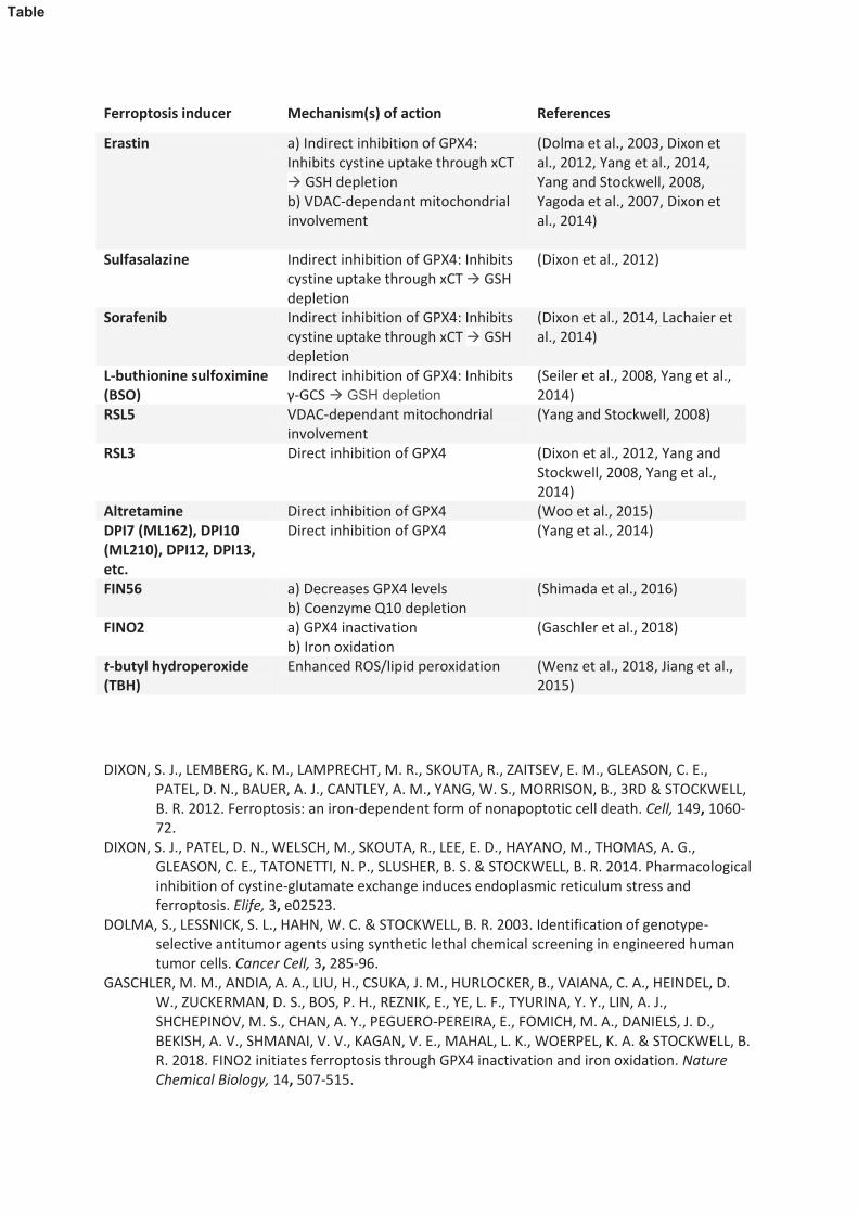
Ferroptosis inducer Mechanism(s) of action References
Erastin a) Indirect inhibition of GPX4:
Inhibits cystine uptake through xCT
à GSH depletion
b) VDAC-dependant mitochondrial
involvement
(Dolma et al., 2003, Dixon et
al., 2012, Yang et al., 2014,
Yang and Stockwell, 2008,
Yagoda et al., 2007, Dixon et
al., 2014)
Sulfasalazine Indirect inhibition of GPX4: Inhibits
cystine uptake through xCT à GSH
depletion
(Dixon et al., 2012)
Sorafenib Indirect inhibition of GPX4: Inhibits
cystine uptake through xCT à GSH
depletion
(Dixon et al., 2014, Lachaier et
al., 2014)
L-buthionine sulfoximine
(BSO)
Indirect inhibition of GPX4: Inhibits
γ-GCS à GSH depletion
(Seiler et al., 2008, Yang et al.,
2014)
RSL5 VDAC-dependant mitochondrial
involvement
(Yang and Stockwell, 2008)
RSL3 Direct inhibition of GPX4 (Dixon et al., 2012, Yang and
Stockwell, 2008, Yang et al.,
2014)
Altretamine Direct inhibition of GPX4 (Woo et al., 2015)
DPI7 (ML162), DPI10
(ML210), DPI12, DPI13,
etc.
Direct inhibition of GPX4 (Yang et al., 2014)
FIN56 a) Decreases GPX4 levels
b) Coenzyme Q10 depletion
(Shimada et al., 2016)
FINO2 a) GPX4 inactivation
b) Iron oxidation
(Gaschler et al., 2018)
t-butyl hydroperoxide
(TBH)
Enhanced ROS/lipid peroxidation (Wenz et al., 2018, Jiang et al.,
2015)
DIXON, S. J., LEMBERG, K. M., LAMPRECHT, M. R., SKOUTA, R., ZAITSEV, E. M., GLEASON, C. E.,
PATEL, D. N., BAUER, A. J., CANTLEY, A. M., YANG, W. S., MORRISON, B., 3RD & STOCKWELL,
B. R. 2012. Ferroptosis: an iron-dependent form of nonapoptotic cell death. Cell, 149, 1060-
72.
DIXON, S. J., PATEL, D. N., WELSCH, M., SKOUTA, R., LEE, E. D., HAYANO, M., THOMAS, A. G.,
GLEASON, C. E., TATONETTI, N. P., SLUSHER, B. S. & STOCKWELL, B. R. 2014. Pharmacological
inhibition of cystine-glutamate exchange induces endoplasmic reticulum stress and
ferroptosis. Elife, 3, e02523.
DOLMA, S., LESSNICK, S. L., HAHN, W. C. & STOCKWELL, B. R. 2003. Identification of genotype-
selective antitumor agents using synthetic lethal chemical screening in engineered human
tumor cells. Cancer Cell, 3, 285-96.
GASCHLER, M. M., ANDIA, A. A., LIU, H., CSUKA, J. M., HURLOCKER, B., VAIANA, C. A., HEINDEL, D.
W., ZUCKERMAN, D. S., BOS, P. H., REZNIK, E., YE, L. F., TYURINA, Y. Y., LIN, A. J.,
SHCHEPINOV, M. S., CHAN, A. Y., PEGUERO-PEREIRA, E., FOMICH, M. A., DANIELS, J. D.,
BEKISH, A. V., SHMANAI, V. V., KAGAN, V. E., MAHAL, L. K., WOERPEL, K. A. & STOCKWELL, B.
R. 2018. FINO2 initiates ferroptosis through GPX4 inactivation and iron oxidation. Nature
Chemical Biology, 14, 507-515.
Table
JIANG, L., KON, N., LI, T., WANG, S. J., SU, T., HIBSHOOSH, H., BAER, R. & GU, W. 2015. Ferroptosis as
a p53-mediated activity during tumour suppression. Nature, 520, 57-62.
LACHAIER, E., LOUANDRE, C., GODIN, C., SAIDAK, Z., BAERT, M., DIOUF, M., CHAUFFERT, B. &
GALMICHE, A. 2014. Sorafenib induces ferroptosis in human cancer cell lines originating
from different solid tumors. Anticancer Res, 34, 6417-22.
SEILER, A., SCHNEIDER, M., FORSTER, H., ROTH, S., WIRTH, E. K., CULMSEE, C., PLESNILA, N.,
KREMMER, E., RADMARK, O., WURST, W., BORNKAMM, G. W., SCHWEIZER, U. & CONRAD,
M. 2008. Glutathione peroxidase 4 senses and translates oxidative stress into 12/15-
lipoxygenase dependent- and AIF-mediated cell death. Cell Metab, 8, 237-48.
SHIMADA, K., SKOUTA, R., KAPLAN, A., YANG, W. S., HAYANO, M., DIXON, S. J., BROWN, L. M.,
VALENZUELA, C. A., WOLPAW, A. J. & STOCKWELL, B. R. 2016. Global survey of cell death
mechanisms reveals metabolic regulation of ferroptosis. Nature Chemical Biology, 12, 497.
WENZ, C., FAUST, D., LINZ, B., TURMANN, C., NIKOLOVA, T., BERTIN, J., GOUGH, P., WIPF, P.,
SCHRODER, A. S., KRAUTWALD, S. & DIETRICH, C. 2018. t-BuOOH induces ferroptosis in
human and murine cell lines. Arch Toxicol, 92, 759-775.
WOO, J. H., SHIMONI, Y., YANG, W. S., SUBRAMANIAM, P., IYER, A., NICOLETTI, P., RODRIGUEZ
MARTINEZ, M., LOPEZ, G., MATTIOLI, M., REALUBIT, R., KARAN, C., STOCKWELL, B. R.,
BANSAL, M. & CALIFANO, A. 2015. Elucidating Compound Mechanism of Action by Network
Perturbation Analysis. Cell, 162, 441-451.
YAGODA, N., VON RECHENBERG, M., ZAGANJOR, E., BAUER, A. J., YANG, W. S., FRIDMAN, D. J.,
WOLPAW, A. J., SMUKSTE, I., PELTIER, J. M., BONIFACE, J. J., SMITH, R., LESSNICK, S. L.,
SAHASRABUDHE, S. & STOCKWELL, B. R. 2007. RAS-RAF-MEK-dependent oxidative cell death
involving voltage-dependent anion channels. Nature, 447, 864-8.
YANG, W. S., SRIRAMARATNAM, R., WELSCH, M. E., SHIMADA, K., SKOUTA, R., VISWANATHAN, V. S.,
CHEAH, J. H., CLEMONS, P. A., SHAMJI, A. F., CLISH, C. B., BROWN, L. M., GIROTTI, A. W.,
CORNISH, V. W., SCHREIBER, S. L. & STOCKWELL, B. R. 2014. Regulation of ferroptotic cancer
cell death by GPX4. Cell, 156, 317-331.
YANG, W. S. & STOCKWELL, B. R. 2008. Synthetic lethal screening identifies compounds activating
iron-dependent, nonapoptotic cell death in oncogenic-RAS-harboring cancer cells. Chem
Biol, 15, 234-45.
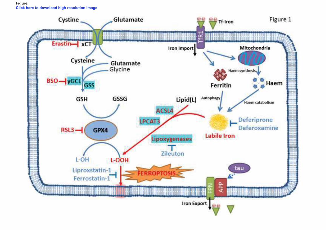
Figure
Click here to download high resolution image