Embed Size (px)
Citation preview
Hospital Physician Board Review Manual www.turner-white.com
Introduction . . . . . . . . . . . . . . . . . . . . . . . . . . . . .1
Pathogenesis . . . . . . . . . . . . . . . . . . . . . . . . . . . . .2
Multiple Myeloma . . . . . . . . . . . . . . . . . . . . . . . . .3
Rare Disorders . . . . . . . . . . . . . . . . . . . . . . . . . .13
Conclusion . . . . . . . . . . . . . . . . . . . . . . . . . . . . .14
Board Review Questions . . . . . . . . . . . . . . . . . . .15
References . . . . . . . . . . . . . . . . . . . . . . . . . . . . .15
Table of Contents
ONCOLOGY BOARD REVIEW MANUAL
Management of Plasma Cell Disorders
Contributor:Brendan M. Weiss, MD Assistant Professor of Medicine, Abramson Cancer Center, Division of Hematology-Oncology, University of Pennsylvania, Philadelphia, PA
STATEMENT OF EDITORIAL PURPOSE
The Hospital Physician Oncology Board Review Manual is a study guide for fellows and practicing physicians preparing for board examinations in oncology. Each manual reviews a topic essential to the current practice of oncology.
PUBLISHING STAFF
PRESIDENT, GROUP PUBLISHERBruce M. White
SENIOR EDITORRobert Litchkofski
EXECUTIVE VICE PRESIDENTBarbara T. White
EXECUTIVE DIRECTOR OF OPERATIONS
Jean M. Gaul
NOTE FROM THE PUBLISHER:This publication has been developed without involvement of or review by the American Board of Internal Medicine.
M a n a g e m e n t o f P l a s m a C e l l D i s o r d e r s
www.turner-white.com Oncology Volume 11, Part 1 1
ONCOLOGY BOARD REVIEW MANUAL
Management of Plasma Cell Disorders
Brendan M. Weiss, MD
INTRODUCTION
The plasma cell disorders are a spectrum of con-ditions that include asymptomatic precursor condi-tions—monoclonal gammopathy of undetermined significance (MGUS) and smoldering multiple my-eloma (SMM)—as well as symptomatic multiple myeloma (MM) and solitary plasmacytoma.1 Other plasma cell disorders include immunoglobulin light chain amyloidosis and POEMS syndrome, which are characterized by a unique set of end-organ manifestations. There are other related plasma cell and B-cell proliferations, such as light chain deposition disease and cryoglobulinemia, that are beyond the scope of this review but are relevant to the hematologist/oncologist and have been re-viewed in detail elsewhere.2
MM is the second most common hematologic ma-lignancy, with approximately 20,000 patients diagnosed annually in the United States.3 The median age at pre-sentation is 72 years, and it is more common in men and in African Americans. In fact, MM is the most com-mon hematologic malignancy in African Americans.
The precursor states MGUS and SMM are asymp- tomatic without end-organ manifestations. MM is
characterized by the accumulation of clonal bone marrow plasma cells and production of mono-clonal immunoglobulins leading to the cardinal end-organ manifestations known by the acronym CRAB: hypercalcemia, renal failure, anemia, and bone disease.4 Hypercalcemia occurs as a result of bone destruction from increased osteoclast ac-tivity stimulated by malignant plasma cells. Renal failure may be associated with hypercalcemia, light chain cast nephropathy, or light chain deposition. Anemia is generally due to the expansion of plas-ma cells in the bone marrow, which may also lead to leukopenia and thrombocytopenia. Myeloma- related bone disease includes osteoporosis, oste-olytic bone lesions, fractures, and bone pain. Additional, less common manifestations of symp-tomatic MM include hypogammaglobulinemia with frequent infections, susceptibility to bleeding, plas-macytomas either extending from bone or in soft tissue sites, and amyloidosis.
Immunoglobulin light chain amyloidosis (AL) may be a primary disorder or may be seen in as-sociation with frank MM. Features of amyloidosis are rare in newly diagnosed MM, but may occur in up to 10% of patients in the relapsed and refrac-
Copyright 2015, Turner White Communications, Inc., Strafford Avenue, Suite 220, Wayne, PA 19087-3391, www.turner-white.com. All rights reserved. No part of this publication may be reproduced, stored in a retrieval system, or transmitted in any form or by any means, mechanical, electronic, photocopying, recording, or otherwise, without the prior written permission of Turner White Communications. The preparation and distribution of this publication are supported by spon-sorship subject to written agreements that stipulate and ensure the editorial independence of Turner White Communications. Turner White Communications retains full control over the design and production of all published materials, including selection of topics and preparation of editorial content. The authors are solely responsible for substantive content. Statements expressed reflect the views of the authors and not necessarily the opinions or policies of Turner White Communications. Turner White Communications accepts no responsibility for statements made by authors and will not be liable for any errors of omission or inaccuracies. Information contained within this publication should not be used as a substitute for clinical judgment.
M a n a g e m e n t o f P l a s m a C e l l D i s o r d e r s
2 Hospital Physician Board Review Manual www.turner-white.com
tory state. AL is characterized by organ dysfunction mediated by the deposition of insoluble amyloid fibrils that are derived from immunoglobulin light chains.5 The manifestations of AL include cardio-myopathy, nephrotic syndrome, peripheral and autonomic neuropathy, macroglossia, and many others.
This article reviews the diagnostic approach to plasma cell disorders, describes the manage-ment of newly diagnosed and relapsed MM, and describes the management of the rare plasma cell disorders AL and POEMS syndrome.
PATHOGENESIS
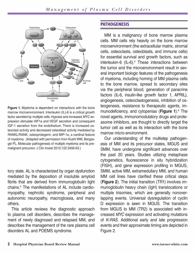
MM is a malignancy of bone marrow plasma cells. MM cells rely heavily on the bone marrow microenvironment (the extracellular matrix, stromal cells, osteoclasts, osteoblasts, and immune cells) for essential survival and growth factors, such as interleukin-6 (IL-6).4 These interactions between the tumor and the microenvironment result in sev-eral important biologic features of the pathogenesis of myeloma, including homing of MM plasma cells to the bone marrow, spread to secondary sites via the peripheral blood, generation of paracrine factors (IL-6, insulin-like growth factor 1, APRIL), angiogenesis, osteoclastogenesis, inhibition of os-teogenesis, resistance to therapeutic agents, im-munodeficiency, and cytopenias (Figure 1).6 The novel agents, immunomodulatory drugs and prote-asome inhibitors, are thought to directly target the tumor cell as well as its interaction with the bone marrow micro-environment.
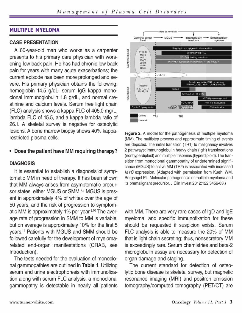
Our understanding of the multistep pathogen-esis of MM and its precursor states, MGUS and SMM, have undergone significant advances over the past 20 years. Studies utilizing metaphase cytogenetics, fluorescence in situ hybridization (FISH), and gene expression profiling in MGUS, SMM, active MM, extramedullary MM, and human MM cell lines have clarified these critical steps (Figure 2). The initial transition (TR1) involves im-munoglobulin heavy chain (IgH) translocations or multiple trisomies, which are generally nonover-lapping events. Universal dysregulation of cyclin D expression is seen in MGUS. The transition from MGUS to MM (TR2) is associated with in-creased MYC expression and activating mutations of K-RAS. Additional early and late progression events and their approximate timing are depicted in Figure 2.
Figure 1. Myeloma is dependent on interactions with the bone marrow microenvironment. Interleukin (IL)-6 is a critical growth factor secreted by multiple cells. Hypoxia and increased MYC ex-pression stimulate HIF1a and VEGF secretion and consequent IGF-1 secretion from the endothelium. There is increased os-teoclast activity and decreased osteoblast activity mediated by RANKL/RANK, osteoprotegerin, and MIP-1a, a cardinal feature of myeloma. (Adapted with permission from Kuehl WM, Bergsa-gel PL. Molecular pathogenesis of multiple myeloma and its pre-malignant precursor. J Clin Invest 2012;122:3456-63.)
M a n a g e m e n t o f P l a s m a C e l l D i s o r d e r s
www.turner-white.com Oncology Volume 11, Part 1 3
MULTIPLE MYELOMA
CASE PRESENTATION
A 60-year-old man who works as a carpenter presents to his primary care physician with wors-ening low back pain. He has had chronic low back pain for years with many acute exacerbations; the current episode has been more prolonged and se-vere. His primary physician obtains the following: hemoglobin 14.5 g/dL, serum IgG kappa mono-clonal immunoglobulin 1.8 g/dL, and normal cre-atinine and calcium levels. Serum free light chain (FLC) analysis shows a kappa FLC of 405.0 mg/L, lambda FLC of 15.5, and a kappa:lambda ratio of 26.1. A skeletal survey is negative for osteolytic lesions. A bone marrow biopsy shows 40% kappa-restricted plasma cells.
• DoesthepatienthaveMMrequiringtherapy?
DIAGNOSIS
It is essential to establish a diagnosis of symp-tomatic MM in need of therapy. It has been shown that MM always arises from asymptomatic precur-sor states, either MGUS or SMM.7,8 MGUS is pres-ent in approximately 4% of whites over the age of 50 years, and the risk of progression to symptom-atic MM is approximately 1% per year.9,10 The aver-age rate of progression in SMM to MM is variable, but on average is approximately 10% for the first 5 years.11 Patients with MGUS and SMM should be followed carefully for the development of myeloma-related end-organ manifestations (CRAB, see Introduction).
The tests needed for the evaluation of monoclo-nal gammopathies are outlined in Table1. Utilizing serum and urine electrophoresis with immunofixa-tion along with serum FLC analysis, a monoclonal gammopathy is detectable in nearly all patients
with MM. There are very rare cases of IgD and IgE myeloma, and specific immunofixation for these should be requested if suspicion exists. Serum FLC analysis is able to measure the 20% of MM that is light chain secreting; thus, nonsecretory MM is exceedingly rare. Serum chemistries and beta-2 microglobulin assay are necessary for detection of organ damage and staging.
The current standard for detection of osteo-lytic bone disease is skeletal survey, but magnetic resonance imaging (MRI) and positron emission tomography/computed tomography (PET/CT) are
Figure 2. A model for the pathogenesis of multiple myeloma (MM). The multistep process and approximate timing of events are depicted. The initial transition (TR1) to malignancy involves 2 pathways: immunoglobulin heavy chain (IgH) translocations (nonhyperdiploid) and multiple trisomies (hyperdiploid). The tran-sition from monoclonal gammopathy of undetermined signifi-cance (MGUS) to active MM (TR2) is associated with increased MYC expression. (Adapted with permission from Kuehl WM, Bergsagel PL. Molecular pathogenesis of multiple myeloma and its premalignant precursor. J Clin Invest 2012;122:3456-63.)
M a n a g e m e n t o f P l a s m a C e l l D i s o r d e r s
4 Hospital Physician Board Review Manual www.turner-white.com
useful in selected circumstances.12,13 MRI is specifi-cally useful for evaluation of paraspinal masses or spinal cord compression. PET/CT is useful in the evaluation of solitary plasmacytomas to detect ad-ditional sites of bone marrow FDG-avid lesions and osteolytic disease. This has become a critical mo-dality in the modern evaluation of solitary plasma-cytomas.14 A bone marrow aspiration and biopsy is necessary to quantify bone marrow PC infiltration and obtain molecular studies for prognosis.
The International Myeloma Working Group (IMWG) criteria for the diagnosis of MM requires the presence of end-organ manifestations; patients without these manifestations are classified as ei-ther MGUS or SMM (Table 2).15 The distinction between MGUS and SMM is made on the basis of the monoclonal immunoglobulin concentration and degree of bone marrow plasmacytosis, but the diagnosis of symptomatic MM can be made at any level of M-protein or bone marrow plasma cell
infiltration. Again, it is imperative that the clinician establishes that these manifestations are related to MM. There are numerous examples of falsely attributing these manifestations to MM when in fact other conditions are present, such as primary hyperparathyroidism accounting for hypercalcemia or anemia secondary to iron deficiency. Patients should not be treated in the absence of MM-related end-organ dysfunction.
The IMWG has developed guidelines for the management of MGUS and SMM and has rec-ommended observation based on the risk of pro-gression to MM.16 Factors that increase the risk of progression from MGUS to MM are monoclonal immunoglobulin concentration ≥1.5 g/dL, abnormal serum FLC ratio, and a non-IgG isotype.17 Patients with a monoclonal gammopathy and the absence of high-risk features for progression can be ob-served every 2 to 3 years. For patients with a sin-gle adverse feature, additional testing including a bone marrow biopsy and skeletal survey is recom-mended. Patients with MGUS should return initially in 3 to 6 months to assess for evidence of progres-sion to active MM and then follow-up annually if not low risk. Patients with SMM should be evaluated every 3 to 6 months depending on risk. Features that increase the risk of progression from SMM to MM are monoclonal immunoglobulin concentration ≥3 g/dL, bone marrow plasma cells ≥10%, and a widely skewed serum FLC ratio (≤0.125 or ≥8).18 During surveillance, patients should have a direct-ed history and physical examination with focus on evidence of MM, lymphoma, or amyloidosis along with standard laboratory tests.
Patients with SMM represent a highly heteroge-neous group of patients—some are progressing to MM at a slow rate similar to high-risk MGUS, and others are early MM patients who have a very high risk of progression to MM in 2 years.11 Identi-
Table1.Diagnostic Tests Used in the Evaluation of Monoclonal Gammopathies
EssentialdiagnostictestsComplete blood count with leukocyte differential
Complete metabolic panel
Serum protein electrophoresis with immunofixation
Quantitative immunoglobulins
24-hour urine protein electrophoresis with immunofixation
Skeletal survey
Serum free light chain analysis
Serum beta-2 microglobulin
Serum albumin
Bone marrow aspirate and biopsy
Bone marrow cytogenetics
Bone marrow fluorescence in situ hybridization (FISH) panel
UsefulinselectedsituationsMagnetic resonance imaging
Positron emission tomography/computed tomography
M a n a g e m e n t o f P l a s m a C e l l D i s o r d e r s
www.turner-white.com Oncology Volume 11, Part 1 5
fying these patients with ultra-high risk SMM who would benefit from earlier intervention prior to the onset of end-organ damage may have substantial benefit for patients.19 The PETHEMA performed a landmark randomized trial of lenalidomide and dexamethasone versus observation in high-risk SMM using the Mayo Clinic and/or PETHEMA bone marrow flow cytometry criteria.20 This study demonstrated both a progression-free and overall survival benefit of lenalidomide and dexametha-sone therapy. At a median follow-up of 40 months, the median time to progression was not reached in the treatment group and was 21 months for observation. Overall survival was also significantly prolonged (hazard ratio for death 0.33, P = 0.03). However, this study has been criticized and has not changed practice for the following reasons: the definition of high-risk SMM using the flow cytometry criteria is not widely available and is technically complex, the treatment arm had in-tensification of treatment at signs of biochemical progression, outcomes on the observation arm were quite poor, and the treatment at progression was not uniform in the experimental arm. Further-more, lenalidomide was not given at the time of progression.
CASECONTINUED
The patient’s back pain resolves with analgesics and physical therapy in 6 weeks. He is diagnosed with SMM and is observed with evaluations every 3 months. Two years after initial consultation, he reports dyspnea and fatigue; he can barely finish a half day of work as a carpenter. He also has a new site of pain in his upper back. Laboratory studies show the following: hemoglobin 7.2 g/dL, creatinine 2.1 mg/dL, IgG kappa 3.8 g/dL, serum kappa FLC 835.5 mg/L, lambda FLC 9.0, kappa:lambda ratio 92.8, beta-2 microglobulin 8.2 µg/mL, albumin 3.2 g/dL. A bone marrow biopsy demonstrates 70% kappa-restricted plasma cells with normal cytoge-netics and FISH testing shows t(14;16) and del(17). A skeletal survey reveals a vertebral compression fracture at T6.
• What is the appropriate management ofnewlydiagnosedMM?
MANAGEMENT
The patient has symptomatic MM based on the presence of anemia, renal failure, and osteolytic bone disease and requires therapy. MM remains incurable, but outcomes have markedly improved
Table2.Diagnostic Criteria for Monoclonal Gammopathies
MonoclonalGammopathyofUndeterminedSignificance
Smoldering MultipleMyeloma
MultipleMyeloma
SolitaryPlasmacytoma
Monoclonal immunoglobulin concentration
<3.0 g/dL ≥3.0 g/dL Any Any
and/or and/or orBone marrow plasma cells (%) <10 ≥10 Any Any
Myeloma-related organ or tissue impairment*
Absent Absent Present A single bone or extraosseous plasmacytoma, but no CRAB
*CRAB features: calcium >11.0 mg/dL, creatinine >2 mg/dL, hemoglobin 2 g below the lower limit of normal or <10.0 g/dL, osteolytic bone lesions or osteoporosis with compression fractures. Other: symptomatic hyperviscosity, recurrent bacterial infections.
M a n a g e m e n t o f P l a s m a C e l l D i s o r d e r s
6 Hospital Physician Board Review Manual www.turner-white.com
in the past 20 years.21 In the mid 1990s, high-dose therapy and autologous stem cell transplantation (HDT/autoSCT) was shown to improve survival compared to conventional chemotherapy. This rep-resented the first major advance in MM since the development of melphalan and prednisone in the 1960s. Subsequently, the development of immu-nomodulatory drugs (IMiDs: thalidomide, lenalido-mide, and pomalidomide) and proteasome inhibi-tors (bortezomib and carfilzomib) has resulted in improved outcomes. Several epidemiologic studies have shown dramatic survival gains.22,23 Based on SEER data, 5-year relative survival in MM has increased from 29% to 35% from the time period 1990–1992 to 2002–2004.24
There are 3 phases in the current approach to treatment of newly diagnosed MM: induction, consolidation, and maintenance.1 The approach to each phase of therapy is individualized based on the features of the myeloma, age, comorbidi-ties, and personal preferences. There are several general considerations for all patients at initial diagnosis. Renal failure at presentation should prompt urgent chemotherapy. In MM patients with renal failure, it is important to maintain volume status and avoid nephrotoxic drugs. The benefit of
plasmapheresis in the management of myeloma-related renal failure remains unclear.25,26 Analge-sia and bisphosphonates for painful bone lesions should be started. Consultation with an orthopedic oncologist may be necessary if there are lesions at high risk for pathologic fracture. Hypercalcemia should be managed with aggressive intravenous fluids and bisphosphonates.
Another critically important decision to be made early in the course of therapy is the patient’s can-didacy for HDT/autoSCT. Generally, patients under the age of 70 without significant comorbidities are candidates for HDT/autoSCT. Melphalan-based in-duction regimens and extensive radiation therapy to bone marrow should be avoided to preserve the option of autologous stem cell collection. Pro-longed therapy with lenalidomide-based induction regimens may also impact stem cell collection and should be done with caution.27,28
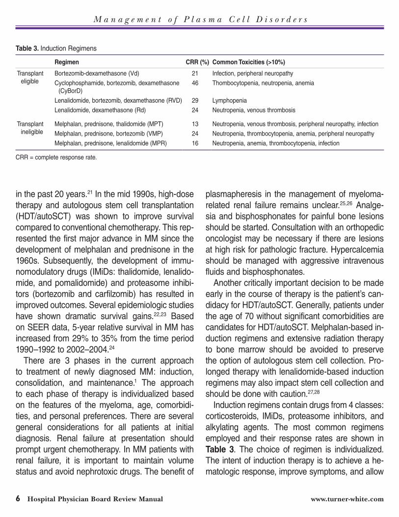
Induction regimens contain drugs from 4 classes: corticosteroids, IMiDs, proteasome inhibitors, and alkylating agents. The most common regimens employed and their response rates are shown in Table 3. The choice of regimen is individualized. The intent of induction therapy is to achieve a he-matologic response, improve symptoms, and allow
Table3.Induction Regimens
Regimen CRR (%) CommonToxicities(>10%)
Transplant eligible
Bortezomib-dexamethasone (Vd) 21 Infection, peripheral neuropathy
Cyclophosphamide, bortezomib, dexamethasone (CyBorD)
46 Thombocytopenia, neutropenia, anemia
Lenalidomide, bortezomib, dexamethasone (RVD) 29 Lymphopenia
Lenalidomide, dexamethasone (Rd) 24 Neutropenia, venous thrombosis
Transplant ineligible
Melphalan, prednisone, thalidomide (MPT) 13 Neutropenia, venous thrombosis, peripheral neuropathy, infection
Melphalan, prednisone, bortezomib (VMP) 24 Neutropenia, thrombocytopenia, anemia, peripheral neuropathy
Melphalan, prednisone, lenalidomide (MPR) 16 Neutropenia, anemia, thrombocytopenia, infection
CRR = complete response rate.
M a n a g e m e n t o f P l a s m a C e l l D i s o r d e r s
www.turner-white.com Oncology Volume 11, Part 1 7
for stem cell collection. Two- or 3-drug induction regimens from the above 4 classes are usually em-ployed. Although 3-drug regimens result in higher response rates, they are associated with increased toxicity. Low-dose dexamethasone is now the stan-dard of care based on a randomized controlled trial comparing lenalidomide with high-dose dexameth-asone (480 mg per month) to lenalidomide and low-dose dexamethasone (160 mg per month).29 The low-dose dexamethasone arm achieved bet-ter overall survival and lower toxicity, including a significantly lower rate of mortality within 4 months of therapy.
Patients who are not candidates for HDT/au-toSCT have several options for induction therapy. Randomized trials have shown that the addition of a novel agent to melphalan and prednisone results in improved outcomes. Melphalan, prednisone, and thalidomide (MPT) has been compared to melphalan and prednisone (MP).4,30 Patients re-ceived 6 cycles of MPT followed by maintenance thalidomide until progression versus MP for 6 cy-cles. MPT was superior, with an event-free survival at 2 years of 54% for MPT compared to 27% for MP (P = 0.0006). The combination of melphalan, prednisone, and bortezomib (VMP) was compared to MP, both administered for 9 cycles without main-tenance therapy.6,31 The partial response rates for VMP and MP were 71% and 35%, respectively. The hazard ratio for overall survival favored bort-ezomib (0.61, P = 0.008).
• HowisprognosisdefinedinMM?
PROGNOSIS
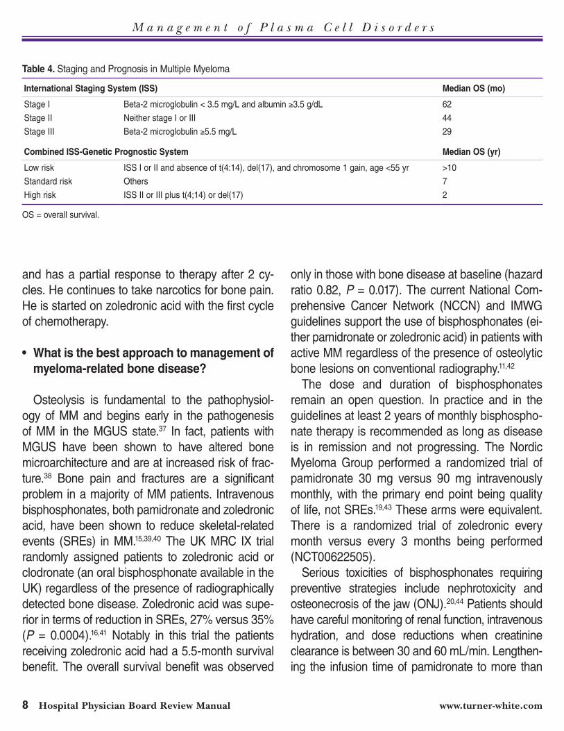
Prognosis in MM is based on both molecular fea-tures of MM and the International Staging System (ISS) (Table4). The ISS uses 2 biomarkers, serum beta-2 microglobulin and serum albumin, and is
simple and more useful than the Durie-Salmon system.7,8,32 Collectively, beta-2 microglobulin and albumin reflect myeloma tumor burden, renal fail-ure (long known to be an independent prognostic factor), and host fitness. Molecular features of MM that have prognostic value include bone marrow karyotype, translocations, chromosome content, and gene expression profiling.9,10,33 There contin-ues to be debate on the molecular classification of MM because the significance of the individual markers changes with the introduction of new therapies. Standard risk molecular features in-clude t(11;14) and hyperdiploidy. High-risk features include del(17), t(14;16), and chromosome 1 gain. Translocation (4;14) is considered intermediate risk. The presence of del(13) is no longer con-sidered an adverse prognostic feature unless it is seen on bone marrow karyotype. Several groups have developed gene expression–based prognos-tic systems, but they are not widely used in clinical practice.34 The IMWG have analyzed outcomes with ISS staging and FISH and have been able to show that the combination of these factors pro-vides robust prognostic information.35 The patients were stratified by ISS stage and the presence of either t(4:14), del(17), or chromosome 1 gain into 3 groups: (a) low risk: ISS I or II and negative FISH; (b) standard risk: ISS III and negative FISH or ISS 1 and positive FISH; and (c) high risk: ISS II/III and positive FISH.11,36 The median survivals for these 3 groups are more than 10 years, 7 years, and 2 years, respectively. At present, there are no spe-cific therapies for specific molecular subgroups of myeloma.
CASECONTINUED
The patient has ISS III myeloma with high-risk molecular features. He begins therapy with cyclo-phosphamide, bortezomib, and dexamethasone
M a n a g e m e n t o f P l a s m a C e l l D i s o r d e r s
8 Hospital Physician Board Review Manual www.turner-white.com
and has a partial response to therapy after 2 cy-cles. He continues to take narcotics for bone pain. He is started on zoledronic acid with the first cycle of chemotherapy.
• Whatisthebestapproachtomanagementofmyeloma-relatedbonedisease?
Osteolysis is fundamental to the pathophysiol-ogy of MM and begins early in the pathogenesis of MM in the MGUS state.37 In fact, patients with MGUS have been shown to have altered bone microarchitecture and are at increased risk of frac-ture.38 Bone pain and fractures are a significant problem in a majority of MM patients. Intravenous bisphosphonates, both pamidronate and zoledronic acid, have been shown to reduce skeletal-related events (SREs) in MM.15,39,40 The UK MRC IX trial randomly assigned patients to zoledronic acid or clodronate (an oral bisphosphonate available in the UK) regardless of the presence of radiographically detected bone disease. Zoledronic acid was supe-rior in terms of reduction in SREs, 27% versus 35% (P = 0.0004).16,41 Notably in this trial the patients receiving zoledronic acid had a 5.5-month survival benefit. The overall survival benefit was observed
only in those with bone disease at baseline (hazard ratio 0.82, P = 0.017). The current National Com-prehensive Cancer Network (NCCN) and IMWG guidelines support the use of bisphosphonates (ei-ther pamidronate or zoledronic acid) in patients with active MM regardless of the presence of osteolytic bone lesions on conventional radiography.11,42
The dose and duration of bisphosphonates remain an open question. In practice and in the guidelines at least 2 years of monthly bisphospho-nate therapy is recommended as long as disease is in remission and not progressing. The Nordic Myeloma Group performed a randomized trial of pamidronate 30 mg versus 90 mg intravenously monthly, with the primary end point being quality of life, not SREs.19,43 These arms were equivalent. There is a randomized trial of zoledronic every month versus every 3 months being performed (NCT00622505).
Serious toxicities of bisphosphonates requiring preventive strategies include nephrotoxicity and osteonecrosis of the jaw (ONJ).20,44 Patients should have careful monitoring of renal function, intravenous hydration, and dose reductions when creatinine clearance is between 30 and 60 mL/min. Lengthen-ing the infusion time of pamidronate to more than
Table4.Staging and Prognosis in Multiple Myeloma
InternationalStagingSystem(ISS) MedianOS(mo)
Stage I Beta-2 microglobulin < 3.5 mg/L and albumin ≥3.5 g/dL 62
Stage II Neither stage I or III 44
Stage III Beta-2 microglobulin ≥5.5 mg/L 29
CombinedISS-GeneticPrognosticSystem MedianOS(yr)
Low risk ISS I or II and absence of t(4:14), del(17), and chromosome 1 gain, age <55 yr >10
Standard risk Others 7
High risk ISS II or III plus t(4;14) or del(17) 2
OS = overall survival.
M a n a g e m e n t o f P l a s m a C e l l D i s o r d e r s
www.turner-white.com Oncology Volume 11, Part 1 9
4 hours can reduce nephrotoxicity. Changes in renal function during therapy should prompt discontinua-tion. The rate of ONJ in the MRC IX trial was ap-proximately 1% per year, but preventive dental care was not mandatory. Preventive dental care reduces the rate of ONJ and is recommended prior to start-ing bisphosphonates as is avoidance of invasive dental procedures.22,23,45
Denosumab, a monoclonal antibody to RANK- ligand approved for use in breast and prostate can-cer metastatic to bone, is contraindicated in MM. A subset of MM patients treated with denosumab had inferior survival compared to the zoledronic acid patients.46 A prospective trial in MM is ongoing.
CASECONTINUED
The patient continues therapy with cyclophos-phamide, bortezomib, and dexamethasone. Dur-ing the last cycle of therapy he develops mild, nonpainful paresthesias in his feet. He is now in a very good partial remission. The patient returns with questions regarding the role of HDT/autoSCT.
• How are the toxicities of induction therapymanaged?
Toxicities of MM induction that require specific management include peripheral neuropathy, ve-nous thromboembolism, and infection. Peripheral neuropathy is common with both bortezomib and thalidomide.47 Bortezomib neuropathy is related to dose, schedule, and mode of administration and is generally reversible. Peripheral neuropathy from thalidomide is cumulative and dose-dependent and is often permanent. Prompt dose reductions are required with development of neuropathy of any grade with thalidomide. In patients with grade 1 or 2 bortezomib-related neuropathy, dose reduc-tion to 1.0 mg/m2 is suggested, or weekly admin-
istration should be considered. For patients who develop grade 3 neuropathy, bortezomib should be held and resumed at 0.7 mg/m2 when the neuropa-thy has resolved to grade 1 or better. A randomized trial of subcutaneous administration compared to intravenous administration of bortezomib showed a dramatic decrease in peripheral neuropathy of all grades (38% vs 53%) and grade 3 (6% vs 16%) peripheral neuropathy.48
There is an increased risk of venous throm-boembolism (VTE) when IMiDs are combined with steroids or anthracyclines.49 The rate of VTE ranges from 20% to 40% without prophylaxis; the highest rates are with combinations that include anthracyclines. A randomized trial comparing as-pirin (100 mg/day), mini-dose warfarin (1.25 mg/day), and enoxaparin (40 mg subcutaneously daily) in patients receiving thalidomide-based regi-mens demonstrated equivalence between aspirin and mini-dose warfarin.50 Therefore, all patients receiving IMiDs in combination with steroids or an-thracyclines should receive VTE prophylaxis with aspirin. For patients with additional risk factors for VTE, one should consider low-molecular-weight heparin at either prophylactic or therapeutic doses.
MM patients are at increased risk of infection due to the underlying disease and therapy. High-dose dexamethasone regimens have a higher risk of infection compared to low-dose regimens.29 Therefore, prophylaxis for opportunistic infection and bacterial infections is less commonly used now that low-dose dexamethasone regimens are standard. Bortezomib is associated with varicella zoster reactivation rates of over 10%, making an-tiviral prophylaxis mandatory.51 For patients with recurrent bacterial infections, intravenous immune globulin is an option, although this practice is based on a study done before the advent of more effective novel agents.52
M a n a g e m e n t o f P l a s m a C e l l D i s o r d e r s
10 Hospital Physician Board Review Manual www.turner-white.com
• WhatisthecurrentroleofHDT/autoSCT?
The place of HDT/autoSCT in the manage-ment of MM continues to evolve in the era of novel drugs.53 The Intergroupe Francophone du Myelome (IFM) first reported improved overall sur-vival with HDT/autoSCT compared to conventional chemotherapy in 1996.54 Several clinical trials comparing HDT/autoSCT to conventional therapy have demonstrated that HDT/autoSCT improves progression-free survival compared to conven-tional therapy, and in some trials there was an overall survival benefit.55 Tandem HDT/autoSCT (2 planned courses of HDT/autoSCT with a 3-month interval) is not clearly superior to a single course of HDT/autoSCT, but this is the subject of ongoing studies.56,57 The standard conditioning regimen is melphalan alone, as a randomized trial comparing melphalan 200 mg/m2 to melphalan 140 mg/m2 along with 8 Gy total body irradiation showed equivalent event-free survival but more toxicity in the irradiation regimen.58 The timing of transplan-tation remains controversial, but overall survival is essentially the same whether it is performed early or at the time of relapse.59 Early transplan-tation is associated with improved time without symptoms, treatment, and treatment-related ad-verse events and thus may be preferred in some patients.60
CASECONTINUED
The patient undergoes HDT/autoSCT with mel-phalan 200 mg/m2 for conditioning. His course is complicated by severe mucositis requiring paren-teral narcotics and intravenous fluids and febrile neutropenia without source. He engrafts neutro-phils and platelets on days 12 and 14, respectively. He is in a complete remission at day 100 following transplant.
• Whatistheroleofmaintenancetherapy?
Due to the inevitable risk of relapse following induction therapy or transplant, there have been a series of studies over the years investigating the long-term use of therapy to maintain remission. Thalidomide increases progression free survival after conventional therapy and HDT/autoSCT, but toxicity is substantial and renders thalidomide intolerable for long-term use.61 The toxicity profile of lenalidomide is more favorable, and this agent has been tested as maintenance therapy following both conventional therapy and HDT/autoSCT. A randomized trial of melphalan, prednisone, and le-nalidomide followed by lenalidomide maintenance (MPR-R), MPR, and MP in transplant-ineligible patients was performed.62 MPR-R demonstrated an improvement in progression-free survival from 14 and 13 months in the MPR and MP arms to 31 months with MPR-R. The IFM and Cancer and Leukemia Group B (CALGB) performed tri-als comparing lenalidomide to placebo following HDT/autoSCT.63,64 Both trials showed improved progression-free survival from approximately 2 years to 4 years. The CALGB trial had an overall survival benefit. An updated analysis of the IFM trial presented at the American Society of Hema-tology annual meeting in 2013 still showed no over-all survival benefit.65 In the IFM trial, there was poor survival following progression in the lenalidomide maintenance group, which suggests the possibility of drug resistance. There are important differences in the IFM trial, notably, only half of patients re-ceived a novel agent during induction, one-quarter of patients received additional cytotoxic (DCEP) induction, and 2 cycles of consolidation full-dose lenalidomide-dexamethasone post transplant were included. Lenalidomide maintenance was contin-ued for a median of 24 months in the IFM trial,
M a n a g e m e n t o f P l a s m a C e l l D i s o r d e r s
www.turner-white.com Oncology Volume 11, Part 1 11
whereas it was continued indefinitely in the CALGB trial.
Maintenance therapy likely prolongs progres-sion-free survival, but it remains unclear if there is an overall survival benefit. The patient subset that is most likely to benefit from maintenance therapy remains unclear. A concerning toxicity in both studies was the increased risk of second primary malignancies. The NCCN recommends discuss-ing these findings with patients prior to deciding on maintenance therapy, whereas the IMWG recom-mends against maintenance therapy.66 Bortezomib during induction and maintenance compared to thalidomide has demonstrated improved progres-sion-free and overall survival without any reported signal of second malignancies.67
CASECONTINUED
The patient starts lenalidomide 10 mg daily and continues zoledronic acid monthly for another 18 months. Approximately 2 years following transplan-tation he fractures his right humerus while lifting a toolbox and undergoes surgical repair. Pertinent laboratory data show: hemoglobin 10.5 g/dL, creatinine 1.7 mg/dL, IgG kappa 1.8 g/dL, kappa
FLC 325.0, lambda FLC 18.0, and kappa:lambda ratio 18.1.
• WhatistheapproachtorelapsedMM?
The approach to relapsed MM is based on the features of the clinical relapse and the patient’s response and toxicity to prior regimens. A critical decision is when to consider treatment for relapse. Patients with asymptomatic rises in monoclonal protein can be observed carefully for the tempo and nature of relapse prior to treatment. How-ever, patients with disease that has been known to behave aggressively or has other high-risk fea-tures should be considered for therapy even with biochemical relapse. It is appropriate to repeat a regimen that the patient previously responded to, particularly if the duration of response was greater than 6 months. It is advisable to change to a different drug class or different combination if the prior regimen did not provide a sufficient response, resulted in rapid relapse, or was intoler-able. There are many regimens available for re-lapsed disease, but preferred regimens are listed in Table5.
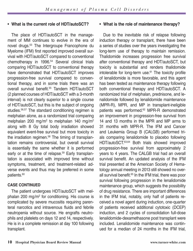
Table5.Regimens for Relapsed Multiple Myeloma
Regimen Study Outcomes
Bortezomib Bortezomib vs high-dose dexamethasone51 ORR: 43% vs 16%1-year OS: 80% vs 67%
Bortezomib–pegylated doxorubicin Bortezomib and pegylated doxorubicin vs Bortezomib68 TTP: 9.3 mo vs 6.5 mo
Lenalidomide-dexamethasone Lenalidomide-dexamethasone vs high-dose dexamethasone69,70 TTP: 11.1 mo vs 4.7 moMedian OS: 29.6 vs 20.2 mo
Carfilzomib Carfilzomib single agent72 DOR: 7.8 moMedian OS: 15.6 mo
Pomalidomide-dexamethasone Pomalidomide-dexamethasone vs high-dose dexamethasone73,74 PFS: 4.0 vs 1.9 moMedian OS: 13.1 vs 8.1 mo
DOR = duration of response; ORR = overall response rate; OS = overall survival; PFS = progression-free survival; TTP = time to progression.
M a n a g e m e n t o f P l a s m a C e l l D i s o r d e r s
12 Hospital Physician Board Review Manual www.turner-white.com
The APEX trial compared bortezomib to high-dose dexamethasone, which demonstrated a com-bined complete response and partial response rate of 38% compared to 18%.51 Survival rates at 1 year were 80% versus 67% (P = 0.0002). These results include about two-thirds of patients crossing over to the bortezomib arm. Bortezomib has been com-bined with liposomal doxorubicin and compared to bortezomib alone, and this combination resulted in an extended time to progression compared to bort-ezomib alone (9.3 versus 6.5 months).68 Lenalido-mide is approved for patients who have relapsed following at least 1 prior line of therapy, based on a North American (MM-009) and International (MM-10) study of lenalidomide with high-dose dexa-methasone compared to high-dose dexametha-sone.69,70 Both of these studies demonstrated improved median time to disease progression in the lenalidomide groups: MM-009 median time to pro-gression was 11.1 months compared to 4.7 months (P < 0.001); MM-010 median time to progression was 11.3 versus 4.7 months (P < 0.001). Median overall survivals were also improved in the lenalid-omide groups in both trials: 29.6 months versus 20.2 (P < 0.001) in MM-009, and in MM-010 the hazard ratio for death was 0.66 (P = 0.03).
Patients who have become refractory to both IMiDs and proteasome inhibitors have a very poor prognosis. The IMWG performed a retrospective study of patients refractory to current therapies including thalidomide, lenalidomide, and bortezo-mib.71 The median overall and event-free survival in these patients was 9 and 5 months, respectively. Therefore, agents active in this population are ur-gently needed. The FDA has approved 2 agents, carfilzomib and pomalidomide, in this population.
Carfilzomib is a second-generation proteasome inhibitor with a potentially improved efficacy and toxicity profile compared to bortezomib. Carfilzo-
mib primarily inhibits the chymotrypsin site of the proteasome, but in higher doses may inhibit the trypsin-like and caspase-like sites. Carfilzomib forms stable and irreversible adducts with the pro-teasome, unlike bortezomib, which is reversible. Optimal inhibition of the proteasome with carfilzo-mib requires consecutive daily dosing. Carfilzomib does not result in significant peripheral neuropa-thy, which is an advantage over bortezomib. The PX 171-003 trial enrolled 266 heavily pre-treated patients (more than 4 lines of therapy and 80% were double refractory).72 Carfilzomib 20 mg/m2 was given twice weekly on days 1 and 2 with dose escalation to 27 mg/m2 on days 8, 9 and 15, 16. The overall response rate was 23.7%, with a median duration of response of 7.8 months, progression-free survival of 3.7 months, and over-all survival of 15.6 months.
The toxicity profile of carfilzomib was notable for a low rate of treatment-emergent peripheral neuropathy (8.3%) despite baseline neuropathy in 77% of patients.72 Severe acute renal failure occurred in 5 of 266 patients. There does appear to be significant cardiopulmonary toxicity with this agent. Five of 24 deaths during the study were considered carfilzomib-related, and 2 of these were cardiac arrests. In addition, one-third of pa-tients experienced mild-moderate dyspnea without detectable lung injury. There is some thought that the dyspnea was related to aggressive hydration, but ongoing studies are evaluating the possibility of direct carfilzomib-related cardiopulmonary toxicity.
Pomalidomide is the third-in-class IMiD and has been approved by the FDA for MM patients treated with at least 2 prior therapies. In multiple phase 2 trials including lenalidomide- and bortezomib-refractory patients, pomalidomide at doses ranging from 2 to 4 mg either daily or daily for 21 days out of 28 days along with dexamethasone 40 mg weekly
M a n a g e m e n t o f P l a s m a C e l l D i s o r d e r s
www.turner-white.com Oncology Volume 11, Part 1 13
demonstrated overall response rates of 25% to 63%.73 A randomized phase 3 study compared pomalidomide 4 mg daily days 1 to 21 and dexa-methasone 40 mg weekly to high-dose dexameth-asone; pomalidomide/low-dose dexamethasone showed improved progression-free survival (4.0 vs 1.9 months, P < 0.001) and improved median over-all survival (13.1 vs 8.1, P = 0.009).74 This is notable even though approximately half of patients in the high-dose dexamethasone arm crossed over to pomalidomide. There was benefit even in patients with high-risk cytogenetics. Importantly, there did not appear to be any impact in the efficacy and toxicity in those with normal versus impaired renal function (creatinine clearance 30–60 mL/min).
RARE DISORDERS
IMMUNOGLOBULINLIGHTCHAINAMYLOIDOSIS
CaseEvaluationA 60-year-old woman presents with an unex-
plained weight loss of 20 pounds, lower extremity edema, dyspnea climbing a single flight of stairs, and several episodes of presyncope. She has seen several doctors without explanation. A nephrologist ordered a 24-hour urine study, which showed 7 g of protein and a small free lambda monoclonal protein.
• What are the next steps in evaluation andmanagement?
The presence of nephrotic-range proteinuria that is mostly albumin and not monoclonal immunoglob-ulin light chain in the presence of additional systemic symptoms should raise suspicion for AL. Suspicion is a critical first step in the diagnosis of AL as many patients go undiagnosed until advanced organ dys-function develops.5 Clinical signs and symptoms that should raise suspicion for AL are unexplained
fatigue, unintentional weight loss, cardiomyopathy, macroglossia, nephrotic syndrome, orthostatic hypo-tension, peripheral or autonomic neuropathy, carpal tunnel syndrome, unexplained bruising (especially periorbital purpura), and hepatomegaly.
The next step in diagnosis is to search for evi-dence of tissue amyloid deposition, which is based on demonstration of a positive Congo red stain in either the involved organ or a surrogate site.75 The most easily accessible surrogate site is the abdominal fat, which is approximately 85% sensi-tive for detection of amyloid deposits. If suspicion remains high, biopsy of an involved organ (eg, heart or kidney) should be pursued. Accurate amy-loid subtyping is necessary to direct therapy and avoid misdiagnosis with hereditary or other forms of amyloidosis due to the commonality of monoclo-nal gammopathies in the general population.76 This may require submission of amyloid-bearing tissue to a reference lab for laser capture microdissection and mass spectrometry.77
Lastly, the extent of organ involvement needs to be assessed by clinical, laboratory, and imaging findings. The outcomes in AL are driven by the extent of cardiac involvement and the aggressive-ness of the plasma cell clone. A staging system in-corporating NT-pro-brain natriuretic peptide (≥1800 pg/mL), cardiac troponin T (≥0.025 ng/mL), and the serum FLC differential (≥180 mg/dL) discrimi-nates patients with very different median survivals: for stage I (no factors) 94.1 months, stage II (any 1 factor) 40.3 months, stage III (any 2 factors) 14 months, and stage IV (all 3 factors) 5.8 months.78
The fundamental treatment principle in AL is to obtain a complete hematologic remission, thereby removing the amyloidogenic precursor protein and allowing for reversal of organ dysfunction. Until the advent of novel drugs, HDT/autoSCT was the most potent anti-plasma cell therapy and was able to
M a n a g e m e n t o f P l a s m a C e l l D i s o r d e r s
14 Hospital Physician Board Review Manual www.turner-white.com
achieve complete hematologic remissions in approxi-mately 40% of patients with variable rates of organ improvement.79,80 However, a randomized trial of HDT/autoSCT compared to standard oral melphalan and dexamethasone performed by the IFM showed better survival in the melphalan-dexamethasone arm.81 This has been attributed to a very high trans-plant-related mortality (TRM) of 24% due to poor patient selection. In the current era, careful selection of patients at experienced centers results in TRM of 5%. Most patients with AL are not fit enough for this procedure and are treated with conventional regi-mens. Fortunately, bortezomib-based regimens can achieve hematologic remission and organ improve-ment rates that are similar to HDT/autoSCT.82–84
POEMSSYNDROME
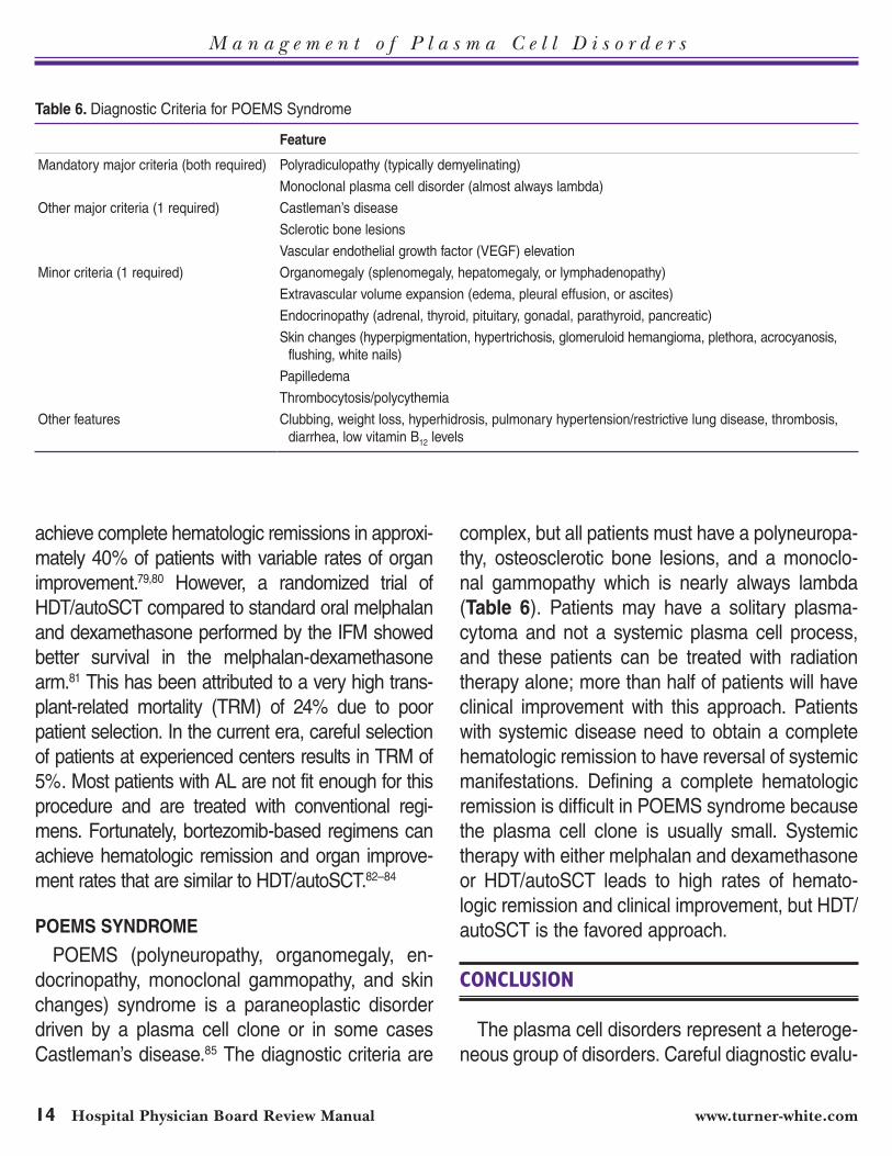
POEMS (polyneuropathy, organomegaly, en-docrinopathy, monoclonal gammopathy, and skin changes) syndrome is a paraneoplastic disorder driven by a plasma cell clone or in some cases Castleman’s disease.85 The diagnostic criteria are
complex, but all patients must have a polyneuropa-thy, osteosclerotic bone lesions, and a monoclo-nal gammopathy which is nearly always lambda (Table 6). Patients may have a solitary plasma-cytoma and not a systemic plasma cell process, and these patients can be treated with radiation therapy alone; more than half of patients will have clinical improvement with this approach. Patients with systemic disease need to obtain a complete hematologic remission to have reversal of systemic manifestations. Defining a complete hematologic remission is difficult in POEMS syndrome because the plasma cell clone is usually small. Systemic therapy with either melphalan and dexamethasone or HDT/autoSCT leads to high rates of hemato-logic remission and clinical improvement, but HDT/ autoSCT is the favored approach.
CONCLUSION
The plasma cell disorders represent a heteroge-neous group of disorders. Careful diagnostic evalu-
Table6.Diagnostic Criteria for POEMS Syndrome
Feature
Mandatory major criteria (both required) Polyradiculopathy (typically demyelinating)
Monoclonal plasma cell disorder (almost always lambda)
Other major criteria (1 required) Castleman’s disease
Sclerotic bone lesions
Vascular endothelial growth factor (VEGF) elevation
Minor criteria (1 required) Organomegaly (splenomegaly, hepatomegaly, or lymphadenopathy)
Extravascular volume expansion (edema, pleural effusion, or ascites)
Endocrinopathy (adrenal, thyroid, pituitary, gonadal, parathyroid, pancreatic)
Skin changes (hyperpigmentation, hypertrichosis, glomeruloid hemangioma, plethora, acrocyanosis, flushing, white nails)
Papilledema
Thrombocytosis/polycythemia
Other features Clubbing, weight loss, hyperhidrosis, pulmonary hypertension/restrictive lung disease, thrombosis, diarrhea, low vitamin B12 levels
M a n a g e m e n t o f P l a s m a C e l l D i s o r d e r s
www.turner-white.com Oncology Volume 11, Part 1 15
ation is critical for accurate diagnosis and manage-ment. The outcomes for MM have improved greatly over the past 20 years with the introduction of HDT/autoSCT and 2 new classes of drugs, IMiDs and proteasome inhibitors.
REFERENCES
1. Palumbo A, Anderson K. Multiple myeloma. N Engl J Med 2011;364:1046–60.
2. Merlini G, Stone MJ. Dangerous small B-cell clones. Blood 2006;108:2520–30.
3. Siegel R, Naishadham D, Jemal A. Cancer statistics, 2012. CA Cancer J Clin 2012;62:10–29.
4. Anderson KC, Carrasco RD. Pathogenesis of myeloma. Annu Rev Pathol 2011;6:249–74.
5. Comenzo RL. How I treat amyloidosis. Blood 2009;114: 3147–57.
6. Kuehl WM, Bergsagel PL. Molecular pathogenesis of mul-tiple myeloma and its premalignant precursor. J Clin Invest 2012;122:3456–63.
7. Weiss BM, Abadie J, Verma P, et al. A monoclonal gam-mopathy precedes multiple myeloma in most patients. Blood 2009;113:5418–22.
8. Landgren O, Kyle RA, Pfeiffer RM, et al. Monoclonal gammopathy of undetermined significance (MGUS) con-sistently precedes multiple myeloma: a prospective study. Blood 2009;113:5412–7.
9. Kyle RA, Therneau TM, Rajkumar SV, et al. Prevalence of monoclonal gammopathy of undetermined significance. N Engl J Med 2006;354:1362–9.
10. Kyle RA, Therneau TM, Rajkumar SV, et al. A long-term study of prognosis in monoclonal gammopathy of undeter-mined significance. N Engl J Med 2002;346:564–9.
11. Kyle RA, Remstein ED, Therneau TM, et al. Clinical course and prognosis of smoldering (asymptomatic) multiple my-eloma. N Engl J Med 2007;356:2582–90.
12. Tan E, Weiss BM, Mena E, et al. Current and future im-aging modalities for multiple myeloma and its precursor states. Leuk Lymphoma 2011;52:1630–40.
13. Dimopoulos M, Terpos E, Comenzo RL, et al. IMWG_Im-aging Leukemia 2009. Leukemia 2009;23:1545–56.
14. Warsame R, Gertz MA, Lacy MQ, et al. Trends and out-comes of modern staging of solitary plasmacytoma of bone. Am J Hematol 2012;87:647–51.
15. International Myeloma Working Group. Criteria for the clas-sification of monoclonal gammopathies, multiple myeloma and related disorders: a report of the International My-eloma Working Group. Br J Haematol 2003;121:749–57.
16. Kyle RA, Durie BGM, Rajkumar SV, et al. Monoclonal gammopathy of undetermined significance (MGUS) and smoldering (asymptomatic) multiple myeloma: IMWG con-sensus perspectives risk factors for progression and guidelines for monitoring and management. Leukemia 2010;24:1121–7.
17. Rajkumar SV. Serum free light chain ratio is an indepen-dent risk factor for progression in monoclonal gammopathy of undetermined significance. Blood 2005;106:812–7.
18. Dispenzieri A, Kyle RA, Katzmann JA, et al. Immunoglobu-lin free light chain ratio is an independent risk factor for progression of smoldering (asymptomatic) multiple my-eloma. Blood 2008;111:785–9.
19. Dispenzieri A, Stewart AK, Chanan-Khan A, et al. Smol-dering multiple myeloma requiring treatment: time for a new definition? Blood 2013;122:4172–81.
20. Mateos M-V, Hernández M-T, Giraldo P, et al. Lenalido-mide plus dexamethasone for high-risk smoldering mul-tiple myeloma. N Engl J Med 2013;369:438–47.
21. Kyle RA, Rajkumar SV. Multiple myeloma. Blood 2008;111:2962–72.
22. Kumar SK, Rajkumar SV, Dispenzieri A, et al. Improved survival in multiple myeloma and the impact of novel thera-pies. Blood 2008;111:2516–20.
23. Kristinsson SY, Landgren O, Dickman PW, Derolf AR, Björkholm M. Patterns of survival in multiple myeloma: a population-based study of patients diagnosed in Sweden from 1973 to 2003. J Clin Oncol 2007;25:1993–9.
24. Kaya H, Peressini B, Jawed I, et al. Impact of age, race and decade of treatment on overall survival in a critical population analysis of 40,000 multiple myeloma patients. Int J Hematol 2012;95:64–70.
25. Clark WF, Stewart AK, Rock GA, et al. Plasma exchange when myeloma presents as acute renal failure: a random-ized, controlled trial. Ann Intern Med 2005;143:777–84.
26. Clark WF, Garg AX. Plasma exchange for myeloma kidney: cast(s) away? Kidney Int 2008;73:1211–3.
27. Kumar S, Dispenzieri A, Lacy MQ, et al. Impact of lenalido-mide therapy on stem cell mobilization and engraftment post-peripheral blood stem cell transplantation in patients with newly diagnosed myeloma. Leukemia 2007;21:2035–42.
28. Kumar S, Giralt S, Stadtmauer EA, et al. Mobilization in myeloma revisited: IMWG consensus perspectives on stem cell collection following initial therapy with thalido-
BOARDREVIEWQUESTIONSTest your knowledge of this topic.
Go to www.turner-white.com and select Oncology from the drop-down menu of specialties.
M a n a g e m e n t o f P l a s m a C e l l D i s o r d e r s
16 Hospital Physician Board Review Manual www.turner-white.com
mide-, lenalidomide-, or bortezomib-containing regimens. Blood 2009;114:1729–35.
29. Rajkumar SV, Jacobus S, Callander NS, et al. Lenalido-mide plus high-dose dexamethasone versus lenalidomide plus low-dose dexamethasone as initial therapy for newly diagnosed multiple myeloma: an open-label randomised controlled trial. Lancet Oncol 2010;11:29–37.
30. Palumbo A, Bringhen S, Caravita T, et al. Oral melphalan and prednisone chemotherapy plus thalidomide compared with melphalan and prednisone alone in elderly patients with multiple myeloma: randomised controlled trial. Lancet 2006;367(9513):825–31.
31. San-Miguel JF, Schlag R, Khuageva NK, et al. Bortezomib plus melphalan and prednisone for initial treatment of mul-tiple myeloma. N Engl J Med 2008;359:906–17.
32. Greipp PR. International staging system for multiple my-eloma. J Clin Oncol 2005;23:3412–20.
33. Fonseca R, Bergsagel PL, Drach J, et al. International My-eloma Working Group molecular classification of multiple myeloma: spotlight review. Leukemia. 2009;23:2210–21.
34. Zhan F. The molecular classification of multiple myeloma. Blood 2006;108:2020–8.
35. Chng W-J, Dispenzieri A, Chim CS, et al. IMWG consen-sus on risk stratification in multiple myeloma. Leukemia 2013;28:269–77.
36. Avet-Loiseau H, Durie BGM, Cavo M, et al. Combining fluorescent in situ hybridization data with ISS staging improves risk assessment in myeloma: an International Myeloma Working Group collaborative project. Leukemia 2013;27:711–7.
37. Minter AR, Simpson H, Weiss BM, Landgren O. Bone dis-ease from monoclonal gammopathy of undetermined sig-nificance to multiple myeloma: pathogenesis, interventions, and future opportunities. Semin Hematol 2011;48:55–65.
38. Farr JN, Zhang W, Kumar SK, et al. Altered cortical micro-architecture in patients with monoclonal gammopathy of undetermined significance. Blood 2014;123:647–9.
39. Berenson JR, Lichtenstein A, Porter L, et al. Long-term pamidronate treatment of advanced multiple myeloma patients reduces skeletal events. Myeloma Aredia Study Group. 1998;16:593–602.
40. Rosen LS, Gordon D, Kaminski M, et al. Long-term ef-ficacy and safety of zoledronic acid compared with pami-dronate disodium in the treatment of skeletal complications in patients with advanced multiple myeloma or breast carcinoma. Cancer 2003;98:1735–44.
41. Morgan GJ, Davies FE, Gregory WM, et al. First-line treat-ment with zoledronic acid as compared with clodronic acid in multiple myeloma (MRC Myeloma IX): a randomised controlled trial. Lancet 2010;376(9757):1989–99.
42. Terpos E, Morgan G, Dimopoulos MA, et al. International
Myeloma Working Group Recommendations for the Treat-ment of Multiple Myeloma-Related Bone Disease. J Clin Oncol 2013;31:2347–57.
43. Gimsing P, Carlson K, Turesson I, et al. Effect of pamidro-nate 30 mg versus 90 mg on physical function in patients with newly diagnosed multiple myeloma (Nordic Myeloma Study Group): a double-blind, randomised controlled trial. Lancet Oncology 2010;11:973–82.
44. Badros A, Terpos E, Katodritou E, et al. Natural history of osteonecrosis of the jaw in patients with multiple myeloma. J Clin Oncol 2008;26:5904–9.
45. Dimopoulos MA, Kastritis E, Bamia C, et al. Reduction of osteonecrosis of the jaw (ONJ) after implementation of preventive measures in patients with multiple myeloma treated with zoledronic acid. Ann Oncol 2008;20:117–20.
46. Henry DH, Costa L, Goldwasser F, et al. Randomized, double-blind study of denosumab versus zoledronic acid in the treatment of bone metastases in patients with ad-vanced cancer (excluding breast and prostate cancer) or multiple myeloma. J Clin Oncol 2011;29:1125–32.
47. Sonneveld P, Jongen JLM. Dealing with neuropathy in plasma-cell dyscrasias. Hematology Am Soc Hematol Educ Program. 2010;2010:423–30.
48. Moreau P, Pylypenko H, Grosicki S, et al. Subcutaneous versus intravenous administration of bortezomib in patients with relapsed multiple myeloma: a randomised, phase 3, non-inferiority study. Lancet Oncol 2011;12:431–40.
49. Kristinsson SY, Pfeiffer RM, Björkholm M, et al. Arterial and venous thrombosis in monoclonal gammopathy of undetermined significance and multiple myeloma: a popu-lation-based study. Blood 2010;115:4991–8.
50. Palumbo A, Cavo M, Bringhen S, et al. Aspirin, warfarin, or enoxaparin thromboprophylaxis in patients with multiple myeloma treated with thalidomide: a phase III, open-label, randomized trial. J Clin Oncol 2011;29:986–93.
51. Richardson PG, Sonneveld P, Schuster MW, et al. Bort-ezomib or high-dose dexamethasone for relapsed multiple myeloma. N Engl J Med 2005;352:2487–98.
52. Chapel HM, Lee M. The use of intravenous immune globu-lin in multiple myeloma. Clin Exp Immunol 1994;97 Suppl 1:21–24.
53. Moreau P, Rajkumar SV. “Should all eligible patients with multiple myeloma receive autologous stem-cell transplant as part of initial treatment?” Leuk Res 2012;36:677–81.
54. Attal M, Harousseau J-L, Stoppa AM, et al. A prospective, randomized trial of autologous bone marrow transplanta-tion and chemotherapy in multiple myeloma. Intergroupe Français du Myélome. N Engl J Med 1996;335:91–7.
55. Blade J, Rosinol L, Cibeira MT, et al. Hematopoietic stem cell transplantation for multiple myeloma beyond 2010 Blood 2010;115:3655–63.
M a n a g e m e n t o f P l a s m a C e l l D i s o r d e r s
www.turner-white.com Oncology Volume 11, Part 1 17
56. Attal M, Harousseau J-L, Facon T, et al. Single versus double autologous stem-cell transplantation for multiple myeloma. N Engl J Med 2003;349:2495–502.
57. Cavo M, Tosi P, Zamagni E, et al. Prospective, randomized study of single compared with double autologous stem-cell transplantation for multiple myeloma: Bologna 96 clinical study. J Clin Oncol 2007;25:2434–41.
58. Moreau P, Facon T, Attal M, et al. Comparison of 200 mg/m(2) melphalan and 8 Gy total body irradiation plus 140 mg/m(2) melphalan as conditioning regimens for peripheral blood stem cell transplantation in patients with newly diagnosed multiple myeloma: final analysis of the Intergroupe Francophone du Myélome 9502 randomized trial. Blood 2002;99:731–5.
59. Fermand JP, Katsahian S, Divine M, et al. High-dose ther-apy and autologous blood stem-cell transplantation com-pared with conventional treatment in myeloma patients aged 55 to 65 years: long-term results of a randomized control trial from the Group Myelome-Autogreffe. J Clin Oncol 2005;23:9227–33.
60. Lévy V, Katsahian S, Fermand JP, Mary JY, Chevret S. A meta-analysis on data from 575 patients with multiple myeloma randomly assigned to either high-dose therapy or conventional therapy. Medicine 2005;84:250–60.
61. Attal M, Harousseau J-L, Leyvraz S, et al. Maintenance therapy with thalidomide improves survival in patients with multiple myeloma. Blood 2006;108:3289–94.
62. Palumbo A, Hajek R, Delforge M, et al. Continuous lenalid-omide treatment for newly diagnosed multiple myeloma. N Engl J Med 2012;366:1759–69.
63. Attal M, Lauwers-Cances V, Marit G, et al. Lenalidomide maintenance after stem-cell transplantation for multiple myeloma. N Engl J Med 2012;366:1782–91.
64. McCarthy PL, Owzar K, Hofmeister CC, et al. Lenalido-mide after stem-cell transplantation for multiple myeloma. N Engl J Med 2012;366:1770–81.
65. Attal M, Lauwers-Cances V, Marit G, et al: Lenalidomide maintenance after stem-cell transplantation for multiple myeloma: Follow-up analysis of the IFM 2005-02 trial. Paper presented at: 55th American Society of Hematology Annual Meeting; December 7-10, 2013; New Orleans, LA. Abstract 406.
66. Multiple Myeloma: NCCN Clinical Practice Guidelines in Oncology. 1–68.
67. Sonneveld P, Goldschmidt H, Rosinol L, et al. Bortezomib-based versus nonbortezomib-based induction treatment before autologous stem-cell transplantation in patients with previously untreated multiple myeloma: a meta-analysis of phase III randomized, controlled trials. J Clin Oncol 2013;31:3279–87.
68. Orlowski RZ, Nagler A, Sonneveld P, et al. Randomized
phase III study of pegylated liposomal doxorubicin plus bortezomib compared with bortezomib alone in relapsed or refractory multiple myeloma: combination therapy improves time to progression. J Clin Oncol 2007;25:3892–901.
69. Weber DM, Chen C, Niesvizky R, et al. Lenalidomide plus dexamethasone for relapsed multiple myeloma in North America. N Engl J Med 2007;357:2133–42.
70. Dimopoulos M, Spencer A, Attal M, et al. Lenalidomide plus dexamethasone for relapsed or refractory multiple myeloma. N Engl J Med 2007;357:2123–32.
71. Kumar SK, Lee JH, Lahuerta JJ, et al. Risk of progression and survival in multiple myeloma relapsing after therapy with IMiDs and bortezomib: a multicenter international myeloma working group study. Leukemia 2012;26:149–57.
72. Siegel DS, Martin T, Wang M, et al. A phase 2 study of single-agent carfilzomib (PX-171-003-A1) in patients with relapsed and refractory multiple myeloma. Blood 2012;120:2817–25.
73. Lacy MQ, McCurdy AR. Pomalidomide. Blood 2013;122: 2305–9.
74. San Miguel J, Weisel K, Moreau P, et al. Pomalidomide plus low-dose dexamethasone versus high-dose dexa-methasone alone for patients with relapsed and refractory multiple myeloma (MM-003): a randomised, open-label, phase 3 trial. Lancet Oncol 2013;14:1055–66.
75. Leung N, Nasr SH, Sethi S. How I treat amyloidosis: the importance of accurate diagnosis and amyloid typing. Blood 2012;120:3206–13.
76. Lachmann HJ, Booth DR, Booth SE, et al. Misdiagnosis of hereditary amyloidosis as AL (primary) amyloidosis. N Engl J Med 2002;346:1786–91.
77. Vrana JA, Theis JD, Dasari S, et al. Clinical diagnosis and typing of systemic amyloidosis in subcutaneous fat aspi-rates by mass spectrometry-based proteomics. Haemato-logica 2014;99:1239–47.
78. Kumar S, Dispenzieri A, Lacy MQ, et al. Revised prognos-tic staging system for light chain amyloidosis incorporating cardiac biomarkers and serum free light chain measure-ments. J Clin Oncol 2012;30:989–95.
79. Skinner M, Sanchorawala V, Seldin DC, et al. High-dose melphalan and autologous stem-cell transplantation in patients with AL amyloidosis: an 8-year study. Ann Intern Med 2004;140:85–93.
80. Cibeira MT, Sanchorawala V, Seldin DC, et al. Outcome of AL amyloidosis after high-dose melphalan and autologous stem cell transplantation: long-term results in a series of 421 patients. Blood 2011;118:4346–52.
81. Jaccard A, Moreau P, Leblond V, et al. High-dose melpha-lan versus melphalan plus dexamethasone for AL amyloi-dosis. N Engl J Med 2007;357:1083–93.
82. Reece DE, Sanchorawala V, Hegenbart U, et al. Weekly
M a n a g e m e n t o f P l a s m a C e l l D i s o r d e r s
18 Hospital Physician Board Review Manual www.turner-white.com
and twice-weekly bortezomib in patients with systemic AL amyloidosis: results of a phase 1 dose-escalation study. Blood 2009;114:1489–97.
83. Venner CP, Lane T, Foard D, et al. Cyclophosphamide, bort-ezomib, and dexamethasone therapy in AL amyloidosis is associated with high clonal response rates and prolonged progression-free survival. Blood 2012;119:4387–90.
84. Jaccard A, Comenzo RL, Hari P, et al. Efficacy of bort-ezomib, cyclophosphamide and dexamethasone in treat-ment of naive patients with high risk cardiac AL amyloi-dosis (Mayo Clinic stage III). Haematologica 2014; 99: 1479–85.
85. Dispenzieri A. How I treat POEMS syndrome. Blood 2012; 119:5650–8.