Embed Size (px)
Citation preview
Approach to Thrombocytopenia in the
Inpatient Setting
Shweta Kurian, MD,
Medical Oncologist and Hematologist
MedStar Franklin Square and MedStar Bel Air
October 16, 2017
1
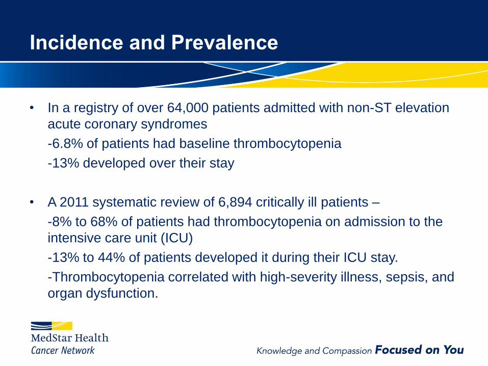
Incidence and Prevalence
• In a registry of over 64,000 patients admitted with non-ST elevation
acute coronary syndromes
-6.8% of patients had baseline thrombocytopenia
-13% developed over their stay
• A 2011 systematic review of 6,894 critically ill patients –
-8% to 68% of patients had thrombocytopenia on admission to the
intensive care unit (ICU)
-13% to 44% of patients developed it during their ICU stay.
-Thrombocytopenia correlated with high-severity illness, sepsis, and
organ dysfunction.
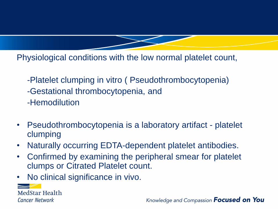
Physiological conditions with the low normal platelet count,
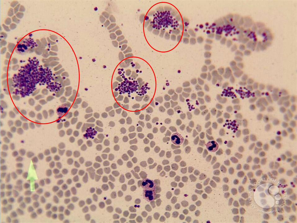
-Platelet clumping in vitro ( Pseudothrombocytopenia)
-Gestational thrombocytopenia, and
-Hemodilution
• Pseudothrombocytopenia is a laboratory artifact - platelet clumping
• Naturally occurring EDTA-dependent platelet antibodies.
• Confirmed by examining the peripheral smear for platelet clumps or Citrated Platelet count.
• No clinical significance in vivo.
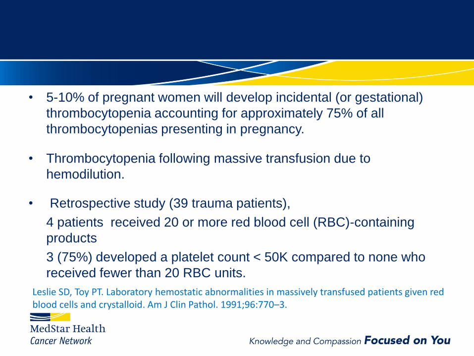
• 5-10% of pregnant women will develop incidental (or gestational)
thrombocytopenia accounting for approximately 75% of all
thrombocytopenias presenting in pregnancy.
• Thrombocytopenia following massive transfusion due to
hemodilution.
• Retrospective study (39 trauma patients),
4 patients received 20 or more red blood cell (RBC)-containing
products
3 (75%) developed a platelet count < 50K compared to none who
received fewer than 20 RBC units.
Leslie SD, Toy PT. Laboratory hemostatic abnormalities in massively transfused patients given red blood cells and crystalloid. Am J Clin Pathol. 1991;96:770–3.
• Major surgery is another common cause
After major surgery, platelet counts typically decline quickly - 1 to 4
days
due to consumption and dilution.
In a study of 581 patients who underwent cardiac surgery with
cardiopulmonary bypass, thrombocytopenia occurred in 56.3% of
patients within 10 days of surgery.
Similar decreases in the platelet counts with other surgeries. Greinacher A, Selleng K. Thrombocytopenia in the intensive care unit patient. Hematology Am Soc Hematol Educ Program. 2010;2010:135–43. Selleng S, Malowsky B, Strobel U, et al. Early-onset and persisting thrombocytopenia in post-cardiac surgery patients is rarely due to heparin-induced thrombocytopenia, even when antibody tests are positive. J Thromb Haemost. 2010;8:30–6.
Causes of Thrombocytopenia in inpatient setting
• Pseudothrombocytopenia
• ITP
• DITP-Drug induced ITP
• Infections( HIV/HepC/ EBV/Sepsis)
• Hypersplenism due to Chronic Liver disease
• Alcohol
• Pregnancy- Preeclampsia/HELLP/DIC
• Multiorgan failure syndrome
• HIT
• TMA- TTP/HUS
• MDS
• DIC
• Marrow infiltration
• Autoimmune Disorders/ APS
• PNH
• Nutrient Deficiencies- B12 /Folate

Cohort of 329 medical and surgical ICU patients with New onset thrombocytopenia
• Sepsis( 48 %)
• Sepsis with documented bacteremia (28 %)
• Liver disease/hypersplenism (18 %)
• Overt DIC (14 %)
• Unknown cause (14 %)
• Infection, other (11 %)
• Primary hematologic disorder (9 %)
• Medications, non-cytotoxic (9 %)
• Medications, cytotoxic (7 %)
• Massive transfusion (7 %)
• Other causes (7 %)
• Alcoholism (5 %)
Commonest Causes in ICU patients
• Adverse outcomes as severity of thrombocytopenia worsens.
Higher degree of thrombocytopenia correlated with the increased
risk of bleeding in the PROTECT trial (adjusted hazard ratios for
mild, moderate, and severe thrombocytopenia were 1.96, 3.52, and
3.54, respectively)
• Moderate and severe (but not mild) thrombocytopenia also
correlated with an increased length of ICU stay and ICU death
• Thrombocytopenia as a Predictor of Death
• In almost all patient populations, thrombocytopenia - an ominous
sign.
- In critically ill patients, thrombocytopenia was an independent
predictor of death in hospital (odds ratio [OR], 2. 1–26. 2) and in the
ICU (OR, 3. 1–4. 2) across six observational studies (N 6,894).
-In patients with acute coronary syndrome, thrombocytopenia,
irrespective of the cause (HIT [0. 3%], glycoproteinIIb/IIIa associated
thrombocytopenia [0. 6%] or other thrombocytopenia [0. 7%])—was
associated with higher risk of bleeding and in-hospital death
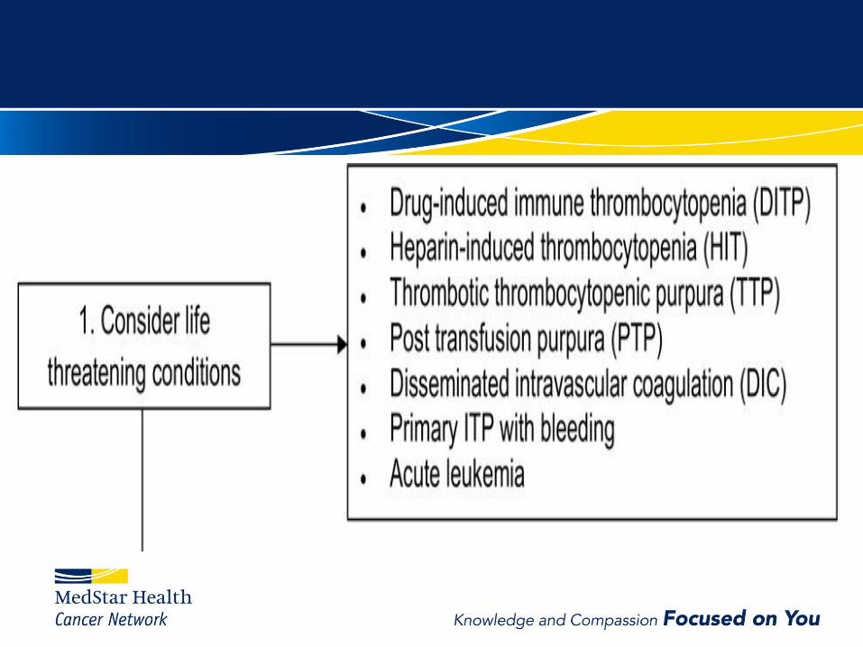
• Association Between Thrombocytopenia and Thrombosis
- Does not protect against thrombosis
• May be associated with increased risk antiphospholipid antibody
syndrome (APS), HIT, and disseminated intravascular coagulation
(DIC).
• Similar rates of venous and arterial thromboembolic events in APS
patients with thrombocytopenia compared patients with normal
platelet counts.
• In a retrospective analysis of 408 patients with HIT, severity of
thrombocytopenia correlated with an increased risk of thrombosis;
over 90% decrease in platelet count had the highest risk (OR, 8. 79
[95% CI, 2. 26–34. 17])
• DIC can present with bleeding and/or thrombotic manifestations
despite moderate to severe thrombocytopenia.
Greinacher A, Farner B, Kroll H, Kohlmann T, Warkentin TE, Eichler P. Clinical features of heparin-induced thrombocytopenia including risk factors for thrombosis. A retrospectiveanalysis of 408 patients. Thromb Haemost. 2005;94:132–5.36. Kitchens CS. Thrombocytopenia and thrombosis in disseminated intravascular coagulation (DIC). Hematology Am Soc Hematol Educ Program. 2009;240–6.
A Rational Approach to the Diagnosis and Management of Thrombocytopenia in the hospitalized Patient, Donald M. Arnolda,b and Wendy Lima Semin Hematol 48:251–258

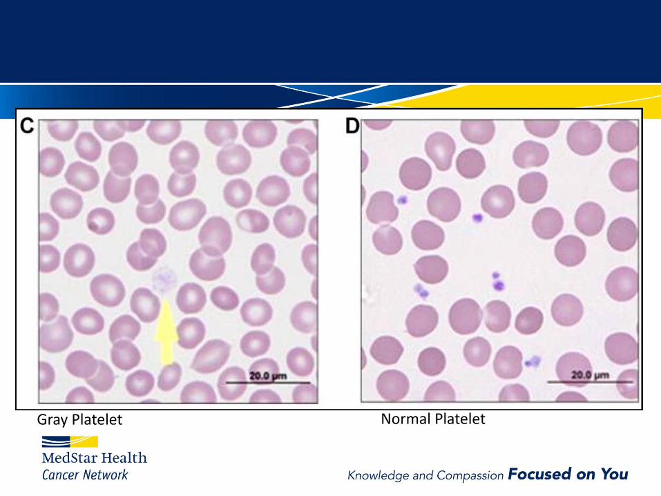
Gray Platelet Normal Platelet

Black arrow- Inclusion body in Neutrophil Red arrow- Giant platelets May- Hegglin anomaly


• LABORATORY TESTING —
• CBC and peripheral blood smear
• B12, Folate
• CMP
• Hepatitis C, HIV
• R/o Infection- Bacterial/Viral
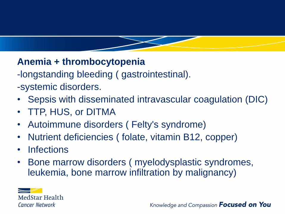
Anemia + thrombocytopenia
-longstanding bleeding ( gastrointestinal).
-systemic disorders.
• Sepsis with disseminated intravascular coagulation (DIC)
• TTP, HUS, or DITMA
• Autoimmune disorders ( Felty's syndrome)
• Nutrient deficiencies ( folate, vitamin B12, copper)
• Infections
• Bone marrow disorders ( myelodysplastic syndromes, leukemia, bone marrow infiltration by malignancy)

Leukocytosis + thrombocytopenia
-infection,
-chronic inflammation, and malignancy.
Leukopenia, anemia, and thrombocytopenia
(Pancytopenia) – MDS/ Leukemia
• Symptoms or findings of systemic autoimmune disorders
- ANA/ LPA
• Thrombosis – Rule out DIC, heparin-induced thrombocytopenia
(HIT), and APS.
- PT/aPTT/D dimer/Fibrinogen/Fibrin Split products/ HIT antibody
and Serotonin Release Assay.
• Schistocyes on the peripheral smear should prompt coagulation
testing (eg, PT, aPTT, fibrinogen),LDH and renal function to
evaluate for DIC, TTP, or HUS
• Hematology Referral:
• Especially for emergencies-
- Suspected TTP/HUS, HIT, Hematologic malignancies-
Acute leukemia/Aplastic anemia.
- Bonemarrow evaluation:
- helpful in some patients if the cause is unclear, or if a
primary hematologic disorder is suspected.

Thrombocytopenia does NOT protect against venous or
arterial thrombosis.
Appropriate use of thromboprophylaxis or anticoagulants –
for patients with mild to moderate thrombocytopenia (>50K)
if indicated.
For patients with more severe thrombocytopenia, decisions
weighing in the risks of bleeding and benefits of
anticoagulation.
ITP
• Isolated Thrombocytopenia
• May present with Bleeding
• Caused by antibody-mediated platelet destruction.
Managemant:
1. IV IG
2. Glucocorticoids
3. Splenectomy
4. Rituximab
5. Eltrombopag/Romiplostim- Thrombopoietic Growth factors
Disseminated intravascular coagulation (DIC)
• A systemic process
• Massive activation of Coagulation and fibrinolysis
• Consumption of clotting factors and platelets,
• Identifying the cause helps manage and halt this process
• Microangiopathic hemolytic anemia with schistocytes on the blood smear.
• Prolonged PT/aPTT
• Low fibrinogen
• Elevated D Dimer/ Fibrin split products
Thrombotic microangiopathy (TMA)
Platelet microthrombi in small vessels leading to organ
damage.
- Thrombotic thrombocytopenic purpura (TTP),
- Complement-mediated TMA (C-TMA), and
- Shiga toxin-mediated Hemolytic Uremic Syndrome (HUS).
Clinical features
• Microangiopathic hemolytic anemia (MAHA),
Schistocytes on the peripheral blood smear
• Thrombocytopenia, which can be severe
• Acute kidney injury
• Neurologic symptoms ranging from mild headache to
seizures and transient focal abnormalities.
• Fever.
32
Thrombotic Micorangiopathy

Challenges with TMAs
-Overlapping clinical presentations,
-But differing pathophysiologies and
• So need for different treatments.
• Potentially life-threatening

Thrombotic Thrombocytopenic Purpura (TTP)
• Severely reduced activity of ADAMTS13, a protease that cleaves
very large von Willebrand factor (VWF) multimers on endothelial
cells.
• Acquired, autoimmune TTP -neutralizing autoantibody – more likely
in a patient without a family history of TTP.
• Hereditary TTP inherited mutation in the ADAMTS13 gene
• ADAMTS13 activity <10 percent.
• Results of ADAMTS13 activity testing often are not immediately
available.
• Urgent plasma exchange (PEX) therapy,
• Removes the autoantibody to ADAMTS13 and supplies functional
ADAMTS13
• For patients known to have hereditary TTP, plasma infusion is
sufficient
• Platelet transfusion should be reserved for treatment of severe
bleeding in a patient with TTP due to the potential increased risk of
thrombosis,
• Platelets should not be withheld in a bleeding patient due to
concerns about this risk.
Complement-mediated TMA - (C-TMA)
(Complement-mediated HUS or "atypical HUS)
- Increased activation of complement on endothelial cells.
- Develop microthrombi in small vessels
- Kidney is often affected- increasing serum creatinine.
- Neutralizing autoantibody or an inherited mutation
- ADAMTS13 activity is ≥10 percent
- stool studies are negative or Shiga toxin-producing organisms.
- Anti-complement therapy Eculizumab.
Shiga toxin-mediated hemolytic uremic syndrome (ST-HUS)
- Enteric infection with an organism that produces the toxin (eg,
enterohemorrhagic Escherichia coli, Shigella)
- Severe abdominal pain and Bloody diarrhea
- Stool testing for Shiga toxin-secreting bacteria
E. coli 0157:H7, Shigella dysenteriae and for Shiga toxin
- Management involves supportive care
Heparin Induced Thrombocytopenia (HIT)
Complication of heparin therapy.
Two types
- Type 1 HIT
develops within the first 2 days after heparin exposure , Platelet
count normalizes with continued heparin therapy.
Nonimmune disorder that results from the direct effect of heparin on
platelet activation.
- Type 2 HIT
Immune-mediated
develops 4-10 days after heparin exposure
life and limb-threatening thrombotic complications
-Heparin Exposure
- Drop in platelet count, particularly if over 50% of the baseline count,
even if the platelet count nadir remains above 150 x 109/L.
- Venous thromboembolism - most common complication.
-Arterial thrombosis (eg, myocardial infarction) may occur.
sometimes termed Heparin-induced thrombocytopenia and
thrombosis (HITT).
• HIT is a Clinicopathologic diagnosis
• Needs clinical feature + lab work for Diagnosis
• Caused by antibodies to complexes of platelet factor 4 (PF4) and
heparin
• The antibodies bind to the PF4-heparin complexes on the platelet
surface and induce platelet activation by cross-linking FcγIIA
receptors.
• Incidence 0.2% in all heparin exposed patients.
• Greater than 50% risk of developing new
thromboembolic events.
• The mortality rate is approximately 20%, and
approximately 10% of patients require amputations or
suffer other major morbidity
Diagnostic tests
Immunoassays – HIT antibody testing
• Identify antibodies against heparin/platelet factor 4 (PF4) complexes.
• Rapid turnaround time and high sensitivity (> 99%).
• Poor specificity (30%-70%) because they also detect non pathogenic antibodies.
• The specificity enhanced by optical density (OD) of the result.
• Higher absolute OD values correlate with a clinical diagnosis of HIT.
• OD of 0.4 to <1.00 indicated a 5% or lower probability of a strongly positive result on functional testing with the serotonin release assay (SRA),
• OD of 2.0 or more resulted in an approximately 90% probability of a strong SRA result.
Functional assays - SRA/HIPA
• Measure the platelet-activating capacity of PF4/heparin-
antibody complexes.
• Functional assays have greater specificity than
immunoassays but are time-consuming
• Based on HIT antibodies causing platelets to aggregate
and release serotonin. Sensitivity 69% to 94%, and
specificity may be as high as 100%
Treatment
• Discontniue heparin
• Direct thrombin Inhibitor
- Argatroban
- Bivalirudin (patients –PCI)
- Fondaparinux (Indirect factor Xa inhibitor)- pregnant
women.

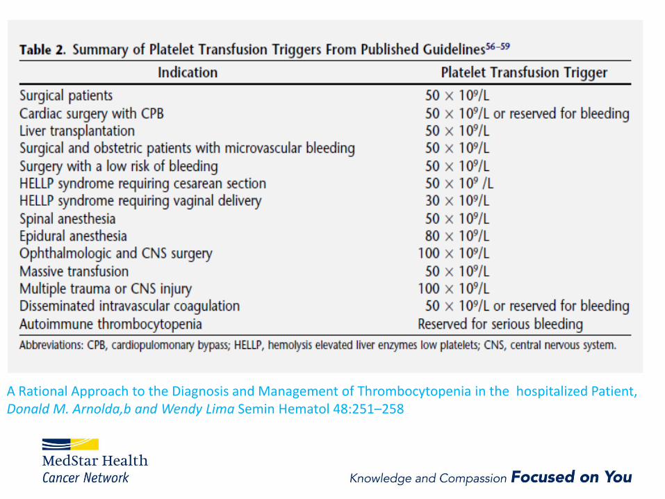
Safe platelet count for invasive procedures
• Most platelet count thresholds for invasive procedures
are based on weak observational evidence.
• Procedures with a greater risk of bleeding are performed
at higher platelet counts.
• Raising the platelet count for an invasive procedure
depend on the underlying condition (Steroids or IVIG for
ITP; platelet transfusion for myelodysplastic syndromes)
• Individuals with impaired platelet function may require
platelet transfusions despite adequate platelet counts
• Correct coagulation abnormalities if present.
A Rational Approach to the Diagnosis and Management of Thrombocytopenia in the hospitalized Patient, Donald M. Arnolda,b and Wendy Lima Semin Hematol 48:251–258
Summary
Thrombocytopenia in inpatient setting – very common
Life-threatening causes should be considered initially
Examination of the Peripheral smear
Temporal relation to drop in platelet count with respect to drug exposure or RBC
transfusion, the severity of thrombocytopenia, and the presence or absence of
Bleeding, symptoms provide clues to the underlying cause.
Platelet transfusions for serious bleeding and
Thrombocytopenia alone is not a contraindication to antithrombotic therapy
weigh risks vs benefits
References
Hui P, Cook DJ, Lim W, et al. The frequency and clinical significance of thrombocytopenia complicating critical illness: a systematic
review. Chest 2011; 139:271.
Strauss R, Wehler M, Mehler K, et al. Thrombocytopenia in patients in the medical intensive care unit: bleeding prevalence,
transfusion requirements, and outcome. Crit Care Med 2002; 30:1765.
Vanderschueren S, De Weerdt A, Malbrain M, et al. Thrombocytopenia and prognosis in intensive care. Crit Care Med 2000;
28:1871.
Arnold DM, Lim W. A rational approach to the diagnosis and management of thrombocytopenia in the hospitalized patient. Semin
Hematol 2011; 48:251.
Greinacher A, Selleng S. How I evaluate and treat thrombocytopenia in the intensive care unit patient. Blood 2016; 128:3032.
Reese JA, Li X, Hauben M, et al. Identifying drugs that cause acute thrombocytopenia: an analysis using 3 distinct methods. Blood
2010; 116:2127.
Williamson DR, Albert M, Heels-Ansdell D, et al. Thrombocytopenia in critically ill patients receiving thromboprophylaxis:
frequency, risk factors, and outcomes. Chest 2013; 144:1207.
Buckley MF, James JW, Brown DE, et al. A novel approach to the assessment of variations in the human platelet count. Thromb
Haemost 2000; 83:480.
Segal JB, Moliterno AR. Platelet counts differ by sex, ethnicity, and age in the United States. Ann Epidemiol 2006; 16:123.
Uptodat: Approach to the adult with unexplained thrombocytopenia
A Rational Approach to the Diagnosis and Management of Thrombocytopenia in the hospitalized Patient, Donald M. Arnolda,b and Wendy Lima Semin Hematol
48:251–258

Thank You!