Embed Size (px)
Citation preview
ORIGINAL RESEARCHpublished: 27 September 2016doi: 10.3389/fmicb.2016.01530
Frontiers in Microbiology | www.frontiersin.org 1 September 2016 | Volume 7 | Article 1530
Edited by:
Olivier Dussurget,
University Paris Diderot, France
Reviewed by:
Glen C. Ulett,
Griffith University, Australia
Pietro Speziale,
Istituto Universitario Di Studi Superiori
Di Pavia, Italy
Ying Zhang,
Johns Hopkins University, USA
*Correspondence:
Nuno Cerca
Specialty section:
This article was submitted to
Microbial Immunology,
a section of the journal
Frontiers in Microbiology
Received: 10 May 2016
Accepted: 12 September 2016
Published: 27 September 2016
Citation:
França A, Pérez-Cabezas B,
Correia A, Pier GB, Cerca N and
Vilanova M (2016) Staphylococcus
epidermidis Biofilm-Released Cells
Induce a Prompt and More Marked In
vivo Inflammatory-Type Response
than Planktonic or Biofilm Cells.
Front. Microbiol. 7:1530.
doi: 10.3389/fmicb.2016.01530
Staphylococcus epidermidisBiofilm-Released Cells Induce aPrompt and More Marked In vivoInflammatory-Type Response thanPlanktonic or Biofilm CellsAngela França 1, 2, Begoña Pérez-Cabezas 3, Alexandra Correia 3, 4, Gerald B. Pier 5,
Nuno Cerca 1* and Manuel Vilanova 2, 3, 4
1 Laboratory of Research in Biofilms Rosário Oliveira, Centre of Biological Engineering, University of Minho, Braga, Portugal,2Departamento de Imuno-Fisiologia e Farmacologia, Instituto de Ciências Biomédicas de Abel Salazar, Universidade do
Porto, Porto, Portugal, 3 Instituto de Investigação e Inovação em Saúde, Universidade do Porto, Porto, Portugal, 4 Instituto de
Biologia Molecular e Celular, Universidade de Porto, Porto, Portugal, 5Division of Infectious Diseases, Department of
Medicine, Brigham and Women’s Hospital/Harvard Medical School, Boston, MA, USA
Staphylococcus epidermidis biofilm formation on indwelling medical devices is frequently
associated with the development of chronic infections. Nevertheless, it has been
suggested that cells released from these biofilms may induce severe acute infections
with bacteraemia as one of its major associated clinical manifestations. However, how
biofilm-released cells interact with the host remains unclear. Here, using a murine model
of hematogenously disseminated infection, we characterized the interaction of cells
released from S. epidermidis biofilms with the immune system. Gene expression analysis
of mouse splenocytes suggested that biofilm-released cells might be particularly effective
at activating inflammatory and antigen presenting cells and inducing cellular apoptosis.
Furthermore, biofilm-released cells induced a higher production of pro-inflammatory
cytokines, in contrast to mice infected with planktonic cells, even though these had a
similar bacterial load in livers and spleens. Overall, these results not only provide insights
into the understanding of the role of biofilm-released cells in S. epidermidis biofilm-related
infections and pathogenesis, but may also help explain the relapsing character of these
infections.
Keywords: S. epidermidis, biofilms, biofilm-released cells, splenocytes transcriptome, pro-inflammatory
cytokines, tissue colonization
INTRODUCTION
Staphylococcus epidermidis is one of the most important etiological agents of device-associatedinfections due to its ability to adhere and form biofilms on the surface of indwellingmedical devices (Vuong and Otto, 2002; Otto, 2009). When compared to planktonic cells,S. epidermidis cells within biofilms are known to be more tolerant to several classes ofantibiotics (Cerca et al., 2005), as well as to the host immune effectors (Cerca et al.,2006; Kristian et al., 2008). Biofilms represent therefore a common cause of recurrent and
França et al. S. epidermidis Biofilm-Released Cells Virulence
relapsing infections (Costerton et al., 1999). Consequently,removal of the infected devices is often required to resolve theseinfections (von Eiff et al., 2002), which results in increasedmorbidity and, occasionally, mortality among infected patients(Otto, 2009). Due to the enormous impact of S. epidermidisbiofilm-related infections on human health, the mechanismsunderlying biofilm formation have been extensively studiedin the last decades. It is currently accepted that biofilmformation involves three main stages: (1) initial adhesion,(2) maturation, and (3) disassembly (Otto, 2012). The laterrefers to the release of bacterial cells from the biofilm to thesurrounding environment, and is the least understood stage ofthe biofilm lifecycle (Boles and Horswill, 2011). Importantly,biofilm disassembly has been associated with the emergenceof severe acute infections such as bacteraemia (Wang et al.,2011) and the embolic events of endocarditis (Pitz et al.,2011). However, despite its clear importance in the clinicalsetting, little is known regarding the phenotype or interactionof these cells with the host immune system. In the first stagesof biofilm formation, planktonic bacteria attached to medicaldevices undergo several physiological modifications that lead tothe biofilm phenotype (Yao et al., 2005). Thus, it was thoughtthat after disassembly biofilm-released cells would quicklyrevert to the planktonic phenotype (Kaplan, 2010; Chua et al.,2014). However, recent reports have shown that cells releasedfrom Pseudomonas aeruginosa (Rollet et al., 2009; Li et al.,2014), Streptococcus mutans (Liu et al., 2013), and Streptococcuspneumoniae (Marks et al., 2013) biofilms present features distinctfrom either the biofilm or planktonic phenotypes, showinghigher virulence potential. Chua and collaborators showed thatP. aeruginosa biofilm-released cells, when compared with theirplanktonic or biofilm counterparts, present higher expressionlevel of genes associated with the bacterium virulence, namelyType 2 Secretion System (TSS) and T3SS psc gene and, moreimportant, they showed that these genes are essential in elicitingfull virulence against macrophages and in the rapid killing ofCaenorhabditis elegans (Chua et al., 2014), respectively. In thecase of S. epidermidis, it is only known that biofilm-releasedcells present higher tolerance than planktonic and biofilmcells to antibiotics (Franca et al., 2016). However, their fullvirulence potential remains unclear. A comprehensive analysisof the interaction between biofilm-released cells and the hostwould clarify their role in the pathogenesis of biofilm-relatedinfections, and help to prevent the pathologic events associatedwith biofilm cells dissemination. Therefore, herein, a murinemodel of hematogenously disseminated infection was used toevaluate the capacity of S. epidermidis biofilm-released cells to(1) induce changes in the transcriptome of murine immunecells within the spleen, (2) stimulate the production of pro-inflammatory cytokines, and (3) colonize and persist in murineorgans. Our results showed that S. epidermidis biofilm-releasedcells induce a prompt and more marked inflammatory-typeresponse than do their planktonic or biofilm counterparts. Inaddition, these findings showed that particular properties of thebiofilm-released cells need to be taken into account to efficientlytarget and treat acute infections originating from S. epidermidisbiofilms.
MATERIALS AND METHODS
Ethics StatementThis study was performed in strict accordance with therecommendations of the European Convention for theProtection of Vertebrate Animals used for Experimentaland Other Scientific Purposes (ETS 123), the 86/609/EECdirective and Portuguese rules (DL 129/92). All experimentalprotocols were approved by the competent national authority(Direcção-Geral de Veterinária), document 023517 (2010.11.25).
MiceFemale BALB/c mice, 8–12 weeks old, were purchased fromCharles River (Barcelona, Spain) and kept under specific-pathogen-free conditions at the Animal Facility of the Institutode Ciências Biomédicas Abel Salazar, Porto, Portugal. Micewere maintained in individually ventilated cages (5 animals percage) with corncob bedding, and under controlled conditions oftemperature (21± 1◦C), relative humidity (between 45 and 65%)and light (12 h light/ dark cycle). Mice had ad libitum access tofood and water. Hiding and nesting materials were provided forenrichment. All procedures such cage changing, water and foodsupply, as well as intravenous injections were always performedduring the day cycle (between 7 and 19 h).
Bacteria and Growth ConditionsThe biofilm forming strain S. epidermidis 9142, isolated from ablood culture (Mack et al., 1994), was used in this study. A singlecolony, from a Tryptic Soy Agar (TSA) plate, was inoculatedinto 2mL of Tryptic Soy Broth (TSB, Liofilchem, Teramo, Italy)and incubated overnight at 37◦C with shaking at 120 rpm. Asuspension with ∼1 × 10⁸ CFU/mL, prepared by adjusting theoptical density (at 640 nm) of the overnight culture to 0.25 ±
0.05, was used to start both planktonic and biofilm cultures.For planktonic cultures 150µL of 1 × 10⁸ CFU/mL bacterialsuspension was inoculated into 10mL of TSB supplemented with0.65% (v/v) glucose (TSB0.65%G) and incubated for 24 h at 37◦Cunder agitation at 120 rpm. Biofilms were grown in 24-wellplates made of polystyrene plastic (Orange Scientific, Braine-l’Alleud, Belgium) by inoculating 15µL of the 1 × 10⁸ CFU/mLbacterial suspension into 1 mL of TSB0.65%G, then incubating at37◦C with agitation at 120 rpm. After 24 h of growth, biofilmswere washed twice with apyrogenic Phosphate Buffered Saline(PBS, Gibco, MD, USA), 1mL of fresh TSB0.65%G was carefullyadded and biofilms allowed to grow, under the same temperatureand agitation conditions, for additional 24 h. Biofilm-releasedcells, (i.e., the cells in the biofilm bulk-fluid), were collected asdescribed before (Franca et al., 2016) from 12 originating biofilmsand pooled together to decrease variability inherent to biofilmgrowth (Sousa et al., 2014). Four biofilms were washed twice withapyrogenic PBS, disrupted and also pooled together to reducevariability. Planktonic (4mL of culture), biofilm and biofilm-released cells were then harvested by centrifugation, suspendedin 4 mL of apyrogenic PBS (Gibco, MD, USA) and sonicated for10 s at 18W (Branson modelW 185 D, Heat Systems Ultrasonics,CT, USA) in order to dissociate cell clusters. Cells viability wasnot reduced by this procedure as determined previously by
Frontiers in Microbiology | www.frontiersin.org 2 September 2016 | Volume 7 | Article 1530
França et al. S. epidermidis Biofilm-Released Cells Virulence
CFU counting and propidium iodide incorporation (Cerca et al.,2011).
Murine Model of HematogenouslyDisseminated InfectionThe inoculum of each of the bacterial populations was adjustedby flow cytometry to 5 × 10⁸ total cells/mL, using SYBR Green(LifeTechnologies, MD, USA)/propidium iodide (Sigma, MO,USA) staining, as optimized before (Cerca et al., 2011). Thenumber of cultivable cells was assessed by CFU counting. Adultmice, randomly allocated to each experimental group, wereinjected intravenously in the lateral tail vein, with the supportof a restrainer, with 0.2mL of 5 × 10⁸ of planktonic, biofilmor biofilm-released cell suspensions. Control mice were injectedintravenously with 0.2mL of apyrogenic PBS. Sample size wasdetermined based on the results of preliminary experiments.It was not possible to perform subsequent mouse studies in ablinded fashion. In order to address the alterations occurringduring the acute phase of infection, the parameters evaluated inthis study were assessed 2, 6, or 14 h after challenging the three S.epidermidis populations.
Serum Collection and Bacterial LoadDetermination in OrgansTwo, 6, and 14 h post-infection, mice were anesthetized withisoflurane (Abbott laboratories, IL, USA) for terminal bloodcollection, and then euthanized by cervical dislocation. Forserum collection, mouse blood was drawn through the retro-orbital route, incubated overnight at 4◦C, and then centrifugedfor 15 min at 4◦C at 16,000 g. Serum was then transferred intoa new tube and stored at −80◦C until further use. Livers andspleens were aseptically removed and immediately transferredinto tissue grinders with, respectively, 3 or 1mL of PBS. Tissueswere homogenized and quantitatively cultured on TSA plates.At all times during the procedure, samples were kept on ice.This experiment was performed 1 (for biofilms cells at all timepoints) to 3 (planktonic and biofilm-released cells, 6 h time point)independent times, with at least 5 animals per infected group.
Cytokines and Chemokines QuantificationTwo, 6, and 14 h post-infection, the levels of the cytokines IL-6,TNF-α and the chemokines CXCL1 (KC), CCL2 (MCP-1), CCL3(MIP-1α), CCL4 (MIP-1β) in mouse serum were quantified ina Bio-Plex R© 200 using the kit Magnetic Custom Multiplex Bio-Plex ProTM Mouse Cytokine Group I assay (Bio-Rad, CA, USA).The procedure was performed following the manufacturer’sinstructions. This experiment was performed 1 (all populationsat 6 and 14 h time point) to 2 (all populations at 2 h time point)independent times, with at least 5 animals per infected group.
Microarray Analysis of Mouse SplenocytesSpleen cells plays a major role in host immune response to blood-born pathogens, working in concert to activate mechanismsrequired for successful resolution of infection. Hence, in orderto address the mechanisms activated during the first contactwith the different S. epidermidis populations, the transcriptomeof splenocytes was analyzed, by microarrays, 2 h after challenge.
In brief, spleens were aseptically removed, transferred to60mm diameter sterile Petri dishes with 9 mL apyrogenic PBSand immediately placed on ice. Thereafter, using two sterilefrosted glass slides, spleens were completely homogenized. Thesuspension was then passed through a sterile column of glasswool to remove fibrous tissue, the number of cells countedby flow cytometry, and 5 × 10⁶ splenocytes harvested by5 min centrifugation at 1200 rpm at 4◦C. Cell pellets wereimmediately suspended in RLT buffer (QIAGEN, Heidelberg,Germany) and stored at −80◦C until the next day. TotalRNA was then isolated using the RNeasy Mini Kit (QIAGEN)following the manufacturer’s instructions. Concentration andpurity was determined using a NanoDropTM1000 and integritywas confirmed using an Agilent 2100 Bioanalyzer (AgilentTechnologies, CA, USA). RNA integrity number values wereabove 8.5 for all samples. This experiment was performed oncewith 2 (control and planktonic cells) to 3 (biofilm and biofilm-released cells) animals per group.
Transcription levels in mouse splenocytes were determinedusing Affymetrix R© Mouse Gene 2.1 ST Array Strip (Affymetrix,MA, USA). RNA was prepared for analysis using AmbionWT Expression Kit (ThermoFisher Scientific, MA, USA) andGeneChip R© WT Terminal Labeling Kit (Affymetrix). Briefly,100 ng of total RNA, containing spiked in Poly-A RNA controls,was used in a reverse transcription reaction to generate first-strand complementary DNA. After second-strand synthesis,double-stranded complementary DNA was used to generatecRNA. cRNA (15µg) was then used for a second cycle of first-strand cDNA synthesis and the resultant single stranded cDNA(5.5µg) was fragmented and end-labeled. Size distribution ofthe fragments was assessed using an Agilent 2100 Bioanalyzer(Agilent Technologies). End-labeled, fragmented cDNA (3.5µg),was then used in a 150µL hybridization cocktail containinghybridization controls (GeneAtlas R© Hybridization, Wash, andStain Kit for WT Array Strips, Affymetrix), of which 120µLwere hybridized on array strips for 20 h at 48◦C. Standard posthybridization wash and double-stain protocols were used on anAffymetrix GeneAtlas system, followed by scanning of the arraystrips.
Microarray Data AnalysisThe arrays were analyzed using Chipster 2 (Kallio et al., 2011)with a custom cdf file in mogene21stmmentrezg.db, as availablefrom Brainarray database (version 17; Sandberg and Larsson,2007). Following Robust Multi-array Average normalization andbiomaRt annotation, differential expression was determined byempirical Bayes two-group test (Smyth, 2004) with Benjamini-Hochberg multiple testing correction and a P-value cut-off of0.05. For further analyses, only genes with fold changes above 1.5were included. The heatmap was constructed using matrix2pnginterface (Pavlidis and Noble, 2003). Venn diagram, createdusing VENNY 2.1 (Oliveros, 2007), was used to identify thegenes that were uniquely and commonly expressed in miceinfected with different S. epidermidis bacterial populations. Geneontology (GO) terms enrichment was assessed using the SearchTool for the Retrieval of Interacting Genes/Proteins (STRING)(version 10; Franceschini et al., 2013). Only gene-sets passing
Frontiers in Microbiology | www.frontiersin.org 3 September 2016 | Volume 7 | Article 1530
França et al. S. epidermidis Biofilm-Released Cells Virulence
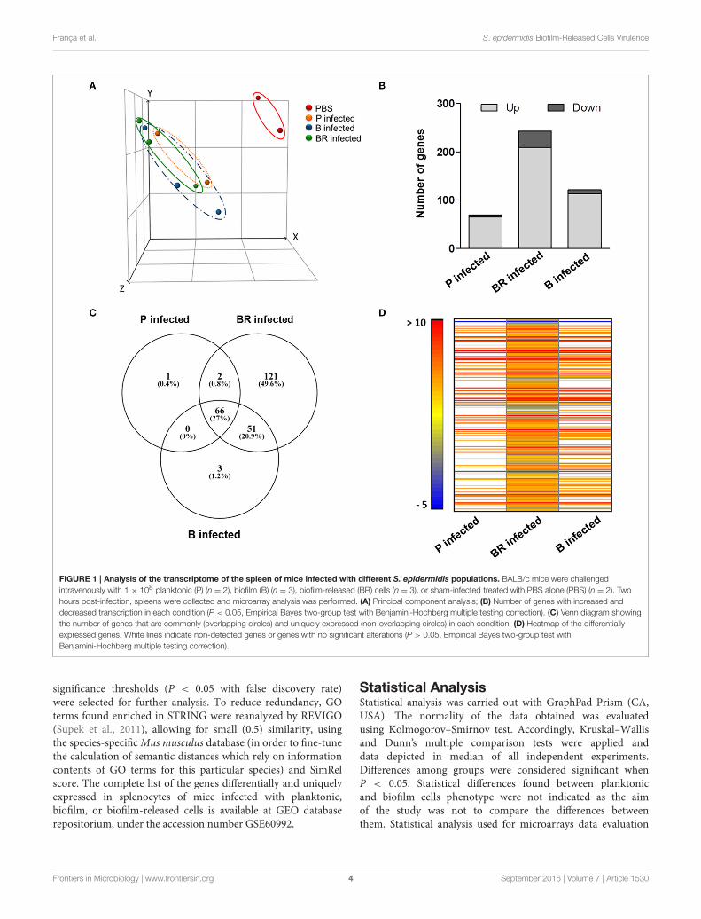
FIGURE 1 | Analysis of the transcriptome of the spleen of mice infected with different S. epidermidis populations. BALB/c mice were challenged
intravenously with 1 × 10⁸ planktonic (P) (n = 2), biofilm (B) (n = 3), biofilm-released (BR) cells (n = 3), or sham-infected treated with PBS alone (PBS) (n = 2). Two
hours post-infection, spleens were collected and microarray analysis was performed. (A) Principal component analysis; (B) Number of genes with increased and
decreased transcription in each condition (P < 0.05, Empirical Bayes two-group test with Benjamini-Hochberg multiple testing correction). (C) Venn diagram showing
the number of genes that are commonly (overlapping circles) and uniquely expressed (non-overlapping circles) in each condition; (D) Heatmap of the differentially
expressed genes. White lines indicate non-detected genes or genes with no significant alterations (P > 0.05, Empirical Bayes two-group test with
Benjamini-Hochberg multiple testing correction).
significance thresholds (P < 0.05 with false discovery rate)were selected for further analysis. To reduce redundancy, GOterms found enriched in STRING were reanalyzed by REVIGO(Supek et al., 2011), allowing for small (0.5) similarity, usingthe species-specificMus musculus database (in order to fine-tunethe calculation of semantic distances which rely on informationcontents of GO terms for this particular species) and SimRelscore. The complete list of the genes differentially and uniquelyexpressed in splenocytes of mice infected with planktonic,biofilm, or biofilm-released cells is available at GEO databaserepositorium, under the accession number GSE60992.
Statistical AnalysisStatistical analysis was carried out with GraphPad Prism (CA,USA). The normality of the data obtained was evaluatedusing Kolmogorov–Smirnov test. Accordingly, Kruskal–Wallisand Dunn’s multiple comparison tests were applied anddata depicted in median of all independent experiments.Differences among groups were considered significant whenP < 0.05. Statistical differences found between planktonicand biofilm cells phenotype were not indicated as the aimof the study was not to compare the differences betweenthem. Statistical analysis used for microarrays data evaluation
Frontiers in Microbiology | www.frontiersin.org 4 September 2016 | Volume 7 | Article 1530
França et al. S. epidermidis Biofilm-Released Cells Virulence
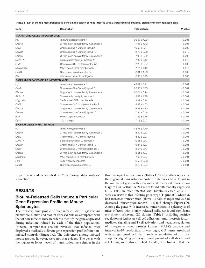
TABLE 1 | List of the top most transcribed genes in the spleen of mice infected with S. epidermidis planktonic, biofilm or biofilm-released cells.
Gene Description Fold change P-value
PLANKTONIC CELLS INFECTED MICE
Irg1 Immunoresponsive gene 1 39.08±4.02 < 0.001
Clec4e C-type lectin domain family 4, member e 17.46±4.73 0.004
Cxcl2 Chemokine (C-X-C motif) ligand 2 16.63±3.64 0.003
Cxcl10 Chemokine (C-X-C motif) ligand 10 8.73±2.98 0.012
Clec5a C-type lectin domain family 5, member a 7.80±0.92 0.003
Slc7a11 Solute carrier family 7, member 11 7.88±2.47 0.013
Ccrl2 Chemokine (C-C motif) receptor-like 2 7.20±0.67 0.006
Mmrgpra2a MAS-related GPR, member A2A 7.15±2.17 0.012
Gpr84 G protein-coupled receptor 84 6.31±1.23 0.009
IIl1rn Interleukin 1 receptor antagonist 5.93±0.95 0.008
BIOFILM-RELEASED CELLS INFECTED MICE
Irg1 Immunoresponsive gene 1 49.02±5.41 < 0.001
Cxcl2 Chemokine (C-X-C motif) ligand 2 25.90±3.85 < 0.001
Clec4e C-type lectin domain family 4, member e 20.24±0.24 < 0.001
Slc7a11 Solute carrier family 7, member 11 13.33±1.99 < 0.001
Mrgpra2a MAS-related GPR, member A2A 9.69±0.14 < 0.001
Ccrl2 Chemokine (C-C motif) receptor-like 2 8.83±1.03 < 0.001
Clec5a C-type lectin domain family 5, member a 8.52±1.10 < 0.001
Cxcl10 Chemokine (C-X-C motif) ligand 10 7.34±0.46 < 0.001
Fpr1 Formyl peptide receptor 1 7.22±1.18 < 0.001
Cd14 CD14 antigen 7.15±0.47 < 0.001
BIOFILM CELLS INFECTED MICE
Irg1 Immunoresponsive gene 1 42.97±4.79 < 0.001
Clec4e C-type lectin domain family 4, member e 18.49±2.67 < 0.001
Cxcl2 Chemokine (C-X-C motif) ligand 2 18.22±3.27 < 0.001
Slc7a11 Solute carrier family 7, member 11 10.51±2.77 < 0.001
Cxcl10 Chemokine (C-X-C motif) ligand 10 10.24±1.37 < 0.001
Ccrl2 Chemokine (C-C motif) receptor-like 2 8.54±0.47 < 0.001
Clec5a C-type lectin domain family 5, member a 8.61±1.07 < 0.001
Mrgpra2a MAS-related GPR, member A2A 7.68±0.87 < 0.001
Fpr1 Formyl peptide receptor 1 6.59±0.83 < 0.001
Gpr84 G protein-coupled receptor 84 6.18±0.37 < 0.001
is particular and is specified in “microarrays data analysis”subsection.
RESULTS
Biofilm-Released Cells Induce a ParticularGene Expression Profile on MouseSplenocytesThe transcriptomic profile of mice infected with S. epidermidisplanktonic, biofilm and biofilm-released cells was compared withthat of non-infected mice in order to identify the genes expressedduring infection induced by each of the three populations.Principal components analysis revealed that infected micedisplayed a markedly different gene expression profile from non-infected controls (Figure 1A). The differences among infectedmouse groups, however, were not that evident. The genes withthe highest or lowest levels of transcription were similar in the
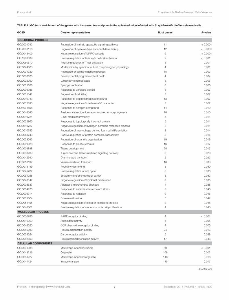
three groups of infected mice (Tables 1, 2). Nevertheless, despitethese general similarities important differences were found inthe number of genes with increased and decreased transcription(Figure 1B). Within the 243 genes found differentially expressed(P < 0.05) in mice infected with biofilm-released cells, 121were exclusive to this infecting phenotype (Figure 1C), where 96had increased transcription (above 1.5-fold change) and 25 haddecreased transcription (above −1.5-fold change; Figure 1D).Among the genes with increased transcription in splenocytes ofmice infected with biofilm-released cells, we found significantenrichment of several GO clusters (Table 3) including positiveregulation of leukocyte cell-cell adhesion, tumor necrosis factor-mediated signaling and T cell activation, and negative regulationof mitogen activated protein kinases (MAPK) cascade andinterleukin-10 production. Interestingly, GO terms associatedwith programmed cell death such as regulation of intrinsicapoptotic signaling pathways, development of cell death, andcell killing were also enriched. Finally, we observed that the
Frontiers in Microbiology | www.frontiersin.org 5 September 2016 | Volume 7 | Article 1530
França et al. S. epidermidis Biofilm-Released Cells Virulence
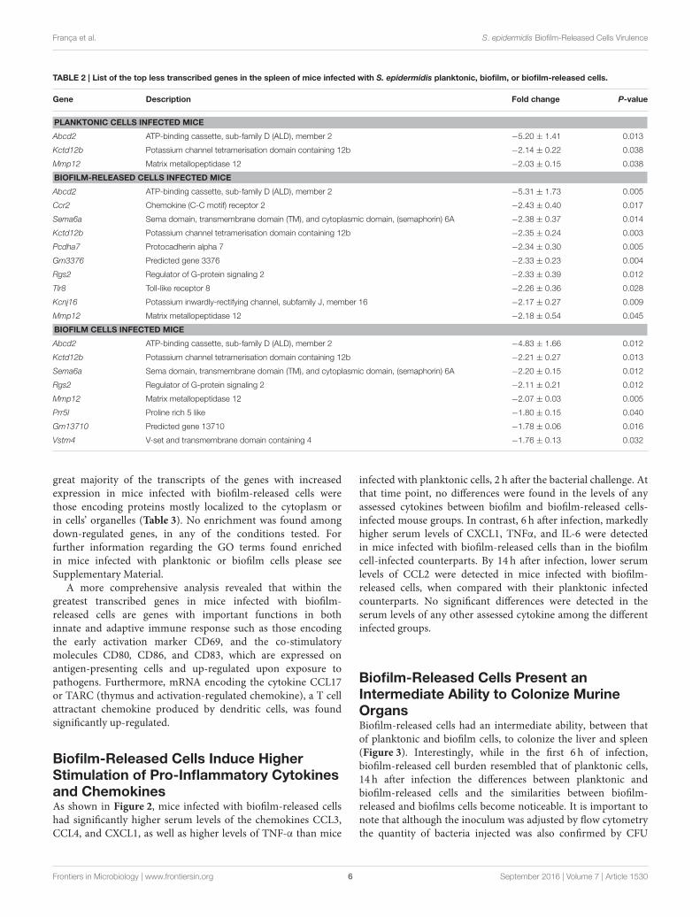
TABLE 2 | List of the top less transcribed genes in the spleen of mice infected with S. epidermidis planktonic, biofilm, or biofilm-released cells.
Gene Description Fold change P-value
PLANKTONIC CELLS INFECTED MICE
Abcd2 ATP-binding cassette, sub-family D (ALD), member 2 −5.20 ± 1.41 0.013
Kctd12b Potassium channel tetramerisation domain containing 12b −2.14 ± 0.22 0.038
Mmp12 Matrix metallopeptidase 12 −2.03 ± 0.15 0.038
BIOFILM-RELEASED CELLS INFECTED MICE
Abcd2 ATP-binding cassette, sub-family D (ALD), member 2 −5.31 ± 1.73 0.005
Ccr2 Chemokine (C-C motif) receptor 2 −2.43 ± 0.40 0.017
Sema6a Sema domain, transmembrane domain (TM), and cytoplasmic domain, (semaphorin) 6A −2.38 ± 0.37 0.014
Kctd12b Potassium channel tetramerisation domain containing 12b −2.35 ± 0.24 0.003
Pcdha7 Protocadherin alpha 7 −2.34 ± 0.30 0.005
Gm3376 Predicted gene 3376 −2.33 ± 0.23 0.004
Rgs2 Regulator of G-protein signaling 2 −2.33 ± 0.39 0.012
Tlr8 Toll-like receptor 8 −2.26 ± 0.36 0.028
Kcnj16 Potassium inwardly-rectifying channel, subfamily J, member 16 −2.17 ± 0.27 0.009
Mmp12 Matrix metallopeptidase 12 −2.18 ± 0.54 0.045
BIOFILM CELLS INFECTED MICE
Abcd2 ATP-binding cassette, sub-family D (ALD), member 2 −4.83 ± 1.66 0.012
Kctd12b Potassium channel tetramerisation domain containing 12b −2.21 ± 0.27 0.013
Sema6a Sema domain, transmembrane domain (TM), and cytoplasmic domain, (semaphorin) 6A −2.20 ± 0.15 0.012
Rgs2 Regulator of G-protein signaling 2 −2.11 ± 0.21 0.012
Mmp12 Matrix metallopeptidase 12 −2.07 ± 0.03 0.005
Prr5l Proline rich 5 like −1.80 ± 0.15 0.040
Gm13710 Predicted gene 13710 −1.78 ± 0.06 0.016
Vstm4 V-set and transmembrane domain containing 4 −1.76 ± 0.13 0.032
great majority of the transcripts of the genes with increasedexpression in mice infected with biofilm-released cells werethose encoding proteins mostly localized to the cytoplasm orin cells’ organelles (Table 3). No enrichment was found amongdown-regulated genes, in any of the conditions tested. Forfurther information regarding the GO terms found enrichedin mice infected with planktonic or biofilm cells please seeSupplementary Material.
A more comprehensive analysis revealed that within thegreatest transcribed genes in mice infected with biofilm-released cells are genes with important functions in bothinnate and adaptive immune response such as those encodingthe early activation marker CD69, and the co-stimulatorymolecules CD80, CD86, and CD83, which are expressed onantigen-presenting cells and up-regulated upon exposure topathogens. Furthermore, mRNA encoding the cytokine CCL17or TARC (thymus and activation-regulated chemokine), a T cellattractant chemokine produced by dendritic cells, was foundsignificantly up-regulated.
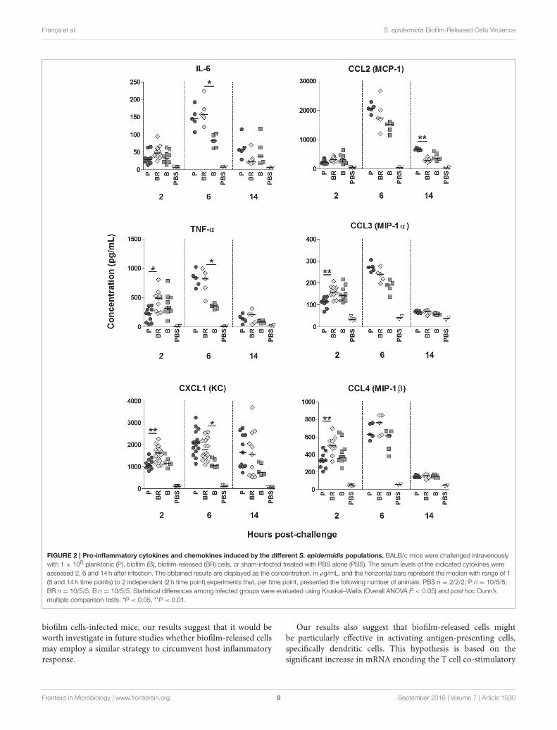
Biofilm-Released Cells Induce HigherStimulation of Pro-Inflammatory Cytokinesand ChemokinesAs shown in Figure 2, mice infected with biofilm-released cellshad significantly higher serum levels of the chemokines CCL3,CCL4, and CXCL1, as well as higher levels of TNF-α than mice
infected with planktonic cells, 2 h after the bacterial challenge. Atthat time point, no differences were found in the levels of anyassessed cytokines between biofilm and biofilm-released cells-infected mouse groups. In contrast, 6 h after infection, markedlyhigher serum levels of CXCL1, TNFα, and IL-6 were detectedin mice infected with biofilm-released cells than in the biofilmcell-infected counterparts. By 14 h after infection, lower serumlevels of CCL2 were detected in mice infected with biofilm-released cells, when compared with their planktonic infectedcounterparts. No significant differences were detected in theserum levels of any other assessed cytokine among the differentinfected groups.
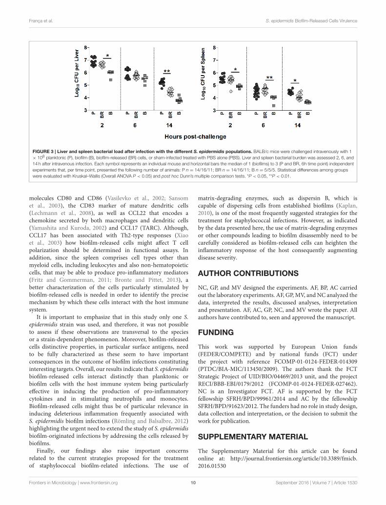
Biofilm-Released Cells Present anIntermediate Ability to Colonize MurineOrgansBiofilm-released cells had an intermediate ability, between thatof planktonic and biofilm cells, to colonize the liver and spleen(Figure 3). Interestingly, while in the first 6 h of infection,biofilm-released cell burden resembled that of planktonic cells,14 h after infection the differences between planktonic andbiofilm-released cells and the similarities between biofilm-released and biofilms cells become noticeable. It is important tonote that although the inoculum was adjusted by flow cytometrythe quantity of bacteria injected was also confirmed by CFU
Frontiers in Microbiology | www.frontiersin.org 6 September 2016 | Volume 7 | Article 1530
França et al. S. epidermidis Biofilm-Released Cells Virulence
TABLE 3 | GO term enrichment of the genes with increased transcription in the spleen of mice infected with S. epidermidis biofilm-released cells.
GO ID Cluster representatives N. of genes P-value
BIOLOGICAL PROCESS
GO:2001242 Regulation of intrinsic apoptotic signaling pathway 11 < 0.0001
GO:2000116 Regulation of cysteine-type endopeptidase activity 12 < 0.0001
GO:0043409 Negative regulation of MAPK cascade 9 < 0.0001
GO:1903039 Positive regulation of leukocyte cell-cell adhesion 9 < 0.001
GO:0050870 Positive regulation of T cell activation 8 0.001
GO:0044003 Modification by symbiont of host morphology or physiology 4 0.001
GO:0031329 Regulation of cellular catabolic process 15 0.003
GO:0010623 Developmental programmed cell death 4 0.004
GO:0002260 Lymphocyte homeostasis 5 0.005
GO:0031638 Zymogen activation 6 0.006
GO:0006986 Response to unfolded protein 5 0.007
GO:0031341 Regulation of cell killing 5 0.007
GO:0010243 Response to organonitrogen compound 13 0.007
GO:0032693 Negative regulation of interleukin-10 production 3 0.007
GO:1901698 Response to nitrogen compound 14 0.010
GO:0048646 Anatomical structure formation involved in morphogenesis 18 0.010
GO:0019724 B cell mediated immunity 5 0.011
GO:0035966 Response to topologically incorrect protein 5 0.011
GO:0010727 Negative regulation of hydrogen peroxide metabolic process 2 0.011
GO:0010743 Regulation of macrophage derived foam cell differentiation 3 0.014
GO:0043243 Positive regulation of protein complex disassembly 3 0.014
GO:0033043 Regulation of organelle organization 19 0.016
GO:0009628 Response to abiotic stimulus 16 0.017
GO:0009888 Tissue development 25 0.017
GO:0033209 Tumor necrosis factor-mediated signaling pathway 3 0.020
GO:0042940 D-amino acid transport 2 0.023
GO:0016192 Vesicle-mediated transport 16 0.030
GO:0018149 Peptide cross-linking 3 0.030
GO:0045787 Positive regulation of cell cycle 8 0.030
GO:0061028 Establishment of endothelial barrier 3 0.032
GO:0048147 Negative regulation of fibroblast proliferation 3 0.035
GO:0008637 Apoptotic mitochondrial changes 4 0.038
GO:0034976 Response to endoplasmic reticulum stress 5 0.046
GO:0009314 Response to radiation 9 0.046
GO:0051604 Protein maturation 7 0.047
GO:0051195 Negative regulation of cofactor metabolic process 2 0.048
GO:0048661 Positive regulation of smooth muscle cell proliferation 4 0.048
MOLECULAR PROCESS
GO:0050786 RAGE receptor binding 4 < 0.001
GO:0016209 Antioxidant activity 6 0.005
GO:0048020 CCR chemokine receptor binding 4 0.005
GO:0046983 Protein dimerization activity 24 0.016
GO:0038024 Cargo receptor activity 5 0.038
GO:0042803 Protein homodimerization activity 17 0.046
CELLULAR COMPONENTS
GO:0031988 Membrane-bounded vesicle 50 < 0.001
GO:0043226 Organelle 108 0.002
GO:0043227 Membrane-bounded organelle 116 0.016
GO:0044424 Intracellular part 115 0.017
(Continued)
Frontiers in Microbiology | www.frontiersin.org 7 September 2016 | Volume 7 | Article 1530
França et al. S. epidermidis Biofilm-Released Cells Virulence
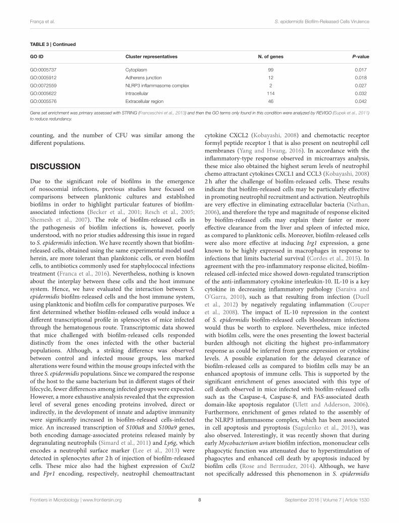
TABLE 3 | Continued
GO ID Cluster representatives N. of genes P-value
GO:0005737 Cytoplasm 99 0.017
GO:0005912 Adherens junction 12 0.018
GO:0072559 NLRP3 inflammasome complex 2 0.027
GO:0005622 Intracellular 114 0.032
GO:0005576 Extracellular region 46 0.042
Gene set enrichment was primary assessed with STRING (Franceschini et al., 2013) and then the GO terms only found in this condition were analyzed by REVIGO (Supek et al., 2011)
to reduce redundancy.
counting, and the number of CFU was similar among thedifferent populations.
DISCUSSION
Due to the significant role of biofilms in the emergenceof nosocomial infections, previous studies have focused oncomparisons between planktonic cultures and establishedbiofilms in order to highlight particular features of biofilm-associated infections (Becker et al., 2001; Resch et al., 2005;Shemesh et al., 2007). The role of biofilm-released cells inthe pathogenesis of biofilm infections is, however, poorlyunderstood, with no prior studies addressing this issue in regardto S. epidermidis infection. We have recently shown that biofilm-released cells, obtained using the same experimental model usedherein, are more tolerant than planktonic cells, or even biofilmcells, to antibiotics commonly used for staphylococcal infectionstreatment (Franca et al., 2016). Nevertheless, nothing is knownabout the interplay between these cells and the host immunesystem. Hence, we have evaluated the interaction between S.epidermidis biofilm-released cells and the host immune system,using planktonic and biofilm cells for comparative purposes. Wefirst determined whether biofilm-released cells would induce adifferent transcriptional profile in splenocytes of mice infectedthrough the hematogenous route. Transcriptomic data showedthat mice challenged with biofilm-released cells respondeddistinctly from the ones infected with the other bacterialpopulations. Although, a striking difference was observedbetween control and infected mouse groups, less markedalterations were found within the mouse groups infected with thethree S. epidermidis populations. Since we compared the responseof the host to the same bacterium but in different stages of theirlifecycle, fewer differences among infected groups were expected.However, a more exhaustive analysis revealed that the expressionlevel of several genes encoding proteins involved, direct orindirectly, in the development of innate and adaptive immunitywere significantly increased in biofilm-released cells-infectedmice. An increased transcription of S100a8 and S100a9 genes,both encoding damage-associated proteins released mainly bydegranulating neutrophils (Simard et al., 2011) and Ly6g, whichencodes a neutrophil surface marker (Lee et al., 2013) weredetected in splenocytes after 2 h of injection of biofilm-releasedcells. These mice also had the highest expression of Cxcl2and Fpr1 encoding, respectively, neutrophil chemoattractant
cytokine CXCL2 (Kobayashi, 2008) and chemotactic receptorformyl peptide receptor 1 that is also present on neutrophil cellmembranes (Yang and Hwang, 2016). In accordance with theinflammatory-type response observed in microarrays analysis,these mice also obtained the highest serum levels of neutrophilchemo attractant cytokines CXCL1 and CCL3 (Kobayashi, 2008)2 h after the challenge of biofilm-released cells. These resultsindicate that biofilm-released cells may be particularly effectivein promoting neutrophil recruitment and activation. Neutrophilsare very effective in eliminating extracellular bacteria (Nathan,2006), and therefore the type and magnitude of response elicitedby biofilm-released cells may explain their faster or moreeffective clearance from the liver and spleen of infected mice,as compared to planktonic cells. Moreover, biofilm-released cellswere also more effective at inducing Irg1 expression, a geneknown to be highly expressed in macrophages in response toinfections that limits bacterial survival (Cordes et al., 2015). Inagreement with the pro-inflammatory response elicited, biofilm-released cell-infected mice showed down-regulated transcriptionof the anti-inflammatory cytokine interleukin-10. IL-10 is a keycytokine in decreasing inflammatory pathology (Saraiva andO’Garra, 2010), such as that resulting from infection (Duellet al., 2012) by negatively regulating inflammation (Couperet al., 2008). The impact of IL-10 repression in the contextof S. epidermidis biofilm-released cells bloodstream infectionswould thus be worth to explore. Nevertheless, mice infectedwith biofilm cells, were the ones presenting the lowest bacterialburden although not eliciting the highest pro-inflammatoryresponse as could be inferred from gene expression or cytokinelevels. A possible explanation for the delayed clearance ofbiofilm-released cells as compared to biofilm cells may be anenhanced apoptosis of immune cells. This is supported by thesignificant enrichment of genes associated with this type ofcell death observed in mice infected with biofilm-released cellssuch as the Caspase-4, Caspase-8, and FAS-associated deathdomain-like apoptosis regulator (Ulett and Adderson, 2006).Furthermore, enrichment of genes related to the assembly ofthe NLRP3 inflammasome complex, which has been associatedin cell apoptosis and pyroptosis (Sagulenko et al., 2013), wasalso observed. Interestingly, it was recently shown that duringearlyMycobacterium avium biofilm infection, mononuclear cellsphagocytic function was attenuated due to hyperstimulation ofphagocytes and enhanced cell death by apoptosis induced bybiofilm cells (Rose and Bermudez, 2014). Although, we havenot specifically addressed this phenomenon in S. epidermidis
Frontiers in Microbiology | www.frontiersin.org 8 September 2016 | Volume 7 | Article 1530
França et al. S. epidermidis Biofilm-Released Cells Virulence
FIGURE 2 | Pro-inflammatory cytokines and chemokines induced by the different S. epidermidis populations. BALB/c mice were challenged intravenously
with 1 × 10⁸ planktonic (P), biofilm (B), biofilm-released (BR) cells, or sham-infected treated with PBS alone (PBS). The serum levels of the indicated cytokines were
assessed 2, 6 and 14 h after infection. The obtained results are displayed as the concentration, in ρg/mL, and the horizontal bars represent the median with range of 1
(6 and 14 h time points) to 2 independent (2 h time point) experiments that, per time point, presented the following number of animals: PBS n = 2/2/2; P n = 10/5/5;
BR n = 10/5/5; B n = 10/5/5. Statistical differences among infected groups were evaluated using Kruskal–Wallis (Overall ANOVA P < 0.05) and post hoc Dunn’s
multiple comparison tests. *P < 0.05, **P < 0.01.
biofilm cells-infected mice, our results suggest that it would beworth investigate in future studies whether biofilm-released cellsmay employ a similar strategy to circumvent host inflammatoryresponse.
Our results also suggest that biofilm-released cells mightbe particularly effective in activating antigen-presenting cells,specifically dendritic cells. This hypothesis is based on thesignificant increase in mRNA encoding the T cell co-stimulatory
Frontiers in Microbiology | www.frontiersin.org 9 September 2016 | Volume 7 | Article 1530
França et al. S. epidermidis Biofilm-Released Cells Virulence
FIGURE 3 | Liver and spleen bacterial load after infection with the different S. epidermidis populations. BALB/c mice were challenged intravenously with 1
× 108 planktonic (P), biofilm (B), biofilm-released (BR) cells, or sham-infected treated with PBS alone (PBS). Liver and spleen bacterial burden was assessed 2, 6, and
14 h after intravenous infection. Each symbol represents an individual mouse and horizontal bars the median of 1 (biofilms) to 3 (P and BR, 6h time point) independent
experiments that, per time point, presented the following number of animals: P n = 14/16/11; BR n = 14/16/11; B n = 5/5/5. Statistical differences among groups
were evaluated with Kruskal–Wallis (Overall ANOVA P < 0.05) and post hoc Dunn’s multiple comparison tests. *P < 0.05, **P < 0.01.
molecules CD80 and CD86 (Vasilevko et al., 2002; Sansomet al., 2003), the CD83 marker of mature dendritic cells(Lechmann et al., 2008), as well as CCL22 that encodes achemokine secreted by both macrophages and dendritic cells(Yamashita and Kuroda, 2002) and CCL17 (TARC). Although,CCL17 has been associated with Th2-type responses (Xiaoet al., 2003) how biofilm-released cells might affect T cellpolarization should be determined in functional assays. Inaddition, since the spleen comprises cell types other thanmyeloid cells, including leukocytes and also non-hematopoieticcells, that may be able to produce pro-inflammatory mediators(Fritz and Gommerman, 2011; Bronte and Pittet, 2013), abetter characterization of the cells particularly stimulated bybiofilm-released cells is needed in order to identify the precisemechanism by which these cells interact with the host immunesystem.
It is important to emphasize that in this study only one S.epidermidis strain was used, and therefore, it was not possibleto assess if these observations are transversal to the speciesor a strain-dependent phenomenon. Moreover, biofilm-releasedcells distinctive properties, in particular surface antigens, needto be fully characterized as these seem to have importantconsequences in the outcome of biofilm infections constitutinginteresting targets. Overall, our results indicate that S. epidermidisbiofilm-released cells interact distinctly than planktonic orbiofilm cells with the host immune system being particularlyeffective in inducing the production of pro-inflammatorycytokines and in stimulating neutrophils and monocytes.Biofilm-released cells might thus be of particular relevance ininducing deleterious inflammation frequently associated withS. epidermidis biofilm infections (Römling and Balsalbre, 2012)highlighting the urgent need to extend the study of S. epidermidisbiofilm-originated infections by addressing the cells released bybiofilms.
Finally, our findings also raise important concernsrelated to the current strategies proposed for the treatmentof staphylococcal biofilm-related infections. The use of
matrix-degrading enzymes, such as dispersin B, which iscapable of dispersing cells from established biofilms (Kaplan,2010), is one of the most frequently suggested strategies for thetreatment for staphylococcal infections. However, as indicatedby the data presented here, the use of matrix-degrading enzymesor other compounds leading to biofilm disassembly need to becarefully considered as biofilm-released cells can heighten theinflammatory response of the host consequently augmentingdisease severity.
AUTHOR CONTRIBUTIONS
NC, GP, and MV designed the experiments. AF, BP, AC carriedout the laboratory experiments. AF, GP,MV, and NC analyzed thedata, interpreted the results, discussed analyses, interpretationand presentation. AF, AC, GP, NC, and MV wrote the paper. Allauthors have contributed to, seen and approved the manuscript.
FUNDING
This work was supported by European Union funds(FEDER/COMPETE) and by national funds (FCT) underthe project with reference FCOMP-01-0124-FEDER-014309(PTDC/BIA-MIC/113450/2009). The authors thank the FCTStrategic Project of UID/BIO/04469/2013 unit, and the projectRECI/BBB-EBI/0179/2012 (FCOMP-01-0124-FEDER-027462).NC is an Investigator FCT. AF is supported by the FCTfellowship SFRH/BPD/99961/2014 and AC by the fellowshipSFRH/BPD/91623/2012. The funders had no role in study design,data collection and interpretation, or the decision to submit thework for publication.
SUPPLEMENTARY MATERIAL
The Supplementary Material for this article can be foundonline at: http://journal.frontiersin.org/article/10.3389/fmicb.2016.01530
Frontiers in Microbiology | www.frontiersin.org 10 September 2016 | Volume 7 | Article 1530
França et al. S. epidermidis Biofilm-Released Cells Virulence
REFERENCES
Becker, P., Hufnagle, W., Peters, G., and Herrmann, M. (2001). Detection of
differential gene expression in biofilm-forming versus planktonic populations
of Staphylococcus aureus using micro-representational-difference analysis.
Appl. Environ. Microbiol. 67, 2958–2965. doi: 10.1128/AEM.67.7.2958-
2965.2001
Boles, B. R., and Horswill, A. R. (2011). Staphylococcal biofilm disassembly. Trends
Microbiol. 19, 449–455. doi: 10.1016/j.tim.2011.06.004
Bronte, V., and Pittet, M. J. (2013). The spleen in local and systemic regulation of
immunity. Immunity 39, 806–818. doi: 10.1016/j.immuni.2013.10.010
Cerca, F., Trigo, G., Correia, A., Cerca, N., Azeredo, J., and Vilanova, M. (2011).
SYBR green as a fluorescent probe to evaluate the biofilm physiological state
of Staphylococcus epidermidis, using flow cytometry. Can. J. Microbiol. 57,
850–856. doi: 10.1139/w11-078
Cerca, N., Jefferson, K. K., Oliveira, R., Pier, G. B., and Azeredo, J. (2006).
Comparative antibody-mediated phagocytosis of Staphylococcus epidermidis
cells grown in a biofilm or in the planktonic state. Infect. Immun. 74,
4849–4855. doi: 10.1128/IAI.00230-06
Cerca, N., Martins, S., Cerca, F., Jefferson, K. K., Pier, G. B., Oliveira, R.,
et al. (2005). Comparative assessment of antibiotic susceptibility of coagulase-
negative staphylococci in biofilm versus planktonic culture as assessed by
bacterial enumeration or rapid XTT colorimetry. J. Antimicrob. Chemother. 56,
331–336. doi: 10.1093/jac/dki217
Chua, S. L., Liu, Y., Yam, J. K., Chen, Y., Vejborg, R. M., Tan, B. G., et al. (2014).
Dispersed cells represent a distinct stage in the transition from bacterial biofilm
to planktonic lifestyles. Nat. Commun. 5:4462. doi: 10.1038/ncomms5462
Cordes, T., Michelucci, A., and Hiller, K. (2015). Itaconic acid: the surprising role
of an industrial compound as a mammalian antimicrobial metabolite. Annu.
Rev. Nutr. 35, 451–473. doi: 10.1146/annurev-nutr-071714-034243
Costerton, J. W., Stewart, P. S., and Greenberg, E. P. (1999). Bacterial biofilms:
a common cause of persistent infections. Science 284, 1318–1322. doi:
10.1126/science.284.5418.1318
Couper, K. N., Blount, D. G., and Riley, E. M. (2008). IL-10: the master
regulator of immunity to infection. J. Immunol. 180, 5771–5777. doi:
10.4049/jimmunol.180.9.5771
Duell, B. L., Tan, C. K., Carey, A. J., Cripps, A. W., and Ulett, G. C. (2012). Recent
insights into microbial triggers of interleukin-10 production in the host and the
impact on infectious disease pathogenesis. FEMS Immunol. Med. Microbiol. 64,
296–313. doi: 10.1111/j.1574-695x.2012.00931.x
Franca, A., Carvalhais, V., Vilanova, M., Pier, G. B., and Cerca, N. (2016).
Characterization of an in vitro fed-batch model to obtain cells released
from S. epidermidis biofilms. AMB Express 6, 23. doi: 10.1186/s13568-016-
0197-9
Franceschini, A., Szklarczyk, D., Frankild, S., Kuhn, M., Simonovic, M., Roth,
A., et al. (2013). STRING v9.1: protein-protein interaction networks, with
increased coverage and integration. Nucleic Acids Res. 41, D808–D815. doi:
10.1093/nar/gks1094
Fritz, J. H., and Gommerman, J. L. (2011). Cytokine/stromal cell networks and
lymphoid tissue environments. J. Interferon. Cytokine Res. 31, 277–289. doi:
10.1089/jir.2010.0121
Kallio, M. A., Tuimala, J. T., Hupponen, T., Klemela, P., Gentile, M., Scheinin, I.,
et al. (2011). Chipster: user-friendly analysis software for microarray and other
high-throughput data. BMC Genomics 12:507. doi: 10.1186/1471-2164-12-507
Kaplan, J. B. (2010). Biofilm dispersal: mechanisms, clinical implications,
and potential therapeutic uses. J. Dent. Res. 89, 205–218. doi:
10.1177/0022034509359403
Kobayashi, Y. (2008). The role of chemokines in neutrophil biology. Front. Biosci.
1, 2400–2407. doi: 10.2741/2853
Kristian, S. A., Birkenstock, T. A., Sauder, U., Mack, D., Gotz, F., and Landmann,
R. (2008). Biofilm formation induces C3a release and protects Staphylococcus
epidermidis from IgG and complement deposition and from neutrophil-
dependent killing. J. Infect. Dis. 197, 1028–1035. doi: 10.1086/528992
Lechmann, M., Shuman, N., Wakeham, A., and Mak, T. W. (2008). The
CD83 reporter mouse elucidates the activity of the CD83 promoter in B,
T, and dendritic cell populations in vivo. Proc. Natl. Acad. Sci. U.S.A. 105,
11887–11892. doi: 10.1073/pnas.0806335105
Lee, P. Y., Wang, J. X., Parisini, E., Dascher, C. C., and Nigrovic, P. A. (2013).
Ly6 family proteins in neutrophil biology. J. Leukoc. Biol. 94, 585–594. doi:
10.1189/jlb.0113014
Li, Y., Petrova, O. E., Su, S., Lau, G. W., Panmanee, W., Na, R., et al.
(2014). BdlA, DipA and induced dispersion contribute to acute virulence and
chronic persistence of Pseudomonas aeruginosa. PLoS Pathog. 10:e1004168. doi:
10.1371/journal.ppat.1004168
Liu, J., Ling, J. Q., Zhang, K., and Wu, C. D. (2013). Physiological properties of
Streptococcus mutansUA159 biofilm-detached cells. FEMSMicrobiol. Lett. 340,
11–18. doi: 10.1111/1574-6968.12066
Mack, D., Nedelmann, M., Krokotsch, A., Schwarzkopf, A., Heesemann, J., and
Laufs, R. (1994). Characterization of transposon mutants of biofilm-producing
Staphylococcus epidermidis impaired in the accumulative phase of biofilm
production: genetic identification of a hexosamine-containing polysaccharide
intercellular adhesin. Infect. Immun. 62, 3244–3253.
Marks, L. R., Davidson, B. A., Knight, P. R., and Hakansson, A. P. (2013).
Interkingdom signaling induces Streptococcus pneumoniae biofilm dispersion
and transition from asymptomatic colonization to disease. MBio 4, e00438–
e00413. doi: 10.1128/mBio.00438-13
Nathan, C. (2006). Neutrophils and immunity: challenges and opportunities. Nat.
Rev. Immunol. 6, 173–182. doi: 10.1038/nri1785
Oliveros, J. C. (2007). VENNY. An Interactive Tool for
Comparing Lists with Venn Diagrams. Available online at:
http://bioinfogp.cnb.csic.es/tools/venny/index.html
Otto, M. (2009). Staphylococcus epidermidis–the ‘accidental’ pathogen. Nat. Rev.
Microbiol. 7, 555–567. doi: 10.1038/nrmicro2182
Otto, M. (2012). Staphylococcal infections: mechanisms of biofilm maturation
and detachment as critical determinants of pathogenicity. Annu. Rev. Med. 64,
175–188. doi: 10.1146/annurev-med-042711-140023
Pavlidis, P., and Noble, W. S. (2003). Matrix2png: a utility for visualizing matrix
data. Bioinformatics 19, 295–296. doi: 10.1093/bioinformatics/19.2.295
Pitz, A. M., Yu, F., Hermsen, E. D., Rupp, M. E., Fey, P. D., and
Olsen, K. M. (2011). Vancomycin susceptibility trends and prevalence of
heterogeneous vancomycin-intermediate Staphylococcus aureus in clinical
methicillin-resistant S. aureus isolates. J. Clin. Microbiol. 49, 269–274. doi:
10.1128/JCM.00914-10
Resch, A., Rosenstein, R., Nerz, C., and Gotz, F. (2005). Differential gene
expression profiling of Staphylococcus aureus cultivated under biofilm
and planktonic conditions. Appl. Environ. Microbiol. 71, 2663–2676. doi:
10.1128/AEM.71.5.2663-2676.2005
Rollet, C., Gal, L., and Guzzo, J. (2009). Biofilm-detached cells, a transition from
a sessile to a planktonic phenotype: a comparative study of adhesion and
physiological characteristics in Pseudomonas aeruginosa. FEMSMicrobiol. Lett.
290, 135–142. doi: 10.1111/j.1574-6968.2008.01415.x
Römling, U., and Balsalbre, C. (2012). Biofilm infections, their resilience to
therapy and innovative treatment strategies. J. Intern. Med. 271, 541–561. doi:
10.1111/joim.12004
Rose, S. J., and Bermudez, L. E. (2014). Mycobacterium avium biofilm attenuates
mononuclear phagocyte function by triggering hyperstimulation and apoptosis
during early infection. Infect. Immun. 83, 405–412. doi: 10.1128/IAI.00820-13
Sagulenko, V., Thygesen, S. J., Sester, D. P., Idris, A., Cridland, J. A., Vajhala, P.
R., et al. (2013). AIM2 and NLRP3 inflammasomes activate both apoptotic
and pyroptotic death via ASC. Cell Death Differ. 20, 1149–1160. doi:
10.1038/cdd.2013.37
Sandberg, R., and Larsson, O. (2007). Improved precision and accuracy for
microarrays using updated probe set definitions. BMC Bioinformatics 8:48. doi:
10.1186/1471-2105-8-48
Sansom, D. M., Manzotti, C. N., and Zheng, Y. (2003). What’s the difference
between CD80 and CD86? Trends Immunol. 24, 314–319. doi: 10.1016/S1471-
4906(03)00111-X
Saraiva, M., and O’Garra, A. (2010). The regulation of IL-10 production by
immune cells. Nat. Rev. Immunol. 10, 170–181. doi: 10.1038/nri2711
Shemesh, M., Tam, A., and Steinberg, D. (2007). Differential gene expression
profiling of Streptococcus mutans cultured under biofilm and planktonic
conditions.Microbiology 153, 1307–1317. doi: 10.1099/mic.0.2006/002030-0
Simard, J. C., Simon, M. M., Tessier, P. A., and Girard, D. (2011). Damage-
associated molecular pattern S100A9 increases bactericidal activity of human
Frontiers in Microbiology | www.frontiersin.org 11 September 2016 | Volume 7 | Article 1530
França et al. S. epidermidis Biofilm-Released Cells Virulence
neutrophils by enhancing phagocytosis. J. Immunol. 186, 3622–3631. doi:
10.4049/jimmunol.1002956
Smyth, G. K. (2004). Linear models and empirical bayes methods for assessing
differential expression in microarray experiments. Stat. Appl. Genet. Mol. Biol.
3:3. doi: 10.2202/1544-6115.1027
Sousa, C., Franca, A., and Cerca, N. (2014). Assessing and reducing sources
of genes expression variability in Staphylococcus epidermidis biofilms.
Biotechniques 57, 295–301. doi: 10.2144/000114238
Supek, F., Bosnjak, M., Skunca, N., and Smuc, T. (2011). REVIGO summarizes
and visualizes long lists of gene ontology terms. PLoS ONE 6:e21800. doi:
10.1371/journal.pone.0021800
Ulett, G. C., and Adderson, E. E. (2006). Regulation of apoptosis by Gram-
positive bacteria: mechanistic diversity and consequences for immunity. Curr.
Immunol. Rev. 2, 119–141. doi: 10.2174/157339506776843033
Vasilevko, V., Ghochikyan, A., Holterman, M. J., and Agadjanyan, M. G. (2002).
CD80 (B7-1) and CD86 (B7-2) are functionally equivalent in the initiation
and maintenance of CD4+ T-cell proliferation after activation with suboptimal
doses of PHA. DNA Cell Biol. 21, 137–149. doi: 10.1089/10445490252925404
von Eiff, C., Peters, G., and Heilmann, C. (2002). Pathogenesis of infections
due to coagulase-negative staphylococci. Lancet Infect. Dis. 2, 677–685. doi:
10.1016/S1473-3099(02)00438-3
Vuong, C., and Otto, M. (2002). Staphylococcus epidermidis infections. Microbes
Infect. 4, 481–489. doi: 10.1016/S1286-4579(02)01563-0
Wang, R., Khan, B. A., Cheung, G. Y., Bach, T. H., Jameson-Lee, M., Kong, K. F.,
et al. (2011). Staphylococcus epidermidis surfactant peptides promote biofilm
maturation and dissemination of biofilm-associated infection in mice. J. Clin.
Invest. 121, 238–248. doi: 10.1172/JCI42520
Xiao, T., Fujita, H., Saeki, H., Mitsui, H., Sugaya, M., Tada, Y., et al. (2003).
Thymus and activation-regulated chemokine (TARC/CCL17) produced by
mouse epidermal Langerhans cells is upregulated by TNF-alpha and IL-4 and
downregulated by IFN-gamma. Cytokine 23, 126–132. doi: 10.1016/S1043-
4666(03)00221-7
Yamashita, U., and Kuroda, E. (2002). Regulation of macrophage-derived
chemokine (MDC, CCL22) production. Crit. Rev. Immunol. 22, 105–114. doi:
10.1615/CritRevImmunol.v22.i2.10
Yang, S. C., and Hwang, T. L. (2016). The potential impacts of formyl peptide
receptor 1 in inflammatory diseases. Front. Biosci. (Elite. Ed). 1, 436–449.
Yao, Y., Sturdevant, D. E., and Otto, M. (2005). Genomewide analysis of
gene expression in Staphylococcus epidermidis biofilms: insights into the
pathophysiology of S. epidermidis biofilms and the role of phenol-soluble
modulins in formation of biofilms. J. Infect. Dis. 191, 289–298. doi:
10.1086/426945
Conflict of Interest Statement: The authors declare that the research was
conducted in the absence of any commercial or financial relationships that could
be construed as a potential conflict of interest.
Copyright © 2016 França, Pérez-Cabezas, Correia, Pier, Cerca and Vilanova. This
is an open-access article distributed under the terms of the Creative Commons
Attribution License (CC BY). The use, distribution or reproduction in other forums
is permitted, provided the original author(s) or licensor are credited and that the
original publication in this journal is cited, in accordance with accepted academic
practice. No use, distribution or reproduction is permitted which does not comply
with these terms.
Frontiers in Microbiology | www.frontiersin.org 12 September 2016 | Volume 7 | Article 1530