Embed Size (px)
Citation preview
Staphylococcus epidermidis agr Quorum-Sensing System: SignalIdentification, Cross Talk, and Importance in Colonization
Michael E. Olson,a Daniel A. Todd,b Carolyn R. Schaeffer,c Alexandra E. Paharik,a Michael J. Van Dyke,a Henning Büttner,e
Paul M. Dunman,d Holger Rohde,e Nadja B. Cech,b Paul D. Fey,c Alexander R. Horswilla
Department of Microbiology, Roy J. and Lucille A. Carver College of Medicine, University of Iowa, Iowa City, Iowa, USAa; Department of Chemistry and Biochemistry,University of North Carolina Greensboro, Greensboro, North Carolina, USAb; Department of Pathology and Microbiology, University of Nebraska Medical Center, Omaha,Nebraska, USAc; Department of Microbiology and Immunology, University of Rochester Medical Center, Rochester, New York, USAd; Institute for Medical Microbiology,Virology and Hygiene, University Medical Centre Hamburg-Eppendorf, Hamburg, Germanye
Staphylococcus epidermidis is an opportunistic pathogen that is one of the leading causes of medical device infections. Globalregulators like the agr quorum-sensing system in this pathogen have received a limited amount of attention, leaving importantquestions unanswered. There are three agr types in S. epidermidis strains, but only one of the autoinducing peptide (AIP) signalshas been identified (AIP-I), and cross talk between agr systems has not been tested. We structurally characterized all three AIPtypes using mass spectrometry and discovered that the AIP-II and AIP-III signals are 12 residues in length, making them thelargest staphylococcal AIPs identified to date. S. epidermidis agr reporter strains were developed for each system, and we deter-mined that cross-inhibitory interactions occur between the agr type I and II systems and between the agr type I and III systems.In contrast, no cross talk was observed between the type II and III systems. To further understand the outputs of the S. epidermi-dis agr system, an RNAIII mutant was constructed, and microarray studies revealed that exoenzymes (Ecp protease and Gehlipase) and low-molecular-weight toxins were downregulated in the mutant. Follow-up analysis of Ecp confirmed the RNAIII isrequired to induce protease activity and that agr cross talk modulates Ecp activity in a manner that mirrors the agr reporter re-sults. Finally, we demonstrated that the agr system enhances skin colonization by S. epidermidis using a porcine model. Thiswork expands our knowledge of S. epidermidis agr system function and will aid future studies on cell-cell communication in thisimportant opportunistic pathogen.
Staphylococcus epidermidis is a human commensal of the skinand mucosal surfaces and is one of the best-known members of
the coagulase-negative staphylococci (CoNS) (1). CoNS are themost frequent cause of hospital-associated infections (HAIs) (2),including central-line-associated infections, and the second mostcommon cause of surgical-site infections and prosthetic-valve in-fective endocarditis (3). Over 150 million intravascular devices areused each year in the United States alone (4), and a major riskfactor for CoNS disease is the presence of a medical implant (5).For many of these infections, S. epidermidis makes up more than80% of the clinical burden of CoNS (3, 6).
The ability of S. epidermidis to colonize implanted devices andform a biofilm is its primary mechanism of pathogenesis (1, 6).The polysaccharide intercellular adhesin (PIA) produced by pro-teins encoded in the icaADBC locus is one of the best-character-ized biofilm determinants (7). More recently, protein-mediatedbiofilm mechanisms have been identified (reviewed in reference6), with the accumulation-associated protein (Aap) being one ofthe best studied (8). Regulation of biofilm formation has been afocus of numerous studies, and some of these investigations havedemonstrated that the agr quorum-sensing system impacts S. epi-dermidis biofilm development. In this regard, strains defective inagr have been shown to display enhanced adherence and biofilmproperties (9, 10) and reduced production of extracellular en-zymes, such as lipases and proteases (10–12). The agr-regulatedlow-molecular-weight toxins, termed phenol-soluble modulins(PSM�s), are known to disrupt S. epidermidis biofilms and to beimportant for dissemination during biofilm infection (13).
The fundamental features of the agr system are conserved in anumber of staphylococcal species, including S. epidermidis (14).
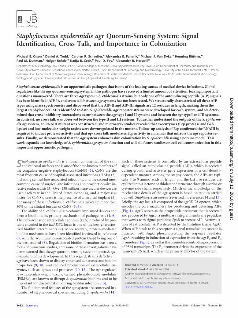
Each of these systems is controlled by an extracellular peptidesignal called an autoinducing peptide (AIP), which is secretedduring growth and activates gene expression in a cell density-dependent manner. Among the staphylococci, the AIPs are typi-cally 7 to 9 amino acids in length, and the last five residues arecyclized into a lactone or thiolactone structure through a serine orcysteine side chain, respectively. Much of the knowledge on themechanistic details of the agr system is based on studies carriedout with Staphylococcus aureus (reviewed in references 14 and 15).Briefly, the agr locus is composed of the agrBDCA operon, whichencodes the core machinery for producing and detecting AIPs(Fig. 1). AgrD serves as the propeptide precursor that is secretedand processed by AgrB, a multipass integral membrane peptidasethat works with signal peptidase SpsB to secrete AIP. Accumula-tion of extracellular AIP is detected by the histidine kinase AgrC.When AIP binds to this receptor, a signal transduction cascade isinitiated, with AgrC phosphorylating the response regulatorAgrA, resulting in induction of expression from the agr P2 and P3
promoters (Fig. 1), as well as the promoters controlling expressionof PSM transcripts. The P3 promoter drives the expression of thetranscript RNAIII, which is the primary effector of the system.
Received 25 May 2014 Accepted 18 July 2014
Published ahead of print 28 July 2014
Address correspondence to Alexander R. Horswill, [email protected].
M.E.O. and D.A.T. contributed equally to this work.
Copyright © 2014, American Society for Microbiology. All Rights Reserved.
doi:10.1128/JB.01882-14
3482 jb.asm.org Journal of Bacteriology p. 3482–3493 October 2014 Volume 196 Number 19
on July 12, 2018 by guesthttp://jb.asm
.org/D
ownloaded from
There remain important unanswered questions about the S.epidermidis agr system. Following the initial description of thelocus (16, 17), it was several years before sequencing studies re-vealed three different classes of S. epidermidis agr systems in clin-ical isolates (18), which are referred to here as agr types I, II, andIII. Although there has been some effort to correlate these classeswith S. epidermidis disease (19–23), the presence of different agrclasses, and their unique AIP signals, has remained largely over-looked. Of these, only the AIP-I structure has been characterized(16). Many studies have focused on prototype biofilm-formingstrain 1457, which encodes an agr type II system, but the structureof S. epidermidis AIP-II is not known.
Our goal in this study was to gain a better understanding of S.epidermidis agr system function and address several open ques-tions. By using bioactivity-guided fractionation and high-resolu-tion mass spectrometry, the AIP signal structures were deter-mined and cross talk between each of S. epidermidis agr systemswas investigated. An RNAIII mutant was constructed in strain1457; the regulatory changes in this mutant were assessed, and askin colonization phenotype was identified. Collectively, these re-sults provide new insights into the molecular details, function,and importance of the S. epidermidis agr system.
MATERIALS AND METHODSGrowth conditions and reagents. Bacterial strains used in this study arelisted in Table 1. Escherichia coli was grown in LB medium, and S. epider-midis cultures were grown in tryptic soy broth (TSB). S. epidermidis cul-tures were grown at 37°C with the appropriate antibiotics at concentra-tions of 10 �g/ml for chloramphenicol (Cam), erythromycin (Erm), andtrimethoprim (Tmp) and 50 �g/ml for kanamycin (Kan), as needed.Chemical reagents were purchased from Sigma-Aldrich (St. Louis, MO)unless otherwise noted. S. epidermidis AIP-II was custom synthesized byAmerican Peptide Company (Vista, CA). All oligonucleotides used in thisstudy are listed in Table 2.
Strain and plasmid constructions. Plasmid constructions were per-formed in E. coli using standard techniques. DNA sequencing was per-formed at the University of Nebraska Medical Center (UNMC) core se-quencing laboratory. Following passage through the restriction-deficientS. aureus strain RN4220 (24), plasmid DNA was electroporated into S.epidermidis strains as previously described (25). Plasmids were movedbetween S. epidermidis strains using bacteriophage 71 transduction aspreviously described (26).
Construction of an RNAIII mutant. The RNAIII allelic replacementvector (pNF41) was designed to have two regions of sequence similarity to
the S. epidermidis chromosome flanking the dihydrofolate reductase(dhfr) gene from plasmid pGO558 (27), which confers resistance to Tmp.The first fragment was a 697-bp piece of the 5= region of RNAIII that wasPCR amplified from S. epidermidis RP62A using oligonucleotides 367 and378 (Table 2). The PCR product was digested with EcoRI and BamHI andwas ligated into pUC19 cut with the same enzymes. The second fragmentwas a 991-bp 3= region of RNAIII amplified from S. epidermidis RP62Awith primers 369 and 379, and this PCR product was digested with SalIand PstI and cloned into the same sites on the pUC19-5=-RNAIII plasmid.The dhfr gene was excised from pGO558 with a SalI digest and was ligatedinto the same site on the RNAIII knockout plasmid to provide Tmp resis-tance. Finally, pROJ6448 (28) was linearized with PstI and inserted intothe plasmid to provide a temperature-sensitive staphylococcal repliconand Erm resistance.
Construction of an ecp mutant. Roughly 1,000-bp regions flankingthe ecp gene were amplified from chromosomal S. epidermidis 1457 DNAusing primers Ecp5_att_for, Ecprev_Eco, 3_for_Eco, and Ecp_att_rev3(Table 2). Amplicons were purified, cleaved using EcoRI, and ligated byT4 ligase. The 2,000-bp ligation product was cloned into pKOR1 using theBP Clonase reaction (Invitrogen), resulting in pKOecp. pKOecp was in-troduced into S. aureus RN4220 by electroporation and, from there, againby electroporation into S. epidermidis mutant 1457-M12 (29). Usingphage A6C, pKOecp was next transduced into S. epidermidis 1457 (8).Allelic replacement was carried out essentially as described previously(30). Putative mutants were screened by PCR, and the loss of ecp wasverified by sequence analysis.
FIG 1 Schematic of the staphylococcal agr locus and S. epidermidis AgrDsequences. The three AgrD types are shown and are divided into the leaderregion, AIP-containing region, and tail region. The highly conserved residuesacross staphylococcal AgrDs are indicated by asterisks. The previously de-duced AIP-I structure is boxed. Also diagramed is the deletion/insertion of thedhfr cassette that disrupts RNAIII in S. epidermidis 1457 to yield the �rnaIII::dhfr mutant used in this study.
TABLE 1 Strains and plasmids
Strain or plasmid DescriptionSource orreference
E. coli DH5� Cloning strain New EnglandBiolabs
S. aureusAH1263 LAC Erms 52RN4220 Restriction deficient strain 24
S. epidermidisATCC 12228 Clinical isolate (ica agr type I) 531457 (AH2490) Clinical isolate (ica� agr type II) 54RP62a Clinical isolate (ica� agr type I) 554804 Clinical isolate (agr type I) Fey collection7237 Clinical isolate (agr type I) Fey collection5183 Clinical isolate (agr type II) Fey collection7022 Clinical isolate (agr type II) Fey collection8595 Clinical isolate (agr type II) Fey collection8099 Clinical isolate (agr type III) Fey collection5794 Clinical isolate (agr type III) Fey collection8247 Clinical isolate (agr type III) Fey collectionAH2673 1457/pCM40 (Ermr) This workAH2491 1457 �rnaIII::dhfr This workAH2589 1457 �ica::dhfr 27AH2924 1457 �ecp This workAH3408 ATCC 12228/pCM40 (Ermr) This workAH3409 8247/pCM40 (Ermr) This work
PlasmidspCM11 PsarA-sGFP Ermr 50pCM40 agr P3-sGFP Ermr This workpGO558 Source of dhfr cassette; Tmpr 27pKOR1 Knockout vector 30pROJ6448 Temp-sensitive replicon 28pUC19 Cloning vector New England
Biolabs
Staphylococcus epidermidis Quorum Sensing
October 2014 Volume 196 Number 19 jb.asm.org 3483
on July 12, 2018 by guesthttp://jb.asm
.org/D
ownloaded from
Construction of S. epidermidis agr reporter strains. The agr P3 pro-moter was PCR amplified from S. aureus strain AH1263 using oligonucle-otides ARH132 and ARH133 (Table 2). The PCR product was digestedwith HindIII and KpnI and cloned into the same sites on plasmid pCM11.The new plasmid was confirmed for agr responsiveness and calledpCM40. Plasmid pCM40 was transformed into S. epidermidis 1457 toconstruct reporter strain AH2673. The reporter plasmid was transducedto other strains using bacteriophage 71 to construct AH3408 and AH3409reporters.
agr P3-sGFP reporter assays. For the agr P3-sGFP time course, anovernight culture of reporter strain AH2673 was diluted to an opticaldensity at 600 nm (OD600) of 0.05 in 20 ml of TSB in 125-ml flasks sup-plemented with 10 �g/ml Erm. For exogenous AIP-II addition, spentmedium was collected from S. epidermidis 1457 by centrifuging 24-h cul-tures at 3,750 rpm and filtering through 0.22-�m filters. The spent me-dium was added to the reporter culture at a final concentration of 10%(vol/vol). Cultures were grown at 37°C with shaking (250 rpm). Measure-ments of the optical density at 600 nm (OD600) and superfolder greenfluorescent protein (sGFP) fluorescence (excitation, 490 nm; emission,520 nm) were obtained hourly for 27 h using a Tecan Infinite M200 platereader. For each measurement, 3 samples of 200 �l were removed fromeach flask and transferred to a 96-well microtiter plate (Corning 3096).The values are reported as relative fluorescence after normalization toOD600 readings. At least three biological replicates were performed foreach condition. Of note, the AH2673 reporter strain was reconstructed forthe time course.
For the cross talk tests, S. epidermidis strains representing different agrsystems were grown for 20 h in TSB, and spent medium was collected andpassed through 0.22-�m filters. The agr reporters for type I (AH3408),type II (AH2673), and type III (AH3409) were grown overnight in 5 ml ofTSB with Erm and subcultured 1:200 into 18-mm tubes with fresh culturemedium. The filtered spent medium collected from S. epidermidis strainswas added to 10% (vol/vol). The reporter cultures were grown for 24 h,and growth (OD600) and sGFP measurements were obtained as describedabove.
Transcriptional profiling. Overnight cultures of S. epidermidis 1457and 1457 �rnaIII::dhfr were diluted 1:100 into fresh TSB and grown at37°C to an OD600 of 8.2 (flask-to-volume ratio, 5:1; shaking at 200 rpm).RNA was converted to cDNA, and microarray analysis was performedaccording to the manufacturer’s instructions (Affymetrix expressionanalysis technical manual; Affymetrix, Inc., Santa Clara, CA) for antisenseprokaryotic arrays as described previously by Beenken and colleagues(31). To ensure reproducibility, cDNA samples from each strain wereprepared from two separate experiments. Each cDNA sample was hybrid-
ized to an S. epidermidis GeneChip. Signal intensity values for each qual-ifier (predicted open reading frame [ORF] and intergenic region) werenormalized to the median signal intensity value for each GeneChip. Sam-ple values were then averaged. Genes for which there was at least a 2-folddifference (P � 0.05, t test) in RNA titer between 1457 and 1457 �rnaIII::dhfr were considered differentially expressed in an rnaIII-dependentmanner.
�-Toxin immunoblots. For the �-toxin immunoblots, strain 1457,strain 1457 supplemented with 40 nM AIP-II, and the 1457 �rnaIII::dhfrmutant were grown in TSB for 21 or 24 h at 37°C. Cells were removed byfiltration through 0.22-�m filters, and spent medium was used for theimmunoblots. Immunoblotting for �-toxin was performed by cross-reac-tivity with rabbit polyclonal antisera to S. aureus �-toxin (Abgent, SanDiego, CA), diluted 1:1000, followed by secondary goat anti-rabbit IgG(1:1,000) conjugated to horseradish peroxidase (Toxin Technology, Sara-sota, FL) as previously described (32).
Ecp activity analysis. A protease assay was developed to follow Ecpactivity using FRET substrate [5-carboxyfluorescein (FAM)-Lys-Leu-Leu-Asp-Ala-Ala-Pro-(QXL520)-OH; AnaSpec, Fremont, CA]. This sub-strate is based on the CXCR2 substrate developed for S. aureus staphopainA (33), and it was suspended to 50 �M using 20 mM Tris (pH 7.4). For Ecpactivity analysis, strains were grown in TSB for 12 h at 37°C. Cells wereremoved by filtration through 0.22-�m filters, and spent medium wassaved for the analysis. A 50-�l portion of collected medium was mixedwith 25 �l of the FRET substrate in a microtiter plate, and fluorescencemeasurements (excitation, 490 nm; emission, 520 nm) were obtained at37°C in a Tecan Infinite M200 plate reader. Reaction rates were recordedas units of fluorescence per minute.
For the agr cross talk impact on Ecp activity, spent medium fromdifferent S. epidermidis strains was prepared as described above for the agrreporter methods. For the Ecp measurements, cultures were prepared asdescribed above except that collected spent medium was added to 10%(vol/vol) of the testing strain at the time of inoculation. Finally, cells wereremoved, and Ecp activity was measured in the samples.
Reverse transcriptase PCR. Overnight cultures were diluted 1:100and grown microaerobically (5:1 [vol/vol], flask-to-medium ratio) in TSBto an OD600 of �7.6. Samples were spun at 6,000 g for 5 min at 4°C.Pellets were resuspended in RLT buffer (Qiagen) with 0.01% �-mercap-toethanol. Cells were lysed by mechanical disruption and the cell debriswas removed by centrifugation at 21,000 g for 15 min at 4°C. Thesupernatant was removed to a new tube with 0.7 volume of ethanol andvortexed. RNA was prepared using the RNeasy kit (Qiagen) according tothe manufacturer’s instructions. cDNA was synthesized from approxi-mately 1.5 �g of RNA using the Transcriptor first-strand cDNA synthesis
TABLE 2 Oligonucleotides used in this study
Name Sequence
367 (3= rnaIII-F) GGAATTCCCGACTAATGCCATAGATAAAAG378 (3= rnaIII-R) CGGGATCCCACCGATTGTAGAAATGATATC369 (5= rnaIII-F) ACGCGTCGACGCCGTGAGTCTCTCCCAAG379 (5= rnaIII-R) AACTGCAGGTAACTAAAGCTTTACCCTAAGCARH132 GTTGTTAAGCTTCTGTCATTATACGATTTAGTACAATCARH133 GTTGTTGGTACCTTAAACAACTCATCAACTATTTTCC2298 (ecp-F) TGTGCTTAAAACGCCACGTA2299 (ecp-R) GTATAGCCGGCACACCAACT2301 (gyrB-F) CTCGAAGCGGTTCGTAAAAG2302 (gyrB-R) TACCACGGCCATTGTCAGTA2547 SERP0736 (psm�1)-F CGGCCTCATTTAGGAGTG2548 SERP0736 (psm�1)-R CGATTCACCATATCAACGCEcp5_att_for GGGGACAAGTTTGTACAAAAAAGCAGGCTTAGCGTGGTGAAGTTAATGATGEcprev_Eco CAACACATGAATTCGCTAGCTTTTGCAACTCTTTCAAATCG3_for_Eco CAACACATGAATTCGCGGCCGCTATTAATATAGAAAGGTGTGCTTATGCEcp_att_rev3 GGGGACCACTTTGTACAAGAAAGCTGGGTAGAAGATATTCATATTAGTGGTGCTG
Olson et al.
3484 jb.asm.org Journal of Bacteriology
on July 12, 2018 by guesthttp://jb.asm
.org/D
ownloaded from
kit (Roche) according to the manufacturer’s instructions using the ran-dom hexamer primer. RT-PCR mixtures contained Quik-Load 2 MidasPCR mix (Monserate Biotech, San Diego, CA), 10 �M primers, and 2 �l ofcDNA. Reactions were run in a MyCycler thermocycler (Bio-Rad) for 20cycles with a 15-s extension. Following agarose gel electrophoresis, DNAbands were visualized by ethidium bromide staining.
Fractionation of S. epidermidis spent medium. S. epidermidis strain5183 was grown in TSB at 37°C with shaking at 200 rpm overnight. Cellswere pelleted by centrifugation at 6,000 g for 5 min. and removed by0.22-�m filtration. The filtered medium was separated by automated flashchromatography using a Teledyne Isco Combiflash Rf system. A RediSepRf Gold C18 column was used with a linear methanol-water gradient start-ing at 10% methanol and ending at 100% methanol over 100 columnvolumes, and the fractions were collected in 25-ml test tubes.
LC-MS identification and quantification of S. epidermidis AIP. Allliquid chromatography-mass spectrometry (LC-MS) analyses were per-formed on a Waters Acquity ultraperformance liquid chromatograph(UPLC) coupled to a Thermo Fisher Scientific LTQ Orbitrap XL massspectrometer with electrospray source. Samples (5 �l) were injected ontoa high-strength silica (HSS) T3 C18 column (Waters Corporation). Anal-yses were conducted at a flow rate of 0.250 ml/min with the followingbinary gradient, where solvent A is 0.1% formic acid in H2O, and solventB is 0.1% formic acid in acetonitrile: 0 to 6 min, from 80% A to 20% A; 6.0to 6.5 min, from 20% A to 80% A; 6.5 to 7.0 min, 80% A (isocratic).
Analyses were conducted in both the positive- and negative-ionmodes. In positive-ion mode, the Orbitrap was operated at a scan range ofm/z 300 to 2,000 with the following settings: tube lens voltage, 110 V;source voltage, 4.50 kV; source current, 100 �A; heated-capillary voltage,20.0 V; heated-capillary temperature, 300.0°C; sheath gas flow rate, 20.0;auxiliary gas flow rate, 0. For negative-ion mode analyses, the Orbitrapwas operated at a scan range of m/z 300 to 2,000 with the following set-tings: tube lens voltage, 100 V; source voltage 4.00 kV; source current, 100�A; heated-capillary voltage, 20.0 V; heated-capillary temperature,275.0°C; sheath gas flow rate, 25.0; auxiliary gas flow rate, 0. For MS-MSanalysis in the negative-ion mode, the precursor masses of 873.34,1,355.60, and 1296.60 (the predicted masses of deprotonated AIP-I, AIP-II, and AIP-III, respectively) were subjected to collision-induced dissoci-ation with activation energy of 35%. The same MS-MS conditions wereused in the positive ion mode, but precursor m/z values for protonatedAIP-I, API-II, and AIP-III (873.34, 1,355.60, and 1,296.60, respectively)were selected.
The AIP-II concentration was calculated using an 8-point calibrationcurve of synthetic AIP-II ranging in concentration from 30.0 �M to 0.3�M by 2-fold dilutions. The calibration curve was generated by plottinglog(peak area) versus log(concentration). Peak area was selected for a m/zof 678.38 (the most intense product peak for mass 1,355.60) from theMS-MS selected ion chromatogram. AIP-II concentrations were calcu-
lated using the slope of the best-fit line obtained by linear regressionanalysis of the calibration curve data.
S. epidermidis colonization of porcine skin. S. epidermidis coloniza-tion of porcine skin was performed as described previously (34). Biopsypunches of 8 mm were taken from the ears of newborn piglets (tissuekindly provided by D. Stoltz). Explanted tissue was washed prior to biopsywith ethanol, and biopsy punches were placed individually on a piece ofsterile gauze in 12-well cell culture plates. The skin biopsy specimens werecultured in Dulbecco’s modified Eagle medium (DMEM) containing hy-drocortisone, 10% fetal bovine serum (FBS), and penicillin-streptomycin(35). The growth medium was added to the wells, bringing the height nearthe apical surface of the biopsy specimen. These samples were incubated at37°C with 5% CO2 for the duration of the experiment, and the culturemedium was changed daily. After 15 h of incubation, 10 �l of bacterialsuspension in saline containing 1.5 104 to 2 104 CFU of wild-type(WT) 1457 or 3.8 104 to 7.5 104 CFU of 1457 �rnaIII::dhfr wasspotted on the apical skin surface. The cultures were allowed to dry ontothe skin surface in a laminar flow hood for 1 h, and the plates were re-turned to the incubator. After 48 h of incubation, the skin biopsy speci-men was placed in1 ml of phosphate-buffered saline (PBS) and vortexedat maximum speed for 1 min. The cells were serially diluted, plated onhigh-salt tryptic soy agar (TSA) (supplemented with 50 g/liter NaCl),incubated overnight at 37°C, and enumerated.
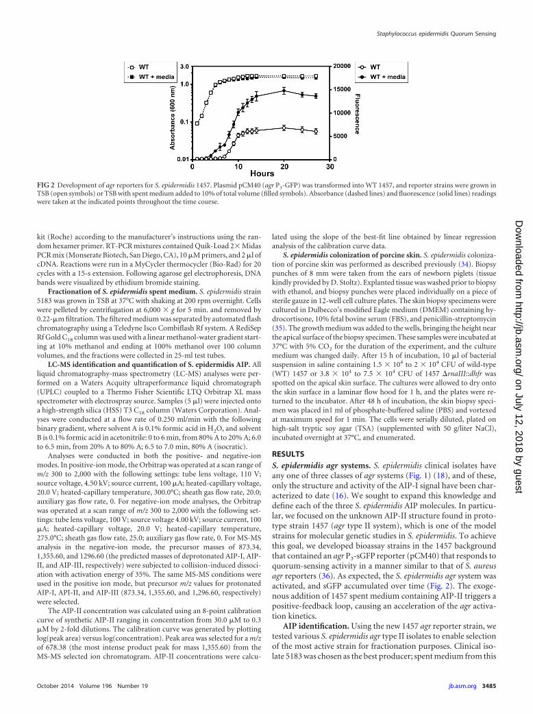
RESULTSS. epidermidis agr systems. S. epidermidis clinical isolates haveany one of three classes of agr systems (Fig. 1) (18), and of these,only the structure and activity of the AIP-I signal have been char-acterized to date (16). We sought to expand this knowledge anddefine each of the three S. epidermidis AIP molecules. In particu-lar, we focused on the unknown AIP-II structure found in proto-type strain 1457 (agr type II system), which is one of the modelstrains for molecular genetic studies in S. epidermidis. To achievethis goal, we developed bioassay strains in the 1457 backgroundthat contained an agr P3-sGFP reporter (pCM40) that responds toquorum-sensing activity in a manner similar to that of S. aureusagr reporters (36). As expected, the S. epidermidis agr system wasactivated, and sGFP accumulated over time (Fig. 2). The exoge-nous addition of 1457 spent medium containing AIP-II triggers apositive-feedback loop, causing an acceleration of the agr activa-tion kinetics.
AIP identification. Using the new 1457 agr reporter strain, wetested various S. epidermidis agr type II isolates to enable selectionof the most active strain for fractionation purposes. Clinical iso-late 5183 was chosen as the best producer; spent medium from this
FIG 2 Development of agr reporters for S. epidermidis 1457. Plasmid pCM40 (agr P3-GFP) was transformed into WT 1457, and reporter strains were grown inTSB (open symbols) or TSB with spent medium added to 10% of total volume (filled symbols). Absorbance (dashed lines) and fluorescence (solid lines) readingswere taken at the indicated points throughout the time course.
Staphylococcus epidermidis Quorum Sensing
October 2014 Volume 196 Number 19 jb.asm.org 3485
on July 12, 2018 by guesthttp://jb.asm
.org/D
ownloaded from
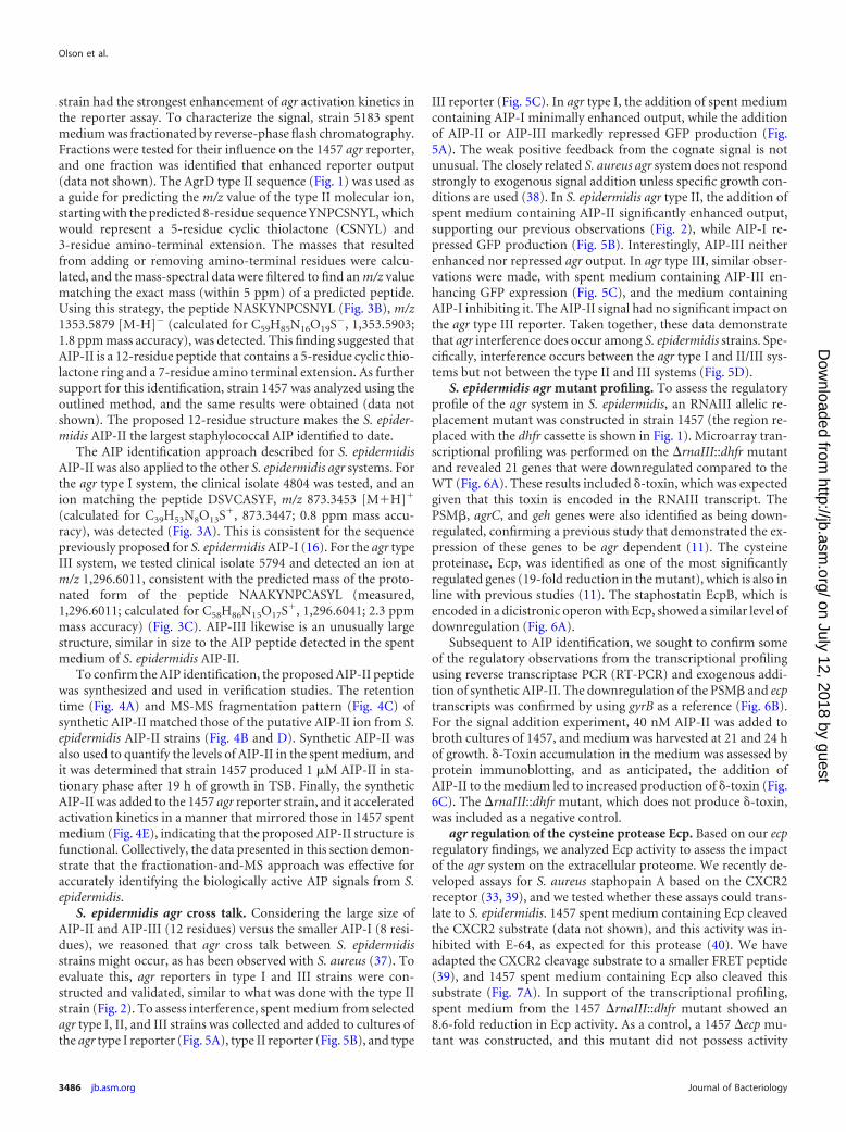
strain had the strongest enhancement of agr activation kinetics inthe reporter assay. To characterize the signal, strain 5183 spentmedium was fractionated by reverse-phase flash chromatography.Fractions were tested for their influence on the 1457 agr reporter,and one fraction was identified that enhanced reporter output(data not shown). The AgrD type II sequence (Fig. 1) was used asa guide for predicting the m/z value of the type II molecular ion,starting with the predicted 8-residue sequence YNPCSNYL, whichwould represent a 5-residue cyclic thiolactone (CSNYL) and3-residue amino-terminal extension. The masses that resultedfrom adding or removing amino-terminal residues were calcu-lated, and the mass-spectral data were filtered to find an m/z valuematching the exact mass (within 5 ppm) of a predicted peptide.Using this strategy, the peptide NASKYNPCSNYL (Fig. 3B), m/z1353.5879 [M-H] (calculated for C59H85N16O19S, 1,353.5903;1.8 ppm mass accuracy), was detected. This finding suggested thatAIP-II is a 12-residue peptide that contains a 5-residue cyclic thio-lactone ring and a 7-residue amino terminal extension. As furthersupport for this identification, strain 1457 was analyzed using theoutlined method, and the same results were obtained (data notshown). The proposed 12-residue structure makes the S. epider-midis AIP-II the largest staphylococcal AIP identified to date.
The AIP identification approach described for S. epidermidisAIP-II was also applied to the other S. epidermidis agr systems. Forthe agr type I system, the clinical isolate 4804 was tested, and anion matching the peptide DSVCASYF, m/z 873.3453 [M�H]�
(calculated for C39H53N8O13S�, 873.3447; 0.8 ppm mass accu-racy), was detected (Fig. 3A). This is consistent for the sequencepreviously proposed for S. epidermidis AIP-I (16). For the agr typeIII system, we tested clinical isolate 5794 and detected an ion atm/z 1,296.6011, consistent with the predicted mass of the proto-nated form of the peptide NAAKYNPCASYL (measured,1,296.6011; calculated for C58H86N15O17S�, 1,296.6041; 2.3 ppmmass accuracy) (Fig. 3C). AIP-III likewise is an unusually largestructure, similar in size to the AIP peptide detected in the spentmedium of S. epidermidis AIP-II.
To confirm the AIP identification, the proposed AIP-II peptidewas synthesized and used in verification studies. The retentiontime (Fig. 4A) and MS-MS fragmentation pattern (Fig. 4C) ofsynthetic AIP-II matched those of the putative AIP-II ion from S.epidermidis AIP-II strains (Fig. 4B and D). Synthetic AIP-II wasalso used to quantify the levels of AIP-II in the spent medium, andit was determined that strain 1457 produced 1 �M AIP-II in sta-tionary phase after 19 h of growth in TSB. Finally, the syntheticAIP-II was added to the 1457 agr reporter strain, and it acceleratedactivation kinetics in a manner that mirrored those in 1457 spentmedium (Fig. 4E), indicating that the proposed AIP-II structure isfunctional. Collectively, the data presented in this section demon-strate that the fractionation-and-MS approach was effective foraccurately identifying the biologically active AIP signals from S.epidermidis.
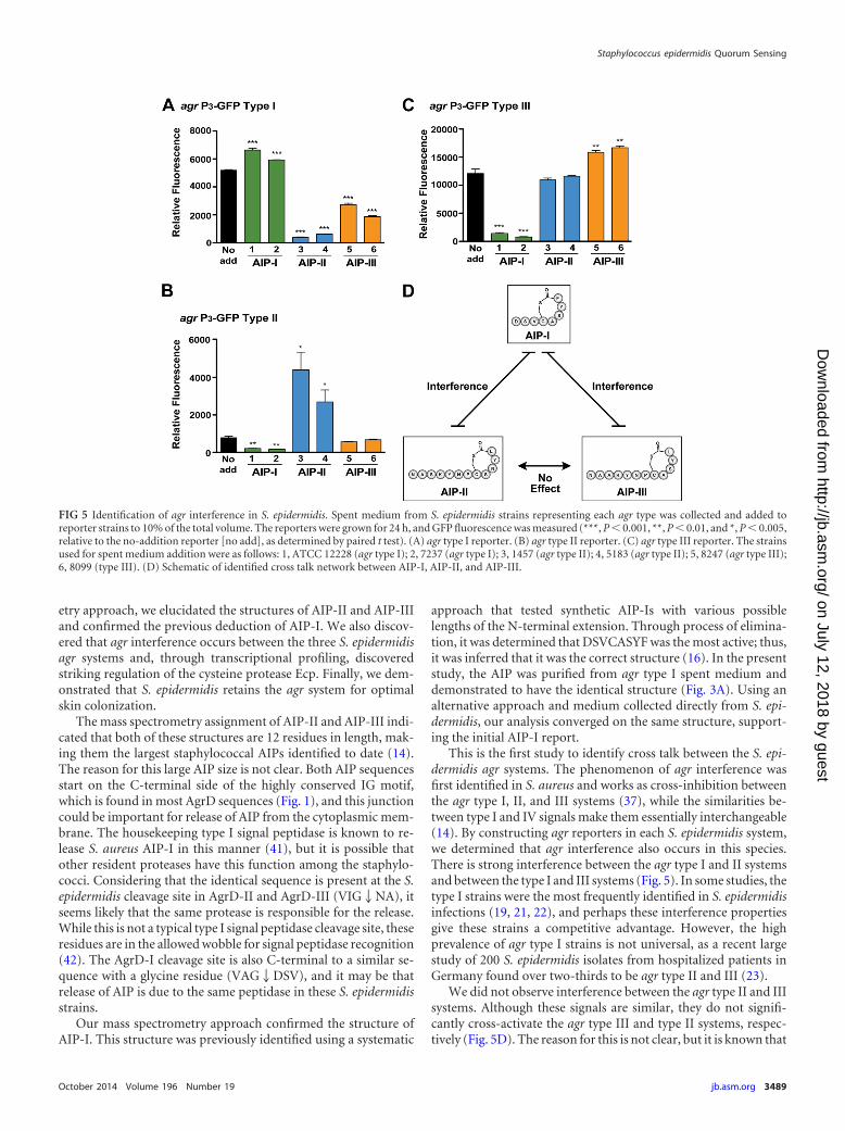
S. epidermidis agr cross talk. Considering the large size ofAIP-II and AIP-III (12 residues) versus the smaller AIP-I (8 resi-dues), we reasoned that agr cross talk between S. epidermidisstrains might occur, as has been observed with S. aureus (37). Toevaluate this, agr reporters in type I and III strains were con-structed and validated, similar to what was done with the type IIstrain (Fig. 2). To assess interference, spent medium from selectedagr type I, II, and III strains was collected and added to cultures ofthe agr type I reporter (Fig. 5A), type II reporter (Fig. 5B), and type
III reporter (Fig. 5C). In agr type I, the addition of spent mediumcontaining AIP-I minimally enhanced output, while the additionof AIP-II or AIP-III markedly repressed GFP production (Fig.5A). The weak positive feedback from the cognate signal is notunusual. The closely related S. aureus agr system does not respondstrongly to exogenous signal addition unless specific growth con-ditions are used (38). In S. epidermidis agr type II, the addition ofspent medium containing AIP-II significantly enhanced output,supporting our previous observations (Fig. 2), while AIP-I re-pressed GFP production (Fig. 5B). Interestingly, AIP-III neitherenhanced nor repressed agr output. In agr type III, similar obser-vations were made, with spent medium containing AIP-III en-hancing GFP expression (Fig. 5C), and the medium containingAIP-I inhibiting it. The AIP-II signal had no significant impact onthe agr type III reporter. Taken together, these data demonstratethat agr interference does occur among S. epidermidis strains. Spe-cifically, interference occurs between the agr type I and II/III sys-tems but not between the type II and III systems (Fig. 5D).
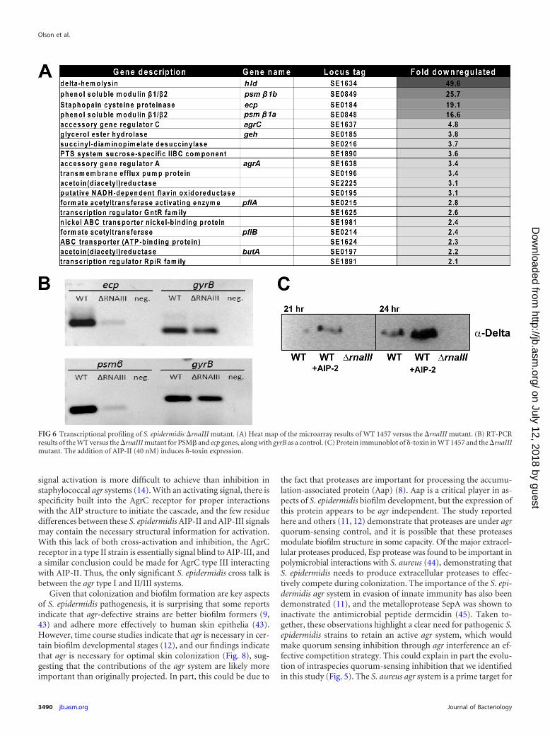
S. epidermidis agr mutant profiling. To assess the regulatoryprofile of the agr system in S. epidermidis, an RNAIII allelic re-placement mutant was constructed in strain 1457 (the region re-placed with the dhfr cassette is shown in Fig. 1). Microarray tran-scriptional profiling was performed on the �rnaIII::dhfr mutantand revealed 21 genes that were downregulated compared to theWT (Fig. 6A). These results included �-toxin, which was expectedgiven that this toxin is encoded in the RNAIII transcript. ThePSM�, agrC, and geh genes were also identified as being down-regulated, confirming a previous study that demonstrated the ex-pression of these genes to be agr dependent (11). The cysteineproteinase, Ecp, was identified as one of the most significantlyregulated genes (19-fold reduction in the mutant), which is also inline with previous studies (11). The staphostatin EcpB, which isencoded in a dicistronic operon with Ecp, showed a similar level ofdownregulation (Fig. 6A).
Subsequent to AIP identification, we sought to confirm someof the regulatory observations from the transcriptional profilingusing reverse transcriptase PCR (RT-PCR) and exogenous addi-tion of synthetic AIP-II. The downregulation of the PSM� and ecptranscripts was confirmed by using gyrB as a reference (Fig. 6B).For the signal addition experiment, 40 nM AIP-II was added tobroth cultures of 1457, and medium was harvested at 21 and 24 hof growth. �-Toxin accumulation in the medium was assessed byprotein immunoblotting, and as anticipated, the addition ofAIP-II to the medium led to increased production of �-toxin (Fig.6C). The �rnaIII::dhfr mutant, which does not produce �-toxin,was included as a negative control.
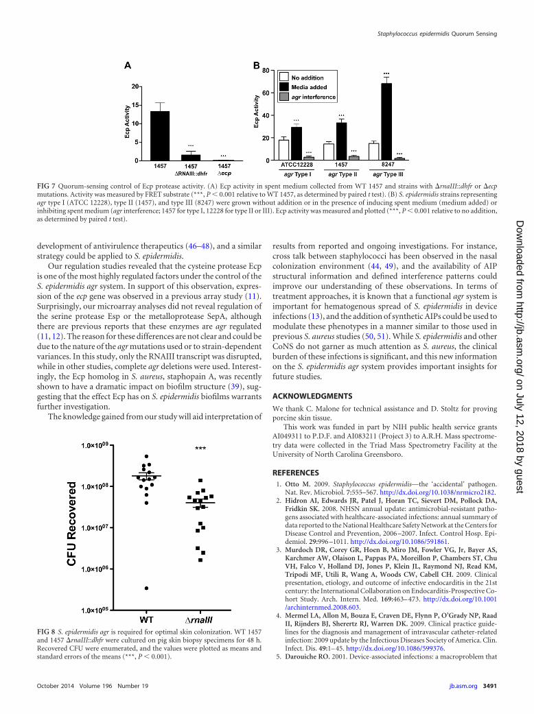
agr regulation of the cysteine protease Ecp. Based on our ecpregulatory findings, we analyzed Ecp activity to assess the impactof the agr system on the extracellular proteome. We recently de-veloped assays for S. aureus staphopain A based on the CXCR2receptor (33, 39), and we tested whether these assays could trans-late to S. epidermidis. 1457 spent medium containing Ecp cleavedthe CXCR2 substrate (data not shown), and this activity was in-hibited with E-64, as expected for this protease (40). We haveadapted the CXCR2 cleavage substrate to a smaller FRET peptide(39), and 1457 spent medium containing Ecp also cleaved thissubstrate (Fig. 7A). In support of the transcriptional profiling,spent medium from the 1457 �rnaIII::dhfr mutant showed an8.6-fold reduction in Ecp activity. As a control, a 1457 �ecp mu-tant was constructed, and this mutant did not possess activity
Olson et al.
3486 jb.asm.org Journal of Bacteriology
on July 12, 2018 by guesthttp://jb.asm
.org/D
ownloaded from
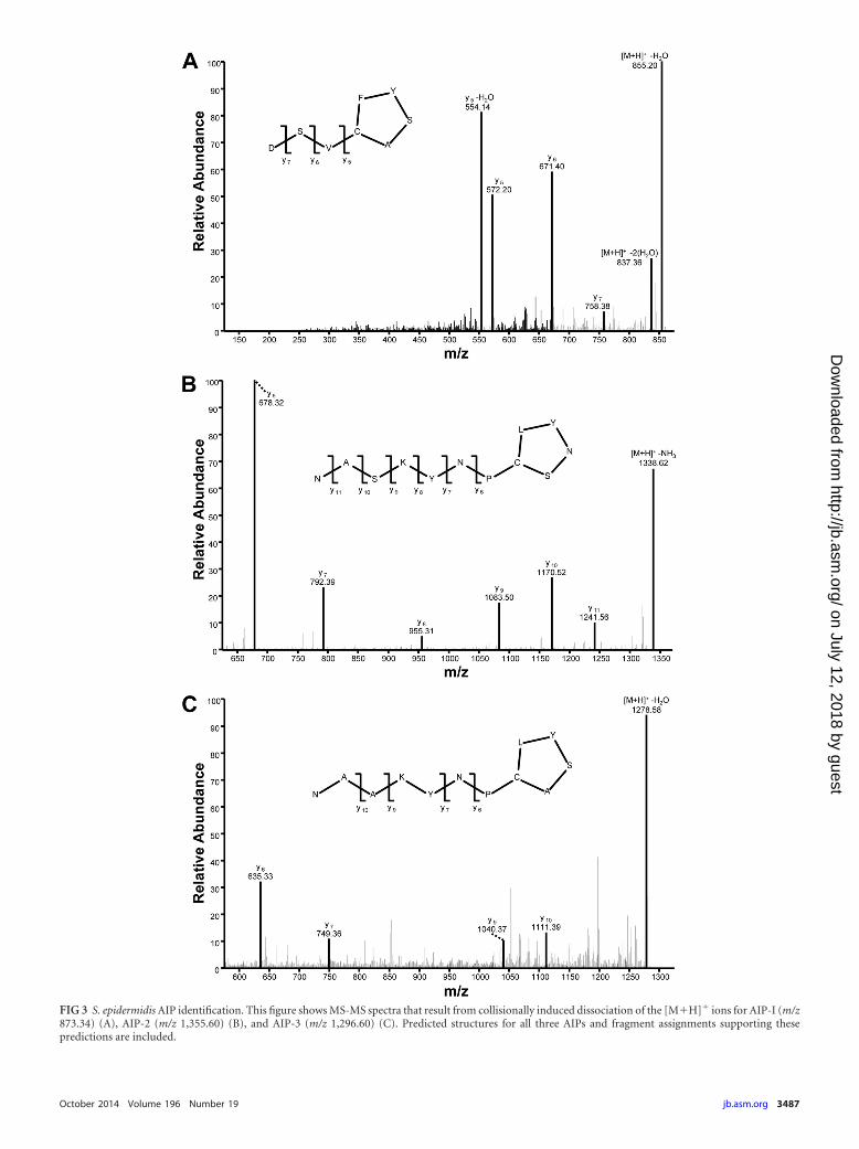
FIG 3 S. epidermidis AIP identification. This figure shows MS-MS spectra that result from collisionally induced dissociation of the [M�H]� ions for AIP-I (m/z873.34) (A), AIP-2 (m/z 1,355.60) (B), and AIP-3 (m/z 1,296.60) (C). Predicted structures for all three AIPs and fragment assignments supporting thesepredictions are included.
October 2014 Volume 196 Number 19 jb.asm.org 3487
on July 12, 2018 by guesthttp://jb.asm
.org/D
ownloaded from
against the FRET substrate (Fig. 7A), demonstrating that no otherproteases contributed to the activity measurements. To furtherassess the role of quorum-sensing regulation in Ecp regulation,spent-medium cross talk tests were performed using informationgleaned from the results outlined in Fig. 5. For each S. epidermidisagr system, spent medium from that agr type added back to thesame strain induced Ecp activity (Fig. 7B). In contrast, spent me-dium from a known interfering strain inhibited activity, resultingin significantly reduced Ecp activity. Taken together, these Ecpfindings support the agr transcriptional reporter (Fig. 5) and mi-croarray results (Fig. 6).
S. epidermidis colonizes pig skin in an agr-dependent man-ner. We hypothesized that S. epidermidis retains the agr systembecause it facilitates successful colonization of the host. Using a
previously developed skin colonization model (34), we comparedthe ability of WT 1457 and the �rnaIII::dhfr mutant to colonizefreshly isolated porcine skin for 2 days. At this time point, a sig-nificant defect in the ability of the �rnaIII::dhfr mutant to colonizewas observed (Fig. 8), indicating that S. epidermidis does requirethe agr system for optimal growth during skin colonization.
DISCUSSION
S. epidermidis is the most frequent cause of device-related infec-tions (6). One of the important regulators that control the pro-duction of S. epidermidis virulence factors is the agr quorum-sens-ing system. Despite the significance of agr, key molecular detailsabout this system have remained unknown, and in this work, weaddressed some of these open questions. Using a mass spectrom-
FIG 4 Confirmation of structure by comparison with synthetic AIP-II. Selected ion chromatograms for the fragment at m/z 678.33 obtained from collisionallyinduced dissociation of the [M�H]� ion at m/z 1,355.60 for both synthetic AIP-II (A) and spent medium from S. epidermidis strain 1457 (B). These chromato-grams show excellent agreement in retention time (RT) between the two ions. As further confirmation, panels C and D demonstrate that the MS-MS spectrumfor synthetic AIP-II (C) matches that of the putative AIP-II ion from S. epidermidis strain 1457. The MS-MS spectra were obtained by collisionally induceddissociation of a precursor ion at m/z 1,355.60 (the [M�H]� ion of AIP-II). (E) As a biological confirmation of the synthetic AIP-II, WT 1457 with the agr P3-GFPreporter was grown with increasing doses of AIP-II, and absorbance and fluorescence readings were taken after 24 h. Relative fluorescence was plotted at theindicated AIP-II concentrations.
Olson et al.
3488 jb.asm.org Journal of Bacteriology
on July 12, 2018 by guesthttp://jb.asm
.org/D
ownloaded from
etry approach, we elucidated the structures of AIP-II and AIP-IIIand confirmed the previous deduction of AIP-I. We also discov-ered that agr interference occurs between the three S. epidermidisagr systems and, through transcriptional profiling, discoveredstriking regulation of the cysteine protease Ecp. Finally, we dem-onstrated that S. epidermidis retains the agr system for optimalskin colonization.
The mass spectrometry assignment of AIP-II and AIP-III indi-cated that both of these structures are 12 residues in length, mak-ing them the largest staphylococcal AIPs identified to date (14).The reason for this large AIP size is not clear. Both AIP sequencesstart on the C-terminal side of the highly conserved IG motif,which is found in most AgrD sequences (Fig. 1), and this junctioncould be important for release of AIP from the cytoplasmic mem-brane. The housekeeping type I signal peptidase is known to re-lease S. aureus AIP-I in this manner (41), but it is possible thatother resident proteases have this function among the staphylo-cocci. Considering that the identical sequence is present at the S.epidermidis cleavage site in AgrD-II and AgrD-III (VIG2NA), itseems likely that the same protease is responsible for the release.While this is not a typical type I signal peptidase cleavage site, theseresidues are in the allowed wobble for signal peptidase recognition(42). The AgrD-I cleavage site is also C-terminal to a similar se-quence with a glycine residue (VAG2DSV), and it may be thatrelease of AIP is due to the same peptidase in these S. epidermidisstrains.
Our mass spectrometry approach confirmed the structure ofAIP-I. This structure was previously identified using a systematic
approach that tested synthetic AIP-Is with various possiblelengths of the N-terminal extension. Through process of elimina-tion, it was determined that DSVCASYF was the most active; thus,it was inferred that it was the correct structure (16). In the presentstudy, the AIP was purified from agr type I spent medium anddemonstrated to have the identical structure (Fig. 3A). Using analternative approach and medium collected directly from S. epi-dermidis, our analysis converged on the same structure, support-ing the initial AIP-I report.
This is the first study to identify cross talk between the S. epi-dermidis agr systems. The phenomenon of agr interference wasfirst identified in S. aureus and works as cross-inhibition betweenthe agr type I, II, and III systems (37), while the similarities be-tween type I and IV signals make them essentially interchangeable(14). By constructing agr reporters in each S. epidermidis system,we determined that agr interference also occurs in this species.There is strong interference between the agr type I and II systemsand between the type I and III systems (Fig. 5). In some studies, thetype I strains were the most frequently identified in S. epidermidisinfections (19, 21, 22), and perhaps these interference propertiesgive these strains a competitive advantage. However, the highprevalence of agr type I strains is not universal, as a recent largestudy of 200 S. epidermidis isolates from hospitalized patients inGermany found over two-thirds to be agr type II and III (23).
We did not observe interference between the agr type II and IIIsystems. Although these signals are similar, they do not signifi-cantly cross-activate the agr type III and type II systems, respec-tively (Fig. 5D). The reason for this is not clear, but it is known that
FIG 5 Identification of agr interference in S. epidermidis. Spent medium from S. epidermidis strains representing each agr type was collected and added toreporter strains to 10% of the total volume. The reporters were grown for 24 h, and GFP fluorescence was measured (***, P � 0.001, **, P � 0.01, and *, P � 0.005,relative to the no-addition reporter [no add], as determined by paired t test). (A) agr type I reporter. (B) agr type II reporter. (C) agr type III reporter. The strainsused for spent medium addition were as follows: 1, ATCC 12228 (agr type I); 2, 7237 (agr type I); 3, 1457 (agr type II); 4, 5183 (agr type II); 5, 8247 (agr type III);6, 8099 (type III). (D) Schematic of identified cross talk network between AIP-I, AIP-II, and AIP-III.
Staphylococcus epidermidis Quorum Sensing
October 2014 Volume 196 Number 19 jb.asm.org 3489
on July 12, 2018 by guesthttp://jb.asm
.org/D
ownloaded from
signal activation is more difficult to achieve than inhibition instaphylococcal agr systems (14). With an activating signal, there isspecificity built into the AgrC receptor for proper interactionswith the AIP structure to initiate the cascade, and the few residuedifferences between these S. epidermidis AIP-II and AIP-III signalsmay contain the necessary structural information for activation.With this lack of both cross-activation and inhibition, the AgrCreceptor in a type II strain is essentially signal blind to AIP-III, anda similar conclusion could be made for AgrC type III interactingwith AIP-II. Thus, the only significant S. epidermidis cross talk isbetween the agr type I and II/III systems.
Given that colonization and biofilm formation are key aspectsof S. epidermidis pathogenesis, it is surprising that some reportsindicate that agr-defective strains are better biofilm formers (9,43) and adhere more effectively to human skin epithelia (43).However, time course studies indicate that agr is necessary in cer-tain biofilm developmental stages (12), and our findings indicatethat agr is necessary for optimal skin colonization (Fig. 8), sug-gesting that the contributions of the agr system are likely moreimportant than originally projected. In part, this could be due to
the fact that proteases are important for processing the accumu-lation-associated protein (Aap) (8). Aap is a critical player in as-pects of S. epidermidis biofilm development, but the expression ofthis protein appears to be agr independent. The study reportedhere and others (11, 12) demonstrate that proteases are under agrquorum-sensing control, and it is possible that these proteasesmodulate biofilm structure in some capacity. Of the major extracel-lular proteases produced, Esp protease was found to be important inpolymicrobial interactions with S. aureus (44), demonstrating thatS. epidermidis needs to produce extracellular proteases to effec-tively compete during colonization. The importance of the S. epi-dermidis agr system in evasion of innate immunity has also beendemonstrated (11), and the metalloprotease SepA was shown toinactivate the antimicrobial peptide dermcidin (45). Taken to-gether, these observations highlight a clear need for pathogenic S.epidermidis strains to retain an active agr system, which wouldmake quorum sensing inhibition through agr interference an ef-fective competition strategy. This could explain in part the evolu-tion of intraspecies quorum-sensing inhibition that we identifiedin this study (Fig. 5). The S. aureus agr system is a prime target for
FIG 6 Transcriptional profiling of S. epidermidis �rnaIII mutant. (A) Heat map of the microarray results of WT 1457 versus the �rnaIII mutant. (B) RT-PCRresults of the WT versus the �rnaIII mutant for PSM� and ecp genes, along with gyrB as a control. (C) Protein immunoblot of �-toxin in WT 1457 and the �rnaIIImutant. The addition of AIP-II (40 nM) induces �-toxin expression.
Olson et al.
3490 jb.asm.org Journal of Bacteriology
on July 12, 2018 by guesthttp://jb.asm
.org/D
ownloaded from
development of antivirulence therapeutics (46–48), and a similarstrategy could be applied to S. epidermidis.
Our regulation studies revealed that the cysteine protease Ecpis one of the most highly regulated factors under the control of theS. epidermidis agr system. In support of this observation, expres-sion of the ecp gene was observed in a previous array study (11).Surprisingly, our microarray analyses did not reveal regulation ofthe serine protease Esp or the metalloprotease SepA, althoughthere are previous reports that these enzymes are agr regulated(11, 12). The reason for these differences are not clear and could bedue to the nature of the agr mutations used or to strain-dependentvariances. In this study, only the RNAIII transcript was disrupted,while in other studies, complete agr deletions were used. Interest-ingly, the Ecp homolog in S. aureus, staphopain A, was recentlyshown to have a dramatic impact on biofilm structure (39), sug-gesting that the effect Ecp has on S. epidermidis biofilms warrantsfurther investigation.
The knowledge gained from our study will aid interpretation of
results from reported and ongoing investigations. For instance,cross talk between staphylococci has been observed in the nasalcolonization environment (44, 49), and the availability of AIPstructural information and defined interference patterns couldimprove our understanding of these observations. In terms oftreatment approaches, it is known that a functional agr system isimportant for hematogenous spread of S. epidermidis in deviceinfections (13), and the addition of synthetic AIPs could be used tomodulate these phenotypes in a manner similar to those used inprevious S. aureus studies (50, 51). While S. epidermidis and otherCoNS do not garner as much attention as S. aureus, the clinicalburden of these infections is significant, and this new informationon the S. epidermidis agr system provides important insights forfuture studies.
ACKNOWLEDGMENTS
We thank C. Malone for technical assistance and D. Stoltz for provingporcine skin tissue.
This work was funded in part by NIH public health service grantsAI049311 to P.D.F. and AI083211 (Project 3) to A.R.H. Mass spectrome-try data were collected in the Triad Mass Spectrometry Facility at theUniversity of North Carolina Greensboro.
REFERENCES1. Otto M. 2009. Staphylococcus epidermidis—the ‘accidental’ pathogen.
Nat. Rev. Microbiol. 7:555–567. http://dx.doi.org/10.1038/nrmicro2182.2. Hidron AI, Edwards JR, Patel J, Horan TC, Sievert DM, Pollock DA,
Fridkin SK. 2008. NHSN annual update: antimicrobial-resistant patho-gens associated with healthcare-associated infections: annual summary ofdata reported to the National Healthcare Safety Network at the Centers forDisease Control and Prevention, 2006 –2007. Infect. Control Hosp. Epi-demiol. 29:996 –1011. http://dx.doi.org/10.1086/591861.
3. Murdoch DR, Corey GR, Hoen B, Miro JM, Fowler VG, Jr, Bayer AS,Karchmer AW, Olaison L, Pappas PA, Moreillon P, Chambers ST, ChuVH, Falco V, Holland DJ, Jones P, Klein JL, Raymond NJ, Read KM,Tripodi MF, Utili R, Wang A, Woods CW, Cabell CH. 2009. Clinicalpresentation, etiology, and outcome of infective endocarditis in the 21stcentury: the International Collaboration on Endocarditis-Prospective Co-hort Study. Arch. Intern. Med. 169:463– 473. http://dx.doi.org/10.1001/archinternmed.2008.603.
4. Mermel LA, Allon M, Bouza E, Craven DE, Flynn P, O’Grady NP, RaadII, Rijnders BJ, Sherertz RJ, Warren DK. 2009. Clinical practice guide-lines for the diagnosis and management of intravascular catheter-relatedinfection: 2009 update by the Infectious Diseases Society of America. Clin.Infect. Dis. 49:1– 45. http://dx.doi.org/10.1086/599376.
5. Darouiche RO. 2001. Device-associated infections: a macroproblem that
FIG 7 Quorum-sensing control of Ecp protease activity. (A) Ecp activity in spent medium collected from WT 1457 and strains with �rnaIII::dhfr or �ecpmutations. Activity was measured by FRET substrate (***, P � 0.001 relative to WT 1457, as determined by paired t test). (B) S. epidermidis strains representingagr type I (ATCC 12228), type II (1457), and type III (8247) were grown without addition or in the presence of inducing spent medium (medium added) orinhibiting spent medium (agr interference; 1457 for type I, 12228 for type II or III). Ecp activity was measured and plotted (***, P � 0.001 relative to no addition,as determined by paired t test).
FIG 8 S. epidermidis agr is required for optimal skin colonization. WT 1457and 1457 �rnaIII::dhfr were cultured on pig skin biopsy specimens for 48 h.Recovered CFU were enumerated, and the values were plotted as means andstandard errors of the means (***, P � 0.001).
Staphylococcus epidermidis Quorum Sensing
October 2014 Volume 196 Number 19 jb.asm.org 3491
on July 12, 2018 by guesthttp://jb.asm
.org/D
ownloaded from
starts with microadherence. Clin. Infect. Dis. 33:1567–1572. http://dx.doi.org/10.1086/323130.
6. Mack D, Davies AP, Harris LG, Jeeves R, Pascoe B, Knobloch JK, RohdeH, Wilkinson TS. 2013. Staphylococcus epidermidis in biomaterial-associated infections, p 25–56. In Moriarty F, Zaat SAJ, Busscher H (ed),Biomaterials associated infection: immunological aspects and antimicro-bial strategies. Springer, New York, NY.
7. Heilmann C, Schweitzer O, Gerke C, Vanittanakom N, Mack D, GotzF. 1996. Molecular basis of intercellular adhesion in the biofilm-formingStaphylococcus epidermidis. Mol. Microbiol. 20:1083–1091. http://dx.doi.org/10.1111/j.1365-2958.1996.tb02548.x.
8. Rohde H, Burdelski C, Bartscht K, Hussain M, Buck F, Horstkotte MA,Knobloch JK, Heilmann C, Herrmann M, Mack D. 2005. Induction ofStaphylococcus epidermidis biofilm formation via proteolytic processing ofthe accumulation-associated protein by staphylococcal and host pro-teases. Mol. Microbiol. 55:1883–1895. http://dx.doi.org/10.1111/j.1365-2958.2005.04515.x.
9. Vuong C, Gerke C, Somerville GA, Fischer ER, Otto M. 2003. Quorum-sensing control of biofilm factors in Staphylococcus epidermidis. J. Infect.Dis. 188:706 –718. http://dx.doi.org/10.1086/377239.
10. Vuong C, Gotz F, Otto M. 2000. Construction and characterization of anagr deletion mutant of Staphylococcus epidermidis. Infect. Immun. 68:1048 –1053. http://dx.doi.org/10.1128/IAI.68.3.1048-1053.2000.
11. Yao Y, Vuong C, Kocianova S, Villaruz AE, Lai Y, Sturdevant DE, OttoM. 2006. Characterization of the Staphylococcus epidermidis accessory-gene regulator response: quorum-sensing regulation of resistance to hu-man innate host defense. J. Infect. Dis. 193:841– 848. http://dx.doi.org/10.1086/500246.
12. Batzilla CF, Rachid S, Engelmann S, Hecker M, Hacker J, Ziebuhr W.2006. Impact of the accessory gene regulatory system (Agr) on extracellu-lar proteins, codY expression and amino acid metabolism in Staphylococ-cus epidermidis. Proteomics 6:3602–3613. http://dx.doi.org/10.1002/pmic.200500732.
13. Wang R, Khan BA, Cheung GY, Bach TH, Jameson-Lee M, Kong KF,Queck SY, Otto M. 2011. Staphylococcus epidermidis surfactant peptidespromote biofilm maturation and dissemination of biofilm-associated in-fection in mice. J. Clin. Invest. 121:238 –248. http://dx.doi.org/10.1172/JCI42520.
14. Thoendel M, Kavanaugh JS, Flack CE, Horswill AR. 2011. Peptidesignaling in the staphylococci. Chem. Rev. 111:117–151. http://dx.doi.org/10.1021/cr100370n.
15. Novick RP, Geisinger E. 2008. Quorum sensing in staphylococci. Annu.Rev. Genet. 42:541–564. http://dx.doi.org/10.1146/annurev.genet.42.110807.091640.
16. Otto M, Sussmuth R, Jung G, Gotz F. 1998. Structure of the pheromonepeptide of the Staphylococcus epidermidis agr system. FEBS Lett. 424:89 –94. http://dx.doi.org/10.1016/S0014-5793(98)00145-8.
17. Van Wamel WJ, van Rossum G, Verhoef J, Vandenbroucke-Grauls CM,Fluit AC. 1998. Cloning and characterization of an accessory gene regu-lator (agr)-like locus from Staphylococcus epidermidis. FEMS Microbiol.Lett. 163:1–9. http://dx.doi.org/10.1111/j.1574-6968.1998.tb13018.x.
18. Dufour P, Jarraud S, Vandenesch F, Greenland T, Novick RP, Bes M,Etienne J, Lina G. 2002. High genetic variability of the agr locus inStaphylococcus species. J. Bacteriol. 184:1180 –1186. http://dx.doi.org/10.1128/jb.184.4.1180-1186.2002.
19. Carmody AB, Otto M. 2004. Specificity grouping of the accessory generegulator quorum-sensing system of Staphylococcus epidermidis is linkedto infection. Arch. Microbiol. 181:250 –253. http://dx.doi.org/10.1007/s00203-003-0644-2.
20. Rosenthal ME, Dever LL, Moucha CS, Chavda KD, Otto M, KreiswirthBN. 2011. Molecular characterization of an early invasive Staphylococcusepidermidis prosthetic joint infection. Microb. Drug Resist. 17:345–350.http://dx.doi.org/10.1089/mdr.2010.0157.
21. Li M, Guan M, Jiang XF, Yuan FY, Xu M, Zhang WZ, Lu Y. 2004.Genetic polymorphism of the accessory gene regulator (agr) locus inStaphylococcus epidermidis and its association with pathogenicity. J. Med.Microbiol. 53:545–549. http://dx.doi.org/10.1099/jmm.0.05406-0.
22. Hellmark B, Soderquist B, Unemo M, Nilsdotter-Augustinsson A. 2013.Comparison of Staphylococcus epidermidis isolated from prosthetic jointinfections and commensal isolates in regard to antibiotic susceptibility,agr type, biofilm production, and epidemiology. Int. J. Med. Microbiol.303:32–39. http://dx.doi.org/10.1016/j.ijmm.2012.11.001.
23. Mertens A, Ghebremedhin B. 2013. Genetic determinants and biofilm
formation of clinical Staphylococcus epidermidis isolates from blood cul-tures and indwelling devises. Eur. J. Microbiol. Immunol. 3:111–119. http://dx.doi.org/10.1556/EuJMI.3.2013.2.4.
24. Nair D, Memmi G, Hernandez D, Bard J, Beaume M, Gill S, FrancoisP, Cheung AL. 2011. Whole-genome sequencing of Staphylococcus aureusstrain RN4220, a key laboratory strain used in virulence research, identi-fies mutations that affect not only virulence factors but also the fitnessof the strain. J. Bacteriol. 193:2332–2335. http://dx.doi.org/10.1128/JB.00027-11.
25. Maliszewski KL, Nuxoll AS. 2014. Use of electroporation and conjugativemobilization for genetic manipulation of Staphylococcus epidermidis.Methods Mol. Biol. 1106:125–134. http://dx.doi.org/10.1007/978-1-62703-736-5_11.
26. Olson ME, Horswill AR. 2014. Bacteriophage transduction in Staphylo-coccus epidermidis. Methods Mol. Biol. 1106:167–172. http://dx.doi.org/10.1007/978-1-62703-736-5_15.
27. Handke LD, Slater SR, Conlon KM, O’Donnell ST, Olson ME, BryantKA, Rupp ME, O’Gara JP, Fey PD. 2007. SigmaB and SarA indepen-dently regulate polysaccharide intercellular adhesin production in Staph-ylococcus epidermidis. Can. J. Microbiol. 53:82–91. http://dx.doi.org/10.1139/w06-108.
28. Projan SJ, Archer GL. 1989. Mobilization of the relaxable Staphylococcusaureus plasmid pC221 by the conjugative plasmid pGO1 involves threepC221 loci. J. Bacteriol. 171:1841–1845.
29. Mack D, Rohde H, Dobinsky S, Riedewald J, Nedelmann M, KnoblochJK, Elsner HA, Feucht HH. 2000. Identification of three essential regu-latory gene loci governing expression of Staphylococcus epidermidis poly-saccharide intercellular adhesin and biofilm formation. Infect. Immun.68:3799 –3807. http://dx.doi.org/10.1128/IAI.68.7.3799-3807.2000.
30. Bae T, Schneewind O. 2006. Allelic replacement in Staphylococcus aureuswith inducible counter-selection. Plasmid 55:58 – 63. http://dx.doi.org/10.1016/j.plasmid.2005.05.005.
31. Beenken KE, Dunman PM, McAleese F, Macapagal D, Murphy E,Projan SJ, Blevins JS, Smeltzer MS. 2004. Global gene expression inStaphylococcus aureus biofilms. J. Bacteriol. 186:4665– 4684. http://dx.doi.org/10.1128/JB.186.14.4665-4684.2004.
32. Kiedrowski MR, Kavanaugh JS, Malone CL, Mootz JM, Voyich JM,Smeltzer MS, Bayles KW, Horswill AR. 2011. Nuclease modulates bio-film formation in community-associated methicillin-resistant Staphylo-coccus aureus. PLoS One 6:e26714. http://dx.doi.org/10.1371/journal.pone.0026714.
33. Laarman AJ, Mijnheer G, Mootz JM, van Rooijen WJ, Ruyken M, MaloneCL, Heezius EC, Ward R, Milligan G, van Strijp JA, de Haas CJ, HorswillAR, van Kessel KP, Rooijakkers SH. 2012. Staphylococcus aureus staph-opain A inhibits CXCR2-dependent neutrophil activation and che-motaxis. EMBO J. 31:3607–3619. http://dx.doi.org/10.1038/emboj.2012.212.
34. Ohnemus U, Kohrmeyer K, Houdek P, Rohde H, Wladykowski E, VidalS, Horstkotte MA, Aepfelbacher M, Kirschner N, Behne MJ, Moll I,Brandner JM. 2008. Regulation of epidermal tight-junctions (TJ) duringinfection with exfoliative toxin-negative Staphylococcus strains. J. Investig.Dermatol. 128:906 –916. http://dx.doi.org/10.1038/sj.jid.5701070.
35. Moll I, Houdek P, Schmidt H, Moll R. 1998. Characterization of epi-dermal wound healing in a human skin organ culture model: accelerationby transplanted keratinocytes. J. Investig. Dermatol. 111:251–258. http://dx.doi.org/10.1046/j.1523-1747.1998.00265.x.
36. Malone CL, Boles BR, Lauderdale KJ, Thoendel M, Kavanaugh JS,Horswill AR. 2009. Fluorescent reporters for Staphylococcus aureus. J.Microbiol. Methods 77:251–260. http://dx.doi.org/10.1016/j.mimet.2009.02.011.
37. Ji G, Beavis R, Novick RP. 1997. Bacterial interference caused by auto-inducing peptide variants. Science 276:2027–2030. http://dx.doi.org/10.1126/science.276.5321.2027.
38. Malone CL, Boles BR, Horswill AR. 2007. Biosynthesis of Staphylococcusaureus autoinducing peptides by using the synechocystis DnaB mini-intein. Appl. Environ. Microbiol. 73:6036 – 6044. http://dx.doi.org/10.1128/AEM.00912-07.
39. Mootz JM, Malone CL, Shaw LN, Horswill AR. 2013. Staphopainsmodulate Staphylococcus aureus biofilm integrity. Infect. Immun. 81:3227–3238. http://dx.doi.org/10.1128/IAI.00377-13.
40. Dubin G, Chmiel D, Mak P, Rakwalska M, Rzychon M, Dubin A. 2001.Molecular cloning and biochemical characterisation of proteases fromStaphylococcus epidermidis. Biol. Chem. 382:1575–1582.
Olson et al.
3492 jb.asm.org Journal of Bacteriology
on July 12, 2018 by guesthttp://jb.asm
.org/D
ownloaded from
41. Kavanaugh JS, Thoendel M, Horswill AR. 2007. A role for type I signalpeptidase in Staphylococcus aureus quorum sensing. Mol. Microbiol. 65:780 –798. http://dx.doi.org/10.1111/j.1365-2958.2007.05830.x.
42. Paetzel M, Karla A, Strynadka NC, Dalbey RE. 2002. Signal peptidases.Chem. Rev. 102:4549 – 4580. http://dx.doi.org/10.1021/cr010166y.
43. Vuong C, Kocianova S, Yao Y, Carmody AB, Otto M. 2004. Increasedcolonization of indwelling medical devices by quorum-sensing mutants ofStaphylococcus epidermidis in vivo. J. Infect. Dis. 190:1498 –1505. http://dx.doi.org/10.1086/424487.
44. Iwase T, Uehara Y, Shinji H, Tajima A, Seo H, Takada K, Agata T,Mizunoe Y. 2010. Staphylococcus epidermidis Esp inhibits Staphylococcusaureus biofilm formation and nasal colonization. Nature 465:346 –349.http://dx.doi.org/10.1038/nature09074.
45. Lai Y, Villaruz AE, Li M, Cha DJ, Sturdevant DE, Otto M. 2007. Thehuman anionic antimicrobial peptide dermcidin induces proteolytic de-fence mechanisms in staphylococci. Mol. Microbiol. 63:497–506. http://dx.doi.org/10.1111/j.1365-2958.2006.05540.x.
46. Cech NB, Horswill AR. 2013. Small-molecule quorum quenchers toprevent Staphylococcus aureus infection. Future Microbiol. 8:1511–1514.http://dx.doi.org/10.2217/fmb.13.134.
47. Gordon CP, Williams P, Chan WC. 2013. Attenuating Staphylococcusaureus virulence gene regulation: a medicinal chemistry perspective. J.Med. Chem. 56:1389 –1404. http://dx.doi.org/10.1021/jm3014635.
48. Gray B, Hall P, Gresham H. 2013. Targeting agr- and agr-like quorumsensing systems for development of common therapeutics to treat multi-ple gram-positive bacterial infections. Sensors (Basel) 13:5130 –5166.http://dx.doi.org/10.3390/s130405130.
49. Lina G, Boutite F, Tristan A, Bes M, Etienne J, Vandenesch F. 2003.Bacterial competition for human nasal cavity colonization: role of Staph-ylococcal agr alleles. Appl. Environ. Microbiol. 69:18 –23. http://dx.doi.org/10.1128/AEM.69.1.18-23.2003.
50. Lauderdale KJ, Malone CL, Boles BR, Morcuende J, Horswill AR. 2010.Biofilm dispersal of community-associated methicillin-resistant Staphylo-coccus aureus on orthopedic implant material. J. Orthop. Res. 28:55– 61.http://dx.doi.org/10.1002/jor.20943.
51. Boles BR, Horswill AR. 2008. agr-mediated dispersal of Staphylococcus au-reus biofilms. PLoS Pathog. 4:e1000052. http://dx.doi.org/10.1371/journal.ppat.1000052.
52. Boles BR, Thoendel M, Roth AJ, Horswill AR. 2010. Identification ofgenes involved in polysaccharide-independent Staphylococcus aureus bio-film formation. PLoS One 5:e10146. http://dx.doi.org/10.1371/journal.pone.0010146.
53. Zhang YQ, Ren SX, Li HL, Wang YX, Fu G, Yang J, Qin ZQ, Miao YG,Wang WY, Chen RS, Shen Y, Chen Z, Yuan ZH, Zhao GP, Qu D,Danchin A, Wen YM. 2003. Genome-based analysis of virulence genes ina non-biofilm-forming Staphylococcus epidermidis strain (ATCC 12228).Mol. Microbiol. 49:1577–1593. http://dx.doi.org/10.1046/j.1365-2958.2003.03671.x.
54. Mack D, Siemssen N, Laufs R. 1992. Parallel induction by glucose ofadherence and a polysaccharide antigen specific for plastic-adherentStaphylococcus epidermidis: evidence for functional relation to intercellu-lar adhesion. Infect. Immun. 60:2048 –2057.
55. Gill SR, Fouts DE, Archer GL, Mongodin EF, Deboy RT, Ravel J, PaulsenIT, Kolonay JF, Brinkac L, Beanan M, Dodson RJ, Daugherty SC, MadupuR, Angiuoli SV, Durkin AS, Haft DH, Vamathevan J, Khouri H, UtterbackT, Lee C, Dimitrov G, Jiang L, Qin H, Weidman J, Tran K, Kang K, HanceIR, Nelson KE, Fraser CM. 2005. Insights on evolution of virulence andresistance from the complete genome analysis of an early methicillin-resistantStaphylococcus aureus strain and a biofilm-producing methicillin-resistantStaphylococcus epidermidis strain. J. Bacteriol. 187:2426 –2438. http://dx.doi.org/10.1128/JB.187.7.2426-2438.2005.
Staphylococcus epidermidis Quorum Sensing
October 2014 Volume 196 Number 19 jb.asm.org 3493
on July 12, 2018 by guesthttp://jb.asm
.org/D
ownloaded from