Embed Size (px)
Citation preview

Staphylococcus aureus secretes a unique class ofneutrophil serine protease inhibitorsDaphne A. C. Stapelsa, Kasra X. Ramyarb, Markus Bischoffc, Maren von Köckritz-Blickweded, Fin J. Mildera,Maartje Ruykena, Janina Eisenbeisc, William J. McWhortere, Mathias Herrmannc, Kok P. M. van Kessela,Brian V. Geisbrechtb,1, and Suzan H. M. Rooijakkersa,1,2
aMedical Microbiology, University Medical Center Utrecht, 3584 CX, Utrecht, The Netherlands; bDepartment of Biochemistry and Molecular Biophysics,Kansas State University, Manhattan, KS 66506; cInstitute of Medical Microbiology and Hygiene, Saarland University Faculty of Medicine and Medical Center,D-66421 Homburg/Saar, Germany; dDepartment of Physiological Chemistry, University of Veterinary Medicine, D-30559 Hannover, Germany; and eSchoolof Biological Sciences, University of Missouri–Kansas City, Kansas City, MO 64110
Edited by Richard P. Novick, New York University School of Medicine, New York, NY, and approved July 31, 2014 (received for review April 28, 2014)
Neutrophils are indispensable for clearing infections with theprominent human pathogen Staphylococcus aureus. Here, we re-port that S. aureus secretes a family of proteins that potentlyinhibits the activity of neutrophil serine proteases (NSPs): neutro-phil elastase (NE), proteinase 3, and cathepsin G. The NSPs, but notrelated serine proteases, are specifically blocked by the extracel-lular adherence protein (Eap) and the functionally orphan Eaphomologs EapH1 and EapH2, with inhibitory-constant values inthe low-nanomolar range. Eap proteins are together essentialfor NSP inhibition by S. aureus in vitro and promote staphylococcalinfection in vivo. The crystal structure of the EapH1/NE complexshowed that Eap molecules constitute a unique class of noncova-lent protease inhibitors that occlude the catalytic cleft of NSPs.These findings increase our insights into the complex pathogene-sis of S. aureus infections and create opportunities to design noveltreatment strategies for inflammatory conditions related to exces-sive NSP activity.
immune evasion | bacteria | phagocytes
Infections with the human pathogen Staphylococcus aureusconstitute a major risk to human health. Although this bacte-
rium harmlessly colonizes more than 30% of the population viathe nose or skin, it causes severe morbidity and mortality uponinvasion of deeper tissues (1). To avert these serious infections,neutrophils play an indispensable role (2). Neutrophil serineproteases (NSPs), including neutrophil elastase (NE), proteinase3 (PR3), and cathepsin G (CG), are important for various neu-trophil functions. Active NSPs are stored within the azurophilicgranules (3), but upon neutrophil activation, they either enterthe nucleus to regulate extracellular trap (NET) formation (4) orthey are released into the extracellular milieu to kill certainbacteria (5), cleave bacterial virulence factors (5, 6), or regulateimmune responses by cleaving chemokines and receptors (7).Recently, a fourth neutrophil serine protease, denoted NSP4,was identified (8).Given the central role of NSPs in neutrophil function, we
wondered whether S. aureus had evolved mechanisms to copewith NSPs. In this study, we discover that S. aureus secretesa family of proteins that specifically and potently block NSPs:extracellular adherence protein (Eap) and the hitherto func-tional orphans Eap-homologue (EapH) 1 and 2. Structuralstudies presented here show that Eap molecules represent aunique class of noncovalent NSP inhibitors that is distinct fromthe well-known chelonianin class of inhibitors. These mecha-nistic insights can initiate development of novel, broad-rangeNSP inhibitors to be used in various inflammatory conditions.Furthermore, these insights increase our understanding of thepathogenicity of S. aureus and underline the exceptional capa-bility of this pathogen to adapt to its host by modulating theimmune response.
ResultsExtracellular Adherence Proteins of S. aureus Inhibit NE. To in-vestigate whether S. aureus secretes inhibitors of NSPs, we in-cubated NE with concentrated culture supernatants of differentS. aureus strains and quantified residual NE activity toward afluorescent peptide substrate. Indeed, we found that NE wasinhibited by supernatants of all tested S. aureus strains (Fig. 1A).Fractionation of the supernatant of S. aureus Newman by ion-exchange and size-exclusion chromatography yielded two proteinbands that corresponded with the NE inhibitory activity. Thesebands were identified by mass spectrometry as Eap and immu-nodominant surface antigen B (IsaB) (Fig. 1B). Further analysisrevealed that Eap is the NE inhibitor, because NE activity wasnot affected by the presence of recombinant IsaB, but fullyblocked by the presence of recombinant Eap (Fig. 1C). Eap isa 50- to 70-kDa protein that consists of multiple (most often fouror five) repetitive EAP domains (11 kDa). The short linkersbetween EAP domains are susceptible to proteolysis (9), whichlikely explains why the band identified as Eap was only approx-imately 25 kDa (Fig. 1B).
Significance
Neutrophils are among the first immune cells to migrate to thesite of infection and clear invading bacteria. They store largeamounts of neutrophil serine proteases (NSPs) that play keyroles in immune defense. Unfortunately, NSPs also contributeto tissue destruction in a variety of inflammatory disorders. Inthis study we discover that the pathogenic bacterium Staphy-lococcus aureus secretes a family of highly potent and specificNSP inhibitors that promote the pathogenicity of this bacte-rium in vivo. From crystallography experiments, we concludethat these proteins constitute a unique class of NSP inhibitors,which can be used to design novel treatment strategies againstexcessive NSP activity. Furthermore, this study significantly in-creases our understanding of the complex nature of S. aureusinfections.
Author contributions: D.A.C.S., M.B., M.v.K.-B., F.J.M., M.H., K.P.M.v.K., B.V.G., and S.H.M.R.designed research; D.A.C.S., K.X.R., M.B., M.R., J.E., W.J.M., and B.V.G. performed research;D.A.C.S., M.B., and B.V.G. analyzed data; D.A.C.S., B.V.G., and S.H.M.R. wrote the paper;M.v.K.-B., F.J.M., M.H., and K.P.M.v.K. interpreted results; and B.V.G. and S.H.M.R. super-vised the project.
The authors declare no conflict of interest.
This article is a PNAS Direct Submission.
Freely available online through the PNAS open access option.
Data deposition: The atomic coordinates have been deposited in the Protein Data Bank,www.pdb.org (PDB ID code 4NZL).1B.V.G. and S.H.M.R. contributed equally to this work.2To whom correspondence should be addressed. Email: [email protected].
This article contains supporting information online at www.pnas.org/lookup/suppl/doi:10.1073/pnas.1407616111/-/DCSupplemental.
www.pnas.org/cgi/doi/10.1073/pnas.1407616111 PNAS | September 9, 2014 | vol. 111 | no. 36 | 13187–13192
MICRO
BIOLO
GY

Eap has previously been reported to mediate bacterial agglu-tination, tissue adherence, and to block neutrophil migration.Although all of these functions require multiple EAP domainslinked in succession (10), we found that NE inhibition is mediatedby individual EAP domains (Fig. S1). Each EAP domain is char-acterized by a beta-grasp fold wherein an alpha-helix is positioneddiagonally across a five-stranded, mixed beta-sheet (11). This fold
is also found in the two S. aureus proteins that are homologous toEap but do not share the above described functions: EapH1(12 kDa) and EapH2 (13 kDa) (11). Likewise, we found thatEapH1 and EapH2 also inhibit NE (Fig. 1C). NE inhibition isspecific for the Eap protein family, because all other tested pro-teins of S. aureus could not inhibit NE (Fig. 1C), including mol-ecules characterized by a similar beta-grasp–type fold as the Eapproteins (Fig. 1C, gray bars) (11, 12).
Eap Proteins Specifically Inhibit Neutrophil Serine Proteases. NEbelongs to the chymotrypsin family of serine proteases, and itsamino acid sequence is most similar to that of PR3 and CG(55% and 37% amino acid identity, respectively). More distantlyrelated chymotrypsin-like proteases include plasmin, plasmakallikrein, and thrombin. Of these six proteases, only the threeNSPs appeared to be dose-dependently inhibited by the Eapproteins (Fig. 2 A–F).Next we determined the inhibitory constant (Ki) values of each
Eap protein versus the three NSPs and also measured the Kivalues of the endogenous human NE/CG inhibitor SLPI (secretedleukocyte protease inhibitor) as a control. The Ki values observedfor all Eap/NSP combinations were in the low-nanomolar range,which is consistent with a very potent inhibition (Fig. 2G). Theyare also within the same order of magnitude as the Ki of SLPI forboth NE and CG. Because previous work has reported a lower Kifor SLPI/NE and SLPI/CG (0.3 nM and 10 nM, respectively) (13),our particular assay system may have even underestimated theinhibitory capacity of the Eap proteins. Importantly, the experi-mentally determined Ki values are all lower than the endogenousexpression levels of the Eap proteins by S. aureus in culture (∼10μg/mL or 200 nM) (14), indicating that Eap inhibition of NSPs isphysiologically relevant.
Eap Proteins Are Essential for NSP Inhibition and Promote StaphylococcalInfection.The genes for the Eap proteins lie interspersed throughoutthe genome, and at least two of three are present in all sequencedS. aureus strains. The eap gene is located upstream, and thereforeoutside, of the beta-hemolysin–converting prophage (phiNM3) thatcontains other immune-evasion proteins like staphylococcal com-plement inhibitor (SCIN) (scn) and chemotaxis inhibitory proteinof S. aureus (CHIPS) (chp) (15, 16) (Fig. 3A). Neither eapH1 noreapH2 lie in close proximity of phage-associated genes. Using se-quential gene deletions by homologous recombination, we con-structed a panel of three isogenic eap mutants in S. aureus strainNewman: Δeap, ΔeapΔH1, and ΔeapΔH1ΔH2 (eap-triple mutant).As a control, the eap-triple mutant was complemented with theindividual genes integrated into their original genomic location(ΔeapΔH1ΔH2 compl.). All isogenic strains showed comparablegrowth in vitro. When incubated with the individual NSPs, sta-tionary-phase supernatant of the WT strain could fully inhibit allthree proteases, but supernatant of the eap-triple mutant had al-most entirely lost this capacity (Fig. 3B). The other mutants showedthat Eap is essential for NE and CG inhibition, but that all Eapproteins together are required for inhibition of PR3. Supernatant ofthe complemented strain showed restored NSP inhibition to levelsequivalent to WT supernatant (Fig. 3B). Although S. aureus wasfound to be resistant to direct killing by NE and CG in vitro (17,18), we examined whether the absence of eap genes might makeS. aureus more prone to direct killing by neutrophils in vitro.Although there was a tendency toward better killing of theeap-triple mutant compared with the WT strain, this difference wasnot significant (Fig. S2).To study the role of the Eap proteins in vivo, we compared the
pathogenicity of the eap-mutant strains in a murine, liver-abscessmodel (19), because the bacterial burden in the liver is known to bea reliable indicator of staphylococcal virulence (20, 21). First, weconfirmed that the purified Eap proteins also block murine NSPsin vitro (Fig. 3C). Then, we injected 1 × 10⁷ bacteria retro-orbitally
Fig. 1. Extracellular adherence protein (Eap) family of S. aureus inhibits NEactivity. (A) Residual activity of 60 nM NE upon incubation with culturesupernatants of multiple S. aureus strains. (B) Silver staining analysis of thefractions after gel filtration (Left). The boxes mark the protein bands ana-lyzed by mass spectrometry and the numbers indicate the sizes of referenceproteins (kilodaltons). The identified peptides are depicted next to the gel(Right). (C) Residual activity of 60 nM NE upon incubation with 100 nM ofthe mass-spectrometry hits Eap and IsaB (hatched bars) and other secretedproteins of S. aureus. Proteins containing a similar beta-grasp-type fold asthe Eap proteins are depicted in gray, and with an unrelated structure inblack. CHIPS, chemotaxis inhibitory protein of S. aureus; Ecb, extracellularcomplement binding protein; Efb, extracellular fibrinogen binding protein;EsxA, ESAT-6 secretion system extracellular protein A; FLIPr, formyl peptidereceptor-like-1 inhibitory protein; SCIN, staphylococcal complement inhibitor;SEA, staphylococcal enterotoxin A; SNase, S. aureus nuclease; SSLs, staphylo-coccal superantigen-like proteins; TSST-1, toxic shock syndrome toxin-1. Dataare representative of two independent experiments (A and B) or represent themean (± SD) of three independent experiments (C). See also Fig. S1.
13188 | www.pnas.org/cgi/doi/10.1073/pnas.1407616111 Stapels et al.
and determined the bacterial loads in the liver tissue after 4 d (Fig.3D). The bacterial loads in the WT and Δeap-infected animals didnot differ significantly. However, the bacterial load in the eap-triplemutant-infected mice was significantly lower than in WT-infectedanimals, strongly suggesting that all Eap proteins together promoteS. aureus virulence in vivo.
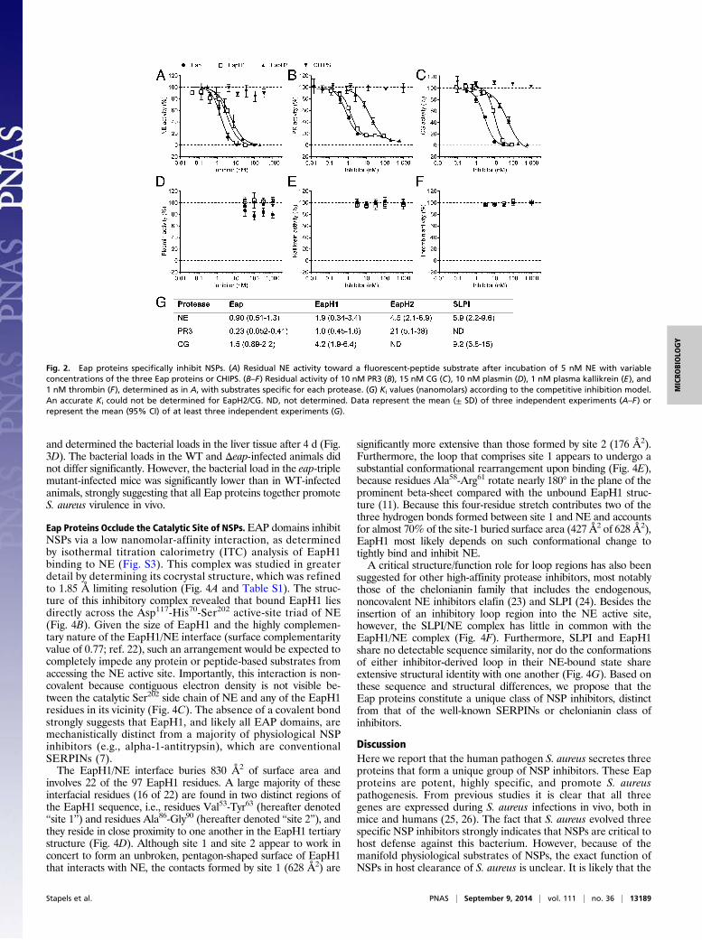
Eap Proteins Occlude the Catalytic Site of NSPs.EAP domains inhibitNSPs via a low nanomolar-affinity interaction, as determinedby isothermal titration calorimetry (ITC) analysis of EapH1binding to NE (Fig. S3). This complex was studied in greaterdetail by determining its cocrystal structure, which was refinedto 1.85 Å limiting resolution (Fig. 4A and Table S1). The struc-ture of this inhibitory complex revealed that bound EapH1 liesdirectly across the Asp117-His70-Ser202 active-site triad of NE(Fig. 4B). Given the size of EapH1 and the highly complemen-tary nature of the EapH1/NE interface (surface complementarityvalue of 0.77; ref. 22), such an arrangement would be expected tocompletely impede any protein or peptide-based substrates fromaccessing the NE active site. Importantly, this interaction is non-covalent because contiguous electron density is not visible be-tween the catalytic Ser202 side chain of NE and any of the EapH1residues in its vicinity (Fig. 4C). The absence of a covalent bondstrongly suggests that EapH1, and likely all EAP domains, aremechanistically distinct from a majority of physiological NSPinhibitors (e.g., alpha-1-antitrypsin), which are conventionalSERPINs (7).The EapH1/NE interface buries 830 Å2 of surface area and
involves 22 of the 97 EapH1 residues. A large majority of theseinterfacial residues (16 of 22) are found in two distinct regions ofthe EapH1 sequence, i.e., residues Val53-Tyr63 (hereafter denoted“site 1”) and residues Ala86-Gly90 (hereafter denoted “site 2”), andthey reside in close proximity to one another in the EapH1 tertiarystructure (Fig. 4D). Although site 1 and site 2 appear to work inconcert to form an unbroken, pentagon-shaped surface of EapH1that interacts with NE, the contacts formed by site 1 (628 Å2) are
significantly more extensive than those formed by site 2 (176 Å2).Furthermore, the loop that comprises site 1 appears to undergo asubstantial conformational rearrangement upon binding (Fig. 4E),because residues Ala58-Arg61 rotate nearly 180° in the plane of theprominent beta-sheet compared with the unbound EapH1 struc-ture (11). Because this four-residue stretch contributes two of thethree hydrogen bonds formed between site 1 and NE and accountsfor almost 70% of the site-1 buried surface area (427 Å2 of 628 Å2),EapH1 most likely depends on such conformational change totightly bind and inhibit NE.A critical structure/function role for loop regions has also been
suggested for other high-affinity protease inhibitors, most notablythose of the chelonianin family that includes the endogenous,noncovalent NE inhibitors elafin (23) and SLPI (24). Besides theinsertion of an inhibitory loop region into the NE active site,however, the SLPI/NE complex has little in common with theEapH1/NE complex (Fig. 4F). Furthermore, SLPI and EapH1share no detectable sequence similarity, nor do the conformationsof either inhibitor-derived loop in their NE-bound state shareextensive structural identity with one another (Fig. 4G). Based onthese sequence and structural differences, we propose that theEap proteins constitute a unique class of NSP inhibitors, distinctfrom that of the well-known SERPINs or chelonianin class ofinhibitors.
DiscussionHere we report that the human pathogen S. aureus secretes threeproteins that form a unique group of NSP inhibitors. These Eapproteins are potent, highly specific, and promote S. aureuspathogenesis. From previous studies it is clear that all threegenes are expressed during S. aureus infections in vivo, both inmice and humans (25, 26). The fact that S. aureus evolved threespecific NSP inhibitors strongly indicates that NSPs are critical tohost defense against this bacterium. However, because of themanifold physiological substrates of NSPs, the exact function ofNSPs in host clearance of S. aureus is unclear. It is likely that the
Fig. 2. Eap proteins specifically inhibit NSPs. (A) Residual NE activity toward a fluorescent-peptide substrate after incubation of 5 nM NE with variableconcentrations of the three Eap proteins or CHIPS. (B–F) Residual activity of 10 nM PR3 (B), 15 nM CG (C), 10 nM plasmin (D), 1 nM plasma kallikrein (E), and1 nM thrombin (F), determined as in A, with substrates specific for each protease. (G) Ki values (nanomolars) according to the competitive inhibition model.An accurate Ki could not be determined for EapH2/CG. ND, not determined. Data represent the mean (± SD) of three independent experiments (A–F) orrepresent the mean (95% CI) of at least three independent experiments (G).
Stapels et al. PNAS | September 9, 2014 | vol. 111 | no. 36 | 13189
MICRO
BIOLO
GY

interpretation of previous animal models trying to address thisquestion (5, 18, 27) has been confounded by the endogenousproduction of Eap proteins. In the future, these conceptuallimitations can be overcome by using the bacterial deletionmutants that are presented here.A key teleological issue remains as to why S. aureus inhibits
NSPs by three distinct proteins. We speculate that the eap genesare most likely differentially expressed during an infection, cre-ating a broader window of opportunity to block NSPs both atdistinct sites of the body and times of infection. In fact, expressionof both eap and eapH1 has been reported to be up-regulated inthe presence of azurophilic-granule contents, whereas eapH2expression was not affected (28). In many ways, this redundancyshows striking parallels with the diverse array of complement-evasion proteins produced by the same organism (29). The ex-pression of multiple proteins with some level of functionalredundancy may be a general principle that underlies S. aureusinnate immune evasion, regardless of the specific host processthat is targeted.From a broader biological perspective, our work suggests that
NSP inhibition by staphylococcal EAP domains has arisen
through a distinct evolutionary trajectory from either the SERPINor chelonianin-class inhibitors found in its human host. In contrastto the SERPINs, EAP domains do not form covalent complexeswith their target(s), they do not appear to undergo large confor-mational changes at sites distal to their inhibitory loop, and theyare relatively small (e.g., EAP domains are about one-fourth themolecular mass of the 44 kDa alpha-1-antitrypsin). Furthermore,although EAP domains are more similar in overall size to activefragments of the chelonianin-class inhibitors SLPI and elafin(∼6 kDa), their fold is entirely different and, importantly, theS. aureus molecules do not require disulfide bonds to constraintheir NSP-inhibitory loop into an active conformation.Despite the many naturally occurring NSP inhibitors found in
the human body, NSPs are known to play a significant role intissue destruction in a variety of inflammatory disorders likecystic fibrosis, chronic obstructive pulmonary disease, emphy-sema, and rheumatoid arthritis (7). As a consequence, targetedNSP inhibition has been considered as a plausible treatment inthese diseases. However, although substantial work has beendone in this area, to date no drug has been registered that targetsall NSPs (7). Here we show that the Eap proteins have a highinhibitory potency and that, whereas many endogenous NSPinhibitors inhibit only two of three NSPs, they inhibit all threeNSPs. Therefore, Eap proteins might serve as a template fordeveloping a novel class of synthetic NSP inhibitors. Althoughdrug development is clearly a long-term goal, the results wepresent here have already increased our understanding of thecomplex nature of S. aureus infections by identifying an effectivemechanism through which this devastating pathogen defendsitself against human innate immunity.
Materials and MethodsScreening for NE Inhibition. S. aureus strains Newman, MW2, USA300, andMu50 were cultured for 8 h in Icove’s Modified Dulbecco’s Medium (Invi-trogen). Culture supernatants were concentrated (100×) on a 3-kDa Amicon-Ultra spin column (Merck Millipore). Supernatants were incubated 1:1 with120 nM NE (Elastin Products Company) in 1 mg/mL BSA-PBS for 60 min at37 °C. Residual NE activity was determined by the conversion of 230 μMsubstrate (AAPV-AMC; Calbiochem) for 60 min, and fluorescence was mea-sured in a Flexstation fluorometer (Molecular Devices). AMC: excitation at360 nm and emission at 460 nm. Final values are expressed as percentage ofNE activity in this assay buffer.
Identification of Eap. Supernatant of S. aureusNewmanwas cultured for 18 h inTodd Hewitt broth and fractionated by using the Äkta system and columns of GEHealthcare. All fractions were analyzed by 15% (wt/vol) SDS/PAGE and silverstaining. Activity of the fractionated supernatant, and of the recombinantproteins (100 nM), was tested as during the screening. See SI Materials andMethods for details.
Purified S. aureus Proteins. Toxic shock syndrome toxin-1 (TSST-1) (Bioconnect),Protein A (Sigma), and staphylococcal enterotoxin A (SEA) (Sigma) wereobtained commercially. All other S. aureus proteins were produced recombi-nantly as His-tagged proteins in Escherichia coli and purified by using nickelaffinity chromatography as described (11, 30–33).
Protease Activity Assays. NE (5 nM), PR3 (10 nM, Elastin Products Company), CG(15 nM, Biocentrum), thrombin (1 nM, Sigma), plasmin (10 nM, Sigma), orplasma kallikrein (1 nM, Innovative Research) were incubated with inhibitorsor culture supernatants for 15 min at room temperature in PBS with 0.05%(vol/vol) Igepal-Ca63 (Sigma). Residual protease activity was measured withprotease-specific substrates at 37 °C in a Fluostar Omega plate reader (BMGLabtech). Data points showing linear substrate conversion were used to de-termine relative protease activity. Thereafter, IC50 values were determined byplotting a sigmoidal curve without constraints, or the Ki was determinedaccording to the competitive inhibitor model by plotting the protease activityin the presence of multiple concentrations of the inhibitors Eap, EapH1, EapH2,or SLPI (R&D Systems) against the multiple concentrations of substrate used (allwith maximum substrate concentrations higher than the determined Km). A listof substrates is found in Table S2.
A
B
C
D
2107 2108 2110eapH1 21112.345.091| |2.350.459
1871 eap tr hlb2.083.996| |2.090.917
scn chp1874
eapH2 0853944.196| |948.870
085008490848 0852
beta-hemolysin converting prophage
Fig. 3. Eap proteins are essential for NSP inhibition and promote staphylo-coccal infection. (A) Schematic overview of the genomic regions of S. aureusNewman in which eap (NWMN_1872), eapH1 (NWMN_2109), and eapH2(NWMN_0851) are located. The location of the beta-hemolysin–convertingphage is also indicated. (B) Residual activity of 5 nM NE (Left), 10 nM PR3(Center), and 15 nM CG (Right) after incubation with culture supernatants ofthe isogenic mutants Δeap, ΔeapΔH1, ΔeapΔH1ΔH2 or the complementedstrain ΔeapΔH1ΔH2 compl. Statistical significance was addressed with anunpaired two-tailed t test. (C) Residual activity of mouse NSPs after in-cubation with buffer, Eap proteins or CHIPS. (D) Murine i.v. infection modelin which mice were infected retro-orbitally with 1 × 10⁷ S. aureus NewmanWT or isogenic eap mutants. Bacterial loads recovered from liver tissues 4 dafter infection (n = 8). Statistical significance was addressed with a non-parametric ANOVA test. Data represent the mean (± SD) of three independentexperiments (B and C) or horizontal bars indicate the median of all observa-tions (D). See also Fig. S2.
13190 | www.pnas.org/cgi/doi/10.1073/pnas.1407616111 Stapels et al.

Generation of Bacterial Mutants. Markerless mutants of eap genes in S. aureusNewman were generated by using the pKOR1 vector (34) with minor mod-ifications. We modified pKOR1 by replacing the lambda recombination cas-sette and the cat (-) and ccdB genes with a conventional multiple cloning site(cloned in ApaI/KpnI, creating the new pKOR1-mcs vector). Subsequently, weligated the 1,000-bp regions immediately upstream and downstream of ourgene of interest by using PCR overlay and cloned this product into pKOR1-mcsin E. coli DC10B (35). After electroporation into S. aureus Newman, allelic re-placement was induced by temperature shift (34). The mutants were gener-ated in a step-wise fashion: We first deleted the eap gene (resulting inMR1811, or Δeap), then eapH1 (MR1852, or ΔeapΔH1) and subsequentlyeapH2 (MR1860, or ΔeapΔH1ΔH2). For complementation of the ΔeapΔH1ΔH2strain (MR1937, or ΔeapΔH1ΔH2 compl.), we also used the pKOR1 system butnow cloned the upstream and downstream regions plus the gene of interest.Homologous recombination restored the presence of the eap genes at theiroriginal location. All mutants were verified by DNA sequencing and pro-duction of secreted Eap was monitored by Western blotting.
Activity of Mouse NSPs in Vitro. Mouse bone-marrow cells were isolated asdescribed before (36). Erythrocytes were lysed with ACK lysis buffer for 3 minat room temperature. The remaining cells were stimulated with 5 μg/mLcytochalasin B (Sigma) for 10 min at room temperature and degranulatedwith 1 μM fMLP (Sigma) for 15 min at 37 °C. The supernatant was used assource of murine NSPs. Diluted supernatant (1:8 for NE, 1:16 for PR3, and 1:2for CG) was incubated with 100 nM of each Eap protein, or CHIPS. The
residual activity was determined with protease-specific substrates (37). Therelative mNSP activity was determined by using the slope of the linear partof the substrate-conversion graph compared with this slope in absence ofinhibitors. A list of substrates is found in Table S2.
Animal Model. The liver abscess model was performed as described, withminor modifications (19). Eight-week-old female C57/BL6 mice (purchasedfrom Charles River) were infected with 100 μL of bacterial suspension (∼1 ×10⁷ CFU) via retro-orbital injection. Infected mice were treated with a dailydose of caprofen (5 mg/kg; Pfizer), and 4 d after infection, mice were eu-thanized with pentobarbital (400 mg/kg; Merial). Their livers were removed,homogenized in PBS, and viable bacteria therein were quantified by serialdilution. The animal experiments were performed as required by the Germanregulations of the Society for Laboratory Animal Science (GV-SOLAS) and theEuropean Health Law of the Federation of Laboratory Animal Science Asso-ciations (FELASA), and were approved by the local governmental animal carecommittee (33.9-42502-04-38/2012).
Macromolecular Crystallography. Crystals of EapH1 bound to human NE wereobtained from vapor diffusion of hanging drops at 20 °C. A sample of theEapH1/NE complex was prepared by mixing a 1:1 molar ratio of eachmonomer, followed by centrifugal buffer exchange into 10 mM Tris atpH 7.4, 50 mM NaCl, and concentration to 5 mg/mL total protein (as judgedby absorbance at 280 nM). Block-shaped crystals of various sizes appearedwithin 1–3 d and grew to their final dimensions over the course of a week
Fig. 4. Eap proteins inhibit by noncovalently occluding the catalytic site of NSPs. (A) Ribbon diagram of the final refined model for the EapH1/NE complex at1.85 Å resolution. NE (black), with the position of its Ser-His-Asp catalytic triad (orange sticks), in complex with EapH1 (cyan), with the position of its NE-contacting residues as either magenta sticks (for site 1) or purple sticks (for site 2). Cyan dotted line represents two residues, Pro67 and Glu68, that could not beaccurately modeled due to poor map quality. (B) Comparison of the EapH1/NE complex with NE where both components are shown as molecular surfaces. NEis shown in black with the location of its catalytic triad in orange, whereas EapH1 is colored as in A, but depicted as a molecular surface. (C) 2Fo-Fc electrondensity map in the region of the NE catalytic site, contoured at 1.5 σ. The locations of the catalytic residue, Ser202, along with His60 (a component of thecharge-relay system) from NE are marked, as is that of Leu59 from EapH1. Proteins are colored as in A. (D) EapH1 colored as in A, but depicted as a molecularsurface (Left) or as a cartoon (Right, for reference). The image has been rotated clockwise in the plane of the page to emphasize the contiguous nature of theNE contact surface, comprised of both site 1 and site 2. (E) Comparison of the loop region comprising EapH1 site 1 in both the free (Left) and NE-bound(Center) states. The image at Right shows a superposition of this region, in both free and bound states. The locations of key residues are indicated. (F)Comparison of the EapH1/NE complex (Left, for reference) with the SLPI/NE complex (Right). Proteins are shown as ribbon diagrams with NE in black, EapH1 incyan, and SLPI in gold. (G) Close-up view of the superposed NE catalytic site (orange sticks) in the presence of inhibitors EapH1 (cyan) and SLPI (gold). PDB IDcode 2Z7F was used to render images of the NE/SLPI structure in F and G. See also Fig. S3 and Table S1.
Stapels et al. PNAS | September 9, 2014 | vol. 111 | no. 36 | 13191
MICRO
BIOLO
GY
from drops consisting of 1-μL complex mixed with 1 μL of reservoir buffer[0.1 M Hepes at pH 7.0, 1.0 M succinic acid, and 1% (wt/vol) polyethyleneglycol-2000] that had been previously diluted with an equal volume ofddH2O, and that were equilibrated over 500 μL of reservoir buffer. Singlecrystals were harvested and flash cooled in liquid nitrogen following a briefsoak in a cryopreservation solution consisting of the reservoir buffer abovesupplemented with 20% (wt/vol) sucrose.
X-ray diffraction data were collected at 1.22 Å wavelength by usingbeamline 22-ID of the Advanced Photon Source, Argonne National Labora-tory. Diffraction data were indexed, integrated, and scaled by usingHKL2000. Crystals of EapH1/NE grew in the space group P61 and containeda single EapH1/NE complex in the asymmetric unit. Structure solution andrefinement were carried out by individual programs as implemented withinthe PHENIX software package (38). Initial phases were obtained by molec-ular replacement by using single copies of NE (PDB ID code 1HNE; ref. 39)and EapH1 (PDB ID code 1YN4; ref. 11) as sequential search models. The finalmodel was completed after iterative cycles of manual building in COOT (40)followed by refinement using PHENIX.REFINE. In the Ramachandran plot,97%of the residuesmodeled occupied favored regions, whereas the remaining3%occupiedallowed regions. Refined coordinates and structure factors (PDB IDcode 4NZL) have been deposited in the Protein Data Bank, Research Collabo-ratory for Structural Bioinformatics, Rutgers University (http://www.rscb.org/).A more complete description of the crystal properties, diffraction data quality,and characteristics of the final model may be found in Table S1.
Statistics. Statistical analyses were performed by using GraphPad Prism 6.0.For determination of residual NSP activity in presence of various bacterialculture supernatants, normality was assumed and an unpaired, two-tailedStudent t test was used. Because the number of data points in the in vivoexperiment did not allow for normality testing and we had to correct formultiple comparison, we used a Krusskal–Wallis test. P < 0.05 was assumedto be statistically significant. ****P < 0.0001; ***P < 0.001; **P < 0.01;*P < 0.05.
ACKNOWLEDGMENTS. We thank Constantin Becker for help with animalexperiments; Ted Bae, Niels Bovenschen and Rolf Urbanus for helpfuldiscussions; Kimberly Jefferson (IsaB), Ted Bae (pKOR1), Ian Monk (DC10B)for providing reagents; the Brenkman laboratory for performing massspectrometry; and Jos van Strijp, Erik Hack, John Lambris, and Daniel Ricklinfor critical reading of the manuscript. This work was supported by a Vidigrant from the Netherlands Organization for Scientific Research (to S.H.M.R.),a Federation of European Microbiological Societies Research Fellowshipgrant (to D.A.C.S.), German Ministry for Education and Research Grant 01KI 07103 Skin Staph (to M.H.), and US National Institutes of Health GrantAI071028 (to B.V.G.). Use of the Advanced Photon Source was supported bythe US Department of Energy, Office of Science, Office of Basic EnergySciences, under Contract No. W-31-109-Eng-38. Data were collected atSoutheast Regional Collaborative Access Team beamlines at the AdvancedPhoton Source, Argonne National Laboratory. A list of supporting memberinstitutions may be found online (www.ser-cat.org/members.html).
1. DeLeo FR, Chambers HF (2009) Reemergence of antibiotic-resistant Staphylococcusaureus in the genomics era. J Clin Invest 119(9):2464–2474.
2. Miller LS, Cho JS (2011) Immunity against Staphylococcus aureus cutaneous infections.Nat Rev Immunol 11(8):505–518.
3. Pham CTN (2006) Neutrophil serine proteases: Specific regulators of inflammation.Nat Rev Immunol 6(7):541–550.
4. Papayannopoulos V, Metzler KD, Hakkim A, Zychlinsky A (2010) Neutrophil elastaseand myeloperoxidase regulate the formation of neutrophil extracellular traps. J CellBiol 191(3):677–691.
5. Belaaouaj A, Kim KS, Shapiro SD (2000) Degradation of outer membrane protein A inEscherichia coli killing by neutrophil elastase. Science 289(5482):1185–1188.
6. Weinrauch Y, Drujan D, Shapiro SD, Weiss J, Zychlinsky A (2002) Neutrophil elastasetargets virulence factors of enterobacteria. Nature 417(6884):91–94.
7. Korkmaz B, Horwitz MS, Jenne DE, Gauthier F (2010) Neutrophil elastase, proteinase3, and cathepsin G as therapeutic targets in human diseases. Pharmacol Rev 62(4):726–759.
8. Perera NC, et al. (2012) NSP4, an elastase-related protease in human neutrophils witharginine specificity. Proc Natl Acad Sci USA 109(16):6229–6234.
9. Hammel M, Nemecek D, Keightley JA, Thomas GJJ, Jr, Geisbrecht BV (2007) TheStaphylococcus aureus extracellular adherence protein (Eap) adopts an elongated butstructured conformation in solution. Protein Sci 16(12):2605–2617.
10. Hussain M, et al. (2008) More than one tandem repeat domain of the extracellularadherence protein of Staphylococcus aureus is required for aggregation, adherence,and host cell invasion but not for leukocyte activation. Infect Immun 76(12):5615–5623.
11. Geisbrecht BV, Hamaoka BY, Perman B, Zemla A, Leahy DJ (2005) The crystal struc-tures of EAP domains from Staphylococcus aureus reveal an unexpected homology tobacterial superantigens. J Biol Chem 280(17):17243–17250.
12. Haas P-J, et al. (2005) The structure of the C5a receptor-blocking domain of chemo-taxis inhibitory protein of Staphylococcus aureus is related to a group of immuneevasive molecules. J Mol Biol 353(4):859–872.
13. Wright CD, Kennedy JA, Zitnik RJ, Kashem MA (1999) Inhibition of murine neutrophilserine proteinases by human and murine secretory leukocyte protease inhibitor.Biochem Biophys Res Commun 254(3):614–617.
14. Palma M, Haggar A, Flock J-I (1999) Adherence of Staphylococcus aureus is enhancedby an endogenous secreted protein with broad binding activity. J Bacteriol 181(9):2840–2845.
15. van Wamel WJB, Rooijakkers SHM, Ruyken M, van Kessel KPM, van Strijp JAG (2006)The innate immune modulators staphylococcal complement inhibitor and chemotaxisinhibitory protein of Staphylococcus aureus are located on β-hemolysin-convertingbacteriophages. J Bacteriol 188(4):1310–1315.
16. Bae T, Baba T, Hiramatsu K, Schneewind O (2006) Prophages of Staphylococcus aureusNewman and their contribution to virulence. Mol Microbiol 62(4):1035–1047.
17. Belaaouaj A, et al. (1998) Mice lacking neutrophil elastase reveal impaired host de-fense against gram negative bacterial sepsis. Nat Med 4(5):615–618.
18. MacIvor DM, et al. (1999) Normal neutrophil function in cathepsin G-deficient mice.Blood 94(12):4282–4293.
19. Li C, et al. (2010) CcpA mediates proline auxotrophy and is required for Staphylo-coccus aureus pathogenesis. J Bacteriol 192(15):3883–3892.
20. Xu SX, et al. (June 9, 2014) Superantigens subvert the neutrophil response to promoteabscess formation and enhance Staphylococcus aureus survival in vivo. Infect Immun,10.1128/IAI.02110-14.
21. Bubeck Wardenburg J, Williams WA, Missiakas D (2006) Host defenses againstStaphylococcus aureus infection require recognition of bacterial lipoproteins. ProcNatl Acad Sci USA 103(37):13831–13836.
22. Lawrence MC, Colman PM (1993) Shape complementarity at protein/protein inter-faces. J Mol Biol 234(4):946–950.
23. Tsunemi M, Matsuura Y, Sakakibara S, Katsube Y (1996) Crystal structure of an elastase-specific inhibitor elafin complexed with porcine pancreatic elastase determined at 1.9 Aresolution. Biochemistry 35(36):11570–11576.
24. Koizumi M, Fujino A, Fukushima K, Kamimura T, Takimoto-Kamimura M (2008)Complex of human neutrophil elastase with 1/2SLPI. J Synchrotron Radiat 15(Pt 3):308–311.
25. Date SV, et al. (2014) Global gene expression of methicillin-resistant Staphylococcusaureus USA300 during human and mouse infection. J Infect Dis 209(10):1542–1550.
26. Joost I, et al. (2009) Transcription analysis of the extracellular adherence protein fromStaphylococcus aureus in authentic human infection and in vitro. J Infect Dis 199(10):1471–1478.
27. Reeves EP, et al. (2002) Killing activity of neutrophils is mediated through activationof proteases by K+ flux. Nature 416(6878):291–297.
28. Palazzolo-Ballance AM, et al. (2008) Neutrophil microbicides induce a pathogensurvival response in community-associated methicillin-resistant Staphylococcus au-reus. J Immunol 180(1):500–509.
29. Spaan AN, Surewaard BGJ, Nijland R, van Strijp JAG (2013) Neutrophils versusStaphylococcus aureus: A biological tug of war. Annu Rev Microbiol 67:629–650.
30. Xie C, et al. (2006) Suppression of experimental autoimmune encephalomyelitis byextracellular adherence protein of Staphylococcus aureus. J Exp Med 203(4):985–994.
31. Rooijakkers SHM, et al. (2007) Staphylococcal complement inhibitor: Structure andactive sites. J Immunol 179(5):2989–2998.
32. de Haas CJC, et al. (2004) Chemotaxis inhibitory protein of Staphylococcus aureus, abacterial antiinflammatory agent. J Exp Med 199(5):687–695.
33. Bestebroer J, et al. (2010) Functional basis for complement evasion by staphylococcalsuperantigen-like 7. Cell Microbiol 12(10):1506–1516.
34. Bae T, Schneewind O (2006) Allelic replacement in Staphylococcus aureus with in-ducible counter-selection. Plasmid 55(1):58–63.
35. Monk IR, Shah IM, Xu M, Tan M-W, Foster TJ (2012) Transforming the untransformable:Application of direct transformation to manipulate genetically Staphylococcus aureusand Staphylococcus epidermidis. MBio 3(2):e00277–e11.
36. Siemsen DW, Schepetkin IA, Kirpotina LN, Lei B, Quinn MT (2007) Neutrophil Isolationfrom Nonhuman Species, Neutrophil Methods and Protocols, eds Quinn MT, DeLeo FR,Bokoch GM (Humana, Totowa, NJ), pp 26–27.
37. Kalupov T, et al. (2009) Structural characterization of mouse neutrophil serine pro-teases and identification of their substrate specificities: Relevance to mouse models ofhuman inflammatory diseases. J Biol Chem 284(49):34084–34091.
38. Zwart PH, et al. (2008) Automated Structure Solution with the PHENIX Suite,Methods Mol Biol, eds Kobe B, Guss M, Huber T (Humana, Totowa, NJ), Vol 426, pp419–435.
39. Navia MA, et al. (1989) Structure of human neutrophil elastase in complex witha peptide chloromethyl ketone inhibitor at 1.84-A resolution. Proc Natl Acad Sci USA86(1):7–11.
40. Emsley P, Lohkamp B, Scott WG, Cowtan K (2010) Features and development of Coot.Acta Crystallogr D Biol Crystallogr 66(Pt 4):486–501.
13192 | www.pnas.org/cgi/doi/10.1073/pnas.1407616111 Stapels et al.