Embed Size (px)
Citation preview
of August 30, 2018.This information is current as
Inflammation In VivoMacrophage Phagocytosis and Attenuate
Biofilms PreventStaphylococcus aureus
and Tammy KielianEngebretsen, Kenneth W. Bayles, Alexander R. HorswillAngle, Amy Aldrich, Stetson H. Williams, Ian L. Lance R. Thurlow, Mark L. Hanke, Teresa Fritz, Amanda
http://www.jimmunol.org/content/186/11/6585doi: 10.4049/jimmunol.1002794April 2011;
2011; 186:6585-6596; Prepublished online 27J Immunol
MaterialSupplementary
4.DC1http://www.jimmunol.org/content/suppl/2011/04/27/jimmunol.100279
Referenceshttp://www.jimmunol.org/content/186/11/6585.full#ref-list-1
, 31 of which you can access for free at: cites 74 articlesThis article
average*
4 weeks from acceptance to publicationFast Publication! •
Every submission reviewed by practicing scientistsNo Triage! •
from submission to initial decisionRapid Reviews! 30 days* •
Submit online. ?The JIWhy
Subscriptionhttp://jimmunol.org/subscription
is online at: The Journal of ImmunologyInformation about subscribing to
Permissionshttp://www.aai.org/About/Publications/JI/copyright.htmlSubmit copyright permission requests at:
Email Alertshttp://jimmunol.org/alertsReceive free email-alerts when new articles cite this article. Sign up at:
Print ISSN: 0022-1767 Online ISSN: 1550-6606. Immunologists, Inc. All rights reserved.Copyright © 2011 by The American Association of1451 Rockville Pike, Suite 650, Rockville, MD 20852The American Association of Immunologists, Inc.,
is published twice each month byThe Journal of Immunology
by guest on August 30, 2018
http://ww
w.jim
munol.org/
Dow
nloaded from
by guest on August 30, 2018
http://ww
w.jim
munol.org/
Dow
nloaded from

The Journal of Immunology
Staphylococcus aureus Biofilms Prevent MacrophagePhagocytosis and Attenuate Inflammation In Vivo
Lance R. Thurlow,*,1 Mark L. Hanke,* Teresa Fritz,* Amanda Angle,* Amy Aldrich,*
Stetson H. Williams,* Ian L. Engebretsen,† Kenneth W. Bayles,* Alexander R. Horswill,‡
and Tammy Kielian*
Biofilms are complex communities of bacteria encased in a matrix composed primarily of polysaccharides, extracellular DNA, and
protein. Staphylococcus aureus can form biofilm infections, which are often debilitating due to their chronicity and recalcitrance to
antibiotic therapy. Currently, the immune mechanisms elicited during biofilm growth and their impact on bacterial clearance
remain to be defined. We used a mouse model of catheter-associated biofilm infection to assess the functional importance of TLR2
and TLR9 in the host immune response during biofilm formation, because ligands for both receptors are present within the
biofilm. Interestingly, neither TLR2 nor TLR9 impacted bacterial density or inflammatory mediator secretion during biofilm
growth in vivo, suggesting that S. aureus biofilms circumvent these traditional bacterial recognition pathways. Several potential
mechanisms were identified to account for biofilm evasion of innate immunity, including significant reductions in IL-1b, TNF-a,
CXCL2, and CCL2 expression during biofilm infection compared with the wound healing response elicited by sterile catheters,
limited macrophage invasion into biofilms in vivo, and a skewing of the immune response away from a microbicidal phenotype as
evidenced by decreases in inducible NO synthase expression concomitant with robust arginase-1 induction. Coculture studies of
macrophages with S. aureus biofilms in vitro revealed that macrophages successful at biofilm invasion displayed limited phago-
cytosis and gene expression patterns reminiscent of alternatively activated M2 macrophages. Collectively, these findings demon-
strate that S. aureus biofilms are capable of attenuating traditional host proinflammatory responses, which may explain why
biofilm infections persist in an immunocompetent host. The Journal of Immunology, 2011, 186: 6585–6596.
Biofilms are adherent communities of bacteria encasedwithin a complex matrix that represents a considerable thera-peutic challenge because organisms within these matrices
are typically recalcitrant to conventional antibiotics. Staphylo-coccus aureus is capable of causing biofilm infections on bothnatural body surfaces including the lung and heart as well asmedical devices such as indwelling catheters and prostheses (1–3).Further complicating this issue is the recent emergence ofcommunity-associated methicillin-resistant S. aureus (CA-MRSA)strains that are able to cause serious infections in otherwise healthyindividuals (4, 5). Although the majority of CA-MRSA isolates
have been recovered from skin and soft tissue infections, the abi-lity of S. aureus to form biofilms presents a significant concern forthe diagnosis and therapeutic treatment of these infections.Although host immune responses to planktonic S. aureus have
been extensively investigated (6–10), little information is cur-rently available regarding host immunity to S. aureus biofilms orhow the bacteria modulate antimicrobial effector mechanismswhen organized in this protective milieu. Due to the communalnature of biofilms, bacteria within them may exhibit distinctproperties compared with planktonic growth phases of the samespecies, which may lead to differences in how biofilms are rec-ognized by the host immune response. To date, the majority ofstudies investigating the innate immune response to biofilms havebeen performed with Pseudomonas aeruginosa and S. epi-dermidis, in which neutrophils have been shown to phagocytizebiofilm-associated bacteria and produce oxidative bursts, albeit atreduced levels compared with planktonic bacteria (11–16). To ourknowledge, only a few reports exist in which leukocyte responsesto S. aureus biofilm have been directly examined, most of whichhave been conducted in vitro (17–19). Therefore, additional mech-anistic studies are needed to advance our understanding of thecross talk between S. aureus biofilm and host innate immunity.Currently, the majority of studies investigating innate immunity to
S. aureus have focused on neutrophils (20–22), whereas macro-phage responses to staphylococcal species have received relativelyless attention. Although neutrophils are important antimicrobialeffectors, their transcriptional capacity for inflammatory cytokine/chemokine production is limited, and their short lifespan requiresconstant neutrophil recruitment due to cell turnover. In comparison,macrophages are more long-lived, produce high levels of proin-flammatory mediators upon bacterial exposure that are critical foramplifying immune cell recruitment/activation cascades, and also
*Department of Pathology and Microbiology, University of Nebraska Medical Cen-ter, Omaha, NE 68198; †Department of Biology, Wayne State College, Wayne, NE68787; and ‡Department of Microbiology, Carver College of Medicine, University ofIowa, Iowa City, IA 52242
1Current address: Department of Microbiology and Immunology, University of NorthCarolina at Chapel Hill Medical School, Chapel Hill, NC.
Received for publication August 18, 2010. Accepted for publication April 3, 2011.
This work was supported by the National Institutes of Health National Institute ofAllergy and Infectious Diseases P01 AI083211 Project 4 (to T.K.) and Project 3 (toA.R.H.).
Address correspondence and reprint requests to Dr. Tammy Kielian, Department ofPathology and Microbiology, University of Nebraska Medical Center, 985900 Ne-braska Medical Center, Omaha, NE 68198-5900. E-mail address: [email protected]
The online version of this article contains supplemental material.
Abbreviations used in this article: 7-AAD, 7-aminoactinomycin D; BMDM, bonemarrow-derived macrophage; CA-MRSA, community-associated methicillin-resis-tant S. aureus; CTO, CellTracker Orange; eDNA, extracellular DNA; erm, erythro-mycin resistance; iNOS, inducible NO synthase; IVIS, In Vivo Imaging System; KO,knockout; PGN, peptidoglycan; PIA, polysaccharide intercellular adhesion; PRR,pattern recognition receptor; qRT-PCR, quantitative real-time RT-PCR; SEM, scan-ning electron microscopy; Tg, transgenic; TSB, trypticase soy broth; UNMC, Uni-versity of Nebraska Medical Center; WT, wild-type.
Copyright� 2011 by The American Association of Immunologists, Inc. 0022-1767/11/$16.00
www.jimmunol.org/cgi/doi/10.4049/jimmunol.1002794
by guest on August 30, 2018
http://ww
w.jim
munol.org/
Dow
nloaded from

exhibit potent phagocytic and antimicrobial effects (23, 24). Inaddition, resident macrophages are present in virtually all tissuesand serve as a critical first line of defense against microbial in-vasion, in part, via bacterial recognition via TLRs, which are dis-cussed below. Therefore, macrophages represent another criticalantimicrobial effector population; however, their responses to S.aureus biofilms remain to be defined.Previous studies have demonstrated that bacterial biofilms, in-
cluding those formed by S. aureus, are encased in a complexmatrix composed of polysaccharides, extracellular DNA (eDNA),and proteins (25–28). It is well known that these microbial struc-tural motifs are recognized by the innate immune system, in part,via the TLR family of pattern recognition receptors (PRRs).Upon activation, TLR signaling leads to the secretion of numerousproinflammatory mediators that serve to recruit and activate ad-ditional immune cell populations to sites of infection (29–35).TLR2 mediates recognition of several pathogen-associated mo-lecular patterns expressed by S. aureus that are released duringnormal bacterial growth and lysis (36–39) including lipoproteins,peptidoglycan (PGN), and lipoteichoic acids (40–42). Numerouscell types express TLR2 including macrophages, dendritic cells,and neutrophils, and recent reports have revealed that phagocy-tosis of S. aureus is required to trigger TLR2-dependent signalingwithin the phagosome and inflammasome activation (43, 44).TLR9 is an intracellular receptor that recognizes unmethylatedCpG motifs characteristic of bacterial DNA (31, 45). Uponphagocytosis and digestion of S. aureus in the phagosome, bac-terial DNA is liberated and engages TLR9. However, eDNA canalso trigger TLR9-dependent activation, which is relevant tobiofilms due to the extensive amount of eDNA contained in thematrix (2, 25, 28). Although several studies have demonstratedthat both TLRs are pivotal for host immune responses to plank-tonic S. aureus and associated pathogen-associated molecularpatterns (7, 46, 47), to our knowledge, the role of TLR2 and TLR9in regulating host immunity to biofilm growth has not yet beenexamined in response to biofilm formed by any bacterial species.Using a mouse model of catheter-associated biofilm infection,
we demonstrate for the first time, to our knowledge, that S. aureusbiofilms actively attenuate traditional antibacterial immune re-sponses, as demonstrated by significant reductions in cytokine/chemokine production associated with biofilm-infected tissuescompared with the wound healing response elicited by sterilecatheters. In agreement with this apparent immune deviation, a rolefor either TLR2 or TLR9 in regulating biofilm infection in vivocould not be demonstrated. Several mechanisms were identified thatcould account for the persistence of biofilm infection includingminimal inducible NO synthase (iNOS) induction concomitant withrobust arginase-1 expression at the host–biofilm interface. In ad-dition, although macrophage infiltration into biofilm infectionswas prominent, immunofluorescence staining revealed that fewcells were able to migrate into the biofilm. Finally, biofilms in-duced significant and rapid cell death in those macrophages thatinvaded deep into the biofilm and programmed macrophages to-ward an alternatively activated M2 phenotype, suggesting addi-tional mechanisms of immune deviation. Collectively, thesestudies demonstrate the unique properties of S. aureus biofilms tocircumvent traditional antimicrobial effector pathways and persistin an immunocompetent host.
Materials and MethodsMouse strains
TLR2 and TLR9 knockout (KO) mice (generously provided by Dr. ShizuoAkira, Osaka University, Osaka, Japan) have been backcrossed with
C57BL/6 mice for a total of eight generations, and C57BL/6 mice (CharlesRiver Laboratories, Frederick, MD) were used as wild-type (WT) controls.Bone marrow-derived macrophages (BMDM) were generated from GFPtransgenic (Tg) mice (C57BL/6-Tg[CAG-EGFP]) (The Jackson Labora-tory, Bar Harbor, ME). The animal use protocol, approved by the Uni-versity of Nebraska Medical Center (UNMC) Animal Care and UseCommittee, is in accord with the National Institutes of Health guidelinesfor the use of rodents.
S. aureus strains
S. aureus strain USA300 LAC was obtained from Dr. Frank DeLeo (Na-tional Institute of Allergy and Infectious Diseases Rocky Mountain Lab-oratories, Hamilton, MT) (48, 49). For in vivo imaging studies, USA300LAC chromosomally transduced with the bacterial luciferase gene lux wasused (Xen 29; Caliper Life Sciences, Hopkinton, MA; USA300 LAC::lux).
In vivo model of S. aureus biofilm infection
The impact of TLR2 or TLR9 on biofilm formation and growth in vivo wasassessed using a mouse model of catheter-associated biofilm infection aspreviously described with minor modifications (50, 51). Briefly, WT, TLR2KO, or TLR9 KO mice received s.c. implants of sterile 1-cm cathetersegments in both flanks under general anesthesia. One catheter was in-oculated with 20 ml (5 3 105 CFU) log-phase USA300 LAC::lux, whereasthe other catheter received an equal volume of PBS to evaluate the foreignbody response. The extent of biofilm formation was monitored longitudi-nally in the same cohort of mice using an In Vivo Imaging System (IVISSpectrum; Caliper Life Sciences) under isoflurane anesthesia, and separategroups of animals were sacrificed at days 3, 7, and 10 postinfection todetermine absolute bacterial burdens associated with catheters and sur-rounding tissues. In experiments designed to assess the ability of identicalS. aureus inoculums to establish biofilm versus s.c. infection in the absenceof an indwelling device, mice received s.c. injections of USA300 LAC::lux(5 3 105 CFU in 20 ml) and processed as described above for biofilminfections.
Scanning electron microscopy
Catheters and associated tissues were harvested from mice at day 10postinfection and fixed with 0.1 M Sorensen’s phosphate buffer containing2% glutaraldehyde and 2% paraformaldehyde. Fixed specimens were cutlongitudinally and washed three times in 0.1 M Sorensen’s phosphatebuffer. The specimens were dehydrated using a graded series of ethanolwashes and critical pointed dried in a Pelco CPD2 critical point dryer (TedPella, Redding, CA). Dried specimens were mounted on aluminum stubswith carbon tabs and colloidal silver paste and sputter coated with gold-palladium using a Hummer VI sputter coater (Anatech, Battle Creek, MI).Samples were viewed using a Quanta 200 scanning electron microscope(FEI, Hillsboro, OR) operated at 25 kV.
Flow cytometry
To compare the degree of macrophage influx into S. aureus biofilms ver-sus s.c. abscesses, FACS analysis was performed. Briefly, abscesses andcatheter-associated tissues were collected at days 3, 7, and 14 postinfectionand minced in HBSS supplemented with 10% FBS (HyClone, Logan, UT)and filtered through a 70-mm nylon mesh cell strainer. The resulting slurrywas digested for 30 min at 37˚C in HBSS supplemented with 2 mg/mlcollagenase type I and 28 U/ml DNAse I (both from Sigma-Aldrich, St.Louis, MO) to obtain a single-cell suspension. Following enzyme neu-tralization, cells were layered onto a discontinuous Percoll gradient (1.03–1.088 g/ml) and centrifuged at 2400 rpm for 20 min at room temperature ina swinging bucket rotor, whereupon the cell interface was collected. Fol-lowing extensive washes and incubation in Fc Block (BD Biosciences, SanDiego, CA) to minimize nonspecific Ab binding to Fc receptors, cells werestained with F4/80-FITC (AbD Serotec, Raleigh, NC) to identify macro-phages. Cells were analyzed using a BD FACSAria (BD Biosciences) withcompensation set based on the staining of each individual fluorochromealone and correction for autofluorescence with unstained cells. Controlsincluded cells incubated with isotype control Abs to assess the degree ofnonspecific staining.
Immunofluorescence staining and confocal microscopy
Biofilm-associated and abscess tissues were collected at days 7 and 14following infection, fixed in 10% formalin, and embedded in paraffin,whereupon paraffin sections (10 mm) were prepared by the UNMC TissueScience Facility for H&E, Gram, and immunofluorescence staining. Sec-tions were deparaffinized in xylene, and a graded series of alcohols and Ag
6586 BIOFILM EVASION OF IMMUNE RESPONSES
by guest on August 30, 2018
http://ww
w.jim
munol.org/
Dow
nloaded from

retrieval was performed by microwaving sections in 10 mM NaCi buffer(pH 6) for 14 min. Next, tissues were rinsed once in 13 PBS (pH 7.4) andstained with Abs specific for arginase-1 (Santa Cruz Biotechnology, SanDiego, CA), iNOS (Abcam, Cambridge, MA), and Iba-1 (macrophage-specific marker; Biocare Medical, Concord, CA). Staining was detectedusing either FITC-conjugated or biotinylated secondary Abs (obtainedfrom Jackson ImmunoResearch Laboratories, West Grove, PA), the latterof which was followed by a streptavidin-594 conjugate (Invitrogen).Confocal images were acquired using a Zeiss 510 META laser scanningconfocal microscope (Carl Zeiss, Thornwood, NY), and staining specificitywas confirmed by the absence of fluorescence signal following incubationof tissues with secondary Abs alone.
Gentamicin protection assays
To determine whether macrophages recovered from biofilm-infected tis-sues harbored viable S. aureus intracellularly, gentamicin protection assayswere performed at day 7 postinfection as previously described (52). Day 7was the only interval examined, as this represented the time point at whichmaximal catheter colonization had occurred. Following macrophage re-covery by FACS, an aliquot of cells was taken to determine total bacterialcounts (extra- and intracellular organisms). A second aliquot of sortedmacrophages was exposed to 100 mg/ml gentamicin for 2 h at 37˚C to killextracellular organisms, whereupon macrophages were washed twice toremove residual gentamicin, lysed using sterile distilled water, and in-tracellular bacterial titers enumerated. To ensure that intracellular bacteriawere sensitive to an antibiotic that can penetrate mammalian cell mem-branes, macrophages were incubated with 1 mg/ml rifampicin. Results areexpressed as the number of viable intracellular S. aureus per 103 biofilm-associated macrophages.
ELISA
To compare the production of inflammatory mediators associated withbiofilm-infected versus sterile catheters of WT, TLR2 KO, and TLR9 KOmice, sandwich ELISA kits were used to quantitate TNF-a, IL-1b, andCCL2 (OptiEIA; BD Biosciences) or CXCL2 (DuoSet; R&D Systems)levels. Results were normalized to the amount of total protein recovered tocorrect for differences in tissue sampling size.
Generation of S. aureus static biofilms in vitro
The USA300 LAC strain was cured of its plasmid encoding erythromycinresistance (erm) to facilitate genetic manipulation. The erm-sensitive var-iant was transformed with plasmid pCM11 to express GFP driven bythe sarA P1 promoter (USA300 LAC-GFP), and plasmid expression wasmaintained with erm selection (10 mg/ml) (53). Our initial investigationof macrophage–S. aureus biofilm interactions used trypticase soy broth(TSB). Although this broth formulation was compatible with biofilmgrowth, it induced changes characteristic of apoptosis in macrophages(data not shown). Therefore, we evaluated the growth kinetics of USA300LAC-GFP in defined macrophage medium (i.e., RPMI 1640 supplementedwith 10% heat-inactivated FBS) compared with TSB. Biofilm maturationwas slightly delayed in macrophage medium compared with equivalentgrowth periods in TSB, and biofilms propagated for either 4 or 6 d wereselected for further analysis based on their differing degrees of structuralmaturity. Specifically, 4-d-old biofilms were considered more immature interms of average thickness (34 mm) and irregular density, whereas 6-d-oldbiofilms were classified as mature based on a relatively uniform thickness(52 mm average), presence of tower structures, and more consistent density(Supplemental Fig. 1). Biofilms were stained with TOTO3 or WGA-AlexaFluor 633 (both from Molecular Probes, San Diego, CA) to visualizeeDNA/dead cells or polysaccharide intercellular adhesion (PIA), re-spectively. TOTO3 staining revealed some eDNA/dead bacteria at thebiofilm base with additional staining located throughout the structure in apunctate staining pattern (Supplemental Fig. 1 and data not shown). Both4- and 6-d-old biofilms also expressed PIA as assessed by WGA staining(data not shown).
To generate static biofilms, sterile two-well glass chamber slides (Nunc,Rochester, NY) were treated with 20% human plasma (kindly provided byDr. Steve Carson, UNMC) in sterile carbonate-bicarbonate buffer (Sigma-Aldrich) overnight at 4˚C to facilitate bacterial attachment (50). Startercultures of USA300 LAC-GFP were prepared from a single bacterialcolony and incubated overnight in macrophage medium supplementedwith 10 mg/ml erm at 37˚C while shaking at 250 rpm under aerobicconditions. The following day, plasma coating buffer was removed andeach chamber inoculated with USA300 LAC-GFP (diluted to an OD600 of0.050; 2 ml/chamber), whereupon bacteria were incubated at 37˚C understatic aerobic conditions for a period of up to 6 d. Medium was carefullyreplenished every 24 h to prevent disruption of the biofilm structure.
Preparation of bone marrow-derived andthioglycollate-elicited peritoneal macrophages
C57BL/6 mice were used to procure thioglycollate-elicited peritonealmacrophages as previously described (54). Elicited macrophages werewashed extensively and labeled with 5 mM CellTracker Orange (CTO;Molecular Probes) for visualization according to the manufacturer’sinstructions. Macrophage cell counts and viability following CTO stainingwere determined using trypan blue dye exclusion. Initial optimizationexperiments verified that CTO labeling did not affect macrophage proin-flammatory responses to planktonic S. aureus or cell viability (data notshown). For experiments to examine the impact of S. aureus biofilms onmacrophage gene expression, BMDM were prepared from GFP Tg miceby flushing long bones with sterile 13 PBS and culturing cells in mac-rophage medium supplemented with 10 mg/ml M-CSF (Invitrogen). FACSanalysis verified that .95% of cells were macrophages based on F4/80staining (data not shown).
Macrophage-biofilm coculture models
CTO-labeled macrophages were added to biofilms at 107 cells/chamberprior to observation by confocal microscopy. Based on enumeration ofbacterial densities within the biofilm, this equated to a multiplicity of in-fection of 10:1 (bacteria/macrophage). Slides were incubated with mac-rophages at 37˚C under static aerobic conditions for either 1 or 24 h priorto observation. To determine how macrophage interactions may vary dur-ing biofilm versus planktonic growth, macrophages were also incubatedwith planktonic bacteria in identical glass chamber slides that had not beenprecoated with human plasma. In some experiments, TOTO3 staining wasperformed to discriminate between viable versus dead macrophages, whichcould be differentiated from dead bacteria based on size. Macrophage–biofilm or –planktonic interactions were visualized at the appropriate timepoint using a Zeiss laser scanning confocal microscope (LSM 510 META;Carl Zeiss). The confocal pinhole was set to obtain an optical sectionthickness of 1 mm. Z-stacks (100–150 mm in thickness) were collectedfrom beneath the glass slide extending to above the point where CTO-labeled macrophages could be detected. Three-dimensional images ofbiofilms and measurements to demonstrate the relative proximity of mac-rophages from the biofilm surface were made using Xen 2007 software(Carl Zeiss). All confocal image files were subjected to a coding strategy toensure that the observer performing quantitations was blinded to treatmentgroup identity to avoid potential bias during analysis.
The impact of S. aureus biofilms on macrophage activation was eval-uated by quantitative real-time RT-PCR (qRT-PCR). Briefly, GFP+ BMDMwere incubated with USA300 LAC static biofilms for 2 h as describedabove or planktonic bacteria for comparison, followed by mechanicaldissociation by trituration. Subsequently, both preparations were incu-bated with the vital dye 7-aminoactinomycin D (7-AAD; eBioscience, SanDiego, CA), and viable macrophages (GFP+, 7-AAD2) were collected byFACS. Total RNA was isolated from sorted macrophages using a TaqManPre-Amp Cells-to-Ct kit (Applied Biosystems, San Diego, CA) for limitingcell numbers and qRT-PCR performed for iNOS, arginase-1, TNF-a, andIL-1b. Results were normalized against cycle thresholds for the house-keeping gene GAPDH and are presented as the fold-change (22DDCt) ofmacrophages exposed to biofilms relative to planktonic bacteria (mean 6SEM from four independent experiments).
Phagocytosis assay
To determine whether macrophages were capable of phagocytizing dis-rupted biofilm material, S. aureus biofilms were mechanically trituratedand fragments incubated with macrophages for phagocytosis assays aspreviously described (55–57). Macrophages were exposed to planktonicbacteria at a 1:10 ratio as a control. Samples were incubated at 37˚C for 30min to allow bacterial uptake by macrophages, whereupon trypsin wasadded at a final concentration of 0.25% for 10 min to remove any residualbacteria at the macrophage surface. Macrophages were washed 33 withPBS to remove remaining bacteria, fixed to glass slides by cytocen-trifugation (CytoSpin; Thermo Scientific), and viewed under 403 oil im-mersion using a Zeiss Observer A.1 fluorescent microscope (Carl Zeiss) toevaluate the presence of intracellular GFP+ bacteria.
Statistics
Significant differences in bacterial titers and cytokine expression betweenexperimental groups were determined using the Student t test with Welch’scorrection for unequal variances, and FACS data were evaluated by one-way ANOVA followed by the Holm-Sidak method for multiple pairwisecomparisons. For all analyses, a p value ,0.05 was considered statisticallysignificant.
The Journal of Immunology 6587
by guest on August 30, 2018
http://ww
w.jim
munol.org/
Dow
nloaded from

ResultsEstablishment of S. aureus biofilms in vivo
Because bacterial biofilms are typically associated with chronicindolent infections, they are presumed to evade the host immuneresponse, yet few studies to date have provided scientific evidence tosubstantiate this phenomenon in vivo, which was the primary ob-jective of this work. We used a mouse model in which biofilms wereestablished on surgically implanted catheters, which mimics biofilmformation associated with infected medical devices in humans (50,51). Evaluation of tissues surrounding infected catheters by Gram-staining revealed evidence of biofilm growth within the catheterlumen as well as the catheter-tissue interface (Fig. 1A, 1B, re-spectively). At the ultrastructural level, scanning electron micros-copy (SEM) also verified biofilm formation at the interface of thecatheter and surrounding host tissue (Fig. 1C). Interestingly, basedon size differentials, it was apparent that host cells were not capableof forming intimate associations with the biofilm (Fig. 1C, arrows).Detailed examination of the catheter lumen by SEM revealed acontiguous bacterial layer with the appearance of tower structures(Fig. 2A, 2B). The interior of these towers was largely hollow, andhigher magnification revealed the presence of an extensive fibernetwork, presumably matrix material that aggregated during theSEM dehydration process (Fig. 2C). These structural features arereminiscent of the tower structures observed during in vitro biofilmgrowth conditions (28, 58). Collectively, these findings demonstratethe formation of a bona fide S. aureus biofilm in vivo, allowing anassessment of the importance of innate immunity in regulating thehost response to biofilm infection.
S. aureus biofilms evade TLR2 and TLR9 recognition in vivo
Because both TLR2 and TLR9 ligands are present within S. aureusbiofilms (i.e., lipoproteins/PGN and eDNA, respectively), we nextexamined what functional role these receptors play in enablingbiofilm recognition utilizing TLR2 and TLR9 KO mice. For thesestudies, we compared the establishment of catheter-associated bio-film formation versus abscesses induced by the s.c. inoculation of S.aureus in the absence of an indwelling device. The rationale for thisapproach was 2-fold: first, it allowed us to evaluate whether TLR2or TLR9 exert differential roles in bacterial clearance betweenbiofilm versus s.c. infections; and second, it enabled an assessmentof the importance of an indwelling device for bacterial persistence.Surprisingly, in vivo imaging studies and bacterial enumerationrevealed that the degree of biofilm infection was qualitatively (Fig.3) and quantitatively (Fig. 4) similar between TLR2 and TLR9 KOmice compared with WT animals. In addition, by comparing theability of identical S. aureus inoculums to develop biofilm versus s.c. infection, we demonstrated an essential role for an indwellingcatheter in establishing sustained infection. Specifically, prominent
bioluminescent signals and bacterial burdens were detected in bio-film infected WT, TLR2 KO, and TLR9 KO mice, whereas minimalbacterial growth was detected in animals inoculated with S. aureusin the absence of an indwelling catheter (Figs. 3, 4). Althoughprevious studies have demonstrated that TLR2 and TLR9 are im-portant for bacterial containment in other models of bloodstreamor s.c. infection, these studies required extremely high bacterialinoculums to show any phenotypes (7, 35). In contrast, our ex-periments used a relatively low infectious dose (i.e., 5 3 105 CFU),which was rapidly cleared and implicates the involvement of al-ternative PRRs. Collectively, these findings highlight the ability ofS. aureus to persist in the context of an indwelling device andcircumvent recognition by traditional PRRs that would normally betriggered by planktonic bacteria. This speaks to the immune eva-siveness of biofilms that is evident from a clinical perspective.
S. aureus biofilms actively attenuate host proinflammatoryresponses
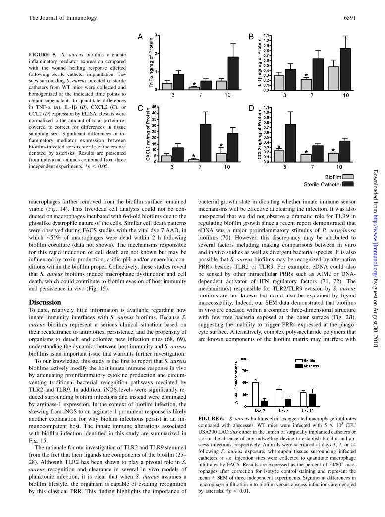
We next evaluated whether S. aureus biofilms would alter proin-flammatory mediator secretion profiles in an attempt to explainwhy these infections persist in an immunocompetent host. Ex-amination of several inflammatory signals responsible for mac-rophage and neutrophil recruitment (CCL2 and CXCL2, respec-tively) and activation (TNF-a and IL-1b) were significantly at-tenuated in biofilm-infected tissues compared with the woundhealing response elicited following the insertion of sterile cathe-ters (Fig. 5). Again, no differences in inflammatory mediator ex-pression were observed between biofilm-infected tissues fromWT, TLR2 KO, or TLR9 KO mice (data not shown), which cor-related with the failure to detect significant differences in bacterialburdens in these animals (Supplemental Figs. 2, 3).To further investigate mechanisms of immune deviation asso-
ciated with biofilm infections, we compared the degree of mac-rophage influx into S. aureus-induced abscesses versus catheter-associated biofilms by FACS. Interestingly, macrophage accumu-lation into biofilm-infected tissues was significantly increased atday 3 postinfection compared with abscesses (Fig. 6). Despite thisearly exaggerated macrophage infiltrate, immunofluorescencestaining demonstrated that the majority of cells remained distantfrom the infection site, with only a few macrophages recruited tothe biofilm surface (Fig. 7). Collectively, these findings demon-strate that S. aureus biofilms evade traditional TLR recognitionand alter the immune response to infection.
Biofilm infections attenuate iNOS and augment arginase-1expression in vivo
Reactive nitrogen intermediates represent a major microbicidalmechanism and are synthesized in macrophages by iNOS (59). In
FIGURE 1. Characterization of S. aureus localization during biofilm development. Catheters and surrounding tissues were isolated from WT mice at day
10 following S. aureus infection and subjected to Gram-staining (A, B) or SEM (C). There was evidence of bacterial growth (purple) within the catheter
lumen (A) as well as host tissue immediately surrounding the catheter (B, arrow). Insets in A and B depict original magnification 310 with red rectangles
indicating the area enlarged at original magnification 340 and asterisks denoting the original location of infected catheters that were nonadherent to glass
slides. Original magnification 3400 SEM image (C) has been pseudocolored to highlight S. aureus biofilm (blue) at the interface between the catheter
(gray) and surrounding host tissue (purple). Host cells located at a distance from the biofilm are indicated by arrows.
6588 BIOFILM EVASION OF IMMUNE RESPONSES
by guest on August 30, 2018
http://ww
w.jim
munol.org/
Dow
nloaded from

contrast, arginase-1 leads to L-proline production, which is a pre-cursor in the collagen biosynthetic pathway (60, 61). Both iNOSand arginase-1 compete for the same substrate (arginine) and, assuch, are differentially expressed (62). Due to the fact that biofilminfections are typically associated with a fibrotic response andbecause of their chronic nature, we investigated whether differ-ences in iNOS and arginase-1 expression would be evident. In-deed, iNOS expression associated with biofilm infections wasreduced compared with abscesses and coincided with robustarginase-1 expression (Fig. 8). In addition, arginase-1 expressionwas detected in macrophages in closest proximity to the biofilm,although other cell type(s) were also arginase positive (Fig. 9).Arginase-1 expression in biofilm-associated macrophages in theabsence of iNOS would be expected to skew the cellular responseaway from bacterial killing and could be another mechanism toaccount for the ability of biofilms to evade clearance.
Biofilms program macrophages toward an alternativelyactivated M2 phenotype
Our in vivo studies had clearly demonstrated that S. aureus bio-films were capable of circumventing traditional antibacterial ef-fector mechanisms. To further examine potential mechanisms re-sponsible for this phenomenon and to simplify the study ofbiofilm–macrophage interactions without confounds from othercell types, we next explored the effects of S. aureus biofilms onmacrophage responses in vitro.To date, few studies have examined the interactions between
S. aureus biofilm and macrophages, which are an important an-tibacterial effector population. To determine whether biofilms in-fluence macrophage activation, gene expression profiles werecompared between macrophages incubated with either S. aureusbiofilms or planktonic bacteria. Similar to our in vivo findings,macrophages cocultured with biofilms demonstrated a gene ex-pression profile reminiscent of alternatively activated M2 cells,namely a reduction in iNOS coincident with a significant increasein arginase-1 expression, whereas no differences in TNF-a or IL-1b were observed (Fig. 10) (63). These results further support theconclusion that biofilm growth reprograms the macrophage re-sponse from a bactericidal M1 to an M2 phenotype, the latter ofwhich is not optimal for maximal bacterial clearance (63, 64).
Macrophages actively phagocytize planktonic S. aureus but notbiofilm-associated bacteria in vitro
Previous studies have suggested that neutrophils are capable ofphagocytizing S. aureus biofilms (18, 19); however, less is knownabout the ability of macrophages to internalize bacteria in a bio-film state. To this end, we incubated primary mouse peritonealmacrophages with either 4- or 6-d-old S. aureus static biofilms andevaluated their phagocytic ability. Interestingly, macrophages in-cubated with S. aureus biofilms for either a 1- or 24-h periodcontained few internalized bacteria (Fig. 11A and data not shown).Similar findings were also obtained with BMDM (data notshown). In contrast to macrophages, intracellular bacteria werereadily discernable in neutrophils cocultured with either 4- or 6-d-old biofilms (Supplemental Fig. 4), demonstrating the sensitivityof confocal analysis for visualizing intracellular organisms. The
FIGURE 2. Evidence of catheter-associated biofilm growth in vivo.
Catheters were isolated from WT mice at day 10 following S. aureus in-
fection and processed for SEM analysis. The smooth surface at the pe-
riphery of the images in A and B represents the catheter with biofilm
visible on the internal face. A, Original magnification 3200 demonstrating
the irregular undulating pattern of the biofilm surface (arrows indicate
tower structures). B, Higher magnification of a tower from the image
shown in A revealing a predominantly hollow interior with numerous cocci
at the margins (original magnification 3800). The image has been pseu-
docolored to highlight S. aureus (gold) and presumably matrix material
(green), which likely aggregated during the SEM dehydration process,
from the remaining biofilm structure (gray; original magnification 3800).
The smooth surface at the upper right represents the catheter (salmon) with
biofilm visible on the internal face. C, Original magnification 33000 of an
S. aureus cluster within the tower depicted in B.
The Journal of Immunology 6589
by guest on August 30, 2018
http://ww
w.jim
munol.org/
Dow
nloaded from

impact of S. aureus biofilms on neutrophil function will be thetopic of a separate manuscript (M. L. Hanke and T. Kielian,manuscript in preparation). The inability of macrophages tophagocytize biofilm-associated bacteria was independent of theage of the biofilm in that intracellular bacteria could not be ob-served in macrophages incubated with either 4- or 6-d-old biofilms(Fig. 11A and data not shown). In addition, macrophages did notsignificantly impact the number of biofilm-associated bacteriaduring the 24-h coincubation period (data not shown). Thesefindings indicate that although macrophages are capable of in-filtrating biofilms, phagocytic mechanisms are not discernableduring this growth state. However, macrophages were capable ofinternalizing disrupted biofilm material (Fig. 11B), suggesting thatthe size and/or physical complexity of the intact biofilm are re-sponsible for its inability to be phagocytized. As expected, mac-rophages were highly phagocytic toward planktonic S. aureus(Fig. 11A), indicating that they were capable of recognizingbacteria in this growth state.In contrast to these in vitro studies, macrophages recovered from
biofilm infections in vivo harbored intracellular S. aureus as dem-onstrated by gentamicin protection assays (Fig. 12). The in-tracellular location of bacteria was confirmed by the ability ofrifampicin, which is capable of crossing the eukaryotic cell mem-brane, to completely eliminate viable organisms (Fig. 12). However,this approach cannot discriminate between uptake of biofilm-associated versus planktonic bacteria, the latter of which occursduring biofilm dispersal in an attempt to colonize new sites of in-fection (28, 65, 66). Collectively, the fact that a large percentage ofmacrophages remain significantly distant from the biofilm surfacein conjunction with reduced iNOS expression suggests that eventhough macrophages exhibit a modest degree of phagocytosis, in-tracellular killing mechanisms are likely compromised.
Macrophage invasion into biofilms in vitro is associated withcell death
Based on the ineffectiveness of the host immune response to clearS. aureus biofilms in vivo, it is likely that the biofilm compromises
numerous antibacterial effector mechanisms to persist in the host.To further investigate such possibilities, we next examinedwhether biofilms adversely affected macrophage survival. In-terestingly, a striking difference in cell morphology was observedbetween macrophages incubated with 4- versus 6-d-old biofilmsfor a 24-h period. Namely, macrophages exposed to immaturebiofilms (i.e., 4-d-old) exhibited a typical rounded morphology,whereas macrophages incubated with mature biofilms (i.e., 6-d-old) appeared ghostlike with a dystrophic morphology as evi-denced by their irregular shape and diminished CTO staining (Fig.13). Because a mature (i.e., 6-d-old) biofilm represents a densematrix network composed of polysaccharides, eDNA, and proteinpolymers, it is likely that this presents the macrophage with asubstrate that is difficult to phagocytize, resulting in a phenome-non termed frustrated phagocytosis (67). Although macrophagesexposed to immature biofilms retained a typical cell appearance,TOTO3 staining revealed that those macrophages most intimatelyassociated with the biofilm were dead, whereas the majority of
FIGURE 3. Visualization of S. aureus biofilm infection using IVIS. WT,
TLR2 KO, and TLR9 KO mice were infected with 5 3 105 CFU USA300
LAC::lux either in the lumen of surgically implanted catheters to establish
biofilm infection or s.c. in the absence of any indwelling device. At the
indicated time points postinfection, mice were subjected to IVIS imaging
to visualize the extent of biofilm or s.c. infection. Images are presented
from one mouse per group that was imaged at all time points to map the
progression of biofilm infection.
FIGURE 4. Infection with an equivalent S. aureus dose leads to the
establishment of catheter-related biofilm infection but rapid clearance from
s.c. sites. WT, TLR2 KO, and TLR9 KO mice were infected with 5 3 105
CFU USA300 LAC::lux either in the lumen of surgically implanted
catheters (Cath) to establish biofilm infection or s.c. (SC) in the absence of
any indwelling device. Animals were sacrificed at the indicated days fol-
lowing S. aureus infection, whereupon host tissues surrounding infected
catheters or s.c. injection sites were homogenized to quantitate bacterial
burdens. Results are expressed as the number of CFU per mg host tissue to
correct for differences in tissue sampling size. Significant differences in
bacterial burdens between biofilm-infected versus s.c. injected mice are
denoted by asterisks. *p , 0.001.
6590 BIOFILM EVASION OF IMMUNE RESPONSES
by guest on August 30, 2018
http://ww
w.jim
munol.org/
Dow
nloaded from
macrophages farther removed from the biofilm surface remainedviable (Fig. 14). This live/dead cell analysis could not be con-ducted on macrophages incubated with 6-d-old biofilms due to theghostlike dystrophic nature of the cells. Similar cell death patternswere observed during FACS studies with the vital dye 7-AAD, inwhich ∼55% of macrophages were dead within 2 h followingbiofilm coculture (data not shown). The mechanisms responsiblefor this rapid induction of cell death are not known but may beinfluenced by toxin production, acidic pH, and/or anaerobic con-ditions within the biofilm proper. Collectively, these studies revealthat S. aureus biofilms induce macrophage dysfunction and celldeath, which could contribute to biofilm evasion of host immunityand persistence in vivo (Fig. 15).
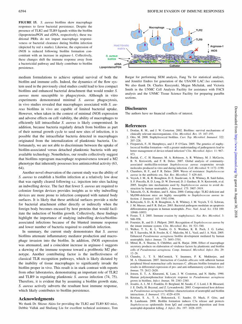
DiscussionTo date, relatively little information is available regarding howinnate immunity interfaces with S. aureus biofilms. Because S.aureus biofilms represent a serious clinical situation based ontheir recalcitrance to antibiotics, persistence, and the propensity oforganisms to detach and colonize new infection sites (68, 69),understanding the dynamics between host immunity and S. aureusbiofilms is an important issue that warrants further investigation.To our knowledge, this study is the first to report that S. aureus
biofilms actively modify the host innate immune response in vivoby attenuating proinflammatory cytokine production and circum-venting traditional bacterial recognition pathways mediated byTLR2 and TLR9. In addition, iNOS levels were significantly re-duced surrounding biofilm infections and instead were dominatedby arginase-1 expression. In the context of biofilm infection, theskewing from iNOS to an arginase-1 prominent response is likelyanother explanation for why biofilm infections persist in an im-munocompetent host. The innate immune alterations associatedwith biofilm infection identified in this study are summarized inFig. 15.The rationale for our investigation of TLR2 and TLR9 stemmed
from the fact that their ligands are components of the biofilm (25–28). Although TLR2 has been shown to play a pivotal role in S.aureus recognition and clearance in several in vivo models ofplanktonic infection, it is clear that when S. aureus assumes abiofilm lifestyle, the organism is capable of evading recognitionby this classical PRR. This finding highlights the importance of
bacterial growth state in dictating whether innate immune sensormechanisms will be effective at clearing the infection. It was alsounexpected that we did not observe a dramatic role for TLR9 inregulating biofilm growth since a recent report demonstrated thateDNA was a major proinflammatory stimulus of P. aeruginosabiofilms (70). However, this discrepancy may be attributed toseveral factors including making comparisons between in vitroand in vivo studies as well as divergent bacterial species. It is alsopossible that S. aureus biofilms may be recognized by alternativePRRs besides TLR2 or TLR9. For example, eDNA could alsobe sensed by other intracellular PRRs such as AIM2 or DNA-dependent activator of IFN regulatory factors (71, 72). Themechanism(s) responsible for TLR2/TLR9 evasion by S. aureusbiofilms are not known but could also be explained by ligandinaccessibility. Indeed, our SEM data demonstrated that biofilmsin vivo are encased within a complex three-dimensional structurewith few free bacteria exposed at the outer surface (Fig. 2B),suggesting the inability to trigger PRRs expressed at the phago-cyte surface. Alternatively, complex polysaccharide polymers thatare known components of the biofilm matrix may interfere with
FIGURE 5. S. aureus biofilms attenuate
inflammatory mediator expression compared
with the wound healing response elicited
following sterile catheter implantation. Tis-
sues surrounding S. aureus infected or sterile
catheters from WT mice were collected and
homogenized at the indicated time points to
obtain supernatants to quantitate differences
in TNF-a (A), IL-1b (B), CXCL2 (C), or
CCL2 (D) expression by ELISA. Results were
normalized to the amount of total protein re-
covered to correct for differences in tissue
sampling size. Significant differences in in-
flammatory mediator expression between
biofilm-infected versus sterile catheters are
denoted by asterisks. Results are presented
from individual animals combined from three
independent experiments. *p , 0.05.
FIGURE 6. S. aureus biofilms elicit exaggerated macrophage infiltrates
compared with abscesses. WT mice were infected with 5 3 105 CFU
USA300 LAC::lux either in the lumen of surgically implanted catheters or
s.c. in the absence of any indwelling device to establish biofilm and ab-
scess infections, respectively. Animals were sacrificed at days 3, 7, or 14
following S. aureus exposure, whereupon tissues surrounding infected
catheters or s.c. injection sites were collected to quantitate macrophage
infiltrates by FACS. Results are expressed as the percent of F4/80+ mac-
rophages after correction for isotype control staining and represent the
mean 6 SEM of three independent experiments. Significant differences in
macrophage infiltration into biofilm versus abscess infections are denoted
by asterisks. *p , 0.01.
The Journal of Immunology 6591
by guest on August 30, 2018
http://ww
w.jim
munol.org/
Dow
nloaded from

optimal engagement of potential ligands with TLRs. However,these possibilities have yet to be investigated at the present time.It is evident that S. aureus biofilms induce macrophage dys-
function at several levels, which provide potential explanations asto why these infections persist in an immunocompetent host. First,although biofilms induce greater macrophage influx during earlyinfection compared with abscesses and harbor intracellular bac-teria, the paucity of iNOS and robust arginase-1 expression sug-gest that bacteria may not be sufficiently cleared. A similar M2-like phenotype was observed in macrophages in vitro followingbiofilm coculture. Second, although biofilms were associated withrobust macrophage accumulation, only a small subset of cellsmigrated toward the biofilm surface and many that expressedarginase-1, which skews macrophages for ineffective killing.Third, our findings clearly demonstrated that biofilms inducedmacrophage death by an as of yet unknown mechanism. Specifi-cally, FACS staining with 7-AAD revealed significant macrophagecell death within 2 h following biofilm exposure. A longer coin-cubation period (i.e., 24 h) led to dystrophic macrophages witha ghostlike appearance, whereas cells cultured with 4-d-old bio-films demonstrated a typical shape even though a high percentage
of these macrophages were dead as revealed by TOTO3 staining.Importantly, macrophages were capable of phagocytizing bacteriafrom mechanically disrupted biofilms regardless of their matura-tion state, suggesting that the large size and/or inability toopsonize intact biofilms could explain the dystrophic macrophagephenotype. The latter possibility is supported by a recent studydemonstrating that IgG and C3b deposition is reduced on thesurface of biofilm-associated S. epidermidis compared withplanktonic bacteria (16). Alternatively, PIA has antiphagocyticproperties, which could also interfere with phagocytic uptake of S.aureus biofilms. It is important to note that the USA300 LACstrain used in the current study expresses PIA as demonstrated byWGA staining, but lacks a capsule (73); therefore, capsule cannotaccount for the antiphagocytic activity of the biofilm. Themechanism(s) that trigger macrophage cell death upon biofilmcontact are not known but could be the result of acidic pH, bac-terial toxins, and/or the hypoxic environment within the biofilmmass. Another question that remains is whether macrophage deathfollowing biofilm exposure is apoptotic or necrotic. Judging by thestriking differences in macrophage morphology during incubationwith immature versus mature biofilms, we speculate that bothtypes of cell death are operative. Specifically, macrophages main-tained their characteristic shape following extended (i.e., 24 h)
FIGURE 9. A subset of biofilm-associated macrophages expresses ar-
ginase-1. Tissues surrounding biofilm-infected catheters were collected
from WT mice at day 7 following S. aureus infection and subjected to
immunofluorescence staining for the macrophage-specific marker Iba-1
(red) and arginase-1 (green) to determine colocalization patterns by con-
focal microscopy (arrows, original magnification320). The position of the
biofilm-infected catheter is denoted by asterisks. Results are representative
of six individual animals.
FIGURE 8. S. aureus biofilms repress iNOS and augment arginase-1
expression. WT mice were infected with 5 3 105 CFU USA300 LAC::lux
either in the lumen of surgically implanted catheters or s.c. in the absence
of any indwelling device to establish biofilm and abscess infections, re-
spectively. Animals were sacrificed at day 7 following S. aureus exposure,
whereupon tissues surrounding infected catheters or s.c. injection sites
were collected and subjected to immunofluorescence staining with argi-
nase-1 (red) and DAPI to identify nuclei (blue) by confocal microscopy
(original magnification 320). Stars represent the location of material
remaining from the catheter lumen, and the diamonds denote the necrotic
abscess core. The border between biofilm and surrounding host tissue is
depicted by arrows. Results are representative of six individual animals.
FIGURE 10. S. aureus biofilms skew macrophage gene expression to-
ward an alternatively activated M2 phenotype. BMDM from GFP Tg mice
were incubated with 6-d-old biofilms or planktonic bacteria for 2 h,
whereupon viable macrophages were purified by FACS and RNA imme-
diately isolated for qRT-PCR analysis. Gene expression levels in macro-
phages exposed to S. aureus biofilms were calculated after normalizing
signals against GAPDH and are presented as the fold-change relative to
macrophages incubated with planktonic bacteria. Results represent the
mean 6 SEM of four independent experiments. *p , 0.05, **p , 0.001.
FIGURE 7. Macrophage migration toward S. aureus biofilms in vivo is
limited. Tissues surrounding biofilm-infected catheters were collected
from WT mice at day 7 following S. aureus infection and subjected to
immunofluorescence staining with Iba-1 to identify macrophages (red) and
DAPI to demarcate nuclei (blue) by confocal microscopy (original mag-
nification 320). Asterisks indicate the location of infected catheters, and
the border between biofilm and surrounding host tissue is denoted by
dashed lines. Results are representative of six individual animals.
6592 BIOFILM EVASION OF IMMUNE RESPONSES
by guest on August 30, 2018
http://ww
w.jim
munol.org/
Dow
nloaded from

coculture with immature biofilms, whereas those macrophagesthat were exposed to mature biofilms for the same interval exhib-ited a wispy, dystrophic morphology reminiscent of necrotic celldeath. Whether apoptosis and/or necrosis are responsible formacrophage death requires clarification in future studies.Our results demonstrating that macrophages are capable of in-
filtrating biofilms are similar to previous reports using mixedleukocyte and purified neutrophil populations with S. aureus
biofilms (17–19). However, several differences were also notedbetween these earlier studies and our own, including the fact thatwe did not observe appreciable phagocytosis of biofilm-associatedS. aureus by macrophages in vitro, whereas studies with neu-trophils reported bacterial uptake within the biofilm, which wealso observed in the current report (Supplemental Fig. 4) (18, 19).In addition, other studies have reported that S. aureus biofilmselicit proinflammatory mediator release, which was not observedin our experiments. The reasons to account for these differencesare not known but could be influenced by several factors includingthe method of biofilm propagation (i.e., static versus flow cell), thetype of immune cell population examined, and/or compatibility of
FIGURE 12. Macrophages recovered from S. aureus biofilm infections
harbor viable intracellular bacteria. Viable macrophages (F4/80+, 7-AAD2)
were recovered from tissues surrounding biofilm infections at day 7
postinfection by FACS, whereupon gentamicin protection assays were
performed to evaluate the presence of viable intracellular bacteria. Con-
trols included sorted macrophages without any antibiotic treatment (to
assess extra- and intracellular bacteria) and macrophages treated with ri-
fampicin, which is capable of penetrating the eukaryotic cell membrane.
Results are expressed as the number of viable S. aureus (CFU per 103
biofilm-associated macrophages) and represent the mean 6 SEM of five
independent experiments.
FIGURE 13. Macrophages exposed to mature S. aureus biofilm exhibit
a dystrophic morphology. Primary macrophages labeled with CTO (yel-
low-orange) were exposed to 4- or 6-d-old USA300 LAC-GFP biofilms
(green) for a 24-h period, whereupon visualization of macrophage mor-
phology was evaluated by confocal microscopy (original magnification
363, 1 mm slice). Macrophages infiltrating immature (4-d-old) biofilms
retained a typical rounded morphology, whereas macrophages infiltrating
mature biofilms (6-d-old) often exhibited a dystrophic ghostlike mor-
phology with minimal retention of CTO (arrows). Results are representa-
tive of six independent experiments.
FIGURE 14. Macrophage viability is dictated by invasion into the
biofilm proper. Four-day-old USA300 LAC-GFP biofilms (average height
∼34 mm) (A) were incubated with primary macrophages labeled with CTO
(B, yellow-orange) for a 24-h period, whereupon macrophage viability was
determined by uptake of the live/dead stain TOTO3 (purple; C). The
majority of dead (TOTO3+) macrophages are within 34 mm of the biofilm
proper (p , 0.05), and a composite image of all staining is shown (D).
Bottom panel, Significant differences between the percentages of TOTO3+
macrophages based on location within the biofilm proper are denoted with
an asterisk. *p , 0.05; n = 218 cells.
FIGURE 11. Macrophages actively phagocytize planktonic S. aureus
but not biofilm-associated bacteria. A, Primary macrophages labeled with
CTO (orange) were exposed to 4-d-old USA300 LAC-GFP biofilms or
planktonic bacteria (green) for a 24-h period, whereupon visualization of
intracellular bacteria was evaluated by confocal microscopy (original
magnification 363, 1 mm slice). Biofilm-associated macrophages exhibi-
ted little evidence of phagocytosis, whereas intracellular bacteria were
readily detectable in macrophages incubated with planktonic organisms.
Results are representative of six independent experiments. In B, 4- and 6-d-
old USA300 LAC-GFP static biofilms were mechanically disrupted by
triturating and incubated with macrophages for 1 h. Planktonic bacteria
were included as a positive control. Fluorescent microscopy of cytospin
preparations shows the ability of macrophages to phagocytize planktonic
(left panel) as well as disassociated bacteria from 4- (center panel) and 6-
d-old (right panel) biofilms. Results are representative of three indepen-
dent experiments.
The Journal of Immunology 6593
by guest on August 30, 2018
http://ww
w.jim
munol.org/
Dow
nloaded from
medium formulations to achieve optimal survival of both thebiofilm and immune cells. Indeed, the dynamics of the flow sys-tem used in the previously cited studies could lead to less compactbiofilms and enhanced bacterial detachment that would render S.aureus more susceptible to phagocytosis. Although in vitroexperiments demonstrated minimal S. aureus phagocytosis,in vivo studies revealed that macrophages associated with S. au-reus biofilms in vivo are capable of limited bacterial uptake.However, when taken in the context of minimal iNOS expressionand adverse effects on cell viability, the ability of macrophages toefficiently kill intracellular S. aureus is likely compromised. Inaddition, because bacteria regularly detach from biofilms as partof their normal growth cycle to seed new sites of infection, it ispossible that the intracellular bacteria detected in macrophagesoriginated from the internalization of planktonic bacteria. Un-fortunately, we are not able to discriminate between the uptake ofbiofilm-associated versus detached planktonic bacteria with anyavailable technology. Nonetheless, our results collectively suggestthat biofilms reprogram macrophage responsiveness toward a M2phenotype that inherently possesses less antimicrobial activity (63,64).Another novel observation of the current study was the ability of
S. aureus to establish a biofilm infection at a relatively low dosethat was rapidly cleared following s.c. injection into sites lackingan indwelling device. The fact that fewer S. aureus are required tocolonize foreign devices provides insights as to why indwellingdevices are more prone to infection compared with natural bodysurfaces. It is likely that these artificial surfaces provide a nichefor bacterial attachment either directly or indirectly when theforeign body becomes encased by host proteins that would facil-itate the induction of biofilm growth. Collectively, these findingshighlight the importance of studying indwelling device/biofilm-associated infections because of the blunted immune responseand lower number of bacteria required to establish infection.In summary, the current study demonstrates that S. aureus
biofilms attenuate inflammatory mediator production and macro-phage invasion into the biofilm. In addition, iNOS expressionwas attenuated, and a coincident increase in arginase-1 suggestsa skewing of the immune response toward a profibrotic M2 phe-notype. Another contributing factor is the ineffectiveness ofclassical TLR recognition pathways, which is likely dictated bythe inability of tissue macrophages to significantly invade thebiofilm proper in vivo. This result is in stark contrast with reportsfrom other laboratories, demonstrating an important role of TLR2and TLR9 in regulating planktonic S. aureus infection (74, 75).Therefore, it is evident that by assuming a biofilm growth state,S. aureus actively subverts the resultant host immune response,which likely contributes to biofilm persistence.
AcknowledgmentsWe thank Dr. Shizuo Akira for providing the TLR2 and TLR9 KO mice,
Debbie Vidlak and Shuliang Liu for excellent technical assistance, Tom
Bargar for performing SEM analysis, Fang Yu for statistical analysis,
and Jennifer Endres for generation of the USA300 LAC::lux construct.
We also thank Dr. Charles Kuszynski, Megan Michalak, and Victoria
Smith in the UNMC Cell Analysis Facility for assistance with FACS
analysis and the UNMC Tissue Science Facility for preparing paraffin
sections.
DisclosuresThe authors have no financial conflicts of interest.
References1. Donlan, R. M., and J. W. Costerton. 2002. Biofilms: survival mechanisms of
clinically relevant microorganisms. Clin. Microbiol. Rev. 15: 167–193.2. Otto, M. 2008. Staphylococcal biofilms. Curr. Top. Microbiol. Immunol. 322:
207–228.3. Fitzpatrick, F., H. Humphreys, and J. P. O’Gara. 2005. The genetics of staphy-
lococcal biofilm formation—will a greater understanding of pathogenesis lead tobetter management of device-related infection? Clin. Microbiol. Infect. 11: 967–973.
4. Burlak, C., C. H. Hammer, M. A. Robinson, A. R. Whitney, M. J. McGavin,B. N. Kreiswirth, and F. R. Deleo. 2007. Global analysis of community-associated methicillin-resistant Staphylococcus aureus exoproteins revealsmolecules produced in vitro and during infection. Cell. Microbiol. 9: 1172–1190.
5. Chambers, H. F., and F. R. Deleo. 2009. Waves of resistance: Staphylococcusaureus in the antibiotic era. Nat. Rev. Microbiol. 7: 629–641.
6. Voyich, J. M., K. R. Braughton, D. E. Sturdevant, A. R. Whitney, B. Saıd-Salim,S. F. Porcella, R. D. Long, D. W. Dorward, D. J. Gardner, B. N. Kreiswirth, et al.2005. Insights into mechanisms used by Staphylococcus aureus to avoid de-struction by human neutrophils. J. Immunol. 175: 3907–3919.
7. Takeuchi, O., K. Hoshino, and S. Akira. 2000. Cutting edge: TLR2-deficient andMyD88-deficient mice are highly susceptible to Staphylococcus aureus in-fection. J. Immunol. 165: 5392–5396.
8. Kobayashi, S. D., K. R. Braughton, A. R. Whitney, J. M. Voyich, T. G. Schwan,J. M. Musser, and F. R. DeLeo. 2003. Bacterial pathogens modulate an apoptosisdifferentiation program in human neutrophils. Proc. Natl. Acad. Sci. USA 100:10948–10953.
9. Foster, T. J. 2005. Immune evasion by staphylococci. Nat. Rev. Microbiol. 3:948–958.
10. Fournier, B., and D. J. Philpott. 2005. Recognition of Staphylococcus aureus bythe innate immune system. Clin. Microbiol. Rev. 18: 521–540.
11. Walker, T. S., K. L. Tomlin, G. S. Worthen, K. R. Poch, J. G. Lieber,M. T. Saavedra, M. B. Fessler, K. C. Malcolm, M. L. Vasil, and J. A. Nick. 2005.Enhanced Pseudomonas aeruginosa biofilm development mediated by humanneutrophils. Infect. Immun. 73: 3693–3701.
12. Mittal, R., S. Sharma, S. Chhibber, and K. Harjai. 2006. Effect of macrophagesecretory products on elaboration of virulence factors by planktonic and biofilmcells of Pseudomonas aeruginosa. Comp. Immunol. Microbiol. Infect. Dis. 29:12–26.
13. Chandra, J., T. S. McCormick, Y. Imamura, P. K. Mukherjee, andM. A. Ghannoum. 2007. Interaction of Candida albicans with adherent humanperipheral blood mononuclear cells increases C. albicans biofilm formation andresults in differential expression of pro- and anti-inflammatory cytokines. Infect.Immun. 75: 2612–2620.
14. Jensen, E. T., A. Kharazmi, K. Lam, J. W. Costerton, and N. Høiby. 1990.Human polymorphonuclear leukocyte response to Pseudomonas aeruginosagrown in biofilms. Infect. Immun. 58: 2383–2385.
15. Jesaitis, A. J., M. J. Franklin, D. Berglund, M. Sasaki, C. I. Lord, J. B. Bleazard,J. E. Duffy, H. Beyenal, and Z. Lewandowski. 2003. Compromised host defenseon Pseudomonas aeruginosa biofilms: characterization of neutrophil and biofilminteractions. J. Immunol. 171: 4329–4339.
16. Kristian, S. A., T. A. Birkenstock, U. Sauder, D. Mack, F. Gotz, andR. Landmann. 2008. Biofilm formation induces C3a release and protectsStaphylococcus epidermidis from IgG and complement deposition and fromneutrophil-dependent killing. J. Infect. Dis. 197: 1028–1035.
FIGURE 15. S. aureus biofilms skew macrophage
responses to favor bacterial persistence. Despite the
presence of TLR2 and TLR9 ligands within the biofilm
(lipoproteins/PGN and eDNA, respectively), these tra-
ditional PRRs do not impart macrophage responsi-
veness or bacterial clearance during biofilm infection
(depicted by red x marks). Likewise, the expression of
iNOS is reduced following biofilm formation con-
comitant with an increase in arginase-1. Collectively,
these changes shift the immune response away from
a bactericidal pathway and likely contribute to biofilm
persistence.
6594 BIOFILM EVASION OF IMMUNE RESPONSES
by guest on August 30, 2018
http://ww
w.jim
munol.org/
Dow
nloaded from

17. Leid, J. G., M. E. Shirtliff, J. W. Costerton, and P. Stoodley. 2002. Humanleukocytes adhere to, penetrate, and respond to Staphylococcus aureus biofilms.Infect. Immun. 70: 6339–6345.
18. Gunther, F., G. H. Wabnitz, P. Stroh, B. Prior, U. Obst, Y. Samstag, C. Wagner,and G. M. Hansch. 2009. Host defence against Staphylococcus aureus biofilmsinfection: phagocytosis of biofilms by polymorphonuclear neutrophils (PMN).Mol. Immunol. 46: 1805–1813.
19. Guenther, F., P. Stroh, C. Wagner, U. Obst, and G. M. Hansch. 2009. Phago-cytosis of staphylococci biofilms by polymorphonuclear neutrophils: S. aureusand S. epidermidis differ with regard to their susceptibility towards the hostdefense. Int. J. Artif. Organs 32: 565–573.
20. Kobayashi, S. D., K. R. Braughton, A. M. Palazzolo-Ballance, A. D. Kennedy,E. Sampaio, E. Kristosturyan, A. R. Whitney, D. E. Sturdevant, D. W. Dorward,S. M. Holland, et al. 2010. Rapid neutrophil destruction following phagocytosisof Staphylococcus aureus. J. Innate Immun. 2: 560–575.
21. Graves, S. F., S. D. Kobayashi, and F. R. DeLeo. 2010. Community-associatedmethicillin-resistant Staphylococcus aureus immune evasion and virulence. J.Mol. Med. 88: 109–114.
22. Anwar, S., L. R. Prince, S. J. Foster, M. K. Whyte, and I. Sabroe. 2009. The riseand rise of Staphylococcus aureus: laughing in the face of granulocytes. Clin.Exp. Immunol. 157: 216–224.
23. Silva, M. T. 2010. Macrophage phagocytosis of neutrophils at inflammatory/infectious foci: a cooperative mechanism in the control of infection and in-fectious inflammation. J. Leukoc. Biol. DOI: 10.1189/jlb.0910536.
24. Silva, M. T. 2010. When two is better than one: macrophages and neutrophilswork in concert in innate immunity as complementary and cooperative partnersof a myeloid phagocyte system. J. Leukoc. Biol. 87: 93–106.
25. Rice, K. C., E. E. Mann, J. L. Endres, E. C. Weiss, J. E. Cassat, M. S. Smeltzer,and K. W. Bayles. 2007. The cidA murein hydrolase regulator contributes toDNA release and biofilm development in Staphylococcus aureus. Proc. Natl.Acad. Sci. USA 104: 8113–8118.
26. Whitchurch, C. B., T. Tolker-Nielsen, P. C. Ragas, and J. S. Mattick. 2002.Extracellular DNA required for bacterial biofilm formation. Science 295: 1487.
27. Allesen-Holm, M., K. B. Barken, L. Yang, M. Klausen, J. S. Webb,S. Kjelleberg, S. Molin, M. Givskov, and T. Tolker-Nielsen. 2006. A charac-terization of DNA release in Pseudomonas aeruginosa cultures and biofilms.Mol. Microbiol. 59: 1114–1128.
28. Mann, E. E., K. C. Rice, B. R. Boles, J. L. Endres, D. Ranjit, L. Chandramohan,L. H. Tsang, M. S. Smeltzer, A. R. Horswill, and K. W. Bayles. 2009. Modu-lation of eDNA release and degradation affects Staphylococcus aureus biofilmmaturation. PLoS ONE 4: e5822.
29. Akira, S., S. Uematsu, and O. Takeuchi. 2006. Pathogen recognition and innateimmunity. Cell 124: 783–801.
30. Hayashi, F., T. K. Means, and A. D. Luster. 2003. Toll-like receptors stimulatehuman neutrophil function. Blood 102: 2660–2669.
31. Bauer, S., C. J. Kirschning, H. Hacker, V. Redecke, S. Hausmann, S. Akira,H. Wagner, and G. B. Lipford. 2001. Human TLR9 confers responsiveness tobacterial DNA via species-specific CpG motif recognition. Proc. Natl. Acad. Sci.USA 98: 9237–9242.
32. Hertz, C. J., S. M. Kiertscher, P. J. Godowski, D. A. Bouis, M. V. Norgard,M. D. Roth, and R. L. Modlin. 2001. Microbial lipopeptides stimulate dendriticcell maturation via Toll-like receptor 2. J. Immunol. 166: 2444–2450.
33. Jones, B. W., T. K. Means, K. A. Heldwein, M. A. Keen, P. J. Hill, J. T. Belisle,and M. J. Fenton. 2001. Different Toll-like receptor agonists induce distinctmacrophage responses. J. Leukoc. Biol. 69: 1036–1044.
34. Kirschning, C. J., and R. R. Schumann. 2002. TLR2: cellular sensor for mi-crobial and endogenous molecular patterns. Curr. Top. Microbiol. Immunol. 270:121–144.
35. Takeuchi, O., K. Hoshino, T. Kawai, H. Sanjo, H. Takada, T. Ogawa, K. Takeda,and S. Akira. 1999. Differential roles of TLR2 and TLR4 in recognition of gram-negative and gram-positive bacterial cell wall components. Immunity 11: 443–451.
36. Mercier, C., C. Durrieu, R. Briandet, E. Domakova, J. Tremblay, G. Buist, andS. Kulakauskas. 2002. Positive role of peptidoglycan breaks in lactococcalbiofilm formation. Mol. Microbiol. 46: 235–243.
37. Cerca, N., K. K. Jefferson, R. Oliveira, G. B. Pier, and J. Azeredo. 2006.Comparative antibody-mediated phagocytosis of Staphylococcus epidermidiscells grown in a biofilm or in the planktonic state. Infect. Immun. 74: 4849–4855.
38. Moscoso, M., E. Garcıa, and R. Lopez. 2006. Biofilm formation by Strepto-coccus pneumoniae: role of choline, extracellular DNA, and capsular poly-saccharide in microbial accretion. J. Bacteriol. 188: 7785–7795.
39. Qin, Z., Y. Ou, L. Yang, Y. Zhu, T. Tolker-Nielsen, S. Molin, and D. Qu. 2007.Role of autolysin-mediated DNA release in biofilm formation of Staphylococcusepidermidis. Microbiology 153: 2083–2092.
40. Morath, S., A. Stadelmaier, A. Geyer, R. R. Schmidt, and T. Hartung. 2002.Synthetic lipoteichoic acid from Staphylococcus aureus is a potent stimulus ofcytokine release. J. Exp. Med. 195: 1635–1640.
41. Weber, J. R., P. Moreillon, and E. I. Tuomanen. 2003. Innate sensors for Gram-positive bacteria. Curr. Opin. Immunol. 15: 408–415.
42. Dziarski, R. 2003. Recognition of bacterial peptidoglycan by the innate immunesystem. Cell. Mol. Life Sci. 60: 1793–1804.
43. Shimada, T., B. G. Park, A. J. Wolf, C. Brikos, H. S. Goodridge, C. A. Becker,C. N. Reyes, E. A. Miao, A. Aderem, F. Gotz, et al. 2010. Staphylococcus aureusevades lysozyme-based peptidoglycan digestion that links phagocytosis,inflammasome activation, and IL-1beta secretion. Cell Host Microbe 7: 38–49.
44. Ip, W. K., A. Sokolovska, G. M. Charriere, L. Boyer, S. Dejardin,M. P. Cappillino, L. M. Yantosca, K. Takahashi, K. J. Moore, A. Lacy-Hulbert,
and L. M. Stuart. 2010. Phagocytosis and phagosome acidification are requiredfor pathogen processing and MyD88-dependent responses to Staphylococcusaureus. J. Immunol. 184: 7071–7081.
45. Hemmi, H., O. Takeuchi, T. Kawai, T. Kaisho, S. Sato, H. Sanjo, M. Matsumoto,K. Hoshino, H. Wagner, K. Takeda, and S. Akira. 2000. A Toll-like receptorrecognizes bacterial DNA. Nature 408: 740–745.
46. Yoshimura, A., E. Lien, R. R. Ingalls, E. Tuomanen, R. Dziarski, andD. Golenbock. 1999. Cutting edge: recognition of Gram-positive bacterial cellwall components by the innate immune system occurs via Toll-like receptor 2. J.Immunol. 163: 1–5.
47. Watanabe, I., M. Ichiki, A. Shiratsuchi, and Y. Nakanishi. 2007. TLR2-mediatedsurvival of Staphylococcus aureus in macrophages: a novel bacterial strategyagainst host innate immunity. J. Immunol. 178: 4917–4925.
48. Diep, B. A., A. M. Palazzolo-Ballance, P. Tattevin, L. Basuino, K. R. Braughton,A. R. Whitney, L. Chen, B. N. Kreiswirth, M. Otto, F. R. DeLeo, andH. F. Chambers. 2008. Contribution of Panton-Valentine leukocidin incommunity-associated methicillin-resistant Staphylococcus aureus pathogenesis.PLoS ONE 3: e3198.
49. Kennedy, A. D., M. Otto, K. R. Braughton, A. R. Whitney, L. Chen,B. Mathema, J. R. Mediavilla, K. A. Byrne, L. D. Parkins, F. C. Tenover, et al.2008. Epidemic community-associated methicillin-resistant Staphylococcus au-reus: recent clonal expansion and diversification. Proc. Natl. Acad. Sci. USA 105:1327–1332.
50. Cassat, J. E., C. Y. Lee, and M. S. Smeltzer. 2007. Investigation of biofilmformation in clinical isolates of Staphylococcus aureus. Methods Mol. Biol. 391:127–144.
51. Rupp, M. E., J. S. Ulphani, P. D. Fey, K. Bartscht, and D. Mack. 1999. Char-acterization of the importance of polysaccharide intercellular adhesin/hemagglutinin of Staphylococcus epidermidis in the pathogenesis ofbiomaterial-based infection in a mouse foreign body infection model. Infect.Immun. 67: 2627–2632.
52. Kielian, T., A. Cheung, and W. F. Hickey. 2001. Diminished virulence of analpha-toxin mutant of Staphylococcus aureus in experimental brain abscesses.Infect. Immun. 69: 6902–6911.
53. Lauderdale, K. J., C. L. Malone, B. R. Boles, J. Morcuende, and A. R. Horswill.2010. Biofilm dispersal of community-associated methicillin-resistant Staphy-lococcus aureus on orthopedic implant material. J. Orthop. Res. 28: 55–61.
54. Liu, S., and T. Kielian. 2009. Microglial activation by Citrobacter koseri ismediated by TLR4- and MyD88-dependent pathways. J. Immunol. 183: 5537–5547.
55. Thurlow, L. R., V. C. Thomas, S. D. Fleming, and L. E. Hancock. 2009. En-terococcus faecalis capsular polysaccharide serotypes C and D and their con-tributions to host innate immune evasion. Infect. Immun. 77: 5551–5557.
56. Drevets, D. A., and P. A. Campbell. 1991. Macrophage phagocytosis: use offluorescence microscopy to distinguish between extracellular and intracellularbacteria. J. Immunol. Methods 142: 31–38.
57. Drevets, D. A., B. P. Canono, P. J. Leenen, and P. A. Campbell. 1994. Genta-micin kills intracellular Listeria monocytogenes. Infect. Immun. 62: 2222–2228.
58. Lauderdale, K. J., B. R. Boles, A. L. Cheung, and A. R. Horswill. 2009. Inter-connections between Sigma B, agr, and proteolytic activity in Staphylococcusaureus biofilm maturation. Infect. Immun. 77: 1623–1635.
59. Kleinert, H., A. Pautz, K. Linker, and P. M. Schwarz. 2004. Regulation ofthe expression of inducible nitric oxide synthase. Eur. J. Pharmacol. 500: 255–266.
60. Curran, J. N., D. C. Winter, and D. Bouchier-Hayes. 2006. Biological fate andclinical implications of arginine metabolism in tissue healing. Wound RepairRegen. 14: 376–386.
61. Wynn, T. A. 2008. Cellular and molecular mechanisms of fibrosis. J. Pathol. 214:199–210.
62. Hesse, M., M. Modolell, A. C. La Flamme, M. Schito, J. M. Fuentes,A. W. Cheever, E. J. Pearce, and T. A. Wynn. 2001. Differential regulation ofnitric oxide synthase-2 and arginase-1 by type 1/type 2 cytokines in vivo:granulomatous pathology is shaped by the pattern of L-arginine metabolism. J.Immunol. 167: 6533–6544.
63. Gordon, S. 2003. Alternative activation of macrophages. Nat. Rev. Immunol. 3:23–35.
64. Benoit, M., B. Desnues, and J. L. Mege. 2008. Macrophage polarization inbacterial infections. J. Immunol. 181: 3733–3739.
65. Boles, B. R., and A. R. Horswill. 2008. Agr-mediated dispersal of Staphylo-coccus aureus biofilms. PLoS Pathog. 4: e1000052.
66. Lauderdale, K. J., C. L. Malone, B. R. Boles, J. Morcuende, and A. R. Horswill.2010. Biofilm dispersal of community-associated methicillin-resistant Staphy-lococcus aureus on orthopedic implant material. J. Orthop. Res. 28: 55–61.
67. Bainton, D. F., R. Takemura, P. E. Stenberg, and Z. Werb. 1989. Rapidfragmentation and reorganization of Golgi membranes during frustratedphagocytosis of immobile immune complexes by macrophages. Am. J. Pathol.134: 15–26.
68. Fatkenheuer, G., O. Cornely, and H. Seifert. 2002. Clinical management ofcatheter-related infections. Clin. Microbiol. Infect. 8: 545–550.
69. Lowy, F. D. 1998. Staphylococcus aureus infections. N. Engl. J. Med. 339: 520–532.
70. Fuxman Bass, J. I., D. M. Russo, M. L. Gabelloni, J. R. Geffner, M. Giordano,M. Catalano, A. Zorreguieta, and A. S. Trevani. 2010. Extracellular DNA:a major proinflammatory component of Pseudomonas aeruginosa biofilms. J.Immunol. 184: 6386–6395.
71. Hornung, V., and E. Latz. 2010. Intracellular DNA recognition. Nat. Rev.Immunol. 10: 123–130.
The Journal of Immunology 6595
by guest on August 30, 2018
http://ww
w.jim
munol.org/
Dow
nloaded from
72. Vilaysane, A., and D. A. Muruve. 2009. The innate immune response to DNA.Semin. Immunol. 21: 208–214.
73. Montgomery, C. P., S. Boyle-Vavra, P. V. Adem, J. C. Lee, A. N. Husain,J. Clasen, and R. S. Daum. 2008. Comparison of virulence in community-associated methicillin-resistant Staphylococcus aureus pulsotypes USA300and USA400 in a rat model of pneumonia. J. Infect. Dis. 198: 561–570.
74. Takeuchi, O., K. Takeda, K. Hoshino, O. Adachi, T. Ogawa, and S. Akira. 2000.Cellular responses to bacterial cell wall components are mediated throughMyD88-dependent signaling cascades. Int. Immunol. 12: 113–117.
75. Gillrie, M. R., L. Zbytnuik, E. McAvoy, R. Kapadia, K. Lee, C. C. Waterhouse,S. P. Davis, D. A. Muruve, P. Kubes, and M. Ho. 2010. Divergent roles of Toll-like receptor 2 in response to lipoteichoic acid and Staphylococcus aureusin vivo. Eur. J. Immunol. 40: 1639–1650.
6596 BIOFILM EVASION OF IMMUNE RESPONSES
by guest on August 30, 2018
http://ww
w.jim
munol.org/
Dow
nloaded from