Embed Size (px)
Citation preview
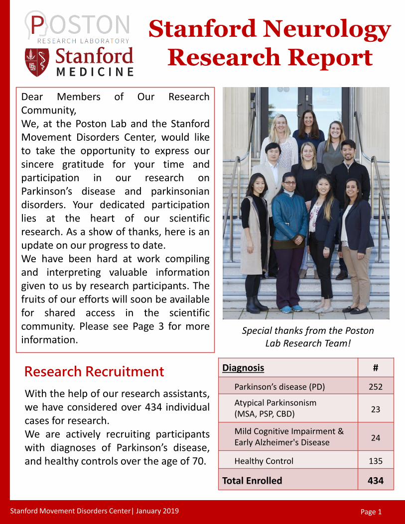
Dear Members of Our ResearchCommunity,We, at the Poston Lab and the StanfordMovement Disorders Center, would liketo take the opportunity to express oursincere gratitude for your time andparticipation in our research onParkinson’s disease and parkinsoniandisorders. Your dedicated participationlies at the heart of our scientificresearch. As a show of thanks, here is anupdate on our progress to date.We have been hard at work compilingand interpreting valuable informationgiven to us by research participants. Thefruits of our efforts will soon be availablefor shared access in the scientificcommunity. Please see Page 3 for moreinformation.
Research Recruitment Diagnosis #
Parkinson’s disease (PD) 252
Atypical Parkinsonism (MSA, PSP, CBD) 23
Mild Cognitive Impairment & Early Alzheimer's Disease 24
Healthy Control 135
Total Enrolled 434
Special thanks from the Poston Lab Research Team!
Page 1 Stanford Movement Disorders Center| January 2019 Page 1
Stanford Neurology Research Report
With the help of our research assistants,we have considered over 434 individualcases for research.We are actively recruiting participantswith diagnoses of Parkinson’s disease,and healthy controls over the age of 70.

Recruitment for New Research Studies!
Stanford Movement Disorders Center| January 2019 Page 2
New PET Study - We recently started recruiting individuals with Parkinson’s
disease for two PET/MRI scans to examine patterns of protein (amyloid) build
up and inflammation (microglial activation) in the brain. The ability to
measure amyloid accumulation and microglial activation provides a critical
opportunity to further the understanding of Parkinson’s disease, monitor
disease progression, and assess disease modifying treatments.
For more information, please contact Marian Shahid at (650) 723-0060
Stanford Alzheimer’s Disease Research Center (ADRC) - The NIH, with the
National Institute for Aging (NIA), approved funding for the new Stanford
Alzheimer’s disease Research Center (ADRC) and we have had a very
successful year of recruitment and data collection! Our Center’s theme is
understanding early memory problems in people with Parkinson’s disease and
Alzheimer’s disease.
For more information, please contact Christina Wyss-Coray at (650)
721-2409 or [email protected].
Pacific Udall Center - In July 2016, Stanford became the lead site for the
Pacific Udall Center: A Morris K Udall Center of Excellence in Parkinson’s
Research, which is one of eight Centers funded by the NIH and the National
Institute for Neurological Disease and Stroke (NINDS). Pathology Chairman Dr.
Tom Montine and Dr. Kathleen Poston are leading the Stanford team.
Together with University of Washington and Oregon Health Sciences
University (OHSU), the Pacific Udall Center’s mission is to understand the
genetic contributions to Parkinson’s disease memory problems and balance
problems.
For more information, please contact Meagan Adams at (650) 721-
5274 or [email protected].

Stanford Movement Disorders Center| January 2019 Page 3
Figure 1: Tiny strokes near fiber tracts in the brain.
People with Parkinson's disease are at risk of developing cognitive impairments; however theexact cause is unknown and it is likely that there are many different contributing causes. Weused data acquired on MRI scans to determine if tiny strokes might play a role. At the 2018Annual Meeting of the American Academy of Neurology and the 2018 Society forNeuroscience Annual Meeting, Dr. Patricia Linortner and Mr. Colin McDaniel showed thatthese tiny strokes increase the risk of having problems with attention, memory and executivefunction, such as the time it takes a person to complete a mental task. These effects becomeespecially apparent when these strokes are larger or next to fiber tracts that transferinformation between different regions of the brain. Treatment of stroke risk factors (like bloodpressure or diabetes) could help slow down the progression of some cognitive difficulties inpeople with Parkinson’s disease, which we plan to investigate in future studies.
Looking at cognition in people with Parkinson’s disease from a differentangle: how do tiny strokes and their location in the brain affect cognition?
Figure 2: Mr. Colin McDaniel presenting at the 2018 Society for Neuroscience Meeting in San Diego, California.

Stanford Movement Disorders Center| January 2019 Page 4
Figure 4: Frontal cortical regions involved in the working memory task.
Ever wondered how our brain works to keep phone numbers in our head until the moment we jot itdown on a piece of paper? It’s done by a brain process called ‘working memory.’ Working memoryrefers to the ability to actively maintain and manipulate information in your mind. You generally relyon this ability when you are keeping track of lists in your head or attending to conversations.We tested a similar process in the MRI scanner in people with Parkinson’s disease, both while theywere on and off their dopaminergic medications. We asked research participants to view a singlenumber probe and respond based on whether it matched with one of the five numbers they sawpreviously (Figure 3). From this study, we found that some individuals’ working memory improvewhen they take their dopaminergic medications, whereas others don't demonstrate suchimprovements. We are currently investigating which clinical characteristics indicate whether anindividual's working memory improves, or does not improve, with dopamine. This can help informhow to best prescribe medications that optimize cognitive functioning.
Maintaining Information
Further, working memory isknown to be facilitated by thefrontal lobes of the brain (asshown by the red activation inthe brain images below) incoordination with other regions,such as the subcortical regions.This ability can be disrupted inParkinson’s disease, which isthought to be driven by adecrease in dopaminergic inputfrom the subcortical regions tothe frontal lobes.
Previously, Dr. Kathleen Poston demonstrated that a subcortical region, the putamen, demonstratescompensatory activity in Parkinson’s disease during this working memory task. Therefore, PostonLab researchers, Dr. Sephira Ryman and Ms. Jeehyun Kim, are in the works to extend this finding andbetter understand the interaction between the putamen and the frontal lobes of the brain.Understanding this system will enable researchers to develop effective means to intervene andimprove cognitive functioning in individuals with Parkinson’s disease.
Figure 3: Sternberg task –the working memory task participants complete inside the MRI scanner.

Stanford Movement Disorders Center| January 2019 Page 5
Figure 5: Ms. Kim presenting at the 2018 Biennial Resting State Brain ConnectivityConference in Montreal, Canada.
How much can a brain at rest tell you?Turns out your resting brain actually tells quite a bit. The 10 minute ‘resting-state’functional MRI scan acquired from our research participants allows us to analyze naturalbrain fluctuations and provides a functional framework underlying basic brain operations.In previous studies, the regions of the brain that control motor activity (termed the ‘motornetwork’) has less synchronized fluctuations in people with Parkinson’s disease, comparedto people who do not have Parkinson’s disease. This could be one mechanism that leads tothe motor symptoms experienced by patients. In the Poston Lab, we are studying whetherthe synchronized fluctuations associated with both cognitive and motor symptoms areinter-linked. We have found that a brain region, called the anterior insula, might beresponsible for this link. The insula is well known for its role in gating of attention andallowing us to switch our attention to different stimuli.While the typical ‘resting-sate’ functional MRI analysis method is to average all of the datacollected over the 10 minute duration of the scan, this average representation might be agross simplification and may miss critical information that can help us better characterizeimportant brain changes over short periods of time. Therefore, we have decided to take anon-stationary approach to analysis. Dr. Christian La and Ms. Nessa Kim are exploring howanalyzing the changes in synchronized fluctuations over time might be a novel metric forunderstanding brain flexibility and overall brain health. This could be particularlyinteresting in people with Parkinson’s disease and helpful in understanding the mechanismunderlying motor and non-motor symptoms. Initial results from this investigation havebeen presented at the 6th Biennial Resting State Brain-Connectivity Conference 2018 inMontreal, Canada, and the manuscript is currently being prepared for publication.

Scientific Papers
Chen KT, Gong E, de Carvalho Macruz FB, Xu J, Boumis A, Khalighi M, Poston KL, Sha SJ, Greicius MD, Mormino E, Pauly JM, Srinivas S, Zaharchuk G. “Ultra-Low-Dose 18F-Florbetaben Amyloid PET Imaging Using Deep Learning with Multi-Contrast MRI Inputs” Radiology. 2018 Dec 11:180940. doi: 10.1148/radiol.2018180940. [Epub ahead of print]https://www.ncbi.nlm.nih.gov/pubmed/30526350
Sundaram S, Müller-Oehring EM, Fama R, Brontë-Stewart HM, Poston KL, Goodcase R, Martin T, Prabhakar V, Karpf J, Schulte T. “Information processing deficit in older adults with HIV infection: A comparison with Parkinson’s disease” Neuropsychology. 2018 Nov 26. doi: 10.1037/neu0000500. [Epub ahead of print]https://www.ncbi.nlm.nih.gov/pubmed/30475047
Kim J, Zhang K, Cai W, YorkWilliams S, Ua Cruadhlaoich MAI, Llanes S, Menon V, Poston KL. “Dopamine-related dissociationof cortical and subcortical brain activations in cognitively unimpaired Parkinson’s disease patients OFF and ON medications”Neuropsychologia. 2018 Jul 21;119:24-33. doi: 10.1016/j.neuropsychologia.2018.07.024.https://www.ncbi.nlm.nih.gov/pubmed/30040957
Cholerton B, Johnson CO, Fish B, Quinn JF, Chung KA, Peterson-Hiller AL, Rosenthal LS, Dawson TM, Albert MS, Hu SC, MataIF, Leverenz JB, Poston KL, Montine TJ, Zabetian CP, Edwards KL. “Sex differences in progression to mild cognitiveimpairment and dementia in Parkinson’s disease” Parkinsonism Relat Disord. 2018 May; 50:29-36. doi:10.1016/j.parkreldis.2018.02.007. Epub 2018 Feb 9.https://www.ncbi.nlm.nih.gov/pubmed/29478836
Simuni T, Siderowf A, Lasch S, Coffey CS, Caspell-Garcia C, Jennings D, Tanner CM, Trojanowski JQ, Shaw LM, Seibyl J, SchuffN, Singleton A, Kieburtz K, Toga AW, Mollenhauer B, Galasko D, Chahine LM, Weintraub D, Foroud T, Tosun D, Poston K,Arnedo V, Frasier M, Sherer T, Chowdhury S, Marek K; Parkinson's Progression Marker Initiative. “Longitudinal change ofclinical and biological measures in early Parkinson’s Disease: Parkinson’s Progression Markers Initiative Cohort” Mov Disord.2018 Mar 23. doi: 10.1002/mds.27361.https://www.ncbi.nlm.nih.gov/pubmed/29572948
Jehangir N, Yu CY, Song J, Shariati MA, Binder S, Beyer J, Santini V, Poston KL, Liao YJ. “Slower saccadic reading inParkinson’s disease” PLoS One. 2018 Jan 24;13(1):e0191005. doi: 10.1371/journal.pone.0191005. eCollection 2018.https://www.ncbi.nlm.nih.gov/pubmed/29364897
Milchenko M, Norris SA, Poston KL, Campbell MC, Ushe M, Perlmutter JS, Snyder AZ. “7T MRI subthalamic nucleus atlas foruse with 3T MRI” J Med Imaging (Bellingham). 2018 Jan;5(1):015002. doi: 10.1117/1.JMI.5.1.015002. Epub 2018 Jan 8.https://www.ncbi.nlm.nih.gov/pubmed/29340288
Ng B, Varoquaux G, Poline JB, Thirion B, Greicius M, Poston KL. “Distinct alterations in Parkinson’s Medication-state andDisease-state Connectivity” NeuroImage: Clinical, 2017 Sep 6;16:575-585.https://www.ncbi.nlm.nih.gov/pmc/articles/PMC5608603/
Hendershott TR, Zhu D, Llanes S, Poston KL. “Domain-specific accuracy of the Montreal Cognitive Assessment subsections inParkinson’s disease” Parkinsonism and Related Disorders, 2017; 38:31-34https://www.ncbi.nlm.nih.gov/pubmed/28215728
Niethammer M, Tang CC, LeWitt PA, Rezai AR, Leehey MA, Ojemann SG, Flaherty AW, Eskandar EN, Kostyk SK, Sarkar A,Siddiqui MS, Tatter SB, Schwalb JM, Poston KL, Henderson JM, Kurlan RM, Richard IH, Sapan CV, Eidelberg D, During MJ,Kaplitt MG, Feigin A. “Long-term follow-up of a randomized AAV2-GAD gene therapy trial for Parkinson’s disease” JCIInsight, 2017; 2(7):e90133https://www.ncbi.nlm.nih.gov/pubmed/28405611
Poston KL, YorkWilliams S, Zhang K, Cai W, Everling D, Tayim FM, Llanes S, Menon V. “Compensatory neural mechanisms incognitively unimpaired Parkinson disease” Annals of Neurology, 2016:79(3):448-463https://www.ncbi.nlm.nih.gov/pubmed/26696272
Stanford Movement Disorders Center| January 2019 Page 6


![Design IT Together / Designing and Initiating Tools for a Citizen-made City [Cerulli /Holder /Udall] Cristina Cerulli Anna Holder Julia Udall Tatjana Schneider](https://img.pdfslide.us/doc/110x75/56649e5e5503460f94b5842e/design-it-together-designing-and-initiating-tools-for-a-citizen-made-city.jpg)