Embed Size (px)
Citation preview
FULLPAPER
www.afm-journal.de
Spontaneous Outcropping of Self-Assembled InsulatingNanodots in Solution-Derived Metallic FerromagneticLa0.7Sr0.3MnO3 Films
By Cesar Moreno, Patricia Abellan, Awatef Hassini, Antoine Ruyter, Angel
Perez del Pino, Felip Sandiumenge, Marie-Jose Casanove, Jose Santiso,
Teresa Puig, and Xavier Obradors*
A new mechanism is proposed for the generation of self-assembled nanodots
at the surface of a film based on spontaneous outcropping of the secondary
phase of a nanocomposite epitaxial film. Epitaxial self-assembled Sr–La oxide
insulating nanodots are formed through this mechanism at the surface of an
epitaxial metallic ferromagnetic La0.7Sr0.3MnO3 (LSMO) film grown on
SrTiO3 from chemical solutions. TEM analysis reveals that, underneath the
La–Sr oxide (LSO) nanodots, the film switches from the compressive out-of-
plane stress component to a tensile one. It is shown that the size and
concentration of the nanodots can be tuned by means of growth kinetics and
through modification of the La excess in the precursor chemical solution. The
driving force for the nanodot formation can be attributed to a cooperative
effect involving the minimization of the elastic strain energy and a
thermodynamic instability of the LSMO phase against the formation of a
Ruddelsden–Popper phase Sr3Mn4O7 embedded in the film, and LSO surface
nanodots. The mechanism can be described as a generalization of the
classical Stranski–Krastanov growth mode involving phase separation. LSO
islands induce an isotropic strain to the LSMO film underneath the island
which decreases the magnetoelastic contribution to the magnetic anisotropy.
1. Introduction
Nanoscale manipulation of functional materials is generating avery broad interest in many fields such as electronics, optics,magnetism, ferroelectricity, or superconductivity owing to thecapability of modifying the fundamental properties or even to
[*] Prof. X. Obradors, C. Moreno, P. Abellan, Dr. A. P. del Pino,Dr. F. Sandiumenge, Dr. J. Santiso, Dr. T. PuigInstitut de Ciencia de Materials de Barcelona, CSICCampus Universitat Autonoma de BarcelonaBellaterra, Catalonia 08193 (Spain)E-mail: [email protected]
Dr. A. Hassini, Dr. A. RuyterLEMA-Universite de Tours, CNRS-CEA UMR 6157Parc de Grandmont, 37000 Tours (France)
Dr. M.-J. CasanoveCNRS, CEMES, Universite de ToulouseUPS, 29 rue J. Marvig, 31055 Toulouse (France)
DOI: 10.1002/adfm.200900095
Adv. Funct. Mater. 2009, 19, 2139–2146 � 2009 WILEY-VCH Verlag GmbH & Co. KGaA, Wein
generate dramatically new functionalities.Nanomagnetism has become a very richarena where new phenomena are beingdiscovered with many potential applica-tions in magnetic recording, spintronics,sensors, biomedicine, etc.[1,2] Also, it hasbeen shown that vortex pinning centers canbe generated in superconducting filmsgrown on top of surface nanotemplates,[3]
similarly tonanocompositefilms.[4] The twogeneral approaches to nanostructure for-mation in surfaces, lithography and self-assembling, have been already found to beextremely successful in metallic and semi-conducting nanostructure generation.[2,5–7]
Complex oxides, instead, have only recentlybecome an object of intense study. Theformation of self-assembled oxide nano-structures by vapor deposition techniqueshave been found to follow either theVolmer–Weber or Stranski–Krastanovmechanisms, similarly to semiconductorsystems,[7] while chemical solution deposi-tion (CSD) has been only recently started to
be investigated as a powerful bottom-up nanofabrication meth-odology.[8,9] Three-dimensional self-organized nanocompositefilms have also very recently appeared as a very rich new arenafor the generation of new physical phenomena, such as multi-ferroic behavior,[10] or to tune their physical properties, such asmagnetoresistance in a percolative transport system.[11]
Among the complex oxides, rare-earth manganites of the typeLn1�xAxMnO3 (Ln is a rare earth,A is a alkaline earth)havebecomean extremely rich family of compounds displaying awide variety ofbehavior, such as ferromagnetism, metallic conductivity, anti-ferromagnetism, colossal magnetoresistance, charge order, phaseseparation, insulating states, etc.[12–14] Additionally, a strong linkamong these states can be induced, either by modified composi-tion, oxygen stoichiometry or strain state, or by several types ofexternal parameters, suchas temperature, pressure,magnetic, andelectric fields, X-ray irradiation, etc. The metallic ferromagneticphase La1�xSrxMnO3 (LSMO) with x� 0.3–0.4 has been foundto be one of the most robust manganites and hence it is oftenselected as amodel system to investigatenanoscale phenomena. Inthe present work, a new strategy of general validity is proposed to
heim 2139
FULLPAPER
www.afm-journal.de
2140
generate self-assembled nanostructures from a nanocompositefilm which generalize the classical Stranski–Krastanov growthmechanism. We propose that the same elastic strain energybehaving as driving force for the formation of nanodots over awetting layer controls the topological arrangement of a phasesegregated system consisting of a wetting layer with nanodots onits surface in the nanocomposite system. The crystalline phasehaving the higher interfacialmisfit will be that outcropping as self-assembled nanodots at the surface of the strained film.
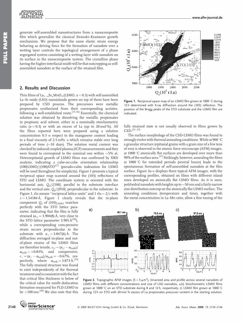
Figure 1. Reciprocal space map of an LSMO film grown at 1000 8C during
12 h determined with X-ray diffraction around the (103) reflection. The
position of the Bragg peaks of the STO substrate and the LSMO film are
indicated.
2. Results and Discussion
Thin films of La1�xSrxMnO3 (LSMO, x¼ 0.3) with self-assembledLa–Sr oxide (LSO) nanoislands grown on top of them have beenprepared by CSD process. The precursors were metallicpropionates synthesized from their corresponding acetates,following a well-established route.[15,16] Essentially, the chemicalsolution was obtained by dissolving the metallic propionatesin propionic acid solvent, either in a nominally stoichiometricratio (x¼ 0.3) or with an excess of La (up to 20mol%). Allthe films reported here were prepared using a solutionconcentration 0.3 M respect to the manganese content leadingto a final viscosity of 2.5mPa � s which remains stable over longperiods of time (�10 days). The solution metal content wascheckedby induced coupled plasma (ICP)measurements and theywere found to correspond to the nominal one within �5% at.Heteroepitaxial growth of LSMO films was confirmed by XRDanalysis, indicating a cube-on-cube orientation relationship(100)LSMO//(100)STO[17] (pseudo-cubic indexation for LSMOwill be used throughout for simplicity). Figure 1 presents a typicalreciprocal space map scanned around the (103) reflections ofSTO and LSMO. The coordinate system is oriented with thehorizontal axis, Qx//[100], parallel to the substrate interfaceand the vertical axis, Qy//[010], perpendicular to the substrate. InFigure 1, rlu means ‘‘reciprocal lattice units’’ and 1 rlu¼ 2/l withl¼ 1.54184 A. Figure 1 clearly reveals that the in-plane
Figure 2. Topographic AFM images (5� 5mm2), binarized area and profile across several nanodots of
LSMO films with different concentrations and size of LSO nanodots. a,b) Stoichiometric LSMO films
grown at 1000 8C on an STO substrate during 8 and 12 h, respectively; c) LSMO film grown at 1000 8Cduring 12 h on STO with 20mol % excess of La propionates precursor content in the starting solution.
component Qx of (103)LSMO matchesperfectly with the STO lattice para-meter, indicating that the film is fullystrained (a//¼ 3.904(8) A, very close tothe STO lattice parameter 3.905 A[18]),while a corresponding com-pressivestrain occurs perpendicular to thesubstrate with a?¼ 3.847(6) A. Thediffraction averaged in-plane and out-of-plane strains of the LSMO filmsare therefore tensile, e//¼ (a//� abulk)/abulk¼þ0.83%, and compressive,e?¼ (a?� abulk)/abulk¼�0.67%, res-pectively, where abulk¼ 3.873 A.[19]
The fully strained structure was foundto exist independently of the thermaltreatment and is consistentwith the factthat critical film thickness is below ofthe critical value for misfit dislocationformation measured for PLD-LSMO tobe >100 nm.[20] We also note that this
� 2009 WILEY-VCH Verlag GmbH &
fully strained state is not usually observed in films grown byCSD.[21–23]
The surface morphology of the CSD-LSMO films was found tostrongly evolvewith thermal annealing conditions.While at 900 8Ca granular structure (epitaxial grains with a grain size of a few tensof nm) is observed in the atomic force microscope (AFM) images,at 1000 8C atomically flat surfaces are developed over more than90%of the surface area.[17] Strikingly, however, annealing thefilmsat 1000 8C for extended periods (several hours) leads to thespontaneous formation of self-assembled nanodots at the filmsurface. Figure 2a–c displays three typical AFM images, with thecorresponding profiles, obtained on films with different islandsizes developed on atomically flat LSMO films. As it is seen,polyhedral nanodotswithheightsup to�50 nmanda fairly narrowsize distribution outcrop on the atomically flat LSMO surface. Theannealing conditions (temperature and time), together withthe metal concentration in La–Mn ratio, allow a fine tuning of the
Co. KGaA, Weinheim Adv. Funct. Mater. 2009, 19, 2139–2146
FULLPAPER
www.afm-journal.de
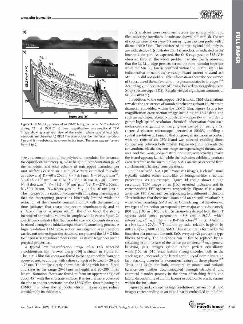
Figure 3. TEM-EELS analysis of an LSMO film grown on an STO substrate
during 12 h at 1000 8C. a) Low magnification cross-sectional TEM
image showing a general view of the system where several interfacial
nanodots are observed; b) EELS line scan across the interfaces nanodot–
film and film–substrate, as shown in the inset. The scan was performed
from 1 to 2.
size and concentration of the polyhedral nanodots. For instance,the equivalent diameter (D), mean height (h), concentration (N) ofthe nanodots, and total volume of outcropped nanodots perunit surface (V) seen in Figure 2a–c were estimated to evolveas follows: a) D� 60� 20 nm, h� 4� 3 nm, N¼ 14 dots mm�2,V� 0.45� 103 nm3mm�2; b) D� 336� 56 nm, h� 40� 10 nm,N¼ 2 dots mm�2, V� 45.2� 103 nm3mm�2; c) D� 270� 60 nm,h� 50� 20 nm, N¼ 8 dots mm�2, V� 114.5� 103 nm3mm�2.The increase of the nanodot volume with annealing time suggeststhat the outcropping process is kinetically limited while thereduction of the nanodot concentration N with the annealingtime indicates that coarsening occurs simultaneously and sosurface diffusion is important. On the other hand, the strongincrease of nanoisland volume in sampleswith La excess Figure 2cclearly demonstrates that the nanodot size and concentration canbe tuned through the initial composition and annealing process. Ahigh resolution TEM cross-section investigation was thereforecarried out to investigate the structural response of the LSMOfilmto the phase segregation process aswell as its consequences on thephysical properties.
A typical low magnification image of a 12 h annealedstoichiometric film, viewed along [010] is shown in Figure 3a.The LSMOfilm thickness was found to change smoothly fromoneobserved area to another with values comprised between�20 and�26 nm. The image clearly shows flat islands with lateral facetsand sizes in the range 20–55 nm in height and 90–280 nm inlength. Nanodots facets are found to form an apparent angle ofabout 45 8 with the substrate surface. It is furthermore observedthat the nanodots penetrate into the LSMOfilm, thus thinning theLSMO film below the nanodots which in some cases reduceconsiderably its thickness.
Adv. Funct. Mater. 2009, 19, 2139–2146 � 2009 WILEY-VCH Verl
EELS analyses were performed across the nanodot–film andfilm–substrate interfaces. Results are shown in Figure 3b. The setof spectra were taken every 3.5 nm using an electron probe with adiameter of 0.5 nm. The positions of the starting andfinal analysesare indicated by 1 (substrate) and 2 (nanodot), as indicated in theinset and the plot. As expected, the O–K edge peak at 532 eV isobserved through the whole profile. It is also clearly observedthat the La M5,4 edge persists across the film–nanodot interfacewhile the Mn L3,2 line is confined within the LSMO layer. Thisindicates that thenanodots have a significant content inLa and lackMn. EELS did not yield reliable information about the occurrenceof Sr because of the unfavorable energies associated to its edges.[24]
Accordingly, theoccurrenceofSrwas checkedbyenergydispersiveX-ray spectroscopy (EDS). Results yielded significant amounts ofSr (20–30wt %).
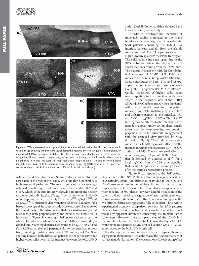
In addition to the outcropped LSO islands, TEM observationsrevealed the occurrence of rounded inclusions, about 10–20 nm indiameter, embedded within the LSMO film. Figure 4a is a lowmagnification cross-section image including an LSO island andsuch an inclusion, labeled Ruddelsden–Popper (R–P). In order togather high spatial resolution chemical information from suchinclusions, energy filtered imaging was carried out using a Cs-corrected electron microscope operated at 200 kV, enabling aspatial resolution of 1 nm. To that purpose, an inclusion in contactwith the roots of an LSO island was selected enabling directcomparison between both phases. Figure 4b and c presents theconventional elastic-electron image corresponding to the analyzedarea, and the La-M5,4-edge distribution map, respectively. Clearly,the island appears La-rich while the inclusion exhibits a contrasteven darker than the surrounding LSMOmatrix, as expected fromstoichiometric balance considerations.
In the analyzed LSMO [010] zone axis images, such inclusionstypically exhibit either cubic-like or tetragonal-like structuralprojections. As an example, Figure 4d and e shows a highresolution TEM image of an [100] oriented inclusion and itscorresponding FFT spectrum, respectively. Figure 4f is a [001]zone axis FFT spectrum corresponding to a different inclusion.This indicates that these inclusions hold an epitaxial relationshipwith the surroundingLSMOmatrix.Considering that theobservedtwo types of projection correspond to twomajor zone axis, namely[001] and [100] or [010], the lattice parameters derived fromtheFFTspectra yield lattice parameters �3.8 and �19.7 A, whichinterestingly fit with the n¼ 2 R–P structure[25] (S.G. I4/mmm,a� b� ap, c� 20 A).[26] Thus, the epitaxial relation is given by(001)[100]R–P//(001)[100]LSMO. This structure is formed by theinsertion of a rock-salt-like unit, SrO, every n (¼2) perovskite-typeblocks, SrMnO3. The Sr cations can in fact be replaced by La,resulting in an increase of the lattice parameters.[26] As a generalbehavior, [001] images exhibit rather perfect crystallinity,while [100] or [010] ones feature strong disorder, both in thestacking sequence and in the lateral continuity of atomic layers. Infact, stacking disorder is a common feature in these phases.[27]
Thus, it is likely that both, structural mismatch and cationicbalance are further accommodated through structural andchemical disorder (mostly in the form of stacking faults andlateral discontinuity of atomic layers) in addition to elastic strainswithin the inclusions.
Figure 5a and c compares high resolution cross-sectional TEMimages corresponding to an island partly embedded in the film,
ag GmbH & Co. KGaA, Weinheim 2141
FULLPAPER
www.afm-journal.de
Figure 4. TEM cross-section analysis of inclusions embedded within the film. a) Low magnifi-
cation image showing the three-phases building the epitaxial system: An (La,Sr)-oxide island, an
embedded R–P type inclusion, and the LSMO film; b,c) correspond to the elastic electron and La-
M5,4 edge filtered images, respectively, of an area including an (La,Sr)-oxide island and a
neighboring R–P type inclusion; d) high resolution image of an R–P inclusion viewed along
its [100] zone axis; e) FFT spectrum corresponding to (d); f) [001] zone axis FFT spectrum
corresponding to an R–P type inclusion different from (d) (not shown).
2142
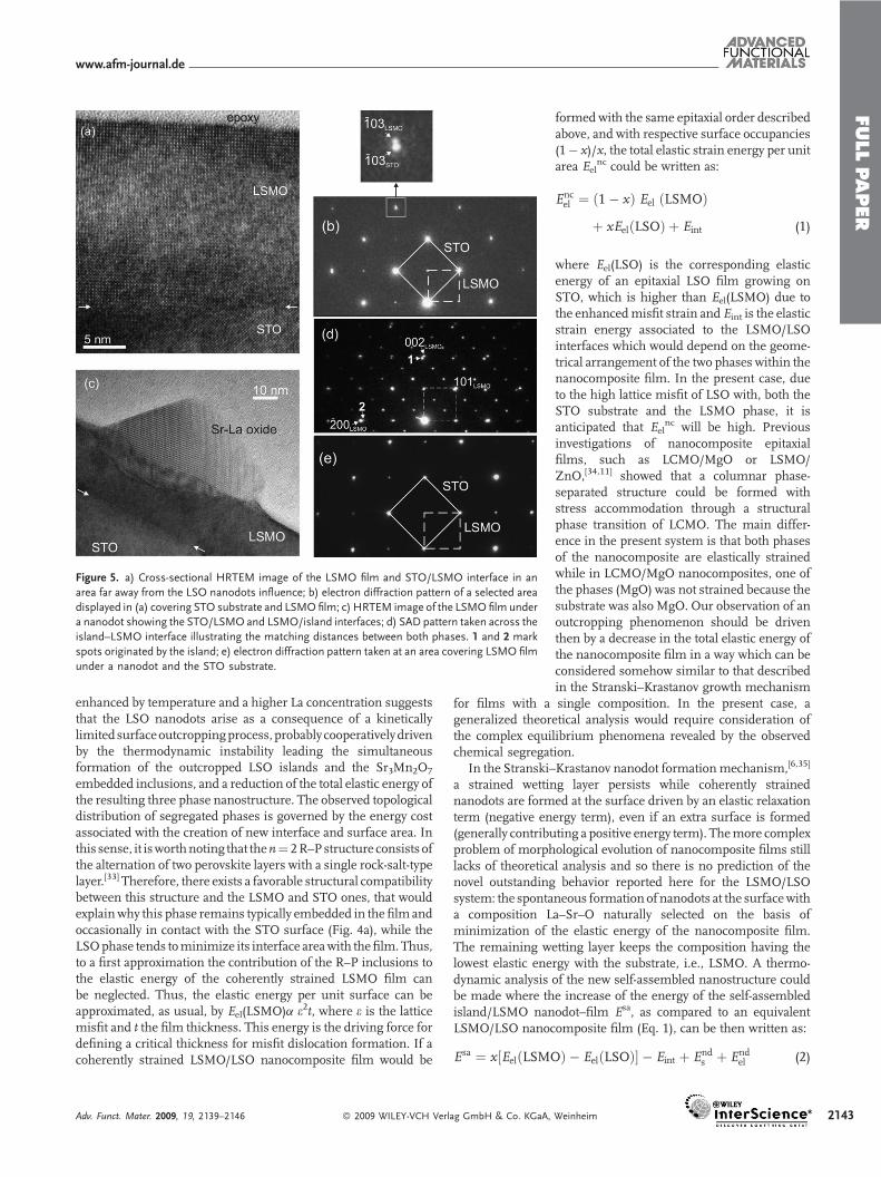
with an island free film region. Strain contrasts can be observedassociated to the root of the island, while the free-film exhibits ahigh structural perfection. The most significant lattice spacingsobtained from thehigh resolution image of the island are: 8.37 and5.41 A,which, to theauthorsknowledge, donot correspondneitherto the sesquioxide Sr0.2La1.8O2.97,
[28] nor to any other Sr–La–Oreportedphase, namely: Sr3La4O9,
[29] La4SrO7,[30] La2Sr2O5,
[31] andLa2SrOx.
[32] A structural determination of these nanodots fallsbeyond the scopeof the present study.However, careful analyses ofthe buried roots of the island reveal that they sustain an epitaxialrelationship both perpendicularly and parallel the film. This isindicated in Figure 5c showing a SAD pattern taken across theisland-film interface, where the coincident lattice is marked by adashed line. Matching distances are found to be d//¼ 4.055 A andd?¼ 4.048 A, parallel and perpendicular to the interface, respec-tively, yielding misfit strains e//¼ 4.7% and e?¼ 4.5%. Spotsplitting between both diffraction patterns are better observed forhigher order reflections, as for instance between the (002)LSMO
� 2009 WILEY-VCH Verlag GmbH & Co. KGaA, Weinheim
and (�200)LSMOspots and those labeled1 and2 for the island, respectively.
In order to investigate the interaction ofmismatch strains originated at the islandinterfacewith those originated at the substrate,SAD patterns containing the LSMO–STOinterface beneath and far from the islandswere compared. The SAD pattern shown inFigure5b corresponds to the island free region.The solid square indicates spots due to theSTO substrate while the dashed squareshows the spots coming from the LSMO film.The pattern is consistent with the rhombohe-dral structure of LSMO (S.G. R-3c) andindicates a cube-on-cube epitaxial relationship.Spots contributed by both, STO and LSMO,appear more intense and are elongatedalong [001], perpendicular to the interface.Careful inspection of higher order spotsreveals splitting in that direction, as demon-strated in the magnified view of the (�103)STOandLSMOreflections.On the other hand,within experimental resolution, the patternindicates complete matching between filmand substrate parallel to the interface, i.e.,a//(LSMO)� a//(STO)¼ 3.905 A. Thus, LSMOfilmregionsnot affectedby the interactionwithnanodots appear under an in-plane tensilestress and the corresponding compressionperpendicular to the substrate, in agreementwith the averaged view provided by X-raydiffraction (Fig. 1). The strain values deter-mined for theLSMOregionsnot affectedby theinteractionwith the nanodots are e//¼þ0.83%and e?¼�1.03%. These values yield a Poissonratio n¼ e?/(e?� 2e//) of 0.38, close tothat determined by Maurice et al.[20] for aL2/3Sr1/3MnO3 film, n¼ 0.34, thus signalingthat the film keeps its structural integrity evenafter the complex segregation process.
Figure 5e corresponds to the SAD pattern
obtained across the LSMO/STO interface in the region beneath anLSO nanodot. Again, the diffraction spots due to the STO andLSMO structures are connected by solid and dashed squares,respectively. In this region, the film also corresponds to arhombohedral LSMO phase. However, careful inspection of thepattern did not reveal any signature of spot splitting or evenelongation in any direction, i.e., diffraction spots coming from thetwo different lattices are apparently fully coincident. Thus, withinexperimental accuracy, comparison between the SAD patternsobtained from regions far from and below the nanodots, do notreveal any apparent difference concerning the in-plane latticeparameters. However, the c-axis parameter of the LSMO filmbecomes tensile stretched below the LSO nanodots by �þ0.83%,resulting in an expanded LSMO unit cell volume DV/V�þ2.5%,as compared to the bulk LSMO unit cell.Results reported above indicate that a complex chemicalsegregation phenomenon lies at the root of the newmechanism ofsurface nanodot formation. The observation of a coarsening effect
Adv. Funct. Mater. 2009, 19, 2139–2146
FULLPAPER
www.afm-journal.de
Figure 5. a) Cross-sectional HRTEM image of the LSMO film and STO/LSMO interface in an
area far away from the LSO nanodots influence; b) electron diffraction pattern of a selected area
displayed in (a) covering STO substrate and LSMOfilm; c) HRTEM image of the LSMO film under
a nanodot showing the STO/LSMO and LSMO/island interfaces; d) SAD pattern taken across the
island–LSMO interface illustrating the matching distances between both phases. 1 and 2 mark
spots originated by the island; e) electron diffraction pattern taken at an area covering LSMO film
under a nanodot and the STO substrate.
enhanced by temperature and a higher La concentration suggeststhat the LSO nanodots arise as a consequence of a kineticallylimited surfaceoutcroppingprocess, probably cooperativelydrivenby the thermodynamic instability leading the simultaneousformation of the outcropped LSO islands and the Sr3Mn2O7
embedded inclusions, and a reduction of the total elastic energy ofthe resulting three phase nanostructure. The observed topologicaldistribution of segregated phases is governed by the energy costassociated with the creation of new interface and surface area. Inthis sense, it isworthnoting that then¼ 2R–Pstructure consists ofthe alternation of two perovskite layers with a single rock-salt-typelayer.[33] Therefore, there exists a favorable structural compatibilitybetween this structure and the LSMO and STO ones, that wouldexplainwhy this phase remains typically embedded in the film andoccasionally in contact with the STO surface (Fig. 4a), while theLSOphase tends tominimize its interface areawith thefilm.Thus,to a first approximation the contribution of the R–P inclusions tothe elastic energy of the coherently strained LSMO film canbe neglected. Thus, the elastic energy per unit surface can beapproximated, as usual, by Eel(LSMO)a e2t, where e is the latticemisfit and t the film thickness. This energy is the driving force fordefining a critical thickness for misfit dislocation formation. If acoherently strained LSMO/LSO nanocomposite film would be
Adv. Funct. Mater. 2009, 19, 2139–2146 � 2009 WILEY-VCH Verlag GmbH & Co. KGaA,
formed with the same epitaxial order describedabove, and with respective surface occupancies(1� x)/x, the total elastic strain energy per unitarea Eel
nc could be written as:
Encel ¼ ð1� xÞ Eel ðLSMOÞ
þ xEelðLSOÞ þ Eint (1)
where Eel(LSO) is the corresponding elasticenergy of an epitaxial LSO film growing onSTO, which is higher than Eel(LSMO) due tothe enhancedmisfit strain and Eint is the elasticstrain energy associated to the LSMO/LSOinterfaces which would depend on the geome-trical arrangement of the two phases within thenanocomposite film. In the present case, dueto the high lattice misfit of LSO with, both theSTO substrate and the LSMO phase, it isanticipated that Eel
nc will be high. Previousinvestigations of nanocomposite epitaxialfilms, such as LCMO/MgO or LSMO/ZnO,[34,11] showed that a columnar phase-separated structure could be formed withstress accommodation through a structuralphase transition of LCMO. The main differ-ence in the present system is that both phasesof the nanocomposite are elastically strainedwhile in LCMO/MgO nanocomposites, one ofthe phases (MgO) was not strained because thesubstrate was also MgO. Our observation of anoutcropping phenomenon should be driventhen by a decrease in the total elastic energy ofthe nanocomposite film in a way which can beconsidered somehow similar to that describedin the Stranski–Krastanov growth mechanism
for films with a single composition. In the present case, ageneralized theoretical analysis would require consideration ofthe complex equilibrium phenomena revealed by the observedchemical segregation.
In the Stranski–Krastanov nanodot formation mechanism,[6,35]
a strained wetting layer persists while coherently strainednanodots are formed at the surface driven by an elastic relaxationterm (negative energy term), even if an extra surface is formed(generally contributing a positive energy term). Themore complexproblem of morphological evolution of nanocomposite films stilllacks of theoretical analysis and so there is no prediction of thenovel outstanding behavior reported here for the LSMO/LSOsystem: the spontaneous formation of nanodots at the surfacewitha composition La–Sr–O naturally selected on the basis ofminimization of the elastic energy of the nanocomposite film.The remaining wetting layer keeps the composition having thelowest elastic energy with the substrate, i.e., LSMO. A thermo-dynamic analysis of the new self-assembled nanostructure couldbe made where the increase of the energy of the self-assembledisland/LSMO nanodot–film Esa, as compared to an equivalentLSMO/LSO nanocomposite film (Eq. 1), can be then written as:
Esa ¼ x½EelðLSMOÞ � EelðLSOÞ� � Eint þ Ends þ End
el (2)
Weinheim 2143
FULLPAPER
www.afm-journal.de
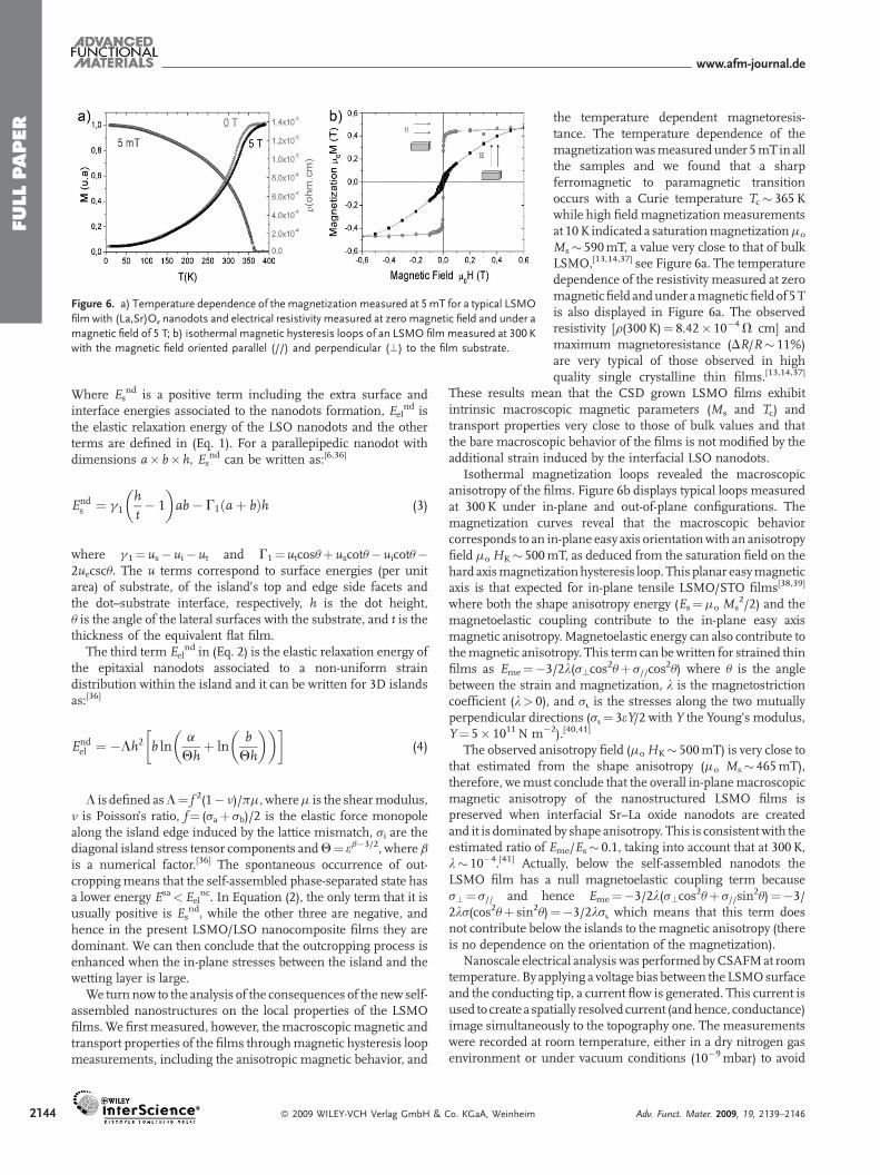
Figure 6. a) Temperature dependence of the magnetization measured at 5mT for a typical LSMO
film with (La,Sr)Ox nanodots and electrical resistivity measured at zero magnetic field and under a
magnetic field of 5 T; b) isothermal magnetic hysteresis loops of an LSMO filmmeasured at 300 K
with the magnetic field oriented parallel (//) and perpendicular (?) to the film substrate.
2144
Where Esnd is a positive term including the extra surface and
interface energies associated to the nanodots formation, Eelnd is
the elastic relaxation energy of the LSO nanodots and the otherterms are defined in (Eq. 1). For a parallepipedic nanodot withdimensions a� b� h, Es
nd can be written as:[6,36]
Ends ¼ g1
h
t� 1
� �ab� G1 aþ bð Þh (3)
where g1¼ us� ui� ut and G1¼ utcosuþ uscotu� uicotu�2uecscu. The u terms correspond to surface energies (per unitarea) of substrate, of the island’s top and edge side facets andthe dot–substrate interface, respectively, h is the dot height,u is the angle of the lateral surfaces with the substrate, and t is thethickness of the equivalent flat film.
The third term Eelnd in (Eq. 2) is the elastic relaxation energy of
the epitaxial nanodots associated to a non-uniform straindistribution within the island and it can be written for 3D islandsas:[36]
Endel ¼ �Lh2 b ln
a
Qhþ ln
b
Qh
� �� �� �(4)
L is defined asL¼ f 2(1� n)/pm, wherem is the shearmodulus,n is Poisson’s ratio, f¼ (saþ sb)/2 is the elastic force monopolealong the island edge induced by the lattice mismatch, si are thediagonal island stress tensor components andQ¼ eb�3/2, where bis a numerical factor.[36] The spontaneous occurrence of out-croppingmeans that the self-assembled phase-separated state hasa lower energy Esa<Eel
nc. In Equation (2), the only term that it isusually positive is Es
nd, while the other three are negative, andhence in the present LSMO/LSO nanocomposite films they aredominant. We can then conclude that the outcropping process isenhanced when the in-plane stresses between the island and thewetting layer is large.
We turn now to the analysis of the consequences of the new self-assembled nanostructures on the local properties of the LSMOfilms.We first measured, however, themacroscopic magnetic andtransport properties of the films throughmagnetic hysteresis loopmeasurements, including the anisotropic magnetic behavior, and
� 2009 WILEY-VCH Verlag GmbH & Co. KGaA, Weinheim
the temperature dependent magnetoresis-tance. The temperature dependence of themagnetizationwasmeasuredunder 5mTin allthe samples and we found that a sharpferromagnetic to paramagnetic transitionoccurs with a Curie temperature Tc� 365Kwhile high field magnetization measurementsat 10 K indicated a saturationmagnetizationmo
Ms� 590mT, a value very close to that of bulkLSMO,[13,14,37] see Figure 6a. The temperaturedependence of the resistivity measured at zeromagneticfield andunder amagneticfieldof 5Tis also displayed in Figure 6a. The observedresistivity [r(300K)¼ 8.42� 10�4V cm] andmaximum magnetoresistance (DR/R� 11%)are very typical of those observed in highquality single crystalline thin films.[13,14,37]
These results mean that the CSD grown LSMO films exhibitintrinsic macroscopic magnetic parameters (Ms and Tc) andtransport properties very close to those of bulk values and thatthe bare macroscopic behavior of the films is not modified by theadditional strain induced by the interfacial LSO nanodots.
Isothermal magnetization loops revealed the macroscopicanisotropy of the films. Figure 6b displays typical loops measuredat 300K under in-plane and out-of-plane configurations. Themagnetization curves reveal that the macroscopic behaviorcorresponds to an in-plane easy axis orientationwith an anisotropyfield moHK� 500mT, as deduced from the saturation field on thehard axismagnetizationhysteresis loop. Thisplanar easymagneticaxis is that expected for in-plane tensile LSMO/STO films[38,39]
where both the shape anisotropy energy (Es¼mo Ms2/2) and the
magnetoelastic coupling contribute to the in-plane easy axismagnetic anisotropy.Magnetoelastic energy can also contribute tothemagnetic anisotropy. This term can bewritten for strained thinfilms as Eme¼�3/2l(s?cos
2uþ s//cos2u) where u is the angle
between the strain and magnetization, l is the magnetostrictioncoefficient (l> 0), and si is the stresses along the two mutuallyperpendicular directions (si¼ 3eY/2 with Y the Young’s modulus,Y¼ 5� 1011N m�2).[40,41]
The observed anisotropy field (moHK� 500mT) is very close tothat estimated from the shape anisotropy (mo Ms� 465mT),therefore, wemust conclude that the overall in-planemacroscopicmagnetic anisotropy of the nanostructured LSMO films ispreserved when interfacial Sr–La oxide nanodots are createdand it is dominated by shape anisotropy. This is consistent with theestimated ratio of Eme/Es� 0.1, taking into account that at 300K,l� 10�4.[41] Actually, below the self-assembled nanodots theLSMO film has a null magnetoelastic coupling term becauses?¼ s// and hence Eme¼�3/2l(s?cos
2uþ s//sin2u)¼�3/
2ls(cos2uþ sin2u)¼�3/2lsi which means that this term doesnot contribute below the islands to themagnetic anisotropy (thereis no dependence on the orientation of the magnetization).
Nanoscale electrical analysis was performedbyCSAFMat roomtemperature. By applying a voltage bias between the LSMOsurfaceand the conducting tip, a current flow is generated. This current isused to create a spatially resolved current (andhence, conductance)image simultaneously to the topography one. The measurementswere recorded at room temperature, either in a dry nitrogen gasenvironment or under vacuum conditions (10�9mbar) to avoid
Adv. Funct. Mater. 2009, 19, 2139–2146
FULLPAPER
www.afm-journal.de
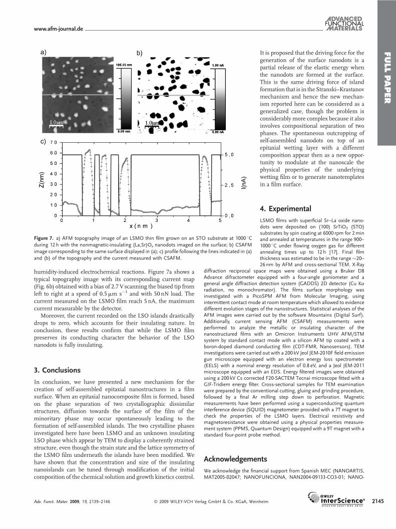
Figure 7. a) AFM topography image of an LSMO thin film grown on an STO substrate at 1000 8Cduring 12 h with the nonmagnetic-insulating (La,Sr)Ox nanodots imaged on the surface; b) CSAFM
image corresponding to the same surface displayed in (a); c) profile following the lines indicated in (a)
and (b) of the topography and the current measured with CSAFM.
humidity-induced electrochemical reactions. Figure 7a shows atypical topography image with its corresponding current map(Fig. 6b) obtained with a bias of 2.7 V scanning the biased tip fromleft to right at a speed of 0.5mm s�1 and with 50 nN load. Thecurrent measured on the LSMO film reach 5 nA, the maximumcurrent measurable by the detector.
Moreover, the current recorded on the LSO islands drasticallydrops to zero, which accounts for their insulating nature. Inconclusion, these results confirm that while the LSMO filmpreserves its conducting character the behavior of the LSOnanodots is fully insulating.
3. Conclusions
In conclusion, we have presented a new mechanism for thecreation of self-assembled epitaxial nanostructures in a filmsurface. When an epitaxial nanocomposite film is formed, basedon the phase separation of two crystallographic dissimilarstructures, diffusion towards the surface of the film of theminoritary phase may occur spontaneously leading to theformation of self-assembled islands. The two crystalline phasesinvestigated here have been LSMO and an unknown insulatingLSO phase which appear by TEM to display a coherently strainedstructure, even though the strain state and the lattice symmetry ofthe LSMO film underneath the islands have been modified. Wehave shown that the concentration and size of the insulatingnanoislands can be tuned through modification of the initialcomposition of the chemical solution and growth kinetics control.
Adv. Funct. Mater. 2009, 19, 2139–2146 � 2009 WILEY-VCH Verlag GmbH & Co. KGaA, Wein
It is proposed that the driving force for thegeneration of the surface nanodots is apartial release of the elastic energy whenthe nanodots are formed at the surface.This is the same driving force of islandformation that is in the Stranski–Krastanovmechanism and hence the new mechan-ism reported here can be considered as ageneralized case, though the problem isconsiderably more complex because it alsoinvolves compositional separation of twophases. The spontaneous outcropping ofself-assembled nanodots on top of anepitaxial wetting layer with a differentcomposition appear then as a new oppor-tunity to modulate at the nanoscale thephysical properties of the underlyingwetting film or to generate nanotemplatesin a film surface.
4. Experimental
LSMO films with superficial Sr–La oxide nano-dots were deposited on (100) SrTiO3 (STO)substrates by spin coating at 6000 rpm for 2minand annealed at temperatures in the range 900–1000 8C under flowing oxygen gas for differentannealing times up to 12 h [17]. Final filmthickness was estimated to be in the range�20–26 nm by AFM and cross-sectional TEM. X-Ray
diffraction reciprocal space maps were obtained using a Bruker D8Advance difractometer equipped with a four-angle goniometer and ageneral angle diffraction detection system (GADDS) 2D detector (Cu Karadiation, no monochromator). The films surface morphology wasinvestigated with a PicoSPM AFM from Molecular Imaging, usingintermittent contact mode at room temperature which allowed to evidencedifferent evolution stages of the nanostructures. Statistical analyses of theAFM images were carried out by the software Mountains (Digital Surf).Additionally, current sensing AFM (CSAFM) measurements wereperformed to analyze the metallic or insulating character of thenanostructured films with an Omicron Instruments UHV AFM/STMsystem by standard contact mode with a silicon AFM tip coated with aboron-doped diamond conducting film (CDT-FMR, Nanosensors). TEMinvestigations were carried out with a 200 kV Jeol JEM-2010F field emissiongun microscope equipped with an electron energy loss spectrometer(EELS) with a nominal energy resolution of 0.8 eV, and a Jeol JEM-2011microscope equipped with an EDS. Energy filtered images were obtainedusing a 200 kV Cs corrected F20-SACTEM Tecnai microscope fitted with aGIF-Tridiem energy filter. Cross-sectional samples for TEM examinationwere prepared by the conventional cutting, gluing and grinding procedure,followed by a final Ar milling step down to perforation. Magneticmeasurements have been performed using a superconducting quantuminterference device (SQUID) magnetometer provided with a 7T magnet tocheck the properties of the LSMO layers. Electrical resistivity andmagnetoresistance were obtained using a physical properties measure-ment system (PPMS, Quantum Design) equipped with a 9T magnet with astandard four-point probe method.
Acknowledgements
We acknowledge the financial support from Spanish MEC (NANOARTIS,MAT2005-02047; NANOFUNCIONA, NAN2004-09133-CO3-01; NANO-
heim 2145
FULLPAPER
www.afm-journal.de
2146
SELECT, CSD2007-00041 FPU, AP2005-4669 FPU), Generalitat deCatalunya (Catalan Pla de Recerca SGR-0029 and CeRMAE), CSIC (PIF-CANNAMUS) and EU (HIPERCHEM, NMP4-CT2005-516858). The Cscorrected F20-FEI electron microscope was used through the Europeanproject ESTEEM (contract no. 026019). Valuable discussions with I.C.Infante are gratefully acknowledged. We also acknowledge electronmicroscopy facilities from Servei de Microscopia de la UAB and Serveide Microscopia de la UB.
Received: January 19, 2009
Published online: June 2, 2009
[1] C. A. Ross, Annu. Rev. Mater. Res. 2001, 31, 203.
[2] H. Zheng, J. Wang, S. E. Lofland, Z. Ma, L. Mohaddes-Ardabili, T. Zhao, L.
Salamanca-Riba, S. R. Shinde, S. B. Ogale, F. Bai, D. Viehland, Y. Jia, D. G.
Schlom, M. Wuttig, A. Roytburd, R. Ramesh, Science 2004, 303, 661.
[3] S. R. Foltyn, L. Civale, J. L. Manus-Driscoll, Q. X. Jia, B. Maiorov, H. Wang,
M. Maley, Nat. Mater. 2007, 6, 631.
[4] J. Gutierrez, A. Llordes, J. Gazquez, M. Gibert, N. Roma, S. Ricart, A.
Pomar, F. Sandiumenge, N. Mestres, T. Puig, X. Obradors, Nat. Mater.
2007, 6, 367.
[5] J. V. Barth, G. Costantini, K. Kern, Nature 2005, 437, 671.
[6] V. A. Shchukin, D. Bimberg, Rev. Mod. Phys. 1999, 71, 1125.
[7] C. Teichert, Phys. Rep. 2002, 365, 335.
[8] M. Gibert, T. Puig, X. Obradors, A. Benedetti, F. Sandiumenge, R. Huhne,
Adv. Mater. 2007, 19, 3937.
[9] M. Gibert, T. Puig, X. Obradors, Surf. Sci. 2007, 601, 2680.
[10] H. M. Zheng, F. Straub, Q. Zhan, P. L. Yang, W. K. Hsieh, F. Zavaliche, Y. H.
Chu, U. Dahmen, R. Ramesh, Adv. Mater. 2006, 18, 2747.
[11] V. Moshnyaga, B. Damaschke, O. Shapoval, A. Belenchuk, J. Faupel, O. I.
Lebedev, J. Verbeeck, G. van Tendeloo, M.Mucksch, V. Tsurkan, R. Tidecks,
K. Samwer, Nat. Mater. 2003, 2, 247.
[12] J. M. D. Coey, M. Viret, S. von Molnar, Adv. Phys. 1999, 48, 167.
[13] M. B. Salamon, M. Jaime, Rev. Mod. Phys. 2001, 73, 583.
[14] Y. Tokura, Rep. Prog. Phys. 2006, 69, 797.
[15] U. Hasenkox, C. Mitze, R. Waser, J. Am. Ceram. Soc. 1997, 80, 2709.
[16] U. Hasenkox, C. Mitze, R. Waser, R. R. Arons, J. Pommer, G. Guntherodt, J.
Electroceram. 1999, 3, 255.
[17] A. Hassini, A. Pomar, J. Gutierrez, M. Coll, N. Roma, C. Moreno, A. Ruyter,
T. Puig, X. Obradors, Supercond. Sci. Technol. 2007, 20, S230.
� 2009 WILEY-VCH Verlag GmbH &
[18] J. Brous, I. Fankuchen, E. Banks, Acta Crystallogr. 1953, 6, 67.
[19] A. Hammouche, E. Siebert, A. Hammou, Mater. Res. Bull. 1989, 24,
367.
[20] J. L. Maurice, F. Pailloux, A. Barthelemy, O. Durand, D. Imhoff, R. Lyonnet,
A. Rocher, J. P. Contour, Philos. Mag. 2003, 83, 3201.
[21] A. Cavallaro, F. Sandiumenge, J. Gazquez, T. Puig, X. Obradors, J. Arbiol,
H. C. Freyhardt, Adv. Funct. Mater. 2006, 16, 1363.
[22] P. A. Langjahr, F. F. Lange, T. Wagner, M. Ruhle, Acta Mater. 1998, 46, 773.
[23] A. Pomar, M. Coll, A. Cavallaro, J. Gazquez, J. C. Gonzalez, N. Mestres, F.
Sandiumenge, T. Puig, X. Obradors, J. Mater. Res. 2006, 21, 1106.
[24] T. Walther, Ultramicroscopy 2003, 96, 401.
[25] S. N. Ruddlesden, P. Popper, Acta Crystallogr. 1958, 11, 54.
[26] R. Seshadri, C. Martin, M. Hervieu, B. Raveau, C. N. R. Rao, Chem. Mater.
1997, 9, 270.
[27] R. Seshadri, M. Hervieu, C. Martin, A. Maignan, B. Domenges, B. Raveau,
A. N. Fitch, Chem. Mater. 1997, 9, 1778.
[28] M. Foex, Bull. Soc. Chim. France 1961, 109.
[29] A. R. Schulze, H. Mullerbuschbaum, Z. Anorg. Allg. Chem. 1980, 471,
59.
[30] International Centre for Diffraction Data No. 00–022–1430, 2008.
[31] International Centre for Diffraction Data No. 00–022–1431, 2008.
[32] International Centre for Diffraction Data No. 00–042–0343, 2008.
[33] G. van Tendeloo, O. I. Lebedev, M. Hervieu, B. Raveau, Rep. Prog. Phys.
2004, 67, 1315.
[34] B. S. Kang, H. Wang, J. L. Manus-Driscoll, Y. Li, Q. X. Jia, I. Mihut, J. B.
Betts, Appl. Phys. Lett. 2006, 88, 192514.
[35] J. Tersoff, F. K. Legoues, Phys. Rev. Lett. 1994, 72, 3570.
[36] J. C. Nie, H. Yamasaki, Y. Mawatari, Phys. Rev. B 2007, 70, 195421.
[37] A. Urushibara, Y. Moritomo, T. Arima, A. Asamitsu, G. Kido, Y. Tokura,
Phys. Rev. B 1995, 51, 14103.
[38] A. M. Haghiri-Gosnet, J. Wolfman, B. Mercey, C. Simon, P. Lecoeur, M.
Korzenski, M. Hervieu, R. Desfeux, G. Baldinozzi, J. Appl. Phys. 2000, 88,
4257.
[39] M. Ziese, H. C. Semmelhack, P. Busch, J. Magn. Magn. Mater. 2002, 246,
327.
[40] B. D. Cullity, Introduction to Magnetic Materials, Addison-Wesley, Reading,
MA 1972.
[41] K. Steenbeck, T. Habisreuther, C. Dubourdieu, J. P. Senateur, Appl. Phys.
Lett. 2002, 80, 3361.
Co. KGaA, Weinheim Adv. Funct. Mater. 2009, 19, 2139–2146